a consensus approach to wound care in … consensus approach to wound care in epidermolysis bullosa...
TRANSCRIPT

AbstractEpidermolysis Bullosa (EB) is a group of inherited diseases with 4 subtypes. This disorder is a model for fragile skin with some affected individuals having chronic, diffi cult to heal wounds. The assessment and treatment of wounds in persons with Epidermolysis Bullosa can be guided by the Wound Bed Preparation model. This holistic patient approach evaluates factors that may delay healing and patient centered concerns including pain, itch and activities of daily living. Local wound care is a challenge to optimize wound debridement, infection/infl ammation and moisture balance. Stalled but healable chronic wounds may be stimulated by advanced therapies (Edge Effect) or optimizing patient care for replacement of defective genes (e.g. increased Type 7 collagen production post bone marrow transplant).
This review evaluates the clinical features that distinguish the various subtypes of Epidermolysis Bullosa, the frequent medical complications and prognosis. The care is best provided by an interprofessional team.
The treatment of chronic wounds is outlined with a quick reference guide of 12 recommendations from the 13 expert panel members. These recommendations have been reviewed by a computer-facilitated modifi ed Delphi process where 15 external reviewers (68.8% of reviewers reported having 11 or more years experience with EB care) independently had a score of 80% strongly agreed or somewhat agreed with each of the recommendations.
A Consensus Approach to Wound Carein Epidermolysis Bullosa
Foreword: Persons with Epidermolysis Bullosa and Wound Bed Preparation
International Experts
A Consensus Approach to Wound Care in Epidermolysis Bullosa
3
4
5
Pope E, Lara-Corrales I, Mellerio JE, Martinez AE, Schultz G, Burrell R, Goodman L, Coutts P, Wagner J, Allen U, Lee M, Tolar J, Sibbald RG
An Expert Panel Report 2011

Supported in part through an educational grant from Molnlycke Health Care.
FRONT COVER IMAGES Application of Apligraf in a patient with recessive dystrophic EB
A person with recessive dystrophic EB had 50% of his back ulcerated for 3 years. He had 2 pieces of a living skin equivalent applied (Apligraf human dermal fibroblasts + bovine type 1 collagen under a human epidermal cell layer). One piece had silver dressing plus moisture-balancing foam, while the other piece had only a moisture-balancing foam. One week later, only the piece with the silver dressing plus foam survived (bacterial balance + moisture balance of wound bed preparation).
3
Persons with Epidermolysis Bullosa (EB), particularly the Recessive
Dystrophic EB (RDEB) and Junctional subtypes, are plagued with lifelong
chronic wounds and infections. This best practice consensus paper examines
the issue of Wound Bed Preparation that was literally “born on the back”
of Alex, a person with EB. We now have extended this knowledge for the
benefit of all persons with EB.
This project united Epidermolysis Bullosa experts with wound healers
(basic scientists, physicians and nurse clinicians) with a pioneer in Bone
Marrow Transplantation. Our aim was to facilitate the knowledge of the
4 different subtypes of Epidermolysis Bullosa so that affected individuals
are recognized early and receive appropriate symptomatic treatment,
including optimal local wound care to prevent life-threatening infections
and failure to thrive. Clinicians also need to be aware of complications such
as cardiomyopathy and squamous cell carcinomas (persistent inflammation
leads to malignant transformation) that may occur even in childhood.
In January 1997, Canada was the first county in the world to approve
an artificial skin substitute grown in vitro (Graftskin: bovine type 1 collagen
with human fibroblasts & an epidermal cell layer). In February 1997,
Alex had 2 pieces of Graftskin (Apligraf, Organogenesis, Canton MA)
placed on the upper part of his extensive back ulceration (covering 50% of
his back for 3 years). One week post application, the skin substitute (with
fluid release slits to maintain contact) under the silver + foam dressing was
intact. In contrast, the other skin substitute piece without the bacterial
balancing silver was destroyed by the local collagenase partly produced by
the pseudomonas organisms. Subsequent applications of both vicryl skin
substitute populated with human fibroblasts (Dermagraft, Advanced
Biohealing, La Jolla CA) & Graftskin, resulted in complete wound
healing of Alex’s back. Although Alex is no longer with us as a result of
cardiomyopathy, his contribution to our better understanding of local
wound care has shaped what clinicians know today.
We have subsequently published the Wound Bed Preparation Model
(WBP: Sibbald et al. 2000, 2003, 2006-7-10, 2011)1-4. The WBP model
includes treating the whole patient, patient centered concerns and local
wound care. Ideally, treatment of the cause involves replacing type 7 col-
lagen for RDEB patients through bone marrow transplants or other gene
modifying therapies. Treating the cause also includes optimizing other
co-factors involved in healing, such as nutrition (e.g. supplements and
early insertion of feeding tubes) and correction of anemia. The
components of local wound care are: DIM before DIME: Debridement,
Infection/Inflammation control, Moisture balance before the Edge
effect for advanced therapies (e.g. skin substitutes for stalled but healable
wounds). Local wound care has been improved with local silicone mesh
products, soft silicone foam coatings and silicone tape.
Wagner et al. published a seminal article in the New England Journal
of Medicine in August 2010 reporting on the first 7 patients with RDEB
undergoing immunoablative chemotherapy and allogeneic stem cell
transplant5. Of the 7 patients, one died of cardiomyopathy pre-transplant &
a second patient died of infection and transplant rejection 183 days post-
transplantation. All recipients had a reduction in new blister formation
between days 30 and 130 post transplantation. Five of the 6 recipients had
an increase in type 7 collagen without forming the complete normally
seen anchoring fibrils.
There are two concerns for transplantation procedures. First, the
small number of individuals eligible for transplant may develop HLA
antigens from the allogeneic cells in the skin transplant increasing the
potential for rejection of a BM transplant. Second, older individuals
with RDEB (especially after age 20) are very susceptible to aggressive
squamous cell carcinomas and the immunosuppression associated with
allogeneic stem-cell transplantation may increase this susceptibility.
We currently have come full circle to apply the principles of wound
bed preparation, specifically for persons with Epidermolysis Bullosa, not
only to optimize healing but also to prevent infections and other compli-
cations pre and post bone marrow transplantation. We are also suggesting
strategies for persons with EB and their circle of care (parents and health-
care providers) in resource poor areas where linkages to EB experts will
help improve diagnosis and treatment.
EB also represents a model for fragile skin found in the elderly, Skin
Changes at Life’s End (SCALE) & other persons suffering from chronic
disease or immunosuppression, therefore the principles discussed in this
document can be applied to other patient populations with skin fragility.
This supplement is dedicated to all persons suffering with EB in
an attempt to improve their quality of life. We would specifically thank
Alex Melkic & Deanna Molinaro from whom we have learned so much.
Persons with Epidermolysis Bullosa and Wound Bed PreparationR. Gary Sibbald and Elena Pope, Co-Chairs on behalf of the Epidermolysis Bullosa–Wound Bed Preparation panel
References:Sibbald et al. WBP 2000 OWM1. Sibbald et al. WBP 2003 OWM2. Sibbald et al.WBP 2006 Wound Care Canada, Advances (2007) WHO (2010)3. Sibbald et al.WBP 2011 Advances and WCC 4. Wagner NEJM 2011 5. R. Gary Sibbald E. Pope

4
Collaboration of...Knowledge and Expertise
International
ExpertsA Consensus Approach
to Wound Care in Epidermolysis Bullosa
Upton Allen, MBBS, MSc, FAAP, FRCPC
Division Head, Infectious Diseases, The Hospital for SickChildren, Toronto/CanadaProfessor, Paediatrics and HealthPolicy Management and EvaluationUniversity of Toronto/Canada
Patricia Coutts, RN
President, Canadian Association of Wound CareRegistered NurseFaculty member & inauguralgraduate, InternationalInterdisciplinary Wound CareCourse (IIWCC), University of Toronto/Canada
Michelle Lee, RN, BScN
Dermatology Wound CareNurse, EB Subspecialty clinic,Hospital for Sick Children,Toronto/CanadaGraduate, InternationalInterdisciplinary Wound CareCourse (IIWCC), University of Toronto
Gregory Schultz, PhD
Researcher, molecular andcellular regulation of woundhealing.University of Florida ResearchFoundation, Professor andDirector, The Institute for WoundResearch, University of Florida/USA
Elena Pope, MD, MSc, FRCPC
The Hospital for Sick ChildrenHead, Section of Dermatology,Division of Paediatric MedicineResearch Institute, Toronto/Canada
R. Gary Sibbald, BSc, MD FRCPC (Med, Derm)
Professor of Medicine,Public Health,University of Toronto/CanadaPresident,World Union of Wound Healing Societies
Robert E Burrell, PhD
Professor and Chair,Department of Biomedical Engineering, Faculties of Engineering & Medicine & DentistryProfessor, Canada ResearchChair in Nanostructured Biomaterials, Chemical and Materials EngineeringFaculty of Engineering,University of Alberta/Canada
Laurie Goodman, RN, BA, MHScN
Director, Mississauga HaltonWound InitiativeCo-Director, InternationalInterdisciplinary Wound CareCourse (IIWCC), University ofToronto/CanadaAdvanced Practice Nurse &Clinical Educator
Anna Elizabeth Martinez, MBBS, MRCP, MRCPCH
Consultant Pediatrician andClinical Lead for the Pediatric EB serviceGreat Ormond Street Hospital, London/UK
Jakub Tolar, MD, PhD
Associate Professor,Department of Pediatrics,University of Minnesota/USABlood and Marrow TransplantationChair, Albert D. & Eva J. CornieaDirector of Stem Cell/Gene Therapies
Irene Lara-Corrales, MD, FRCPC(Derm)
Assistant Professor,Pediatrics, University of Toronto/CanadaPediatric DermatologistInterdisciplinary ClinicHospital for Sick Children,with EpidermolysisBullosa/Wound Care interest
Jemima Mellerio, BSc, MD, FRCP
Consultant Dermatologist andClinical Lead for the Adult EB Service, Guy’s & St Thomas’ HospitalGreat Ormond Street Hospital,London/UKSt John’s Institute of Dermatology
John Wagner, MD
Director, Blood and MarrowTransplantationProfessor, Department of Pediatrics, University of Minnesota/USAPrincipal Investigator, ClinicalStem Cell Trials for SevereEpidermolysis Bullosa
EB Team and Affiliates

5
Introduction
Epidermolysis Bullosa (EB) is a group of inherited diseases characterized by mechanical fragility of the skin and mucous membranes. There are four different subtypes of EB resulting from structural protein gene mutations at the cutaneous basement membrane zone (BMZ) or the relatively rare, suprabasal cell-cell adhesion desmosomal proteins. The recent consensus classification divides EB into four main types according to the plane of blister formation at the dermal-epidermal junction (DEJ) (simplex, junctional, dystrophic and mixed), with each type determined by the specific protein defect1. The severity of mucocutaneous and other organ disease varies considerably between types of EB. In the various subtypes of EB, mucocutaneous and other organ distribution is largely determined by the nature of mutations and the gene penetration resulting in different expression profiles of causative EB genes2,3.
When possible, the complex care of patients with EB is
best provided by an interprofessional specialized team. In the absence of a cure, supportive wound care and early recognition and treatment of complications are the mainstays of patient management.
Wound care in EB populations poses unique challenges. A highly individualized treatment plan is needed for each patient due to the clinical variability of the many types and subtypes of EB. Overall cost to the family and health units must be considered in deciding how to treat the widespread skin involvement com-mon to many patients. The clinical decision process is further complicated by the myriad of available wound care products.
To date, there are no specific wound care guidelines that address the unique problems of the EB population. A group of inter- national experts in the field of EB, wound care, microbiology,
infectious diseases and bone marrow transplantation met for three days in Alton, Ontario, Canada and developed a list of 12 recommendations for addressing wound care in patients with EB. Using an online-based modified Delphi method of gen-erating consensus, the list was translated into a survey that was completed by 15 other experts around the world (Table 1) and then further refined into a list of recommendations (Table 2).
A. Treat the Cause
The cutaneous clinical findings in all types of EB are the result of skin fragility leading to bullae formation. There is a wide spectrum of presentations, varying from very mild forms [e.g. EB simplex (EBS), dominant dystrophic EB (DDEB)] to disfiguring disabling and life-limiting disease [e.g. Junctional EB (JEB), recessive dystrophic (RDEB)]. The clinical presentation, morbidity, and mortality of this group of conditions are the result of skin involvement and mucosal blis-tering (eyes4,5, oral mucosa , gastrointestinal tract6, genitourinary tract7,8, respiratory tract). In addition, anemia, cardiomyopathy9,10, renal failure, respiratory failure, osteoporosis including fractures13, chronic malnutrition and growth failure13 are frequently encoun-tered in severe cases.
While traumatic blistering is the norm in EB, the extent, depth, and healing of the wounds is subject to wide inter-patient varia-tions largely dependent on the type of EB. It is well recognized that many milder EBS patients have limited blistering and healing is the norm with minimal skin care or systemic intervention. In contrast, severe generalized RDEB patients have chronic,
non-healing ulcers covering large areas of the skin that lead to
scarring and contractures. However, the severity, extent and location of blisters are subject to, but not predictive of the disease type. This is important to keep in mind particularly in the first few months of life, as the extent of the skin blistering can be un-derestimated. The ability to heal is also influenced by other factors such as bacterial load, malnutrition and low levels of hemoglobin. Wound Bed Preparation is a framework for assessment, diagnosis and treatment of wounds along the continuum toward optimal healing14. This process includes the treatment of the cause and patient centered concerns prior to instituting best practices for local wound care (DIM before DIME: Debridement, Infection and Inflammation, Moisture Balance & Edge Effect for healable but “stalled” wounds).
Table 1
EB EXTERNAL REVIEWERS
Karen Wiss (USA)Edward Barrett (Canada)Rosemarie Watson (Ireland)Anna Bruckner (USA)Annmarie Ormonde (Italy)Anne Lucky (USA)Michelle Lee (Canada)Gerry Kelly-Mancuso (USA)
Celia Moss (UK)Agnes Schwieger (Canada)May El Hachem (Italy)Louise Fret-Lalonde (Canada)Dedee Murrell (Australia)Andrew Lin (Canada)Francis Palisson (Chile)

6
Main Themes Specific Themes Specific Recommendations
A. Treat the cause 1. Assess the patient’s ability to heal Evaluate EB type specific involvement (simplex, junctional, dystrophic, Kindler syndrome) •and co-morbiditiesConsider age of the patient•Assess nutrition status: growth centiles, BMI •Monitor hemoglobin levels•
2. Develop individualized goals and plan of care
Low hemoglobin consider: Fe supplementation, transfusion(s) •Low albumin: protein supplements, feeding tube, etc.•Address other specific sub-type involvement•
B. Patient centered concerns
3. Address and support management of patient centered concerns to enable healing
Pain:World Health Organization pain ladder for nociceptive pain •Neuropathic pain: consider tricyclics, gabapentin, pregabulin•Local or topical approaches•Non-pharmacological approaches •
Itch (only partly histamine mediated)Combine non-sedating H1 antihistamine in the morning with sedating preparations at night •Consider liquid quick onset preparations for breakthrough•
Activities of Daily Living Consider rehabilitation consult•
4. Provide education and support to the patient/parent and their circle of care to increase treatment adherence*
Build confidence with patient and their circle of care individuals, to increase adherence•Develop interprofessional team •Explore the support from established EB centers•
Consult: ebcare network ([email protected])•dEBra foundations (www.debra-international.org; http://www.debra.org/international)•
C. Local wound care 5. Assess wound locations and characteristics
Location•Target wound or wounds •Longest length x widest width at right angles •MEASURE mnemonic•
6. Gently cleanse wounds with low toxicity solutions
Saline, water or acetic acid (0.5%-1.0)•Consider baths, whirlpool +/- with salt, bleach, other antimicrobials•
7. Debridement Drain blisters with a sterile needle to prevent tracking BUT LEAVE ROOF ON BLISTER •Consider non-traumatic conservative debridement of slough •
8. Assess and treat Superficial critical colonization (NERDS) & abnormal inflammation •Deep/surrounding tissue infection (STONEES) / generalized inflammation •
9. Select an appropriate dressing/topical therapy based on the subtype of EB
Autolytic debridement – alginates, hydrogels •Superficial critical colonization –silver, honey, PHMB•Moisture balance with silicone coatings to prevent trauma, pain •
10. Evaluate the expected rate of healing or reassess wound goals of care
Reassess individuals not healing at the expected rate: Low hemoglobin•Low albumin•Infection •Systemic organ compromise •
11. Edge effect: If a wound is stalled or the edge/other areas appear atypical, consider a skin biopsy to rule out squamous cell carcinoma or other complications prior to considering active therapeutic options
Determine if wound is healable but stalled •Consider advanced or active therapies •
Skin grafts •Living skin equivalents (beware of potential HLA sensitization for future bone marrow •transplant and other procedures)**Biological agents •
D. Provide organizational support
12. Consider a health care system support structure including specialized nurses, interprofessional clinics and a struc-tured approach to new cases
Each new case needs diagnosis and typing/subtyping ASAP•Develop health care system support for new patients (existing models)•Individual patients need community virtual interprofessional team •http://www.internationalebforum.org •
Table 2: Wound Care Recommendations for Persons with Epidermolysis Bullosa
*For professionals requiring further support contact DEBRA or other established EB centers**If cellular therapy candidate (identify early, especially JEB): Use filtered blood products; consider the risk of HLA exposure with any cellular products (e.g. allogeneic skin grafting); optimization of the vaccine strategies for potentially immunocompromised individuals

7
1. Assess the patient’s ability to heal
1.1. Evaluate EB type specific involvement (simplex, junctional, dystrophic, Kindler syndrome) and co-morbidities.
In EB simplex (EBS), trauma-induced separation occurs within the epidermis. In the majority of cases this is at the level of basal keratinocytes, resulting from autosomal dominant mutations in either of the basal keratin genes (KRT5 or KRT14, encoding keratin 5 or 14 respectively)15. Patients with the milder local-ized form of EBS (Weber-Cockayne) have predominantly acral blisters on the palms and soles, exacerbated by heat and friction. The more severe, generalized Dowling-Meara form of EBS is characterized by a classical presentation with grouped blisters that extend at the periphery resembling a string of pearls and acral blisters that lead to painful keratoderma. Dowling-Meara EBS may be very severe in infancy with a tendency for slow improve-ment in blistering over time. EBS with muscular dystrophy is an autosomal recessive form of EB starting with blistering from birth, followed by progressive muscle weakness after a variable interval of up to three decades. Mutations in the plectin gene (PLEC1) cause a deficiency of plectin, a hemidesmosomal inner plaque protein also expressed in skeletal muscle16. Rare recessive forms of EBS with suprabasal cleavage are caused by mutations in the desmosomal genes PKP1 (encoding plakophilin 1, which causes skin fragility-ectodermal dysplasia syndrome)17 or DSP1 (encoding desmoplakin, resulting in a severe form of acantholytic EB) that is generally fatal in the neonatal period18.
Junctional EB (JEB) is autosomal recessively inherited and charac-terized by blister formation at the level of the lamina lucida. Muta-tions in one of the three genes encoding laminin 332 (LAMA3, LAMB3, LAMC2) result in either the very severe Herlitz form of JEB (where severe mucocutaneous fragility, airway involvement and failure to thrive usually result in death in the first year or two of life), or the non-Herlitz form that may be severe in infancy but is generally compatible with a normal life-span19. The Herlitz vari-ant of JEB has a pathognomonic presentation with peri-orificial blistering, exuberant hypergranulation tissue and periungal involve-ment with nail shedding. The diaper region is often particularly difficult to manage. Frequently, the presences of large denuded areas are difficult to protect from the urine and feces. When the blister-ing is very extensive, these patients have significant percutaneous fluid losses which can lead to anemia, hypoalbuminemia, electrolyte imbalances and increased risk of bacterial sepsis. Non-Herlitz JEB
can also arise from mutations in the type XVII collagen gene
(COL17A1)20. Dental enamel defects, nail involvement, patchy scarring alopecia and ocular involvement are common. A very rare form of JEB associated with gastrointestinal atresia, usually affecting the pylorus, EB with pyloric atresia (EB-PA), is caused by mutations in the genes encoding α6 or ß4 integrin (ITGA6 or ITGB4) or, very rarely, plectin21. These proteins are co-expressed in gastroin-testinal epithelia accounting for the phenotype of gut involvement. Most cases succumb in the early days or weeks although a milder clinical picture is recognized in a minority.
Dystrophic EB (DEB) may be dominantly or recessively inherited, but all forms are caused by mutations in the gene that encode type VII collagen (COL7A1), the major component of anchor-ing fibrils22. In contrast to other forms of EB, scarring and milia formation are a hallmark of DEB. The location of the blisters is variable from patient to patient but tends to affect trauma-prone areas. Patients with severe forms of RDEB commonly present with chronic wounds (lasting months, sometimes years) affecting large body surface areas. As the chances of critical colonization/infec-tion increases with wound chronicity most of these patients have wounds that are “stalled” in an inflammatory phase which further delays healing. Esophageal strictures are a frequent complication, particularly in RDEB, which is generally more severe than domi-nant forms. Involvement of other mucosa membranes, notably the mouth and eyes, may result in blistering, ulceration and scarring.
Figure 1: Wound Bed Preparation adapted to the Person with Epidermolysis Bullosa.
8
Patients with DEB, particularly severe generalized recessive DEB, are at increased risk of developing aggressive cutaneous squamous cell carcinomas from adolescence onwards. This is the major cause of death in this subgroup of DEB patients23.
Kindler syndrome (KS) is a rare autosomal recessive genoderma-tosis in which skin fragility early in life is gradually replaced by poikiloderma, scarring and photosensitivity of the skin along with gingival inflammation24. It is now included as a form of EB termed ‘mixed’, due to the variable plane of cleavage that can be seen ultrastructurally with a combination or all three levels occur-ring: simplex, junctional and dystrophic separation.
Before deciding on a particular wound care management strategy it is important to be able to take an inventory of the body surface area (BSA) affected, the type(s) of skin involvement (intact blisters, erosions, chronic wounds; exudative, non-exudative type) and the presence of critical colonization/infection. Ideal methods of serial assessment of wounds in EB patients are lacking. In addition, most patients are very reluctant to expose their entire skin at each visit. Often the care team needs to negotiate a rotating skin examination schedule that allows for the entire skin to be carefully inspected at least every six months. Serial photography may be beneficial for wounds that are particularly problematic. Signs of local infection such as increased redness, local pain, odour and exudate should be documented for each problematic wound.
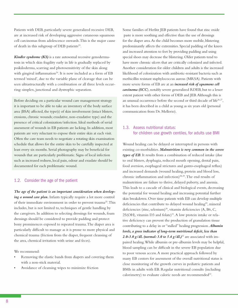
1.2. Consider the age of the patient
The age of the patient is an important consideration when develop-
ing a wound care plan. Infants typically require a lot more control of their immediate environment in order to prevent trauma25. This includes, but is not limited to, techniques of gentle handling by the caregivers. In addition to selecting dressings for wounds, foam dressings should be considered to provide padding and protect bony prominences exposed to repeated trauma. The diaper area is particularly difficult to manage as it is prone to more physical and chemical trauma (friction from the diaper, frequent cleansing of the area, chemical irritation with urine and feces).
We recommend:Removing the elastic bands from diapers and covering them •with a non-stick material. Avoidance of cleansing wipes to minimize friction•
Some families of Herlitz JEB patients have found that zinc oxide paste is more soothing and effective than the use of dressings for the diaper area. As the child becomes more mobile, blistering predominantly affects the extremities. Special padding of the knees and increased attention to feet by providing padding and using special shoes may decrease the blistering. Older patients tend to have more chronic ulcers that are critically colonized and infected. Another consideration for older children and adults is the increased likelihood of colonization with antibiotic-resistant bacteria such as methicillin resistant staphylococcus aureus (MRSA). Patients with more severe forms of EB are at an increased risk of squamous cell
carcinoma (SCC), notably severe generalized RDEB, but to a lesser extent patient with other forms of DEB and JEB. Although this is an unusual occurrence before the second or third decade of life26,27, it has been described in a child as young as six years old (personal communication from Dr. Mellerio).
1.3. Assess nutritional status: for children use growth centiles, for adults use BMI
Wound healing can be delayed or interrupted in persons with existing co-morbidities. Malnutrition is very common in the severe
types of EB. It results from a combination of reduced intake (due to oral blisters, dysphagia, reduced mouth opening, dental pain, food aversion, esophageal strictures and gastro-esophageal reflux) and increased demands (wound healing, protein and blood loss, chronic inflammation and infection)28,29. The end results of malnutrition are failure to thrive, delayed puberty, and anemia. This leads to a cascade of clinical and biological events, decreasing the potential for wound healing and increasing potential further skin breakdown. Over time patients with EB can develop multiple deficiencies that contribute to delayed wound healing29, mineral deficiencies (zinc, selenium)30, vitamin deficiencies (A, B6, C, 25(OH), vitamin D3 and folate)31. A low protein intake or rela-tive deficiency can prevent the production of granulation tissue contributing to a delay in or “stalled” healing progression. Albumin
levels, a gross indicator of long-term nutritional deficit, less than
2.0-3.0 g/dL (normal: 3.0 to 5.4 g/dL)32 are associated with im-paired healing. While albumin or pre-albumin levels may be helpful, blood sampling can be difficult in the severe EB population due to poor venous access. A more practical approach followed by many EB centers for assessment of the overall nutritional status is close monitoring of the growth curves in pediatric patients and BMIs in adults with EB. Regular nutritional consults (including calorimetry) to evaluate caloric needs are recommended33.

9
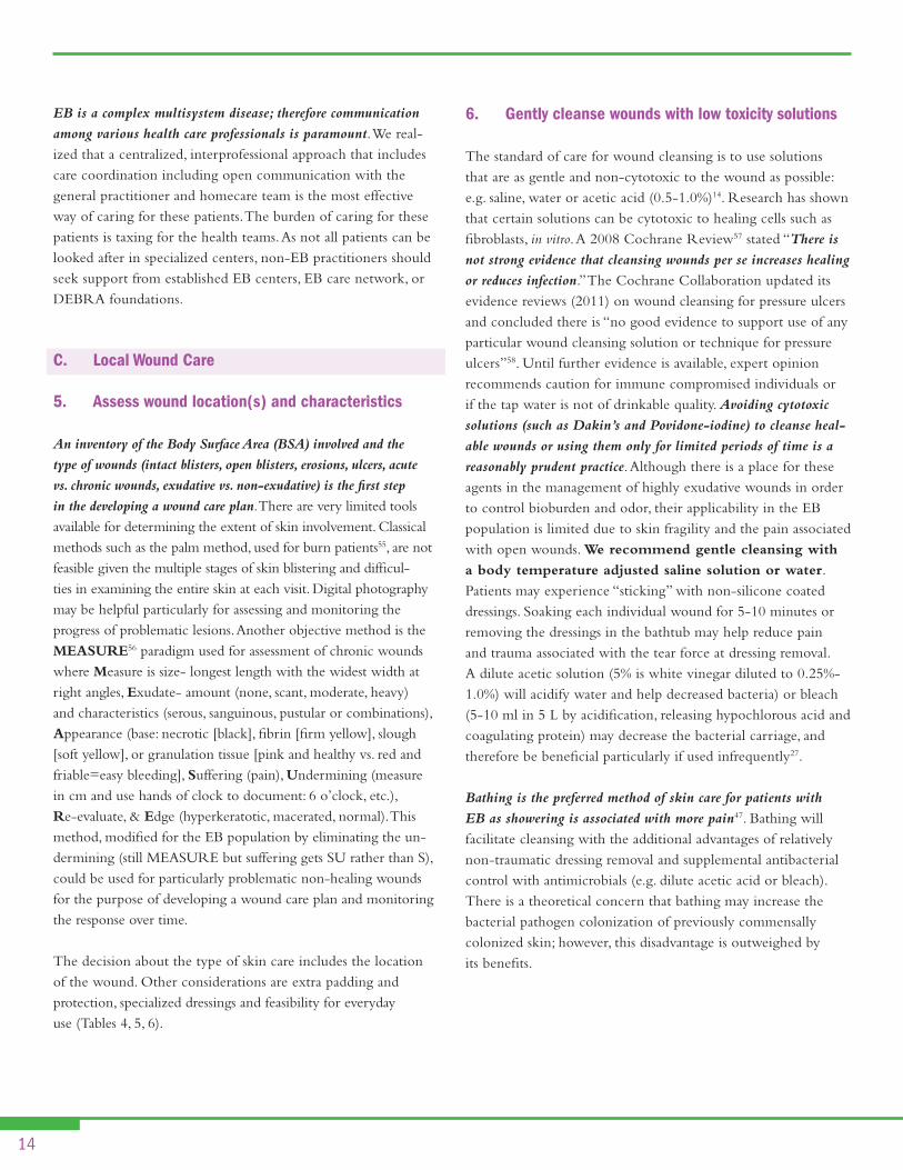
EB Simplexheel-prick blistering
EBSblistering and keratoderma
EBSDowling-Meara type
“herpetiform blistering”
Dominant DEBmilia formation
Recessive DEBchronic neck blistering
RDEBsquamous cell carcinoma
in a chronic ulcer
JEBhand blistering
JEBextensive neonatal blisters
EPIDERMOLYSIS BULLOSA
Type Specific Presentations
Figure 2:

10
1.4. Monitor hemoglobin levels: ideally over 80 g/L
Anemia is a frequent and serious complication of the severe types of EB such as RDEB and JEB. While its mechanisms are not fully understood multiple factors are likely involved:
increased blood loss through the skin and GI tract•decreased red cell production secondary to chronic •inflammationiron deficiency and decreased iron utilization•deficiencies of vitamin B12, folate and other minerals• 27
For persons with pressure ulcers, it has been proposed that hemo-globin less than 100 g/L may cause impaired wound healing due to decreased tissue oxygenation34. The low hemoglobin levels in EB patients are one of the factors that are likely to be associated with delay or “stalled” healing in these patients.
The panel’s consensus for EB related wounds was to aim for hemoglobin levels of 80 g/L or above as this is a more attainable goal and may avoid potential long-term complications of iron overload from intravenous iron therapy or red cell transfusions.
2. Develop Individualized Goals and Plan of Care
The interprofessional team should develop an individualized wound care plan after a thorough comprehensive assessment. The plan must be tailored to the individual and take into consideration his/her unique biopsychosocial needs.
Biopsychosocial Need Considerations
Individual personal preferences
Reflect, respect & integrate experiences •and feedback from the patient and those in circle of care
Risk factor EB subtype specific•
Comorbidities Anemia•Malnutrition•Cardiomyopathy•
Quality of life issues Pain management •Frequency of the dressing changes•Interference with daily activities•Schooling and/or employment•
Support systems/ circle of care
Consider who performs the skin care•Access to home care•Specialized EB teams•
Access to care Specialized EB interprofessional team•
Individualized patient preference must be respected and reflected in the plan of wound care35. Sackett et al. recognized three dimen-sions of equal importance: best available scientific evidence, clinical expertise and patient preference. It is common for EB families to have “time-tested” exceptional wound care routines that do not necessarily follow the currently accepted medical wisdom. Over the years we found that a flexible approach that includes the family and patient preferences is most likely to increase adherence, satisfaction with care, and improved outcomes.
With the new exciting, disease modifying cellular therapies that are currently emerging36, it is also important to maximize the chances of each patient being a potential candidate for these therapies. Early recognition of JEB-Herlitz is one example.
Care for Potential Candidates of Disease Modifying Cellular Therapies
Maintain overall health by preventing, recognizing & treating disease related complications
Anemia•Malnutrition•Cardiomyopathy•
Minimize risks of exposure to antibodies
Use filtered blood products•Consider risks of HLA exposure •with cellular products such as allogeneic skin grafting
Optimizing vaccination strategies for potentially immune-compromised individuals
Ensure compliance with vaccination •schedule before procedures
The wound care plan should be clearly outlined in a written docu-ment that is given to the family and copied to the health records, family practitioner and home care personnel. The care plan should also be evaluated and updated regularly.
2.1. Low hemoglobin consider: Fe supplementation, transfusion(s)
To date, there is not an ideal management strategy for dealing with
anemia in EB patients. A pathogenic based approach is sensible, but not always possible. Using adequate skin care and preventing/treating infection can minimize blood losses through the skin. Oral iron supplementation for correction of iron deficiency is widely used but its individual effectiveness varies. Patients fre-quently report gastrointestinal upset and constipation as reasons for non-adherence. Poor iron absorption may further limit how well replacements works27. Intravenous iron37 plus erythropoi-etin has been documented as being beneficial in small studies of

11
RDEB patients38. Blood transfusions should be considered for cases where Hgb levels are consistent below 80 mg/dL and/or for symptomatic patients who do not respond to other measures.
2.2 Treat low albumin: optimize nutrition, protein supplements, feeding tube
In order to optimize nutritional status, EB patients may need a
gastrostomy tube (G-tube). Although there are no clear guidelines for use in the EB population, indications for G-tube placement include: recurrent aspiration due to uncoordinated swallowing, ongoing difficulties with oral intake including administration of medication, significant weight loss or failure to gain weight and “falling off ” growth curves for infants and children29. Gastrosto-mies are reported in up to 40% of patients with JEB-Herlitz and in 4.2% of patients with RDEB39, although these numbers are currently likely to be higher40. With a G-tube in place it is easier to provide supplementary feeding at night while encouraging oral intake during the day. Supplementation of identified deficiencies is commonly suggested by many EB centers and 6 to 12 monthly monitoring to identify them is endorsed29.
B. Patient Centered Concerns
3. Address and support management of patient centered concerns to enable healing
3.1. Pain
Patients have a disease, but experience an illness and related suf-fering. This is particularly applicable to pain.
Pain is the most common symptom experienced by patients with EB, irrespective of the subtype.
The frequency and severity of pain is often proportional to the disease severity, with up to 50% of the patients with the most extensive type of EB (RDEB) experiencing daily pain >5 (0 to 10 scale)41. While the cause of pain in EB is multifacto-rial, the skin and related EB lesions are by far the most significant source of pain. The pain can occur at rest due to pressure exerted on blisters and denuded skin, secondary infection, friction and shear from bandages with physical movements. Pain can also be triggered or exacerbated by trauma during dressing changes or bathing and other activities of daily living. The assessment of
pain is conducted using various standardized tools that are age appropriate e.g. faces scales for young children and a visual analog scale or 0 to 10 numerical rating scale42. Multidimensional pain instruments should be considered for a comprehensive evalu-ation of pain and its impact on patients. Behavioral indicators of pain have also been developed for infants and those who cannot verbalize their pain. These tools are easy to administer in a variety of clinical circumstances, including at home by the family mem-bers. In order to develop an adequate pain management approach, pain levels should be recorded before, during and after dressing changes, bathing and other painful interventions43. Consideration of other patient-related factors (anxiety, previous experiences, lack of comfort, depression) is important in the under-standing of pain experience. These factors should be recognized and treated separately from wound-related pain. The impact of chronic unrelenting pain can be devastating, eroding the individual’s quality of life and constituting a significant amount of stress for patients and their families. Each patient is unique and may have individual preferences for approaches to manage pain including the use of medication (long acting, short acting, nociceptive and neuropathic agents) and other non-drug and complementary strategies (e.g. acupuncture, music, distraction, massage). It is considered best prac-tice to assess pain on an ongoing basis, to compare pain between dressing changes to acute pain that is repetitive at dressing change or incidental pain with procedures including surgical debridement. Regular pain assessments also provide the temporal pattern and identify potential aggravating and alleviating factors for pain44.
Increased levels of stress have been demonstrated to lower pain thresholds and decreased tolerance. The result is a vicious cycle of pain, stress/anxiety, anticipation of pain, and worsening of pain. Increased stress also activates the hypothalamic-pituitary adrenal (HPA) axis producing hormones that modulate the immune system, thus compromising normal wound healing45.
The approach to pain in an EB patient includes preventative and therapeutic modalities46. Prevention strategies include, but are not limited to, the use of protective atraumatic dressings, padding of the trauma prone areas, releasing fluid from tense blisters, avoiding adhesive dressings or skin adhesive products, removing dressings in water to hydrate the surface and limit friction with removal and treatment of skin infections.
Adjunctive non-medication pain therapies that have been de-scribed as being beneficial include physical modalities such as ice packs, vibration, distraction, relaxation and music therapy.

12
In circumstances where pain is anticipated (e.g. dressing changes), adjunctive procedures should be considered in conjunction with medical therapy. Therapeutic pain medication can be delivered topically and systemically (Table 3). Some potentially useful topical options include: addition of salt to the bath water to make it isotonic47; and using dressings with silicone contact surfaces to prevent pain and trauma on removal (e.g. Mepitel®, Mepilex®, Molnlycke, Sweden) or expert opinion on the use of dressings that have analgesics (Foam dressing with slow release ibuprofen: Biatin-IBU, Coloplast, Denmark not available in the USA).
In order to decide on the best pharmaceutical intervention, the mechanism of pain should be explored. In general, wound-
associated pain is both nociceptive and stimulus-dependent
(gnawing, aching tender, throbbing) vs. neuropathic or non-
stimulus-dependent or spontaneous pain (burning, stinging,
shooting, stabbing). Nociceptive pain is treated with the WHO pain ladder medication starting with aspirin and non-steroidal anti-inflammatories and then progressing to weak and strong narcotics (Table 3)48.
Short acting agents are often used to determine the dose of longer-acting agents with the short-acting agents then reserved for breakthrough pain including administration 30 to 60 minutes
before dressing change. Neuropathic pain often responds to tricyclic agents, particularly second-generation agents high in anti-noradrenalin activity. Nortriptyline and desipramine are often better than amitriptyline. For non-responders results may be better with alternative agents such as gabapentin, pregabalin or other anti-epileptics48. Procedural pain (e.g. before dressing changes, bathing) should be managed with an interprofessional approach, reflected upon and communicated to those within the circle of care. This is especially important in the care of EB children as they often required re-peated unpleasant procedures. If procedural pain is not adequately managed, long-lasting negative outcomes may result and the ther-apeutic relationship could be jeopardized, therefore care planning is essential to procedural pain management. Oral sucrose 24% is a useful, short-acting analgesic that is effective for children under two years of age49. For older children and adults, acetaminophen or morphine administered 30 minutes prior to the procedure may be used. Non-pharmacological modalities (Table 3) are also helpful in combination with the pharmacological measures listed above. In the general population, music therapy has been found to improve patients’ moods when experiencing anxiety during care delivery50 and for depression51. Music therapy has been used successfully in several EB clinics to manage the pain and anxiety associated with wound care assessments.
Pain Management Strategies Goals/Types Actions
Preventative Avoid traumaAvoid blister expansionPrevent local infection
Protection, use foam dressings, use soft sleeping and seating surfaces•Clothing and shoe modification•Release fluid from blister, maintain roof of blister over affected area•Cover open areas•Control local colonization•Use of hand cleansers by caregivers prior to dressing changes•
Therapeutic Pharmacological
Non-pharmacological
Nociceptive:mild pain: Acetaminophen ± NSAIDS•moderate pain: Acetaminophen ± NSAIDS + morphine•severe pain: Acetaminophen + NSAIDS + morphine/other strong opioids•
Neuropathic:tricyclics (nortriptyline, desipramine), gabapentin, pregabulin, other anti-epileptics•relaxation/distraction•biofeedback•physical modalities (e.g. vibration, cooling)•
Table 3: Pain Management Strategies
NSAIDS = non-steroidal anti-inflammatory drugs

13
3.2. Itch Itch, also known as pruritus, can be a most distressing experi-
ence for any individual with skin disorders and is often affiliated
with depression, sleep impairment and overall distress 52. Kini et al.(2011) commented on their study findings: “Chronic pruritus has a substantial impact on Quality of Life (QoL), one that may be comparable to that of pain. The severity of symptoms and the use of support networks are the main factors that determine the degree to which patients are affected by their symptoms. Address-ing support networks in addition to developing new therapies may improve the QoL of itchy patients.”
Itch is a common symptom in the EB population. The exact mechanism is not known: abnormal persistent skin inflamma-tion, overheating due to dressings, local sensitizers, and the use of systemic opioids that are histamine releasers are all potential contributors46. The pruritus leads to more skin blistering from skin trauma, which in turn exacerbates the pruritus. Itch is often a poorly controlled symptom in most patients with EB as there is not one modality that targets all potential pathogenic components including histamine, slow reacting substances or prostaglandins. Management should start with a thorough history to identify the timing when itch is more severe and factors that will exacerbate itch. Occasionally changing the topical routine (switching dressings or discontinuing topical antibiotics) may be sufficient to bring the pruritus down to a manageable level. Establishing the time of the day when itching is most significant is also important. Itching at night may be related to body overheating and can be treated with sedating antihistamines (e.g. Hydroxyzine) or a tricyclic antidepressant with prominent H1 antihistamine action (e.g. doxepin). Apart from sedation, the advantage of using tricy-clics is their mild antidepressant effects (doxepin at higher doses).
Daytime pruritus requires the addition of a non-sedating antihis-tamine H1-blocker such as Cetirizine or Loratadine. Using H1 antihistamines from different chemical classes can give a syner-gistic effect on the competitive blocking of the H1 blocker with some other agents such as Ketotophen having additional mast cell stabilizing properties as well. Liquid preparations are always preferable for breakthroughs as they have a shorter onset of action, are easier to swallow and can be administered via G-tubes. For persistent itching there are anecdotal reports of successful use of Ondansetron or low dose Gabapentin46. Clinicians should be
aware of potential sensitizers that can trigger itch in dressing and other topical preparation. According to studies fragrance is one of the most common skin sensitizers53.
3.3. Activities of daily living Pain, odor, mobility limitations have a significant impact on the
EB patients and their daily living. The disease burden may in-clude a difficulty performing personal care, difficulty in engaging in school or employment activities, increased financial burden and ultimately impacts on engagement in and enjoyment of life. Depression and anxiety are also common54 and further contribute to social isolation. Fostering independence and safety during activities of daily living may help to decrease the burden of disease, and reduce dependence on others. Significant environ-
mental modifications (e.g., special seating in baths, wheelchairs,
footwear) have to be put in place to diminish the amount of
trauma and suffering that these patients experience and to make
their environment functional for their needs. A rehabilitation consult early on with frequent re-evaluations is recommended. EB can have a detrimental aspect on individual’s image of self. In addition to appearance, odour and leakage from wounds often prohibit patients from engaging in social activities that people would take for granted. It is important to remember that most patients grow up with the disease and the impact of social isola-tion and disruption of daily routine (e.g. schooling) on personality development can be tremendous.
4. Provide education and support to the patient/parent and their circle of care to increase treatment adherence (compliance)
In order to provide support and education to an EB patient, one has to gain the trust of the patient and his/her family by
developing a therapeutic relationship. This occurs when trust, communication and open dialogue allow the patient and those in their circle of care to understand that each person involved has a meaningful contribution. Having a patient assist with the decision-making process provides reassurance that the team is working with them. A constant dialogue with all key players is very important. EB families often develop highly individualized unconventional routines that are very difficult to change either because of the feasibility of implementing new treatment modali-ties or because of a learned distrust of the medical community.
14
EB is a complex multisystem disease; therefore communication
among various health care professionals is paramount. We real-ized that a centralized, interprofessional approach that includes care coordination including open communication with the general practitioner and homecare team is the most effective way of caring for these patients. The burden of caring for these patients is taxing for the health teams. As not all patients can be looked after in specialized centers, non-EB practitioners should seek support from established EB centers, EB care network, or DEBRA foundations.
C. Local Wound Care
5. Assess wound location(s) and characteristics
An inventory of the Body Surface Area (BSA) involved and the
type of wounds (intact blisters, open blisters, erosions, ulcers, acute
vs. chronic wounds, exudative vs. non-exudative) is the first step
in the developing a wound care plan. There are very limited tools available for determining the extent of skin involvement. Classical methods such as the palm method, used for burn patients55, are not feasible given the multiple stages of skin blistering and difficul-ties in examining the entire skin at each visit. Digital photography may be helpful particularly for assessing and monitoring the progress of problematic lesions. Another objective method is the MEASURE56 paradigm used for assessment of chronic wounds where Measure is size- longest length with the widest width at right angles, Exudate- amount (none, scant, moderate, heavy) and characteristics (serous, sanguinous, pustular or combinations), Appearance (base: necrotic [black], fibrin [firm yellow], slough [soft yellow], or granulation tissue [pink and healthy vs. red and friable=easy bleeding], Suffering (pain), Undermining (measure in cm and use hands of clock to document: 6 o’clock, etc.), Re-evaluate, & Edge (hyperkeratotic, macerated, normal). This method, modified for the EB population by eliminating the un-dermining (still MEASURE but suffering gets SU rather than S), could be used for particularly problematic non-healing wounds for the purpose of developing a wound care plan and monitoring the response over time.
The decision about the type of skin care includes the location of the wound. Other considerations are extra padding and protection, specialized dressings and feasibility for everyday use (Tables 4, 5, 6).
6. Gently cleanse wounds with low toxicity solutions
The standard of care for wound cleansing is to use solutions that are as gentle and non-cytotoxic to the wound as possible: e.g. saline, water or acetic acid (0.5-1.0%)14. Research has shown that certain solutions can be cytotoxic to healing cells such as fibroblasts, in vitro. A 2008 Cochrane Review57 stated “There is
not strong evidence that cleansing wounds per se increases healing
or reduces infection.” The Cochrane Collaboration updated its evidence reviews (2011) on wound cleansing for pressure ulcers and concluded there is “no good evidence to support use of any particular wound cleansing solution or technique for pressure ulcers”58. Until further evidence is available, expert opinion recommends caution for immune compromised individuals or if the tap water is not of drinkable quality. Avoiding cytotoxic
solutions (such as Dakin’s and Povidone-iodine) to cleanse heal-
able wounds or using them only for limited periods of time is a
reasonably prudent practice. Although there is a place for these agents in the management of highly exudative wounds in order to control bioburden and odor, their applicability in the EB population is limited due to skin fragility and the pain associated with open wounds. We recommend gentle cleansing with
a body temperature adjusted saline solution or water. Patients may experience “sticking” with non-silicone coated dressings. Soaking each individual wound for 5-10 minutes or removing the dressings in the bathtub may help reduce pain and trauma associated with the tear force at dressing removal. A dilute acetic solution (5% is white vinegar diluted to 0.25%-1.0%) will acidify water and help decreased bacteria) or bleach (5-10 ml in 5 L by acidification, releasing hypochlorous acid and coagulating protein) may decrease the bacterial carriage, and therefore be beneficial particularly if used infrequently27. Bathing is the preferred method of skin care for patients with
EB as showering is associated with more pain47. Bathing will facilitate cleansing with the additional advantages of relatively non-traumatic dressing removal and supplemental antibacterial control with antimicrobials (e.g. dilute acetic acid or bleach). There is a theoretical concern that bathing may increase the bacterial pathogen colonization of previously commensally colonized skin; however, this disadvantage is outweighed by its benefits.

15
Type of Wound/indication Primary dressing Secondary dressing Topical therapy
Protection Soft silicone foams•Modified absorbent pads •Lipidocolloid dressings•Contact layers•
Burn net to keep in place •(if feasible)
None•
Open non-exudative Soft silicone foams•Modified absorbent pads •Lipidocolloid dressings•Contact layers•
Burn net to keep in place •(if feasible)
None•
Exudative Soft silicone foams•Lipidocolloid dressings•Hydrofibers•
Burn net to keep in place •(if feasible)
Topical antibiotics •(avoid allergens)
Eschar Hydrogels•Biosynthetic cellulose•
Foams•Modified absorbent pads•
None•
Critically colonized or infected Contact layer + Ag or other anti-microbial
Atraumatic foams •Hydrofibers•Alginates•
Foams•Modified absorbent pads•
Topical antibiotics •(avoid allergens)
Pain Soft silicone coating •(e.g. Safetac® Technology)Biosynthetic cellulose•Hydrogel sheets•
Foams•Modified absorbent pads•
Topical NSAIDs•
Itch Soft silicone coating •Biosynthetic celluloseHydrogel sheets•
Foams•Modified absorbent pads•
Short course of topical •mid-potency corticosteroids
Hypergranulation Contact layer +/- silver or other •antimicrobial
Foams•Modified absorbent pads•
Short course of topical potent •corticosteroids
Table 4: Dressing choices according to indications/type of wounds

16
Type of Wound/indication Primary dressing Secondary dressing Topical therapy
Foams Mepilex®• 3 Mepilex Lite®• 3 Mepilex Border®• 3
Mepilex Border Lite®• 3
PolyMem• 2 (expert opinion from panel members)
Contains silicone layer to make these non-adherent•
Generally made from hydrophilic polyurethane•Non-occlusive. Semi-permeable surface allows •exudate into the dressing and foam traps moisture
Allow large amounts of fluid and wound •drainage to be absorbedProvide padding and protection to •woundsDepending on the amount of exudate, •can be left in place up to 7 daysSome require secondary dressing to •hold in placeBordered dressing may sometimes be too •sticky and should be used with caution
Hydrogels Gels:•Duoderm• 4 Intrasite• 5
Sheets (Cool dressings):•ActiFoamCool• 6
Intrasite Conformable• 5
Made out of insoluble polymers that expand in •water and hydrate woundsProvide autolytic debridement•
For wounds with minimal or no exudate•Due to hydrating capacity, these offer •cooling effect and may aid in relief of pain, itch and discomfort
Alginates (calcium or calcium/sodium)
Kaltostat• 7 Made of non-woven fibers derived from seaweed•Turn into a non-sticky gel when in contact with •wound drainage
Requires exudate•Does not work on dry wounds or •wounds with escharCalcium alginate dressings release •calcium ions that help stop bleeding
Hydrofibers Aquacel• 7 Made out of sodium carboxymethyl- cellulose that •when in contact with wound drainage becomes a gel and provides a moist environment
More absorbent than alginates•Consider in wounds with heavy drainage•
Modified absorbent pads Telfa• 8
Restore• 1
Mesorb• 3
Thin layer of absorbent cotton fibers that are •enclosed in a sleeve of perforated polyethylene terephthalate and sealed along two edgesA plastic film prevents dressing from adhering •to wound surface and perforated surface allows passage of exudate into the pad
Contact layers Mepitel®• 3 Mepitac®•Silflex• 9 Adaptic touch• 10 Siltape• 9
Protective, inert material that allows non-traumatic •removal (Mepitel® scientific studies, others expert opinion)
Biosynthetic cellulose Suprasorb X• 11 Dressing consisting of cellulose, water and •0.085% chlorhexidine gluconate (preservative) that has ability to both absorb and donate moisture
Also considered a cooling dressing, •aids in pain reduction and adding moisture to woundsMay also reduce itch•
Lipidocolloid dressings Urgotul• 12
Restore (North American •equivalent to Urgotul)
Composed of an open weave polyester mesh •impregnated with hydrocolloid polymers dispersed within petrolatumWhen in contact with exudate, the hydrocolloid •polymers are hydrated and constitute with the petrolatum a lipidocolloid interface that provides a non-adherent surface
For wounds with exudate. Also used for •protection of vulnerable areas
Table 5: Dressings categories, properties, indications
1 Hollister,2 Ferris Mfg.,3 Molnlycke Health Care,4 ConvaTec,5 Smith & Nephew,6 Activa Healthcare,7 ConvaTec,8 Kendall Company Ltd, 9 Advancis Medical,10 Systagenix,11 Activa Healthcare,12 Urgo, 13 3M Healthcare

17
Location Dressing / topical therapy Properties Expert comment (opinion)
Perianal area Restore contact layer• Autolytic debridement•Provides moisture•
Difficult to keep in place•Can be used to line the diaper •
Intrasite conformable • Autolytic debridement•Provides moisture•
Difficult to keep in place•Can be used to line the diaper•
Bepanthen (ointment with Pro Vitamin B5)• Aids in moisture balance•
Cavilon (liquid barrier film)• Creates breathable, transparent •coating on the skin
Does not sting •Alcohol free•
Emollin 50/50 emollient spray •(CD Medical Ltd) (white soft paraffin and liquid paraffin)
Water-repellent•Provides barrier protection•
Does not sting•
Oral mucosa BioXtra (salivary substitute)• Provides moisture•
Difflam Spray (active ingredient is •benzydamine hydrochloride, and NSAID)
Reduces pain and inflammation•Also acts as local anesthetic•
Corsodyl (mouthwash •containing chlorhexidine)
Provides antiseptic and •disinfectant properties
Gelclair (bioadherent oral gel)• Creates barrier that protects •nerve endings, reducing pain
Can be used prior to meals•
Feeding tube sites AMD- PHMB foam fenestrated disc •dressing (antimicrobial foam dressing)
Moisture balance•Contains antiseptic •(Polyhexamethylene biguanide, •PHMB) (Effective against MRSA, VRE, gram + and gram – bacte-ria, fungi and yeast
4% sucralfate mixed with Cavilon• Protectant•
PC/C lines, fixator Mepitac®, Mepitel® Adaptic touch, Siltape • Non-stick•
Adhesives Medical adhesive remover (Hollister)•Appeel (CliniMed Ltd)or Niltac •(North American equivalent)Adhesive remover spray (Coloplast)•
Adhesive remover is temporary.•These sprays are silicone-based.•
Retention bandage Tubifast™•Acti-Wrap cohesive retention •bandage(Activa Healthcare)
Secures dressings in place• Useful to fix small dressings•
Table 6: Dressing choices/topical therapy for special locations/indications

18
7. Debridement Initially, wounds in EB patients present as blisters. To prevent blisters from enlarging, it is important to carefully puncture the blister with a sterile needle to release the inner fluid and prevent blister extension with fluid tracking. It may be necessary to punc-ture the blister at various sites to optimize the fluid release. The fluid should be allowed to drain on its own, as excessive pressure at the site may lead to further extension of the blister ⁄bulla. The overlying skin should never be removed as it acts like a natural dressing and aids healing, reducing pain, and minimizing the risk of exogenous infection.
When the wound is covered with a firm dehydrated eschar or soft slough the normal healing process is impaired. A firm eschar
serves as a pro-inflammatory stimulus that inhibits healing, while the slough acts as a culture media for bacterial prolifera-tion debridement. Debridement promotes healing by removing senescent cells that are deficient in cellular activities and remov-ing biofilms that maintain the inflammatory process59. Debride-ment in the EB population—in contrast to methods for other chronic wounds—should be extremely gentle and whenever possible involve non-physical methods (i.e. autolytically using hydrogel or calcium alginate dressings).
8. Assess and treat
8.1. Superficial critical colonization & abnormal inflammation
Chronic wounds contain bacteria; however, the presence of bacteria
obtained from a surface swab does not define infection. The bacte-rial implications for healing are dependent on the bacterial load and virulence. Contamination refers to smaller bacterial loads on the wound surface and colonization refers to the establishment of bacterial colonies in the tissue, usually without interfering with healing. Critical colonization occurs when the bacterial prolifera-tion causes local damage and the wound to get “stuck” or stalled precluding healing. Infection is determined by the overall bacte-rial load, typically defined as >105 colonies per gram of tissue (i.e. 1.0x10⁶ or higher), the nature of the invading bacteria and most importantly host resistance60. Surface critical colonization and deep and surrounding skin infection are clinical diagnoses. The mnemonics NERDS© and STONEES©61,62, which repre-sent the two levels of bacterial damage or infection, have been
validated for use in chronic wounds. New or increasing pain is a symptom that worsens with bacterial damage to cause superficial critical colonization or deep and surrounding wound margin infection as validated by Gardiner et al14.
Any three NERDS criteria are required for superficial critical colonization and the need for a topical antimicrobial:
Non-healing: The wound is not getting larger or smaller •Exudate is increasing (host response to noxious damage)•Red friable tissue (indicating over production of blood vessels •due to increased VEGF production stimulated by bacteria)Debris: New dead slough on the surface that needs to be •distinguished from shed epidermal blister roof Smell: This indicates the presence of gram negatives and •anaerobic organisms
For deeper or surrounding skin infection and systemic therapy any 3 of the STONEES criteria are required:
Size increasing: This is due to bacteria destroying the wound •margin and/or base Temperature: infrared thermometry is a good measure •of surrounding tissue change (mirror image >3 degree Fahrenheit warmer)Os is Latin for bone: exposed or probing to bone •New areas of breakdown that indicate satellite involvement •with the wound Erythema and or Edema of the surrounding skin that are •the clinical signs of cellulitis Exudate increase as in NERDS •Smell as in NERDS: If exudate/smell are present, an •additional criteria is needed to define bacterial damage as superficial, deep or both
Although these concepts need to be validated for persons with EB, the need for 3 criteria is useful to help distinguish infection from persistent inflammation. Persons with EB may not demonstrate all the classical signs of deep infection but these signs should help to identify more subtle changes associated with bacterial damage. The most common bacteria isolated from chronic and most likely EB wounds are gram-positive organisms (Staphylococcus Aureus and Streptococci species), gram negatives (Pseudomonas aeruginosa) and anaerobes (personal communication from R. Gary Sibbald). Documenting critical colonization/infection in the EB population is rarely needed.

19
Skin swabs are indicated only to determine antibiotic selection in cases where multi-resistant organisms or non-responsive infection is suspected. A proper technique is the Levine tech-nique, i.e. rotating a bacterial swab 360 degrees over a clinically normal area of skin with just enough pressure to extract fluid63. The swab can be pre-moistened with the transport media if the wound surface is relatively dry. This technique requires that the wound surface be cleansed prior to collection64. Critical colonization can be controlled with topical agents. The bacterial load may be reduced by bathing with diluted bleach, applying compresses or using sprays with diluted vinegar64. Lipid-stabilized hydrogen peroxide cream (Crystacide available in the UK) is well tolerated and effective when applied directly on the wound or on the dressing that comes in contact with the colo-nized wound25,63. Topical antibiotics (e.g. Polysporin, Fucidic Acid, Mupirocin) should be used only for short periods of time and rotated every two to six weeks to prevent resistance, and clini-cians should watch for sensitization. In addition, the use of potent topical sensitizers (e.g. neomycin) should be discouraged. There are a variety of dressings containing silver, honey, iodine in a cadexomer carbohydrate and polyethylene glycol slow release for-mulation, and PHMB (PolyHexaMethyleneBiguinide) that may facilitate a decrease of critical colonization. (Tables 4, 6). The use of antimicrobial dressings should be reviewed at regular intervals, preferably every two to four weeks, and discontinued if critical colonization has been corrected or if there is not a demonstrable beneficial effect14. There is currently a great tendency to overuse antimicrobial dressings, which is not cost effective. Silver’s broad spectrum of antimicrobial activity can be used in critically colonized chronic wounds that have the ability to heal. Silver must be ionized to exert an antimicrobial effect. Ionized silver requires an aqueous or water environment and should not be used in a maintenance or non-healable wound where the desired outcome is the combination of moisture reduction and bacterial reduction14. Silver should not be in close proximity to any oil-based products (e.g., petrolatum, zinc oxide), where the oil molecules may interfere with the ionization of the silver14. Prod-ucts that produce a continuous supply of ionized silver are likely to be more effective and higher levels of silver release are often necessary to treat pseudomonas14. The amount of silver released from these dressings is a fraction of the silver released from silver sulfadiazine cream formulations. Silver sulfadiazine has been asso-ciated with argyria (permanent silver deposits in the dermis with a blue discoloration to the skin) and silver dressings have been
associated with periwound staining but there are no published reports of argyria for patients that have not had silver sulfadiazine cream. There are anecdotal reports of high serum levels of silver after silver dressing use in Epidermolysis Bullosa (personal com-munication from Jemima Mellerio); therefore, despite the fact that they release less silver than silver sulfadiazine cream, their pro-longed use should be discouraged especially if used on large areas or on individuals with a large surface area to total body weight. There is evidence of effectiveness of other topical antibacterial agents in other wound indications including medical grade honey products (ointments, dressings). The honey preparations provide short-term benefit (low pH, high osmolality, hydrogen peroxide release) but their use can increase local pain and may temporarily increase exudate levels65. Once the honey becomes diluted with wound fluid, malodour will be associated with bacterial growth, including gram negatives and anaerobes. 8.2. Deep/surrounding tissue infection/
generalized inflammation The signs of deep and surrounding tissue infection are outlined
in the mnemonic STONEES where 3 or more criteria are an
indication for systemic antimicrobial therapy. Regional and con-stitutional signs and symptoms (lymphadenopathy, fever, malaise) are important for prompt antimicrobial therapy initiation and the potential need for parental therapy. Empirical use of systemic antibiotics that cover common pathogens is recommended. The antibiotic choice can be further refined once the bacterial swab results identifying pathogenic organisms and their antimicrobial sensitivities. Administration via oral route may be sufficient, although longer treatment durations may be needed. Bacterial
swabs positive for Streptococcus should be treated even in the absence
of overt clinical infection due to the risk of complications (sepsis,
toxin mediated disease, nephropathy)64.
For chronic non-healing wounds or frequent blister formation,
systemic, low-dose anti-inflammatory antibacterial agents may
be used for longer periods of time (e.g. trimethoprim, macrolides,
and doxycycline). These agents are useful in controlling low grade bacterial damage combined with local and systemic inflammation12 and have not commonly been associated with bacterial resistance (personal communication from Elena Pope). An alternating schedule of two to three months between any two or three agents is prudent.
20
9. Select an appropriate dressing/topical therapy that is appropriate for the needs of the patient and the caregiver based on the subtype of EB
The approach for dressing choices should be based on the type of
EB, extent and location of the wound, dressing frequency, cost and
availability. It is important to remember that given the various types of wounds that a particular EB patient may experience at one given time, the choice of dressing should be individualized for each wound. Chronic wounds are the most difficult ones to manage in patients with EB. Chronic wounds may be “stalled” in the inflammatory stage14. These wounds demonstrate marked increased activity of inflammatory cells and associated mediators such as matrix metalloproteinase (MMPs) and elastase14. Wound healing is stalled because degradation of extracellular matrix and growth factors occurs more rapidly when their synthesis is hindering the wound from progressing towards the proliferative phase and ultimately re-epithelialization14,66. Moore et al. reported that the longer a wound remains in the
inflammatory phase, the more cellular defects are detected with
potential delayed healing66. Recently, there has been a renewal of interest in wound diagnostic testing that will result in tests for increased metalloproteases at the bedside14 and there are wound dressings with oxidized reduced collagen and cellulose that can trap metalloproteases and these dressings can be combined with antimicrobials such as silver. In the Sibbald cube14, these special-ized dressings can be combined antimicrobials depending on the presence of the NERDS (superficial antibacterial dressing criteria) or STONEES (systemic antibiotic) where the presence of increased inflammation can be treated topically or systemically. Appropriate moisture is required to facilitate the action of growth
factors, cytokines, and the migration of cells including fibroblasts and
keratinocytes. Moisture equilibrium is a delicate balancing act. Excessive moisture can potentially cause damage to the skin sur-rounding a wound, leading to maceration of the keratin, increased bacterial proliferation and potential breakdown. Conversely, inadequate moisture in the wound environment can impede cel-lular activities and promote eschar formation resulting in delayed and poor quality of wound healing. A moisture-balanced wound environment is maintained primarily by using ‘modern” dressings with occlusive, semi-occlusive, absorptive, hydrating, and haemo-static characteristics, depending on the drainage of the wound bed. Tables 4 and 5 provide a practical approach to wound depen-dent dressing choices for the EB population.
10. Evaluate the expected rate of healing or reassess wound goals of care (including potential maintenance status)
It is noted that a wound size reduction of 20% to 40% in two and four weeks is likely to be a reliable predictor of healing67,68. In a study with persons with diabetic foot ulcers using complete healing as the end point, a 30% reduction at week four was a good predictor for healing by week 1269. One measure of heal-
ing is clinical observation of the edge of the wound: nonhealing
wounds often have a cliff like edge instead of the tapered sandy
shore of a beach with a gradually sloping contour that is often
purple due to new epithelialization. If the wound edge is not migrating after appropriate wound bed preparation (debride-ment, bacterial balance, moisture balance) and healing is stalled, then advanced therapies should be considered to overcome the non-advancing Edge effect70. This may be considered after other causes and co-factors of delayed healing have been ruled out. Clinicians need to remember that wound healing is not always the primary outcome. Complete healing is particularly difficult
in severe types of EB. Other wound-related outcomes such as less pain, reduced bacterial load, fewer dressing changes with decreased exudate and odour, and/or an improved quality of life, may be more attainable.
11. Edge effect: If a wound is stalled or the edge or other areas appear atypical, consider a skin biopsy to rule out squamous cell carcinoma or other complications prior to considering active therapeutic options
Squamous cell carcinoma (SCC) is a major cause of morbidity and
mortality in patients with EB, particularly those with RDEB. The cumulative risk of developing SCC in severe generalized RDEB by age 55 is 90.1%, much higher than that of the general aged matched population of the United States (9%-14% among men and 4%-9% in women)71. SCC tends to occur much earlier in the EB population is multifocal and more aggressive, leading to increased mortality (over 55% of severe generalized RDEB patients die from SCC by 40 years of age)72,23. As chronicity is the norm in many EB patients, a high degree of suspicion is required at the sites of chronic blistering, which may in fact be SCC. Any wounds that enlarge rapidly, have increased pain, appearance changes on serial photographic documentation or “feel different” for the patient should be biopsied73.

21
D. Provide Organizational Support
12. Consider a health care system support structure including specialized nurses, interprofessional clinics and a structured approach to new cases
EB is not “just a skin disorder”, therefore treating it requires
involvement of a dedicated team with expertise in all aspects of care. The expression “it takes a village” (Figure 3) clearly illustrates not only the number of people who may need to be involved in patient’s care, but also emphasizes that practitioners need to part-ner with patients and family in order to provide the best care.
Over the past decade EB specialized clinics have been opened worldwide in 16 countries. These clinics provide an interpro-fessional model of care utilizing the expertise of allied health professionals (nurses, physicians, occupational therapists, physical therapist, PT, social workers, dietitian, music therapists, etc.). These valuable teams of EB experts offers aspects of patient care that crosses all affected healthcare systems with strong community linkages. Isolated cases can be overwhelming to health practitioners particularly when referral to an established EB center is not feasible. Access to international EB experts via http://www.internationalebforum.org is possible and has changed the fabric of pre-existing professional isolation.
Other resources for patients and practitioners are DEBRA foundations that exist in many countries.
The birth of a child with EB is a traumatic event for a family. We find that imparting early education about the disease, determining the type/subtype of EB as soon as possible, and providing ongoing support from knowledgeable practitioners, allows a family to regroup and focus on providing the best care to their baby. We also find that a flexible approach at each visit that involves the family’s agenda is more likely to lead to good care and building trust.
Conclusion
EB is one of the most complex diseases in medicine with severe EB types having devastating effects on the quality of life & life span of affected patients and their families. Wound care requires an inter-professional coordinated approach that addresses the patient as a whole. We have brought together experts in the field of EB, the science of wound care & clinical wound care practice to provide the best available approaches for optimal wound care to the EB individuals. Until a definitive cure becomes available, these practices’ goals are to minimize suffering, improve wound healing and prepare patients for potential corrective procedures thereby improving the lives of afflicted individuals.
Figure 3: EB Care Team

22
Fine JD, Eady RA, Bauer EA, Bauer JW, Bruckner-Tuderman L, Heagerty A, 1. Hintner H, Hovnanian A, Jonkman MF, Leigh I, McGrath JA, Mellerio JE, Murrell DF, Shimizu H, Uitto J, Vahlquist A, Woodley D, Zambruno G. The classification of inherited epidermolysis bullosa (EB): Report of the third international consensus meeting on diagnosis and classification of EB. J Am Acad Dermatol. 2008; 58: 931-50.
Fine JD, Mellerio JE. Extracutaneous manifestations and complications of 2. inherited epidermolysis bullosa. Part I. Epithelial associated tissues. J Am Acad Dermatol. 2009a; 61:367-84.
Fine JD, Mellerio JE. Extracutaneous manifestations and complications of 3. inherited epidermolysis bullosa. Part II. Other organs. J Am Acad Dermatol. 2009b; 61: 387-402.
Fine JD, Johnson LB, Weiner M, Stein A, Cash S, Deleoz J, Devries DT, 4. Suchindran C. Eye involvement in inherited epidermolysis bullosa: experi-ence of the national epidermolysis bullosa registry. Am J Ophthalmol. 2004; 138:254-62.
Matsumoto Y, Dogru M , Tsubota K. Ocular surface findings in hallopeau-5. siemens subtype of dystrophic epidermolysis bullosa: report of a case and literature review. Cornea. 2005; 24:474-9.
Wright JT, Fine JD, Johnson L. Dental caries risk in hereditary epidermolysis 6. bullosa. Pediatr Dent. 1994; 16:427-32.
Freeman EB, Koglmeier J, Martinez AE, Mellerio JE, Haynes L, Sebire NJ, 7. Lindley KJ, Shah N. Gastrointestinal complications of epidermolysis bullosa in children. Br J Dermatol. 2008; 158:1308-14.
Fine JD, Johnson LB, Weiner M , Suchindran C. Gastrointestinal compli-8. cations of inherited epidermolysis bullosa: cumulative experience of the national Epidermolysis bullosa registry. J Pediatr Gastroenterol Nutr. 2008; 46:147-58.
Chan SM, Dillon MJ, Duffy PG, Atherton DJ. Nephro-urological complica-9. tions of epidermolysis bullosa in paediatric patients. Br J Dermatol. 2007; 156:143-7.
Fine JD, Johnson LB, Weiner M, Stein A, Cash S, DeLeoz J, Devries DT, 10. Suchindran C. Genitourinary complications of inherited Epidermolysis bullosa: experience of the national Epidermolysis bullosa registry and review of the literature. J Urol. 2004; 172:2040-4.
Sidwell RU, Yates R, Atherton D. Dilated cardiomyopathy in dystrophic 11. epidermolysis bullosa. Arch Dis Child. 2000; 83:59-63.
Lara-Corrales I, Mellerio JE, Martinez AE, Green A, Lucky AW, Azizkhan 12. RG, Murrell DF, Agero AL, Kantor PF, Pope E. Dilated cardiomyopathy in epidermolysis bullosa: A retrospective, multicenter study. Pediatr Dermatol. 2010; 27(3): 238-43.
Martinez AE, Allgrove J, Brain C. Growth and pubertal delay in patients 13. with epidermolysis bullosa. In: Epidermolysis bullosa: part II - diagnosis and management, Murrell DF, Thiers BH, editors. Dermatol Clin. 28, 2. Philadelphia:Saunders; 2010.
Sibbald RG, Goodman L, Woo KY, Krasner DL, Smart H, Gulnaz T, Ayello 14. EA, Burrell RE, Keast DH, Mayer D, Norton L. Special Considerations in Wound Bed Preparation 2011: An Update. Advances in skin & wound care. 2011; 24(9):415-436.
Coulombe PA, Kerns ML, Fuchs E. Epidermolysis bullosa simplex: a para-15. digm for disorders of tissue fragility. J Clin Invest. 2009; 119:1784-93.
Pfendner E, Rouan F, Uitto J. Progress in epidermolysis bullosa: the pheno-16. typic spectrum of plectin mutations. Exp Dermatol. 2005; 14:241-9.
McGrath JA, Mellerio JE. Ectodermal dysplasia-skin fragility syndrome. 17. Dermatol Clin. 2010a; 28:125-9.
McGrath JA, Bolling MC, Jonkman MF. Lethal acantholytic epidermolysis 18. bullosa. Dermatol Clin. 2010b; 28:131-5.
Nakano A, Chao SC, Pulkkinen L, Murrell D, Bruckner-Tuderman L, 19. Pfendner E, Uitto J. Laminin 5 mutations in junctional epidermolysis bullosa: molecular basis of herlitz vs. non-herlitz phenotypes. Hum Genet. 2002; 110:41-51.
Kiritsi D, Kern JS, Schumann H, et al. Molecular mechanisms of phenotypic 20. variability in junctional epidermolysis bullosa. J Med Genet. 2011; 48:450-7.
Chung HJ, Uitto J. Epidermolysis bullosa with pyloric atresia. 21. Dermatol Clin. 2010; 28:43-54.
Van den Akker PC, Jonkman MF, Rengaw T, Bruckner-Tuderman L, Has C, 22. Bauer JW, Klausegger A, Zambruno G, Castiglia D, Mellerio JE, McGrath JA, van Essen AJ, Hofstra RM, Swertz MA. The international dystrophic epider-molysis bullosa patient registry: an online database of dystrophic epidermoly-sis bullosa patients and their COL7A1 mutations. Hum Mutat. 2011.
Fine JD, Johnson LB, Weiner M, Li KP, Suchindram C. Epidermolysis bullosa 23. and the risk of life-threatening cancers: the national eb registry experience, 1986-2006. J Am Acad Dermatol. 2009; 60:203-11.
Lai-Cheong JE, McGrath JA. Kindler syndrome. 24. Dermatol Clin. 2010; 28:119-24.
Denyer JE. Wound management for children with epidermolysis bullosa. 25. Dermatology Clin. 2010; 28: 256-264.
Shivaswarmy, Nanjundaswarmy K, Sumathy, Kunneth T, Shyamprasad, Lak-26. shminarayana A, Ranganathan, Chandrasekharan. Squamous cell carcinoma complicating epidermolysis bullosa in a 6-year-old girl. Int J Dermatol. 2009; 48: 731-733.
Mellerio JE. Weiner M, Denyer JE, Pillay EI, Lucky AW, Bruckner A, Palisson 27. F. Medical management of epidermolysis bullosa: proceedings of the iind international symposium on epidermolysis bullosa, Santiago, Chile 2005. Intern J Dermatol. 2007; 46:795-800.
Allman S, Haynes L, MacKinnon P, Atherton DJ. Nutrition in dystrophic 28. epidermolysis bullosa. Pediatr Dermatol. 1992; 9:231-8.
Haynes l. Nutrition for children with epidermolysis bullosa. 29. Dermatol Clin. 2010; 28: 289-301.
Fine JD, Tamura T, Johnson L. Blood vitamin and trace metal levels in 30. epidermolysis bullosa. Arch Dermatol. 1989; 125:374-9.
Ingen-Housz-Oro S, Blanchet-Bardon C, Vrillat M , Dubertret L. 31. Vitamin and trace metal levels in recessive dystrophic epidermolysis bullosa. J Eur Acad Dermatol Venereol. 2004; 18:649-53.
Albumin-serum: MelinePlus Medical Encyclopeia. Nlm.nih.gov.retrieved 32. 2011-06-04. Available online at http://www.nlm.nih.gov/medlineplus/ency/article/003480.htm. Last accessed on:
Hubbard L, Haynes L, Sklar M, Martinez AE, Mellerio JE. The challenges of 33. meeting nutritional requirements in children and adults with epidermolysis bullosa: proceedings of a multidisciplinary team study day. Clin Exp Dermatol. 2011; 36(6):579-84.
References

23
Keast DH, Fraser C. Treatment of chronic skin ulcers in individuals with 34. anemia of chronic disease using recombinant human erythropoietin (EPO): a review of four cases. Ostomy Wound Manage. 2004; 50(10):64-70.
Sackett DL, Strauss SE, Richardson WS, Rosenberg W, Haynes RB. Evidence-35. based medicine: how to practice and teach EBM. 2nd ed. Edinburgh, Scotland: Churchill Livingston; 2000.
Wagner JE, Ishida-Yamamoto A, McGrath JA, Hordinsky M, Keene DR, 36. Woodley DT, Chen M, Riddle MJ, Osborn MJ, Lund T, Dolan M, Blazar BR, Tolar J. Bone marrow transplantation for recessive dystrophic epidermo-lysis bullosa. NEJM. 2010; 363(7): 629-39.
Atherton DJ, Cox I, Hann I. Intraveous iron hydroxide-sucrose complex for 37. anemia in epidermolysis bullosa. BJD. 1999; 140:773
Fridge JL, Vichinsky EP. Correction of the anemia of epidermolysis bullosa 38. with intravenous iron and erythropoietin. J Pediatr. 1998; 132(5):871-3.
Freeman EB, Köglmeier J, Martinez AE, Mellerio JE, Haynes L, Sebire NJ, 39. Lindley KJ, Shah N. Gastrointestinal complications of epidermolysis bullosa in children. Br J Dermatol. 2008; 158:1308-14.
Haynes L, Mellerio JE and Martinez AE. Gastrostomy feeding in children 40. with epidermolysis bullosa; consideration of key issues. Pediatr Dermatol. 2011. (pending publication). In press.
Fine JD, Johnson LB, Weiner M, Stein A, Cash S, DeLeoz J, Devries DT, 41. Suchindran C. Genitourinary complications of inherited epidermolysis bullosa: experience of the national epidermylosis bullosa registry and review of the literature. J Urol. 2004; 172:2040-4.
National Institute of Health Pain Consortium. Available online at http://42. painconsortium.nih.gov/pain_scales/index.html. Last accessed on:
Woo KY, Coutts PM, Price P, Harding K, Sibbald RG. A randomized cross-43. over investigation of pain at dressing change comparing 2 foam dressings. Adv Skin Wound Care. 2009; 22(7):304-10.
Krasner D. The chronic wound pain experience: a conceptual model. 44. Ostomy Wound Manage. 1995; 41(3):20-5.
Guo S, DiPietro LA. Factors affecting wound healing. 45. J Dent Res. 2010; 89(3): 219–229.
Goldschneider KR, Lucky AW. Pain management in epidermolysis bullosa. 46. Dermatol Clin. 2010; 28:273-82.
Arbackle A. Bathing for individuals with epidermolysis bullosa. 47. Dermatol Clin. 2010; 265-7.
Price P, Fogh K, Glynn C, Krasner DL, Osterbrink J, Sibbald RG. Managing 48. painful chronic wounds: the wound pain management model. Int Wound J. 2007; 4(1):4-15.
Anand KJ. International evidence-based group for neonatal pain; consen-49. sus statement for the prevention and management of pain in the newborn. Archives of Pediatric Adolescent Medicine. 2001;155: 173-180.
Evans D. The effectiveness of music as an intervention for hospital patients: a 50. systematic review. Journal of Advanced Nursing. 2002;37(1):8-18.
Maratos A, Gold C, Wang X, Crawford M. Music therapy for depression. Co-51. chrane Database of Systematic Reviews 2008, Issue 1. Art. No.:CD004517. DOE:10.1002/146518458.CD004517.pub2
Kini SP, DeLong LK, Veledar E, McKenzie-Brown AM, Schaufele M, Chen 52. SC. Arch Dermatolog. Published online June 16, 2011. Doi:10.1001/arch-dermatol.2011.178
Sibbald RG, Cameron J. Dermatological aspects of wound care. In: Krasner 53. DL, Rodeheaver GT, Sibbald RG, editors. Chronic Wound Care: A clinical source book for healthcare professionals (3rd edition). HMP Communications, Wayne PA, 2001.
Tabolli S, Pagliarello C, Uras C, Di Pietro C, Zambruno G, Castiglia D, Sam-54. pogna F, Abeni D. Family burden in epidermolysis bullosa is independent of disease type/subtype. Acta Derm Venereol. 2010; 90(6): 607-11.
Hettiaratchy S, Papini R. Initial assessment of a major burn. 55. BMJ. 2004; 329:7457:101-104.
Keast DH, Bowering CK, Evans AW, Mackean GL, Burrows C, D’Souza L. 56. MEASURE: A proposed assessment framework for developing best practice recommendations for wound assessment. Wound Repair Regen. 2004; 12(3 Suppl):1-17.
Fernandez R, Griffiths R. Water for wound cleansing. 57. Cochrane Database Syst Rev. 2008 Jan 23:CD003861
Moore ZEH, Cowman S. Wound cleansing for pressure ulcers. 58. Co-chrane Database of Systematic Reviews. 2005; (4). Article CD004983. DOI: 10.1002/14651858.CD004983.pub2.
Kirshen C, Woo K, Ayello EA, Sibbald RG. Debridement: a vital compo-59. nent of wound bed preparation. Adv Skin Wound Care. 2006; 19(9):506-17; quiz 517-9.
Sibbald RG, Goodman L, Woo KY, et al. Special considerations in wound 60. bed preparation 2011: an update. Adv Skin Wound Care. 2011; 24(9):415-436.
Sibbald RG, Woo K, Ayello EA. Increased bacterial burden and infection: the 61. story of the NERDS and STONES. Adv Skin Wound Care. 2006; 19:447-461.
Woo K, Sibbald RG. A cross-sectional validation study of using NERDS 62. and STONEES to assess bacterial burden. Ostomy Wound Manage. 2009; 55(8):40-48.
Gardner SE, Frantz RA, Saltzman CL, Hillis SL, Park H, Scherubel M. 63. Diagnostic validity of three swab techniques for identifying chronic wound infection. Wound Repair Regen. 2006; 14(5):548-557.
Mellerio JE. Infection and colonization in epidermolysis bullosa. 64. Dermtol Clin. 2010; 28:267-71.
Mellerio JE. Infection and colonization in epidermolysis bullosa. 65. Dermtol Clin. 2010; 28:265-66.
Moore K, McCallion R, Searle RJ, Stacey MC, Harding KG. Prediction and 66. monitoring the therapeutic response of chronic dermal wounds. Int Wound J. 2006; 3(2):89-96. Review.
Kantor J, Margolis DJ. A multicentre study of percentage change in venous 67. leg ulcer area as a prognostic index of healing at 24 weeks. Br J Dermatol. 2000;142(5):960-964.
Margolis DJ, Allen-Taylor L, Hoffstad O, et al. The accuracy of venous 68. leg ulcer prognostic models in a wound care system. Wound Repair Regen. 2004;12:163-168.
Sheehan P, Jones P, Caselli A, Giurini JM, Veves A. Percent change in 69. wound area of diabetic foot ulcers over a 4-week period is a robust pre-dictor of complete healing in a 12-week prospective trial. Diabetes Care 2003;26(6):1879-1882.
Woo K, Ayello EA, Sibbald RG. The edge effect: current therapeutic options 70. to advance the wound edge. 2009; 41-57.
Fine JD, Johnson LB, Suchindran C. Cancer and inherited epidermolysis 71. bullosa. Baltimore: John Hopkins University Press; 1999.
Fine JD, Johnson LB, Weiner M, Suchindran C. Cause-specific risks of child-72. hood death in inherited epidermolysis bullosa. J Pediatr. 2008; 152:276-80.
Venugopal SS, Murrell FD. Treatment of skin cancers in epidermolysis 73. bullosa. Dermatol Clin. 2010; 28: 283-87.
Arbuckle, HA. Epidermolysis bullosa care in the United States. 74. Dermatol Clin. 2010; 28(2); 387-438.

Debridement Infl ammation/Infection Moisture Balance Edge EffectClinical presentation
Wound bed covered with •black eschar or loose necrotic sloughExudate level may range •from nil to high depending on wound bed dynamics
Chronic Infl ammation•Superfi cial increase in Bacterial Burden •(NERDS)
• Non-healing state or deterioration of wound condition • Exudate level ↑ • Red wound bed, bleeds easily • Debris In wound • Smell ↑
Deep compartment infection (• STONEES) • Size ↑ • Temperature ↑ • OS (probes to or exposed bone) • New areas of breakdown • Exudate, Erythema, Edema • Smell ↑
Wounds may present with varying levels •of exudate from nil to copious and from serous and serosanquineous to viscous or purulent depending on etiology, concomi-tant factors such as edema, infl ammation, infection, etc. and nature and degree of tissue damage
Epithelium fails to migrate across •a fi rm and level granulation baseEpidermal edge may have a •steep, cliff-like appearance or may be rolled underUndermining maybe present•
Critical considerations
Assess healability•Remove debris where •appropriate to increase rate of healingNon-viable tissue may •prolong the infl ammatory process and provide a medium for bacterial growth
Identify cause and co-factors•Bacterial damage can extend beyond the •local wound bedExtensive bacterial damage results in •deeper and surrounding skin compartment infection that usually requires systemic antimicrobial treatmentInfl ammation and infection inhibit collagen •synthesis and epidermal migration and may lead to increased tissue damageInfection prolongs infl ammatory phase•Bacterial toxins in exudate may inhibit the •wound repair process
Assess healability. A moist wound •environment may be contraindicated in non-healable or maintenance woundsFluid exuded from a wound is not inert. It •has specifi c biologic and chemical proper-ties that can hasten or prolong healing timeA moist wound environment hastens the •healing process and promotes growth of new tissueExcess moisture in the wound bed can impair •the healing process and damage surrounding skin leading to peri-wound macerationPromote optimum moisture balance•
Keratinocytes produce growth •factors and play an important role in wound healingAbnormal keratinocytes do not •respond to wound healing signalsIf a chronic wound is not 30% •smaller at week 4, despite optimal local wound care it is unlikely to heal by week 12 and advanced therapies should be considered
Patient centered concerns
Manage trauma and pain•Facilitate patient •empowermentAddress quality of life issues•
Manage trauma and pain•Facilitate patient empowerment•Address quality of life issues•
Manage trauma and pain including •peri-wound maceration and potential for skin strippingAddress quality of life and facilitate •patient empowerment
Address quality of life and •facilitate patient empowerment, adherence and co-adherenceManage Trauma and Pain including •peri-wound maceration and potential for skin stripping
Local wound care Assess wound history and •physical characteristicsDebride healable wounds•Assess and treat for in-•creased bacterial burden
Differentiate healability; classify as •healable, maintenance or non-healableDetermine if bacterial imbalance exists •and if the increased bacterial burden is in the superfi cial compartment or a deep compartment infection, or bothSupport natural cleansing mechanisms •of the woundDecrease bacterial load•Protect against further invasion of organisms•
Select dressing appropriate to exudate level •to promote optimal moisture balanceEvaluate need to fi ll cavity or dead space•Matched dressing characteristics to wound •management requirements including fl uid handling capacity, dressing change frequency and peri-wound skin health
Consider cellular products and •other complementary therapiesSupport the cellular products with •appropriate wound dressing to optimize management element relative to DIME paradigm
Treatment goals Support effective debridement•Minimize risk of infection•Promote patient comfort•
Support natural cleansing mechanisms •of woundDecrease bacterial load•Protect against further invasion •of organisms
Maintain optimal moisture balance•Protect the wound bed and support healing•Prevent contamination from external sources•Manage absorbed exudate and prevent •contamination of external environment
Enhanced cellular migration•Stimulate healing process in •chronic wounds that have stalledRestore cellular function•Support favourable wound •healing environmentProtect peri-wound area•
Product and treatment options
Surgical/Sharp debridement•Autolytic debridement •Hydrogel®
Hypergel®•Mechanical debridement•Biological debridement •– Maggot therapyEnzymatic debridement•
Non-healable and maintenance wounds• Topical AntisepticsInfl ammation• Hypertonic Saline
Mesalt®•Superfi cial compartment bacterial imbalance• Antibacterial dressings
Mepilex Ag®, Mepilex Border®, •Melgisorb Ag®
Deep compartment infection• Systemic antimicrobial therapyAntibacterial dressings
Mepilex Ag®, Mepilex Border®, •Melgisorb Ag®
Moderately to highly absorbent• Foam• – Mepilex®, Mepilex Border®Alginates & Hydofi bers• – Melgisorb®Dry hypertonic• – Mesalt®Absorbent & Composite• – Mesorb® Alldress
Low absorbent• Lite Foams• – Mepilex Lite®, Mepilex Border Lite®Hydrocolloid•Acrylic•
Non-absorbent• Wound Contact Layers• – Mepitel®, Mepilex Transfer®Transparent Film• – Mepore Film®
Hydrating• Hydrogel• – Normlgel®
Acellular preparations• Growth factors•Extracellular matrices•Matrix metalloproteinases•
Cellular therapies• Grafting•Autologous grafts•Epidermal, dermal & •composite products
Complementary therapies• Hyperbaric oxygen•NPWT•
Supporting products• Refer to product listing under •moisture balance
Pain and Wound Bed Preparation: From patient centered concerns to DIME
D I M E