a contemporary review of mycoplasma hyopneumoniae control strategies · 2017-09-01 · a...
TRANSCRIPT

iA Contemporary Review of Mycoplasma hyopneumoniae Control Strategies
A CONTEMPORARY REVIEW OF MYCOPLASMA HYOPNEUMONIAE CONTROL STRATEGIESESTABLISHINGHERD STATUS
CONTROLMEASURES
MONITORINGINTERVENTION
STRATEGIES
DIAGNOSTICS
RISKMANAGEMENT
MYCOPLASMAHYOPNEUMONIAE

TABLE OF CONTENTS1. Introduction . . . . . . . . . . . . . . . . . . . . . . . . . 1
2. Establishing Herd Status Classification Criteria for Breeding Herds . . . . . . . . . . . . . . . . 5
3. Diagnostics . . . . . . . . . . . . . . . . . . . . . . . . . . 10 § Current Trends and Diagnostic Tools . . . . . . . . . . . . . . . . . . 11
§ Choosing the Right Test . . . . . . . . . . . . . . . . . . . . . . . . . 17
4. Risk Management . . . . . . . . . . . . . . . . . . . . . . 21 § Gilt Acclimation . . . . . . . . . . . . . . . . . . . . . . . . . . . . . . 22
§ The Sow Farm . . . . . . . . . . . . . . . . . . . . . . . . . . . . . . . 25
§ Growing Pigs . . . . . . . . . . . . . . . . . . . . . . . . . . . . . . . 28
5. Control Measures . . . . . . . . . . . . . . . . . . . . . . 31 § Biosecurity . . . . . . . . . . . . . . . . . . . . . . . . . . . . . . . . . 32
§ Breeding Stock Views on M. hyopneumoniae Control and Elimination . . . . . . . . . . . . . . . . . . . . . . . 34
§ Vaccination . . . . . . . . . . . . . . . . . . . . . . . . . . . . . . . . 37
§ Medication . . . . . . . . . . . . . . . . . . . . . . . . . . . . . . . . . 41
§ Elimination . . . . . . . . . . . . . . . . . . . . . . . . . . . . . . . . 43
6. Monitoring Intervention Strategies . . . . . . . . . . . 48
ABBREVIATIONS USED IN THIS REPORT
ADG average daily gain IAV-S influenza A virus in swine (formerly swine influenza virus)
PCV-2 porcine circovirus type 2
BAL bronchoalveolar lavage MH Mycoplasma hyopneumoniae PRDC porcine respiratory disease complex
ELISA enzyme linked immunosorbent assay
MPS mycoplasmal pneumonia of swine PRRS porcine reproductive and respiratory syndrome
EP enzootic pneumonia PCR polymerase chain reaction PRRSV porcine reproductive and respiratory syndrome virus
ESTABLISHINGHERD STATUS
CONTROLMEASURES
MONITORINGINTERVENTION
STRATEGIES
DIAGNOSTICS
RISKMANAGEMENT
MYCOPLASMAHYOPNEUMONIAE

INTRODUCTION
1A Contemporary Review of Mycoplasma hyopneumoniae Control Strategies

2A Contemporary Review of Mycoplasma hyopneumoniae Control Strategies
INTRODUCTIONLucina Galina Pantoja, DVM, PhD Pork Technical Services Zoetis Inc. Hendersonville, Tennessee
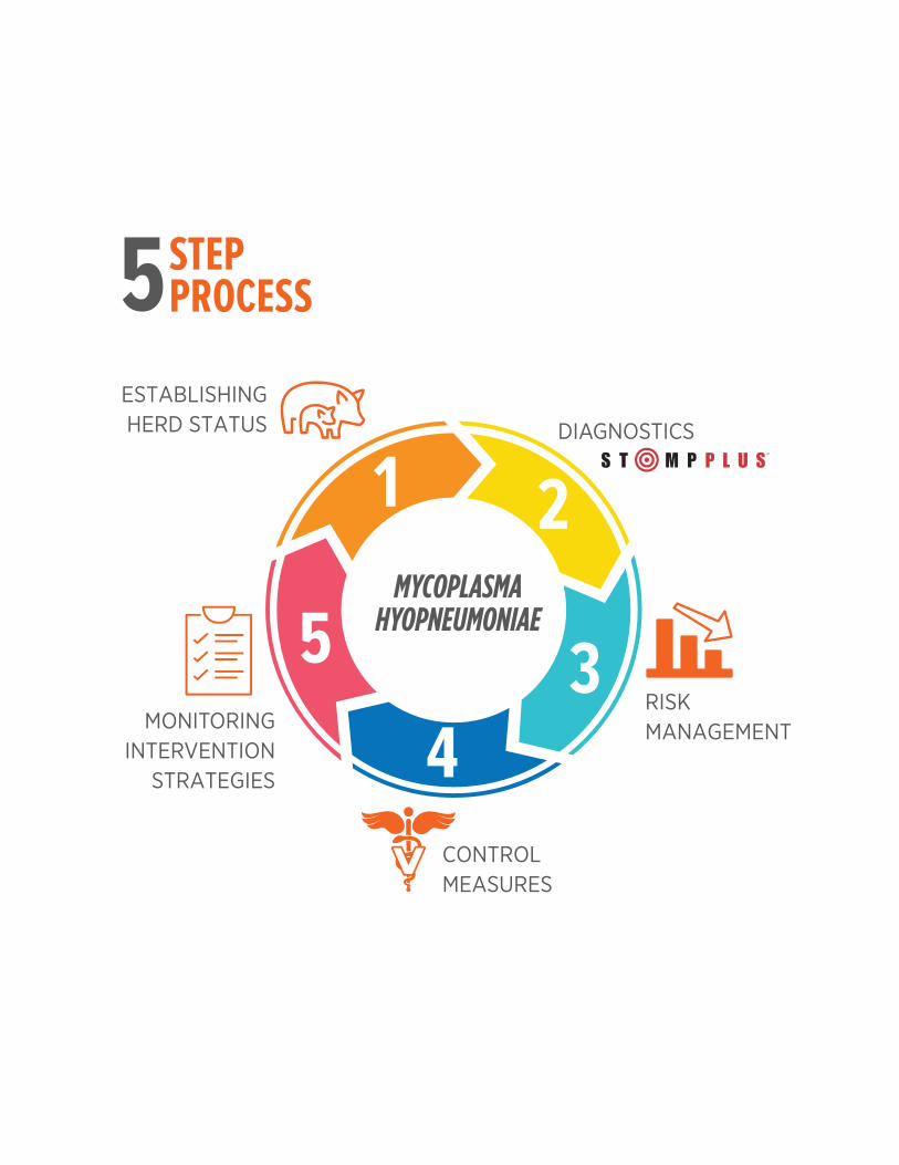
PURPOSE OF THE REVIEWRespiratory infection caused by Mycoplasma hyopneumoniae (MH) has been a well-known feature of swine production for decades . However, a comprehensive review of the disease has not been published in the peer-reviewed literature since 2008 .1 Despite a long acquaintance with MH and its principal clinical manifestation, enzootic pneumonia (EP), some gaps in our understanding of the disease and its control still remain and new information on diagnosis and management of MH infection has recently emerged . The purpose of this review is to provide a comprehensive update on MH . This includes a proposed case definition for MH disease in individual pigs, herd status classification criteria for breeding herds and information that fills several knowledge gaps involving MH herd infection, such as what defines a herd that is stable versus one that is unstable, and what is the appropriate role of anti-infective agents and vaccination in MH treatment and control versus MH elimination . The review is structured as a five-step approach for managing MH in the production setting, as follows:
§ Establishing herd status classification criteria for breeding herds
§ Diagnosis of MH infection
§ Risk management
§ Control measures
§ Monitoring intervention strategies
The review is authored by a distinguished panel of experts from academia, industry, and clinical practice . Collectively, they represent decades of diagnostic and clinical experience with swine infectious respiratory disease and MH specifically, and have authored numerous peer-reviewed reports on these topics . Their expertise ensures an up-to-date review with insights that can be readily applied by swine practitioners .
CLINICAL AND ECONOMIC RELEVANCE OF MHMycoplasmas, the smallest self-replicating bacteria, are distinguished by the lack of a cell wall that is present in most bacteria .2 As a result, they are resistant to beta-
lactam antimicrobials, which disrupt cell-wall synthesis . Other classes of antimicrobials, notably tetracyclines and macrolides, have activity against MH .3 Lack of a cell wall renders MH and other mycoplasmas susceptible to environmental extremes and disinfectants . Surface antigens in the lipid membrane surrounding the MH organism bind to receptors in the swine respiratory tract . Colonization of the respiratory cilia represents a unique niche for MH, resulting in persistent airway presence and creating a population of infected, asymptomatic animals that continually expose other pigs . The herd effect consists of recurring clinical episodes of EP, or when co-pathogens are involved, porcine respiratory disease complex (PRDC), occurring primarily in mid-finishing to market-weight pigs .4
Enzootic pneumonia in these animals is characterized by the dry, non-productive cough, labored breathing, and pneumonia familiar to most swine producers . PCR studies have demonstrated MH infection lasting in some cases from 15 to 30 weeks after initial exposure,4-6 ensuring long-term presence within herds . The economic consequences of this scenario include chronic reduction in feed efficiency and increased opportunity for co-infection with other respiratory pathogens, leading to the more severe PRDC syndrome .7,8 It has been estimated that for every 10% increase in swine lung tissue affected by pneumonia, there is a 41-gram decrease in average daily gain and a 16 .7-increase in days to slaughter weight .9 MH-associated EP has been shown to reduce growth rate of infected pigs by 16% and feed conversion by 14% .10 More conservative estimates place the reduction in growth rate during the finishing phase at 6% .11
The impact of MH on productivity is compounded by the extended duration of infection that typically occurs . Chronicity of MH lesions is due to several factors . MH has the ability to alter the host immune response . Studies have shown that macrophages activated by MH-infected pigs undergo marked reduction in phagocytic response when challenge exposed to a secondary pathogen .12 In addition, MH is able to vary the genetic expression of its surface antigens, which may allow it to evade immune recognition following host infection .7,13 Finally, the cellular and humoral immune response to MH appears to provide incomplete protection: although lesion severity is reduced in vaccinated pigs, MH airway colonization persists .7,14-16 Chronic MH infection in the form of high morbidity, low mortality respiratory disease has been estimated to affect up to 80% of pigs worldwide .5
The traditional view is that MH colonization leads to clinical disease . However, with the improved sensitivity

3A Contemporary Review of Mycoplasma hyopneumoniae Control Strategies
and specificity of modern diagnostic tests and a better understanding of pig colonization patterns at weaning, that position is now a matter of debate . The current understanding is that infection can occur without the economic losses associated with EP . To illustrate, the economic impact of uncomplicated MH infection versus co-infection with other swine respiratory pathogens was recently evaluated in a large U .S . production system . The cost of uncomplicated MH infection was <$1 .00 per pig versus approximately $10 per pig in MH-positive sites co-infected with swine influenza virus or porcine reproductive and respiratory syndrome (PRRS) virus .17
LIMITATIONS OF TRADITIONAL CONTROL METHODSMH control in segregated systems has been attempted by several widely practiced methods . These include herd vaccination programs, early vaccination of suckling pigs,18 antimicrobial treatments at peak transmission times,7 movement of pigs in all in-all out fashion and multi-site farm production .19 In endemic environments, these practices used individually or in combination have failed to fully control clinical and subclinical MH-associated disease .
In the late 1990s, breeding stock companies began producing MH-negative gilts to populate new farms . Today, the North American swine industry averages a 50% gilt replacement rate . This means that MH-negative gilts are introduced into MH-positive systems . Vaccination is commonly used during the gilt acclimation period . However, inactivated MH vaccines do not fully prevent colonization or stop shedding .3 As a result, vaccinated gilts can be infected after entering the breeding herd and become symptomatic or asymptomatic carriers and persistent MH shedders .
Contact exposure to MH-positive, carrier pigs or inoculation with virulent MH is sometimes used to for immunization of gilts . However, approximately 240-250 days are required to achieve total clearance of MH following natural exposure .4 This makes it impossible to introduce non-shedding gilts into the breeding herd at around 26 weeks of age . In such cases, gilts will have their first litters while shedding MH to their piglets, which in turn infect other pigs at weaning when litters are commingled . MH transmission continues to occur up to 200 days post-exposure, typically resulting in clinical signs in exposed pigs during the finishing stages .4
Disease dynamics in a population are strongly influenced by human intervention . Effective disease control requires a commitment by the producer to avoid practices that can exacerbate transmission . These include premature or unnecessary cross-fostering of newborn piglets, inconsistent vaccination that leaves pockets of susceptible animals, stressors such as double stocking, inadequate pen space,
failure to diagnose or control concurrent infectious diseases, and inappropriate timing of antimicrobial treatment . Other, less apparent factors, such as host genetic susceptibility, may also compromise efforts to control MH-associated disease .
Later sections of this review will discuss approaches to MH disease control, including vaccination, medication, and elimination .
PROPOSED CASE DEFINITIONA case definition is an epidemiological tool that describes the clinical and diagnostic criteria for an individual to be considered positive for a particular disease entity . For example, a case definition for clinical PCV-2 infection (known as porcine circovirus associated disease, or PCVAD) has been widely accepted . The PCVAD case definition requires that pigs (1) exhibit characteristic clinical signs, (2) have characteristic histologic lesions, and (3) have diagnostically confirmed presence of PCV-2 in tissue from characteristic lesions .20 Individuals or groups can be categorized as suspect, probable, or confirmed depending on the extent to which they conform to a case definition .
The individual components of a proposed MH case definition are discussed in detail in the diagnostics sections of this report, authored by Dr . Kent Schwartz . Below is a proposed case definition for positively diagnosing an individual pig with MH-associated disease based on the PCVAD model and Dr . Schwartz’s diagnostic criteria:
1) Clinical signs: The principal clinical sign is a dry hacking cough, exacerbated by physical exertion, predominantly in the late grower (9-16 weeks of age) or finisher phases of production (>16 weeks of age) . Feed consumption may be normal or moderately depressed . Uncomplicated MH infection and clinical manifestations are self-limiting, usually within 8 weeks, and mortality is negligible . There is a seasonal tendency toward greater prevalence in the fall . Likelihood of MH infection is greater in non-vaccinated herds . Fever is typically absent except in cases of co-infection . When co-infections are present, PRDC is a more severe complication of MH infection .
2) Gross and histopathologic lesions: Characteristic gross and histopathologic lesions are present in clinically affected pigs . Gross lung lesions generally consist of sharply demarcated cranioventral lobular distribution of gray or reddish consolidation . The amount of lung involved is highly variable but often does not exceed 10% of total lung volume . Characteristic histopathologic lesions consist of lobular distribution of peribronchiolar and perivascular lymphocytic cuffing . Alveoli and airways may contain serous fluid with a few macrophages and neutrophils .

4A Contemporary Review of Mycoplasma hyopneumoniae Control Strategies
Airway epithelium is intact, and sometimes slightly hyperplastic . Lymphocytic cuffs become more prominent as disease progresses .7
3) MH-specific laboratory diagnosis: Tissue samples from characteristic lung lesions are positive for MH based on immunohistochemistry, fluorescent antibody, in situ hybridization, or PCR testing . Culturing is also confirmatory but is used less often than the other methods .7
If all three components of the proposed case definition are present in an individual pig, a confirmed diagnosis of MH-associated EP or mycoplasmal pneumonia of swine (MPS) can be made . Determining the status in a herd is different and will be addressed in the next section .
REFERENCES1 . Maes D, Segales J, Meyns T, et al . Control of
Mycoplasma hyopneumoniae infection in pigs . Vet Microbiol 2008;126:297-309 .
2 . Razin S, Yogev D, Naoth Y . Molecular biology and pathogenicity of mycoplasmas . Microbiol Mol Biol Rev 1998;62:1094-1156 .
3 . Maes D . Mycoplasma hyopneumoniae infections in pigs: update on epidemiology and control . In: Proceedings 21st International Pig Veterinary Society Congress, Vancouver, Canada . 2010;30-35 .
4 . Pieters M, Pijoan C, Fano E, et al . An assessment of the duration of Mycoplasma hyopneumoniae infection in an experimentally infected population of pigs . Vet Microbiol 2009;134:261-266 .
5 . Fano E, Pijoan C, Dee S . Dynamics and persistence of Mycoplasma hyopneumoniae infection in pigs . Can J Vet Res 2005;69:223-228 .
6 . Mattsson JG, Bergström K, Wallgren P, et al . Detection of Mycoplasma hyopneumoniae in nose swabs from pigs by in vitro amplification of the 16S rRNA gene . J Clin Microbiol 1995;33:893-897 .
7 . Thacker E . Mycoplasmal diseases . In: Straw BE, Zimmerman JJ, D’Allaire S, et al, eds . Diseases of Swine, 9th ed . Oxford, UK: Blackwell Publishing Ltd; 2004:701-717 .
8 . Opriessnig T, Thacker EL, Yu S, et al . Experimental reproduction of postweaning multisystemic wasting syndrome in pigs by dual infection with Mycoplasma hyopneumoniae and porcine circovirus type 2 . Vet Pathol 2004;41:624-640 .
9 . Hill MA, Scheidt AB, Teclaw RF, et al . Association between growth indicators and volume of lesions in lungs from pigs at slaughter . Am J Vet Res 1992;53:2221-2223 .
10 . Pointon AM, Byrt D, Heap P . Effect of enzootic pneumonia of pigs on growth performance . Aust Vet J 1985;62:13-18 .
11 . Rautiainen E, Virtala AM, Wallgren P, et al . Varying effects of infections with Mycoplasma hyopneumoniae on the weight gain recorded in three different multisource fattening pig herds . J Vet Med B Infect Dis Vet Public Health 2000;47:461-469 .
12 . Caruso JP, Ross RF . Effects of Mycoplasma hyopneumoniae and Actinobacillus (Haemophilus) pleuropneumoniae infections on alveolar macrophage functions in swine . Am J Vet Res 1990;51:227-231 .
13 . Wise KS, Kim MF . Major membrane surface proteins of Mycoplasma hyopneumoniae selectively modified by covalently bound lipid . J Bacteriol 1987;169:5546-5555 .
14 . DeBey MC, Ross RF . Ciliostasis and loss of cilia induced by Mycoplasma hyopneumoniae in porcine tracheal organ cultures . Infect Immun 1994;62:5312-5318 .
15 . Desrosiers R . A review of some aspects of the epidemiology, diagnosis, and control of Mycoplasma hyopneumoniae infections . J Swine Health Prod 2001;9:233-237 .
16 . Sibila M, Bernal R, Torrents D, et al . Effect of sow vaccination against Mycoplasma hyopneumoniae on sow and piglet colonization and seroconversion, and pig lung lesions at slaughter . Vet Microbiol 2008;127:165-170 .
17 . Haden DC, Painter T, Fangman T, et al . Assessing production parameters and economic impact of swine influenza, PRRS and Mycoplasma hyopneumoniae on finishing pigs in a large production system . In: Proceedings 43rd Annual Meeting Am Assoc Swine Veterinarians, Denver, Colorado . 2012:75–76 .
18 . Martelli P, Terreni M, Guazzetti S, et al . Antibody response to Mycoplasma hyopneumoniae infection in vaccinated pigs with or without maternal antibodies induced by sow vaccination . J Vet Med B Infect Dis Vet Public Health 2006;53:229-233 .
19 . Harris H, Alexander T . Methods of disease control . In: Straw B, D’Allaire S, Mengeling W, Taylor D, eds . Diseases of Swine, 8th ed . Ames, Iowa: Iowa State University Press; 1999:1077-1110 .
20 . Sorden SD . Update on porcine circovirus and postweaning multisystemic wasting syndrome (PMWS) . Swine Health Prod 2000;8:133-136 .

ESTABLISHING HERD STATUS CLASSIFICATION CRITERIA FOR BREEDING HERDS
5A Contemporary Review of Mycoplasma hyopneumoniae Control Strategies

6A Contemporary Review of Mycoplasma hyopneumoniae Control Strategies
ESTABLISHING HERD STATUS CLASSIFICATION CRITERIA FOR BREEDING HERDSLucina Galina Pantoja, DVM, PhD Pork Technical Services Zoetis Inc. Hendersonville, Tennessee
IMPORTANCE OF A CLASSIFICATION SYSTEM Currently, a standardized terminology describing the M. hyopneumoniae (MH) status of breeding herds has not been established . The benefits of a clear and concise classification system are manifold: facilitating communication between swine producers, veterinarians, diagnosticians and breeding stock companies, evaluating strategies for disease control and supporting regional control and elimination efforts . The objective of this document is to propose an MH herd status classification system that incorporates comprehensible diagnostic criteria based on biologically relevant features of MH . This herd status classification was created utilizing the input from the Zoetis Veterinary Advisory Board, a group of twenty U .S . swine veterinarians who provide veterinary service to swine operations in the United States . Furthermore, the authors followed the terminology for classifying herds by PRRSV infection status defined by Holtkamp and collaborators .1
HERD STATUS CLASSIFICATION The proposed herd-status classification focuses on the sow herd, a population of animals which includes the breeding animals and their offspring . If a gilt development unit (GDU) is located within the premises, the GDU will be considered part of the breeding herd and the group of animals with the lower MH health status in the premises will determine the status of the entire site .
Four major herd-status categories are proposed for breeding herds: positive unstable (I), positive stable (II), provisionally negative (III), and negative (IV) (Table 2 .1) .
Positive unstable (I): breeding herds experiencing MH-associated clinical signs and MH is detected within the respiratory tract of weaning-age pigs . These populations may be serologically positive . Herds going through an MH outbreak in breeding animals and those that have not performed the necessary testing are considered category I .
Positive stable (II): breeding herds that do not show MH-associated clinical signs and pulmonary lesions . These herds show a reduced prevalence of MH in the upper respiratory tract of weaning-age pigs (<10% of positive laryngeal swabs) for at least 90 days . To fall into this category 4 consecutive samplings of 45 laryngeal swabs from weaning–age pigs every 30 days should reveal no more than 10% of pigs positive in each sampling . The breeding herd population is serologically positive . Herds undergoing elimination efforts (i .e . herd closure) fall into this category .
Provisionally negative (III): breeding herds that do not show MH-associated clinical signs and MH is not detected, but the population remains serologically positive . Category III herds should have at least 90 days of no positive results from weaning-age pigs in samples from the respiratory tract (4 consecutive samplings of 30 laryngeal swabs every 30 days) . Negative but vaccinated herds are considered category III . These commercial farms are not infected but implement continued use of MH vaccine in replacement gilts that entered the breeding herd due to the high cost of an acute outbreak in a naive farm .
Negative (IV): breeding herds that do not show MH-associated clinical signs, MH is not detected in any type of sample and the population is serologically negative . Herds undergoing elimination efforts should have been category III for at least one year and the herd would have rolled over to fall into category IV . Newly established herds and those that went through complete depopulation and repopulation efforts fall within category IV, after no positive serology results at least 60 days after placement of animals . A minimum of 30 sow samples (estimated prevalence of 10%) and ideally 57 samples (estimated prevalence of 5%) have to be collected to detect one positive .

7A Contemporary Review of Mycoplasma hyopneumoniae Control Strategies
CONSIDERATIONS
DIAGNOSTIC CRITERIA
MH-associated clinical signs with disease are defined as a dry non-productive cough, exacerbated by physical exertion . Furthermore, other clinical signs may be present: fever, decreased appetite and labored breathing .2 Clinical signs are not pathognomonic for MH and other respiratory pathogens must be ruled out . Microscopic lesions consist of lobular distribution of peribronchiolar and perivascular lymphocytic cuffing .2 Alveoli and airways may contain serous fluid with a few macrophages and neutrophils . Airway epithelium is intact, and sometimes slightly hyperplastic .2 Microscopic lesions are nonspecific and can be similar to those observed with viral agents .
Detection of the agent within lung lesions or the upper respiratory tract can be achieved using several tests . Immunohistochemistry (IHC), fluorescent antibody (FA), in situ hybridization (ISH) and polymerase chain reaction (PCR) testing are routinely utilized by diagnostic laboratories . However, PCR is recommended as the ideal testing method . Although culturing is confirmatory, it is less practical than the other methods and not frequently used .
The selection of the optimal sampling site in live animals will likely be affected by timing of infection . For example, it is possible that recently infected pigs harbor MH in the nasal cavity whereas non-acute pigs are more likely to harbor MH in the larynx, trachea or deeper sections of the respiratory tract . Current literature suggests laryngeal swabs as the preferred sample type for MH over nasal swabs and tracheo-bronchial lavage .3 However, these methods were evaluated under experimental conditions where pigs were inoculated intratracheally, bypassing the nasal cavity . Finally, seroconversion within a population can take several weeks to be detected by ELISA2,4 and therefore timing should be considered .
For sample size calculations several factors must be considered: the assumed true prevalence, the level of precision, the sensitivity and specificity of the diagnostic test and the population size . For the proposed classification, to detect a 10% prevalence of MH infection in weaning-age pigs (i .e ., for a positive stable herd), at least 45 samples should be collected, from a population of 1000 pigs .5 On the other hand, when the objective is to detect at least one positive, at least 30 samples should be collected, assuming the prevalence of MH is 10% (i .e ., provisionally negative) or
57 samples if 5% (i .e ., negative) prevalence is assumed .5 Both of these calculations assume nearly perfect tests . But diagnostic tests available today for MH are not perfect . Therefore, veterinarians should recognize these limitations when considering sample sizes and interpreting diagnostic results .
DEFINITION OF STABILITY
A positive correlation has been reported between the presence of MH in the upper respiratory tract at weaning and the extension of pulmonary lesions at slaughter .6 Based on those findings, reasonable goals for control should be to produce litters that are either negative or with low MH prevalence at weaning . This classification has taken the suggested cutoff point of 10% of infected weaning-age pigs to determine if a population has reached stability . This herd-status classification can be further refined as more information about prevalence at weaning and disease in the finisher stages becomes available .
Introduction of replacement gilts in the breeding herd plays a critical role in the MH stability a herd . Negative breeding herds that introduced positive replacement gilts are likely to become positive unstable (I) or stable (II) . Positive breeding herds that introduced positive or negative replacements are likely to be positive unstable (I) or stable (II) . Provisionally negative or negative herds that introduced negative replacement gilts are likely to maintain their provisionally negative (III) or negative (IV) status .
GROWING-PIG HERD CLASSIFICATIONA herd-status classification in the growing-finishing herd may be useful in the future . There is no clear definition of what an unstable growing-finishing site is . Recent reports suggest that a specific coughing index and serology thresholds have been associated with MH disease in finishing pigs . A coughing index (calculated as the percentage of pigs coughing per minute of observation) ≥2 .5% and an MH-ELISA seroprevalence >50% was reported to be an indicator of enzootic pneumonia, and therefore, could be an indicator of lack of stability, in finishing pigs .7
CONCLUSIONThis MH herd-status classification is a first proposal to be utilized as a foundation for discussions in the swine industry . It is the authors’ desire to initiate collaboration efforts, to identify areas of improvement, and to recognize areas that warrant further research .

8A Contemporary Review of Mycoplasma hyopneumoniae Control Strategies
KNOWLEDGE GAPS § An industry consensus of what makes a herd MH-
stable and MH-unstable .
§ Industry consensus on MH categories .
§ Threshold for MH colonization prevalence in weaned pigs as an indicator for disease at the finishing sites, validated in different commercial environments .
§ Coughing index associated with clinical MH disease for segregated operations common in North America .
§ Relevance of coughing index at the sow farm .
§ Threshold for MH sample-to-positive (S/P) ratios in finishing pigs and the percentage of seropositivity associated with clinical MH disease .
REFERENCES1 . Holtkamp DJ, Polson DD, Torremorell M .
Terminology for classifying swine herds by porcine reproductive and respiratory syndrome virus status . J Swine Health and Prod 2011;19(1):44-56 .
2 . Thacker E . Mycoplasmal diseases . In: Straw BE, Zimmerman JJ, D’Allaire S, et al, eds . Diseases of Swine, 9th ed . Oxford, UK: Blackwell Publishing Ltd; 2004:701-717 .
3 . Pieters M, and Rovira A . Comparison of various samples types for detection of Mycoplasma hyopneumoniae in recently infected pigs . Proceedings Allen D. Leman Swine Conference, St . Paul, MN . 2013;75-76 .
4 . Sibila M, et al . Chronological study of Mycoplasma hyopneumoniae infection, seroconversion and associated lung lesions in vaccinated and non-vaccinated pigs . Vet Microbiol 2007;122:97-107 .
5 . EpiTools epidemiological calculators . Accessed December 2015 . http://epitools .ausvet .com/au/content .php?page=PrevalenceSS .
6 . Fano E, et al . Effect of Mycoplasma hyopneumoniae colonization at weaning on disease severity in growing pigs . Can J Vet Res 2007;71:195-200 .
7 . Nathues H, et al . Value of the clinical examination in diagnosing enzootic pneumonia in fattening pigs . Vet J 2012;193:443-447 .

9A Contemporary Review of Mycoplasma hyopneumoniae Control Strategies
TABLE 2.1. MH BREEDING HERD-STATUS CLASSIFICATION CRITERIA*
Herd CategoryCriteria
Description and Diagnostic RequirementsClinical Signs
Antigen Detection in Respiratory Tract
Serology
Positive unstable (I)
Yes Yes Maybe
Clinical signs consistent with MH are present and MH is detected within lesions and/or in the respiratory tract .
Recently infected populations may be serologically negative .
Untested herds are category I by default .
Positive stable (II)
No
Yes
<10% of weaning-age pigs positive for antigen within respiratory tract
Yes
MH characteristic clinical signs or lesions are not detected .
At least 90 days of less than 10% of due-to-be-weaned pigs positive by PCR in 4 consecutive samplings of 45 laryngeal swabs at days 0, 30, 60 and 90 .*
Provisionally negative (III)
No No Yes
At least 90 days of negative results from weaning-age pigs by PCR in 4 consecutive samplings of 30 laryngeal swabs at days 0, 30, 60 and 90 .*
Herds undergoing MH elimination, no clinical signs or positive tests from respiratory tract of replacement animals, at least 60 days after the initial introduction .
Negative but vaccinated herds are considered category III .
Negative (IV)
No No No
At least one year as category III and have gone through a complete sow rollover . At completion of rollover testing must take place .
Newly established herds and herds that underwent complete depopulation and repopulation .
A minimum of 30 serum samples from sows should test negative by ELISA .*
*http://epitools.ausvet.com/au/content.php?page=PrevalenceSS. Accessed December 2015.

DIAGNOSTICS
10A Contemporary Review of Mycoplasma hyopneumoniae Control Strategies
11A Contemporary Review of Mycoplasma hyopneumoniae Control Strategies
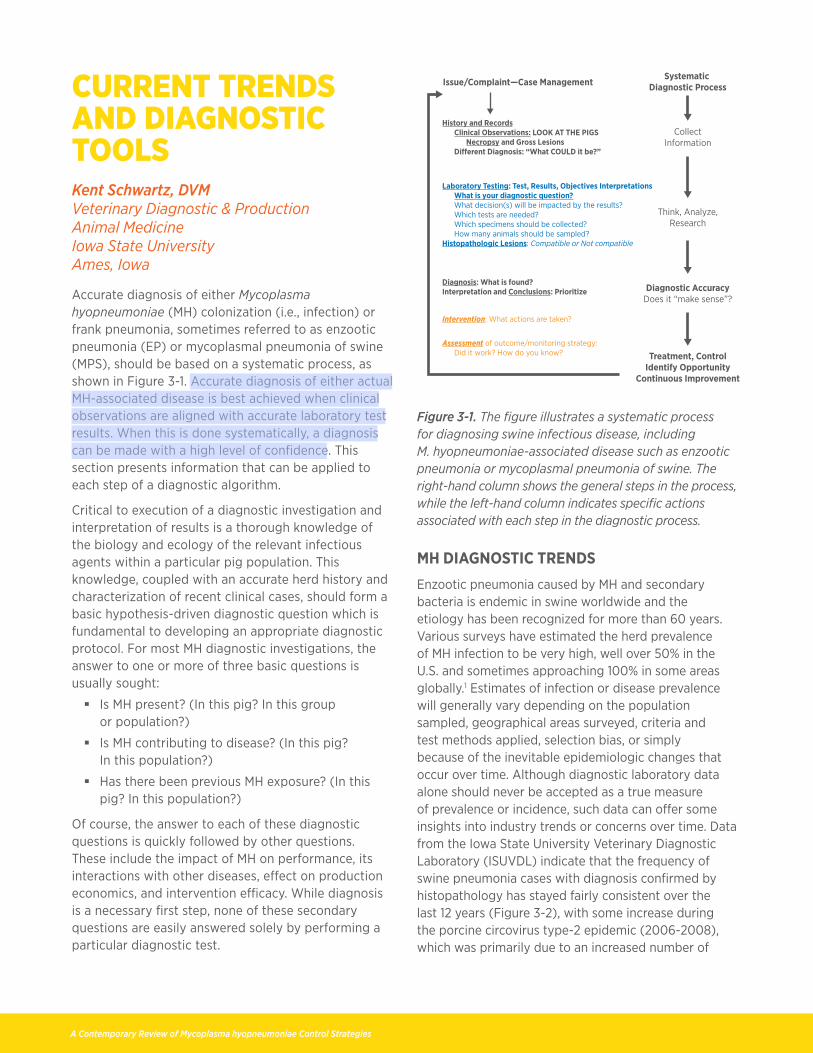
CURRENT TRENDS AND DIAGNOSTIC TOOLSKent Schwartz, DVM Veterinary Diagnostic & Production Animal Medicine Iowa State University Ames, Iowa
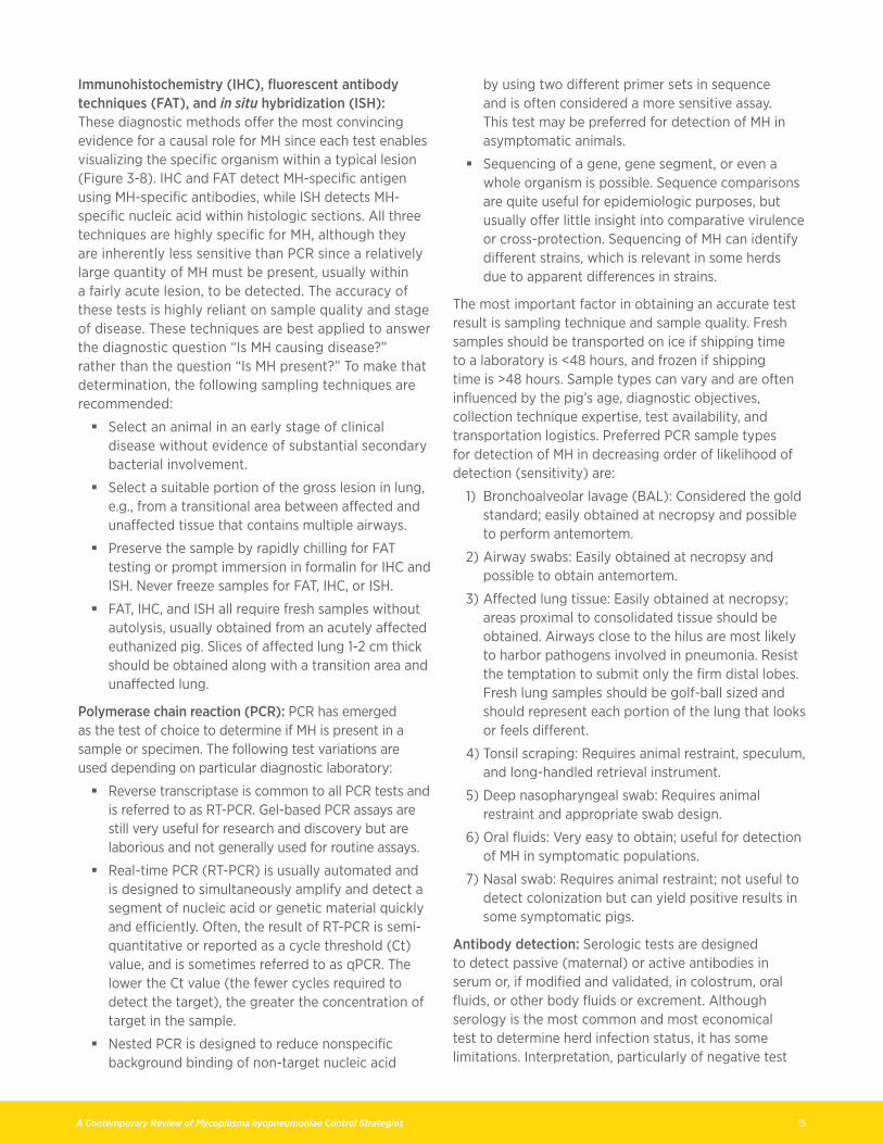
Accurate diagnosis of either Mycoplasma hyopneumoniae (MH) colonization (i .e ., infection) or frank pneumonia, sometimes referred to as enzootic pneumonia (EP) or mycoplasmal pneumonia of swine (MPS), should be based on a systematic process, as shown in Figure 3-1 . Accurate diagnosis of either actual MH-associated disease is best achieved when clinical observations are aligned with accurate laboratory test results . When this is done systematically, a diagnosis can be made with a high level of confidence . This section presents information that can be applied to each step of a diagnostic algorithm .
Critical to execution of a diagnostic investigation and interpretation of results is a thorough knowledge of the biology and ecology of the relevant infectious agents within a particular pig population . This knowledge, coupled with an accurate herd history and characterization of recent clinical cases, should form a basic hypothesis-driven diagnostic question which is fundamental to developing an appropriate diagnostic protocol . For most MH diagnostic investigations, the answer to one or more of three basic questions is usually sought:
§ Is MH present? (In this pig? In this group or population?)
§ Is MH contributing to disease? (In this pig? In this population?)
§ Has there been previous MH exposure? (In this pig? In this population?)
Of course, the answer to each of these diagnostic questions is quickly followed by other questions . These include the impact of MH on performance, its interactions with other diseases, effect on production economics, and intervention efficacy . While diagnosis is a necessary first step, none of these secondary questions are easily answered solely by performing a particular diagnostic test .
Systematic Diagnostic ProcessIssue/Complaint—Case Management
CollectInformation
Think, Analyze,Research
Diagnostic AccuracyDoes it “make sense”?
Treatment, ControlIdentify Opportunity
Continuous Improvement
History and Records Clinical Observations: LOOK AT THE PIGS Necropsy and Gross Lesions Di�erent Diagnosis: “What COULD it be?”
Laboratory Testing: Test, Results, Objectives Interpretations What is your diagnostic question? What decision(s) will be impacted by the results? Which tests are needed? Which specimens should be collected? How many animals should be sampled?Histopathologic Lesions: Compatible or Not compatible
Diagnosis: What is found?Interpretation and Conclusions: Prioritize
Intervention: What actions are taken?
Assessment of outcome/monitoring strategy: Did it work? How do you know?
Figure 3-1. The figure illustrates a systematic process for diagnosing swine infectious disease, including M. hyopneumoniae-associated disease such as enzootic pneumonia or mycoplasmal pneumonia of swine. The right-hand column shows the general steps in the process, while the left-hand column indicates specific actions associated with each step in the diagnostic process.
MH DIAGNOSTIC TRENDS Enzootic pneumonia caused by MH and secondary bacteria is endemic in swine worldwide and the etiology has been recognized for more than 60 years . Various surveys have estimated the herd prevalence of MH infection to be very high, well over 50% in the U .S . and sometimes approaching 100% in some areas globally .1 Estimates of infection or disease prevalence will generally vary depending on the population sampled, geographical areas surveyed, criteria and test methods applied, selection bias, or simply because of the inevitable epidemiologic changes that occur over time . Although diagnostic laboratory data alone should never be accepted as a true measure of prevalence or incidence, such data can offer some insights into industry trends or concerns over time . Data from the Iowa State University Veterinary Diagnostic Laboratory (ISUVDL) indicate that the frequency of swine pneumonia cases with diagnosis confirmed by histopathology has stayed fairly consistent over the last 12 years (Figure 3-2), with some increase during the porcine circovirus type-2 epidemic (2006-2008), which was primarily due to an increased number of

12A Contemporary Review of Mycoplasma hyopneumoniae Control Strategies
cases submitted . Table 3-1 shows a fairly consistent 12-year pattern where (1) MH diagnoses varied within a 6% range (5-11%) of total swine pneumonia cases, (2) swine influenza virus (IAV-S) had a secular increase from 16% to 36%, and (3) porcine reproductive and respiratory syndrome (PRRS) virus diagnoses remained relatively constant within the 26-32% range with the exception of 2011, when 39% of pneumonia cases had PRRSV involvement . The ISUVDL data show that of the three etiologic agents, MH is the least common while IAV-S and PRRSV are more common and have a roughly equivalent frequency of diagnosis . Any presumptive diagnosis of clinical pneumonia in swine should consider the relative frequency of these three major infectious pathogens, although all three are very common infections .
Number of Swine Pneumonia Cases Diagnosedat ISUVDL (2003 – 2014)
0
5000
10000
15000
2014
2013
2012
2011
2010
2009
2008
2007
2006
2005
2004
2003
2003 2004 2005 2006 2007 2008 2009 2010 2011 2012 2013 2014
Figure 3-2. The chart shows the number of swine pneumonia cases based on lung histopathology performed at the Iowa State University Veterinary Diagnostic Laboratory (ISUVDL). The total never dropped appreciably below 5,000 cases for any year since 2003, but exceeded 10,000 cases in some years.
TABLE 3-1. ETIOLOGY OF SWINE PNEUMONIA CASES AS DETERMINED BY HISTOPATHOLOGY DIAGNOSED BY ISUVDL (2003-2014)
Causative Agent and Percentage of Cases
Year MH IAV-S PRRSV
2003 8% 16% 29%
2004 7% 15% 26%
2005 6% 17% 26%
2006 7% 16% 29%
2007 5% 15% 27%
2008 6% 18% 30%
2009 8% 22% 29%
2010 10% 26% 32%
2011 11% 31% 39%
2012 9% 33% 32%
2013 8% 37% 31%
2014 8% 36% 32%
IAV-S = influenza A virus in swine; ISUVDL = Iowa State University Veterinary Diagnostic Laboratory
MH = Mycoplasma hyopneumoniae
PRRSV = porcine reproductive and respiratory syndrome virus.
Figures 3-3 and 3-4 further define the epidemiology of MPS . MH is more frequently diagnosed in finisher stages (>16 week of age) than in the grower (9-16 weeks) or nursery (3-9 weeks) phases (Figure 3-3) . In addition, MPS has a seasonal tendency, with a greater frequency in the fall compared to other seasons (Figure 3-4) .
90%
2003 2004 2005 2006 2007 2008 2009 2010 2011 2012 2013 2014
80%
70%
60%
50%
40%
30%
20%
10%
0%
% Nursery
% Grower% Finisher
Figure 3-3. The chart shows the frequency of MH-associated swine pneumonia by stage of production, based on Iowa State University Veterinary Diagnostic Laboratory data for the 12 years from 2003-14. The trend suggests that the frequency of MH diagnoses in case of porcine pneumonia increases in the finishing phase and declines in nursery and grower pigs.

13A Contemporary Review of Mycoplasma hyopneumoniae Control Strategies
ISUVDL Cases of Swine MH (2003 – 2014)
0
100
200
300
400
500
600
700
800
Dec
Nov
Oct
Sep
Aug
Jul
Jun
May
Apr
Mar
Feb
Jan
JAN FEB MAR APR MAY JUN JULY AUG SEP OCT NOV DEC
Figure 3-4. Iowa State University Veterinary Diagnostic Laboratory (ISUVDL) data compiled over a 12-year span indicate that swine MH diagnoses vary by season with the highest frequency of diagnosis occurring in the fall.
FACTORS AFFECTING MH INFECTION A conceptual understanding of MH exposure, colonization, host response and the approximate time line of post-exposure events is essential for appropriate test selection and interpretation of results . When considering the dynamics of MH infection, it is helpful to understand that there is extreme variability in these factors among individuals and populations . Transmission of MH between pigs is highly variable and difficult to predict . MH can colonize the cilia of the swine respiratory tract with no clinical signs for weeks before other risk factors exacerbate disease expression . Clinical MPS signs, and presumably transmission, can be slow (over weeks to months) or fairly explosive (in days to weeks) depending on host susceptibility, bacterial loads (infectious dose), strain virulence, and presence of exacerbating co-infections, environmental stress or genetic risk factors .
Experimental MH infection of healthy, naive pigs has a somewhat more predictable course than natural disease . Clinical signs (coughing) may become apparent as early as 10 days (usually at 14-21 days) after intratracheal inoculation . Coughing corresponds to the onset of detectable gross and microscopic lesions . In uncomplicated MH infections, lesions and coughing generally persist for about four weeks or so in individual pigs before gradually subsiding to subclinical status . Gross lesions can resolve within 10-12 weeks in uncomplicated experimental MH infections . If secondary bacterial pathogens localize in the MH-compromised lung, clinical
signs and lesions will be much more severe, sometimes fatal, and can persist for months .
Co-infections involving MH and other respiratory pathogens are very common . Historically, EP was defined as pneumonia in which concurrent and common bacterial co-infections are present with MH as primary initiator of disease . Bacteria identified by ISUVDL, roughly in order of decreasing frequency, include: Pasteurella multocida, streptococci, actinobacilli, and Haemophilus, with Trueperella or Bordetella co-infections common .
In the last 25 years, co-infections with viruses have become common as well . PRRSV, IAV-S, and sometimes PCV-2 are often present in pneumonic lungs and contribute to pneumonia . A thorough and complete diagnostic process therefore becomes essential before causation can be determined . The tools for confirming a role for co-infections are quite similar to those specific for MH . Clinical assessment, gross lesions, histopathology, and laboratory tests for specific agents are now routinely performed by many production systems and diagnostic laboratories . Monitoring of herds by means of necropsy, oral fluid sampling, and serology for co-infections are often incorporated as part of the routine diagnostic protocol .
COMMON DIAGNOSTIC TOOLS Diagnostic laboratories often differ in the repertoire of laboratory tests offered as well as in methodology of various tests performed . Some variation among tests and laboratories is expected but can be disconcerting when not anticipated . Although each laboratory accredited by the American Association of Veterinary Laboratory Diagnosticians has quality assurance protocols in place, inherent differences in tests and their interpretation means that comparison of numerical results from different laboratories should not be made .
Diagnostic tools can be broadly defined to include performance records and historical information useful for problem definition and monitoring interventions (discussed in another section of this report) . Observational diagnostic evidentiary tools can also include clinical observations and gross lesions . Laboratory testing usually implies laboratory tests to detect specific analytes, including those for MH .
OBSERVATIONAL DIAGNOSTIC TOOLS
Clinical Diagnosis: Clinical diagnosis by observation is very useful to determine if disease is present, to characterize clinical signs, and to estimate the extent and magnitude of clinical disease . A clinical diagnosis implies that the observer actually examines pigs in

14A Contemporary Review of Mycoplasma hyopneumoniae Control Strategies
their environment at various times over a period of time to identify illness, abnormalities, and risk factors . Colonization with MH is more likely to achieve clinical disease status when there are concurrent infections, when immunologically naive pigs are infected or when air quality is compromised .
Although clinical signs of MH infection can occur in pigs of any age, most clinical cases of pneumonia will be in pigs in the late grower or finisher period . The severity of disease in individual pigs or within a herd depends on the animals’ innate or acquired resistance and concurrent health challenges . The predominant sign is a hacking cough, most commonly occurring during exercise or at the start of the day . Fever is not a feature with MH infection alone but may occur when co-infections are present . Feed consumption is only modestly decreased . MH-associated clinical disease is generally self-limiting unless pigs are immunologically naive or if there are complicating infections or abnormal environmental stresses present . When co-infections are present, porcine respiratory disease complex (PRDC) emerges as a more severe complication of MH infection . In such cases, death loss can escalate dramatically . Co-infections may occur either sequentially or simultaneously with MH . Common co-infections in PRDC include viruses (e .g ., PRRSV, IAV-S, PCV2) or those bacteria with pathogenic potential residing in the nasopharynx (e .g ., Pasteurella multocida, Streptococcus suis, Haemophilus parasuis, Actinobacillus spp . or Bordetella bronchiseptica) .
Gross Lesions: Gross lesions present at necropsy in cases of MH-associated pneumonia (Figure 3-5) will generally be lungs with sharply demarcated cranioventral lobular distribution of gray or reddish firmness (i .e ., consolidation, hepatization) . Affected tissues usually include cranioventral portions of one or more of the apical, intermediate, cardiac, and perhaps some of the caudal lobes . The amount of lung involved is highly variable but often does not exceed 10% of total lung volume in uncomplicated cases, although the variation within the population will range from ≥30% to no detectable lesions . In acute cases, there may be marked parenchymal and interlobular edema in consolidated portions of lung . As disease progresses, affected tissue becomes purple-to-gray and rubbery . Cloudy mucus exudes from airways of cut surfaces . When other bacteria are involved, generally relatively more lung volume is affected, with greater firmness, more purulent exudate in airways, and with tracheobronchial lymph nodes being more prominent . In chronic or resolving cases, there may be interlobular fibrosis and a wrinkled
appearance of lung parenchyma . Uncomplicated cases may resolve in 8 weeks .
Histopathology: Histopathological evaluation by microscopic examination of lesions is useful to identify tissue changes that are compatible with MH involvement and to implicate other respiratory pathogens . Histopathology is performed on formalin-fixed lung sections, preferably from affected euthanized pigs, since pigs that succumb naturally often have confounding co-infections . Lung sections approximately 1x3x3 cm should be collected from every portion of lung that looks or feels different and should include transitional portions as well as visible airways . Usually, this will be from areas of lung nearer the hilus (the area of tracheal bifurcation) rather than just the consolidated tips of various lobes .
Peribronchiolar and perivascular lymphocytic cuffing is expected with MH but is not pathognomonic for MH since chronic antigenic stimulation from a variety of infectious insults can result in lymphocyte cuffing . In early stages of infection there may be lobular distribution of loose cuffing of airways and blood vessels with lymphocytes and fewer macrophages (Figure 3-6) . Alveoli and airways may contain serous fluid with a few macrophages and neutrophils . Airway epithelium is intact, and sometimes slightly hyperplastic . Later, lymphocytic cuffs become more prominent and may contain nodules or follicles (Figure 3-7) . Although microscopic lesions can be quite characteristic in uncomplicated cases, co-infections can obscure or obfuscate interpretation .
MH-SPECIFIC LABORATORY TESTS
MH-specific laboratory tests are critical to establish a disease diagnosis after assessment of clinical signs, gross lesions and histopathology . Specific testing is also useful to identify asymptomatic carriers or to detect antibodies as a tell-tale sign of endemic infections . Tools that demonstrate the presence of MH rely on visualizing the organism (culturing, microscopy) or detection of nucleic acid or antigen specific for MH .
Culturing: Isolation and culturing of MH is tedious, requiring 4-8 weeks for detection . Culturing also requires expensive media and specialized techniques, and is frequently compromised by contamination with other, faster-growing Mycoplasma spp . Due to these disadvantages, culturing is seldom attempted for routine MH diagnosis . Samples for isolation of other bacteria should consist of fresh, 5 cm-diameter portions of gross lesion (consolidated) in lung, packaged separately from other tissues, and chilled .
15A Contemporary Review of Mycoplasma hyopneumoniae Control Strategies
Immunohistochemistry (IHC), fluorescent antibody techniques (FAT), and in situ hybridization (ISH): These diagnostic methods offer the most convincing evidence for a causal role for MH since each test enables visualizing the specific organism within a typical lesion (Figure 3-8) . IHC and FAT detect MH-specific antigen using MH-specific antibodies, while ISH detects MH-specific nucleic acid within histologic sections . All three techniques are highly specific for MH, although they are inherently less sensitive than PCR since a relatively large quantity of MH must be present, usually within a fairly acute lesion, to be detected . The accuracy of these tests is highly reliant on sample quality and stage of disease . These techniques are best applied to answer the diagnostic question “Is MH causing disease?” rather than the question “Is MH present?” To make that determination, the following sampling techniques are recommended:
§ Select an animal in an early stage of clinical disease without evidence of substantial secondary bacterial involvement .
§ Select a suitable portion of the gross lesion in lung, e .g ., from a transitional area between affected and unaffected tissue that contains multiple airways .
§ Preserve the sample by rapidly chilling for FAT testing or prompt immersion in formalin for IHC and ISH . Never freeze samples for FAT, IHC, or ISH .
§ FAT, IHC, and ISH all require fresh samples without autolysis, usually obtained from an acutely affected euthanized pig . Slices of affected lung 1-2 cm thick should be obtained along with a transition area and unaffected lung .
Polymerase chain reaction (PCR): PCR has emerged as the test of choice to determine if MH is present in a sample or specimen . The following test variations are used depending on particular diagnostic laboratory:
§ Reverse transcriptase is common to all PCR tests and is referred to as RT-PCR . Gel-based PCR assays are still very useful for research and discovery but are laborious and not generally used for routine assays .
§ Real-time PCR (RT-PCR) is usually automated and is designed to simultaneously amplify and detect a segment of nucleic acid or genetic material quickly and efficiently . Often, the result of RT-PCR is semi-quantitative or reported as a cycle threshold (Ct) value, and is sometimes referred to as qPCR . The lower the Ct value (the fewer cycles required to detect the target), the greater the concentration of target in the sample .
§ Nested PCR is designed to reduce nonspecific background binding of non-target nucleic acid
by using two different primer sets in sequence and is often considered a more sensitive assay . This test may be preferred for detection of MH in asymptomatic animals .
§ Sequencing of a gene, gene segment, or even a whole organism is possible . Sequence comparisons are quite useful for epidemiologic purposes, but usually offer little insight into comparative virulence or cross-protection . Sequencing of MH can identify different strains, which is relevant in some herds due to apparent differences in strains .
The most important factor in obtaining an accurate test result is sampling technique and sample quality . Fresh samples should be transported on ice if shipping time to a laboratory is <48 hours, and frozen if shipping time is >48 hours . Sample types can vary and are often influenced by the pig’s age, diagnostic objectives, collection technique expertise, test availability, and transportation logistics . Preferred PCR sample types for detection of MH in decreasing order of likelihood of detection (sensitivity) are:
1) Bronchoalveolar lavage (BAL): Considered the gold standard; easily obtained at necropsy and possible to perform antemortem .
2) Airway swabs: Easily obtained at necropsy and possible to obtain antemortem .
3) Affected lung tissue: Easily obtained at necropsy; areas proximal to consolidated tissue should be obtained . Airways close to the hilus are most likely to harbor pathogens involved in pneumonia . Resist the temptation to submit only the firm distal lobes . Fresh lung samples should be golf-ball sized and should represent each portion of the lung that looks or feels different .
4) Tonsil scraping: Requires animal restraint, speculum, and long-handled retrieval instrument .
5) Deep nasopharyngeal swab: Requires animal restraint and appropriate swab design .
6) Oral fluids: Very easy to obtain; useful for detection of MH in symptomatic populations .
7) Nasal swab: Requires animal restraint; not useful to detect colonization but can yield positive results in some symptomatic pigs .
Antibody detection: Serologic tests are designed to detect passive (maternal) or active antibodies in serum or, if modified and validated, in colostrum, oral fluids, or other body fluids or excrement . Although serology is the most common and most economical test to determine herd infection status, it has some limitations . Interpretation, particularly of negative test

16A Contemporary Review of Mycoplasma hyopneumoniae Control Strategies
results, can be quite challenging when performed on individual animals or asymptomatic herds . Because of the unique MH colonization “niche” on the respiratory cilia, colonization can occur without predictable onset of antibody response . Animals may be colonized for long periods of time without detectable seroconversion . Commercial ELISA tests are most often used for antibody detection . All have very good specificity (positive predictive value) but can lack sensitivity . This is primarily due to lack of sufficient systemic immune stimulation early in infection or when pigs are superficially colonized . Complement fixation tests utilized in the last century have yielded to the more commonly used ELISA tests including HerdCheck® (IDEXX) ELISA, Tween20 ELISA, and Oxoid ELISA .
REFERENCES1 . Thacker EL, Minion FC . Mycoplasmosis . In:
Zimmerman JJ, Karriker LA, Schwartz KJ, et al, eds . Diseases of Swine, 10th ed . Oxford, UK: Wiley-Blackwell; 2012:779-797 .
Figure 3-5. Typical gross lesion with uncomplicated MH is sharply demarcated cranioventral lobular distribution of gray or reddish firmness.
Figure 3-6. Peribronchiolar and perivascular lymphocytic cuffing with mild accumulations of serous fluid and neutrophils in airways and adjacent alveoli is compatible but not pathognomonic for MH.
Figure 3-7. In later stages of MH infection, lymphocytic cuffs become more prominent and may contain lymphoid nodules or follicles.
Figure 3-8. MH can be detected by immunohistochemistry on cilia within airways in early stages of disease.

17A Contemporary Review of Mycoplasma hyopneumoniae Control Strategies
CHOOSING THE RIGHT TESTKent Schwartz, DVM Veterinary Diagnostic & Production Animal Medicine Iowa State University Ames, Iowa
EVALUATING THE ROLE OF MH IN CLINICAL DISEASETwo basic questions surrounding Mycoplasma hyopneumoniae (MH) diagnosis that can be answered by laboratory testing are: “Is MH causing disease in a pig or pig population?” or “Is MH colonizing a pig or a population without causing clinical disease?” This section discusses the first of these two questions . Unfortunately, bacterial and viral co-infections are quite common in modern production systems . A truly accurate diagnosis can be challenging and should consider non-infectious risk factors that contribute to pneumonia . Laboratory testing is essential to confirm presence of MH and to rule out a role for other pathogens . Three criteria necessary to confirm a diagnosis of MH-associated pneumonia are (1) characteristic gross lesions from clinically affected pigs, (2) characteristic histopathologic lesions, and (3) demonstration of MH in affected portions of lung .
CLINICAL DIAGNOSIS
Clinical signs previously discussed (e .g ., hacking cough exacerbated by activity or arousal) are quite typical in uncomplicated cases and are expected to occur in the late grower or finisher period . In such cases, gross lung lesions from animals with typical clinical signs are highly suggestive of MH and are often sufficient for field diagnosis . When respiratory co-infections are present, PRDC emerges as a more severe complication of MH infection . Careful clinical evaluation will aid in determining the number and types of diagnostic tests to identify involvement of MH and other respiratory pathogens .
PATHOLOGICAL DIAGNOSIS
Gross and microscopic lesions, while not specific to for MH, are necessary to confirm presence of disease (i .e ., lesions) compatible with this commonly detected pathogen . Because neither gross nor microscopic lesions
are pathognomonic for MH, it is necessary to obtain an MH-specific confirmation using the laboratory methods and sampling techniques .
INTERPRETATION OF DIAGNOSTIC RESULTS
Interpretation of diagnostic results is fairly straightforward . Diagnosis of MH-associated pneumonia is confirmed if gross and microscopic lesions are characteristic of MH and if MH is demonstrated to be present in affected lung . Additional confidence is gained if these observations align with typical clinical signs or other indirect laboratory test results . Because MH infection is commonplace and varies considerably in severity, translating laboratory diagnostic results into a herd-level picture of pathologic and economic impact of MH-associated disease requires additional investigative tools .
Once clinical MH-associated disease is confirmed, several diagnostic approaches can help resolve herd epidemiologic issues . These include determining disease prevalence, time of onset, and the impact of interventions . The following methods can help answer these important post-diagnostic questions:
§ Systematic and objective clinical assessment in grow-finish phase, also known as syndromic surveillance, can define the onset, duration, and magnitude of clinical signs in selected groups of pigs . This approach offers a framework for identifying which pigs to monitor and diagnostically evaluate for identification of other etiologies .
§ Slaughter checks are useful, particularly if clinical signs are present later in the finishing period, after 18 weeks of age . Determination of lesion prevalence, severity, and variation (i .e ., percent lung involvement) and the opportunity to sample lungs to confirm etiology can be insightful for diagnosis and monitoring .
§ Routine necropsy with or without laboratory submission is useful for identifying general causes of mortality by gross examination and ensuring that clinical impressions and gross lesions are consistent . Necropsy can also help avoid mis-diagnoses by obtaining timely identification of unexpected causes of mortality .
§ Serologic profiles can detect changes in MH prevalence over time and determine when significant MH challenges occur . Interpretation of serologic data should consider the age and vaccination status of the animals sampled, and epidemiologic trends affecting the herd . Longitudinal and cross-sectional serum sampling can determine when MH is circulating or causing

18A Contemporary Review of Mycoplasma hyopneumoniae Control Strategies
clinical disease .
§ PCR analysis of oral fluids, while not particularly sensitive, has been used to confirm the presence of MH when clinical signs are present . This sample type does not reliably determine if MH is colonizing pigs in the sample population, but will often be positive when pigs are clinically affected and coughing .
EVALUATING MH COLONIZATION IN NON-CLINICAL POPULATIONS Another purpose of MH diagnostic testing is to answer the question, “Is MH colonizing an individual animal or the herd?” Monitoring infection status is very important for MH-negative seedstock suppliers or other populations where colonization with MH is not expected . In positive herds, there is growing suspicion that the percent of pigs colonized (prevalence of colonization) at weaning is predictive of clinical disease severity during the grow-finish period . Piglets can be colonized from dams (sow-to-piglet transmission) but the percentage of piglets colonized at weaning is usually quite low . Moreover, there is a great deal of variation in infection status of pigs among different groups and over time . Although it is well-recognized that MH is transmitted from pig-to-pig, the rates of transmission and colonization are quite variable and frequently much lower than expected when compared to most viruses or bacteria . Not all pigs will be colonized in MH-positive groups even after 5 months of being housed together .
There is value in identifying presumed negative MH colonized pigs as early and quickly as possible . However, an accurate diagnosis of asymptomatic MH colonization or very early infection is daunting, mostly because of the unusual ecological niche of MH . Cilia on the respiratory mucosa are the obligatory site of MH colonization . Practically speaking, it is possible for pigs to be exposed to organisms but not become colonized . Or pigs may be exposed and colonized with MH for long periods of time with no overt disease or detectable host response . Colonization can be patchy within the bronchial tree with relatively low numbers of organisms present . As a result, there is no predictable time line for seroconversion after colonization or for the reliable detection of MH in tissues or other samples by PCR .
Proving an animal or a population negative for MH colonization with 100% confidence is not possible with antemortem testing . Genomic testing with PCR methods or antibody detection with ELISA are the only practical tools for detecting MH in asymptomatic animals . In colonized but completely asymptomatic,
presumed negative pigs, the probability of detecting MH in a single sample from one animal is low . This can be compensated for by optimizing sample type and/or testing more animals . Epidemiologic considerations include the number of animals to sample, the expected prevalence, confidence interval expectations, sample types, sample size (number of samples), test method used, and the sensitivity and specificity of a particular diagnostic test . Unfortunately, because MH prevalence is unknown but generally assumed to be low and the desired confidence is usually high, a large sample size is usually required . The value of pooling samples to decrease cost of testing is unknown . PCR is the usual test used, which can be expensive when used for an extended monitoring period . Long-term monitoring with PCR involves the labor to collect a large number of samples, which are often difficult to obtain . Still, there appears to be enough value in monitoring populations that it is frequently performed .
Two types of testing are available to attempt detection of MH colonization or early-stage infection in asymptomatic pigs: PCR detection of nucleic acid and ELISA testing for presence of specific antibody .1-3 PCR tests generally are quite sensitive, with nested PCR or RT-PCR often preferred . The principal factor limiting sensitivity of detection is not the test but the sample type, sample quality, sample quantity, or the sample preservation . In general, the closer the sample obtained is to the area of the tracheal bifurcation (hilus), the greater the likelihood of detection of MH by PCR .
Colonized pigs found falsely negative by PCR tests are not unusual, simply because the organism is not in the sample . This is because, in most asymptomatic pigs, MH colonizes cilia of medium-sized and larger airways and is not shed in oral fluids or nasal secretions in detectable levels . For increasing the likelihood of identifying true MH positives, BAL or brush-swabs of airways at the hilus are probably the best sample types . Both sample types are difficult although not impossible to obtain antemortem . Preferred antemortem sample types, in order of likelihood of detecting an MH-colonized pig, are BAL, brush swab from large airways, swabs from large airways, rinsate from an upper tracheal swab or aspirate, tonsil scrapings, deep nasopharyngeal swabs, and nasal swabs . PCR tests are invariably applied to such samples, with nested PCR considered to be somewhat more sensitive than RT-PCR and in overall sensitivity .
Clinicians are sometimes in doubt about which PCR method is best for diagnosing MH from clinical cases . PCR is used to evaluate samples from either symptomatic or asymptomatic animals of unknown MH
19A Contemporary Review of Mycoplasma hyopneumoniae Control Strategies
infection status . Any of the PCR methods can confirm the presence of MH in a pneumonic lesion . PCR tests used for detection of MH in diseased animals (i .e ., in lung tissue samples) do not need to be as sensitive as those designed to detect mere colonization in absence of clinical disease or lesions . Diseased lungs are expected to have sufficient quantities of target nucleic acid to be easily detectable by routine PCR . For detection of MH in lesions, important considerations are selection of animals to be sampled (those with clinical signs and gross lesions), location of sample (transitional area between consolidation and less affected lung with large airways included), and the preservation of the sample . Samples should be transported on ice if shipping time to a laboratory is <48 hours, and frozen if shipping time is >48 hours .
Serology is useful to detect MH exposure in nonclinical populations, although sensitivity is often limited in these animals . Seroconversion does not occur for some weeks after colonization and then only sporadically in colonized pigs . However, detection of a positive animal is sufficient to warrant concern that the population is colonized . Serology is of particular value in the following cases:
§ Presumptively negative sow farms can be monitored by antibody detection in colostrum of dams .
§ Serum antibodies measured during the growing-finishing phases or at marketing can detect MH infections occurring during the grow-finish period .
Non-vaccinated pigs that are serologically positive are likely infected . Prevalence may be quite low but the high specificity of serology testing suggests that positive animals should not be ignored . Detection of seropositive animals in a vaccinated population is more difficult to interpret . Experience with serology tests and nuances of vaccination timing are required to determine whether seropositive animals are infected or merely vaccinated .
KNOWLEDGE GAPSDespite an abundance of observational and laboratory testing capabilities for MH, improvements in diagnostic tests, sample types, and sampling strategy are always desirable, specifically as noted in the following sections .
DIAGNOSIS OF SUBCLINICAL INFECTION
§ The ecology of MH is characterized by the organism’s relatively low numbers and stationary location on cilia deep in the respiratory tract and
its ability to escape detection of adaptive immune surveillance mechanisms for extended periods . For this reason, sensitivity of detection of early infection is limited . Improvement in sampling and sample type as well as improvement in sensitivity of PCR are needed .
§ Serologic tests perhaps are not sensitive enough to detect early-onset antibodies that may result from subclinical infection . Perhaps there are other epitopes or assays that could be utilized to improve sensitivity .
§ There may be other methods to assess early subclinical infections, for example, a cell-mediated response involving lymphocyte stimulation assays, skin tests or other less convenient, more costly, and less understood tests .
DIAGNOSIS OF CLINICAL DISEASE IN MIXED INFECTIONS
§ Because pneumonia in swine is multifactorial, current diagnostic testing often cannot discern the impact of the MH contribution to pneumonia in an individual pig or in a population of pigs where MH is endemic . Detection of presence of MH is not the same as diagnosis of disease caused by MH . Histologic lesions are not pathognomonic and have considerable overlap with chronic pneumonia from other causes . Criteria for assigning cause probably need to be standardized . More work with quantitative PCR (qPCR) is warranted .
§ Diagnostic test requests vary with age of pig, stage of infection and stage of lesions or lesion resolution . Transmission apparently is fairly slow and there is very broad distribution of individuals infected, clinically affected, and degree of infection severity over time . It may be possible to correlate qPCR with some of these factors .
§ Finding MH-associated pneumonia in a sample pig does not confirm that similar lesions and impact are present in all pigs . Extrapolation of diagnostic testing results from an individual pig or biased sampling to other populations of pigs is often not accurate and could be improved .
DIAGNOSIS OF MH CLINICAL DISEASE IMPACT IN INDIVIDUAL PIGS AND POPULATIONS
§ Production processes and metrics could be correlated with diagnostic tools and statistically

20A Contemporary Review of Mycoplasma hyopneumoniae Control Strategies
validated over time .
§ The impact of a single agent such as MH in a production setting should be determined in relation to all factors, infectious and noninfectious, that affect measured parameters . Figure 3-9 shows an example of the author’s opinion of how the total impact of relevant diseases and management factors can be estimated within the context of revenue opportunity .
§ Systematic evaluations need to be done in real-time with infection monitoring in multiple groups or sites . Systematic collection of oral fluids and serum and population-based sampling along with statistical process control will need to be refined .
RELATIVE IMPACT CALCULATORINSULT
PERCENT CONTRIBUTION
$34.00 OPPORTUNITY LOSS / PIG
$ / PIG PERCENT CATEGORY
$23 .12Death loss Vaccine
Medications Variation
Systemic Disease
PRRSV 10% $3 .40
23% SystemicPCV2 3% $1 .02
Ss, HPS, MHR, etc . 10% $3 .40
Respiratory Disease
IAV-S 6% $2 .04
17% RespiratoryMPH 3% $1 .02
Pm, SS, A. suis, HPS, etc .
8% $2 .72
Locomotory Cumulative Insults 8% $2 .72 8% Lameness
Enteric Disease
Colitis 2% $0 .68
20% Enteric
Lawsonla 5% $1 .70
Salmonella 3% $1 .02
E. coli 5% $1 .70
Viral 5% $1 .70
$2 .38 Feed Issues Feed/Feeding Particle size/ feed loss 7% $2 .38
32%May not be
the infectious disease!
$3 .06 UnrecognizedManagement
Accident / Trauma Unknown
Many 3% $1 .02
Several 1% $0 .34
Unknown 5% $1 .70
$5 .44 Management Logistics, people, transport, sort loss, etc . 16% $5 .44
$34 .00 Total 100% $34 .00
Figure 3-9. A relative impact calculator such as the one shown in this example considers broadly relevant management and disease factors in determining the economic opportunity of MH in a herd setting.4
REFERENCES1 . Bates J . The use of antemortem tracheobronchial
mucus collection technique for Mycoplasma hyopneumoniae . In: Proceedings 44th Annual Meeting Am Assoc Swine Veterinarians, Poster 66 . Orlando, Florida . 2015 .
2 . Fablet C, Marois C, Kobisch M, et al . Estimation of the sensitivity of four sampling methods for Mycoplasma hyopneumoniae detection in live pigs using a Bayesian approach . Vet Microbiol 2010;143:238-245 .
3 . Erlandson K, Thacker B, Wegner M, et al . Evaluation of three serum antibody ELISA tests for Mycoplasma hyopneumoniae . J Swine Health Prod 2005; 13:198-203 .
4 . Schwartz K . 2015 . PCV2: Tools for assessing the subclinical impact . In: Proceedings 46th Annual Meeting Am Assoc Swine Veterinarians, Orlando, Florida . 2015 .

RISK MANAGEMENT
21A Contemporary Review of Mycoplasma hyopneumoniae Control Strategies

22A Contemporary Review of Mycoplasma hyopneumoniae Control Strategies
GILT ACCLIMATIONJim Lowe, DVM, MS, Dip ABVP (Food Animal) Lowe Consulting, Ltd. Albers, Illinois
Dr. Lowe is a Visiting Instructor Department of Integrated Food Animal Medicine Systems College of Veterinary Medicine University of Illinois at Urbana-Champaign
ROLE OF GILTS IN INTRA-HERD INFECTION DYNAMICSWhile there are many things that we think we know about Mycoplasma hyopneumoniae (MH) under field conditions, there is a paucity of peer-reviewed literature to support most of the conventional wisdom of veterinarians in managing MH’s role in PRDC . Our inability to accurately assess infection dynamics limits our ability to design evidence-based MH management strategies for gilt populations . This has resulted in many divergent and sometimes conflicting approaches to controlling MH in modern production systems . In this section, I discuss five principles (“key messages”) that I consider critical for effectively controlling MH . These focus on the status of gilts at the time of entry to the sow farm .
Control of MH-associated PRDC in late finishing will not be achieved until the MH infection status of gilts at the time of entry is adequately managed . As I have previously stated, “fix the gilts, fix Mycoplasma” .1 The number of pigs infected at weaning determines the disease load from MH in growing pigs . The number of pigs infected at weaning is directly related to the number of sows shedding MH at the time of farrowing . Females that are infected in the 200 days prior to farrowing are likely to shed MH to their piglets, and gilts are the most likely animals to be infected in the 200 days prior to farrowing . Moreover, vaccination is not an effective tool when used as a sole or primary approach to disease control .
Key Message 1: The number of pigs infected at weaning determines the disease load from MH in growing pigs . The first step in understanding the potential impact of an infectious disease in a population is to understand the root mechanisms of the disease source and its transmission . In the case of MH, a slow growing organism with a long incubation prior to emergence of clinical signs, infection early in life is an important factor in disease progression . Without a sufficient period
for the organism to grow in the host prior to harvest, clinically important disease does not occur . This means that for disease to occur in the population, increased transmission early in the growing period is important . There are multiple ways to promote transmission but the simplest is to have large numbers of infected pigs in the population at weaning . This hypothesis was tested by investigators at the University of Minnesota who clearly demonstrated that increasing the number of infected piglets at weaning produced earlier and more severe respiratory disease associated with MH .2
Key Message 2: Shedding sows means infected pigs . While it is possible that piglets could become infected with MH from horizontal transmission during transport or immediately after weaning, this appears to be an unlikely source of infection . The dam is the most logical source of infection in pigs at weaning for most diseases, and MH is no exception . There have been numerous studies on the impact of sow infection on the infection and antibody status of piglets at weaning .3-5 In a recent review of the epidemiology of MH, Sibila et al concluded that sow-to-pig transmission is a likely driver of clinical disease in infected herds .6 It is logical to assume from the available evidence that controlling this transmission link is the key to improving clinical outcomes in late finishing pigs .
Key Message 3: Females that are infected in the 200 days prior to farrowing are likely to shed MH to their piglets . The available evidence suggests that, although reported infection rates in piglets at weaning are low, at least some sows are shedding MH to their piglets .3-5 This raises the question of why some sows are more likely to shed MH than others . There is clear evidence in the literature that animals that are within 200 days of MH exposure and infection are capable of infecting other pigs .7 This means that any female infected within the last 200 days is capable and likely to infect her offspring, but those that are infected more than 200 days previously are not likely to do so . Thus, the ability of a sow to shed MH is strongly dependent on the post-infection interval .
Research has not definitively established MH reinfection and shedding patterns, but it is speculated that once animals are infected with MH they are unlikely to be reinfected or to shed the organism again . This assumption suggests that if the herd does not have new exposures to MH for at least 200 days, then shedding will stop . In the case of MH elimination programs, herd closure for extended periods of time has been shown to stop sow-to-piglet transmission .8

23A Contemporary Review of Mycoplasma hyopneumoniae Control Strategies
Message 4: Gilts are the most likely animals to be infected in the 200 days prior to farrowing . In typical breeding herds with continuous or intermittent introduction and removals, new animals are the most likely to be infected within 200 days of farrowing . There are two potential ways for this to occur either before or after introduction into the herd . In the first scenario, naive gilts are introduced into an infected herd and become infected during the gestation period . In the second scenario, gilts are recently infected prior to arrival and have not cleared the organism prior to farrowing . Both of these scenarios have the same clinical outcome, namely transmission to first-parity litters and late-finishing PRDC in susceptible animals .
An alternative hypothesis for MH-associated PRDC is that co-factors cause enhanced transmission from sow-to-pig or among post-weaned pigs . There is little or no evidence that this occurs . For example, it has been demonstrated that PRRS virus infection does not enhance MH transmission in pigs,9 suggesting that co-infections are unlikely to exacerbate transmission of MH in susceptible pig populations .
Message 5: Vaccination is not effective when used as the sole or primary tool for disease control . There is clear evidence that conventional killed MH bacterins do not alter the transmission rates of MH in populations .7,10 This means that while an MH bacterin may minimize or prevent clinical signs from MH in pigs,11-19 it does not prevent infection or reduce the number of infected pigs in a herd setting . Although MH bacterins remain a valuable tool for mitigating clinical EP or PRDC, they will not solve the root cause of MH infection or minimize the impact of MH on profitability .
GILT ACCLIMATION The objective in commercial swine herds is to have gilts that are not shedding MH at the time of farrowing in order to reduce the rate of MH infection in their offspring . Practically speaking, there are four scenarios for replacement gilt (RG) introduction in commercial swine herds that need to be managed in order to minimize the impact of MH on downstream pig flow . In all four scenarios, it is critical that acclimation addresses all of the other microorganisms that can cause production losses in the herd . The strategies outlined are exclusive to MH and need to be incorporated into an individualized, whole-herd, evidence-based acclimation plan that considers other conditions and constraints present .
SCENARIO 1
MH-Naive RGs, MH-Naive Breeding Herd: This is the simplest scenario to manage . In this case, the acclimation program needs to be preceded by a period of isolation and testing to insure that the RGs are free from MH . This can be a challenge because all MH testing strategies suffer from relatively low sensitivity and specificity . However, coupling incoming RG monitoring with a comprehensive source-herd monitoring strategy can produce confidence in the naive status of the RGs . No additional acclimation processes are necessary in this case .
SCENARIO 2
MH-Naive RGs, MH-Infected Breeding Herd: This scenario is increasingly common due to the steady increase in MH-naive sources of replacement breeding stock . In this scenario, it is critical that gilts are exposed to MH no later than 200 days pre-farrowing, which is at least 90 days prior to breeding . This means that RGs have to be delivered at less than 90 kg (200 lbs) of bodyweight to the acclimation barn to achieve enough time from exposure to farrowing while still maintaining target breeding weights . In addition, because it is difficult to maintain a consistent MH source for exposure, a continuous-flow acclimation barn is almost always necessary to achieve successful exposure . Exposure can take from 1 to 30 days after entry, so extended acclimation times produce more consistent exposure patterns than very short (close to calculated minimums) acclimation periods . This means that introduction of RGs no heavier than 25 kg (55 lbs) is necessary to allow for isolation and testing prior to entry into an acclimation facility .
SCENARIO 3
MH-Infected RGs, MH-Infected Breeding Herd: While this scenario appears to be simple it is often the most complex to manage . For this model to be successful it is necessary for the RGs to be exposed using the same time line as Scenario 2 . This is often more difficult than expected because MH-infected herds have variable rates of MH shedding from sow to pig . This means that the timing of exposure is not consistent in the RGs coming from that herd . To insure that MH infection occurs early enough during the development period, RGs have to be delivered to the acclimation barn at less than 90 kg (200 lbs) of bodyweight in order to achieve enough time from exposure to farrowing while still achieving target breeding weights . In addition, it is difficult to maintain a consistent MH source for exposure, so a continuous-flow acclimation barn is almost always necessary to achieve
24A Contemporary Review of Mycoplasma hyopneumoniae Control Strategies
successful exposure . Exposure can take from 1 to 30 days after entry, so extended acclimation times produce more consistent exposure patterns than very short (close to calculated minimums) acclimation periods . This means that introduction of gilts no heavier than 25 kg (55 lbs) is necessary to allow for isolation and testing prior to entry into an acclimation facility .
SCENARIO 4
MH Infected RG, MH Naïve Breeding Herd: This scenario cannot be successfully managed with any degree of repeatability and therefore should be avoided at all cost . The introduction of the MH infected gilts into a population naïve to MH will induce an outbreak of acute MH in the breeding herd and the resulting pig flow as the RG will shed MH to the naïve animals after introduction . If MH positive gilts are to enter an MH naïve herd, they need to have enough time to clear the infection . The only way to reliably prove that the animals are no longer infected is to wait until the RG no longer have any antibodies against MH as animals that are still carriers will always have an antibody titer (the converse is not true; all antibody positive animals are not infected as there is a period between MH clearance and antibodies decaying below detectable levels) . Other diagnostic modalities have a high enough false negative rate (low sensitivity) to not be useful in determining infection status .
KNOWLEDGE GAPSFor long term success in MH control, understanding infection dynamics within herds is critical . Matching infection status of replacements with that of the herd (i .e ., MH-positive replacements with an MH-positive herd, MH-negative replacements with an MH-negative herd) is the first step . In infected herds, understanding MH transmission patterns during gilt development is necessary to implement potential solutions to MH endemic or acute disease . As an industry, we know relatively little about what drives the changes in MH infection timing within gilt-provider and sow herds . While we know what the problem is, we do not currently have enough knowledge to develop evidence-based solutions for consistent, effective control of MH-associated disease .
REFERENCES1 . Lowe JF . Mycoplasma hyopneumoniae: Gilts,
are they the problem? In: Proceedings Allen D. Leman Swine Conference, Minneapolis, Minnesota . 2012;39:83-85 .
2 . Fano E, Pijoan C, Dee S, et al . Effect of Mycoplasma hyopneumoniae colonization at weaning on disease severity in growing pigs . Can J Vet Res 2007;71:195-200 .
3 . Grosse Beilage E, Rohde N, Krieter J . Seroprevalence and risk factors associated with seropositivity in sows from 67 herds in north-west Germany infected with Mycoplasma hyopneumoniae . Prev Vet Med 2009;88:255-263 .
4 . Sibila M, Nofrarias M, Lopez-Soria S, et al . Exploratory field study on Mycoplasma hyopneumoniae infection in suckling pigs . Vet Microbiol 2007;121:352-356 .
5 . Rautiainen E, Wallgren P . Aspects of the transmission of protection against Mycoplasma hyopneumoniae from sow to offspring . J Vet Med B Infect Dis Vet Public Health 2001;48:55-65 .
6 . Sibila M, Pieters M, Molitor T, et al . Current perspectives on the diagnosis and epidemiology of Mycoplasma hyopneumoniae infection . Vet J 2009;181:221–231 .
7 . Pieters M, Fano E, Pijoan C, et al . An experimental model to evaluate Mycoplasma hyopneumoniae transmission from asymptomatic carriers to unvaccinated and vaccinated sentinel pigs . Can J Vet Res 2010;74:157-160 .
8 . Wallgren P, Sahlander P, Hasslebäck G, et al . Control of infections with Mycoplasma hyopneumoniae in swine herds by disrupting the chain of infection, disinfection of buildings and strategic medical treatment . Zentralbl Veterinarmed B 1993;40:157-169 .
9 . Van Alstine WG, Stevenson GW, Kanitz CL . Porcine reproductive and respiratory syndrome virus does not exacerbate Mycoplasma hyopneumoniae infection in young pigs . Vet Microbiol 1996;49:297-303 .
10 . Meyns T, Dewulf J, de Kruif A, et al . Comparison of transmission of Mycoplasma hyopneumoniae in vaccinated and non-vaccinated populations . Vaccine 2006;24:7081-7086 .

25A Contemporary Review of Mycoplasma hyopneumoniae Control Strategies
11 . Okada M, Sakano T, Senna K, et al . Evaluation of Mycoplasma hyopneumoniae inactivated vaccine in pigs under field conditions . J Vet Med Sci 1999;61:1131-135 .
12 . Pallares FJ, Gómez S, Ramis G, et al . Vaccination against swine enzootic pneumonia in field conditions: effect on clinical, pathological, zootechnical and economic parameters . Vet Res 2000;31:573-582 .
13 . Dawson A, et al ., Studies of the field efficacy and safety of a single-dose Mycoplasma hyopneumoniae vaccine for pigs . Vet Rec 2002;151:535-538 .
14 . Jensen CS, Ersboll AK, Nielsen JP . A meta-analysis comparing the effect of vaccines against Mycoplasma hyopneumoniae on daily weight gain in pigs . Prev Vet Med 2002;54:265-278 .
15 . Siugzdaite J, Garlaite K, Urbsiene D . Evaluation of antibody formation, daily weight gain and meat quality after vaccination of piglets against Mycoplasma hyopneumoniae . Acta Vet Hung 2003;51:273-281 .
16 . Moreau IA, Miller GY, Bahnson PB . Effects of Mycoplasma hyopneumoniae vaccine on pigs naturally infected with M. hyopneumoniae and porcine reproductive and respiratory syndrome virus . Vaccine 2004;22:2328-2333 .
17 . Baccaro MR, Hirose F, Umehara O, et al . Comparative efficacy of two single-dose bacterins in the control of Mycoplasma hyopneumoniae in swine raised under commercial conditions in Brazil . Vet J 2006;172:526-531 .
18 . Tzivara A, Kritas SK, Bourriel AR, et al . Efficacy of an inactivated aqueous vaccine for the control of enzootic pneumonia in pigs infected with Mycoplasma hyopneumoniae . Vet Rec 2007;160:225-229 .
19 . Maes D, Segales J, Meyns T, et al . Control of Mycoplasma hyopneumoniae infections in pigs . Vet Microbiol 2008;126:297-309 .
THE SOW FARMMaria Pieters, DVM, PhD Department of Veterinary Population Medicine College of Veterinary Medicine University of Minnesota St. Paul, Minnesota
As discussed in the preceding section, gilt acclimation is crucial for effective Mycoplasma hyopneumoniae (MH) management in reproductive herds . Sow farm management will be made either easier or more difficult depending on the work that is performed with the newly introduced gilts . An important consideration in sow herd management is the farm facilities . Although often overlooked and with not much science behind it, facilities design and potential options (the availability to rent other facilities, for example) usually dictates the optimal movement of animals and how long gilts can be housed before they are allowed into the sow farm .
EFFECT OF SOW PARITY ON MH SHEDDINGSow parity influences MH shedding .1,2 Young females have a 32% MH prevalence, intermediate-parity females 23%, and old-parity females 20% . There is an ongoing discussion on the real influence of parity on sow shedding of MH . Work by Fano et al showed that dams from all parities (including a group of parity ≥4 sows) were able to shed MH to their progeny .1 However, this study also showed that younger parities shed more microorganisms than older parities, presumably due to their immune status . In reality, shedding by parity is not a black-and-white scenario like some would suppose . Sow parity should not be associated specifically with shedding, because shedding or infection status has nothing to do with pig’s age (there is no scientific basis for MH age-susceptibility) . Rather, shedding is a timing issue, influenced by when the dam is exposed . For example, a dam can have its initial or subsequent farrowings without being exposed to MH, then become infected later on . Thus, a late-parity dam could become MH-positive after it has been at the sow farm for more than a year .

26A Contemporary Review of Mycoplasma hyopneumoniae Control Strategies
EFFECT OF DAM’S INFECTION STATUS ON SHEDDINGThe assumption is sometimes made that MH reinfection does not take place on a sow farm . It is also often assumed that there is only one MH strain circulating and that once a dam has been exposed and has recovered from the disease, it remains immune for life . Data from a European study suggested that reinfection can occur when different MH strains infect the same pig .3 However, the investigators found that the period between the first and second infections was very short, raising the possibility that a longer interval following initial infection may allow protection to develop . To my knowledge, no data on reinfection has been generated for sow farms, a topic that invites further inquiry .
The dam’s MH infection status (positive, negative) or exposure to MH-positive sows influences shedding .4,5 We have investigated how the MH-infection status of the dam at weaning affects the colonization of the weaned litter .5 Our results indicated that in weaning groups where no dams were shedding MH (as detected by PCR in nasal swabs), the probability of piglets being colonized at weaning was extremely low . In the case of piglets that were suckling MH-positive dams, their probability of being MH-positive by the end of the lactation period was high, regardless of piglets’ age . In weaning groups where a portion of dams were positive for MH, a similar proportion of piglets was colonized at weaning age, an outcome that was true for piglets that were suckling negative dams . It is important to note that a piglet’s likelihood of being MH-positive increased with its age in all groups .
LACTATION MANAGEMENTBandrick et al at the University of Minnesota successfully demonstrated the transfer of MH-specific cell-mediated immunity from vaccinated dams to their progeny .6 In that study, antibodies and immune cells were identified in piglets after they suckled . Placentation in pigs does not allow for transfer of immunity from the dam to the piglets in utero . A baseline was established by collecting samples before suckling to compare with samples collected after suckling . The potential of piglets’ immune cells to react to the same antigen used in the vaccine administered to their dams was demonstrated, showing that cells were not only transferred but that they were responsive to the original antigen .
A later study by the same group demonstrated that lactation management can significantly affect natural passive antibody transfer .7 The investigators showed
that antibody transfer occurs from the dam to the piglet regardless of the origin of the antibodies . That is, piglets from one dam could absorb antibodies even if they suckled from a different dam . It is important to note that a window of time for antibody absorption was observed, with absorption occurring up to 20 hours after birth, but not occurring 24 hours after birth .
Cell-mediated immunity was transferred from dam to litters in a different way than maternal antibodies . Immune cells from the dam were absorbed only when piglets suckled from their own mothers .7 The investigators showed that cross-fostered suckling piglets were unable to absorb cells from adoptive mothers, regardless of when they suckled after birth . Thus, proper piglet management is important for successful transfer of antibodies and immune cells from the mother to the piglet . Farm managers should ensure that all piglets suckle from their biological mothers during the first hours after birth . Cross-fostering, if needed, should be performed only once, after piglets have suckled from their own dam . It is important to note that this study was performed using dams in an endemically infected farm that were vaccinated twice prior to farrowing . Outcomes could be different on farms where an infection occurs but no booster vaccination is performed .
Duration of lactation should be judged from a management standpoint regardless of the pathogen in question . On one hand, piglets should remain with their dams for an ample period of time in order to get a good start and be able to thrive in the nursery environment . On the other hand, reducing the time that piglets share with the dams is critical for lower MH colonization levels . I base the benefits of a shorter period of time with the dam on the fact that programs like medicated early weaning (MEW) and segregated early weaning (SEW) have historically been successful for production of negative piglets from positive sow herds . MEW and SEW were widely used in past decades to consistently produce MH-negative litters . A more recent study suggested that piglets that remained with their dams for shorter periods had lower MH colonization than piglets that remained for longer periods with their dams .5 However, recommending a specific duration of the lactation period should consider other factors not discussed here, and should be done with caution .
VERTICAL MH TRANSMISSION AND IMPACT ON COLONIZATION AT WEANINGA study by Fano et al indicated that the level of MH colonization at weaning was a predictor of disease severity in pigs at slaughter age .1 The study was

27A Contemporary Review of Mycoplasma hyopneumoniae Control Strategies
conducted in segregated, all-in, all-out systems designed to minimize lateral infections . The investigators showed that colonization prevalence at weaning (evaluated as the independent variable) had a significant effect on serum prevalence, PCR prevalence, and lung lesions at slaughter age . It should be noted that similar studies replicating this data are not available . Nevertheless, the study has led the industry towards a different approach to disease control, which does not start in the grow-finish phase when clinical signs are typically observed, but during lactation when piglets become colonized from their dams .
Proper gilt acclimation is a basic component of sow-farm management and determines the infection dynamics in the sow herd . Ample interaction between sows and gilts will ensure that older dams will expose younger dams to MH . The time that piglets spend with the dams should be minimized without compromising piglet performance later on . Vaccination of dams has been shown to decrease MH-colonization prevalence at weaning .8 However, another study found that while vaccination of dams did not reduce colonization in weaned litters, litters from vaccinated dams had less evidence of clinical disease at slaughter age .9
Cross-fostering should be kept to a minimum and should only be performed once, after all piglets have suckled from their biological mothers . Split suckling of cross-fostered litters is recommended to ensure all piglets have access to their mother’s colostrum . Non-peer reviewed data indicate that antibiotic treatment of dams can reduce MH prevalence at weaning .10
KNOWLEDGE GAPSThe proper gilt acclimation period for minimizing MH colonization has not been conclusively determined . Despite extensive experimentation, no science-based protocol has been developed . The progression of MH infection is quite slow, so that achieving good exposure within the entire herd remains a significant challenge . The role of MH prevalence at weaning as a disease predictor should be better defined in replicate studies to ensure that intervention is timely . Lactation management can be further refined, particularly as it relates to cross-fostering . A shorter time with the dam would generate cleaner litters, but this approach cannot be applied indiscriminately, and other factors need to be taken into consideration .
REFERENCES1 . Fano E, Pijoan C, Dee S, et al . Effect of
Mycoplasma hyopneumoniae colonization at weaning on disease severity in growing pigs . Can J Vet Res 2007;71:195-200 .
2 . Pieters M, Payne B, Rendahl A . Investigating risk factors for Mycoplasma hyopneumoniae piglet colonization during the lactation period . In: Proceedings Allen D. Leman Swine Conference, Minneapolis, Minnesota . 2013;233 .
3 . Villarreal I, Maes D, Meyns T, et al . Infection with a low virulent Mycoplasma hyopneumoniae isolate does not protect piglets against subsequent infection with a highly virulent M. hyopneumoniae isolate . Vaccine 2009;27:1875-1879 .
4 . Pieters M . Risk factors for M. hyopneumoniae piglet colonization during the lactation period . In: Proceedings 23rd International Pig Veterinary Society, Cancun, Mexico . 2014;93 .
5 . Pieters M, Cline GS, Payne BJ, et al . Intra-farm risk factors for Mycoplasma hyopneumoniae colonization at weaning age . Vet Microbiol 2014;172:575-580 .
6 . Bandrick M, Pieters M, Pijoan C, et al . Passive transfer of maternal Mycoplasma hyopneumoniae-specific cellular immunity to piglets . Clin Vaccine Immunol 2008;15:540-543 .
7 . Bandrick M, Pieters M, Pijoan C, et al . Effect of cross-fostering on transfer of maternal immunity to Mycoplasma hyopneumoniae to piglets . Vet Rec 2011;168:100 .
8 . Ruiz A, Utrera V, Pijoan C . Effect of Mycoplasma hyopneumoniae sow vaccination on piglet colonization at weaning . J Swine Health Prod 2003;11:131-135 .
9 . Sibila M, Bernal R, Torrents D, et al . Effect of sow vaccination against Mycoplasma hyopneumoniae on sow and piglet colonization and seroconversion, and pig lung lesions at slaughter . Vet Microbiol 2008;127:165-170 .
10 . Holst S, Yeske P, Leuwerke B, et al . Effect of pre-farrow administration of tulathromycin injectable solution on Mycoplasma hyopneumoniae prevalence in suckling pigs at birth and weaning . In: Proceedings 44th Annual Meeting Am Association Swine Veterinarians, San Diego, California . 2013;90 .
28A Contemporary Review of Mycoplasma hyopneumoniae Control Strategies
GROWING PIGSMaria Pieters, DVM, PhD Department of Veterinary Population Medicine College of Veterinary Medicine University of Minnesota St. Paul, Minnesota
FACTORS INFLUENCING MH TRANSMISSION The role and interactions of the various factors affecting Mycoplasma hyopneumoniae (MH) transmission in growing pigs are not known with complete certainty, and this aspect of swine production invites further investigation . Other aspects of MH transmission have been better defined . For example, in a recent study involving 100 herds, European investigators found that contact between fattening pigs of different ages during restocking of compartments increased the risk for the occurrence of EP in a herd . A second finding was that farms that used living animals to expose gilts during acclimation were less likely to test positive for MH and less likely to develop clinical symptoms of EP in fattening pigs .1
It is believed that different strains of MH have different transmission rates, although there is no evidence to this effect . Belgian researchers demonstrated that different MH strains do exist in nature and that they have different degrees of virulence .2 Meyns et al showed that different MH strains grow at a different rate in vitro,3 and Villarreal et al showed that lesions develop at different times depending on which MH strain pigs were infected with .4 Vaccination has not been shown to affect the rate of MH transmission .5,6 Pigs can become colonized and develop clinical disease regardless of vaccination status . Housing and management are thought to play a significant role in MH transmission, but their effect has not been sufficiently measured to permit conclusions on the impact of environment and management intervention .
The MH reproduction rate expressed as a R0 value is very low, 1 .6 in nursery pigs (a R0 of 1 indicates that one infected pig can be expected to infect one other pig during a specified period) . While this suggests that it is difficult to infect all MH-exposed pigs in a group, the low R0 value in controlled experimental conditions is not equivalent to what can be expected in a production environment . Producers should keep in mind that the MH transmission rate remains similar in vaccinated and unvaccinated pigs .
A decade ago, Carlos Pijoan hypothesized that there were three possible MH transmission scenarios based on the herd’s MH prevalence at weaning . Three MH prevalence levels were proposed, low (0-5% prevalence at weaning), medium (6-20%), or high (>20%) .7 Although these scenarios are still considered relevant, the swine industry has not designed disease control strategies directed at each scenario . Although Pijoan’s cut-offs were somewhat arbitrary, practitioners may find them useful in predicting the extent of clinical disease and risk that can be anticipated on endemically infected farms . Stated another way, the clinical presentation of disease is dependent on a given colonization level in piglets at weaning .
Multi-site production has been used as a means of MH control because age separation of pigs will interrupt the infection chain and result in reduced transmission of infectious agents, including MH . In such cases, horizontal transmission within groups assumes a more relevant role . Although the speed of MH transmission is very slow, it does occur in a given percentage of pigs and is responsible for the maintenance of MH in the herd, even in the face of vaccination or antibiotic treatment . MH has exceptional persistence in infected pigs, up to 7 months for strains of moderate virulence .8 Because of its potential for chronicity, it is assumed that MH-infected pigs will reach slaughter age as carriers of the pathogen . This, of course, means that infected pigs can meanwhile transmit MH to others, even during the chronic phase of infection . Thus, a pig shedding MH during a four-month grow-finish period can in theory infect several other pigs . The low R0 value seen for pigs in the nursery phase may underestimate the transmission rate during the grow-finishing phase .
Ideally, pigs should be managed in a way that ensures no contact between young and old animals . In an all-in, all-out system, maintaining a maximum age difference of 3 weeks within one group is advisable .9 Just as in the case of other infectious agents, a higher chance of MH transmission occurs in groups of pigs with multiple ages . However, continuous flow management and contact of pigs of different ages are necessary for MH exposure of young gilts at gilt development units, which is beneficial in herds where mass exposure is attempted .
The existence of different MH strains with varying virulence levels has been demonstrated in vitro and in vivo .2,4,10
However, at present there is no diagnostic test that can differentiate the virulence of an MH clinical sample or even an isolate . Experimental infection and detailed examination of clinical signs and lesions is the only method that has been successfully used to evaluate virulence .

29A Contemporary Review of Mycoplasma hyopneumoniae Control Strategies
Genetic characterization of MH has advanced our understanding of the epidemiology of this pathogen but does not predict the virulence of a given isolate .10,11 Two principal methods can be used for genetic characterization of clinical samples: P146 sequencing and multiple loci VNTR analysis (MLVA) .10-13 These two methods have identified various MH strains in individual pigs and have allowed for tracking of variants within and between herds .14 It may be too early to realize the full potential of genetic analysis for improved control of field infections, but MH strain genetics are starting to be evaluated in applied research studies .
Co-infections with MH and other swine infectious respiratory pathogens often occur in the field and have been re-created in experimental settings .15,16 Published studies present a highly variable picture of what can be expected from MH co-infections in production settings . Co-infections with PRRS virus, IAV-S, and PCV-2 have been confirmed,16-19 but the actual role and effect of individual pathogens in a co-infected pig is not clear-cut and is a topic of ongoing debate .
RECOMMENDATIONS FOR MINIMIZING MH TRANSMISSION AMONG GROWING PIGSMH control in the growing pig starts at birth . Dams that are not shedding MH have litters with lower MH prevalence at weaning, which will be reflected in milder or subclinical disease . Thus, sow vaccination or antimicrobial medication potentially decreases MH infection prevalence in weaned pigs, even in endemically infected farms . Timing is the key to successful strategic antibiotic treatment .20 Antibiotic treatment decreases bacterial load, which potentially translates into lower transmission . However, MH clearance cannot be achieved with antibiotic treatment alone, even during the chronic phase of infection .21
KNOWLEDGE GAPS Better identification of factors that affect MH transmission during the grow-finish phase would potentially translate into improved control strategies . No good data currently exist on susceptibility or resistance associated with MH genetic variations . Specific control strategies for Carlos Pijoan’s MH prevalence levels would perhaps improve disease control in higher-prevalence herds . The role of co-infections in horizontal MH transmission would shed light on the importance and impact of controlling respiratory pathogens other than MH .
REFERENCES1 . Nathues H, Chang YM, Wieland B, et al . Herd-level
risk factors for the seropositivity to Mycoplasma hyopneumoniae and the occurrence of enzootic pneumonia among fattening pigs in areas of endemic infection and high pig density . Transbound Emerg Dis 2014;61:316-328 .
2 . Vicca J, Stakenborg T, Maes D, et al . Evaluation of virulence of Mycoplasma hyopneumoniae field isolates . Vet Microbiol 2003;97:177-190 .
3 . Meyns T, Maes D, Calus D, et al . Interactions of highly and low virulent Mycoplasma hyopneumoniae isolates with the respiratory tract of pigs . Vet Microbiol 2007;120:87-95 .
4 . Villarreal I, Maes D, Vranckx K, et al . Effect of vaccination of pigs against experimental infection with high and low virulence Mycoplasma hyopneumoniae strains . Vaccine 2011;29:1731-1735 .
5 . Meyns T, Dewulf J, de Kruif A, et al . Comparison of transmission of Mycoplasma hyopneumoniae in vaccinated and non-vaccinated populations . Vaccine 2006;24:7081-7086 .
6 . Pieters M, Fano E, Pijoan C, et al . An experimental model to evaluate Mycoplasma hyopneumoniae transmission from asymptomatic carriers to unvaccinated and vaccinated sentinel pigs . Can J Vet Res 2010;74:157-60 .
7 . Pijoan, A controversial view of Mycoplasma hyopneumoniae . In: Proceedings Allen D. Leman Swine Conference, Minneapolis, Minnesota . 2005;114-116 .
8 . Pieters M, Pijoan C, Fano E, et al . An assessment of the duration of Mycoplasma hyopneumoniae infection in an experimentally infected population of pigs . Vet Microbiol 2009;134:261-266 .
9 . Maes D, Segales J, Meyns T, et al . Control of Mycoplasma hyopneumoniae infection in pigs . Vet Microbiol . 2008;126: 297-309 .
10 . Vranckx K, Maes D, Calus D, et al . Multiple-locus variable-number tandem-repeat analysis is a suitable tool for differentiation of Mycoplasma hyopneumoniae strains without cultivation . J Clin Microbiol 2011;49:2020-2023 .
11 . Dos Santos LF, Sreevatsan S, Torremorell M, et al . Genotype distribution of Mycoplasma hyopneumoniae in swine herds from different geographical regions . Vet Microbiol 2015;175:374-381 .
30A Contemporary Review of Mycoplasma hyopneumoniae Control Strategies
12 . Mayor D, Zeeh F, Frey J, et al . Diversity of Mycoplasma hyopneumoniae in pig farms revealed by direct molecular typing of clinical material . Vet Res 2007;38:391-398 .
13 . Vranckx K, Maes D, Sacristán Rdel P, et al . A longitudinal study of the diversity and dynamics of Mycoplasma hyopneumoniae infections in pig herds . Vet Microbiol 2012;156:315-321 .
14 . Galina-Pantoja L, Pettit K, Dos Santos L, et al . Mycoplasma hyopneumoniae genetic variability within a swine operation . J Vet Diagn Invest Accepted for publication .
15 . Thacker EL, Halbur PG, Ross RF, et al . Mycoplasma hyopneumoniae potentiation of porcine reproductive and respiratory syndrome virus-induced pneumonia . J Clin Microbiol 1999;37:620-627 .
16 . Sibila M, Fort M, Nofrarías M, et al . Simultaneous porcine circovirus type 2 and Mycoplasma hyopneumoniae co-inoculation does not potentiate disease in conventional pigs . J Comp Pathol 2012;147:285-295 .
17 . Thanawongnuwech R, Thacker B, Halbur P, et al . Increased production of proinflammatory cytokines following infection with porcine reproductive and respiratory syndrome virus and Mycoplasma hyopneumoniae . Clin Diagn Lab Immunol 2004;11:901-908 .
18 . Opriessnig T, Giménez-Lirola LG, Halbur PG . Polymicrobial respiratory disease in pigs . Anim Health Res Rev 2011;12:133-148 .
19 . Deblanc C, Robert F, Pinard T, et al . Pre-infection of pigs with Mycoplasma hyopneumoniae induces oxidative stress that influences outcomes of a subsequent infection with a swine influenza virus of H1N1 subtype . Vet Microbiol 2013;162:643-651 .
20 . Thacker E, Thacker B, Wolff T . Efficacy of a chlortetracycline feed additive in reducing pneumonia and clinical signs induced by experimental Mycoplasma hyopneumoniae challenge . J Swine Health Prod 2004;14:140-144 .
21 . Painter T, Kuhn M, Wolff T, et al . Efficacy and duration of infection study for RespiSure and Draxxin against a Mycoplasma hyopneumoniae challenge in swine . In: Proceedings Allen D. Leman Swine Conference, Minneapolis, Minnesota . 2012;39:225 .

CONTROL MEASURES
31A Contemporary Review of Mycoplasma hyopneumoniae Control Strategies
32A Contemporary Review of Mycoplasma hyopneumoniae Control Strategies
BIOSECURITYMaria Jose Clavijo, DVM, PhD PIC North America Hendersonville, Tennessee
RISK FACTORS FOR MH TRANSMISSION Mycoplasma hyopneumoniae (MH) airborne transmission has been repeatedly documented and transport of infectious MH organisms by aerosol has been reported for up to 5 .7 mi (9 .2 Km) .1,2 Several risk factors for MH re-infection have been documented in Swiss swine herds .3 Farms in proximity to infected neighboring farms were more likely to become re-infected by aerosol transmission, compared to those with MH-negative neighbors . Similarly, farms located near livestock transportation parking sites were more likely to become infected, suggesting that these sites might be the source from which aerosols are spread . That scenario emphasizes the importance of proper cleaning and disinfection of transport vehicles and equipment . Finishing-only farms were more likely to become infected compared to breeding-only herds . Lastly, farms that purchased animals from multiple sources were at higher risk of re-infection compared to a single source .3 In contrast, farms that utilized previously infected animals for gilt acclimation were less likely to test positive for MH .4
The role of fomites in MH transmission has seldom been addressed in the published literature . A pilot study evaluating the role of personnel as mechanical vectors for MH found that farm personnel who had previous contact with infected pigs failed to transmit the bacterium to naive pigs after the use of standard hygiene protocols prior to entering the herd .5 There is no information on the role of fomites in spread of MH .
The extent and role of MH infection in the wild boar population is unknown but presents another possible source of transmission . A recent study found that the estimated prevalence of MH in wild boars from various regions in Switzerland was 26 .2% (95% CI 23 .3-29 .2%),6 demonstrating the widespread presence of the bacterium in feral populations . Not only is this prevalence surprisingly high, but it is indicative of the ubiquitous presence of MH in the worldwide pig population . Although the study suggests the wild boar population is an unanticipated source of infection, the authors argued that the likelihood of transmission from wild boars to domestic pigs was low due to small wild boar population densities and the low probability of contact between the two populations . Nonetheless, a clear risk exists if such wild populations are allowed to expand .
Once a farm has been exposed, the most frequent source of infection for naive pigs is infected pigs within the herd via direct oral-nasal contact . Piglets commonly acquire the bacterium from their dams and subsequently become the source of horizontal transmission to post-weaning penmates .7 Within sow herd populations, there is evidence that younger-parity females (gilts, p1 and p2) are more likely to carry MH in their respiratory tract compared to older-parity sows, thus perpetuating the disease in the herd .8,9 This observation has led practitioners to focus on gilts when designing a herd stabilization program .10 However, other studies have failed to find such parity difference .7
Studies in Europe and North America have identified potential risk factors for the increased probability of MH-positive piglets at weaning . In a German study, herds with high gilt replacement (>120 head per year), multiple pens per farrowing room (>16), and lack of batch farrowing (sows entering previously unemptied rooms) were associated with an increased probability of MH infection in weaning-age piglets .4 Similarly, Pieters and colleagues observed that the probability of piglet colonization at weaning was influenced by MH shedding by the sow during lactation .11 In these studies, it was also observed that as piglet age increased so did the probability of MH colonization .11,12 Recently, Heiko and colleagues developed a mathematical model to assess the impact of several risk factors .13 Exposure to live pigs during gilt acclimation, piglet vaccination, and weaning at 21 days were associated with lower intra-herd transmission .
In general, survivability of mycoplasmas in the environment is hindered by their lack of a cell wall .14 However, it has been shown how various Mycoplasma species such as M. bovis and M. agalactiae are able to persist within the host or survive in the environment by the formation of biofilms (“bacteria attached to a substratum, or each other, often surrounded by an extracellular polysaccharide matrix”) .15 The formation and utilization of biofilms by MH has not been studied to date, nor has survival time of MH in different bio-matrices been evaluated . Understanding these bio-processes could potentially shed light on the persistence of MH in its obligatory swine host and subsequently aid in the implementation of control strategies .
In a recent pilot study carried out in Germany, PCR analysis showed that 14% (16/108) of farmers from MH-positive herds were MH-positive in the nasal cavity .16 Although viability of these bacteria was not evaluated it raises the question of whether humans contribute to the spread of MH within swine populations .

33A Contemporary Review of Mycoplasma hyopneumoniae Control Strategies
The numerous MH risk factors described here make it apparent that prevention and control of MH should be based on a farm-specific, multi-level approach . Proper gilt acclimation, strategic medication and vaccination play an important role in breeding herd stability and subsequent MH prevalence at weaning .
KNOWLEDGE GAPS § The survivability of MH in the environment has not
been definitively determined .
§ Estimation of the MH prevalence in U .S . wild boar populations is lacking .
§ There is little information on the role of fomites in the intra-herd spread of MH . Likewise, the impact of human nasal carriage on MH transmission requires further investigation .
§ The possibility of MH reinfection in pigs is still unclear, especially in the case of a heterologous challenge .
REFERENCES1 . Fano E, Pijoan C, Dee S . Dynamics and persistence
of Mycoplasma hyopneumoniae infection in pigs . Can J Vet Res 2005;69:223-228 .
2 . Otake S, Dee S, Corzo C, et al . Long-distance airborne transport of infectious PRRSV and Mycoplasma hyopneumoniae from a swine population infected with multiple viral variants . Vet Microbiol 2010;145:198-208 .
3 . Hege R, Zimmermann W, Scheidegger R, et al . Incidence of reinfections with Mycoplasma hyopneumoniae and Actinobacillus pleuropneumoniae in pig farms located in respiratory-disease-free regions of Switzerland – identification and quantification of risk factors . Acta Vet Scand 2002;43:145-156 .
4 . Nathues H, Woeste H, Doehring S, et al . Herd specific risk factors for Mycoplasma hyopneumoniae infections in suckling pigs at the age of weaning . Acta Vet Scand 2013;55:30 .
5 . Batista L, Pijoan C, Ruiz A, et al . Assessment of transmission of Mycoplasma hyopneumoniae by personnel . J Swine Health Prod 2004;12:75-77 .
6 . Batista Linhares M, Belloy L, Origgi FC, et al . Investigating the role of free-ranging wild boar (Sus scrofa) in the re-emergence of enzootic pneumonia in domestic pig herds: A pathological, prevalence and risk-factor study . PLoS One 2015;10:e0119060 .
7 . Calsamiglia M, Pijoan C . Colonisation state and colostral immunity to Mycoplasma hyopneumoniae of different parity sows . Vet Rec 2000;146:530-532 .
8 . Cardona AC, Pijoan C, Utrera V, et al . Prevalence of Mycoplasma hyopneumoniae in different parity cull sows . In: Proceedings Allen D. Leman Swine Conference Abstracts, Minneapolis, Minnesota . 2003;38 .
9 . Fano E, Pijoan C, Dee S, et al . Assessment of the effect of sow parity on the prevalence of Mycoplasma hyopneumoniae in piglets at weaning . In: Proceedings 19th International Pig Veterinary Society Congress, Copenhagen, Denmark . 2002;96 .
10 . Lowe, JF . Mycoplasma hyopneumoniae: Gilts, are they the problem? In: Proceedings Allen D. Leman Swine Conference, Minneapolis, Minnestota . 2012;39:83-85 .
11 . Pieters M, Cline GS, Payne BJ . Intra-farm risk factors for Mycoplasma hyopneumoniae colonization at weaning age . Vet Microbiol 2014;72:575-580 .
12 . Nathues H, Doehring S, Woeste H, et al . Individual risk factors for Mycoplasma hyopneumoniae infections in suckling pigs at the age of weaning . Acta Vet Scand 2013;55: 44 .
13 . Nathues H, Fournie G, Wieland B, et al . Compartmental mathematical model of the within-herd transmission of M. hyopneumoniae in a pig herd . In: Proceedings 23rd International Pig Veterinary Society Congress, Cancun, Mexico . 2014;92 .
14 . Razin S, Yogev D, Naot Y . Molecular biology and pathogenicity of mycoplasmas . Microbiol Mol Biol Rev 1998;62:1094-1156 .
15 . McAuliffe L, Ellis RJ, Miles K, et al . Biofilm formation by Mycoplasma species and its role in environmental persistence and survival . Microbiology 2006;152(Pt 4):913-922 .
16 . Nathues H, Woeste H, Doehring S, et al . Detection of Mycoplasma hyopneumoniae in nasal swabs sampled from pig farmers . Vet Rec 2012;170:623 .

34A Contemporary Review of Mycoplasma hyopneumoniae Control Strategies
BREEDING STOCK VIEWS ON M. HYOPNEUMONIAE CONTROL AND ELIMINATION Maria Jose Clavijo, DVM, PhD PIC North America Hendersonville, Tennessee
INTRODUCTION OF NEGATIVE GILTS INTO POSITIVE SYSTEMS.Control of Mycoplasma hyopneumoniae (MH) is typically based on achieving sow-herd stabilization by means of effective gilt acclimation protocols, strategic medication, and vaccination . The overarching goal is to minimize dam shedding of MH . This reduces the number of positive piglets at weaning, which is a predictor for MH clinical disease .
A challenging aspect of MH disease control is the process of acclimation of incoming negative gilts . Practitioners have relied on live, infected animals (i .e ., cull gilts, actively coughing pigs) to expose replacement gilts in continuous-flow gilt development units (GDUs) . Other potential approaches include exposing breeding stock to infected lung homogenates and live MH culture . However, the efficacy of these strategies has not been evaluated and available information comes only from empirical data .1,2
Following exposure, gilts should be vaccinated and medicated . After a 30-to-60 day “cooling” period, time that allows for development of active immunity and a decrease in MH shedding, gilts are introduced into the breeding herd . If gilt exposure is unsuccessful, susceptible gilts can become infected from exposure to shedding sows and develop the disease . In turn, they can serve as the source of infection for piglets or other susceptible females, hence destabilizing the breeding herd . Furthermore, since animals can shed MH for as long as 214 to 254 days, a short cooling period will not entirely prevent the introduction of actively shedding animals into the breeding herd .1,3
ESTABLISHING AN MH-NEGATIVE HERD
Industry-driven pressures coupled with the objective of creating an MH-negative genetic nucleus have led breeding-stock companies to develop a number of strategies to obtain MH-negative herds . Treatment of sows and piglets in the context of medicated early weaning (MEW), early weaning, and modified early weaning (Isowean) programs all require the early segregation of weaned piglets to off-site facilities . These highly successful strategies are the foundation of multi-site swine production systems and allow for a reduction in the transmission of pathogens as well as increased pig productivity .4
In the U .S . swine industry, most multipliers and nucleus farms are MH-negative . In the event of an outbreak, the standard procedure is to carry out an elimination program . Table 5-1 outlines a typical MH elimination protocol for multi-site production systems . The protocol begins by establishing a diagnostic baseline to understand the proportion of exposed individuals in the breeding herd . Depending on the exposure level in the breeding herd (sow herd and GDU), the practitioner can determine an exposure period that aims to ensure that all breeding animals become infected with MH prior to the start of the herd-closure period . As discussed before, this is the most challenging aspect of MH elimination since (1) there are currently no reliable or consistent methods of ensuring infection of all animals in a given population and (2) the 254 days of potential herd closure will begin when the last female becomes infected .
An important step is MH exposure verification by means of diagnostic sampling . Detection of serum antibodies via ELISA is the most frequent diagnostic test performed for MH . Although these tests are highly specific, they have a low sensitivity, especially during the first 5-6 weeks post-infection, ranging from 37-49%, meaning that 5 or 6 out of 10 recently infected MH-positive animals can be expected to test negative .5 Furthermore, studies have found that experimental infection of pigs with different MH strains results in variable diagnostic test results .6 Thus, caution should be used when interpreting individual test results, and serology should be used to assess herd exposure status based on multiple test samples .
Various sample types have been used to detect MH from the respiratory tract of live pigs . These include nasal, oropharyngeal, tracheo-bronchial and laryngeal swabs, tracheo-bronchial lavage, and oral fluids . In one study, tracheo-bronchial lavage and swabbing were more sensitive compared to oropharyngeal and nasal swabbing at detecting MH in naturally infected pigs .7 In the same study, nasal swabs appeared to be the least
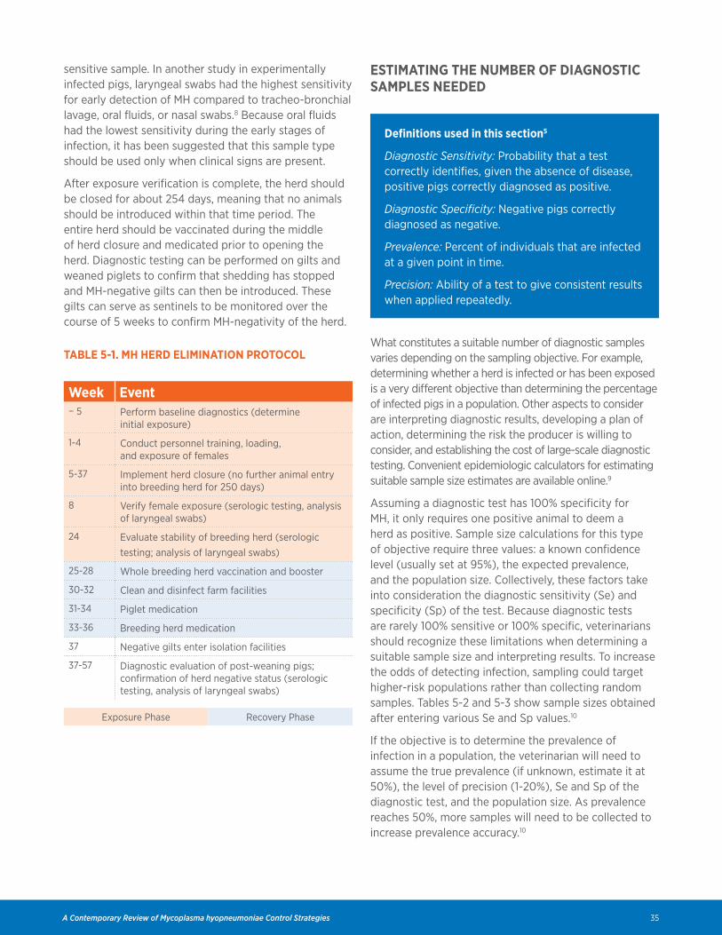
35A Contemporary Review of Mycoplasma hyopneumoniae Control Strategies
sensitive sample . In another study in experimentally infected pigs, laryngeal swabs had the highest sensitivity for early detection of MH compared to tracheo-bronchial lavage, oral fluids, or nasal swabs .8 Because oral fluids had the lowest sensitivity during the early stages of infection, it has been suggested that this sample type should be used only when clinical signs are present .
After exposure verification is complete, the herd should be closed for about 254 days, meaning that no animals should be introduced within that time period . The entire herd should be vaccinated during the middle of herd closure and medicated prior to opening the herd . Diagnostic testing can be performed on gilts and weaned piglets to confirm that shedding has stopped and MH-negative gilts can then be introduced . These gilts can serve as sentinels to be monitored over the course of 5 weeks to confirm MH-negativity of the herd .
TABLE 5-1. MH HERD ELIMINATION PROTOCOL
Week Event− 5 Perform baseline diagnostics (determine
initial exposure)
1-4 Conduct personnel training, loading, and exposure of females
5-37 Implement herd closure (no further animal entry into breeding herd for 250 days)
8 Verify female exposure (serologic testing, analysis of laryngeal swabs)
24 Evaluate stability of breeding herd (serologic testing; analysis of laryngeal swabs)
25-28 Whole breeding herd vaccination and booster
30-32 Clean and disinfect farm facilities
31-34 Piglet medication
33-36 Breeding herd medication
37 Negative gilts enter isolation facilities
37-57 Diagnostic evaluation of post-weaning pigs; confirmation of herd negative status (serologic testing, analysis of laryngeal swabs)
Exposure Phase Recovery Phase
ESTIMATING THE NUMBER OF DIAGNOSTIC SAMPLES NEEDED
Definitions used in this section5
Diagnostic Sensitivity: Probability that a test correctly identifies, given the absence of disease, positive pigs correctly diagnosed as positive .
Diagnostic Specificity: Negative pigs correctly diagnosed as negative .
Prevalence: Percent of individuals that are infected at a given point in time .
Precision: Ability of a test to give consistent results when applied repeatedly .
What constitutes a suitable number of diagnostic samples varies depending on the sampling objective . For example, determining whether a herd is infected or has been exposed is a very different objective than determining the percentage of infected pigs in a population . Other aspects to consider are interpreting diagnostic results, developing a plan of action, determining the risk the producer is willing to consider, and establishing the cost of large-scale diagnostic testing . Convenient epidemiologic calculators for estimating suitable sample size estimates are available online .9
Assuming a diagnostic test has 100% specificity for MH, it only requires one positive animal to deem a herd as positive . Sample size calculations for this type of objective require three values: a known confidence level (usually set at 95%), the expected prevalence, and the population size . Collectively, these factors take into consideration the diagnostic sensitivity (Se) and specificity (Sp) of the test . Because diagnostic tests are rarely 100% sensitive or 100% specific, veterinarians should recognize these limitations when determining a suitable sample size and interpreting results . To increase the odds of detecting infection, sampling could target higher-risk populations rather than collecting random samples . Tables 5-2 and 5-3 show sample sizes obtained after entering various Se and Sp values .10
If the objective is to determine the prevalence of infection in a population, the veterinarian will need to assume the true prevalence (if unknown, estimate it at 50%), the level of precision (1-20%), Se and Sp of the diagnostic test, and the population size . As prevalence reaches 50%, more samples will need to be collected to increase prevalence accuracy .10
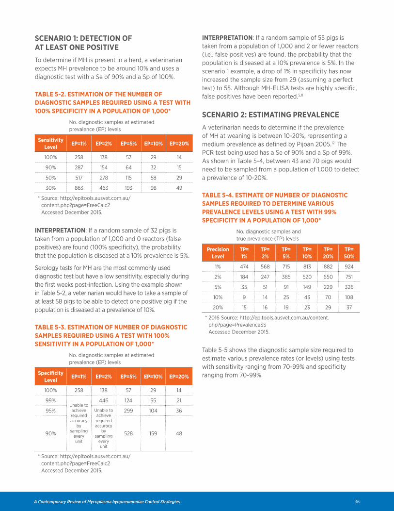
36A Contemporary Review of Mycoplasma hyopneumoniae Control Strategies
SCENARIO 1: DETECTION OF AT LEAST ONE POSITIVE To determine if MH is present in a herd, a veterinarian expects MH prevalence to be around 10% and uses a diagnostic test with a Se of 90% and a Sp of 100% .
TABLE 5-2. ESTIMATION OF THE NUMBER OF DIAGNOSTIC SAMPLES REQUIRED USING A TEST WITH 100% SPECIFICITY IN A POPULATION OF 1,000*
No . diagnostic samples at estimated prevalence (EP) levels
Sensitivity Level
EP=1% EP=2% EP=5% EP=10% EP=20%
100% 258 138 57 29 14
90% 287 154 64 32 15
50% 517 278 115 58 29
30% 863 463 193 98 49
* Source: http://epitools .ausvet .com .au/ content .php?page=FreeCalc2 Accessed December 2015 .
INTERPRETATION: If a random sample of 32 pigs is taken from a population of 1,000 and 0 reactors (false positives) are found (100% specificity), the probability that the population is diseased at a 10% prevalence is 5% .
Serology tests for MH are the most commonly used diagnostic test but have a low sensitivity, especially during the first weeks post-infection . Using the example shown in Table 5-2, a veterinarian would have to take a sample of at least 58 pigs to be able to detect one positive pig if the population is diseased at a prevalence of 10% .
TABLE 5-3. ESTIMATION OF NUMBER OF DIAGNOSTIC SAMPLES REQUIRED USING A TEST WITH 100% SENSITIVITY IN A POPULATION OF 1,000*
No . diagnostic samples at estimated prevalence (EP) levels
Specificity Level
EP=1% EP=2% EP=5% EP=10% EP=20%
100% 258 138 57 29 14
99%Unable to achieve required accuracy
by sampling
every unit
446 124 55 21
95% Unable to achieve required accuracy
by sampling
every unit
299 104 36
90% 528 159 48
* Source: http://epitools .ausvet .com .au/ content .php?page=FreeCalc2 Accessed December 2015 .
INTERPRETATION: If a random sample of 55 pigs is taken from a population of 1,000 and 2 or fewer reactors (i .e ., false positives) are found, the probability that the population is diseased at a 10% prevalence is 5% . In the scenario 1 example, a drop of 1% in specificity has now increased the sample size from 29 (assuming a perfect test) to 55 . Although MH-ELISA tests are highly specific, false positives have been reported .5,11
SCENARIO 2: ESTIMATING PREVALENCE A veterinarian needs to determine if the prevalence of MH at weaning is between 10-20%, representing a medium prevalence as defined by Pijoan 2005 .12 The PCR test being used has a Se of 90% and a Sp of 99% . As shown in Table 5-4, between 43 and 70 pigs would need to be sampled from a population of 1,000 to detect a prevalence of 10-20% .
TABLE 5-4. ESTIMATE OF NUMBER OF DIAGNOSTIC SAMPLES REQUIRED TO DETERMINE VARIOUS PREVALENCE LEVELS USING A TEST WITH 99% SPECIFICITY IN A POPULATION OF 1,000*
No . diagnostic samples and true prevalence (TP) levels
Precision Level
TP= 1%
TP= 2%
TP= 5%
TP= 10%
TP= 20%
TP= 50%
1% 474 568 715 813 882 924
2% 184 247 385 520 650 751
5% 35 51 91 149 229 326
10% 9 14 25 43 70 108
20% 15 16 19 23 29 37
* 2016 Source: http://epitools .ausvet .com .au/content .php?page=PrevalenceSS Accessed December 2015 .
Table 5-5 shows the diagnostic sample size required to estimate various prevalence rates (or levels) using tests with sensitivity ranging from 70-99% and specificity ranging from 70-99% .

37A Contemporary Review of Mycoplasma hyopneumoniae Control Strategies
TABLE 5-5. ESTIMATE OF NUMBER OF DIAGNOSTIC SAMPLES REQUIRED TO DETERMINE A PREVALENCE OF 10% IN A POPULATION OF 1,000 WITH VARYING SE AND SP LEVELS*
No . diagnostic samples and sensitivity (Se) levels
Test Specificity
Level
Se= 70%
Se= 80%
Se= 90%
Se= 95%
Se= 99%
Se= 0.99
70% 351 260 198 175 159 156
80% 225 171 135 121 111 109
90% 126 100 82 75 70 69
99% 56 48 43 40 38 38
0 .99 50 44 39 36 35 34
* Source: http://epitools .ausvet .com .au/content .php?page=PrevalenceSS Accessed December 2015 .
REFERENCES1 . Dalquist L . Mycoplasma hyopneumoniae
acclimation: Overcoming challenges in the field . Proceedings Allen D. Leman Swine Conference, St . Paul, Minnesota, 2014 . Available at: https://docs .google .com/file/d/0BzGsnfsQ28heRWN4OHVaZTBab2c/edit . Accessed December 2015 .
2 . Hollis W . Mycoplasma hyopneumoniae field experiences . In: Proceedings Allen D. Leman Swine Conference Minneapolis, Minnesota, 2014 . Available at: https://docs .google .com/file/d/0BzGsnfsQ28hebUlrdVJUVUUtYkU/edit . Accessed December 2015 .
3 . Pieters M, Pijoan C, Fano E, et al . An assessment of the duration of Mycoplasma hyopneumoniae infection in an experimentally infected population of pigs . Vet Microbiol 2009;134:261-266 .Harris DL . Multisite production: A review of 15 years lessons learned or forgotten . In: Proceedings Swine Disease Conference, Ames, Iowa . 2003 .
4 . Erlandson KR, Evans RB, Thacker BJ, et al . Evaluation of three serum antibody enzyme-linked immunosorbent assays for Mycoplasma hyopneumoniae . J Swine Health Prod 2005;13:198-203 .
5 . Strait EL, Madsen ML, Minion FC, et al . Real-time PCR assays to address genetic diversity among strains of Mycoplasma hyopneumoniae . J Clin Microbiol 2008;46:2491-2498 .
6 . Fablet C, Marois C, Kobisch M, et al . Estimation of the sensitivity of four sampling methods for Mycoplasma hyopneumoniae detection in live pigs using a Bayesian approach . Vet Microbiol 2010;143:238-245 .
7 . Pieters M, Rovira A . Comparison of various sample types for detection of Mycoplasma hyopneumoniae in recently infected pigs . In: Proceedings Allen D. Leman Swine Conference, Minneapolis, Minnesota, 2013 .
8 . EpiTools epidemiological calculators . AusVet Animal Health Services . Available at: http://epitools .ausvet .com .au/content .php?page=home . Accessed December 2015 .
9 . Garder I . Analysis and use of diagnostic data . In: Zimmerman JJ, Karriker LA, Schwartz KJ, et al, eds . Diseases of Swine, 10th ed . Oxford, UK: Wiley-Blackwell; 2012:94-105 .
10 . Johnson, E . Minton B . Test characteristics when verifying Mycoplasma hyopneumoniae status in expected negative herds . In: Proceedings Allen D Leman Swine Conference, Minneapolis, Minnesota . 2009;185 .
11 . Pijoan C . A controversial view of Mycoplasma hyopneumoniae . In: Proceedings Allen D. Leman Swine Conference, Minneapolis, Minnesota . 2005;114-117 .
VACCINATIONJoseph F. Connor, DVM, MS Carthage Veterinary Service, Ltd. Carthage, Illinois
MH VACCINES AND THE GOAL OF VACCINATIONMycoplasma hyopneumoniae (MH) vaccines can be effective in reducing the economic impact of the chronic and clinical forms of MH-associated disease . Although MH vaccines can help reduce MH shedding, they do not prevent colonization or infection .1-4 Commercial and autogenous MH vaccines are both used . At least six companies manufacture inactivated MH vaccines in the U .S ., either as single-dose or two-dose products . There is no information to indicate that there is a relevant MH strain differentiation among vaccines, suggesting that the differences in these products are restricted

38A Contemporary Review of Mycoplasma hyopneumoniae Control Strategies
to antigen concentration and the adjuvant . Vaccine preferences are largely based on controlled research studies and field experience .5
Autogenous vaccines have a low market penetration because of the lack of MH strain differentiation among field isolates, difficultly in growing the organism, and absence of controlled research studies substantiating the value of autogenous MH agents . Autogenous vaccines often have the same adjuvants used in commercial vaccines due to commonality in manufacturing . If strain differentiation and its relevance become clearly defined, requests for autogenous vaccines may increase .
VACCINATION TIMING Timing of MH vaccination continues to be vigorously debated . In U .S . herds, vaccination at weaning or immediately after weaning is the optimum time to avoid maternal antibody interference, help protect pigs prior to anticipated exposure, and achieve reasonable duration of immunity .6 A number of studies have evaluated early piglet vaccination .7-9 Piglet immunity is primarily cell-mediated, so interference by circulating maternal antibodies is not expected to interfere with the innate immune response . Interpretation of these studies is confounded by low stimulation of circulating antibodies after vaccination and the short duration of the MH serologic response . Studies suggest that cellular transfer from dam to litter is valuable in protecting piglets .4,10
Timing of vaccination of suckling piglets reflects the industry trend to segregated production systems . Producers and veterinarians find it more convenient to vaccinate piglets at the sow farm, which helps ensure greater vaccination compliance and allows more convenient monitoring of vaccine administration . For example, improved consistency could be expected on a 5,000-sow farm where 3,000 pigs per week are vaccinated compared to individual wean-to-finish technicians vaccinating 3,000 pigs every six months . It would be common for a sow herd to flow pigs from one to 20 finishing sites across a wide geographical area . The advantages of vaccination at the sow farm, in addition to monitoring the process, can include improved inventory control, a consistent storage environment, and consistency of the vaccination process . Studies have shown equivalent efficacy in late-suckling piglet vaccination programs whether one- or two-dose regimens are used .7,9 To ensure that piglets receive at least one vaccine dose, producers and veterinarians may be more comfortable with a two-dose regimen . In the suckling phase, the most common procedure is for each pig to be handled, so there is typically a very high rate of
vaccination compliance in this population .
CHOICE OF VACCINE DURING AN OUTBREAKMH vaccination decisions during an outbreak are generally similar to what occurs with an ongoing vaccination program . Since MH infection has a very low replication rate, vaccination of the population during an active outbreak could provide some degree of efficacy or herd immunity for the population as a whole . The veterinarian must critically evaluate MH prevalence and the extent and time of onset of clinical activity when deciding to vaccinate in the face of an outbreak . Since it takes 2-4 weeks for the immune response to vaccination to occur, administering vaccination concurrently with water-soluble or feed medication could be considered as a transmission-reducing strategy .11
LINKING VACCINATION TO PREVALENCEStudies by University of Minnesota investigators have shown that MH prevalence varies widely in suckling pig populations .3 Vaccination is likely to be most successful in low- and medium-prevalence populations of suckling-pigs and early post-weaned populations . High-prevalence populations may respond better to an early suckling-pig vaccination strategy or early vaccination combined with oral antibiotics administered immediately after weaning . Because of MH prevalence variability among groups, to achieve a consistent vaccination response, early suckling pig vaccination is recommended . Factors influencing MH prevalence include introduction of MH-negative replacement gilts of various ages, a 50% or greater annual replacement rate, reduced birth weights associated with increased litter size, increase in suckling pig age, age variation within a room or group, variation in maternal immunity in gilts compared to sows, and variation in transmission rate between parities .12-15
NUMBER OF VACCINE DOSESVaccination with either a single- or two-dose regimen has equivalent efficacy in late suckling piglets . Producers or veterinarians may favor a two-dose vaccination program on the assumption that all pigs will receive at least one vaccine dose even if 100% compliance with the multi-dose regimen is not achieved . In the suckling phase, the most common procedure is for each pig to be individually handled, resulting in very high vaccination compliance .

39A Contemporary Review of Mycoplasma hyopneumoniae Control Strategies
COMBINATION VACCINESA number of companies manufacture a bivalent MH and PCV-2 vaccine which has the advantages of reducing the number of injections, more convenient handling of products, and administration efficiency . Manufacturers’ studies and field experience suggest that these MH combination vaccines have similar efficacy as monovalent MH vaccines .16
IMMUNE RESPONSE IN LITTERS FROM VACCINATED DAMSTwo recent studies have shown that vaccinated dams have reduced dam-to-piglet and sow-to-sow transmission of MH .15,17 This outcome indicates that sow-herd vaccination can be a key component of an effective control strategy . Although there are concerns that vaccination of sows will interfere with piglet vaccination due to maternal antibody interference, no published studies have shown this to be true .
IMPACT OF CONCURRENT VACCINATION FOR MH AND PRRSCoinfections with MH and PRRSV are common in swine production systems . Thacker et al reported that MH infection potentiates PRRSV-induced disease,18 suggesting that the impact of controlling MH in PRRSV-infected production units warrants further study . Manufacturer studies indicated that concurrent vaccination for MH and PRRSV does not interfere with efficacy of MH vaccination,19,20 findings that were independently corroborated .21 A greater concern is the effect that wild-type PRRSV has in pigs vaccinated for MH . It is speculated that because of the prevalence of PRRSV in sow herds, weaned piglets will often be PRRSV-viremic for 8 to 50 weeks, resulting in a less robust immune response to MH vaccination . Vaccination of viremic pigs also creates the possibility of PRRSV transmission via needle injection . This scenario would likely result in an immediate PRRSV viremia in exposed piglets and potentially reduce the immune response to other vaccines .
Timing of MH vaccination should be critically evaluated in relation to PRRSV prevalence at birth and weaning . For a PRRSV-viremic or infected population, it would be advantageous to vaccinate the suckling pigs at a very early age, 1 to 3 days . University investigators studying the effects of MH vaccine on pigs naturally infected with MH and PRRSV reported that regardless of the timing of PRRSV seroconversion, MH vaccination improved production performance .22
CHALLENGE-OF-IMMUNITY TRIALSMH efficacy studies sometimes use a contact-exposure model to challenge vaccinated and control pigs . Because of MH’s low replication rate, transmission from infected seeder pigs frequently results in a lower infection rate than what probably occurs in field populations . The low MH transmission rate following experimental MH challenge reduces the economic consequences of challenge studies compared to what occurs in commercial settings following natural exposure . A study has utilized preliminary viral exposure prior to MH challenge to improve transmission and susceptibility .18 It is important to remember that vaccination is expected to provide an economic return over-and-above vaccine and labor costs . A study indicates that properly administered MH vaccines have the capability of improving average daily gain (ADG) and feed conversion (FC) and reduce mortality,23 but vaccination should be given under circumstances that maximize the potential for an optimum response .
EXPECTATIONS AND RECOMMENDATIONS FOR VACCINATION MH vaccination has the potential to help reduce vertical and horizontal MH transmission and to reduce the economic losses resulting from subpar ADG and FC and production losses and treatment costs due to disease morbidity and mortality . These outcomes should be tempered by the understanding that vaccination does not prevent MH colonization or infection, potentially allowing MH transmission and low but irreducible levels of chronic and clinical disease .
Whether to administer multiple or a single dose should be based in part on the producer preferences . Administration timing is highly dependent on the MH prevalence in weaned pigs and any evidence, diagnostic or clinical, of concurrent viral and other bacterial infections . Because of the unpredictability and wide variation in MH prevalence in weaned litters, any pig vaccination program should be based on diagnostic surveillance . A calculation of MH vaccination return on investment should consider variables such as number of vaccine doses, vaccination timing, which population to vaccinate, and the diagnostic basis for vaccination .
KNOWLEDGE GAPSThere is little information on the real cost of uncomplicated MH infection in U .S . swine herds . Likewise, there is little return-on-investment data on the

40A Contemporary Review of Mycoplasma hyopneumoniae Control Strategies
value of vaccinating pig populations with low, moderate, or high prevalence of MH infection . Whether vaccination is effective against multiple MH strains is also an open question . Diagnostic monitoring of late-gestation gilts may provide direction on prevalence to guide optimum timing of vaccination .
REFERENCES1 . Maes D, Deluyker H, Verdonck M, et al . Effects of
vaccination against Mycoplasma hyopneumoniae in herds with continuous system . Zentralbl Veterinarmed B 1998;45:495-505 .
2 . Maes D, Deluyker H, Verdonck M, et al . Effect of vaccination against Mycoplasma hyopneumoniae in pig herd with all-in all-out system . Vaccine 1999;17:1024–1034 .
3 . Fano E, Pijoan C, Dee S, et al . Effect of Mycoplasma hyopneumoniae colonization at weaning on disease severity in growing pigs . Can J Vet Res 2007;71:195-200 .
4 . Pieters M, Fano E, Dee SA, et al . Transmission of Mycoplasma hyopneumoniae to vaccinated and unvaccinated replacement gilts from persistently infected pigs . 2006 . In: Proceedings 19th International Pig Veterinary Society Congress, Copenhagen, Denmark . 2006;102 .
5 . Strait EL, Rapp-Gabrielson VJ, Erickson BZ, et al . Efficacy of a Mycoplasma hyopneumoniae bacterin in pigs challenged with two contemporary pathogenic isolates of M. hyopneumoniae . J Swine Health Prod 2008;16:200-206 .
6 . Thacker BJ, Boettcher TB, Anderson TS, et al . Influence of passive immunity on serological responses to Mycoplasma hyopneumoniae vaccination . Swine Research Report 1997, Paper 45, Iowa State University, Ames, Iowa . Available at: http://lib .dr .iastate .edu/swinereports_1997/45 . Accessed December 2015 .
7 . Greiner L, Connor JF, Lowe JF . Comparison of Mycoplasma hyopneumoniae vaccination . In: Proceedings of the 42nd Annual meeting Am Assoc Swine Veterinarians, Phoenix, Arizona, 2011: 245-248 .
8 . Kaiser T, Yonkers TK, Asmus MD, Taylor LP . Mycoplasma hyopneumoniae vaccine administered to pigs at one day of age . In: Proceedings of the 39th Annual Meeting Am Assoc Swine Veterinarians, Dallas, TX, 2009, 157-160 .
9 . Lilie K, Ritzmann M, Erber M, Heinritzi K . Study on the effect and tolerance of an inactivated one shot vaccine against Mycoplasma hyopneumoniae in pigs at different ages in a farm naturally infected . Der Praktishche Tierartz 87, 2006: Heft 7, 548-552 .
10 . Molitor TW, Brandrick, M, Pieters M, et al . Maternal transfer of immunity: Role in Mycoplasma hyopneumoniae . In: Proceedings Allen D. Leman Swine Conference, Minneapolis, Minnesota . 2007;75-78 .
11 . Opriessnig T, Thacker E, Halbur PG . Chlortetracycline is effective in reducing lesions in pigs coinfected with Mycoplasma hyopneumoniae and porcine circovirus type 2 . In: Proceedings 19th International Pig Veterinary Society Congress, Copenhagen, Denmark . 2006;302 .
12 . Fano E, Pijoan C, Dee SA, et al . Assessment of the effect of sow parity on the prevalence of Mycoplasma hyopneumoniae in piglets at weaning . In: Proceedings 19th International Pig Veterinary Society Congress, Copenhagen, Denmark . 2006;96 .
13 . Fano E, Pijoan C, Deen J, et al . Prevalence of Mycoplasma hyopneumoniae in piglets at weaning as a predictor on the severity of the disease in growing pigs . In: Proceedings 19th International Pig Veterinary Society Congress, Copenhagen, Denmark . 2006;441 .
14 . Pieters M, Baido SK, Molitor TW . The effect of passive immunity to Mycoplasma hyopneumoniae and an extended lactation period on piglet immune development . In: Proceedings Allen D. Leman Swine Conference, Minneapolis, Minnesota . 2012;39:224 .
15 . Ruiz A, Batista L, Pijoan C . Effect of different vaccination protocols in Mycoplasma hyopneumoniae infection . In: Proceedings 17th International Pig Veterinary Society Congress, Ames, Iowa . 2002;334 .
16 . Cowles B, Greiner D, Nitzel GP, et al . Comparative evaluation of one and two doses PCV/M. hyopneumoniae vaccination protocols in swine challenged with PCV and M. hyopneumoniae . In: Proceedings of the 45th Annual Meeting Am Assoc Swine Veterinarians, Dallas, TX . 2014: 385-386
17 . Sibila M, Bernal R, Torrents D, et al . Effect of sow vaccination against Mycoplasma hyopneumoniae on sow and piglet colonization and seroconversion and pig lung lesions at slaughter . Vet Microbiol 2008;127:165-170 .
18 . Thacker, EL, Halbur RG,Ross RF, et al . Mycoplasma hyopneumoniae potentiation of porcine reproductive and respiratory syndrome virus-induced pneumonia . J Clin Microbiol 1999;37:620-627 .
19 . Blood S, Fangman T, Cline G . Performance of weaned pigs vaccinated with 3FLEX compared to pigs vaccinated with Ingelvac CircoFLEX-MycoFLEX and Ingelvac PRRS MLV in separate injections . In: Proceedings Allen D. Leman Swine Conference, Minneapolis, Minnesota . 2011;263 .

41A Contemporary Review of Mycoplasma hyopneumoniae Control Strategies
20 . Haiwick G, Eichmeyer M, Roof M, et al . Trivalent vaccine mixture protects against simultaneous challenge with M. hyopneumoniae, PCV2, and PRRS virus . In: Proceedings Allen D. Leman Swine Conference, Minneapolis, Minnesota . 2010; 176 .
21 . Drexler CS, Witvliet MH, Raes M, et al . Efficacy of combined porcine reproductive and respiratory syndrome virus and Mycoplasma hyopneumoniae vaccination in piglets . Vet Rec 2010;166:70-74 .
22 . Moreau IA, Miller GY, Bahnson PB . Effects of Mycoplasma hyopneumoniae vaccine on pigs naturally infected with M. hyopneumoniae and porcine reproductive and respiratory syndrome virus . Vaccine 2004;22:2328-2333 .
23 . Pieters M, Fano E, Pijoan C, et al . An experimental model to evaluate Mycoplasma hyopneumoniae transmission from asymptomatic carriers to unvaccinated and vaccinated sentinel pigs . Can J Vet Res 2010;74:157-160 .
MEDICATIONDoug King, DVM formerly with Zoetis Pork Technical Services
Lucina Galina Pantoja, DVM, PhD Pork Technical Services Zoetis Inc. Hendersonville, Tennessee
There are a variety of medications available for treating and controlling Mycoplasma hyopneumoniae (MH) in swine . Although MH is resistant to ß-lactams, other classes of antimicrobials, namely tetracyclines, macrolides and lincosamides, have activity against MH . Certain dosage forms of some anti-infective agents have activity against MH and are sometimes used even though they do not have a label indication for swine EP .
MEDICATIONS USED FOR MHFor control and treatment of respiratory disease including MH infections in pigs, tetracyclines, macrolides, lincosamides, pleuromutilins, fluoroquinolones, florfenicol, aminoglycosides and aminocyclitols have all been used worldwide .1 The anti-infective agents described below are in widespread use in North America for MH treatment, control, or elimination .
LINCOMYCIN
Multiple studies have confirmed that lincomycin reduces the incidence and severity of MH .2-4 Indications and dosing for approved lincomycin formulations available in the U .S . are as follows:
Lincomix® Feed Medication
§ Fed at 200 grams per ton of complete feed for 21 days .
§ Indicated for reduction in the severity of mycoplasmal pneumonia of swine (MPS) .
CHLORTETRACYCLINE
Evidence that MH disease can be controlled by medication with tetracycline antibiotics has been documented .2,5,6 In the U .S ., commercial products that contain chlortetracycline do not have a label approval for MH . The use of medicated feeds for extralabel uses is strictly prohibited in the U .S . A new federal regulation, effective in December, 2016, will require veterinary oversight in the form of a “VFD” or Veterinary Feed Directive for the use of medically important antibiotics in animal feeds . Under this new regulation, extra-label use of medicated feeds will continue to be prohibited even under the order or supervision of a veterinarian .
TULATHROMYCIN
Tulathromycin (Draxxin®, Zoetis) is the first triamilide, a macrolide subclass developed as a highly bioavailable, long-acting antimicrobial for treatment of common respiratory pathogens in pigs and cattle, including MH .8 Pigs treated with tulathromycin in randomized trials had significant improvements in clinical respiratory disease and mortality compared to control pigs .9,10 In experimental settings, tulathromycin-treated pigs had significantly improved lung lesion scores, clinical response, and weight gain compared to untreated controls .11
DRAXXIN® INJECTABLE SOLUTION
This antimicrobial is given at 2 .5 mg/kg for treatment and control of swine respiratory disease associated with MH . Holst et al demonstrated that Draxxin® given to dams just prior to farrowing (day 112) reduced MH shedding as evidenced by lower prevalence of organisms in sows at farrowing, number of piglets at birth, and number of pigs at weaning .12 Although an impact on MH shedding was demonstrated, a study by Painter et al showed that vaccination alone or in combination with Draxxin® did not result in the elimination of MH from experimentally challenged pigs sooner than would occur naturally .13
42A Contemporary Review of Mycoplasma hyopneumoniae Control Strategies
MEDICATION FOR MH TREATMENTField observations suggest that severe clinical disease associated with MH is most often associated with viral co-infections (PRRS, IAV-S, PCV-2) or in MH-exposed animals that were previously naive . In such cases, porcine respiratory disease complex (PRDC) involving primary MH infection often results in death . Individual-pig treatment via injectable medication may be warranted at the onset of clinical signs . There is some evidence that MH field isolates have resistance to macrolides, lincosamides, and fluoroquinolones,2 although antimicrobial resistance has not yet posed a major problem for treatment of MH infections .
Mass medication is indicated for treatment of MH infection until clinical signs resolve . Thereafter, feed medications are often used as convalescent therapy, until clinical signs resolve, or until pigs are marketed . Because pig performance is negatively impacted before the onset of overt clinical signs, it is advisable to consider MH control in populations that have a portion of the animals showing clinical signs .
MEDICATION FOR MH CONTROL Vertical transmission from dam to offspring of MH has been documented by multiple investigators .14,15 Fano et al further determined that piglets from young sows were more likely to have a higher prevalence at weaning than piglets from intermediate and older sows .15 In 2007, Fano and Pijoan demonstrated that MH prevalence at weaning was correlated with the severity of respiratory disease in the grow-out period .16 Besides vaccination, several treatment strategies should be considered in an unstable sow herd (i .e ., a herd where clinical signs of MH infection are present) to help mitigate vertical transmission of MH and help reduce prevalence at weaning . These include pre-farrowing sow medication, early medication at weaning, and feed medication after weaning . In contrast, for a stable sow herd experiencing less vertical MH transmission and a low prevalence at weaning, feed-medication is typically administered strategically in order to help prevent clinical disease . Due to the slow transmission of MH and delayed expression of clinical signs within groups of pigs, antibody response (seroconversion) is generally a lagging indicator of exposure . The interval between infection and seroconversion varied from 2 weeks (in experimental infection) to 8 weeks (in field infections) .17 Therefore, medication , for example using Lincomix® fed at 200 grams per ton of complete feed as the sole ration for 21 consecutive days, to mitigate expression of disease is for agreement with singular subject medication typically administered 4-8 weeks in advance of seroconversion .
MEDICATION FOR MH ELIMINATIONAfter a long period of herd closure (i .e ., no new animal introduction) to negate MH circulation in a sow herd, whole-herd injectable medication using injectable Draxxin® or Draxxin® 25 (2 .5 mg/kg of body weight) in pigs or sows and feed medication with Lincomix® (200 g/ton for 21 consecutive days) is typically instituted as an insurance measure to eliminate any lingering viable organisms . Elimination without herd closure has been attempted with a lower degree of success by using two whole-herd (all sows and pigs) injections of Draxxin® or Draxxin® 25 .
IMPORTANT SAFETY INFORMATION Withdraw DRAXXIN/DRAXXIN 25 five (5) days prior to slaughter . Do not use in animals known to be hypersensitive to the product . See full Prescribing Information on pages 48-51 .
REFERENCES1 . Maes D . Mycoplasma hyopneumoniae infection
in pigs: update on epidemiology and control . In: Proceedings of the 21st International Pig Veterinary Society Congress, Vancouver, Canada . 2010;30-35 .
2 . Vicca J, Stakenborg VJ, Maes D, et al . In vitro susceptibilities of Mycoplasma hyopneumoniae field isolates . Antimicrob Agents Chemother 2004;48:4470-4472 .
3 . Van Buren J W . Lincomycin and swine mycoplasmal pneumonia indications and efficacy . In: Proceedings Am Assoc Swine Practitioners, Cincinnati, Ohio . 1983;29-40 .
4 . Graham R, Lens S, Jansegers L . The effect of lincomycin as medicated feed on reduction of incidence and severity of mycoplasmal pneumonia in growing swine . In: Proceedings 8th International Pig Veterinary Society Congress Vet Society Congress, Ghent, Belgium . 1984;119 .
5 . Whittlestone P . The role of mycoplasmas in the production of pneumonia in the pig . In: Pathogenic Mycoplasmas. Amsterdam: Associated Scientific Publishers;1972:263–283 .
6 . Thacker EL, Nilubol D, Halbur PG, et al . Efficacy of aureomycin chlortetracycline (CTC) granulated premix in decreasing Mycoplasma hyopneumoniae-potentiated PRRSV pneumonia . In: Proceedings 20th International Pig Veterinary Society Congress, Durban, South Africa . 2008;208 .
7 . Thacker EL, Thacker BJ, Wolff T . Efficacy of a chlortetracycline feed additive in reducing pneumonia and clinical signs induced by experimental Mycoplasma hyopneumoniae challenge . J Swine Health Prod 2006;14:140-144 .

43A Contemporary Review of Mycoplasma hyopneumoniae Control Strategies
8 . Evans NA . Tulathromycin: an overview of a new triamilide antibiotic for livestock respiratory disease . Vet Ther 2005;6:83-95 .
9 . Nanjiani IA, McKelvie J, Benchaoui HA, et al . Evaluation of the therapeutic activity of tulathromycin against swine respiratory disease on farms in Europe . Vet Ther 2005;6:203-313 .
10 . Nutsch RG, Hart FJ, Rooney KA, et al . Efficacy of tulathromycin injectable solution for the treatment of naturally occurring swine respiratory disease . Vet Ther 2005;6:214-224 .
11 . McKelvie J, Morgan JH, Nanjiani IA, et al . Evaluation of tulathromycin for the treatment of pneumonia following experimental infection of swine with Mycoplasma hyopneumoniae . Vet Ther 2005;6:197-202 .
12 . Holst S, Yeske P, Leuwerke B, et al . Effect of pre-farrow administration of tulathromycin injectable solution on Mycoplasma hyopneumoniae prevalence in suckling pigs at birth and weaning . In: Proceedings 44th Annual Meeting Am Association Swine Veterinarians, San Diego, California . 2013;90 .
13 . Painter T, Kuhn M, Wolff T, et al . Efficacy and duration of infection study for Respisure and Draxxin against a Mycoplasma hyopneumoniae challenge in swine . In: Proceedings Allen D. Leman Swine Conference, Minneapolis, Minnesota . 2012;39:225 .
14 . Clark LK, Scheidt AB, Armstrong CH, et al . The effect of all-in/all-out management on pigs from a herd with enzootic pneumonia . Vet Med 1991;86:946-951 .
15 . Ross RF . Mycoplasmal diseases . In : Straw B, D’Allaire S, Mengeling W, et al, eds . Diseases of Swine, 8th ed . Ames, Iowa: Iowa State University Press; 1999:495-509 .
16 . Fano E, Pijoan C, Dee S, et al . Effect of Mycoplasma hyopneumoniae colonization at weaning on disease severity in growing pigs . Can J Vet Res 2007;71:195-200 .
17 . Calsamiglia M, Pijoan C, Bosch GJ . Profiling Mycoplasma hyopneumoniae in farms using serology and a nested PCR technique . J Swine Health Prod 1999;7:263-268 .
ELIMINATION Paul Yeske DVM, MS Swine Vet Center St. Peter, Minnesota
Mycoplasma hyopneumoniae (MH) continues to be one of the most prevalent and economically significant respiratory pathogens affecting the swine industry .1 MH is the etiologic agent of enzootic pneumonia, a chronic respiratory disease in swine characterized by a chronic, non-productive cough .2,3 Economic losses related to MH are associated with decreased feed efficiency, reduced average daily gain (ADG), and increased medication costs .1 Tables 5-6 . 5-7, 5-8, and 5-9 detail the differences in performance ADG, feed conversion efficiency, percent mortality, percent pigs marketed, feed grade medication cost, and other medication costs in selected populations of MH-negative versus MH-positive pigs where all pigs had similar genetics and nutrition . The single-year (Table 5-6) and multi-year (Table 5-9) performance data were based on close-out records for 1 million pigs per year, 75% of which were MH-negative and 25% of which were MH-positive . All herds were PRRS virus negative at the time of nursery placement . Approximately 50% of pigs from either group became PRRS virus positive during the late finishing stages, reflecting what typically occurs in production settings .
Estimates based on 2013 data collection (Table 5-7) and 2007-14 data (Table 5-9) indicate that the opportunity cost for a finishing pig with MH ranged from $3 .61 to $7 .30 . The 8-year data from 2007-2014 shows a much lower opportunity cost ($3 .61) compared to the opportunity cost for 2013 alone ($7 .30) . The wide difference is no doubt due to the fact that the multi-year data did not over-represent any single year, and also indicates how production variables can vary widely on a year-to-year basis . The comparisons in the tables affirm the considerable economic impact of MH-negative status and the strong incentive to have an MH-negative herd .
The economic cost of MH-associated disease has been the primary driver for producers to consider elimination . Many herds maintain an MH-negative status for extended periods, demonstrating that elimination can have long-term benefits . This has encouraged more herds to implement MH elimination after considering return on investment and the amount of time that herds have been able to stay MH-negative . Table 5-10 illustrates the success of MH elimination programs and the amount of time that they have been able to stay negative .

44A Contemporary Review of Mycoplasma hyopneumoniae Control Strategies
MH ELIMINATION PROTOCOLS Several MH elimination protocols have been developed and can be implemented to eradicate MH from a herd or flow . The following MH elimination protocols have been described, (1) depopulation and repopulation, (2) herd closure and medication, (3) whole herd medication without herd closure, and (4) change of flow in a parity-segregated flow system .
Depopulation and repopulation is the most direct approach for MH elimination . This protocol involves removal of the entire breeding herd and restocking with MH-negative replacements .4 Advantages of depopulation and repopulation include the ability to eliminate more than one disease at once and the opportunity to improve genetics .4 However, there is a complete loss of production from the time the breeding herd is liquidated until replacement females begin farrowing . The duration of lost production can be reduced with an off-site breeding project, which of course incurs additional costs . Total depopulation of the breeding herd may be undesirable on farms with animals that have a high genetic potential (i .e ., genetic nucleus or multiplier farms) .
The herd closure and medication approach has been adapted from a European model and from herd closures done for PRRS control .5,6 Herd closure and medication is often done to simultaneously eliminate both PRRS virus and MH . The basis of this procedure is to close the herd after all replacement animals have been exposed to MH and not make any additions for 240 days . Work done by Pijoan and Peters demonstrated that the 240-day herd closure period is the time required for animals to reliably discontinue MH shedding following natural infection .7 Based on this information, gilts are accumulated for the closure into the herd when possible or maintained at an off-site breeding project . Gilts that will be added to the herd in the future must be infected with MH to “start the clock ticking” . Gilts should be exposed at least by 84 days of age to MH to minimize the likelihood of MH shedding (Figure 5-1) . This can be done over a 1-2 month period and is a critical step in the process to ensure success . Once the closure period is completed, herd immunity is boosted every 90 days by means of whole-herd vaccination . Seven weeks before MH-negative replacements are scheduled to arrive, the whole herd is medicated (sows and piglets) . Although different antimicrobial combinations have been used, a common approach had been to use water-soluble lincomycin in drinking water for the sows and injectable antimicrobials for the piglets at birth and at 14 days . However, the use of water-soluble lincomycin for MH control is off-
label and should be replaced with an on-label use of lincomycin in feed . Figure 5-2 shows a detailed time-and-event protocol for the closure and medication process, including an off-site breeding project . The protocol can be used as a checklist to ensure that all steps in the process are completed in accordance with a hypothetical time line .
Effective in December, 2016, the use of all medically important antimicrobials (those important in human and animal health) in feed will require a Veterinary Feed Directive (VFD) issued by a veterinarian, and use of medically important antimicrobials in water will require a veterinary prescription . Lincomix® is among the products that will require a VFD .
Whole-herd medication is a protocol that allows for much faster MH elimination from the herd but one that has a lower success rate . This procedure does not involve herd closure and is generally done when gilts have just entered the farm . Instead of herd closure, all animals are treated on site followed by treatment of all sows 2 weeks later . All treated piglets are weaned off site and MH-negative replacements are introduced into the herd .
The parity-segregated flow protocol offers a unique opportunity to eliminate MH by using the pig flow to allow for immune sows . Since older-parity sows should not be shedding MH and have not been exposed to younger, MH-shedding sows, the older sows should be MH-negative and capable of flowing negative replacement animals into the herd . The flow is then reversed, following older MH-immune sows to a parity 1 site until 240 days have elapsed for the last MH-positive gilts that were introduced into the parity 1 herd . Once the process is completed, the flow can return to normal . Some herds adopting this approach use medication with the change of flow to improve the odds for success .
Table 5-10 shows the economic benefits of these elimination protocols and the average period of time that MH-negative herds have been able to remain negative . The longest period a herd in this data base has stayed MH-negative is 11 years following elimination . Elimination protocols have been applied in farrow-to-wean farms of various sizes, with the largest herd being 8,000 sows . An algorithm has been developed to document the cost of MH in a herd versus the cost of an elimination protocol for purposes of calculating return on investment .8,9 This tool has been very useful in detailing multiple scenarios for owners as they look at possible MH elimination protocols .
There are various ways to eliminate MH from a swine herd . Because each herd is different, individual producers will have their own goals for an elimination plan based on the MH-exposure risk levels that they are willing to accept . As the
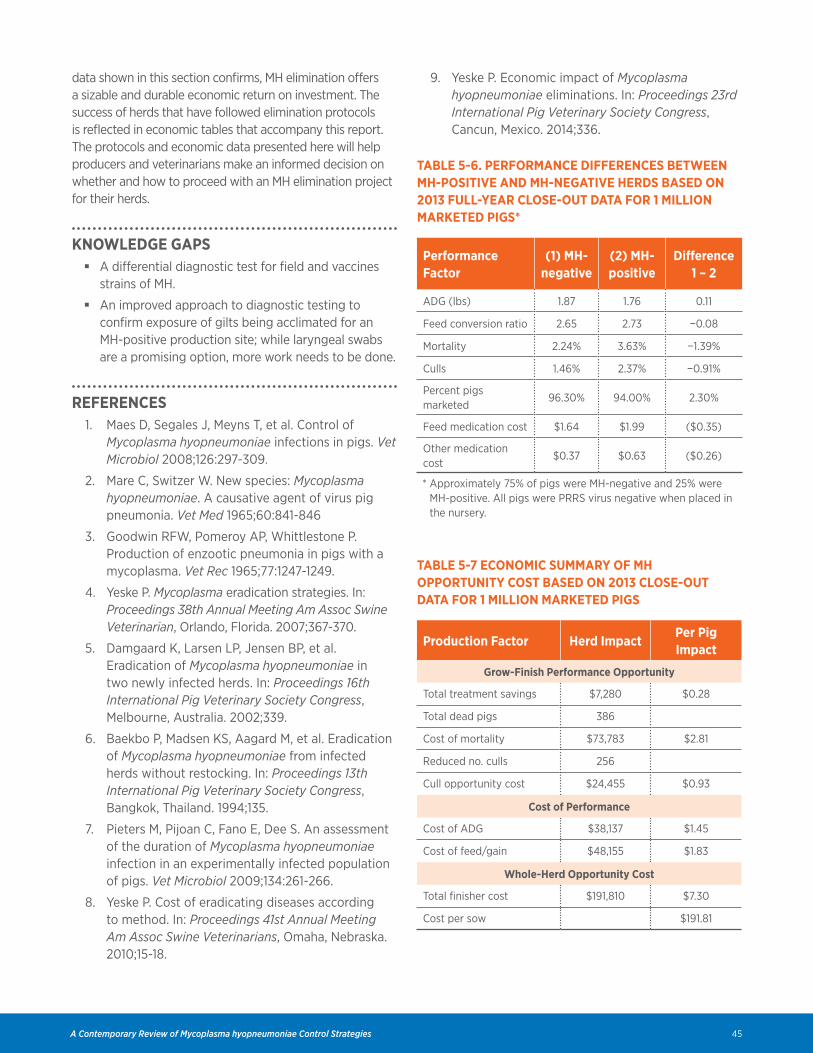
45A Contemporary Review of Mycoplasma hyopneumoniae Control Strategies
data shown in this section confirms, MH elimination offers a sizable and durable economic return on investment . The success of herds that have followed elimination protocols is reflected in economic tables that accompany this report . The protocols and economic data presented here will help producers and veterinarians make an informed decision on whether and how to proceed with an MH elimination project for their herds .
KNOWLEDGE GAPS § A differential diagnostic test for field and vaccines
strains of MH .
§ An improved approach to diagnostic testing to confirm exposure of gilts being acclimated for an MH-positive production site; while laryngeal swabs are a promising option, more work needs to be done .
REFERENCES1 . Maes D, Segales J, Meyns T, et al . Control of
Mycoplasma hyopneumoniae infections in pigs . Vet Microbiol 2008;126:297-309 .
2 . Mare C, Switzer W . New species: Mycoplasma hyopneumoniae . A causative agent of virus pig pneumonia . Vet Med 1965;60:841-846
3 . Goodwin RFW, Pomeroy AP, Whittlestone P . Production of enzootic pneumonia in pigs with a mycoplasma . Vet Rec 1965;77:1247-1249 .
4 . Yeske P . Mycoplasma eradication strategies . In: Proceedings 38th Annual Meeting Am Assoc Swine Veterinarian, Orlando, Florida . 2007;367-370 .
5 . Damgaard K, Larsen LP, Jensen BP, et al . Eradication of Mycoplasma hyopneumoniae in two newly infected herds . In: Proceedings 16th International Pig Veterinary Society Congress, Melbourne, Australia . 2002;339 .
6 . Baekbo P, Madsen KS, Aagard M, et al . Eradication of Mycoplasma hyopneumoniae from infected herds without restocking . In: Proceedings 13th International Pig Veterinary Society Congress, Bangkok, Thailand . 1994;135 .
7 . Pieters M, Pijoan C, Fano E, Dee S . An assessment of the duration of Mycoplasma hyopneumoniae infection in an experimentally infected population of pigs . Vet Microbiol 2009;134:261-266 .
8 . Yeske P . Cost of eradicating diseases according to method . In: Proceedings 41st Annual Meeting Am Assoc Swine Veterinarians, Omaha, Nebraska . 2010;15-18 .
9 . Yeske P . Economic impact of Mycoplasma hyopneumoniae eliminations . In: Proceedings 23rd International Pig Veterinary Society Congress, Cancun, Mexico . 2014;336 .
TABLE 5-6. PERFORMANCE DIFFERENCES BETWEEN MH-POSITIVE AND MH-NEGATIVE HERDS BASED ON 2013 FULL-YEAR CLOSE-OUT DATA FOR 1 MILLION MARKETED PIGS*
Performance Factor
(1) MH-negative
(2) MH-positive
Difference 1 – 2
ADG (lbs) 1 .87 1 .76 0 .11
Feed conversion ratio 2 .65 2 .73 −0 .08
Mortality 2 .24% 3 .63% −1 .39%
Culls 1 .46% 2 .37% −0 .91%
Percent pigs marketed
96 .30% 94 .00% 2 .30%
Feed medication cost $1 .64 $1 .99 ($0 .35)
Other medication cost
$0 .37 $0 .63 ($0 .26)
* Approximately 75% of pigs were MH-negative and 25% were MH-positive . All pigs were PRRS virus negative when placed in the nursery .
TABLE 5-7 ECONOMIC SUMMARY OF MH OPPORTUNITY COST BASED ON 2013 CLOSE-OUT DATA FOR 1 MILLION MARKETED PIGS
Production Factor Herd ImpactPer Pig Impact
Grow-Finish Performance Opportunity
Total treatment savings $7,280 $0 .28
Total dead pigs 386
Cost of mortality $73,783 $2 .81
Reduced no . culls 256
Cull opportunity cost $24,455 $0 .93
Cost of Performance
Cost of ADG $38,137 $1 .45
Cost of feed/gain $48,155 $1 .83
Whole-Herd Opportunity Cost
Total finisher cost $191,810 $7 .30
Cost per sow $191 .81

46A Contemporary Review of Mycoplasma hyopneumoniae Control Strategies
TABLE 5-8. PERFORMANCE DIFFERENTIAL PER 1,000 SOWS BETWEEN MH-POSITIVE AND MH-NEGATIVE GROUPS BASED ON 2007-14 CLOSE-OUT DATA FOR 1 MILLION MARKETED PIGS PER YEAR.
Performance Factor
(1) MH-negative
(2) MH-positive
Difference 1 – 2
Finishing mortality 3 .0% 3 .8% −0 .8%
Finishing culls 1 .7% 1 .9% −0 .2%
Total pigs sold 27,376 27,081 295
Per-pig cost of treatment
$1 .04 $1 .54 ($0 .50)
Finishing ADG (lbs) 1 .80 1 .72 0 .08
Feed conversion ratio 2 .73 2 .75 −0 .02
TABLE 5-9. ECONOMIC SUMMARY OF OPPORTUNITY COST OF MH INFECTION BASED ON 2007-14 CLOSE-OUT DATA FOR 1 MILLION MARKETED PIGS PER YEAR*
Economic Factor Herd ImpactPer Pig Impact
Grow-Finish Performance Opportunity
Treatment savings $14,750 $0 .54
Total mortality 236
Mortality cost $36,393 $1 .35
No . culls 54
Cull opportunity $4,245 $0 .16
Cost of Performance
Cost of ADG $30,006 $1 .10
Cost of feed/gain $12,728 $0 .46
Whole-Herd Opportunity Cost
Total finisher cost $98,722 $3 .61
Cost per sow $98 .72
* Approximately 75% of pigs were MH-negative and 25% were MH-positive . All pigs were PRRS virus-negative when placed in the nursery .
TABLE 5-10. SUCCESS ANALYSIS OF TWO MH ELIMINATION METHODS FOR 2003-15 PRODUCTION DATA
Elimination Method
Outcome Parameter
Herd Closure (n=63 herds)
Medication (n=20 herds)
Total
No . sows 130,088 47,450 177,638
No . herds 46 20 66
Percent MH-negative @ 1 yr
98% 58% 88%
Percent MH-negative >1 yr
91% 53% 80%
Herds negative 41 10 51
Ave . no . months MH-negative
38 28 35
Days 126 Gestation 114 Days
240 Days to Stop Shedding
WeaningStable Pigs(Breed 210 Days Minimum)
(Minimum of Age 84 Days)Exposing Gilts
Figure 5-1. Time line for introducing MH-negative gilts into an MH-positive herd. The process assumes a 240-day period for discontinuance of MH shedding in gilts that have been exposed to MH-positive animals. Breeding occurs at 210 days of age and farrowing of pigs with an MH-low level of colonization at 240 days.

47A Contemporary Review of Mycoplasma hyopneumoniae Control Strategies
HERD CLOSURE PROTOCOL FOR MH ELIMINATION IN A HYPOTHETICAL HERD, INCLUDING AN OFF-SITE BREEDING PROJECT
Farm:
Date Day Activity Product DosageWithdrawal Interval
9/16/2014 Last gilt’s birth date
10/26/2014 1 Herd closure N/A N/A N/A
11/25/2014 31 Confirmed MH exposure complete N/A N/A N/A
11/26/2014 32 Whole-herd vaccination (sow unit & GDU) RespiSure-ONE® 2 mL 21 days
2/23/2015 121 Whole-herd vaccination (sow unit & GDU) RespiSure-ONE® 2 mL 21 days
3/20/2015 148 Vaccinate gilts at off-site breeding project RespiSure-ONE® 2 mL 21 days
4/3/2015 162 Vaccinate gilts at off-site breeding project RespiSure-ONE® 2 mL 21 days
4/12/2015 171
• Select test gilts in GDU
• Test selected gilts in all pens
• Maintain appropriate records for re-testing
N/A N/A N/A
4/17/2015 176 Begin off-site breeding project N/A N/A N/A
5/21/2015 210 Wash gestation facility N/A N/A N/A
5/24/2015 213 Whole-herd vaccination (sow unit & GDU) RespiSure-ONE® 2 mL 21 days
5/24/2015 213
• Select test gilts in GDU
• Test selected gilts in all pens
• Maintain appropriate records for re-testing
N/A N/A N/A
6/4/2015 224 Whole sow-herd feed medication Lincomix®200 gr/ton for 21
daysnone
6/4/2015 224 Anti-infective treatment of all pigs on site Draxxin® 25 1 mL/22 lbs BW
• 5 days
• 33 days for export
6/5/2015 225 Piglet treatment at birth Draxxin® 25 0 .2 mL/4 lbs BW
• 5 days
• 33 days for export
6/25/2015 238
• Stop whole-herd water medication
• Begin Denagard® CTC® in feed for lactating and gestating sows
Denagard® CTC®1,600 g/ton CTC®,
35 g/ton Denagard®14 days
6/25/2015 238Begin anti-infective treatment for all pigs 14 days and older
Draxxin® 25 0 .7 mL/15 lbs BW 33 days
7/2/2015 252 Last piglets treated at birth N/A N/A N/A
7/15/2015 265 Remove all culls from sow herd N/A N/A N/A
7/16/2015 266 Last piglet treatment @ 14 days of age N/A N/A N/A
7/23/2015 273 MH elimination completed N/A N/A N/A
7/24/2015 274Date pigs are expected to be MH-negative (240 days since herd was confirmed MH-positive)
N/A N/A N/A
7/24/2015 274 Negative gilts enter breeding herd N/A N/A N/A
*Denagard® CTC® is used to control other swine respiratory pathogens .
GDU = gilt development unit; NA = not applicable .
Figure 5-2. A time line and protocol for M. hyopneumoniae elimination using the herd-closure and medication method.

48A Contemporary Review of Mycoplasma hyopneumoniae Control Strategies
Antibiotic100 mg of tulathromycin/mLFor use in beef cattle (including suckling calves), non-lactating dairy cattle (including dairy calves), veal calves, and swine. Not for use in female dairy cattle 20 months of age or older.CAUTION: Federal (USA) law restricts this drug to use by or on the order of a licensed veterinarian.DESCRIPTIONDRAXXIN Injectable Solution is a ready-to-use sterile parenteral preparation containing tulathromycin, a semi-synthetic macrolide antibiotic of the subclass triamilide. Each mL of DRAXXIN contains 100 mg of tulathromycin as the free base in a 50% propylene glycol vehicle, monothioglycerol (5 mg/mL), with citric and hydrochloric acids added to adjust pH.DRAXXIN consists of an equilibrated mixture of two isomeric forms of tulathromycin in a 9:1 ratio. Structures of the isomers are shown below.Figure 1.
The chemical names of the isomers are (2R,3S,4R,5R,8R,10R, 11R,12S,13S,14R)-13-[[2,6-dideoxy-3-C-methyl-3-Ο-methyl- 4-C-[(propylamino) methyl]-α-L-ribo-hexopyrano-syl]oxy]- 2-ethyl-3,4,10-trihydroxy-3,5,8,10,12,14-hexamethyl-11- [[3,4,6-trideoxy-3-(dimethylamino)-β-D-xylo-hexopyrano-syl]-oxy]-1-oxa-6-azacyclopentadecan-15-one and (2R,3R,6R, 8R,9R,10S,11S,12R)-11-[[2,6-dideoxy-3-C-methyl-3-Ο-methyl-4-C-[(propylamino)methyl]-α-L-ribo-hexopyrano-syl]oxy]-2-[(1R,2R)-1,2-dihydroxy-1-methylbutyl]-8-hydroxy- 3,6,8,10,12-pentamethyl-9-[[3,4,6-trideoxy-3-(dimethylamino)- β-D-xylo-hexopyranosyl]oxy]-1-oxa-4-azacyclotridecan-13-one, respectively.INDICATIONSBeef and Non-Lactating Dairy CattleBRD – DRAXXIN Injectable Solution is indicated for the treatment of bovine respiratory disease (BRD) associated with Mannheimia haemolytica, Pasteurella multocida, Histophilus somni, and Mycoplasma bovis; and for the control of respiratory disease in cattle at high risk of developing BRD associated with Mannheimia haemolytica, Pasteurella multocida, Histophilus somni, and Mycoplasma bovis.IBK – DRAXXIN Injectable Solution is indicated for the treatment of infectious bovine keratoconjunctivitis (IBK) associated with Moraxella bovis.Foot Rot – DRAXXIN Injectable Solution is indicated for the treatment of bovine foot rot (interdigital necrobacillosis) associated with Fusobacterium necrophorum and Porphyromonas levii.Suckling Calves, Dairy Calves, and Veal CalvesBRD - DRAXXIN Injectable Solution is indicated for the treatment of BRD associated with M. haemolytica, P. multocida, H. somni, and M. bovis.SwineDRAXXIN Injectable Solution is indicated for the treatment of swine respiratory disease (SRD) associated with Actinobacillus pleuropneumoniae, Pasteurella multocida, Bordetella bronchiseptica, Haemophilus parasuis, and Mycoplasma hyopneumoniae; and for the control of SRD associated with Actinobacillus pleuropneumoniae, Pasteurella multocida, and Mycoplasma hyopneumoniae in groups of pigs where SRD has been diagnosed.DOSAGE AND ADMINISTRATIONCattleInject subcutaneously as a single dose in the neck at a dosage of 2.5 mg/kg (1.1 mL/100 lb) body weight (BW). Do not inject more than 10 mL per injection site.
®
(tulathromycin) Injectable Solution
Table 1. DRAXXIN Cattle Dosing Guide Animal Weight Dose Volume (Pounds) (mL) 100 1.1 200 2.3 300 3.4 400 4.5 500 5.7 600 6.8 700 8.0 800 9.1 900 10.2 1000 11.4
SwineInject intramuscularly as a single dose in the neck at a dosage of 2.5 mg/kg (0.25 mL/22 lb) BW. Do not inject more than 2.5 mL per injection site.
Table 2. DRAXXIN Swine Dosing Guide Animal Weight Dose Volume (Pounds) (mL) 15 0.2 30 0.3 50 0.6 70 0.8 90 1.0 110 1.3 130 1.5 150 1.7 170 1.9 190 2.2 210 2.4 230 2.6 250 2.8 270 3.1 290 3.3
CONTRAINDICATIONSThe use of DRAXXIN Injectable Solution is contraindicated in animals previously found to be hypersensitive to the drug.WARNINGSFOR USE IN ANIMALS ONLY.NOT FOR HUMAN USE.KEEP OUT OF REACH OF CHILDREN.NOT FOR USE IN CHICKENS OR TURKEYS.
RESIDUE WARNINGSCattleCattle intended for human consumption must not be slaughtered within 18 days from the last treatment. Do not use in female dairy cattle 20 months of age or older.SwineSwine intended for human consumption must not be slaughtered within 5 days from the last treatment.
PRECAUTIONSCattleThe effects of DRAXXIN on bovine reproductive performance, pregnancy, and lactation have not been determined. Subcutaneous injection can cause a transient local tissue reaction that may result in trim loss of edible tissue at slaughter.SwineThe effects of DRAXXIN on porcine reproductive performance, pregnancy, and lactation have not been determined. Intramuscular injection can cause a transient local tissue reaction that may result in trim loss of edible tissue at slaughter.ADVERSE REACTIONSCattleIn one BRD field study, two calves treated with DRAXXIN at 2.5 mg/kg BW exhibited transient hypersalivation. One of these calves also exhibited transient dyspnea, which may have been related to pneumonia.SwineIn one field study, one out of 40 pigs treated with DRAXXIN at 2.5 mg/kg BW exhibited mild salivation that resolved in less than four hours.CLINICAL PHARMACOLOGYAt physiological pH, tulathromycin (a weak base) is approximately 50 times more soluble in hydrophilic than hydrophobic media. This solubility profile is consistent with
the extracellular pathogen activity typically associated with the macrolides.1 Markedly higher tulathromycin concentrations are observed in the lungs as compared to the plasma. The extent to which lung concentrations represent free (active) drug was not examined. Therefore, the clinical relevance of these elevated lung concentrations is undetermined.Although the relationship between tulathromycin and the characteristics of its antimicrobial effects has not been characterized, as a class, macrolides tend to be primarily bacteriostatic, but may be bactericidal against some pathogens.2 They also tend to exhibit concentration independent killing; the rate of bacterial eradication does not change once serum drug concentrations reach 2 to 3 times the minimum inhibitory concentration (MIC) of the targeted pathogen. Under these conditions, the time that serum concentrations remain above the MIC becomes the major determinant of antimicrobial activity. Macrolides also exhibit a post-antibiotic effect (PAE), the duration of which tends to be both drug and pathogen dependent. In general, by increasing the macrolide concentration and the exposure time, the PAE will increase to some maximal duration. Of the two variables, concentration and exposure time, drug concentration tends to be the most powerful determinant of the duration of PAE.Tulathromycin is eliminated from the body primarily unchanged via biliary excretion.1 Carbon, C. 1998. Pharmacodynamics of Macrolides, Azalides, and Streptogramins: Effect on Extracellular Pathogens. Clin. Infect. Dis., 27:28-32.2 Nightingale, C.J. 1997. Pharmacokinetics and Pharma-codynamics of Newer Macrolides. Pediatr. Infect. Dis. J., 16:438-443.CattleFollowing subcutaneous administration into the neck of feeder calves at a dosage of 2.5 mg/kg BW, tulathromycin is rapidly and nearly completely absorbed. Peak plasma concentrations generally occur within 15 minutes after dosing and product relative bioavailability exceeds 90%. Total systemic clearance is approximately 170 mL/hr/kg. Tulathromycin distributes extensively into body tissues, as evidenced by volume of distribution values of approximately 11 L/kg in healthy rumi-nating calves. 3 This extensive volume of distribution is largely responsible for the long elimination half-life of this compound [approximately 2.75 days in the plasma (based on quantifiable terminal plasma drug concentrations) versus 8.75 days for total lung concentrations (based on data from healthy animals)]. Linear pharmacokinetics are observed with subcutaneous doses ranging from 1.27 mg/kg BW to 5.0 mg/kg BW. No pharmacokinetic differences are observed in castrated male versus female calves.3 Clearance and volume estimates are based on intersubject comparisons of 2.5 mg/kg BW administered by either subcutaneous or intravenous injection.SwineFollowing intramuscular administration to feeder pigs at a dosage of 2.5 mg/kg BW, tulathromycin is completely and rapidly absorbed (Tmax ~0.25 hour). Subsequently, the drug rapidly distributes into body tissues, achieving a volume of distribution exceeding 15 L/kg. The free drug is rapidly cleared from the systemic circulation (CLsystemic = 187 mL/hr/kg). However, it has a long terminal elimination half-life (60 to 90 hours) owing to its extensive volume of distribution. Although pulmonary tulathromycin concentrations are substantially higher than concentrations observed in the plasma, the clinical significance of these findings is undetermined. There are no gender differences in swine tulathromycin pharmacokinetics.MICROBIOLOGYCattleTulathromycin has demonstrated in vitro activity against Mannheimia haemolytica, Pasteurella multocida, Histophilus somni, and Mycoplasma bovis, four pathogens associated with BRD; against Moraxella bovis associated with IBK; and against Fusobacterium necrophorum and Porphyromonas levii associated with bovine foot rot.The MICs of tulathromycin against indicated BRD and IBK pathogens were determined using methods recommended by the Clinical and Laboratory Standards Institute (CLSI, M31-A2). The MICs against foot rot pathogens were also determined using methods recommended by the CLSI (M11-A6). All MIC values were determined using the 9:1 isomer ratio of this compound.

49A Contemporary Review of Mycoplasma hyopneumoniae Control Strategies
normal respiration, and rectal temperature of < 104°F on Day 7. The treatment success rate was significantly greater (P ≤ 0.05) in DRAXXIN-treated pigs (70.5%) compared to saline-treated pigs (46.1%). M. hyopneumoniae was isolated from 106 saline-treated and non-treated sentinel pigs in this study.Two induced infection model studies were conducted to confirm the effectiveness of DRAXXIN against M. hyopneumoniae. Ten days after inoculation intranasally and intratracheally with a field strain of M. hyopneumoniae, 144 pigs were treated with either DRAXXIN (2.5 mg/kg BW) intramuscularly or an equivalent volume of saline. Pigs were euthanized and necropsied 10 days post-treatment. The mean percentage of gross pneumonic lung lesions was statistically significantly lower (P < 0.0001) for DRAXXIN-treated pigs than for saline-treated pigs in both studies (8.52% vs. 23.62% and 11.31% vs. 26.42%).The effectiveness of DRAXXIN for the control of SRD was evaluated in a multi-location natural infection field study. When at least 15% of the study candidates showed clinical signs of SRD, all pigs were enrolled and treated with DRAXXIN (226 pigs) or saline (227 pigs). Responses to treatment were evaluated on Day 7. Success was defined as a pig with normal attitude, normal respiration, and rectal temperature of < 104°F. The treatment success rate was significantly greater (P < 0.05) in DRAXXIN-treated pigs compared to saline-treated pigs (59.2% vs. 41.2%).ANIMAL SAFETYCattleSafety studies were conducted in feeder calves receiving a single subcutaneous dose of 25 mg/kg BW, or 3 weekly subcutaneous doses of 2.5, 7.5, or 12.5 mg/kg BW. In all groups, transient indications of pain after injection were seen, including head shaking and pawing at the ground. Injection site swelling, discoloration of the subcutaneous tissues at the injection site and corresponding histopathologic changes were seen in animals in all dosage groups. These lesions showed signs of resolving over time. No other drug-related lesions were observed macroscopically or microscopically.An exploratory study was conducted in feeder calves receiving a single subcutaneous dose of 10, 12.5, or 15 mg/kg BW. Macroscopically, no lesions were observed. Microscopically, minimal to mild myocardial degeneration was seen in one of six calves administered 12.5 mg/kg BW and two of six calves administered 15 mg/kg BW.A safety study was conducted in preruminant calves 13 to 27 days of age receiving 2.5 mg/kg BW or 7.5 mg/kg BW once subcutaneously. With the exception of minimal to mild injection site reactions, no drug-related clinical signs or other lesions were observed macroscopically or microscopically.SwineSafety studies were conducted in pigs receiving a single intramuscular dose of 25 mg/kg BW, or 3 weekly intramuscular doses of 2.5, 7.5, or 12.5 mg/kg BW. In all groups, transient indications of pain after injection were seen, including restless-ness and excessive vocalization. Tremors occurred briefly in one animal receiving 7.5 mg/kg BW. Discoloration and edema of injection site tissues and corresponding histopathologic changes were seen in animals at all dosages and resolved over time. No other drug-related lesions were observed macroscopically or microscopically.STORAGE CONDITIONSStore at or below 25°C (77°F)HOW SUPPLIEDDRAXXIN Injectable Solution is available in the following package sizes:50 mL vial100 mL vial250 mL vial500 mL vialNADA 141-244, Approved by FDA
To report a suspected adverse reaction or to request a safety data sheet call 1-888-963-8471. For additional information about adverse drug experience reporting for animal drugs, contact FDA at 1-888-FDA-VETS or online at http://www.fda.gov/AnimalVeterinary/SafetyHealth.For additional DRAXXIN product information call: 1-888-DRAXXINor go to www.DRAXXIN.com
032908ZOA&PMade in Brazil Revised: February 2014
Distributed by:Zoetis Inc. Kalamazoo, MI 49007
BRD - The MICs of tulathromycin were determined for BRD isolates obtained from calves enrolled in therapeutic and at-risk field studies in the U.S. in 1999. In the therapeutic studies, isolates were obtained from pre-treatment nasopharyngeal swabs from all study calves, and from lung swabs or lung tissue of saline-treated calves that died. In the at-risk studies, isolates were obtained from nasopharyngeal swabs of saline-treated non-responders, and from lung swabs or lung tissue of saline- treated calves that died. The results are shown in Table 3.IBK - The MICs of tulathromycin were determined for Moraxella bovis isolates obtained from calves enrolled in IBK field studies in the U.S. in 2004. Isolates were obtained from pre-treatment conjunctival swabs of calves with clinical signs of IBK enrolled in the DRAXXIN and saline-treated groups. The results are shown in Table 3.Foot Rot - The MICs of tulathromycin were determined for Fusobacterium necrophorum and Porphyromonas levii obtained from cattle enrolled in foot rot field studies in the U.S. and Canada in 2007. Isolates were obtained from pre-treatment interdigital biopsies and swabs of cattle with clinical signs of foot rot enrolled in the DRAXXIN and saline-treated groups. The results are shown in Table 3.Table 3. Tulathromycin minimum inhibitory concentration (MIC) values* for indicated pathogens isolated from field studies evaluating BRD and IBK in the U.S. and from foot rot field studies in the U.S. and Canada.
Indicated Date No. of MIC50 ** MIC90** MIC range pathogen isolated isolates (μg/mL) (μg/mL) (μg/mL) Mannheimia haemolytica 1999 642 2 2 0.5 to 64
Pasteurella multocida 1999 221 0.5 1 0.25 to 64
Histophilus somni 1999 36 4 4 1 to 4
Mycoplasma ≤ 0.063 bovis 1999 43 0.125 1 to > 64 Moraxella bovis 2004 55 0.5 0.5 0.25 to 1 Fusobacterium ≤ 0.25 necrophorum 2007 116 2 64 to > 128 Porphyromonas ≤ 0.25 levii 2007 103 8 128 to > 128* The correlation between in vitro susceptibility data and clinical effectiveness is unknown.** The lowest MIC to encompass 50% and 90% of the most susceptible isolates, respectively.
SwineIn vitro activity of tulathromycin has been demonstrated against Actinobacillus pleuropneumoniae, Pasteurella multocida, Bordetella bronchiseptica, Haemophilus parasuis, and Mycoplasma hyopneumoniae.The MICs of tulathromycin against indicated SRD pathogens were determined using methods recommended by the Clinical and Laboratory Standards Institute (CLSI, M31-A and M31-A3). MICs for Haemophilus parasuis were determined using Veterinary Fastidious Medium and were incubated up to 48 hours at 35 to 37°C in a CO2-enriched atmosphere. All MIC values were determined using the 9:1 isomer ratio of this compound. Isolates obtained in 2000 and 2002 were from lung samples from saline-treated pigs and non-treated sentinel pigs enrolled in Treatment of SRD field studies in the U.S. and Canada. Isolates obtained in 2007 and 2008 were from lung samples from saline-treated and DRAXXIN-treated pigs enrolled in the Control of SRD field study in the U.S. and Canada. The results are shown in Table 4.Table 4. Tulathromycin minimum inhibitory concentration (MIC) values* for indicated pathogens isolated from field studies evaluating SRD in the U.S. and Canada. Indicated Date No. of MIC50 ** MIC90** MIC range pathogen isolated isolates (μg/mL) (μg/mL) (μg/mL) Actinobacillus 2000-2002 135 16 32 16 to 32 pleuropneumoniae 2007-2008 88 16 16 4 to 32 Haemophilus 0.25 parasuis 2000-2002 31 1 2 to > 64 Pasteurella 2000-2002 55 1 2 0.5 to > 64 multocida 2007-2008 40 1 2 ≤ 0.03 to 2 Bordetella bronchiseptica 2000-2002 42 4 8 2 to 8
* The correlation between in vitro susceptibility data and clinical effectiveness is unknown.** The lowest MIC to encompass 50% and 90% of the most susceptible isolates, respectively.
EFFECTIVENESSCattleBRD – In a multi-location field study, 314 calves with naturally occurring BRD were treated with DRAXXIN. Responses to treatment were compared to saline-treated controls. A cure was defined as a calf with normal attitude/activity, normal respiration, and a rectal temperature of ≤ 104°F on Day 14. The cure rate was significantly higher (P ≤ 0.05) in DRAXXIN-treated calves (78%) compared to saline-treated calves (24%). There were two BRD-related deaths in the DRAXXIN-treated calves compared to nine BRD-related deaths in the saline-treated calves.Fifty-two DRAXXIN-treated calves and 27 saline-treated calves from the multi-location field BRD treatment study had Mycoplasma bovis identified in cultures from pre-treatment nasopharyngeal swabs. Of the 52 DRAXXIN-treated calves, 37 (71.2%) calves were categorized as cures and 15 (28.8%) calves were categorized as treatment failures. Of the 27 saline- treated calves, 4 (14.8%) calves were categorized as cures and 23 (85.2%) calves were treatment failures.A Bayesian meta-analysis was conducted to compare the BRD treatment success rate in young calves (calves weighing 250 lbs or less and fed primarily a milk-based diet) treated with DRAXXIN to the success rate in older calves (calves weighing more than 250 lbs and fed primarily a roughage and grain-based diet) treated with DRAXXIN. The analysis included data from four BRD treatment effectiveness studies conducted for the approval of DRAXXIN in the U.S. and nine contemporaneous studies conducted in Europe. The analysis showed that the BRD treatment success rate in young calves was at least as good as the BRD treatment success rate in older calves. As a result, DRAXXIN is considered effective for the treatment of BRD associated with M. haemolytica, P. multocida, H. somni, and M. bovis in suckling calves, dairy calves, and veal calves.In another multi-location field study with 399 calves at high risk of developing BRD, administration of DRAXXIN resulted in a significantly reduced incidence of BRD (11%) compared to saline-treated calves (59%). Effectiveness evaluation was based on scored clinical signs of normal attitude/activity, normal respiration, and a rectal temperature of ≤ 104°F on Day 14. There were no BRD-related deaths in the DRAXXIN-treated calves compared to two BRD-related deaths in the saline-treated calves. Fifty saline-treated calves classified as non-responders in this study had Mycoplasma bovis identified in cultures of post-treatment nasopharyngeal swabs or lung tissue.Two induced infection model studies were conducted to confirm the effectiveness of DRAXXIN against Mycoplasma bovis. A total of 166 calves were inoculated intratracheally with field strains of Mycoplasma bovis. When calves became pyrexic and had abnormal respiration scores, they were treated with either DRAXXIN (2.5 mg/kg BW) subcutaneously or an equivalent volume of saline. Calves were observed for signs of BRD for 14 days post-treatment, then were euthanized and necropsied. In both studies, mean lung lesion percentages were statistically significantly lower in the DRAXXIN-treated calves compared with saline-treated calves (11.3% vs. 28.9%, P = 0.0001 and 15.0% vs. 30.7%, P < 0.0001).IBK – Two field studies were conducted evaluating DRAXXIN for the treatment of IBK associated with Moraxella bovis in 200 naturally-infected calves. The primary clinical endpoint of these studies was cure rate, defined as a calf with no clinical signs of IBK and no corneal ulcer, assessed on Days 5, 9, 13, 17, and 21. Time to improvement, defined as the first day on which a calf had no clinical signs of IBK in both eyes, provided that those scores were maintained at the next day of observation, was assessed as a secondary variable. At all time points, in both studies, the cure rate was significantly higher (P < 0.05) for DRAXXIN-treated calves compared to saline-treated calves. Additionally, time to improvement was significantly less (P < 0.0001) in both studies for DRAXXIN-treated calves compared to saline-treated calves.Foot Rot - The effectiveness of DRAXXIN for the treatment of bovine foot rot was evaluated in 170 cattle in two field studies. Cattle diagnosed with bovine foot rot were enrolled and treated with a single subcutaneous dose of DRAXXIN (2.5 mg/kg BW) or an equivalent volume of saline. Cattle were clinically evaluated 7 days after treatment for treatment success, which was based on defined decreases in lesion, swelling, and lameness scores. In both studies, the treatment success percentage was statistically significantly higher in DRAXXIN-treated calves compared with saline-treated calves (60% vs. 8%, P < 0.0001 and 83.3% vs. 50%, P = 0.0088).SwineIn a multi-location field study to evaluate the treatment of naturally occurring SRD, 266 pigs were treated with DRAXXIN. Responses to treatment were compared to saline-treated controls. Success was defined as a pig with normal attitude,
50A Contemporary Review of Mycoplasma hyopneumoniae Control Strategies
(tulathromycin injection)Injectable Solution
Antibiotic25 mg of tulathromycin/mLFor use in suckling calves, dairy calves, veal calves, and swine. Not for use in ruminating cattle.
CAUTION: Federal (USA) law restricts this drug to use by or on the order of a licensed veterinarian.
DESCRIPTIONDRAXXIN 25 Injectable Solution is a ready-to-use sterile parenteral preparation containing tulathromycin, a semi- synthetic macrolide antibiotic of the subclass triamilide. Each mL of DRAXXIN 25 contains 25 mg of tulathromycin as the free base in a 50% propylene glycol vehicle, monothioglycerol (5 mg/mL), citric acid (4.8 mg/mL) with hydrochloric acid and sodium hydroxide added to adjust pH. DRAXXIN 25 consists of an equilibrated mixture of two isomeric forms of tulathromycin in a 9:1 ratio.
The chemical names of the isomers are (2R,3S,4R,5R,8R,10R,11R,12S,13S,14R)-13-[[2,6-dideoxy-3-C-methyl-3-Ο-methyl-4-C-[(propylamino) methyl]-α-L-ribohexopyrano-syl]oxy]-2-ethyl-3,4,10-trihydroxy-3,5,8,10,12,14-hexam-ethyl-11-[[3,4,6-trideoxy-3-(dimethylamino)-β-D-xylo-hexopyranosyl]-oxy]-1-oxa-6-azacyclopentadecan-15-one and (2R,3R,6R,8R,9R,10S,11S,12R)-11-[[2,6-dideoxy-3-C-methyl-3-Ο-methyl-4-C-[(propylamino)methyl]-α-L-ri-bohexopyrano-syl]oxy]-2-[(1R,2R)-1,2-dihydroxy-1-methylbutyl]-8-hydroxy-3,6,8,10,12-pentamethyl-9-[[3,4,6-tri-deoxy-3-(dimethylamino)-β-D-xylohexopyranosyl]oxy]-1-oxa-4-azacyclotridecan-13-one, respectively.
INDICATIONSSwineDRAXXIN 25 Injectable Solution is indicated for the treatment of swine respiratory disease (SRD) associated with Actinobacillus pleuropneumoniae, Pasteurella multocida, Bordetella bronchiseptica, Haemophilus parasuis, and Mycoplasma hyopneumoniae; and for the control of SRD associated with Actinobacillus pleuropneumoniae, Pasteurella multocida, and Mycoplasma hyopneumoniae in groups of pigs where SRD has been diagnosed.
Suckling Calves, Dairy Calves, and Veal CalvesBRD - DRAXXIN 25 Injectable Solution is indicated for the treatment of bovine respiratory disease (BRD) associated with Mannheimia haemolytica, Pasteurella multocida, Histophilus somni, and Mycoplasma bovis.
DOSAGE AND ADMINISTRATIONSwineInject intramuscularly as a single dose in the neck at a dosage of 2.5 mg/kg (1 mL/22 lb) Body Weight (BW). Do not inject more than 4 mL per injection site.
Table 1. DRAXXIN 25 Swine Dosing Guide (25 mg/mL)
Animal Weight Dose Volume (Pounds) (mL) 4 0.2 10 0.5 15 0.7 20 0.9 22 1.0 25 1.1 30 1.4 50 2.3 70 3.2 90 4.0
CalvesInject subcutaneously as a single dose in the neck at a dosage of 2.5 mg/kg (1 mL/22 lb) body weight (BW). Do not inject more than 11.5 mL per injection site.
Table 2. DRAXXIN 25 Calf Dosing Guide (25 mg/mL)
Animal Weight Dose Volume (Pounds) (mL) 50 2.3 75 3.4 100 4.5 150 7.0 200 9.0 250 11.5
CONTRAINDICATIONSThe use of DRAXXIN 25 Injectable Solution is contraindicated in animals previously found to be hypersensitive to the drug.
WARNINGSFOR USE IN ANIMALS ONLY.NOT FOR HUMAN USE.KEEP OUT OF REACH OF CHILDREN.NOT FOR USE IN CHICKENS OR TURKEYS.
RESIDUE WARNINGSSwineSwine intended for human consumption must not be slaughtered within 5 days from the last treatment.CalvesCalves intended for human consumption must not be slaughtered within 22 days from the last treatment with DRAXXIN 25 Injectable Solution. This drug is not for use in ruminating cattle.
PRECAUTIONSSwineThe effects of Draxxin 25 Injectable Solution on porcine reproductive performance, pregnancy, and lactation have not been determined. Intramuscular injection can cause a transient local tissue reaction that may result in trim loss of edible tissue at slaughter.CattleThe effects of Draxxin 25 Injectable Solution on bovine reproductive performance, pregnancy, and lactation have not been determined. Subcutaneous injection can cause a transient local tissue reaction that may result in trim loss of edible tissue at slaughter.
ADVERSE REACTIONSSwineIn one field study, one out of 40 pigs treated with DRAXXIN Injectable Solution (100 mg/mL) at 2.5 mg/kg BW exhibited mild salivation that resolved in less than four hours.CalvesIn one BRD field study, two calves treated with DRAXXIN Injectable Solution (100 mg/mL) at 2.5 mg/kg BW exhibited transient hypersalivation. One of these calves also exhibited transient dyspnea, which may have been related to pneumonia.
Post Approval Experience The following adverse events are based on post approval adverse drug experience reporting for DRAXXIN Injectable Solution (100 mg/mL). Not all adverse events are reported to the FDA CVM. It is not always possible to reliably estimate the adverse event frequency or establish a causal relationship to product exposure using these data. The following adverse events are listed in decreasing order of reporting frequency in cattle: Injection site reactions and anaphylaxis/anaphylactoid reactions. For a complete listing of adverse reactions for DRAXXIN Injectable Solution or DRAXXIN 25 Injectable Solution reported to the CVM see: http://www.fda.gov/AnimalVeterinary.
CLINICAL PHARMACOLOGYAt physiological pH, tulathromycin (a weak base) is approximately 50 times more soluble in hydrophilic than lipophilic media. This solubility profile is consistent with the extracellular pathogen activity typically associated with the macrolides.1 Markedly higher tulathromycin concentrations are observed in the lung parenchyma as compared to the plasma, and these elevated concentrations can remain in lung tissue for several days beyond that which can be measured in the plasma. However the clinical relevance of these elevated lung concentrations is undetermined.
As a class, macrolides tend to be primarily bacteriostatic, but may be bactericidal against some pathogens.2 When acting as a cidal compound, they tend to exhibit concentration independent killing; the rate of bacterial eradication does not change once serum drug concentrations reach 2 to 3 times the minimum inhibitory concentration (MIC) of the targeted pathogen. Under these conditions, the time that serum concentrations remain above the MIC becomes the major determinant of antimicrobial activity. Macrolides also exhibit a post-antibiotic effect (PAE), the duration of which tends to be both drug and pathogen dependent. In general, by increasing the macrolide concentration and the exposure time, the PAE will increase to some maximal duration.3 Tulathromycin is eliminated from the body primarily unchanged via biliary excretion.1 Carbon, C. 1998. Pharmacodynamics of Macrolides, Azalides, and Streptogramins: Effect on Extracellular Patho-
gens. Clin. Infect. Dis., 27:28-32.2 Nightingale, C.J. 1997. Pharmacokinetics and Pharmacodynamics of Newer Macrolides. Pediatr. Infect. Dis. J.,
16:438-443.3 Andes D, Anon J, Jacobs MR, Craig WA. (2004). Application of pharmacokinetics and pharmacodynamics to
antimicrobial therapy of respiratory tract infections. Clin Lab Med., 24:477-502.
SwineFollowing intramuscular (IM) administration to feeder pigs at a dosage of 2.5 mg/kg BW, tulathromycin is nearly completely absorbed, with peak plasma concentrations achieved within ~0.25 hr. The volume of distribution exceeds 15 L/kg, which is consistent with extensive tissue binding. This large distribution volume results in a long terminal elimination half- life (60 to 90 hours) despite a rapid systemic free drug clearance (187 mL/kg/hr). There are no gender differences in swine tulathromycin pharmacokinetics.
Comparative Bioavailability SummaryDespite slightly lower peak concentrations with DRAXXIN 25 Injectable Solution, a single IM dose of 2.5 mg tulathromycin/kg BW of either DRAXXIN Injectable Solution (100 mg/mL) or DRAXXIN 25 Injectable Solution (25 mg/mL) resulted in comparable tulathromycin total systemic exposure. Therefore, DRAXXIN 25 Injectable Solution is considered to be therapeutically equivalent to DRAXXIN Injectable Solution when administered to swine by IM injection at a dose of 2.5 mg tulathromycin/kg BW.
CalvesFollowing subcutaneous (SC) administration into the neck of feeder calves at a dosage of 2.5 mg/kg BW, tulath-romycin is nearly completely absorbed, with peak plasma concentrations achieved within ~0.25 hr. The volume of distribution exceeds 11 L/kg4, which is consistent with extensive tissue binding. This large distribution volume results in a long terminal elimination half- life of more than 100 hours, despite a rapid systemic free drug clearance (170 mL/kg/hr). No pharmacokinetic differences are observed in castrated male versus female calves.
Comparative Bioavailability SummaryDespite lower peak concentrations with DRAXXIN 25 Injectable Solution, a single SC dose of 2.5 mg tulathromycin/kg BW of either DRAXXIN Injectable Solution (100 mg/mL) or DRAXXIN 25 Injectable Solution (25 mg/mL) resulted in comparable total systemic tulathromycin exposure. Therefore, DRAXXIN 25 Injectable Solution is considered to be therapeutically equivalent to DRAXXIN Injectable Solution when administered to calves by SC injection at a dose of 2.5 mg tulathromycin/kg BW.4 Clearance and volume estimates are based on intersubject comparisons of 2.5 mg/kg BW administered by either
subcutaneous or intravenous injection.
MICROBIOLOGYSwineTulathromycin has demonstrated in vitro activity against A. pleuropneumoniae, P. multocida, B. bronchiseptica, H. parasuis, and M. hyopneumoniae. The MICs of tulathromycin against indicated pathogens collected from field studies were determined using methods recommended by the Clinical and Laboratory Standards Institute (CLSI, M31-A and M31-A3). MICs for H. parasuis were determined using Veterinary Fastidious Medium and were incu-bated up to 48 hours at 35 to 37°C in a CO2-enriched atmosphere. These values are represented in Table 3, below.
Table 3. Tulathromycin minimum inhibitory concentration (MIC) values* for indicated pathogens isolated from field studies evaluating SRD in the U.S. and Canada. Indicated Date No. of MIC50** MIC90** MIC pathogen isolated isolates (μg/mL) (μg/mL) range (μg/mL)
Actinobacillus 2000-2002 135 16 32 16 to 32 pleuropneumoniae 2007-2008 88 16 16 4 to 32 Haemophilus 2000-2002 31 1 2 0.25 to > 64 parasuis Pasteurella 2000-2002 55 1 2 0.5 to> 64 multocida 2007-2008 40 1 2 ≤ 0.03 to 2 Bordetella 2000-2002 42 4 8 2 to 8 bronchiseptica
*The correlation between in vitro susceptibility data and clinical effectiveness is unknown.** The lowest MIC to encompass 50% and 90% of the most susceptible isolates, respectively.
CalvesTulathromycin has demonstrated in vitro activity against M. haemolytica, P. multocida, H. somni, and M. bovis, four pathogens associated with BRD. The MICs of tulathromycin against indicated pathogens collected from field studies using DRAXXIN Injectable Solution (100 mg/mL) were determined using methods recommended by the CLSI (M31-A2). These values are represented in Table 4, below.

51A Contemporary Review of Mycoplasma hyopneumoniae Control Strategies
Table 4. Tulathromycin minimum inhibitory concentration (MIC) values* for indicated pathogens isolated from field studies evaluating BRD in the U.S.
Indicated Date No. of MIC50** MIC90** MIC pathogen isolated isolates (μg/mL) (μg/mL) range (μg/mL) Mannheimia 1999 642 2 2 0.5 to 64 haemolytica Pasteurella 1999 221 0.5 1 0.25 to 64 multocida Histophilus 1999 36 4 4 1 to 4 somni Mycoplasma 1999 43 0.125 1 ≤ 0.063 to bovis > 64
* The correlation between in vitro susceptibility data and clinical effectiveness is unknown.** The lowest MIC to encompass 50% and 90% of the most susceptible isolates, respectively.
EFFECTIVENESSSwinePlasma concentrations of tulathromycin administered as DRAXXIN Injectable Solution (100 mg/mL) or as DRAXXIN 25 Injectable Solution were demonstrated to be therapeutically equivalent (see CLINICAL PHARMACOLOGY, Comparative Bioavailability Summary). Therefore effectiveness studies conducted with DRAXXIN Injectable Solution (100 mg/mL) support the effectiveness for DRAXXIN 25 Injectable Solution.
In a multi-location field study to evaluate the treatment of naturally occurring SRD, 266 pigs were treated with DRAXXIN Injectable Solution (100 mg/mL). Responses to treatment were compared to saline-treated controls. Success was defined as a pig with normal attitude, normal respiration, and rectal temperature of < 104°F on Day 7. The treatment success rate was significantly greater (P ≤ 0.05) in DRAXXIN-treated pigs (70.5%) compared to saline-treated pigs (46.1%). M. hyopneumoniae was isolated from 106 saline-treated and non-treated sentinel pigs in this study.
Two induced infection model studies were conducted to confirm the effectiveness of DRAXXIN Injectable Solution (100 mg/mL) against M. hyopneumoniae. Ten days after inoculation intranasally and intratracheally with a field strain of M. hyopneumoniae, 144 pigs were treated with either DRAXXIN (2.5 mg/kg BW) intramuscularly or an equivalent volume of saline. Pigs were euthanized and necropsied 10 days post-treatment. The mean percentage of gross pneumonic lung lesions was statistically significantly lower (P < 0.0001) for DRAXXIN-treated pigs than for saline-treated pigs in both studies (8.52% vs. 23.62% and 11.31% vs. 26.42%).
The effectiveness of DRAXXIN Injectable Solution (100 mg/mL) for the control of SRD was evaluated in a multi- location natural infection field study. When at least 15% of the study candidates showed clinical signs of SRD, all pigs were enrolled and treated with DRAXXIN (226 pigs) or saline (227 pigs). Responses to treatment were evaluated on Day 7. Success was defined as a pig with normal attitude, normal respiration, and rectal temperature of < 104°F. The treatment success rate was significantly greater (P < 0.05) in DRAXXIN-treated pigs compared to saline-treated pigs (59.2% vs. 41.2%).
CalvesPlasma concentrations of tulathromycin administered as DRAXXIN Injectable Solution (100 mg/mL) or as DRAXXIN 25 Injectable Solution were demonstrated to be therapeutically equivalent (see CLINICAL PHARMACOLOGY, Comparative Bioavailability Summary). Therefore effectiveness studies conducted with DRAXXIN Injectable Solution (100 mg/mL) support the effectiveness for DRAXXIN 25 Injectable Solution.
BRD - In a multi-location field study, 314 calves with naturally occurring BRD were treated with DRAXXIN Injectable Solution (100 mg/mL). Responses to treatment were compared to saline-treated controls. A cure was defined as a calf with normal attitude/activity, normal respiration, and a rectal temperature of ≤ 104°F on Day 14. The cure rate was significantly higher (P ≤ 0.05) in DRAXXIN-treated calves (78%) compared to saline-treated calves (24%). There were two BRD-related deaths in the DRAXXIN-treated calves compared to nine BRD-related deaths in the saline-treated calves.
Fifty-two DRAXXIN Injectable Solution (100 mg/mL)-treated calves and 27 saline-treated calves from the multi- location field BRD treatment study had Mycoplasma bovis identified in cultures from pre-treatment nasopharyngeal swabs. Of the 52 DRAXXIN-treated calves, 37 (71.2%) calves were categorized as cures and 15 (28.8%) calves were categorized as treatment failures. Of the 27 saline-treated calves, 4 (14.8%) calves were categorized as cures and 23 (85.2%) calves were treatment failures.
A Bayesian meta-analysis was conducted to compare the BRD treatment success rate in young calves (calves weighing 250 lbs or less and fed primarily a milk-based diet) treated with DRAXXIN Injectable Solution (100 mg/mL) to the success rate in older calves (calves weighing more than 250 lbs and fed primarily a roughage and grain-based diet) treated with DRAXXIN. The analysis included data from four BRD treatment effectiveness studies conducted for the approval of DRAXXIN Injectable Solution (100 mg/mL) in the U.S. and nine contemporaneous studies conducted in Europe. The analysis showed that the BRD treatment success rate in young calves was at least as good as the BRD treatment success rate in older calves. As a result, DRAXXIN Injectable Solution (100 mg/mL) was considered effective for the treatment of BRD associated with M. haemolytica, P. multocida, H. somni, and M. bovis in suckling calves, dairy calves, and veal calves.
Two induced infection model studies were conducted to confirm the effectiveness of DRAXXIN Injectable Solution (100 mg/mL) against Mycoplasma bovis. A total of 166 calves were inoculated intratracheally with field strains of Mycoplasma bovis. When calves became pyrexic and had abnormal respiration scores, they were treated with either DRAXXIN (2.5 mg/kg BW) subcutaneously or an equivalent volume of saline. Calves were observed for signs of BRD for 14 days post-treatment, then were euthanized and necropsied. In both studies, mean lung lesion per-centages were statistically significantly lower in the DRAXXIN-treated calves compared with saline-treated calves (11.3% vs. 28.9%, P = 0.0001 and 15.0% vs. 30.7%, P < 0.0001).
ANIMAL SAFETYSwinePlasma concentrations of tulathromycin administered as DRAXXIN Injectable Solution (100 mg/mL) or as DRAXXIN 25 Injectable Solution were demonstrated to be therapeutically equivalent (see CLINICAL PHARMACOLOGY, Comparative Bioavailability Summary). Therefore systemic target animal safety studies conducted with DRAXXIN Injectable Solution support the systemic safety for DRAXXIN 25 Injectable Solution.
Safety studies were conducted in pigs receiving a single intramuscular dose of 25 mg/kg BW, or 3 weekly intramus-cular doses of 2.5, 7.5, or 12.5 mg/kg BW (both studies utilized DRAXXIN Injectable Solution (100 mg/mL)). In all groups, transient indications of pain after injection were seen, including restlessness and excessive vocalization. Tremors occurred briefly in one animal receiving 7.5 mg/ kg BW. Discoloration and edema of injection site tissues and corresponding histopathologic changes were seen in animals at all dosages and resolved over time. No other drug-related lesions were observed macroscopically or microscopically.
Sixteen growing pigs were injected with either saline or DRAXXIN 25 Injectable Solution as a single injection of 4 mL. Injection site observations included two instances of erythema in the DRAXXIN 25-treated group on Day 1 post-injection. No heat, sensitivity, firmness, necrosis, drainage, or swelling was observed at any injection sites in either treatment group. The gross and microscopic findings in the DRAXXIN 25-treated group were consistent with inflammatory changes induced by injections and were considered to be mild or moderate with progression to macroscopic resolution by Day 28 post-injection and microscopic resolution by Day 42 post-injection.
CalvesPlasma concentrations of tulathromycin administered as DRAXXIN Injectable Solution (100 mg/mL) or as DRAXXIN 25 Injectable Solution were demonstrated to be therapeutically equivalent (see CLINICAL PHARMACOLOGY, Comparative Bioavailability Summary). Therefore effectiveness studies conducted with DRAXXIN Injectable Solution support the effectiveness for DRAXXIN 25 Injectable Solution.
A safety study was conducted in feeder calves receiving DRAXXIN Injectable Solution (100 mg/mL) as a single subcutaneous dose of 25 mg/kg BW, or 3 weekly subcutaneous doses of 2.5, 7.5, or 12.5 mg/kg BW. In all groups, transient indications of pain after injection were seen, including head shaking and pawing at the ground. Injection site swelling, discoloration of the subcutaneous tissues at the injection site and corresponding histopathologic changes were seen in animals in all dosage groups. These lesions showed signs of resolving over time. No other drug-related lesions were observed macroscopically or microscopically.
An exploratory study was conducted in feeder calves receiving DRAXXIN Injectable Solution (100 mg/mL) as a single subcutaneous dose of 10, 12.5, or 15 mg/kg BW. Macroscopically, no lesions were observed. Microscopically, minimal to mild myocardial degeneration was seen in one of six calves administered 12.5 mg/kg BW and two of six calves administered 15 mg/kg BW.
A safety study was conducted in preruminant calves 13 to 27 days of age receiving DRAXXIN Injectable Solution (100 mg/mL) at 2.5 mg/kg BW or 7.5 mg/kg BW once subcutaneously. With the exception of minimal to mild injection site reactions, no drug-related clinical signs or other lesions were observed macroscopically or micro-scopically.
Sixteen growing cattle were injected with either saline (eight animals) as a single injection of 11.5 mL or DRAXXIN 25 Injectable Solution (eight animals) as a single injection of either 2.5 mg/kg BW or a dose volume of 11.5 mL (whichever volume was higher). One calf in the DRAXXIN 25-treated group was observed to have firmness at the injection site for a single day. Two DRAXXIN 25-treated calves exhibited injection site swelling. In one calf, the swelling resolved within 48 hours. In the other calf, the swelling was observed over a three-day period, after which the calf underwent a scheduled necropsy, preventing further injection site observations. No injection site swelling was observed in saline-treated animals. At necropsy, three of the saline-treated calves and five of the DRAXXIN 25-treated calves had altered tissue present at the injection site. The gross and microscopic findings in the DRAXXIN 25-treated group were consistent with inflammatory changes induced by injections, were considered to be mild to marked, and progressed to macroscopic resolution and microscopic resolution by Day 42 post-injection.
STORAGE CONDITIONS:Store at or below 25°C (77°F). Use within 90 days of first vial puncture.
HOW SUPPLIEDDRAXXIN 25 Injectable Solution is available in the following package sizes:50 mL vial100 mL vial250 mL vial
NADA 141-349, Approved by FDA
Distributed by:Zoetis Inc.Kalamazoo, MI 49007
To report a suspected adverse reaction or to request a safety data sheet call 1-888-963-8471. For additional information about adverse drug experience reporting for animal drugs, contact FDA at 1-888-FDA-VETS or online at http://www.fda.gov/AnimalVeterinary/SafetyHealth.For additional DRAXXIN 25 product information call: 1‑888‑DRAXXIN or go to www.DRAXXIN.com
060005AAA&PMade in Brazil Revised: September 2014
MONITORING INTERVENTION STRATEGIES
52A Contemporary Review of Mycoplasma hyopneumoniae Control Strategies

53A Contemporary Review of Mycoplasma hyopneumoniae Control Strategies
MONITORING INTERVENTION STRATEGIESDoug King, DVM formerly with Zoetis Pork Technical Services
Kent Schwartz, DVM Veterinary Diagnostic & Production Animal Medicine Iowa State University Ames, Iowa
Monitoring Mycoplasma hyopneumoniae (MH) intervention strategies is challenging in many respects . Diagnostic interpretation of an MH-positive or an MH-negative serology or PCR test result is often ambiguous . The particular animals sampled, sample types collected, and test methods used can all affect results and interpretation . Development of MH antibodies significantly lags infection and clinical signs . Serum antibody levels can also be affected by prior vaccination or by cross-reaction with M. flocculare . Furthermore, because the MH organism tends to reside deep within pulmonary bronchioles, detection of MH via PCR from the more proximal regions of the respiratory tract (accessed by nasal swabs or oral fluids) may occur only in acute or severe infections . In addition, MH involvement can be exacerbated by non-infectious factors (ventilation, air quality, stocking density) as well as by infectious agents (PRRSV, IAV-S, PCV-2 virus, others) . Collectively, these confounding factors make precise determination of MH involvement and the evaluation of intervention strategies very difficult .
Several diagnostic approaches can help determine when MH infection occurs, when clinical disease occurs, infection prevalence, and the epidemiologic impact of MH-associated disease . All of these are helpful parameters for monitoring the impact of interventions to treat, control, or eliminate MH . Fortunately, MH monitoring methods can be leveraged by using them to monitor other endemic infectious diseases of swine .
SCENARIOS WHERE INTERVENTION MONITORING IS APPROPRIATEMost often, MH interventions are initiated based on herd observation and history, morbidity, mortality, clinical signs (especially coughing), response to previous
treatments as well as historical and current diagnostic results . Increasingly, swine practitioners are challenged to more objectively evaluate and measure the impact of interventions within production systems . Because each herd or flow has different disease challenges and production nuances, a universal monitoring protocol to assess MH (or any infectious disease) intervention is not available, nor should it be . A diagnostic approach to monitor populations over time to better understand intervention efficacy or disease interactions should consist of a thoughtful, customized, herd-specific, systematic and consistent-yet-flexible plan .
Monitoring the efficacy of MH-management interventions should be customized for the following four population groups:
1) Non-vaccinated, MH-naive populations that are expected to remain negative (e .g ., seed stock) .
2) Naive or MH-negative populations at high risk for infection (e .g ., wean-to-finish pigs in endemic areas or naive breeding animals entering a positive herd) .
3) MH-positive populations with a low prevalence of colonization (<10%) at weaning .
4) MH-positive populations with a medium or high prevalence of colonization (>10%) at weaning .
Although types of diagnostic tests, testing strategy, and basic monitoring concepts are often similar for these four populations, there are differences as noted in the following sections .
MONITORING MH-NAIVE OR NEGATIVE POPULATIONS EXPECTED TO REMAIN NEGATIVE Monitoring an MH-naive population such as a seed-stock farm to pinpoint the first true MH-positive animal is challenging because of the difficulty of detecting animals with subclinical or silent infections . In pristine environments, very few animals are colonized . Colonization, when it does occur, is usually with low numbers of MH organisms and colonized pigs may not seroconvert due to the low challenge exposure and superficial (cilia) location of colonization . Approaches to monitoring in this situation include some combination of the following:
§ Detection of MH antibodies in colostrum from gilts at farrowing .
§ Monitoring offspring to market-weight with serology .
§ Clinical monitoring for clinical signs (cough) and syndromic respiratory disease with accompanying diagnostic investigation of such occurrences .

54A Contemporary Review of Mycoplasma hyopneumoniae Control Strategies
§ Necropsy of all pneumonia cases and laboratory testing of diagnostic samples .
§ PCR testing of tonsil scrapings from market-weight pigs .
§ PCR testing of deep nasopharyngeal or tracheal samples (swabs or rinsates) at weaning can be diagnostically useful, but is usually not warranted since MH prevalence is expected to be very low in recently infected but asymptomatic alleged MH-naive populations .
MONITORING MH-NAIVE OR NEGATIVE POPULATIONS AT HIGH RISK OF INFECTION When risk of lateral infection is low, there is little reason for management intervention in MH-naive or negative populations . However, practitioners should be mindful that when naive herds or offspring from naive sow farms are exposed to MH, severe clinical signs with high morbidity and some degree of mortality, as well as significant production losses, typically occur . Monitoring MH-negative status is fairly straightforward in non-vaccinated herds by use of serology and aggressive diagnostic investigation of respiratory cases . However, the decision is often made to vaccinate sows and/or growing pigs in MH-negative herds in order to avoid the potentially catastrophic effects of MH-associated disease in a naive herd . Even in vaccinated herds, medication and other treatment measures will likely be needed if an outbreak occurs .
In vaccinated herds, serological responses expressed as ELISA S:P (sample-to-positive ratio) values may vary, depending on particular vaccine used .1,2 With experience, practitioners may develop confidence in their ability to differentiate ELISA S:P values in vaccinated, unexposed animals versus S:P values associated with MH infection (Yeske, personal communication) . When pneumonia occurs in MH-negative systems, a proper sample submission to a diagnostic laboratory can determine if MH is involved (detection of MH with a compatible microscopic lesion) as well as rule in or out contributions by other infectious respiratory agents . If the population has become MH-positive, the next step is to determine if MH has a primary role or if other agents are involved . Approaches for monitoring of MH-positive populations are discussed in the following two scenarios .
MONITORING MH-POSITIVE POPULATIONS WITH LOW COLONIZATION PREVALENCE (<10%) AT WEANING For populations with low MH prevalence, most management interventions are intended to minimize or eliminate clinical signs during the growing period and
to optimize performance (i .e ., ADG, feed conversion) by mitigating the subclinical impact of infection . Within the sow herd, vaccination or natural exposure via acclimation of incoming gilts and ongoing vaccination of sows will ensure uniform immunity and minimize vertical transmission . Vaccination of growing pigs has been shown to provide performance benefits .3 Litters should be monitored for clinical signs and diagnostic tissue samples can be evaluated for MH involvement . Prevalence at weaning is determined by PCR performed on statistical sampling of tracheal swabs or rinsates pooled by litter . Sequential or cross-sectional serologic snapshots can determine when MH circulation is typically occurring in the post-weaning period, again with interpretation made in context of vaccination product and timing . Response to medication pulses administered to suppress clinical signs such as dry coughing can be indirect measures of MH disease pressure . Production performance will be optimized by administering medication pulses 4-6 weeks ahead of seroconversion as detected by periodic serologic testing during the post-weaning phase . These types of management interventions would be deemed successful if significant clinical signs are not evident and subsequent serologic snapshots show later MH seroconversion in a low percentage of individual animals .
MONITORING MH-POSITIVE POPULATIONS WITH A HIGH PREVALENCE OF COLONIZATION (>10%) AT WEANING Populations with a relatively high level of MH-prevalence are more likely to have significant clinical involvement, with severity influenced by non-infectious stressors or concurrent infectious agents . In such cases, vaccination and medication are typically used to mitigate the impact of MH . Diagnostic interpretation and close-out performance must be carefully scrutinized in order to evaluate effectiveness of vaccination or medication protocols . Slaughter checks are sometimes used to assess the extent of pneumonic lesions depending on when peak MH pathology occurs during the grow-finish period . Lesion severity at slaughter may be reduced due to natural resolution if MH disease is occurring prior to 18 weeks of age . Assessing gross lesions with routine necropsy of mortalities or at slaughter can be a gauge, but keep in mind that other infectious agents can also produce gross lung lesions of “cranioventral consolidation” that resemble mycoplasmal pathology . Another approach that may be more objective and appropriate for today’s large production systems is to sacrifice several pigs in the mid- to late-finishing phase for gross examination and MH histologic evaluation .4 If
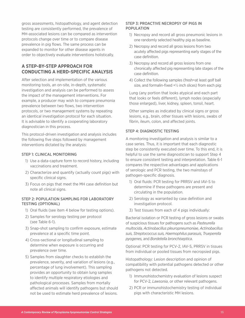
55A Contemporary Review of Mycoplasma hyopneumoniae Control Strategies
gross assessments, histopathology, and agent detection testing are consistently performed, the prevalence of MH-associated lesions can be compared as intervention protocols change over time or to compare disease prevalence in pig flows . The same process can be expanded to monitor for other disease agents in order to objectively evaluate interventions holistically .
A STEP-BY-STEP APPROACH FOR CONDUCTING A HERD-SPECIFIC ANALYSISAfter selection and implementation of the various monitoring tools, an on-site, in-depth, systematic investigation and analysis can be performed to assess the impact of the management interventions . For example, a producer may wish to compare pneumonia prevalence between two flows, two intervention protocols, or two management systems by repeating an identical investigation protocol for each situation . It is advisable to identify a cooperating laboratory diagnostician in this process .
This protocol-driven investigation and analysis includes the following five steps followed by management interventions dictated by the analysis:
STEP 1: CLINICAL MONITORING
1) Use a data-capture form to record history, including vaccinations and treatment .
2) Characterize and quantify (actually count pigs) with specific clinical signs .
3) Focus on pigs that meet the MH case definition but note all clinical signs .
STEP 2: POPULATION SAMPLING FOR LABORATORY TESTING (OPTIONAL)
1) Oral fluids (see item 4 below for testing options) .
2) Samples for serology testing per protocol (see Table 6-1) .
3) Snap-shot sampling to confirm exposure, estimate prevalence at a specific time point .
4) Cross-sectional or longitudinal sampling to determine when exposure is occurring and prevalence over time .
5) Samples from slaughter checks to establish the prevalence, severity, and variation of lesions (e .g ., percentage of lung involvement) . This sampling provides an opportunity to obtain lung samples to identify multiple respiratory etiologies and pathological processes . Samples from mortally affected animals will identify pathogens but should not be used to estimate herd prevalence of lesions .
STEP 3: PROACTIVE NECROPSY OF PIGS IN POPULATION
1) Necropsy and record all gross pneumonic lesions in one randomly selected healthy pig as baseline .
2) Necropsy and record all gross lesions from two acutely affected pigs representing early stages of the case definition .
3) Necropsy and record all gross lesions from one chronically affected pig representing late stages of the case definition .
4) Collect the following samples (fresh=at least golf ball size, and formalin-fixed =½ inch slices) from each pig:
Lung (any portion that looks atypical and each part that looks or feels different), lymph nodes (especially those enlarged), liver, kidney, spleen, tonsil, heart .
Other samples as indicated by clinical signs or gross lesions, e .g ., brain, other tissues with lesions, swabs of fibrin, ileum, colon, and affected joints .
STEP 4: DIAGNOSTIC TESTING
A monitoring investigation and analysis is similar to a case series . Thus, it is important that each diagnostic step be consistently executed over time . To this end, it is helpful to use the same diagnostician to support Step 4 to ensure consistent testing and interpretation . Table 6-1 compares the respective advantages and applications of serologic and PCR testing, the two mainstays of pathogen-specific diagnosis .
1) Oral fluids: PCR testing for PRRSV and IAV-S to determine if these pathogens are present and circulating in the population .
2) Serology as warranted by case definition and investigation protocol .
3) Test tissues from each of 4 pigs individually:
Bacterial isolation or PCR testing of gross lesions or swabs of suspicious tissues for pathogens such as Pasteurella multocida, Actinobacillus pleuropneumoniae, Actinobacillus suis, Streptococcus suis, Haemophilus parasuis, Trueperella pyogenes, and Bordetella bronchiseptica .
Optional: PCR testing for PCV-2, IAV-S, PRRSV in tissues from individual or pooled tissues from necropsied pigs .
Histopathology: Lesion description and opinion of compatibility with potential pathogens detected or other pathogens not detected .
1) Immunohistochemistry evaluation of lesions suspect for PCV-2, Lawsonia, or other relevant pathogens .
2) PCR or immunohistochemistry testing of individual pigs with characteristic MH lesions .
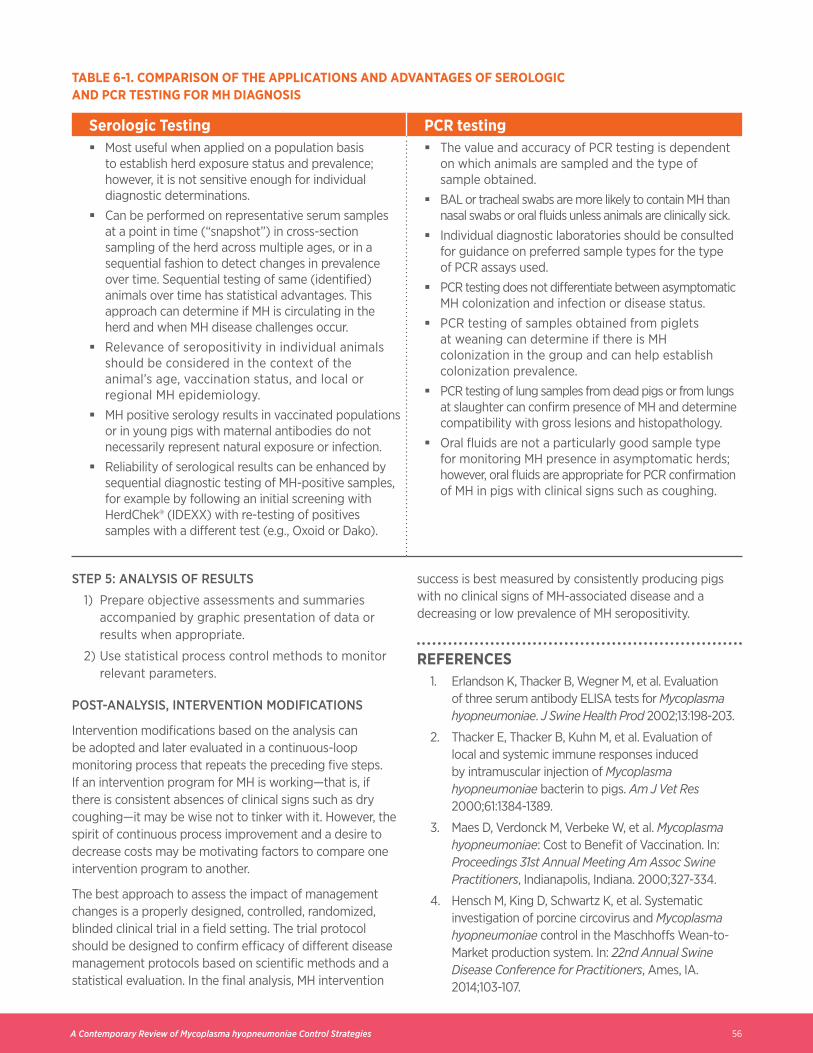
56A Contemporary Review of Mycoplasma hyopneumoniae Control Strategies
TABLE 6-1. COMPARISON OF THE APPLICATIONS AND ADVANTAGES OF SEROLOGIC AND PCR TESTING FOR MH DIAGNOSIS
Serologic Testing PCR testing § Most useful when applied on a population basis
to establish herd exposure status and prevalence; however, it is not sensitive enough for individual diagnostic determinations .
§ Can be performed on representative serum samples at a point in time (“snapshot”) in cross-section sampling of the herd across multiple ages, or in a sequential fashion to detect changes in prevalence over time . Sequential testing of same (identified) animals over time has statistical advantages . This approach can determine if MH is circulating in the herd and when MH disease challenges occur .
§ Relevance of seropositivity in individual animals should be considered in the context of the animal’s age, vaccination status, and local or regional MH epidemiology .
§ MH positive serology results in vaccinated populations or in young pigs with maternal antibodies do not necessarily represent natural exposure or infection .
§ Reliability of serological results can be enhanced by sequential diagnostic testing of MH-positive samples, for example by following an initial screening with HerdChek® (IDEXX) with re-testing of positives samples with a different test (e .g ., Oxoid or Dako) .
§ The value and accuracy of PCR testing is dependent on which animals are sampled and the type of sample obtained .
§ BAL or tracheal swabs are more likely to contain MH than nasal swabs or oral fluids unless animals are clinically sick .
§ Individual diagnostic laboratories should be consulted for guidance on preferred sample types for the type of PCR assays used .
§ PCR testing does not differentiate between asymptomatic MH colonization and infection or disease status .
§ PCR testing of samples obtained from piglets at weaning can determine if there is MH colonization in the group and can help establish colonization prevalence .
§ PCR testing of lung samples from dead pigs or from lungs at slaughter can confirm presence of MH and determine compatibility with gross lesions and histopathology .
§ Oral fluids are not a particularly good sample type for monitoring MH presence in asymptomatic herds; however, oral fluids are appropriate for PCR confirmation of MH in pigs with clinical signs such as coughing .
STEP 5: ANALYSIS OF RESULTS
1) Prepare objective assessments and summaries accompanied by graphic presentation of data or results when appropriate .
2) Use statistical process control methods to monitor relevant parameters .
POST-ANALYSIS, INTERVENTION MODIFICATIONS
Intervention modifications based on the analysis can be adopted and later evaluated in a continuous-loop monitoring process that repeats the preceding five steps . If an intervention program for MH is working—that is, if there is consistent absences of clinical signs such as dry coughing—it may be wise not to tinker with it . However, the spirit of continuous process improvement and a desire to decrease costs may be motivating factors to compare one intervention program to another .
The best approach to assess the impact of management changes is a properly designed, controlled, randomized, blinded clinical trial in a field setting . The trial protocol should be designed to confirm efficacy of different disease management protocols based on scientific methods and a statistical evaluation . In the final analysis, MH intervention
success is best measured by consistently producing pigs with no clinical signs of MH-associated disease and a decreasing or low prevalence of MH seropositivity .
REFERENCES1 . Erlandson K, Thacker B, Wegner M, et al . Evaluation
of three serum antibody ELISA tests for Mycoplasma hyopneumoniae . J Swine Health Prod 2002;13:198-203 .
2 . Thacker E, Thacker B, Kuhn M, et al . Evaluation of local and systemic immune responses induced by intramuscular injection of Mycoplasma hyopneumoniae bacterin to pigs . Am J Vet Res 2000;61:1384-1389 .
3 . Maes D, Verdonck M, Verbeke W, et al . Mycoplasma hyopneumoniae: Cost to Benefit of Vaccination . In: Proceedings 31st Annual Meeting Am Assoc Swine Practitioners, Indianapolis, Indiana . 2000;327-334 .
4 . Hensch M, King D, Schwartz K, et al . Systematic investigation of porcine circovirus and Mycoplasma hyopneumoniae control in the Maschhoffs Wean-to-Market production system . In: 22nd Annual Swine Disease Conference for Practitioners, Ames, IA . 2014;103-107 .

57A Contemporary Review of Mycoplasma hyopneumoniae Control Strategies

58A Contemporary Review of Mycoplasma hyopneumoniae Control Strategies
All brands are the property of Zoetis Inc ., its affiliates and/or licensors . ©2016 Zoetis Inc . All rights reserved .Printed in the USA . TI-01782