treatment and control of - department of reproduction ... · treatment and control of mycoplasma...
TRANSCRIPT
Treatment and control of Mycoplasma hyopneumoniae
infections
Rubén del Pozo Sacristán

A mis padres
Everyone is a genius.
But if you judge a fish on its ability to climb a tree,
it will live its whole life believing that it is stupid.
Albert Einstein
Todo el mundo es un genio.
Pero si judgas a un pez por su habilidad de escalar un árbol,
pasará toda su vida pensando que es estúpido.
Albert Einstein

Treatment and control of
Mycoplasma hyopneumoniae infections
Dissertation submitted in fulfilment of the requirements for the degree of Doctor of
Philosophy (PhD) in Veterinary Sciences, Faculty of Veterinary Medicine, Ghent University
Rubén del Pozo Sacristán
Promoters:
Prof. dr. D. Maes
Prof. dr. F. Haesebrouck
Ghent University
Faculty of Veterinary Medicine
Department of Reproduction, Obstetrics and Herd Health
Unit of Porcine Health Management
Department of Pathology, Bacteriology and Avian Diseases
Merelbeke, 2014

The cover photo was designed by Rubén del Pozo Sacristán.
This thesis was financially supported by MSD, Elanco and Zoetis

5
Table of Contents
LIST OF ABBREVIATIONS ................................................................................................... 7
Chapter 1. GENERAL INTRODUCTION ............................................................................... 9
Review of the literature .................................................................................................. 11
1.1. Characteristics of Mycoplasma hyopneumoniae ................................................ 12
1.2. Pathogenesis of Mycoplasma hyopneumoniae infections .................................. 13
1.3. Epidemiology of Mycoplasma hyopneumoniae ................................................. 15
1.4. Clinical signs, lesions and porcine respiratory disease complex ....................... 17
1.5. Diagnosis............................................................................................................ 22
1.6. Treatment and control of Mycoplasma hyopneumoniae infections ................... 27
1.6.1. Optimization of management practices and housing conditions ................ 27
1.6.2. Antimicrobial treatment .............................................................................. 29
1.6.3. Vaccination against Mycoplasma hyopneumoniae ..................................... 43
1.6.4. Preventive medication vs vaccination ......................................................... 51
Chapter 2. AIMS OF THE STUDY ........................................................................................ 67
Chapter 3. EXPERIMENTAL STUDIES ............................................................................... 71
3.1. Efficacy of florfenicol injection in the treatment of Mycoplasma hyopneumoniae
induced respiratory disease in pigs .......................................................................... 73
3.2. Efficacy of early Mycoplasma hyopneumoniae vaccination against mixed
respiratory disease in older fattening pigs ............................................................... 85
3.3. Efficacy of in-feed medication with chlortetracycline in a farrow-to-finish herd
against a clinical outbreak of respiratory disease in fattening pigs ....................... 111
Chapter 4. GENERAL DISCUSSION .................................................................................. 135
SUMMARY .......................................................................................................................... 157
SAMENVATTING ............................................................................................................... 163
CURRICULUM VITAE ......................................................................................................... 169
BIBLIOGRAPHY ................................................................................................................. 173
ACKNOWLEDGEMENTS .................................................................................................. 179

7
LIST OF ABBREVIATIONS
ADG Average daily weight gain
ANOVA Analysis of variance
BAL Broncho-alveolar lavage
CCU Color changing units
DNA Deoxyribonucleic acid
ELISA Enzyme-linked immunosorbent assay
EU European Union
FCR Feed conversion ration
ID Intradermal
IF Immunofluorescence
Ig Immunoglobulins
IM Intramuscular
MIC Minimum inhibitory concentration
nPCR nested Polymerase chain reaction
NV Non-vaccinated
NS Nasal swabs
OD Optical density
PCR Polymerase chain reaction
PCV-2 Porcine circovirus type 2
ppm parts per million
PRDC Porcine respiratory disease complex
PRRSV Porcine reproductive and respiratory syndrome virus
RDS Respiratory disease score
qPCR quantitative Polymerase chain reaction
SD Standard deviation
SIV Swine influenza virus
TBS Tracheo-bronchial swabs
U.K. United Kingdom
U.S. United States
V Vaccinated


Ch. 1 GENERAL INTRODUCTION
Chapter 1. GENERAL INTRODUCTION
General introduction

11
Ch. 1 GENERAL INTRODUCTION
REVIEW OF THE LITERATURE
Respiratory disease in pigs is one of the most important disease conditions in intensive
pig production systems worldwide. Mycoplasma hyopneumoniae is the primary agent of
enzootic pneumonia, a chronic respiratory disease in pigs resulting from mixed respiratory
infections with M. hyopneumoniae and one or more secondary bacterial pathogens (Maes et
al., 2008a). M. hyopneumoniae is present in most of the countries with intensive swine
production and is also considered as one of the main pathogens involved in the porcine
respiratory disease complex (PRDC). The multifactorial aetiology of the PRDC includes both
viral and bacterial pathogens, and is also influenced by management and environmental
conditions (Opriessnig et al., 2011). Both EP and PRDC lead to major economic losses due to
the reduced growth, increased mortality and feed conversion, costs for antimicrobials and
immunoprophylaxis and increased time to market (Maes et al., 2008a).
M. hyopneumoniae lacks a cell wall and has a small genome encoding only a few
biosynthetic pathways. The pathogenesis of M. hyopneumoniae infections includes various
steps: adhesion of the respiratory tract, modulation of the immune system leading to damage
of pulmonary tissue and persistence in the respiratory tract, as well as interaction with other
infectious agents.
Non-productive coughing is the most obvious clinical sign. Macroscopic lesions are
characterized by catarrhal bronchopneumonia. They consist of red to purplish consolidated
areas on the cranial-ventral parts of the apical, cardiac, accessory and diaphragmatic lobes.
The diagnosis of enzootic pneumonia is generally made at herd level rather than at individual
level (Thacker and Minion, 2012; Taylor, 2013).
Since eradication of M. hyopneumoniae from infected herds is complicated and difficult
to achieve, control of the infections is considered the best strategy. Control measures include
optimization of housing and management practices, antimicrobial treatment and vaccination.
This review aims to summarize the current knowledge on M. hyopneumoniae infections and
enzootic pneumonia, with emphasis on antimicrobial agents used for treatment and vaccines
used for control this disease.

12
Ch. 1 GENERAL INTRODUCTION
1.1. CHARACTERISTICS OF MYCOPLASMA HYOPNEUMONIAE
Mycoplasmas are taxonomically classified as members of the class Mollicutes, a group
of bacteria characterized by the lack of a cell wall. Mycoplasma cells are mostly spherical
and range from 0.3 to 0.8 µm in size (Razin, 2006). They have a small genome (893-920
kilobase pairs) (Dybvig and Voelker, 1996), which encodes a limited number of genes,
resulting in a few biosynthetic pathways (Razin et al., 1998). Because of this limited
metabolism, mycoplasmas need to obtain essential metabolites from the host or growth
environment (Thacker and Minion, 2012).
The first isolation of M. hyopneumoniae was performed in 1965 in the U.S. (Mare and
Switzer, 1965) and the U.K. (Goodwin et al., 1965). In the 70‟s of the previous century,
important research was carried out to determine the main characteristics of this organism. M.
hyopneumoniae is very sensitive to environmental conditions due to the absence of a cellular
wall (Goodwin, 1972). In 1973, the J-strain was isolated (Whittlestone, 1973), which is still
considered as the reference strain. Due to its slow and fastidious growth in vitro (Friis, 1974),
a special culture medium supplemented with serum, Friis medium, is necessary for isolation
(Friis, 1975). Using electron microscopy, M. hyopneumoniae appears as round or oval cells
with a mean diameter ranging from 0.5 to 0.8 µm (Tajima and Yagihashi, 1982; Blanchard et
al., 1992).
The whole genome sequence of several M. hyopneumoniae strains has been published
(strains J, 232, 7448 and 168) (Liu et al., 2011; Minion et al., 2004; Vasconcelos et al.,
2005). Recent studies using molecular techniques have revealed a large diversity of M.
hyopneumoniae isolates at genomic (Stakenborg et al., 2006b; Strait et al., 2008; Nathues et
al., 2011), and proteomic (Vicca et al., 2003; Calus et al., 2007; Pinto et al., 2007) level.
Different M. hyopneumoniae isolates also have different virulence characteristics, as assessed
by experimental infections in pigs (Vicca et al., 2003; Villarreal et al., 2009).
13
Ch. 1 GENERAL INTRODUCTION
1.2. PATHOGENESIS OF MYCOPLASMA HYOPNEUMONIAE
INFECTIONS
M. hyopneumoniae is a host specific organism that only infects pigs. The pathogenesis
of M. hyopneumoniae infections includes various steps: colonization of the respiratory tract,
modulation of the immune system leading to damage of pulmonary tissue and persistence in
the respiratory tract. This complex pathogenesis may also be affected, mainly under field
conditions, by the interaction with other infectious agents.
Colonization of the respiratory tract begins with the adherence of M. hyopneumoniae
to the ciliated epithelial cells of the trachea, bronchi and bronchioles (Blanchard et al., 1992).
This attachment to the mucosal surface of the ciliated epithelium is a pre-requisite for
initiation of disease (Tajima and Yagihashi, 1982) and is mainly enabled by various proteins
(e.g. P97, P102, P116, P135, P159, P216) and other cell surface structures. A complete
description of these multifunctional adhesins and their binding abilities can be found in
Simionatto et al. (2013). The binding of M. hyopneumoniae to the cilia provokes ciliostasis,
clumping and finally loss of cilia (DeBey and Ross, 1994). The impaired ciliary activity leads
to a reduction of the mucosal clearance of the respiratory tract. Once the first non-specific
defense barrier of the lung is damaged, further multiplication of the M. hyopneumoniae and
colonization of the airways occur. The loss of the cilia is predominantly limited to the
airways from cranio-ventral lobes of the lungs (apical and cardiac) and this distribution is
associated also with the main location of macroscopic lesions of pneumonia (Mebus and
Underdahl, 1977).
It has been shown that M. hyopneumoniae is able to modulate both innate and
adaptive immune responses (Thacker, 2001), leading to evasion of host defenses and
establishment of a chronic and persistent infection (Razin et al., 1998). The exact mechanism
of modulation of the immune response is not yet fully elucidated (Simecka, 2005), but it is
generally accepted that the immune response plays a role in the development of lesions.
Immediately after colonization, there is a massive infiltration of peribronchiolar and
perivascular lymphohistiocytic cells (Morris et al., 1995). Alveolar macrophages and
lymphocytes are the predominant cells. M. hyopneumoniae has the ability to alter the function
of these cells. The phagocytic capacity of the macrophages is impaired, resulting in a reduced
clearance, and consequently, in a chronic colonization of the respiratory tract by M.
hyopneumoniae (Caruso and Ross, 1990). In addition, M. hyopneumoniae stimulates
macrophages to produce proinflammatory cytokines, which leads to an inflammatory

14
Ch. 1 GENERAL INTRODUCTION
response and lung damage (Thacker and Minion, 2012). A general immunosuppression has
been suggested since M. hyopneumoniae may also alter the function of lymphocytes
(Kishima and Ross, 1985).
M. hyopneumoniae usually persists for a long time in the animal. Sorensen et al. (1997)
demonstrated the presence of M. hyopneumoniae in the respiratory tract by means of
Polymerase Chain Reaction (PCR) up to 85 days post-infection. Using nested PCR (nPCR),
Fano et al. (2005) detected M. hyopneumoniae in nasal swabs up to 185 days post-infection.
More recently, Pieters et al. (2009) showed that M. hyopneumoniae can be isolated from the
respiratory tract of potential infectious carriers up to 200 days post-infection, it can persist up
to 214 days post-infection, and total clearance is only complete by 254 days post-infection.
The complete pathogenesis of M. hyopneumoniae infections is not yet fully understood,
since not all respiratory tract infections with M. hyopneumoniae lead to pneumonia. Other
factors, such as virulence of the strain involved (Zielinski and Ross, 1990; Vicca et al., 2003;
Meyns et al., 2007; Villarreal et al., 2009) and interactions with other respiratory pathogens
(Ciprián et al., 1994; Opriessnig et al., 2011), may play a decisive role in the development of
the disease.

15
Ch. 1 GENERAL INTRODUCTION
1.3. EPIDEMIOLOGY OF MYCOPLASMA HYOPNEUMONIAE
1.3.1. Occurrence of M. hyopneumoniae infections
M. hyopneumonie is present in almost all countries with an intensive swine production
(Sibila et al., 2004a; Fano et al., 2007; Maes et al., 2008a; Martínez et al., 2009; Fraile et al.,
2010; Meyns et al., 2011; Fablet et al., 2012b; Nathues et al., 2012b), and within herds,
infections may occur in all the production phases, namely breeding animals, suckling and
weaned piglets, as well as fattening pigs.
The prevalence of M. hyopneumoniae in suckling pigs assessed by nPCR on nasal
swabs ranges from 0.5 to 10 per cent (Sibila et al., 2007a; Sibila et al., 2007b; Villarreal et
al., 2010). The prevalence is higher in weaned piglets, namely from 0 to 51 per cent (Sibila
et al., 2004a; Fano et al., 2007; Sibila et al., 2007b). Even though infections may already
occur during suckling period, in most pig herds, the highest infection levels of M.
hyopneumoniae occur during the grow-finishing period (Sibila et al., 2004a). The dynamics
of infection vary largely between herds, and are influenced by different environmental
conditions, such as management and housing conditions and the production system of the
herd. The potential of the sows to shed M. hyopneumoniae is one of the key points on the
epidemiology of this agent, not only because of the transmission to their offspring
(Calsamiglia and Pijoan, 2000), but also in the maintaining of the infection within the herd.
There is however not much information available on occurrence of M. hyopneumoniae in
breeding animals (Sibila et al., 2009). The prevalence of M. hyopneumoniae in sows
assessed by antibodies in sera ranges from 27 to 65 per cent (Sibila et al., 2007a; Grosse
Beilage et al., 2009).
1.3.2. Transmission of M. hyopneumoniae
The transmission of M. hyopneumoniae can take place by different routes.
Transmission between herds occurs mainly by the introduction of infected animals or
airborne. Regarding the transmission within herds, distinction can be made between vertical
(from the sow to the offspring) and horizontal transmission (between pigs of the same or
different pens).
Vertical transmission
Vertical transmission occurs predominantly via nose-to-nose contact (Calsamiglia and
Pijoan, 2000). To date, in-utero or lactogenic transmission has not been reported (Maes et al.,
16
Ch. 1 GENERAL INTRODUCTION
2008b). Moore et al. (2001) suggested a reduction of the severity of pneumonia lesions in
slaughter pigs born from parity 2 sows when compared with pigs born from parity 1 sows.
Recent epidemiological studies confirmed these observations and have demonstrated that the
piglets from younger sows (gilts and parity 1-2) have more risk of being infected with M.
hyopneumoniae compared to piglets from older sows (Fano et al., 2007). Although the risk is
lower than in younger sows, older sows may still transmit M. hyopneumoniae to their
offspring (Calsamiglia and Pijoan, 2000; Grosse Beilage et al., 2009).
Horizontal transmission
a. Direct contact:
Nose-to-nose contact between penmates or even between pigs of different pens may
result in spread of M. hyopneumoniae from infected to susceptible animals (Thacker and
Minion, 2012). Using transmission experiments under experimental conditions, Meyns et al.,
(2004) showed that one infected pig may be able to infect one littermate during a nursery
period of six weeks. Similar results were obtained by Villarreal et al,. (2011) in transmission
experiments under field conditions.
b. Indirect contact:
Airborne transmission has been shown to be important for M. hyopneumoniae.
Goodwin (1985) stated that M. hyopneumoniae-free herds may become infected with M.
hyopneumoniae if infected herds are located within a distance of 3.2 km. Cardona et al.
(2005) showed under experimental conditions using aerosols that transmission of M.
hyopneumoniae is possible over 150 m. More recent studies have demonstrated that M.
hyopneumoniae may be transmitted over much larger distances namely 4.7 km (Dee et al.,
2009) and 9.2 km (Otake et al., 2010).
The implementation of standard hygiene measures of the personnel when entering the
herd may reduce the risk of transmission of M. hyopneumoniae between herds (Batista et al.,
2004). More recently, an observational study performed during four years illustrated that one-
night downtime period prevents the entry of M. hyopneumoniae in a herd not only by
personnel, but also by fomites (Pitkin et al., 2011). In general, mechanical vectors, such
fomites and personnel, are considered to be of limited importance in the transmission of M.
hyopneumoniae.

17
Ch. 1 GENERAL INTRODUCTION
1.4. CLINICAL SIGNS, LESIONS AND PORCINE RESPIRATORY
DISEASE COMPLEX
1.4.1. Clinical signs
In general, two distinct forms of clinical disease can be observed, namely endemic and
epidemic disease (Thacker and Minion, 2012; Taylor, 2013).
Endemic mycoplasmosis, commonly known as enzootic pneumonia, is the form most
frequently observed. In many cases, infections are subclinical (Clark et al., 1991). In case of
clinical disease, a non-productive coughing is the most obvious clinical sign. Coughing may
be present in nursery, grower and finisher pigs and usually a considerable percentage of the
pigs are affected. Coughing may disappear after 2-3 weeks, but it can also persist throughout
the whole fattening period.
Under experimental infections, clinical signs are mainly characterized by slight fever,
followed by dry coughing. Coughing appears from 10 to 14 days post-infection, reaches a
maximum peak at about 4-5 weeks, after which it disappears gradually (Whittlestone 1972;
Kobisch et al., 1993).
Under field conditions, herds that are only infected with M. hyopneumoniae are rarely
seen. Mostly, mixed infections occur. In case of secondary bacterial infections, the severity of
the clinical signs increases, namely a severe respiratory distress including fever, labored
breathing and prostration, and reduced appetite. This leads to an increase of the feed
conversion ratio, lower average daily weight gain, and more weight variation between the
pigs.
Epizootic mycoplasmosis is uncommon. It occasionally occurs when M.
hyopneumoniae enters into a negative, immunologically naive herd. The spread of the disease
occurs rapidly and all age groups may be affected. Coughing, acute respiratory distress, fever
and deaths may be present (Thacker and Minion, 2012).
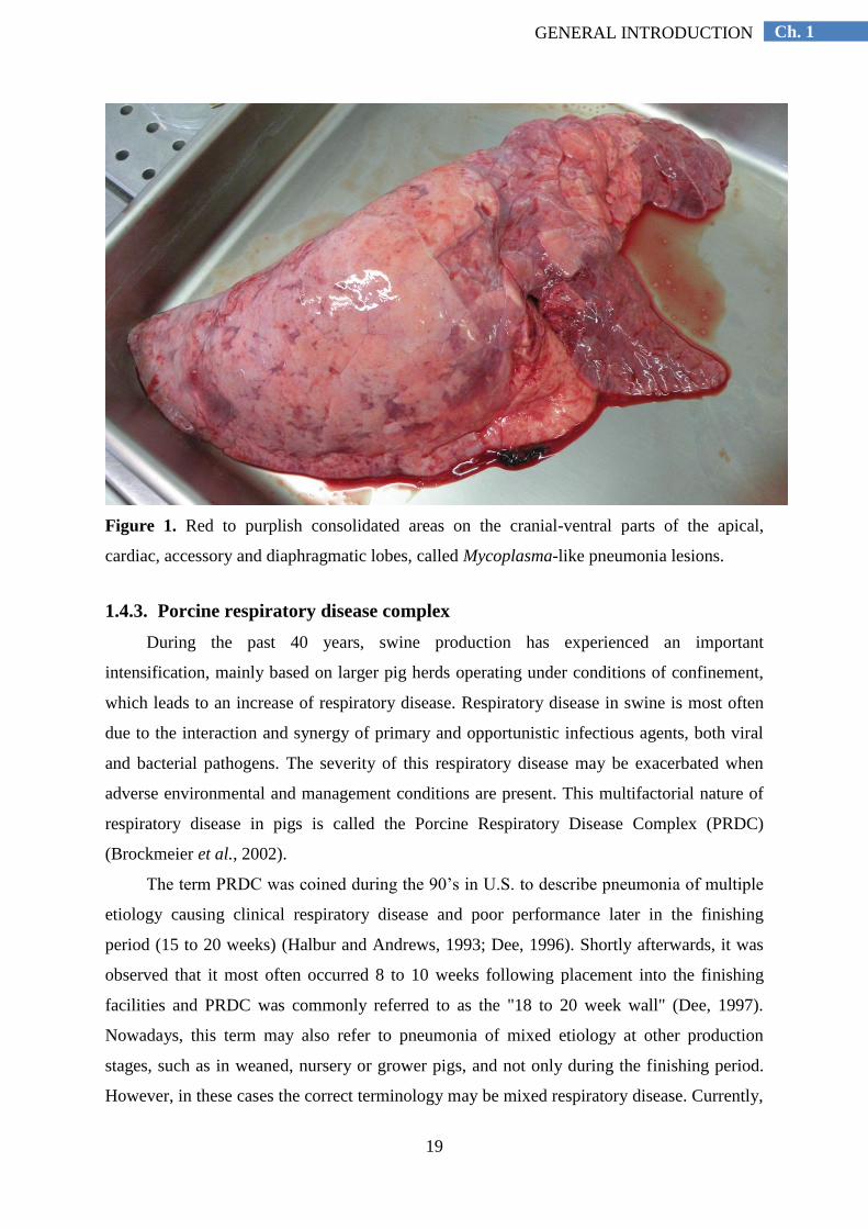
1.4.2. Lesions
M. hyopneumoniae infection can lead to different gross lesions depending on the course
of the disease. The most common macroscopic lesion in chronic M. hyopneumoniae
infections is characterized by red to purplish consolidated areas on the cranial-ventral parts of
the apical, cardiac, accessory and diaphragmatic lobes. These lesions are commonly called
Mycoplasma-like pneumonia lesions (active lesions) (Figure 1) (Whittlestone, 1972; Kobisch
et al., 1993). They may appear from 7 days after experimental infection onwards and reach a
18
Ch. 1 GENERAL INTRODUCTION
maximum size at about 4 weeks post-infection (Whittlestone, 1972; Kobisch et al., 1993).
These uncomplicated lesions are characterized by the presence of a catarrhal exudate in the
airways, a uniform colour of the parenchyma of the lung and a “meaty” consistency. The
pneumonia lesions may be recovered after 7-10 weeks, and by that time, red to purplish
interlobular scar retractions of connective tissue, called fissures, are present (old lesions)
(Whittlestone, 1972; Kobisch et al., 1993).
Under field conditions, when M. hyopneumoniae infections are complicated by
secondary bacterial pathogens, the pneumonia lesions occupy a large surface of the lung, and
are characterized by the presence of mucopurulent exudate in the airways, a more firm
consistency and an inconsistent greyish colour of the parenchyma (Osborne et al., 1981).
However, Mycoplasma-like pneumonia lesions at slaughter are not pathognomonic for
infections with M. hyopneumoniae. Apart from M. hyopneumoniae, it is well known that
infections e.g. with viruses and in particular swine influenza virus, may cause similar
pneumonia lesions (Thacker et al., 2001, Sibila et al., 2009).
Adhesive pleurisy is a common finding in slaughter pigs reared under field conditions
and has been associated with respiratory disease (Fraile et al., 2010; Meyns et al., 2011;
Fablet et al., 2012b; Merialdi et al., 2012). It is defined as fibrotic adherence between the
visceral and parietal membranes of the pleural sac. Although adhesive pleurisy is not a
characteristic lesion of M. hyopneumoniae infections, Meyns et al. (2011) found a positive
association between higher prevalence of pneumonia and the presence of pleurisy at
slaughter.
Histopathological examination of the pneumonia lesions infected with M.
hyopneumoniae revealed neutrophil and lymphocyte infiltration around the airways and
alveoli (Whittlestone, 1972). As the infection progresses, broncho-interstitial pneumonia
develops. The accumulation of mononuclear cells, mainly lymphocytes and macrophages, is
more evident and results in the formation of lymphocytic cuffs around the airways, which is
commonly known as perivascular and peribronchiolar lymphohistiocytic infiltration and
nodule formation (Blanchard et al., 1992; Morris et al., 1995). In cases of enzootic
pneumonia, a neutrophilic exudate may be present in the lumen of airways and alveoli.
19
Ch. 1 GENERAL INTRODUCTION
Figure 1. Red to purplish consolidated areas on the cranial-ventral parts of the apical,
cardiac, accessory and diaphragmatic lobes, called Mycoplasma-like pneumonia lesions.
1.4.3. Porcine respiratory disease complex
During the past 40 years, swine production has experienced an important
intensification, mainly based on larger pig herds operating under conditions of confinement,
which leads to an increase of respiratory disease. Respiratory disease in swine is most often
due to the interaction and synergy of primary and opportunistic infectious agents, both viral
and bacterial pathogens. The severity of this respiratory disease may be exacerbated when
adverse environmental and management conditions are present. This multifactorial nature of
respiratory disease in pigs is called the Porcine Respiratory Disease Complex (PRDC)
(Brockmeier et al., 2002).
The term PRDC was coined during the 90‟s in U.S. to describe pneumonia of multiple
etiology causing clinical respiratory disease and poor performance later in the finishing
period (15 to 20 weeks) (Halbur and Andrews, 1993; Dee, 1996). Shortly afterwards, it was
observed that it most often occurred 8 to 10 weeks following placement into the finishing
facilities and PRDC was commonly referred to as the "18 to 20 week wall" (Dee, 1997).
Nowadays, this term may also refer to pneumonia of mixed etiology at other production
stages, such as in weaned, nursery or grower pigs, and not only during the finishing period.
However, in these cases the correct terminology may be mixed respiratory disease. Currently,
20
Ch. 1 GENERAL INTRODUCTION
it is generally accepted that when M. hyopneumoniae is the primary agent of respiratory
disease and other secondary bacteria take advantage of this situation, the resulting disease is
called enzootic pneumonia. On the other hand, in case of PRDC, M. hyopneumoniae plays a
central role together with viral pathogens such as Porcine Reproductive and Respiratory
Syndrome Virus (PRRSV), Porcine Circovirus type 2 (PCV-2) and Swine Influenza Virus
(SIV), whereas other viruses and opportunistic bacteria are considered as secondary actors
(Opriessnig et al., 2011).
1.4.3.1. Polymicrobial nature
Numerous studies have illustrated during the past years the polymicrobial nature of
PRDC (Morrison et al., 1985a; Loeffen et al., 1999; Bochev, 2007; Moorkamp et al., 2008;
Hansen et al., 2010; Opriessnig et al., 2011; Fablet et al., 2012a; Ticó et al., 2013). There is
an enormous variety of microorganisms associated with PRDC and they can be categorized
as commensal microflora vs potentially pathogenic agents; primary vs secondary or
opportunistic pathogens. There is no clear distinction between commensal and potential
pathogenic agents, since different studies categorized the same agent as either commensal or
pathogenic (VanAlstine, 2012). Primary pathogens are capable of evading respiratory defense
mechanisms from the host and establishing infection and disease on their own, whereas
secondary pathogens take advantages of the damage caused by primary pathogens to establish
infections and exacerbate respiratory disease (Brockmeier et al., 2002). In general, primary
pathogens, such as viral agents and M. hyopneumoniae, may damage the epithelium of the
respiratory tract and decrease the local and systemic defense mechanisms, favoring invasion,
colonization and establishment of the infection by secondary agents, mainly bacteria. Primary
pathogens involved in PRDC include PRRSV, PCV-2, SIV, Aujeszky‟s Disease Virus, M.
hyopneumoniae, Actinobacillus pleuropneumoniae and Bordetella bronchiseptica. Other
pathogens include Paramyxovirus, Porcine Cytomegalovirus, Porcine Respiratory
Coronavirus, Torque teno sus virus, Actinobacillus suis, Haemophilus parasuis, M. hyorhinis,
Pasteurella multocida, Streptococcus suis and Trueperella pyogenes (Opriessnig et al.,
2011). Major efforts have been done to study and understand the interaction between
different respiratory pathogens under experimental conditions. Although the reproduction of
disease has been easily achieved by the use of single challenge infection models, it is not
always possible to reproduce disease in dual infections or co-infection experiments. A review
was published by Opriessnig et al. (2011) where a wide variety of interactions between
different viral and bacterial pathogens is discussed.

21
Ch. 1 GENERAL INTRODUCTION
Under field conditions, it is very rare to observe a clinical case of respiratory disease in
which only one primary pathogen is involved. Frequently, one or more primary agents are
present and cause primary infections which become complicated with opportunistic bacteria
that aggravate the respiratory disease (VanAlstine, 2012). A study published in the U.S.
including three case reports of PRDC (Harms et al., 2002) illustrated a low morbidity and
mortality. Postmortem examinations revealed a high extent of pneumonia lesions and
bronchopneumonia as major gross and microscopic findings, respectively. Concurrent
infections of PCV-2 combined with PRRSV, SIV and M. hyopneumoniae were described.
1.4.3.2. Non-infectious factors
Apart from its polymicrobial nature, PRDC also depends largely on many non-
infectious factors. Identifying and reducing risk factors may lead to a reduction of the
transmission of pathogens, less stress provoked to the animal by hostile environments
(physical, climate and air quality factors) and less direct impairment of the respiratory tract
(Opriessnig et al., 2011). A short description of non-infectious factors is presented in section
1.6 of this thesis. Details on the influence of management and environmental factors on
respiratory disease are also well described in the review papers by Stärk (2000) and Maes et
al. (2008a).

22
Ch. 1 GENERAL INTRODUCTION
1.5. DIAGNOSIS
Diagnosis of enzootic pneumonia is generally established at group level rather than at
individual level (Thacker and Minion, 2012; Taylor, 2013). The presence of coughing in
fattening pigs combined with chronic bronchopneumonia lesions at slaughter, as well as poor
performance and feed efficiency during the fattening period may lead to a presumptive
diagnosis of enzootic pneumonia. However, definitive diagnosis is based on detection of
(parts of) M. hyopneumoniae. Differential diagnosis should consider other possible causes of
coughing, as well as bacteria and viruses causing Mycoplasma-like pneumonia lesions (Sibila
et al., 2009).
1.5.1. Clinical signs
Non-productive coughing, especially in fattening pigs, being more evident after
boosting and characterized by persistence in the population is a typical sign of enzootic
pneumonia.
Several methods have been described to assess the severity of coughing under
experimental and field conditions. Halbur et al. (1996) described a Respiratory Disease Score
(RDS) to quantify the severity of coughing under experimental conditions. The RDS could
range from 0 to 6: 0 (no coughing), 1 (mild coughing after encouraged move), 2 (mild
coughing in rest), 3 (moderate coughing after encouraged move), 4 (moderate coughing at
rest), 5 (severe coughing after encouraged move), 6 (severe coughing at rest). This method
has been used in recent transmission and pathogenesis studies, as well as in the assessment of
efficacy of vaccines and antimicrobial treatments (Vicca et al., 2003; Meyns et al., 2004;
Vicca et al., 2005; Meyns et al., 2006; Meyns et al., 2007). Under field conditions, a method
that quantifies the number of pigs coughing in a given period of time has also been described
(Maes et al., 1999; Mateusen et al., 2001). The pigs were moved up and the number of pigs
coughing per pen during 10 minutes was counted. A RDS was calculated by dividing the
number of pigs per pen that coughed during 10 min, by the total number of pigs in that pen,
multiplied by 100. A similar method has been used by Nathues et al. (2012a). These authors
confirmed that a quantitative assessment of the onset of coughing is useful for the diagnosis
of enzootic pneumonia and is correlated with the infection pattern of M. hyopneumoniae
assessed by PCR.

23
Ch. 1 GENERAL INTRODUCTION
1.5.2. Macroscopic lung lesions
Examination of the lungs of slaughter pigs is an excellent method to monitor the
respiratory health in a herd under field conditions. If possible, checks should include visual
examination and palpation in a well-illuminated place, and the lesions should be sketched
onto a diagram, which may be followed by image analysis (VanAlstine, 2012). The
prevalence and extent of the lesions, not only pneumonia, but also adhesive pleurisy, nodules,
abscesses, pericarditis, etc., may provide reliable information regarding the severity of the
respiratory disease.
However, slaughterhouse examinations also have limitations. Lesion assessement is
subjective and the lesions are generally not pathognomic (Sibila et al., 2009). Subclinical
infections may be not detected and lesions in young animals may be healed at the moment of
slaughter (Regula et al., 2000). Also, the fast speed of slaughtering in modern slaughter
facilities makes it more difficult to perform reliable slaughter checks. Although a mimimum
of 30 randomly selected pigs from the same slaughter batch has been shown to provide
reliable information at herd level (Straw et al., 1989), it is generally accepted that it is better
to evaluate more pigs. In recent epidemiological studies performed in Europe, in which
slaughter examinations were carried out in order to estimate the prevalence and severity of
lung lesions, an average of 127 (Meyns et al., 2011), 106 (Fraile et al., 2010), 100 (Merialdi
et al., 2012) and 30 (Fablet et al., 2012) pigs per batch were investigated. The low number of
the last study may be due to the large amount of additional bacteriological investigations
carried out on these lungs.
Different scoring systems have been described in the past years to monitor enzootic
pneumonia under experimental (Hanan et al., 1982), as well as field conditions (Madec et al.,
1982; Morrison et al., 1985b; Straw et al., 1986; Christensen et al., 1999). Only the methods
used in this thesis will be described here. The scoring system described by Hannan et al.
(1982) is appropriate to carefully quantify lung lesions caused by experimental infection with
M. hyopneumoniae. The score ranges from 0 (no lesions) to 35 (all lung tissue affected). This
method is especially designed for Mycoplasma-like pneumonia lesions, since they are mainly
located in the cranial-ventral parts of the lungs, and this score gives the same significance to
each of the 7 lobes, namely 5 points/lobe out of the total are of the lung (35 points). The lung
lesion score from Morrison et al (1985b) is appropriate for field evaluation. It is based on the
total percentage of affected lung tissue, with the different lung lobes representing the

24
Ch. 1 GENERAL INTRODUCTION
following percentage of the total lung surface area: apical (10 per cent), cardiac (7 per cent),
accessory (6 per cent) and diaphragmatic (30 per cent).
1.5.3. Microscopic lung lesions
Microscopic findings of peribronchiolar and perivascular lymphohistiocytic infiltration
and nodule formation are not specific of M. hyopneumoniae infections, but they may be
helpful for a final diagnosis. Two methods can be performed to measure the severity and the
extent of the mycoplasmal lesions.
The severity of peribronchiolar and perivascular lymphohistiocytic infiltration and
nodule formation related to M. hyopneumoniae induced pneumonia lesions may be scored by
using light microscopy according to the method described by Morris et al. (1995). The score
ranges from 1 (limited cellular infiltrates around bronchioles) to 5 (severe infiltration of
lymphocytic cells around bronchioles and interstitium, and inflammatory cell exudates into
the airways) (Fig. 2).
The extent of pneumonia can also be measured by calculating the percentage of tissue
occupied by air. Using a camera software and light microscopy, a picture is taken from a
microscopic field including a sample of lung lesion. By means of an automatic image
analysis system, the percentage of air in each picture is calculated. This percentage is
negatively correlated to the number of infiltrating cells, the amount of atelectasis,
intrabronchiolar-intrabronchial exudates, intra-alveolar exudates and/or oedema and
proliferation of type II cells. The advantage of this method is that the measurements are done
in an objective way.
1.5.4. Demonstration of M. hyopneumoniae infections
Bacteriological culture from fresh lung tissue is considered as the gold standard for
diagnosis of enzootic pneumonia. However, due to the fastidious growth of M.
hyopneumoniae, the very time-consuming isolation procedure, the fact that special media are
required and the low sensitivity of the procedure, bacteriological culture is not used for
routine diagnosis of M. hyopneumoniae (Friis, 1975).
The detection of M. hyopneumoniae antigen in lung tissue can be performed by
immunofluorescence (IF) and by immunohistochemistry. Immunofluorescence is a semi-
quantitative assay to assess the amount of M. hyopneumoniae-organisms in the airways
(Kobisch et al., 1978). Commonly used scores range from 0 to 3: 0 (no IF), 1 (limited IF), 2
(moderate IF) and 3 (intense IF) (Vicca et al., 2003). The limitations of this technique include

25
Ch. 1 GENERAL INTRODUCTION
the laborious and time-consuming procedure, the diagnosis can only be done post-mortem
and the semi-quantitative outcome obtained, which does not allow to accurately quantify the
load of M. hyopneumoniae organisms.
The detection of M. hyopneumoniae DNA by PCR testing is currently widely used to
investigate the potential role of this agent in respiratory disease. The accurate detection level
of infection, as well as the relatively rapid and inexpensive characteristic of this method, have
favored its establishment in routine diagnostic labs (Thacker and Minion, 2012). PCR assays
are suitable for detection of M. hyopneumoniae in a wide range of samples, including lung
tissue, nasal and trachea-bronchial swabs, as well as broncho-alveolar lavage fluid. Different
PCR assays have been described. Nested PCR has an excellent sensitivity, even capable to
detect as few as 4 or 5 DNA copies (Stärk et al., 1998a). Recently, a real-time (quantitative)
PCR (qPCR) has been developed which allows to quantify the number of DNA copies per ml
(Marois et al., 2010). Even the problem of identifying multiple mycoplasmas that grow in
culture broth has been solved with the development of a multiplex PCR (Stakenborg et al.,
2006a). Although this increased accuracy allows detection of M. hyopneumoniae earlier than
seroconversion, contamination of the samples should be avoided as this may result in false
positive results (Thacker and Minion, 2012).
Serology is commonly used to detect serum antibodies against M. hyopneumoniae
(Calsamiglia et al., 1999; Thacker, 2004) at group or herd level. Commercial ELISAs
generally have a good specificity. However, the sensitivity of these assays may be low,
ranging from 37 to 49 per cent, which results in a high percentage of false negatives
(Thacker, 2004). Consequently, the use of ELISAs is specially recommended for monitoring
the M. hyopneumoniae status of a population, and, therefore, careful interpretation at
individual level is required.
1.5.5. Molecular typing
Molecular typing techniques (e.g. pulsed-field gel electrophoresis, amplified fragment
length polymorphism, randomly amplified polymorphic DNA, PCR-random fragment length
polymorphism, multiple-locus variable number of tandem repeats analysis) have been
developed recently and they are mainly used to investigate the diversity of M.
hyopneumoniae strains (Stakenborg et al., 2005b; 2006b).

26
Ch. 1 GENERAL INTRODUCTION
2a 2b 2c 2d 2e
Figure 2. Microscopic scoring system to evaluate the severity of peribronchiolar and perivascular lymphohistiocytic infiltration and nodule
formation related with M. hyopneumoniae induced pneumonia lesions (Morris et al., 1995). The following scores are represented in the picture:
(2a) score 1 [limited cellular infiltrates (macrophages and lymphocytes) around bronchioles, with airways and alveolar spaces free of cellular
exudates]; (2b) score 2 (light to moderate infiltrates with mild diffuse cellular exudates into airways); (2c) score 3, (2d) score 4 and (2e) score 5
(mild, moderate and severe lesions characteristic of broncho-interstitial pneumonia, centered around bronchioles but extending to the
interstitium, with lymphofollicular infiltration and mixed inflammatory cell exudates). Scores 1 and 2 are considered non M. hyopneumoniae-
related, whereas scores 3 to 5 are suggestive of M. hyopneumoniae infection.

27
Ch. 1 GENERAL INTRODUCTION
1.6. TREATMENT AND CONTROL OF MYCOPLASMA
HYOPNEUMONIAE INFECTIONS
The control of M. hyopneumoniae infections can be accomplished by optimization of
management practices and housing conditions, antimicrobial treatment and vaccination (Maes
et al., 2008a).
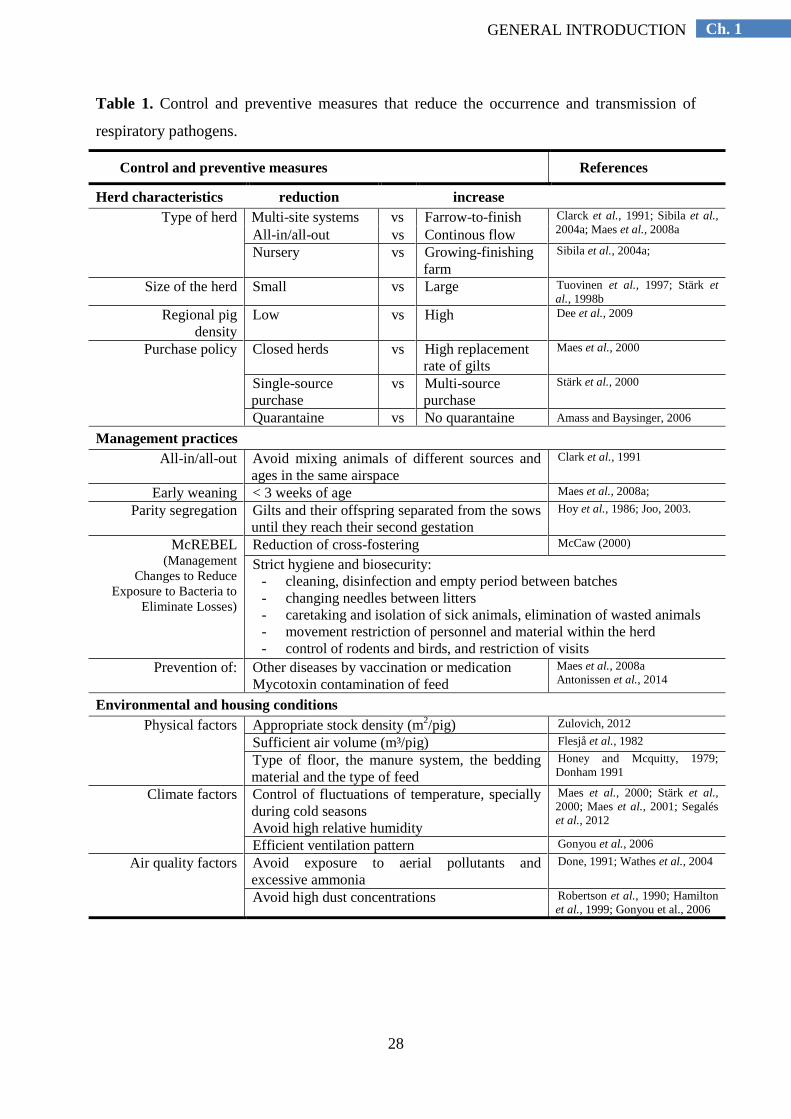
1.6.1. Optimization of management practices and housing conditions
Improvement of the management practices and the environment of the animals is of
crucial importance in the control of M. hyopneumoniae infections. Most authors classify non-
infectious risk factors in three categories (management, environment and pig factors) (Stärk
2000; Brockmeier et al., 2002; Opriessnig et al., 2011), but it is clear that in practice, many
factors may interact with each other. A short description of the main control and preventive
measures that reduce the occurrence and transmission of respiratory pathogens is presented in
table 1.
28
Ch. 1 GENERAL INTRODUCTION
Table 1. Control and preventive measures that reduce the occurrence and transmission of
respiratory pathogens.
Control and preventive measures References
Herd characteristics reduction increase
Type of herd Multi-site systems vs Farrow-to-finish Clarck et al., 1991; Sibila et al.,
2004a; Maes et al., 2008a All-in/all-out vs Continous flow
Nursery vs Growing-finishing
farm
Sibila et al., 2004a;
Size of the herd Small vs Large Tuovinen et al., 1997; Stärk et
al., 1998b
Regional pig
density
Low vs High Dee et al., 2009
Purchase policy Closed herds vs High replacement
rate of gilts
Maes et al., 2000
Single-source
purchase
vs Multi-source
purchase
Stärk et al., 2000
Quarantaine vs No quarantaine Amass and Baysinger, 2006
Management practices
All-in/all-out Avoid mixing animals of different sources and
ages in the same airspace
Clark et al., 1991
Early weaning < 3 weeks of age Maes et al., 2008a;
Parity segregation Gilts and their offspring separated from the sows
until they reach their second gestation
Hoy et al., 1986; Joo, 2003.
McREBEL (Management
Changes to Reduce
Exposure to Bacteria to
Eliminate Losses)
Reduction of cross-fostering McCaw (2000)
Strict hygiene and biosecurity:
- cleaning, disinfection and empty period between batches
- changing needles between litters
- caretaking and isolation of sick animals, elimination of wasted animals
- movement restriction of personnel and material within the herd
- control of rodents and birds, and restriction of visits
Prevention of: Other diseases by vaccination or medication
Mycotoxin contamination of feed
Maes et al., 2008a
Antonissen et al., 2014
Environmental and housing conditions
Physical factors Appropriate stock density (m2/pig) Zulovich, 2012
Sufficient air volume (m³/pig) Flesjå et al., 1982
Type of floor, the manure system, the bedding
material and the type of feed
Honey and Mcquitty, 1979;
Donham 1991
Climate factors Control of fluctuations of temperature, specially
during cold seasons
Avoid high relative humidity
Maes et al., 2000; Stärk et al.,
2000; Maes et al., 2001; Segalés
et al., 2012
Efficient ventilation pattern Gonyou et al., 2006
Air quality factors Avoid exposure to aerial pollutants and
excessive ammonia
Done, 1991; Wathes et al., 2004
Avoid high dust concentrations Robertson et al., 1990; Hamilton
et al., 1999; Gonyou et al., 2006

29
Ch. 1 GENERAL INTRODUCTION
1.6.2. Antimicrobial treatment
This section summarizes the current knowledge on antimicrobial agents used for the
treatment of M. hyopneumoniae infections, the route of administration of these antimicrobials,
as well as the presence of antimicrobial resistance. Although antimicrobials are capable of
controlling M. hyopneumoniae infections, complete elimination of the organism from the
respiratory tract cannot be achieved by medication (Thacker and Minion, 2012).
1.6.2.1. Antimicrobials
Potentially active antimicrobials against M. hyopneumoniae include tetracyclines,
macrolides, lincosamides, pleuromutilins, florfenicol, aminoglycosides, aminocyclitols and
fluoroquinolones (Vicca, 2005; Maes et al., 2008a; AMCRA, 2013). Only the last two agents
have mycoplasmacidal effects (Hannan et al., 1989). To control and treat respiratory disease,
tetracyclines (doxycycline), potentiated sulfonamides (sulfadiazine-trimethoprim) and
macrolides (tylosin and tilmicosin) are mostly used. Although sulfadiazine-trimethoprim is not
effective against M. hyopneumoniae, it may be useful for treatment of secondary bacterial
infections, often present during enzootic pneumonia. An overview of the main antimicrobials
effective against M. hyopneumoniae is presented in this section, including pharmacokinetic and
pharmacodynamic characteristics, as well as the mode of action and spectrum of activity (Table
2).
1. Tetracyclines
Most of the tetracyclines can be administered per os, although intramuscular and
intravenous formulations may also be used. Oral bioavailability may be lower when
administered in feed, since bivalent cations (calcium, magnesium, iron) have a chelating effect
on tetracyclines and, therefore, absorption and activity is reduced (Luthman and Jacobsson,
1983). Oral bioavailability when administered via the drinking water may also be reduced by
the water acidity and bivalent cations (iron) of the water pipes (Prescott, 2000b). Some
tetracyclines are more lipophilic than other members, such as doxycycline, and therefore
diffuse easily through most tissues, biological barriers and cell membranes. The degree of
liposolubility largely varies between the different tetracyclines and determines their tissue
distribution and elimination rate. Based on the duration of the effect, tetracyclines can be
classified in three different groups: short acting (chlortetracycline, tetracycline,
oxytetracycline), medium duration (demeclocycline, metacycline) and long acting
(doxycycline, minocycline) (Lemos, 2002). Susceptibility testing has demonstrated that some

30
Ch. 1 GENERAL INTRODUCTION
coliforms (Burch 2013), Mycoplasma hyopneumoniae (Inamoto et al., 1994), A.
pleuropneumoniae (Vanni et al., 2012), S. suis (De Jong et al., 2014), H. parasuis (De la
Fuente et al., 2007) and P. multocida (De Jong et al., 2014) have acquired resistance to
tetracyclines. Generally, tetracyclines are widely used in the treatment of M. hyopneumoniae
infections, as well as to treat and prevent atrophic rhinitis and other respiratory infections in
pigs caused by A. pleuropneumoniae and P. multocida.
2. Macrolides
Most of the macrolides can be administered orally or parenterally. They are basic
compounds very lipophilic, they are well absorbed from the intestine and have a good tissue
distribution (Vicca, 2005). This high liposolubility enables macrolides to diffuse easily through
biological barriers, and to reach therapeutic concentrations in most of the tissues, often many
times higher than serum concentrations (Lemos, 2002). Once macrolides are distributed
through the tissues, they accumulate intracellularly in the lysozomes of the phagocytes
(Scorneaux and Shryock, 1998). Acquired resistance to macrolides has been described under
field conditions in M. hyopneumoniae (Stakenborg et al., 2005a).
Generally, macrolides are recommended to treat M. hyopneumoniae infections, as well as
pneumonia and respiratory infections caused by other bacteria, such as A. pleuropneumoniae,
B. bronchiseptica, H. parasuis, and P. multocida.
3. Lincosamides
Lincosamides are alkaline, lipophilic compounds, which favors absorption through the
gastrointestinal tract and diffusion through biological barriers, leading to high tissue
concentrations. Lincomycin is indicated in the treatment of M. hyopneumoniae infections.
Acquired resistance to lincosamides has been described under field conditions in M.
hyopneumoniae (Stakenborg et al., 2005a).
4. Pleuromutilins
Pleuromutilins present an excellent absorption after oral administration in monogastric
mammals (Prescott, 2000c), a high bioavailability (around 90 per cent) and achieve high tissue
concentrations in the lung (Burch, 2012). Pleuromutilins are often used to treat PRDC.
Tiamulin and valnemulin may be used for the treatment of M. hyopneumoniae infections.
However, it is better to restrict their use to control Brachyspira infections (AMCRA, 2013),
since only a few effective antimicrobials are still available for the treatment of swine dysentery
due to the increased antimicrobial resistance.

31
Ch. 1 GENERAL INTRODUCTION
Table 2. Mode of action and spectrum of activity of the main antimicrobials effective against M. hyopneumoniae.
Family of
antimicrobials Mode of action Spectrum References
Tetracyclines chlortetracycline
doxycycline
oxytetracycline
Inhibition of protein synthesis by binding reversely to 30S ribosomal subunit, which
interferes with association of aminoacyl-tRNA to
mRNA-ribosomal complex
Broad-spectrum; bacteriostatic Gram+, Gram-, chlamydiae,
mycoplasmas, rickettsiae, protozoa
Aronson, 1980; Riviere et
al., 1991; Prescott, 2000b;
Chopra and Roberts, 2001
Macrolides erythromycin
tildipirosin, tilmicosin
tulathromycin
tylosin, tylvalosin
Inhibition of protein synthesis by binding reversely to 50S ribosomal subunit, which
interferes with translocation of peptides, hence with
growth of peptide chain and results in dissociation of
peptydil-tRNA from ribosome
Bacteriostatic Gram+, selected Gram- (Pasteurella,
Mannheimia, Leptospira,
Campylobacter, Actinobacillus), some
anaerobes and Mycoplasma spp.
Adams, 2001; Vester and
Douthwaite, 2001; Vicca,
2005
Lincosamides lincomycin
Inhibition of protein sysnthesis cfr. Macrolides
Bacteriostatic. Gram+ and anaerobes,
but less effective against Gram- and
mycoplasmas than macrolides
Prescott, 2000c
Pleuromutilins tiamulin
valnemulin
Inhibition of protein sysnthesis cfr. Macrolides
Bacteriostatic. Gram+, anaerobes and
mycoplasmas, but less effective against
Gram- than macrolides. Tiamulin: active
against Gram- (Actinobacillus,
Bordetella, Brachyspira, Haemophilus,
Mycoplasma and Pasteurella).
Weisblum, 1998; Aitken
et al., 1999; Vester and
Douthwaite, 2001;
Lemos, 2002
Amphenicols florfenicol
Inhibition of protein sysnthesis cfr. Macrolides
Broad-spectrum; bacteriostatic Gram+, Gram-, chlamydiae,
mycoplasmas, rickettsiae and anaerobes
Cannon et al., 1990;
Sams, 1994; Priebe and
Schwarz, 2003; Shin et
al., 2005
Aminoglycosides apramycin, gentamicin,
neomycin, streptomycin
Aminocyclitols
spectinomycin
Inhibition of protein sysnthesis by binding irreversibly 30S ribosomal subunit and
blocking tRNA translation. Translation fidelity is
decreased by disruption of translocation step of protein
synthesis and by induction of translation errors
Broad-spectrum; bactericidal Some Gram + (Mycobacteria,
Staphylococcus spp.), many aerobic
Gram- (Spirochetes) and mycoplasmas
Calvert et al., 1985;
Brown, 1988; Malik et
al., 1994; Riviere and
Spoo, 1995; Kotra et al.,
2000; Prescott, 2000a;
Lemos, 2002;
Fluoroquinolones danofloxacin
enrofloxacin
flumequine
marbofloxacin
Abolition DNA supercoiling and replication by inhibition of the topoisomerase II, DNA gyrase and
topoisomerase IV
Broad-spectrum; bactericidal Some Gram+, most Gram-, chlamydiae,
mycobacteria and mycoplasmas
Arai et al., 1992; Roberts,
1992; Brown, 1996;
Hannan et al., 1997;
Papich and Riviere, 2001;
Lees and Shojaee
AliAbadi, 2002
32
Ch. 1 GENERAL INTRODUCTION
5. Florfenicol
Florfenicol can be administered orally or parenterally (Liu et al., 2003; Ciprián et al.,
2012). This lipophilic drug has a wide tissue distribution. Immediately after intramuscular
administration, high initial blood levels are reached, leading to a quick initial response.
Duration of therapeutic plasma level may last more than 53 h after intramuscular
administration (Liu et al., 2003).
The use of florfenicol is indicated for the treatment of bacterial pneumonia and
associated respiratory infections caused by A. pleuropneumoniae, B. bronchiseptica, H.
parasuis, M. hyopneumoniae, M. hyosynoviae, P. multocida, Salmonella enterica and S. suis
(Burch, 2012).
6. Aminoglycosides and aminocyclitols
Aminoglycosides can be administered orally or parenterally. Due to their low lipophilic
degree, diffusion through cell membranes is limited and distribution impaired (Giroux et al.,
1995). They are rapidly and well absorbed from intramuscular and subcutaneous routes of
administration (Hammond, 1953; Brown and Riviere, 1991). When administered
parenterally, aminoglycosides easily penetrate in lung parenchyma and bronchial secretions
(Saux et al., 1986; Goldstein et al., 2002). Therefore, they are specially indicated in the
treatment of M. hyopneumoniae infections. However, they are very poorly absorbed after oral
administration (Brown and Riviere, 1991). Therefore, the oral route is mainly used to treat
enteric infections, and parenteral treatment for other infections.
7. Fluoroquinolones
Fluroquinolones can be administered via oral, intramuscular or subcutaneous route.
Fluoroquinolones are strongly lipophilic and therefore a high tissue distribution is reached
with good penetration through biological barriers. In monogastric animals, orally
administered fluoroquinolones are absorbed quickly and completely (80-100 per cent) (Inui et
al., 1998) and maximum plasma concentrations are reached one hour after intake. Oral
absorption is high, but it can be affected by divalent and trivalent cations. Absorption from
parenteral administration is rapid and nearly complete (Cabanes et al., 1992; Gavrielli et al.,
1995; Kaartinen et al., 1995; Brown, 1996; Mengozzi et al., 1996; Nielsen and Gyrd-Hansen,
1997; Richez et al., 1997; Bailey et al., 1998). They achieve concentrations that are at least as
high as plasma in most tissues (Papich and Riviere, 2001) and are rapidly accumulated in
macrophages and neutrophils.

33
Ch. 1 GENERAL INTRODUCTION
Fluoroquinolones can be utilized for the treatment of M. hyopneumoniae, as well as
other respiratory infections (P. multocida).
1.6.2.2. Antimicrobial use and routes of administration
The intensification of swine production in the past years has increased the use of
antimicrobials to maintain pig health. Nowadays, the use of antimicrobial remains necessary
to control respiratory disease (Mateu and Martin, 2001; Schwarz et al., 2001; McEwen and
Fedorka-Cray, 2002; Timmerman et al., 2006; Maes et al., 2008a; Callens et al., 2012).
Based on the objective of the therapeutic medication, antimicrobial use can be divided
in three categories: treatment, metaphylactic, prophylactic. Metaphylaxis is defined as the
mass medication of a group of animals when some of these animals are clinically diseased
while others are subclinically infected (Vicca, 2005). Prophylaxis is defined as the mass
medication of a group of animals to prevent a possible clinical outbreak during high-risk
periods for disease (Vicca, 2005). The high-risk period of M. hyopneumoniae infections
occurrence under European production conditions is known to be after transfer of animals to
the finishing facilities (10 weeks of age) (Léon et al., 2001). A recent study in Belgian pig
herds (Callens et al., 2012) demonstrated that antimicrobials are frequently utilized
therapeutically but also metaphylactically or prophylactically. This study illustrated a 7 and
93 per cent of metaphylactic and prophylactic use, respectively, in Belgian pig herds.
However, when compared with a similar previous report (Timmerman et al., 2006)
(metaphylactic: 44 per cent; prophylactic: 56 per cent), an increased number of prophylactic
group treatments and a drastic decrease of the portion of metaphylactic group treatments to
the total number of group treatments were observed along the time. These studies have also
described an increased number of treatment incidents in 2010 when compared with 2006 in
Belgium. Therefore, these results clearly show the need for a reduction of group level
prophylactic antimicrobial use (Callens et al., 2012). It also further confirms previous
observation which indicated that in modern pig production, animals are mostly treated as a
group and not as single individuals (Vicca, 2005; Timmerman et al., 2006; Burch, 2012).
Antimicrobial group treatments are predominantly applied via in-feed or in-water
medication because oral administration is less laborious and stressful compared to parenteral
administration. Oral administration is the most common method of administering
antimicrobials at group level. It can be quantified as treatment incidences (TI) based on the
animal daily dose pig (ADDpig: average maintenance dose per day per kg pig of a drug used
for its main indication) and the used daily dose pig (UDDpig: administered dose per day per

34
Ch. 1 GENERAL INTRODUCTION
kg pig of a drug) (Callens et al., 2012). Also parenteral treatment can be quantified in this
way. It has been shown that oral administrations are mostly underdosed (Timmerman et al.,
2006).
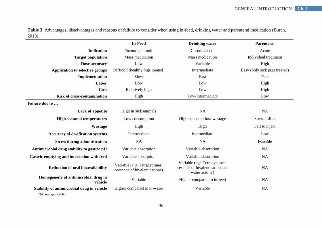
It is generally accepted that in-feed medication is the most common method of
administering antimicrobials at group level. However, it presents various disadvantages,
which may be common to both (in-feed and via drinking water) oral administrations (Table
3). First, a proportion of the feed provided to the pigs is wasted and not consumed by them.
Gonyou and Lou (1998) and Van Heugten and Van Kempen (1999) showed that wasted feed
can range from 5 to 6 per cent. Therefore, to avoid underdosing, the dosage of antimicrobial
should be calculated based on real feed intake, and not on total feed provided (Gottlob et al.,
2007). Second, the absorption and metabolism of some antimicrobials agents depend on the
pH of the stomach and the solubility of the agent. Therefore, a perfect knowledge of the
pharmacokinetic properties is required for the selection of the correct antimicrobial. Third, in-
feed medication is not recommended for the treatment of acute infections (e.g. A.
pleuropneumoniae), where a quick administration is of crucial importance for the success of
the therapy (Pijpers et al., 1990; Henry and Apley, 1999; Friendship, 2000). However, it is
especially suited for endemic or chronic diseases, such as enzootic pneumonia. Fourth, it
appears that a not depreciable amount of medication is wasted or not ideally administered,
since not only sick animals, but also healthy pigs receive medication. This can be justified by
the intention to treat also penmates that are at-risk to become infected, especially in herds
with a high stocking density.
Antimicrobial group treatment can also be implemented via drinking water. Currently,
administration of antimicrobials given as soluble formulations in the drinking water is
becoming more popular. The development of more reliable dosing/water-proportioner
machines has favored this increased use. It is especially recommended for the treatment of
acute diseases. Urgent group treatment can be done in a quick way and sick pigs often
continue to drink, even when they refuse feed. An important limitation is the antimicrobial
wastage due to water disappearance. Water disappearance is defined as the overall usage of
water, including water intake and wastage. Water wastage is mainly caused by either the type
of drinking system and/or playing with the drinking nipple both intentionally (out of
boredom) or unintentionally (highly dense stocked pens) (Brumm and Heemstra, 2000). For
example, nipple drinkers waste 50 per cent more than bowl drinkers and nipple drinkers that
are activated from any angle waste more that the ones that are activated from a forward angle
(Brumm and Heemstra, 2000; Gottlob et al., 2007). Water intake may also be largely

35
Ch. 1 GENERAL INTRODUCTION
influenced by the season. Therefore, appropriate follow-up of water usage efficiency is a
prerequisite for achieving correct therapeutic doses (Henry and Apley, 1999).
Group treatment can also be applied by parenteral administration. Advantages and
disadvantages of in-feed, drinking water and the parenteral route of administration are
presented in table 3. Callens et al. (2012) have illustrated a change in the relative importance
of the different routes of administration of group treatments in Belgian pig herds, when
compared with 2003 (Timmerman et al., 2006). The oral group treatments appeared to have
been replaced by long-acting injectable group treatments. Injectable group treatments are
mainly used in suckling pigs, and according to Timmerman et al. (2006) and Callens et al.
(2012), overdosing often takes place.
Individual treatments are usually applied by parenteral administration. Data from
Callens et al. (2012) revealed that in most of the Belgian herds antimicrobials at individual
level were administered. Individual treatments are frequently practiced in pigs suffering from
acute disease by parenteral injection during onset and first days of disease. It often results in a
good response against the disease, especially when acute outbreaks of respiratory disease
occur and appetite of sick animals is diminished (Pijpers, 1990; Henry and Apley, 1999).
The control of the M. hyopneumoniae infections by group medication can be
accomplished by strategic administration of antimicrobials. It is considered as a
prophylactic practice in swine (Karriker et al., 2012). It is usually applied by oral
administration. Strategic medication may reduce the consequences of the disease and the
infection load, but it does not prevent pigs from becoming infected with M. hyopneumoniae
(Thacker et al., 2006). In addition, the symptoms may reappear after cessation of the therapy.
Strategic medication during extended periods of time is not recommended. The risk for
antimicrobial residues in pig carcasses at slaughter and the development of antimicrobial
resistance in pathogens and bacteria belonging to the normal microbiota are major concerns
in Europe (Maes et al., 2008a). Currently, several countries of the EU (Denmark, Sweden,
The Netherlands, Germany) are running different programs at national level to monitor and
reduce the use of antimicrobials in livestock (Callens et al., 2012).
36
Ch. 1 GENERAL INTRODUCTION
Table 3. Advantages, disadvantages and reasons of failure to consider when using in-feed, drinking water and parenteral medication (Burch,
2013).
In-Feed Drinking water Parenteral
Indication Enzootic/chronic Chronic/acute Acute
Target population Mass medication Mass medication Individual treatment
Dose accuracy Low Variable High
Application to selective groups Difficult (healthy pigs treated) Intermediate Easy (only sick pigs treated)
Implementation Slow Fast Fast
Labor Low Low High
Cost Relatively high Low High
Risk of cross-contamination High Low/Intermediate Low
Failure due to …
Lack of appetite High in sick animals NA NA
High seasonal temperatures Low consumption High consumption/ wastage Stress inflict
Wastage High High Fail to inject
Accuracy of dosification systems Intermediate Intermediate Low
Stress during administration NA NA Possible
Antimicrobial drug stability to gastric pH Variable absorption Variable absorption NA
Gastric emptying and interaction with feed Variable absorption Variable absorption NA
Reduction of oral bioavailability Variable (e.g. Tetracyclines:
presence of bivalent cations)
Variable (e.g. Tetracyclines:
presence of bivalent cations and
water acidity)
NA
Homogeneity of antimicrobial drug in
vehicle Variable Higher compared to in-feed NA
Stability of antimicrobial drug in vehicle Higher compared to in-water Variable NA
NA: not applicable
37
Ch. 1 GENERAL INTRODUCTION
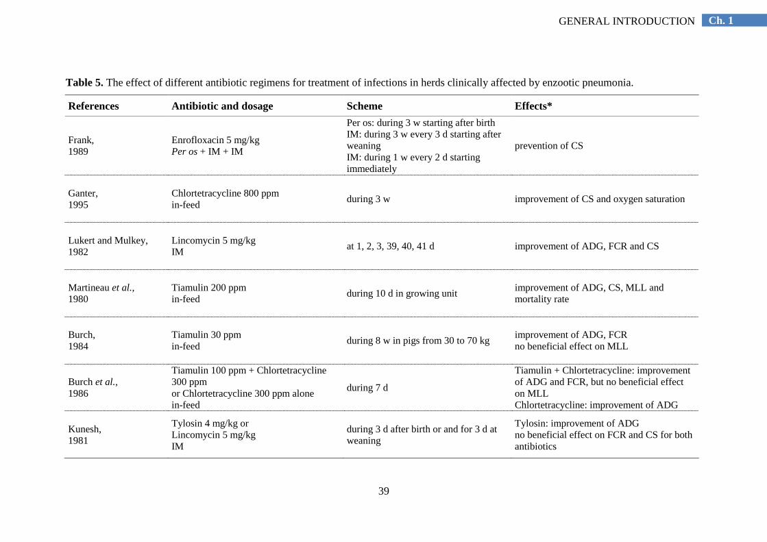
1.6.2.3. Efficacy of antimicrobials against M. hyopneumoniae infections under
experimental and field conditions
Various studies have been performed in the past years to assess the efficacy of
commercial antimicrobials used for the control and treatment of M. hyopneumoniae
infections. A summary of the principal results obtained from these studies published in peer-
reviewed journals is given in tables 4 and 5. Since these studies were conducted under
different conditions and many variables were included, comparison of results is complicated.
However, it can be concluded that for the majority of antimicrobials tested, performance
losses, clinical signs and lung lesions were reduced in treated animals. Nevertheless, M.
hyopnemumonia could still be isolated from treated animals.

38
Ch. 1 GENERAL INTRODUCTION
Table 4. The effect of different antibiotic regimens for treatment of experimental infections with Mycoplasma hyopneumoniae.
References Antibiotic and dosage Scheme Effects*
Hannan and Goodwin,
1990
6-chloro analogue of Norfloxacin
400 or 200 ppm Norfloxacin 100
ppm in-feed
during 3 w starting 1 m after
infection
improvement of ADG and FCR improvement of LL by 6-chloro analogue at 400 ppm
only
Schuller et al., 1977
Tiamulin 100 or 200 ppm in-feed
during 10 d starting 2 d before
infection
improvement of ADG and FCR LL were not prevented Mh could still be isolated
Goodwin, 1979
Tiamulin 50 mg/kg in-feed
during 10 d starting 1 m after
infection improvement of ADG, CS and LL Mh could still be isolated
Kobisch and Sibelle,
1982 Tiamulin 240 ppm in-feed
during 10 d starting 10 d after
infection improvement of ADG, FCR, CS, MLL and mLL
Hsu et al., 1983
Tiamulin 10, 20, 30 ppm in-feed
during 28 d starting 14 d after
infection improvement of ADG and FCR no beneficial effect on CS and LL
Ross and Cox, 1988
Tiamulin 60, 120, 180 ppm in-water
during 10 d starting 11 d after
infection no beneficial effect on ADG, FCR, CS, MLL and mLL
Hannan et al., 1982
Tylosin tartrate 50 mg/kg combined
with Tiamulin 10 mg/kg in-water during 10 d starting 14 d after
infection (with lung homogenate)
improvement of MLL Mh could still be isolated less secondary bacteria
Clarck et al., 1998
Tilmicosin 363 ppm in-feed
during 21 d starting 7 d before
infection improvement of CS no beneficial effect on ADG and LL
Thacker et al., 2006
Chlortetracycline 500 ppm in-feed
during 14 d starting 3 d before or
10 d after infection
3 d before infection: improvement of CS, LL both regimens: reduction of Mh organisms, but it could
still be isolated
Vicca et al., 2005
Tylosin tartrate 100 mg/kg in-feed
during 21 d starting 12 d after
infection
no beneficial effect on ADG improvement of CS and MLL Mh could still be isolated
Ciprián et al., 2012
Florfenicol 20 ppm in-feed
during 35 d starting the day of
infection improvement of ADG and LL Mh could still be isolated
McKelvie et al. 2005
Tulathromycin 2.5 mg/kg IM Enrofloxacin 5 mg/kg IM
during 1 or 3 d starting 5 d after
infection Tulathromycin: improvement weight gain, CS,LL Enrofloxacin: improvement of CS, LL
Le Carrou et al., 2006
Marbofloxacin 1 or 2 mg/kg IM
during 3 d starting 27 and 4 d
after infection no beneficial effect on ADG, MLL Mh could still be isolated
* Adapted from Vicca (2005); IM: intramuscular; d: day(s); w: week(s); m: month(s); ADG: average daily gain; FCR: feed conversion ratio; LL: lung lesions; CS: clinical signs; MLL:
macroscopic LL; mLL: microscopic LL;
39
Ch. 1 GENERAL INTRODUCTION
Table 5. The effect of different antibiotic regimens for treatment of infections in herds clinically affected by enzootic pneumonia.
References Antibiotic and dosage Scheme Effects*
Frank, 1989
Enrofloxacin 5 mg/kg Per os + IM + IM
Per os: during 3 w starting after birth IM: during 3 w every 3 d starting after
weaning IM: during 1 w every 2 d starting
immediately
prevention of CS
Ganter, 1995
Chlortetracycline 800 ppm in-feed
during 3 w improvement of CS and oxygen saturation
Lukert and Mulkey, 1982
Lincomycin 5 mg/kg IM
at 1, 2, 3, 39, 40, 41 d improvement of ADG, FCR and CS
Martineau et al., 1980
Tiamulin 200 ppm in-feed
during 10 d in growing unit improvement of ADG, CS, MLL and
mortality rate
Burch, 1984
Tiamulin 30 ppm in-feed
during 8 w in pigs from 30 to 70 kg improvement of ADG, FCR no beneficial effect on MLL
Burch et al., 1986
Tiamulin 100 ppm + Chlortetracycline
300 ppm or Chlortetracycline 300 ppm alone in-feed
during 7 d
Tiamulin + Chlortetracycline: improvement
of ADG and FCR, but no beneficial effect
on MLL Chlortetracycline: improvement of ADG
Kunesh, 1981
Tylosin 4 mg/kg or Lincomycin 5 mg/kg IM
during 3 d after birth or and for 3 d at
weaning
Tylosin: improvement of ADG no beneficial effect on FCR and CS for both
antibiotics

40
Ch. 1 GENERAL INTRODUCTION
References Antibiotic and dosage Scheme Effects*
Binder et al., 1993
Tilmicosin 300 ppm in-feed
during 9 or 14 d improvement of ADG, CS and less
secondary bacteria
LeGrand and Kobisch, 1996
Tiamulin 200 ppm + Chlortetracycline
600 ppm in-feed
pulse medication: 2 d treatment/2 w
during fattening period lower prevalence on MLL no beneficial effect on severity of MLL
Kavanagh, 1994
Tiamulin 30 ppm + Oxytetracycline 300
ppm in-feed
pulse medication: 2 or 3 d treatment/w
during fattening period
improvement of ADG and FCR lower prevalence and severity of MLL financial benefit
Jouglar et al., 1993
Tiamulin 40 ppm + Oxytetracycline 300
ppm in-feed
pulse medication: 2 d treatment/w
during fattening period
improvement of ADG and mortality rate no beneficial effect on FCR and severity of
LL
Bousquet et al., 1998
Marbofloxacin 1 or 2 mg/kg IM
during 3 d starting 27 and 4 d after
infection no beneficial effect on ADG, MLL Mh could still be isolated
Nanjani et al., 2005
Tulathromycin 2.5 mg/kg IM Tiamulin 15mg/kg IM Florfenicol 15mg/kg IM
at 0 d at 0, 1, 2 d at 0, 2 d
similar beneficial effect for all 3
antimicrobials on ADG
Nutsch et al., 2005
Tulathromycin 2.5 mg/kg IM Ceftiofur 3mg/kg IM
at 0 d at 0, 1, 2 d
similar beneficial effect for both
antimicrobials on cure and mortality rate
Mateusen et al., 2001
Vaccination (two-shot) Tilmicosin 200 ppm in-feed
at 4 and 22 d of age during 3 w (34-55
d) and 2 w (77-98 d) similar improvement of ADG and FCR, CS,
MLL
Mateusen et al., 2002
Vaccination (two-shot) Lincomycin 200 ppm in-feed Lincomycin 200 ppm + Vaccination
at 4 and 28 d of age during 3 w in
fattening period similar improvement of ADG and FCR, CS,
MLL
* Adapted from Vicca (2005); IM: intramuscular; d: day(s); w: week(s); m: month(s); ADG: average daily gain; FCR: feed conversion ratio; LL: lung lesions; CS: clinical signs; MLL:
macroscopic LL; mLL: microscopic LL;

41
Ch. 1 GENERAL INTRODUCTION
1.6.2.4. Antimicrobial susceptibility of M. hyopneumoniae
Few data is available concerning the antimicrobial susceptibility and resistance for M.
hyopneumoniae. Additionally, the fastidious growth of M. hyopneumoniae and its time
consuming isolation, complicate the consistency of susceptibility testing and the
interpretation of results.
Standard broth and agar dilution methods used for susceptibility testing of other
bacteria have been adapted for use with mycoplasmas. The „International Research
Programme on Comparative Mycoplasmology (IRPCM)‟ suggested the adoption of
„Guidelines and recommendations for antimicrobial minimum inhibitory concentration
testing against veterinary mycoplasma species‟ (Hannan, 2000).
Nowadays, both broth dilution and microbroth dilution techniques have been adapted
for mycoplasmas and are the most widely used. The test consists on the inoculation of a
standardized number of Mycoplasma-organisms in 96-well microtiter plates. These 96-well
microtiter plates are prepared with serial doubling concentrations of antimicrobial agents and
then, a mycoplasmal medium is added to these broth dilutions. The mycoplasmal medium
contains a constant number of microorganisms and a substrate (glucose, arginin or urea)
which is metabolized by mycoplasmas. The degradation of this medium results in a pH
change visible as color change, which implies mycoplasma growth. The presence of
increasing antibiotic concentrations permits to determine the lowest antimicrobial
concentration that inhibits mycoplasma growth. The minimum inhibitory concentration
(MIC) is defined as the lowest concentration of an antimicrobial agent that prevents a color
change at the same moment that the color in the control without antibiotic has changed, hence
the lowest concentration that prevents mycoplasma growth (Vicca, 2005). The study of
antimicrobial susceptibility by means of MIC determinations for M. hyopneumoniae has
various limitations. Bacterial contamination may occur, resulting in turbidity or color change
in broth controls. Since MIC determination of M. hyopneumoniae is time consuming and may
last up to 14 days, MICs of unstable antibiotics (chlortetracycline, valnemulin) are unreliable.
In addition, there are no specific clinical breakpoints for mycoplasmas.
Antimicrobial resistance in Mycoplasma spp. may be categorized in two types: intrinsic
or acquired resistance. Intrinsic, innate or natural resistance may be defined as the relative
insensitivity of all members of a bacterial species or genus against an antimicrobial agent
(Vicca, 2005). Members of the class Mollicutes lack a cell wall and are therefore resistant to
all β-lactam antibiotics, such as penicillins and cephalosporins, as well as antibiotics targeting

42
Ch. 1 GENERAL INTRODUCTION
the cell wall, like glycopeptides. Additionally, mycoplasmas are also resistant to polymyxins,
sulfonamides, trimethoprim, naladixic acid and rifampin. Within Mycoplasma species,
macrolides present specific patterns of innate resistance depending on the structure of the
lactone ring. The 14-membered lactone ring macrolides are ineffective against M.
hyopneumoniae, M. flocculare, M. bovis, M. pulmonis and M. fermentans (Williams, 1978;
Tanner et al., 1993; Chambaud et al., 2001; Bébéar and Bébéar, 2002; Francoz et al., 2005),
whereas M. pneumoniae and M. genitalium are susceptible to this subgroup of macrolides
(Bébéar et al., 2000; Kenny and Cartwright, 2001). In general terms, there are three main
biochemical mechanisms of both intrinsic and acquired antimicrobial resistance in
mycoplasmas, e.g. active efflux mechanisms, enzymatic modification of the drug or alteration
in the drug target site. A complete overview of the biochemical mechanisms involved in the
antimicrobial acquired resistance for Mycoplasma spp. was published by Vicca (2005).
In vitro susceptibility of M. hyopneumoniae to antimicrobials used in pig veterinary
medicine has been studied. Apart of intrinsic antimicrobial resistance, acquired antimicrobial
resistance of M. hyopneumoniae has also been documented. Inconsistent results have been
reported in susceptibility testing of tetracyclines (Williams, 1978; Etheridge et al., 1979;
Yamamoto and Koshimizu, 1984). Shortly afterwards, Inamoto et al. (1994) reported
acquired antimicrobial resistance to tetracyclines (chlortetracycline and oxytetracycline)
occurring in M. hyopneumoniae field strains isolated in Japan between 1970 and 1990. More
recently also acquired antimicrobial resistance to macrolides (tylosin, tilmicosin),
lincosamides (lincomycin) and fluoroquinolones (enrofloxacin, flumequine) (Vicca et al.,
2004) has been documented.
Despite some reports on resistance against tetracyclines, macrolides, lincosamides and
fluoroquinolones in M. hyopneumoniae (Stakenborg et al., 2005a; Le Carrou et al., 2006;
Vicca et al., 2007), resistance against other antimicrobials has not yet been detected. This
indicates that antimicrobial resistance does not constitute a major problem for treatment of M.
hyopneumoniae infections (Vicca et al., 2004; Maes et al., 2008a).

43
Ch. 1 GENERAL INTRODUCTION
1.6.3. Vaccination against Mycoplasma hyopneumoniae
1.6.3.1. Commercial vaccines
Vaccination against M. hyopneumoniae is practiced in most of the swine producting
countries, being in some cases applied in more than 70 per cent of the herds (Maes et al.,
2008a). Vaccination with commercial bacterins is considered as an important and effective
tool to control M. hyopneumoniae infections.
Commercial bacterins, consisting of inactivated, adjuvanted whole-cell preparations,
have been commonly demonstrated efficacious to control the disease. The benefit of
vaccination is based on reduction of losses caused by poor performance (average daily gain,
ADG; 2-8 per cent), poor feed efficiency (feed conversion ratio; 2-5 per cent) and mortality
(Maes et al., 2008a). A shorter time to reach market weight, decreased clinical signs and lung
lesions and lower costs for antimicrobial medication and vaccination are contributors to this
economic benefit (Dohoo and Montgomery 1996; Jensen et al., 2002; Maes et al., 2003;
Maes et al., 2008a). A summary of experiments conducted under field conditions and the
benefits observed after vaccination is presented in table 6.
Despite all these advantages, commercial vaccines present various limitations. Thacker
et al. (2000) suggested that current bacterins provided only partial protection and do not
prevent colonization of the respiratory tract by M. hyopneumoniae. However, Sibila et al.
(2007b) also demonstrated that vaccination may reduce the infection level in a herd. This
corroborates previous observations from Haesebrouck et al. (2004), which indicated that
maximum beneficial effects of vaccination are reached several months after the initiation of
vaccination. Recent studies demonstrated that vaccination significantly reduced the number
of M. hyopneumoniae organisms in the lungs of experimentally infected pigs (Vranckx et al.,
2012b), but the fact that M. hyopneumoniae DNA could still be detected by PCR confirmed
previous results from experimental and field transmission studies (Meyns et al., 2006,
Villarreal et al., 2011). These studies showed a limited reduction in transmission in
vaccinated pigs. Therefore, all these findings suggest that, similarly to antimicrobials,
vaccination alone does not prevent infection, and it is not able to eliminate M.
hyopneumoniae from infected pig herds.
During the past 15 years, few studies have investigated the immune mechanisms
involved in induction of protection after vaccination with bacterins. All these studies
indicated that both systemic and mucosal immune responses play a central role in the control
of M. hyopneumoniae infections. However, the exact mechanism of protection conferred by
44
Ch. 1 GENERAL INTRODUCTION
vaccination against M. hyopneumoniae is not yet fully understood. Thacker et al. (1998)
compared the level of protection of four different commercial bacterins induced after
experimental challenge. All four induced the production of specific antibodies in serum, but
only a partial protection against clinical signs was conferred. Djordjevic et al. (1997) further
confirmed this observation. A reduction in pneumonia was observed after vaccination, but
antibody concentrations (in serum and respiratory tract washings) were not correlated with
protection from the disease. Nowadays it is generally accepted that the presence and
concentration of serum antibodies are not correlated with protection. Therefore, this is not
suitable for the evaluation of protective immunity (Maes et al., 2008a; Marchioro et al.,
2013).
Because M. hyopneumoniae is a mucosal pathogen which mainly adheres to the cilia of
the epithelial cells from the respiratory tract, local M. hyopneumoniae-specific antibodies
seem to play an important role in protection. Thacker et al. (2000) suggested that both local
mucosal antibodies and systemic cell-mediated immunity responses are important for
protection. These observations were confirmed by Marchioro et al. (2013). These authors
illustrated the induction of both local and systemic immune responses involving both specific
antibodies and cellular immunity following vaccination against M. hyopneumoniae with a
commercially available bacterin.
An inconsistent immune response and protection conferred by different commercial
vaccines has been reported (Thacker et al., 1998). This variability may be due to several
factors, such as different adjuvants included by commercial vaccines and possible antigenic
differences (e.g. absence of specific epitopes) between the vaccine strain and the strains
circulating in the field (Marchioro et al., 2013).
1.6.3.2. Strategies of vaccination
Many studies have been conducted in order to investigate the efficacy of the
commercial bacterins under field conditions (Jensen et al., 2002). However, to decide
whether vaccination in a specific herd should be implemented has to be based not only on
efficacy parameters, but also on a cost-benefit analysis (Maes et al., 2003). Type of the herd,
production system, infection pattern, health status of the population at risk, as well as easy
implementation at practical level are main variables playing a role in the design of a
successful vaccination strategy.

45
Ch. 1 GENERAL INTRODUCTION
First, it has to be decided which population at risk should be vaccinated. In practice in
Europe, sow vaccination is rarely practiced, whereas the main strategy of M. hyopneumoniae
control is vaccination of piglets before or at weaning (Haesebrouck et al., 2004).
Vaccination of sows ensures immunization of the newborn piglets through the passage
of maternal derived antibodies via colostrum. However, colostral immunity does not prevent
colonization of the piglets with M. hyopneumoniae (Thacker et al., 2000; Sibila et al., 2008).
The main objective of vaccinating sows is to decrease vertical transmission from the dam to
the off-spring via nose-to-nose contact (Ruiz et al., 2003).
Vaccination of gilts before entering into the sow herd may avoid destabilization of the
herd immunity (Bargen, 2004).
Vaccination of suckling piglets (early vaccination; <4 weeks of age) is often practiced
in single-site herds, where early infections are common (Maes et al., 2008a). Because M.
hyopneumoniae infections can take place in piglets already during the first (from 1 to 3)
weeks of life (Calsamiglia and Pijoan 2000, Ruiz et al., 2003, Fano et al., 2007, Sibila et al.,
2007a, Nathues et al., 2010, Villarreal et al., 2010, 2011, Vranckx et al., 2012a, Fablet et al.,
2012a), and because vaccination is most likely effective if active immunity can be established
before the exposure to the pathogen, vaccination is commonly applied in suckling piglets.
When compared with weaned piglets, suckling piglets are less infected with pathogens, such
as PRRSV and PCV-2, which may cause problems after weaning and interfere with the
establishment of a protective immune response to M. hyopneumoniae vaccination (Maes et
al., 2008a). A limitation of early vaccination is the presence of maternal antibodies and its
possible interference with vaccine efficacy (grosse Beilage et al., 2005; Martelli et al., 2006;
Bandrick et al., 2008). However, the majority of the pig herds in Europe vaccinate piglets in
the presence of maternal antibodies and the efficacy is apparently not affected (Martelli et al.,
2006; Maes et al., 2008a).
Vaccination of weaners/growers/early fattening pigs (late vaccination; between 4 and
10 weeks) is frequently applied in multi-site systems, where late infections are more common
(Maes et al., 2008a). Late vaccination may evade the possibility of interference with
maternally derived antibodies. However, it may elongate the age-window between decline of
maternal immunity and age of infection, leading to a higher risk of infection before
immunization. In addition, nursery pigs may already be infected with M. hyopneumoniae,
since the onset of infection may vary between herds and also within a herd among successive
batches (Sibila et al., 2004b, 2007b, Fano et al., 2007, Segalés et al., 2012).

46
Ch. 1 GENERAL INTRODUCTION
Vaccination programs have become even more complicated with the introduction of
single-dose formulations in addition to traditional two-dose formulations. The first
commercially available bacterins indeed required two intramuscular injections and were
demonstrated efficacious to control the disease. However, with the appearance of single-dose
vaccines, which were confirmed as equally efficacious as the two-dose formulations (Kriakis
et al., 2001; Dawson et al., 2002), the newer bacterins became more popular. This was
mainly due to the reduced labor costs, easy implementation at farm level, less stress inflicted
to the pigs, while a similar protection is conferred when compared with two-shot vaccines
(Roof et al., 2001; Roof et al., 2002; Alexopoulos et al., 2004; Lillie et al., 2004; Baccaro et
al., 2006; Greiner et al., 2011). Despite the acceptable efficacy of single-dose vaccines, there
are still some situations in which two-dose formulations may be indicated (Yeske et al.,
2001). These situations include critical periods (pigs placed in fall months) and production
systems (multi-source, multi-age, continuous flow) with serious M. hyopneumoniae
challenges. It is also recommended in large herds with unstable situation to PRRSV, as well
as when vaccination compliance is doubtful (Yeske et al., 2001).
Variation in protection between different bacterins may be associated with the different
adjuvants. Although all commercial bacterins are made from inactivated M. hyopneumoniae
cells, they differ in the type of adjuvant (aluminium hydroxide, carbopol, mineral oil or
biodegradable oil) and the solution included. Vaccines containing carbopol or aluminium
hydroxide are produced as water based suspensions, whereas the oil adjuvant vaccines are
produced as different emulsions. It is generally accepted that aqueous-based adjuvants
produce a lesser stimulation of the immune system, and, consequently re-vaccination (two-
shot) is needed. Oil-based adjuvants are more reactive and often take a longer time to be
processed. Therefore, they provide a prolonged release of the antigen and a better stimulation
of the immune system. Generally speaking, bacterins containing oil-based adjuvants are
capable to stimulate the immune system over time, mimicking conventional two-shot
vaccines. Oil-based adjuvants can be divided in two types: oil in water emulsions and water
in oil emulsions. Oil in water emulsions present relatively good immune stimulating
properties, but a quick release of the antigen, which may shorten the duration of immune
stimulation. In order to achieve a long term protection, commercial vaccines include mineral
oil, which may cause significant local reactions. These local side effects may be minimized
by water in oil emulsions, which are made of very small droplets of antigenic medium
dispersed in biodegradable oil. This formulation based in biodegradable oil ensures an initial
fast immune response and a subsequent slow release of the antigen. This provides a smooth

47
Ch. 1 GENERAL INTRODUCTION
but long lasting stimulation of the immune system. Therefore, the onset of immunity is
modulated by the progressive liberation of the antigen after only a few days and this
immunity is maintained for a long period, compared to other adjuvant models (Herbach,
personal communication, 2005; Aucouturier et al., 2002).
The efficacy of all different commercial vaccines has been widely demonstrated during
the last years. Numerous clinical trials both under experimental and field conditions have
been carried out. Numerous parameters of comparison have been included. A summary of the
experiments carried out in the past is presented in table 6. It includes type of vaccine, doses
and age of vaccination, conditions of the experiment, parameters of evaluation, and
significant results obtained.
The route of vaccine administration has also to be taken into account when designing
a vaccination strategy. Traditionally, intramuscular administration has been used. Apart from
intramuscular vaccination, the effect of intraperitoneal (Sheldrake et al., 1991), oral (Weng et
al., 1992), aerosol (Murphy et al., 1993), intranasal (Shimoji et al., 2003) and intradermal
(Jones et al., 2005) vaccination against M. hyopneumoniae has also been investigated.
Nowadays, intradermal vaccination has arisen as an alternative to intramuscular
administration. It has been shown to be as efficacious as the intramuscular route in reducing
performance losses and providing a better protection against clinical signs and lung lesions
(Tassis et al., 2012). Main advantages of intradermal administration include lower risk of
disease transmission through the application (no needles) and the consistent delivery of the
vaccine. The lower volume and higher dispersion of the antigen during administration is
associated with a total reduction of side effects at the injection site and a decrease of
condemnation rate of carcasses at slaughter. Less pain and stress is inflicted to the animals
and it is considered to be a safe procedure for the operators. Disadvantages of intradermal
vaccination include the high cost for purchasing and maintaining the device for the ID
application, and the fact that training of the person using it is needed.
The use of combination vaccines has increased in the last years. Combination vaccines
are those that contain antigens from different pathogens in the same formulation. Combined
vaccination may maximize vaccine benefits, while minimizing costs, labor and the stress
inflicted to the animals (Drexler et al., 2010). Although they are widely used in U.S., they
lack popularity in Europe. A few studies have been carried out to investigate the effect of
different combinations of vaccines against different pathogens [PRRSV and M.
hyopneumoniae (Drexxler et al., 2010); PRRSV, M. hyopneumoniae and A.
pleuropneumoniae (Delisle and Rigaut, 2007); PCV-2 and PRRSV (Bretey et al., 2010);

48
Ch. 1 GENERAL INTRODUCTION
PCV-2 and M. hyopneumoniae (Hayes and Saltzman, 2009)]. These studies showed that
vaccines against different pathogens can be combined without compromising the efficacy and
safety, when used according to the manufacturer‟s instructions.
1.6.3.3. Development of new vaccines
Commercial vaccines are whole cell, adjuvanted bacterins (Okada et al., 1999).
Nowadays, the availability of complete genome sequences has contributed to a new
approach in vaccine development (Scarselli et al., 2005), called reverse vaccinology
(Rappuoli, 2001). The reverse vaccinology approach starts from the genomic sequence and,
by computer analysis, predicts those antigens that are most likely to be vaccine candidates.
This method may be used for the development of both subunit and DNA vaccines.
The use of recombinant proteins contributed to the development of subunit vaccines.
The recombinant subunit vaccines are based on fractions of the organism and are produced
using heterologous protein expression systems. These heterologous proteins can be produced
in e.g. Escherichia coli cells and, once expressed and purified, can be administered to the
animals. Specific antigens of M. hyopneumoniae include surface proteins (P46, P65, P97),
cytosolic proteins (P36) and functional enzymes (L-lactate dehydrogenase, ribonucleotide
reductase) (Kim et al., 1990; Strasser et al., 1991; Futo et al., 1995; Zhang et al., 1995; Fagan
et al., 1996), and may be considered as potential vaccine candidates.
Recently, several experimental recombinant subunit vaccines have been developed and
tested for immune responses in mice (Marchioro et al., 2012) or pigs (Marchioro et al.,
unpublished). Excellent reviews have been published by Simionatto et al. (2013) and
Marchioro (2013). Significant immune responses have been demonstrated in mice and pigs
immunized with these experimental vaccines. Thus, recombinant subunit vaccines might
represent a promising strategy for developing more effective vaccines against M.
hyopneumoniae.
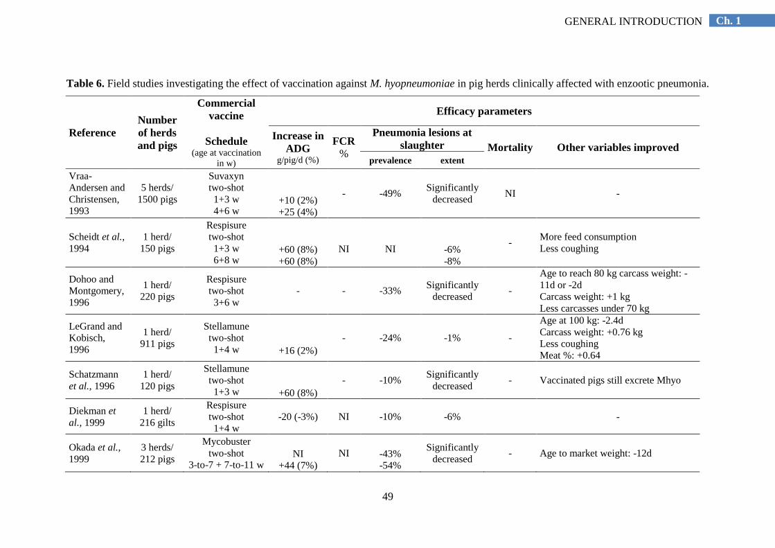
49
Ch. 1 GENERAL INTRODUCTION
Table 6. Field studies investigating the effect of vaccination against M. hyopneumoniae in pig herds clinically affected with enzootic pneumonia.
Reference
Number
of herds
and pigs
Commercial
vaccine
Schedule (age at vaccination
in w)
Efficacy parameters
Increase in
ADG g/pig/d (%)
FCR
%
Pneumonia lesions at
slaughter Mortality Other variables improved
prevalence extent
Vraa-
Andersen and
Christensen,
1993
5 herds/
1500 pigs
Suvaxyn two-shot 1+3 w 4+6 w
+10 (2%) +25 (4%)
- -49% Significantly
decreased NI -
Scheidt et al.,
1994 1 herd/
150 pigs
Respisure two-shot 1+3 w 6+8 w
+60 (8%) +60 (8%)
NI
NI
-6% -8%
- More feed consumption Less coughing
Dohoo and
Montgomery,
1996
1 herd/
220 pigs
Respisure two-shot 3+6 w
- - -33% Significantly
decreased -
Age to reach 80 kg carcass weight: -
11d or -2d Carcass weight: +1 kg Less carcasses under 70 kg
LeGrand and
Kobisch,
1996
1 herd/
911 pigs
Stellamune two-shot 1+4 w
+16 (2%)
- -24% -1% -
Age at 100 kg: -2.4d Carcass weight: +0.76 kg Less coughing Meat %: +0.64
Schatzmann
et al., 1996 1 herd/
120 pigs
Stellamune two-shot 1+3 w
+60 (8%)
- -10% Significantly
decreased - Vaccinated pigs still excrete Mhyo
Diekman et
al., 1999 1 herd/
216 gilts
Respisure two-shot 1+4 w
-20 (-3%) NI -10% -6%
-
Okada et al.,
1999 3 herds/
212 pigs
Mycobuster two-shot
3-to-7 + 7-to-11 w
NI
+44 (7%)
NI
-43% -54%
Significantly
decreased - Age to market weight: -12d
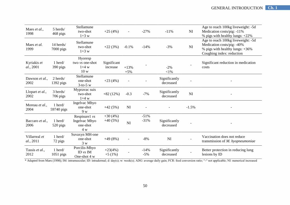
50
Ch. 1 GENERAL INTRODUCTION
Maes et al.,
1998 5 herds/
468 pigs
Stellamune two-shot 1+3 w
+25 (4%) - -27% -11% NI Age to reach 100kg liveweight: -5d Medication costs/pig: -11% % pigs with healthy lungs: +22%
Maes et al.
1999 14 herds/
7000 pigs
Stellamune two-shot 1+3 w
+22 (3%) -0.1% -14% -3% NI
Age to reach 100kg liveweight: -5d Medication costs/pig: -40% % pigs with healthy lungs: +36% Coughing index: reduction
Kyriakis et
al., 2001 1 herd/
390 pigs
Hyoresp two vs one-shot
1+4 w 10 w
Significant
increase
+13% +5%
-
-2% <1%
- Significant reduction in medication
costs
Dawson et al.,
2002 2 herds/
1392 pigs
Stellamune one-shot 3-to-5 w
+23 (4%) - - Significantly
decreased - -
Llopart et al.,
2002 3 herds/
706 pigs
Mypravac suis two-shot 1+4 w
+82 (12%) -0.3 -7% Significantly
decreased NI -
Moreau et al.,
2004 1 herd/
59740 pigs
Ingelvac Mhyo one-shot
9 w +42 (5%) NI - - -1.5% -
Baccaro et al.,
2006 1 herd/
520 pigs
Respisure1 vs
Ingelvac Mhyo one-shot
4 w
+30 (4%) +40 (5%)
NI
-51% -31%
Significantly
decreased - -
Villarreal et
al., 2011 1 herd/
72 pigs
Suvaxyn MH-one one-shot
3 w +49 (8%) - -8% NI -
Vaccination does not reduce
transmission of M. hyopneumoniae
Tassis et al.,
2012 1 herd/
1051 pigs
Porcilis-Mhyo ID vs IM
One-shot 4 w
+23(4%) +5 (1%)
- -14% -5%
Significantly
decreased -
Better protection in reducing lung
lesions by ID
* Adapted from Maes (1998); IM: intramuscular; ID: intradermal; d: day(s); w: week(s); ADG: average daily gain; FCR: feed conversion ratio; “-“ not applicable; NI: numerical increased
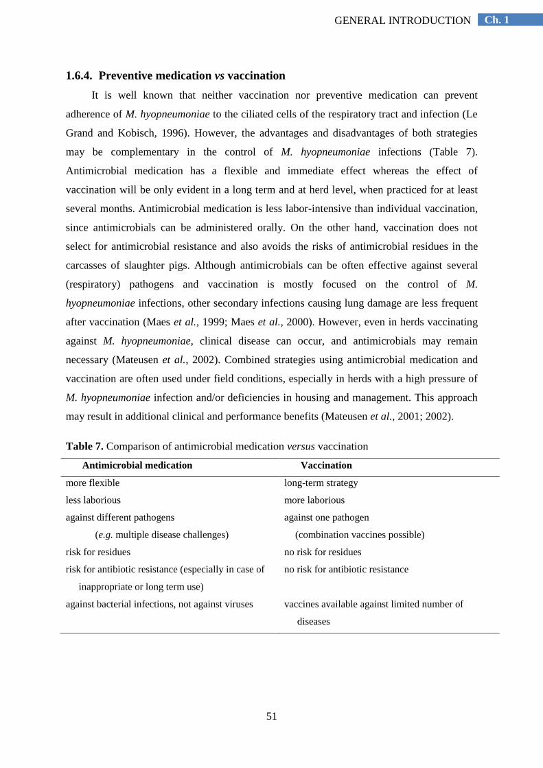
51
Ch. 1 GENERAL INTRODUCTION
1.6.4. Preventive medication vs vaccination
It is well known that neither vaccination nor preventive medication can prevent
adherence of M. hyopneumoniae to the ciliated cells of the respiratory tract and infection (Le
Grand and Kobisch, 1996). However, the advantages and disadvantages of both strategies
may be complementary in the control of M. hyopneumoniae infections (Table 7).
Antimicrobial medication has a flexible and immediate effect whereas the effect of
vaccination will be only evident in a long term and at herd level, when practiced for at least
several months. Antimicrobial medication is less labor-intensive than individual vaccination,
since antimicrobials can be administered orally. On the other hand, vaccination does not
select for antimicrobial resistance and also avoids the risks of antimicrobial residues in the
carcasses of slaughter pigs. Although antimicrobials can be often effective against several
(respiratory) pathogens and vaccination is mostly focused on the control of M.
hyopneumoniae infections, other secondary infections causing lung damage are less frequent
after vaccination (Maes et al., 1999; Maes et al., 2000). However, even in herds vaccinating
against M. hyopneumoniae, clinical disease can occur, and antimicrobials may remain
necessary (Mateusen et al., 2002). Combined strategies using antimicrobial medication and
vaccination are often used under field conditions, especially in herds with a high pressure of
M. hyopneumoniae infection and/or deficiencies in housing and management. This approach
may result in additional clinical and performance benefits (Mateusen et al., 2001; 2002).
Table 7. Comparison of antimicrobial medication versus vaccination
Antimicrobial medication Vaccination
more flexible long-term strategy
less laborious more laborious
against different pathogens
(e.g. multiple disease challenges)
against one pathogen
(combination vaccines possible)
risk for residues no risk for residues
risk for antibiotic resistance (especially in case of
inappropriate or long term use)
no risk for antibiotic resistance
against bacterial infections, not against viruses vaccines available against limited number of
diseases
52
Ch. 1 GENERAL INTRODUCTION
REFERENCES
Adams, H.R. (2001). Macrolides. In: Adams, H.R. (Ed.). Veterinary Pharmacology and Therapeutics. Iowa
State University Press, Ames, USA, pp. 876-882.
Aitken, I.A., Morgan, J.H., Dalziel, R., Burch, D.G.S. and Ripley, P.H. (1999). Comparitive in vitro activity of
valnemulin against porcine bacterial pathogens. The Veterinary Record 144, 128.
Alexopoulos, C., Kritas, S.K., Papatsas, I., Papatsiros, V.G., Tassis, P.D. and Kyriakis, S.C. (2004). Efficacy of
one and two shot vaccines for the control of enzootic pneumonia (EP) in a pig unit suffering from respiratory
syndrome due to EP, PRRS and PMWS. Proceedings of the 18th
International Pig Veterinary Society
Congress. Hamburg, Germany, June 27 to July 1, p 449.
Amass, S. and Baysinger, A. (2006). Swine disease transmission and prevention. In: Straw, B.E., Zimmerman,
J.J., D‟Allaire, S., Taylor, D.J. (Eds.). Diseases of Swine. Blacwell Publishing Ltd., Oxford, UK, pp. 1075–
1098.
AMCRA (2013). Richtlijnen voor gebruik van antibacterïele middelen per indicatie. In: AMCRA VZW (Eds).
Formularium voor verantwoord gebrik van antibacterïele middelen in de varkenshouderij. Merelbeke,
Belgium, pp. 11-13.
Antonissen, G., Martel, A., Pasmans, F., Ducatelle, R., Verbrugghe, E., Vandenbroucke, V., Li, S.,
Haesebrouck, F., Van Immerseel, F. and Croubels, S. (2014). The impact of Fusarium mycotoxins on human
and animal host susceptibility to infectious diseases. Toxins 6, 430-452.
Arai, S., Gohara, Y., Kuwano, K. and Kawashima, T. (1992). Antimycoplasmal activities of new quinolones,
tetracyclines, and macrolides against Mycoplasma pneumoniae. Antimicrobial Agents and Chemotherapy 36,
1322-1324.
Aronson, A.L. (1980). Pharmacotherapeutics of the newer tetracyclines. Journal of the American Veteterinary
Medicine Association 176, 1061-1068.
Aucouturier, J., Dupuis, L., Deville, S., Ascarateil, S. and Ganne, V. (2002). Montanide ISA 720 and 51: a new
generation of water in oil emulsions as adjuvants for human vaccines. Expert Review of Vaccines 1, 111-118.
Baccaro, M.R., Hirose, F., Umehara, O., Gonçalves, L.C.B., Doto, D.S., Paixão, R., Shinya, L.T. and Moreno,
A.M. (2006). Comparative efficacy of two single-dose bacterins in the control of Mycoplasma
hyopneumoniae in swine raised under commercial conditions in Brazil. The Veterinary journal, 172, 526–31.
Bailey, T.A, Sheen, R.S., Silvanose, C., Samour, J.H., Garner, A. and Harron, D.W. (1998). Pharmacokinetics
of enrofloxacin after intravenous, intramuscular, and oral administration in houbara bustard (Chlamydotis
undulata macqueenii). Journal of Veterinary Pharmacology and Therapeutics 21, 288-297.
Bandrick, M., Pieters, M., Pijoan, C. and Molitor, T.W. (2008). Passive transfer of maternal Mycoplasma
hyopneumoniae-specific cellular immunity to piglets. Clinical and vaccine immunology, 15, 540–543.
Bargen, L. (2004). A system response to an outbreak of enzootic pneumonia in grow/finish pigs. Canadian
Veterinary Journal 45, 856–859.
Batista, L., Pijoan, C., Ruiz, A., Utrera, V. and Dee, S. (2004). Assessment of transmission of Mycoplasma
hyopneumoniae by personnel. Journal of Swine Health and Production, 12, 75–77.
Bébéar, C.M., Renaudin, H., Bryskier, A. and Bébéar, C. (2000). Comparative activities of telithromycin,
levofloxacin, and other antimicrobials against human mycoplasmas. Antimicrobial Agents and
Chemotherapy 44, 1980-1982.
Bébéar, C.M. and Bébéar, C. (2002). Antimycoplasmal agents. In: Razin, S., Herrmann, R. (Eds.). Molecular
biology and pathogenicity of mycoplasmas. Kluwer Academic/Plenum Publishers, New York, pp. 545-566.
Binder, S., Le, N.B, Berner, H. and Bauer, J. (1993). The effectiveness of tilmicosin in respiratory diseases of
swine. Berliner and Münchener Tierärztliche Wochenschrift 106, 6-9.
Blanchard, B., Vena, M.M., Cavalier, A, Le Lannic, J., Gouranton, J. and Kobisch, M. (1992). Electron
microscopic observation of the respiratory tract of SPF piglets inoculated with Mycoplasma hyopneumoniae.
Veterinary microbiology, 30, 329–341.
Bochev, I. (2007). Porcine respiratory disease complex (PRDC): a review. I. Etiology, epidemiology, clinical
forms and pathoanatomical features. Bulgarian Journal of Veterinary Medicine 2, 131–146.
Bousquet, E., Pommier, P., Wessel-Robert, S., Morvan, H., Benoit-Valiergue, H. and Laval, A. (1998). Efficacy
of doxycycline in feed for the control of pneumonia caused by Pasteurella multocida and Mycoplasma
hyopneumoniae in fattening pigs. The Veterinary Record 143, 269–272.
53
Ch. 1 GENERAL INTRODUCTION
Bretey, K., Husa, J., Diaz, E., Philips, R., Edler, R. and Hartsook, G. (2010). Performance benefits resulting
from vaccination with PRRS MLV. American Association of Swine Veterinarians, 1, 279–280.
Brockmeier, S. L., Halbur, P. G. and Thacker, E. L. (2002). Porcine Respiratory Disease Complex. In: Brogden,
K.A. and Guthmiller, J.M. (Eds.). Polymicrobial diseases. Washington DC, ASM Press.
Brown, S.A. (1988). Treatment of gram-negative infections. The Veterinary Clinics of North America. Small
Animall Practice 18, 1141-1165.
Brown, S.A. and Riviere, J.E. (1991). Comparative pharmacokinetics of aminoglycoside antibiotics. Journal of
Veterinary Pharmacology Therapeutics 14, 1-35.
Brown, S.A. (1996). Fluoroquinolones in animal health. Journal of Veterinary Pharmacology Therapeutics 19,
1-14.
Brumm, M.C. and Heemstra, J.M. (2000). Impact of feeders and drinker devices on pig performance, water use,
and manure volume. Journal of Swine Health Production 8, 51–57
Burch, D. (1984). Tiamulin feed premix in the improvement of growth performance of pigs in herds severely
affected with enzootic pneumonia. The Veterinary Record 114, 209-211.
Burch, D., Jones, G., Heard, T. and Tuck, R. (1986). The synergistic activity of tiamulin and in-feed treatment
of bacterially complicated enzootic pneumonia in fattening pigs. The Veterinary Record 119, 108-112.
Burch, D.G.S. (2012). Examination of the pharmacokinetic/pharmacodynamic (PK/PD) relationships of orally
administered antimicrobials and their correlation with the therapy of various bacterial and mycoplasmal
infections in pigs. Royal College of Veterinary Surgeons, London. Thesis.
Burch, D.G.S. (2013). Antimicrobial drug use in swine. In: Giguère, S., Prescott, J.F. and Dowling, P.M. (Eds).
Antimicrobial Therapy in Veterinary Medicine. John Wiley & Sons, pp. 553-568.
Cabanes, A., Arboix, M., Garcia Anton, J.M. and Reig, M. (1992). Pharmacokinetics of enrofloxacin after
intravenous and intramuscular injection in rabbits. American Journal of Veterinary Research 53, 2090-2093.
Callens, B., Persoons, D., Maes, D., Laanen, M., Postma, M., Boyen, F., Haesebrouck, F., Butaye, P., Catry, B.
and Dewulf, J. (2012). Prophylactic and metaphylactic antimicrobial use in Belgian fattening pig herds.
Preventive veterinary medicine 106, 53–62.
Calsamiglia, M., Pijoan, C. and Bosch, G. (1999). Profiling Mycoplasma hyopneumoniae in farms using
serology and a nested PCR techinique. Swine Health and Production 7, 263-268.
Calsamiglia, M. and Pijoan, C. (2000). Colonisation state and colostral immunity to Mycoplasma
hyopneumoniae of different parity sows. The Veterinary Record, 146, 530-532.
Calus, D., Baele, M., Meyns, T., de Kruif, A., Butaye, P., Decostere, A., Haesebrouck, F. and Maes, D. (2007).
Protein variability among Mycoplasma hyopneumoniae isolates. Veterinary Microbiology 120, 284–291.
Calvert, C.A., Greene, C.E. and Hardie, E.M. (1985). Cardiovascular infections in dogs: epizootiology, clinical
manifestations, and prognosis. Journal of American Veterinary Medicine Association 187, 612-616.
Cannon, M., Harford, S. and Davies, J. (1990). A comparative study on the inhibitory actions of
chloramphenicol, thiamphenicol and some fluorinated derivatives. Journal of Antimicrobial Chemotherapy
26, 307-317.
Cardona, A.C., Pijoan, C. and Dee, S. (2005). Assessing Mycoplasma hyopneumoniae aerosol movement at
several distances. The Veterinary Record 156, 91-92.
Caruso, J.P. and Ross, R.F. (1990). Effects of Mycoplasma hyopneumoniae and Actinobacillus (Haemophilus)
pleuropneumoniae infections on alveolar macrophage functions in swine. American Journal of Veterinary
Research 51, 227-231.
Chambaud, I., Heilig, R., Ferris, S., Barbe, V., Samson, D., Galisson, F., Moszer, I., Dybvig, K., Wroblewski,
H., Viari, A., Rocha, E.P. and Blanchard, A. (2001). The complete genome sequence of the murine
respiratory pathogen Mycoplasma pulmonis. Nucleic Acids Research 29, 2145-2153.
Chopra, I. and Roberts, M. (2001). Tetracycline antibiotics: mode of action, applications, molecular biology,
and epidemiology of bacterial resistance. Microbiology and molecular biology reviews 65, 232-260.
Christensen, G., Sørensen, V. and Mousing, J. (1999). Diseases of respiratory system. In: Straw, B.E., D‟Allaire,
S., Mengeling, W., Taylor, D.J. (Eds.). Diseases of Swine. Iowa University Press, Ames, Iowa, pp. 913–
940.
Ciprián, A., Cruz, T.A. and de la Garza, M. (1994). Mycoplasma hyopneumoniae: interaction with other agents
in pigs, and evaluation of immunogens. Archives of Medical Research 25, 235-239.

54
Ch. 1 GENERAL INTRODUCTION
Ciprián, A., Palacios, J.M., Quintanar, D., Batista, L., Colmenares, G., Cruz, T., Romero, A., Schnitzlein, W.
and Mendoza, S. (2012). Florfenicol feed supplemented decrease the clinical effects of Mycoplasma
hyopneumoniae experimental infection in swine in México. Research in Veterinary Science 92, 191–196.
Clark, L., Freeman, M., Scheidt, A. and Knox, K. (1991). Investigating the transmission of Mycoplasma
hyopneumoniae in a swine herd with enzootic pneumonia. Veterinary Medicine 86, 543–550.
Clark, K., Wu, CH.C., Van Alstine, W.G. and Knox, K.E. (1998). Evaluation of the effectiveness of a macrolide
antibiotic on reduction of respiratory pathogens in 12 and 21 day weaned pigs. Journal of Swine Health and
Production 6, 257– 262
Dawson, A., Harvey, R.E., Thevasagayam, S.J., Sherington, J. and Peters, A.R. (2002). Studies of the field
efficacy and safety of a single-dose Mycoplasma hyopneumoniae vaccine for pigs. The Veterinary Record
151, 535–538.
De Jong, A., Thomas, V., Simjee, S., Moyaert, H., El Garch, F., Maher, K., Morrissey, I., Butty, P., Klein, U.,
Marion, H., Rigaut, D. and Vallé M. (2014). Antimicrobial susceptibility monitoring of respiratory tract
pathogens isolated from diseased cattle and pigs across Europe: The VetPath study. Veterinary
Microbiology. doi: 10.1016/j.vetmic.2014.04.008. [Epub ahead of print]
De la Fuente, A.J., Tucker, A.W., Navas, J., Blanco, M., Morfia, S.J. and Gutiérrez-Martín, C.B. (2007).
Antimicrobial susceptibility patterns of Haemophilus parasuis from pigs in the United Kingdom and Spain.
Veterinary Microbiology 120, 184-191.
DeBey, M.C. and Ross, R.F. (1994). Ciliostasis and loss of cilia induced by Mycoplasma hyopneumoniae in
porcine tracheal organ cultures. Infection and Immunity 62, 5312-5318.
Dee S.A. (1996). The porcine respiratory disease complex: Are subpopulations important? Swine Health and
Production 4, 147-149.
Dee, S.A. (1997). Porcine Respiratory Disease Complex − the "18 Week Wall". In: Proceedings of the 28th
Meeting of American Association of Swine Practitioners, Quebec, pp. 465−466.
Dee, S., Otake, S., Oliveira, S., and Deen, J. (2009). Evidence of long distance airborne transport of porcine
reproductive and respiratory syndrome virus and Mycoplasma hyopneumoniae. Veterinary research 40, 39.
Deslise, G. and Rigaut, M. (2007). Combined PRRS, Mhyo and App vaccination in piglets: results from a field
trial in France. In: Proceedings of the 5th International Symposium on Emerging and Re-emerging Pig
Diseases - Krakow, Poland, 24th-27th June, p. 212.
Diekman, M.A., Scheidt, A.B., Grant, A.L., Kelly, D.T., Sutton, A.L., Martin, T.G. and Cline, T.R. (1999).
Effect of vaccination against Mycoplasma hyopneumoniae on health, growth, and pubertal status of gilts
exposed to moderate ammonia concentrations in all-in-all-out versus continuous-flow system, swine health.
Swine Health Production 7, 55–61.
Djordjevic, S.P., Eamens, G.J., Romalis, L.F., Nicholls, P.J., Taylor, V. and Chin, J. (1997). Serum and mucosal
antibody responses and protection in pigs vaccinated against Mycoplasma hyopneumoniae with vaccines
containing a denatured membrane antigen pool and adjuvant. Australian Veterinary Journal 75, 504–511.
Dohoo, I.R. and Montgomery, M.E. (1996). A field trial to evaluate a Mycoplasma hyopneumoniae vaccine:
effects on lung lesions and growth rates in swine. Canadian Veterinary Journal 37, 299-302.
Done, S.H. (1991). Environmental factors affecting the severity of pneumonia in pigs. The Veterinary Record
128, 582–586.
Donham, K.J. (1991). Association of environmental air contaminants with disease and productivity in swine.
American Journal of Veterinary Research 52, 1723-1730.
Drexler, C.S., Witvliet, M.H., Raes, M., Van de Laar, M., Eggen, A.A. and Thacker, E.L. (2010). Efficacy of
combined porcine reproductive and respiratory syndrome virus and Mycoplasma hyopneumoniae vaccination
in piglets. The Veterinary Record, 166, 70–74.
Dybvig, K. and Voelker, L.L. (1996). Molecular biology of Mycoplasmas. Annual Reviews Microbiology, 25–
57.
Etheridge, J., Lloyd, L. and Cottew, G. (1979). Resistance of Mycoplasma hyopneumoniae to chlortetracycline.
Australian Veterinary Journal 55, 40.
Fablet, C., Marois-Créhan, C., Simon, G., Grasland, B., Jestin, A., Kobisch, M., Madec, F. & Rose, N. (2012a)
Infectious agents associated with respiratory diseases in 125 farrow-to-finish pig herds: A cross-sectional
study. Veterinary Microbiology 157, 152-163.
55
Ch. 1 GENERAL INTRODUCTION
Fablet, C., Marois, C., Dorenlor, V., Eono, F., Eveno, E., Jolly, J.P., Le Devendec, L., Kobisch, M., Madec, F.
and Rose, N. (2012b). Bacterial pathogens associated with lung lesions in slaughter pigs from 125 herds.
Research in Veterinary Science 93, 627–630.
Fagan, P.K., Djordjevic, S.P., Eamens, G.J., Chin, J. and Walker, M.J. (1996). Molecular characterization of a
ribonucleotide reductase (nrdF) gene fragment of Mycoplasma hyopneumoniae and assessment of the
recombinant product as an experimental vaccine for enzootic pneumonia. Infection and Immunology 64,
1060-1064.
Fano, E., Pijoan, C. and Dee, S. (2005). Dynamics and persistence of Mycoplasma hyopneumoniae infection in
pigs. Canadian Journal of Veterinary Research 69, 223–228.
Fano, E., Pijoan, C., Dee, S. and Deen, J. (2007). Effect of Mycoplasma hyopneumoniae colonization at weaning
on disease severity in growing pigs. Canadian Journal of Veterinary Research 71, 195–200.
Flesjå, K.I., Forus, I.B. and Solberg, I. (1982). Pathological lesions in swine at slaughter. V. Pathological lesions
in relation to some environmental factors in the herds. Acta Veterinaria Scandinavica 23, 169–183.
Fraile, L., Alegre, A., López-Jiménez, R., Nofrarías, M. and Segalés, J. (2010). Risk factors associated with
pleuritis and cranio-ventral pulmonary consolidation in slaughter-aged pigs. Veterinary journal 184, 326–
333.
Francoz, D., Fortin, M., Fecteau, G. and Messier, S. (2005). Determination of Mycoplasma bovis susceptibilities
against six antimicrobial agents using the E test method. Veterinary Microbiology 105, 57-64
Frank, H. (1989). Zur Therapie und Prophylaxe der Enzootischen Pneumonie des Schweines mit Baytril®.
Wiener Tierarztliche Monatschrift 76, 312.
Friendship, R.M. (2000). Antimicrobial drug use in swine. In: Prescott, J.F., Baggot, J.D. and Walker, R.D.
(Eds.). Antimicrobial Therapy in Veterinary Medicine. Iowa State University Press, Ames, pp. 603-616.
Friis, N.F., (1974). Mycoplasmas in pigs with special regard to the respiratory tract. DSR Forlag. Royal
Veterinary and Agricultural University, Copenhagen. Thesis.
Friis, N.F. (1975). Some recommendations concerning primary isolation of Mycoplasma suipneumoniae and
Mycoplasma flocculare: a survey. Nordisk Veterinaer Medicin 27, 337-339.
Futo, S., Seto, Y., Mitsuse, S., Mori, Y., Suzuki, T. and Kawai, K. (1995). Molecular cloning of a 46-kilodalton
surface antigen (P46) gene from Mycoplasma hyopneumoniae: direct evidence of CGG codon usage for
arginine. Journal of Bacteriology 177, 1915-1917.
Ganter, M., Dudziak, D. and Delbeck, F. (1995). Treatment of swine with chronic pneumonia with
chlortetracycline-medicated feed. Deutsche Tierärztliche Wochenschrift 102, 44-49.
Gavrielli, R., Yagil, R., Ziv, G., Creveld, C.V. and Glickman, A. (1995). Effect of water deprivation on the
disposition kinetics of enrofloxacin in camels. Journal of Veterinary Pharmacology Therapeutics 18, 333-
339.
Giroux D., Sirois G. and Martineau G.P. (1995). Gentamicin pharmacokinetics in newborn and 42-day-old male
piglets. Journal of Veterinary Pharmacology and Therapeutics 18, 407-412.
Goodwin, R.F.W., Pomeroy, A.P. and Whittlestone, P. (1965). Production of enzootic pneumonia in pigs with a
mycoplasma. The Veterinary Record 77, 1247–1249.
Goodwin, R.F.(1972). The survival of Mycoplasma suipneumoniae in liquid medium, on solid medium and in
pneumonic tissue. Research in Veterinary Science 13, 203-204.
Goodwin, R.F.W. (1979). Activity of tiamulin against Mycoplasma suipneumoniae and enzootic pneumoniae of
pigs. The Veterinary Record 104, 194-195.
Goodwin, R.F. (1985). Apparent reinfection of enzootic-pneumonia-free pig herds: search for possible causes.
The Veterinary Record 116, 690-694.
Goldstein, I., Wallet, F., Robert, J., Becquemin, H., Marquette, C.H. and Rouby, J.J. (2002). Lung tissue
concentrations of nebulized amikacin during mechanical ventilation in piglets with healthy lungs. American
Journal of Respiratory and Critical Care Medicine 165, 171-175.
Gonyou, H.W. and Lou, Z. (1998). In: Grower/finisher feeders: design, behaviour and performance. Saskatoon,
Saskatchewan, Canada: Prairie Swine Centre Inc, pp. 1–77.
Gonyou, H., Lemay, S. and Zhang, Y. (2006). Effects of the environment on productivity and disease. In: Straw,
B.E., Zimmerman, J.J., D‟Allaire, S., Taylor, D.J. (Eds.). Diseases of Swine. Blacwell Publishing Ltd.,
Oxford, UK, pp. 1027–1038.

56
Ch. 1 GENERAL INTRODUCTION
Gottlob, R.O., Dritz, S.S., Tokach, M.D., DeRouchey, J.M., Goodband, R.D., Nelssen, J.L., Hastad, C.W.,
Groesbeck, C.N., Neill, C.R. (2007). Effects of water-based antimicrobials on growth performance of
weanling pigs. Journal of Swine Health Production 15, 198–205.
Gottschalk, M. (2012). Actinobacillosis. In: Zimmerman, J.J., Karriker, L.A., Ramírez, A., Schwartz, K.J.,
Stevenson, G.W. (Eds.). Diseases of Swine. Wiley-Blackwell, Ames, Iowa, USA, pp. 653-669.
Greiner, L., Connor, J.F. and Lowe, J.F. (2011). Comparison of Mycoplasma hyopneumoniae vaccination. In:
Proceedings of the 42nd
Annual Meeting of the American Association of Swine Veterinarians, Phoenix,
Arizona, March 5 to 8, p 245-248.
Grosse Beilage, E. and Schreiber, A. (2005). Impfung von Sauen gegen Mycoplasma hyopneumoniae mit
Hyoresp®/ Vaccination of sows against Mycoplasma hyopneumoniae with Hyoresp®. Deutsche tierärztliche
Wochenschrift 112, 256-261.
Grosse Beilage, E., Rohde, N. and Krieter, J. (2009). Seroprevalence and risk factors associated with
seropositivity in sows from 67 herds in north-west Germany infected with Mycoplasma hyopneumoniae.
Preventive veterinary medicine 88, 255–263.
Haesebrouck, F., Pasmans, F., Chiers, K., Maes, D., Ducatelle, R., and Decostere, A. (2004). Efficacy of
vaccines against bacterial diseases in swine: what can we expect? Veterinary microbiology 100, 255–268.
Halbur, P.G., Paul, P.S. and Andrews, J.J. (1993). Viral contributors to the porcine respiratory disease complex.
In: Proceedings of the 24th Annual Meeting of the American Association of Swine Practitioners, Kansas
City, pp. 343−350.
Halbur, P.G., Paul, P.S., Meng, X.J., Lum, M.A., Andrews, J.J. and Rathje, J.A. (1996). Comparative
pathogenicity of nine US porcine reproductive and respiratory syndrome virus (PRRSV) isolates in a five-
week-old cesarean-derived, colostrum-deprived pig model. Journal of veterinary diagnostic investigation 8,
11–20.
Hamilton, T.D.C., Roe, J.M., Hayes, C.M., Jones, P., Pearson, G.R., Webster, A.J.F., (1999). Contributory and
exacerbating roles of gaseous ammonia and organic dust in the etiology of atrophic rhinitis. Clinical and
Diagnostic Laboratory Immunology 6, 199 - 203.
Hammond, P.B. (1953). Dihydrostreptomycin dose-serum level relationships in cattle. Journal of the American
Veterinary Medical Association 122, 203-206.
Hannan, P.C., Bhogal, B.S. and Fish, J.P. (1982). Tylosin tartrate and tiamutilin effects on experimental piglet
pneumonia induced with pneumonic pig lung homogenate containing mycoplasmas, bacteria and viruses.
Research in Veterinary Science 33, 76–88.
Hannan, P.C.T., O‟Hanlon, P.J. and Rogers, N.H. (1989). In vitro evaluation of various quinolone antibacterial
agents against veterinary mycoplasmas and porcine respiratory bacterial pathogens. Research in Veterinary
Science 46, 202-211.
Hannan, P.C.T. and Goodwin, R.F.W. (1990). Treatment of experimental enzootic pneumonia of the pig by
norfloxacin or its 6-chloro analogue. Research in Veterinary Science 49, 203-210.
Hannan, P.C.T., Windsor, G.D., de Jong, A., Schmeer, N. and Stegeman, M. (1997). Comparative
susceptibilities of various animal-pathogenic mycoplasmas to fluoroquinolones. Antimicrobial Agents and
Chemotherapy 41, 2037-2040.
Hannan, P.C.T. (2000). Guidelines and recommendations for antimicrobial minimum inhibitory concentration
testing against veterinary mycoplasma species. Veterinary Research 31, 373-395.
Hansen, M.S., Pors, S.E., Jensen, H.E., Bille-Hansen, V., Bisgaard, M., Flachs, E.M. and Nielsen, O.L. (2010).
An investigation of the pathology and pathogens associated with porcine respiratory disease complex in
Denmark. Journal of comparative pathology, 143, 120–31.
Harms, P.A., Halbur, P.G. and Sorden, S.D. (2002). Three cases of porcine respiratory disease complex
associated with porcine circovirus type 2 infection. Journal of Swine Health and Production 10, 27-30.
Hayes, P.W. and Saltzman, R. (2009). Efficacy evaluation of a mixed Mycoplasma hyopneumoniae bacterin and
a porcine circovirus type 2 vaccine. American Association of Swine Veterinarians, 299–300.
Henry, S.C. and Apley, M. (1999). Therapeutics. In: Straw, B.E., D‟Allaire, S., Mengeling, W.L., Taylor, D.J.
(Eds). Diseases of Swine. Iowa State University Press, Ames, USA, pp. 1155-1162.
Herbach, N. (personal communication) (2005). International Pig Topics 20, 5.
Honey, L.F. and Mcquitty, J.B. (1979). Some physical factors affecting dust concentrations in a pig facility.
Canadian Agricultural Engineering 21, 9-14.

57
Ch. 1 GENERAL INTRODUCTION
Hoy, S., Mehlhorn, G., Hörügel, K., Dorn, W., Eulenberger, K. and Johannsen, U. (1986). Der Einfluss
ausgewählter endogener Faktoren auf das Auftreten entzündlicher Lungenveränderungen bei Schweinen.
Mh. Veerinary. Medicine 41, 397–400.
Hsu, F.S., Yeh, T.P. and Lee, C.T. (1983). Tiamulin feed medication for the maintenance of weight gains in the
presence of mycoplasmal pneumonia in swine. Journal of Animal Science 57, 1474-1478.
Inamoto, T., Takahashi, H., Yamamoto, K., Nakai, Y. and Ogimoto, K. (1994). Antibiotic susceptibility of
Mycoplasma hyopneumoniae isolated from swine. The Journal of Veterinary Medical Science 56, 393-394.
Inui, T., Taira, T., Matsushita, T. and Endo, T. (1998). Pharmacokinetic properties and oral bioavailabilities of
difloxacin in pig and chicken. Xenobiotica 28, 887-893.
Jensen, C.S., Ersbøll, A.K. and Nielsen, J.P. (2002). A meta-analysis comparing the effect of vaccines against
Mycoplasma hyopneumoniae on daily weight gain in pigs. Preventive veterinary medicine 54, 265–278.
Jones, G.F., Rapp-Gabrielson, V.,Wilke, R., Thacker, E.L., Thacker, B.J., Gergen, L., Sweeney, D. and
Wasmoen, T. (2005). Intradermal vaccination for Mycoplasma hyopneumoniae. Journal of Swine Health
Production 13,19-27.
Joo, H.S. (2003). Segregated parity production: a potential method to improve production and health.
International Pigletter 23, 5–6.
Jouglar, J., Borne, P., Sionneau, G., Bardon, T. and Eclache, D. (1993). Evaluation de l‟intérêt de l‟association
tiamuline-oxytétracycline dans la prévention des bronchopneumonies du porc charcutier. Revue de Médécine
Vétérinaire 144, 981-988.
Kaartinen, L., Salonen, M., Alli, L. and Pyörälä, S. (1995). Pharmacokinetics of enrofloxacin after single
intravenous, intramuscular, and subcutaneous injections in lactating cows. Journal of Veterinary
Pharmacology Therapeutics 18, 357-362.
Karriker, A.L., Coetzee, J., Friendship, R.M. & Prescott, J.F. (2012). Drug pharmacology, therapy and
prophylaxis. In: Zimmerman, J.J., Karriker, L.A., Ramírez, A., Schwartz, K.J., Stevenson, G.W. (Eds.).
Diseases of Swine. Wiley-Blackwell, Ames, Iowa, USA, p 106-118
Kavanagh, N. (1994). The effect of pulse medication with a combination of tiamulin and oxytetracycline on the
performance of fattening pigs in a herd infected with enzootic pneumonia. Irish Veterinary Journal 47, 58-
61.
Kenny, G.E. and Cartwright, F.D. (1994). Susceptibilities of Mycoplasma hominis, Mycoplasma pneumoniae,
and Ureaplasma urealyticum to new glycylcyclines in comparison with those to older tetracyclines.
Antimicrobial Agents and Chemotherapy 38, 2658-2632.
Kim, M.F., Heidari, M.B., Stull, S.J., McIntosh, M.A. and Wise, K.S. (1990). Identification and mapping of an
immunogenic region of Mycoplasma hyopneumoniae p65 surface lipoprotein expressed in Escherichia coli
from a cloned genomic fragment. Infection and Immunology 58, 2637-2643.
Kishima, M. and Ross, R.F. (1985). Suppressive effect of nonviable Mycoplasma hyopneumoniae on
phytohemagglutinin-induced transformation of swine lymphocytes. American Journal of Veterinary
Research 46, 2366-2368.
Kobisch, M., Tillon, J.P., Vannier, P., Magueur, S., Morvan, P. (1978). Pneumonie enzootique à Mycoplasma
suipneumoniae chez le porc: diagnostic rapide et recherche d‟anticorps. Récueil de Médecine Vétérinaire
154, 847-852.
Kobisch, M. and Sibelle, C. (1982). Evaluation de l‟efficacité de la tiamuline chez des porcelets infectés
expérimentalement par Mycoplasma hyopneumoniae. Receuil de Médécine Vétérinaire 158, 375-381.
Kobisch, M., Blanchard, B., Portier, M. and Le Potier, M. (1993). Mycoplasma hyopneumoniae infection in
pigs: duration of the disease and resistance to reinfection. Veterinary Research 24, 67-77.
Kotra, L.P., Haddad, J. and Mobashery, S. (2000). Aminoglycosides: perspectives on mechanisms of action and
resistance and strategies to counter resistance. Antimicrobial Agents and Chemotherapy 44, 3249-3256.
Kunesh, J. (1981). A comparison of two antibodies in treating mycoplasma pneumonia in swine. Veterinary
Medicine. Small Animal Clinics 76, 871-872.
Kyriakis, S.C., Alexopoulos, C., Vlemmas, J., Sarris, K., Lekkas, S., Koutsoviti-Papadopoulou, M. and
Saoulidis, K. (2001). Field study on the efficacy of two different vaccination schedules with HYORESP in a
Mycoplasma hyopneumoniae-infected commercial pig unit. Journal of Veterinary Medicine B. Infectious
Disease in Veterinary Public Health 48, 675-684.

58
Ch. 1 GENERAL INTRODUCTION
Le Carrou, J., Laurentie, M., Kobisch, M. and Gautier-Bouchardon, A.V. (2006). Persistence of Mycoplasma
hyopneumoniae in experimentally infected pigs after marbofloxacin treatment and detection of mutations in
the parC gene. Antimicrobial agents and chemotherapy 50, 1959–1966.
Lees, P. and Shojaee AliAbadi, F. (2002). Antimicrobianos que inhiben la función de los ácidos nucleicos. In:
Botana, L.M., Landoni, F., Martín-Jiménez, T. (Eds). Farmacología y Terapeútica Veterinaria. McGraw-
Hill/Interamericana, Madrid, pp. 484-492.
Le Grand, A. and Kobisch, M. (1996). Comparaison de l‟utilisation d‟un vaccin et d‟un traitement antibiotique
séquentiel dans un élevage infecté par Mycoplasma hyopneumoniae. Veterinary Research 27, 241-253.
Léon, E.A., Madec, F., Taylor, N.M. and Kobisch, M. (2001). Seroepidemiology of Mycoplasma
hyopneumoniae in pigs from farrow-to-finish farms. Veterinary Microbiology 78, 331-341.
Lemos, M.L. (2002). Antimicrobianos que inhiben la síntesis de proteínas. In: Botana, L.M., Landoni, F.,
Martín-Jiménez, T. (Eds). Farmacología y Terapeútica Veterinaria. McGraw-Hill/Interamericana, Madrid,
pp. 468-483.
Lillie, K., Ritzmann, M. and Heinritzi, K. (2004). Field study on the effect of the early single dose Mycoplasma
hyopneumoniae vaccination (Stellamune® One) in a naturally infected herd. In: Proceedings of the 18
th
International Pig Veterinary Society Congress, Hamburg, Germany, June 27 to July 1, p. 414.
Liu, J., Fung, K.F., Chen, Z., Zeng, Z. and Zhang, J. (2003). Pharmacokinetics of florfenicol in healthy pigs and
in pigs experimentally infected with Actinobacillus pleuropneumoniae. Antimicrobial Agents and
Chemotherapy 47, 820-823.
Liu, W., Feng, Z., Fang, L., Zhou, Z., Li, Q., Li, S., Luo, R., Wang, L., Chen, H., Shao, G. and Xiao, S. (2011).
Complete genome sequence of Mycoplasma hyopneumoniae strain 168. Journal of Bacteriology 193, 1016–
1017.
Llopart, D., Casal, J., Clota, J., Navarra, I., March, R., Riera, P. and Artigas C. (2002). Evaluation of the field
efficacy and safety of a Mycoplasma hyopneumoniae vaccine in finishing pigs. The Pig Journal 49, 70-83.
Loeffen, W.L., Kamp, E.M., Stockhofe-Zurwieden, N., Van Nieuwstadt, A.P., Bongers, J.H., Hunneman, W.A.,
Elbers, A.R., Baars, J., Nell, T. and Van Zijderveld, F.G. (1999). Survey of infectious agents involved in
acute respiratory disease in finishing pigs. The Veterinary Record 145, 123–129.
Lukert, P. and Mulkey, G. (1982). Treatment of mycoplasmosis in young swine. Modern Veterinary Practice
63, 107-110.
Luthman, J. and Jacobsson, S.O. (1983). The availability of tetracyclines in calves. Nordisk Veterinaer Medicin
35, 292-299.
Madec, F. and Kobisch, M. (1982). Bilan lesionnel des pumons de porcs charcutiers a l‟abattoir. Journées de la
Recherche Porcine en France 14, 405–412.
Maes, D., Deluyker, H., Verdonck, M., Castryck, F., Miry, C., Lein, A., Vrijens, B. and de Kruif., A. (1998).
The effect of vaccination against Mycoplasma hyopneumoniae in pig herds with a continuous production
system. Journal of Veterinary Medicine. Seire B, 45, 495–505.
Maes, D., Deluyker, H., Verdonck, M., Castryck, F., Miry, C., Vrijens, B., Verbeke, W., Viaene, J. and de
Kruif, A. (1999). Effect of vaccination against Mycoplasma hyopneumoniae in pig herds with an all-in/all-
out production system. Vaccine 17, 1024-1034.
Maes, D., Deluyker, H., Verdonck, M., Castryck, F., Miry, C., Vrijens, B. and de Kruif, A. (2000). Herd factors
associated with the seroprevalences of four major respiratory pathogens in slaughter pigs from farrow-to-
finish pig herds. Veterinary Research 31, 313-327.
Maes, D., Chiers, K., Haesebrouck, F., Laevens, H., Verdonck, M. and de Kruif, A. (2001). Herd factors
associated with the seroprevalences of Actinobacillus pleuropneumoniae serovars 2, 3 and 9 in slaughter
pigs from farrow-to-finish pig herds. Veterinary Research 32, 409-419.
Maes, D., Verbeke, W., Vicca, J., Verdonck, M. and de Kruif, A. (2003). Benefit to cost of vaccination against
Mycoplasma hyopneumoniae in pig herds under Belgian market conditions from 1996 to 2000. Livestock
Production Science 83, 85–93.
Maes, D., Segales, J., Meyns, T., Sibila, M., Pieters, M. and Haesebrouck, F. (2008a). Control of Mycoplasma
hyopneumoniae infections in pigs. Veterinary Microbiology 126, 297–309.
Maes, D., Nauwynck, H., Rijsselaere, T., Mateusen, B., Vyt, P., de Kruif, A. and Van Soom, A. (2008b).
Diseases in swine transmitted by artificial insemination: an overview. Theriogenology 70, 1337–1345.

59
Ch. 1 GENERAL INTRODUCTION
Malik, R., Hunt, G.B., Goldsmid, S.E., Martin, P., Wigney, D.I. and Love D.N. (1994). Diagnosis and treatment
of pyogranulomatous panniculitis due to Mycobacterium smegmatis in cats. Journal of Small Animal
Practice 35, 524-530.
Marchioro, S.B., Simionatto, S., Galli, V., Conceição, F.R., Brum, C.B., Fisch, A., Gomes, C.K., Dellagostin
O.A. (2012). Production and characterization of recombinant transmembrane proteins from Mycoplasma
hyopneumoniae. Veterinary Microbiology 155, 44-52.
Marchioro, S.B., Maes, D., Flahou, B., Pasmans, F., Del Pozo Sacristán, R., Vranckx, K., Melkebeek, V., Cox,
E., Wuyts, N. and Haesebrouck, F. (2013). Local and systemic immune responses in pigs intramuscularly
injected with an inactivated Mycoplasma hyopneumoniae vaccine. Vaccine 31, 1305–1311.
Marchioro, S.B. (2013). Immune responses following vaccination of pigs and mice against Mycoplasma
hyopneumoniae. Faculty of Veterinary Medicine. Ghent University. Thesis.
Mare, C.J. and Switzer, W.P. (1965). New Species: Mycoplasma hyopneumoniae: A causative agent of virus pig
pneumonia. Veterinary Medicine. Small Animal Clinics 60, 841-846.
Marois, C., Dory, D., Fablet, C., Madec, F. and Kobisch, M. (2010). Development of a quantitative Real-Time
TaqMan PCR assay for determination of the minimal dose of Mycoplasma hyopneumoniae strain 116
required to induce pneumonia in SPF pigs. Journal of Applied Microbiology 108, 1523-1533.
Martelli, P., Terreni, M., Guazzetti, S. and Cavirani, S. (2006). Antibody response to Mycoplasma
hyopneumoniae infection in vaccinated pigs with or without maternal antibodies induced by sow
vaccination. Journal of veterinary medicine. B, Infectious diseases and veterinary public health 53, 229–233.
Martineau, G.P. (1980). Bronchopneumonie enzootique du porc: Amélioration de l‟index pulmonaire après
traitement par la tiamuline. Annales de Médécine Vétérinaire 124, 281.
Martínez, J., Peris, B., Gómez, E.A. and Corpa, J.M. (2009). The relationship between infectious and non-
infectious herd factors with pneumonia at slaughter and productive parameters in fattening pigs. Veterinary
journal 179, 240–246.
Mateu, E. and Martin, M. (2001). Why is anti-microbial resistance a veterinary problem as well? Journal of
Veterinary Medicine. Serie B 48, 569–581.
Mateusen, B., Maes, D., Hoflack, G., Verdonck, M. and de Kruif, A. (2001). A comparative study of the
preventive use of tilmicosin phosphate (Pulmotil premix) and Mycoplasma hyopneumoniae vaccination in a
pig herd with chronic respiratory disease. Journal of veterinary medicine. B, Infectious diseases and
veterinary public health 48, 733–741.
Mateusen, B., Maes, D., Van Goubergen, M., Verdonck, M. and de Kruif, A. (2002). Effectiveness of treatment
with lincomycin hydrochloride and/or vaccination against Mycoplasma hyopneumoniae for controlling
chronic respiratory disease in a herd of pigs. The Veterinary Record 151, 135-140.
McCaw, M.B. (2000). Effect of reducing crossfostering at birth on piglet mortality and performance during an
acute outbreak of porcine reproductive and respiratory syndrome. Swine Health and Production 8, 15-21.
McEwen, S.A.and Fedorka-Cray, P.J. (2002). Antimicrobial use and resistance in animals. Clinical Infectious
Diseases 3, 93–106.
McKelvie, J., Morgan, J.H., Nanjiani, I.A., Sherington, J., Rowan, T.G. and Sunderland, S.J. (2005). Evaluation
of tulathromycin for the treatment of pneumonia following experimental infection of swine with
Mycoplasma hyopneumoniae. Veterinary Therapeutics 6, 197-202.
Mebus, C.A. and Underdahl, N.R. (1977). Scanning electron microscopy of trachea and bronchi from
gnotobiotic pigs inoculated with Mycoplasma hyopneumoniae. American Journal of Veterinary Research 38,
1249-1254.
Mengozzi, G., Intorre, L., Bertini, S. and Soldani, G. (1996). Pharmacokinetics of enrofloxacin and its
metabolite ciprofloxacin after intravenous and intramuscular administration in sheep. American Journal of
Veterinary Research 57, 1040-1043.
Merialdi, G., Dottori, M., Bonilauri, P., Luppi, A., Gozio, S., Pozzi, P., Spaggiari, B. and Martelli, P. (2012).
Survey of pleuritis and pulmonary lesions in pigs at abattoir with a focus on the extent of the condition and
herd risk factors. Veterinary journal 193, 234–239.
Meyns, T., Maes, D., Dewulf, J., Vicca, J., Haesebrouck, F., and de Kruif, A. (2004). Quantification of the
spread of Mycoplasma hyopneumoniae in nursery pigs using transmission experiments. Preventive
veterinary medicine 66, 265–275.

60
Ch. 1 GENERAL INTRODUCTION
Meyns, T., Dewulf, J., de Kruif, A., Calus, D., Haesebrouck, F. and Maes, D. (2006) Comparison of
transmission of Mycoplasma hyopneumoniae in vaccinated and non-vaccinated populations. Vaccine 24,
7081-7086.
Meyns, T., Maes, D., Calus, D., Ribbens, S., Dewulf, J., Chiers, K., de Kruif, A., Cox, E., Decostere, A. and
Haesebrouck, F. (2007). Interactions of highly and low virulent Mycoplasma hyopneumoniae isolates with
the respiratory tract of pigs. Veterinary microbiology 120, 87–95.
Meyns, T., Van Steelant, J., Rolly, E., Dewulf, J., Haesebrouck, F. and Maes, D. (2011). A cross-sectional study
of risk factors associated with pulmonary lesions in pigs at slaughter. Veterinary journal 187, 388–392.
Minion, F.C., Lefkowitz, E.J., Madsen, M.L., Cleary, B.J., Swartzell, S.M. and Mahairas, G.G. (2004). The
genome sequence of Mycoplasma hyopneumoniae strain 232, the agent of swine mycoplasmosis. Journal of
Bacteriology 186, 7123–7133.
Moore, C. (2005). Parity segregation. In: Proceedings of London Swine Conference – Production at the Leading
Edge, London, UK, April 6 to 7, pp 61-67.
Moorkamp, L., Nathues, H., Spergser, J., Tegeler, R. and Grosse Beilage, E. (2008). Detection of respiratory
pathogens in porcine lung tissue and lavage fluid. Veterinary journal 175, 273–275.
Moreau, I.A., Miller, G.Y. and Bahnson, P.B. (2004). Effects of Mycoplasma hyopneumoniae vaccine on pigs
naturally infected with Mycoplasma hyopneumoniae and porcine reproductive and respiratory syndrome
virus. Vaccine 22, 2328-2333.
Morris, C., Gardner, I., Hietala, S., Carpenter, T., Anderson, R. and Parker, K. (1995). Seroepidemiologic study
of natural transmission of Mycoplasma hyopneumoniae in a swine herd. Preventive Veterinary Medicine 21,
323–337.
Morrison, R.B., Pijoan, C., Hilley, H.D. and Rapp, V. (1985a). Microorganisms associated with pneumonia in
slaughter weight swine. Canadian Journal of Comparative Medicine 49, 129–137.
Morrison, R.B., Hilley, H.D. and Leman, A.D. (1985b). Comparison of methods for assessing the prevalence
and extent of pneumonia in market weight Swine. The Canadian Veterinary Journal 26, 381–384.
Murphy, D., Van Alstine, W., Clark, L., Albregts, S. and Knox, K. (1993). Aerosol vaccination of pigs against
Mycoplasma hyopneumoniae infection. American Journal of Veterinary Research 54, 1874-1880.
Nanjiani, I.A., McKelvie, J., Benchaoui, H.A., Godinho, K.S., Sherington, J., Sunderland, S.J., Weatherley, A.J.
and Rowan, T.G. (2005). Evaluation of the therapeutic activity of tulathromycin against swine respiratory
disease on farms in Europe. Veterinary Therapeutics 6, 203-213.
Nathues, H., Kubiak, R., Tegeler, R. and Grosse Beilage, E. (2010). Occurrence of Mycoplasma hyopneumoniae
infections in suckling and nursery pigs in a region of high pig density. The Veterinary Record 166, 194-198.
Nathues, H., Grosse Beilage, E., Kreienbrock, L., Rosengarten, R. and Spergser, J. (2011). RAPD and VNTR
analyses demonstrate genotypic heterogeneity of Mycoplasma hyopneumoniae isolates from pigs housed in a
region with high pig density. Veterinary Microbiology, 152(3-4), 338–45.
Nathues, H., Spergser, J., Rosengarten, R., Kreienbrock, L. and Grosse Beilage E. (2012a). Value of the clinical
examination in diagnosing enzootic pneumonia in fattening pigs. The Veterinary Journal 193, 443-447.
Nathues, H., Chang, Y.M., Wieland, B., Rechter, G., Spergser, J., Rosengarten, R., Kreienbrock, L. and Grosse
Beilage, E. (2012b). Herd-level risk factors for the seropositivity to Mycoplasma hyopneumoniae and the
occurrence of Enzootic Pneumonia among fattening pigs in areas of endemic infection and high pig density.
Transboundary Emerging Diseases, doi: 10.1111/tbed.12033.
Nielsen, P. and Gyrd-Hansen, M. (1997). Bioavailability of enrofloxacin after oral administration to fed and
fasted pigs. Pharmacology & Toxicology 80, 246-250.
Nutsch, R.G., Hart, F.J., Rooney, K.A., Weigel, D.J., Kilgore, W.R. and Skogerboe, T.L. (2005). Efficacy of
tulathromycin injectable solution for the treatment of naturally occurring Swine respiratory disease.
Veterinary Therapeutics 6, 214-224.
Okada, M., Sakano, T., Senna, K., Maruyama, T., Murofushi, J., Okonogi, H. and Sato, S. (1999). Evaluation of
Mycoplasma hyopneumoniae inactivated vaccine in pigs under field conditions. Journal of Veterinary
Medicine Science 61, 1131-1135.
Opriessnig, T., Giménez-Lirola, L.G. and Halbur, P.G. (2011). Polymicrobial respiratory disease in pigs. Animal
health research reviews 12, 133–148.
61
Ch. 1 GENERAL INTRODUCTION
Osborne, A.D., Saunders, J.R. and Sebunya, T.K. (1981). An abattoir survey of the incidence of pneumonia in
Saskatchewan Swine and an investigation of the microbiology of affected lungs. Canadian Veterinary
Journal 22, 82-85.
Otake, S., Dee, S., Corzo, C., Oliveira, S. and Deen, J. (2010). Long-distance airborne transport of infectious
PRRSV and Mycoplasma hyopneumoniae from a swine population infected with multiple viral variants.
Veterinary microbiology 145, 198–208.
Papich, M.G. and Riviere, J.E. (2001). Fluoroquinolone antimicrobial drugs. In: Adams HR (Ed.). Veterinary
Pharmacology and Therapeutics. Ames, Iowa State University Press, USA, pp. 898-912.
Pieters, M., Pijoan, C., Fano, E. and Dee, S. (2009). An assessment of the duration of Mycoplasma
hyopneumoniae infection in an experimentally infected population of pigs. Veterinary Microbiology 134,
261-266.
Pijpers, A., Vernooy, J., Leengoed, L. and Verheijden, J. (1990). Feed and water consumption in pigs following
an Actinobacillus plreuropneumoniae challenge. In: Proceedings of 11th
International Pig Veterinary Society
Congress, Lausanne, Switzerland, 6:39.
Pinto, P.M., Chemale, G., de Castro, L.A., Costa, A.P., Kich, J.D., Vainstein, M.H., Zaha, A. and Ferreira, H.B.
(2007). Proteomic survey of the pathogenic Mycoplasma hyopneumoniae strain 7448 and identification of
novel post-translationally modified and antigenic proteins. Veterinary Microbiology 121, 83–93.
Pitkin, A., Otake, S. and Dee, S. (2011). A one-night downtime period prevents the spread of porcine
reproductive and respiratory syndrome virus and Mycoplasma hyopneumoniae by personnel and fomites
(boots and coveralls). Journal of Swine Health and Production 19, 345–348.
Prescott, J.F. (2000a). Aminoglycosides and aminocyclitols. In: Prescott, J.F., Baggot, J.D., Walker, R.D. (Eds).
Antimicrobial Therapy in Veterinary Medicine. Iowa State University Press, Ames, pp.191-228.
Prescott, J.F. (2000b). Tetracyclines. In: Prescott, J.F., Baggot, J.D., Walker, R.D. (Eds). Antimicrobial Therapy
in Veterinary Medicine. Iowa State University Press, Ames, USA, pp. 275-289.
Prescott, J.F. (2000c). Lincosamides, Macrolides and Pleuromutilins. In: Prescott, J.F., Baggot, J.D., Walker,
R.D. (Eds). Antimicrobial Therapy in Veterinary Medicine. Iowa State University Press, Ames, USA, pp.
229-262.
Priebe, S. and Schwarz, S. (2003). In vitro activities of florfenicol against bovine and porcine respiratory tract
pathogens. Antimicrobial Agents and Chemotherapy 47, 2703-2705.
Rappuoli, R. (2001). Reverse vaccinology, a genome-based approach to vaccine development. Vaccine 19,
2688-2691.
Razin, S., Yogev, D. and Naot, Y. (1998). Molecular biology and pathogenicity of Mycoplasmas. Microbiology
and Molecular Biology Reviews 62, 1094–1156.
Razin, S. (2006). The Genus Mycoplasma and Related Genera (Class Mollicutes). In: Dworkin, M., Falkow, S.,
Rosenberg, E., Schleifer, K.-H. and Stackebrandt, E. (Eds.). The Prokaryotes. New York, Springer, USA, pp.
836–904.
Regula, G., Lichtensteiger, C.A., Mateus-Pinilla, N.E., Scherba, G., Miller, G.Y. and Weigel, R.M. (2000).
Comparison of serologic testing and slaughter evaluation for assessing the effects of subclinical infection on
growth in pigs. Journal of American Veterinary Medicine Association 217, 888-895.
Richez, P., Monlouis, J.D., Dellac, B. and Daube, G. (1997). Validation of a therapeutic regimen for
enrofloxacin in cats on the basis of pharmacokinetic data. Journal of Veterinary Pharmacology Therapy 20,
152-153.
Riviere, J., Craigmill, A.L. and Sundlof, S.F. (1991). In: Handbook of comparative pharmacokinetics and
residues of veterinary antimicrobials. Boca Raton, FL, CRC Press, Inc, pp. 175-226.
Riviere, J.E. and Spoo, J.W. (1995). Aminoglycoside antibiotics. In: Adams, H.R. (Ed.). Veterinary
pharmacology and therapeutics. Iowa State University Press, Ames, USA, pp. 797-819.
Roberts, M.C. (1992). Antibiotic resistance. In: Maniloff, J., McElhaney, R.N., Finch, L.R. and Baseman, J.B.
(Eds.). Mycoplasmas: molecular biology and pathogenesis. American Society for Microbiology,
Washington, D.C. pp. 513-524.
Robertson, J.F., Wilson, D. and Smith, W.J. (1990). Atrophic rhinitis: the influence of the aerial environment.
Animal Production 50, 173-182.
62
Ch. 1 GENERAL INTRODUCTION
Roof, M., Burkhart, K. and Zuckermann, F.A. (2001). Evaluation of the immune response and efficacy of 1 and
2 dose commercial Mycoplasma hyopneumoniae bacterins. In: Proceedings of the 32nd
Annual Meeting of
the American Association of Swine Veterinarians, Nashville, Tennessee, February 27 to 27, pp. 163-167.
Roof, M., Burkhart, K. and Zuckermann, F. (2002) Pig efficacy study comparing Mycoplasma vaccines used at
one and 2 doses. In: Proceedings of the 17th
International Pig Veterinary Society Congress, Ames, Iowa,
June 2 to 5, p 395.
Ross, R. and Cox, D. (1988). Evaluation of tiamulin for treatment of mycoplasmal pneumonia in swine. Journal
of the American Veterinary Medical Association 193, 441-446.
Ruiz, A.R., Utrera, V. and Pijoan, C. (2003). Effect of Mycoplasma hyopneumoniae sows vaccination on piglet
colonization at weaning. Journal of Swine Health and Production 11, 131-135.
Sams RA. (1994). Florfenicol: chemistry and metabolism of a novel broad-spectrum antibiotic. In: Proceedings
of the XVIII World Buiatrics Congress. Bologna, Italy, pp. 13-17.
Saux, M.C., Crockett, R., Fourtillan, J.B., Leng, B. and Couraud, L. (1986). Diffusion of amikacin in the lungs.
Pathologie - Biologie 34, 113-117.
Scarselli, M., Giuliani, M.M., Adu-Bobie, J., Pizza, M. and Rappuoli, R. (2005). The impact of genomics on
vaccine design. Trends Biotechnology 23, 84-91.
Schatzmann, E., Keller, H., Grest, P., Lorenz, D. and Burri, W. (1996). Feldversuche mit einer Vakzine gegen
die Enzootische Pneumonie (EP) der Schweine. Schweiz. Arch. Tierheilkd. 138: 483-489.
Scheidt, A., Mayrose, V., Van Alstine, W., Clark, L., Cline, T. and Einstein M. (1994). The effects of
vaccinating pigs for mycoplasmal pneumonia in a swine herd affected by enzootic pneumonia. Swine Health
Production 2, 7-11.
Schuller, W., Neumeister, E. and Vogl, D. (1977). Zur Sanierung von mit Enzootischer Pneumonie versuchten
Schweinebestanden. Wiener Tierärztliche Monatschrift 64, 156-160.
Schwarz, S., Kehrenberg, C. and Walsh, T.R. (2001). Use of antimicrobial agents in veterinary medicine and
food animal production. International Journal of Antimicrobial Agents 17, 431–437.
Scorneaux, B. and Shryock, T.R. (1998). Intracellular accumulation, subcellular distribution and efflux of
tilmicosin in swine phagocytes. Journal of Veterinary Pharmacology and Therapeutics 21, 257-268.
Segalés, J., Valero, O., Espinal, A., López-Soria, S., Nofrarías, M., Calsamiglia, M. and Sibila, M. (2012).
Exploratory study on the influence of climatological parameters on Mycoplasma hyopneumoniae infection
dynamics. International journal of biometeorology, 56, 1167–1171.
Sheldrake, R., Gardner, I., Saunders, M. and Romalis, L., (1991). Intraperitoneal vaccination of pigs to control
Mycoplasma hyopneumoniae. Research in Veterinary Science 51, 285-291.
Shimoji, Y., Oishi, E., Muneta, Y., Nosaka, H. and Mori Y. (2003). Vaccine efficacy of the attenuated
Erysipelothrix rhusiopathiae YS-19 expressing a recombinant protein of Mycoplasma hyopneumoniae P97
adhesin against mycoplasmal pneumonia of swine. Vaccine 21, 532-537.
Shin, S.J., Kang, S.G., Nabin, R., Kang, M.L. and Yoo, H.S. (2005). Evaluation of the antimicrobial activity of
florfenicol against bacteria isolated from bovine and porcine respiratory disease. Veterinary Microbiology
106, 73-77.
Sibila, M., Calsamiglia, M., Vidal, D., Badiella, L., Aldaz, A. and Jensen, J. C. (2004a). Dynamics of
Mycoplasma hyopneumoniae infection in 12 farms with different production systems. Canadian journal of
veterinary research 68, 12–18.
Sibila, M., Calsamiglia, M., Nofrarías, M., López-Soria, S., Espinal, A., Segalés, J., Riera, P., Llopart, D. and
Artigas, C. (2004b). Longitudinal study of Mycoplasma hyopneumoniae infection in naturally infected pigs.
In: Proceedings of the 18th
IPVS Congress, Hamburg, Germany, June 27 to July 1, p 169.
Sibila, M, Nofrarías, M., López-Soria, S., Segalés, J., Riera, P., Llopart, D., and Calsamiglia, M. (2007a).
Exploratory field study on Mycoplasma hyopneumoniae infection in suckling pigs. Veterinary microbiology
121, 352–356.
Sibila, M., Nofrarías, M., López-Soria, S., Segalés, J., Valero, O., Espinal, A. and Calsamiglia, M. (2007b).
Chronological study of Mycoplasma hyopneumoniae infection, seroconversion and associated lung lesions in
vaccinated and non-vaccinated pigs. Veterinary microbiology 122, 97–107.
Sibila, M., Bernal, R., Torrents, D., Riera, P., Llopart, D., Calsamiglia, M. and Segales, J. (2008). Effect of sow
vaccination against Mycoplasma hyopneumoniae on sow and piglet colonization and seroconversion, and pig
lung lesions at slaughter. Veterinary Microbiology 127, 165-170.

63
Ch. 1 GENERAL INTRODUCTION
Sibila, M., Pieters, M., Molitor, T., Maes, D., Haesebrouck, F. and Segalés, J. (2009). Current perspectives on
the diagnosis and epidemiology of Mycoplasma hyopneumoniae infection. The Veterinary journal 181, 221–
231.
Simecka, J.W. (2005). Immune responses following Mycoplasma infection. In: Blanchard, A., Browning, G.
(Eds.), Mycoplasmas Molecular Biology Pathogenicity and Strategies for Control. Horizon Biosciences,
Norfolk, UK, pp. 485-534.
Simionatto, S., Marchioro, S.B., Maes, D. and Dellagostin, O.A. (2013). Mycoplasma hyopneumoniae: From
disease to vaccine development. Veterinary microbiology 165, 234–242.
Sorensen, V., Ahrens, P., Barfod, K., Feenstra, A.A., Feld, N.C., Friis, N.F., Bille-Hansen, V., Jensen, N.E. and
Pedersen, M.W. (1997). Mycoplasma hyopneumoniae infection in pigs: duration of the disease and
evaluation of four diagnostic assays. Veterinary Microbiology 54, 23–34.
Stakenborg, T., Vicca, J., Butaye, P., Maes, D., Minion, F.C., Peeters, J., de Kruif, A. and Haesebrouck, F.
(2005a). Characterization of In Vivo acquired resistance of Mycoplasma hyopneumoniae to macrolides and
lincosamides. Microbial Drug Resistency 11, 290-294.
Stakenborg, T., Vicca, J., Butaye, P., Maes, D., Peeters, J., de Kruif, A. and Haesebrouck, F. (2005b). The
diversity of Mycoplasma hyopneumoniae within and between herds using pulsed-field gel electrophoresis.
Veterinary Microbiology 109, 29-36.
Stakenborg, T., Vicca, J., Butaye, P., Imberechts, H., Peeters, J., de Kruif, A., Haesebrouck, F. and Maes, D.
(2006a). A multiplex PCR to identify porcine mycoplasmas present in broth cultures. Veterinary Research
Communications 30, 239-247.
Stakenborg, T., Vicca, J., Maes, D., Peeters, J., de Kruif, A., Haesebrouck, F. and Butaye, P. (2006b).
Comparison of molecular techniques for the typing of Mycoplasma hyopneumoniae isolates. Journal of
Microbiological Methods 66, 263-275.
Stärk, K.D., Nicolet, J. and Frey, J. (1998a). Detection of Mycoplasma hyopneumoniae by air sampling with a
nested PCR assay. Applied and Environmental Microbiology 64, 543-548.
Stärk, K.D.C. Pfeiffer, D.U. and Morris, R.S. (1998b). Risk factors for respiratory diseases in New Zealand pig
herds. New Zealand Veterinary Journal 46, 3–10.
Stärk, K.D. (2000). Epidemiological investigation of the influence of environmental risk factors on respiratory
diseases in swine--a literature review. The Veterinary Journal 159, 37–56.
Strait, E.L., Madsen, M.L., Minion, F.C., Christopher-Hennings, J., Dammen, M., Jones, K.R. and Thacker, E.L.
(2008). Real-time PCR assays to address genetic diversity among strains of Mycoplasma hyopneumoniae.
Journal of Clinical Microbiology 46, 2491–2498.
Strasser, M., Frey, J., Bestetti, G., Kobisch, M. and Nicolet, J. (1991). Cloning and expression of a species-
specific early immunogenic 36-kilodalton protein of Mycoplasma hyopneumoniae in Escherichia coli.
Infection and Immunology 59, 1217-1222.
Straw, B.E., Backstrom, L. and Leman, A.D. (1986). Examination of swine at slaughter. Part II. Findings at
slaughter and their significance. Compendium on Continuing Education for the Practicing Veterinarian 8,
106–112.
Straw, B.E., Tuovinen V.K. and Bigras-Poulin, M. (1989). Estimation of the cost of pneumonia in swine herds.
Journal of American Veterinary Medical Association 195, 1702–1706.
Tajima, M. and Yagihashi, T. (1982). Interaction of Mycoplasma hyopneumoniae with the porcine respiratory
epithelium as observed by electron microscopy. Infection and immunity 37, 1162–1169.
Tanner, A.C., Erickson, B.Z. and Ross, R.F. (1993). Adaptation of the Sensititre® broth microdilution technique
to antimicrobial susceptibility testing of Mycoplasma hyopneumoniae. Veterinary Microbiology 36, 301-306.
Tassis, P.D., Papatsiros, V.G., Nell, T., Maes, D., Alexopoulos, C., Kyriakis, S.C., Tzika, E.D. (2012). Clinical
evaluation of intradermal vaccination against porcine enzootic pneumonia (Mycoplasma hyopneumoniae).
The Veterinary Record 170, 261.
Taylor, D.J. (2013). Bacterial diseases. In: Taylor, D.J. (Ed.). Pig diseases. Glasgow, UK. pp 188-198.
Thacker, E.L., Thacker, B.J., Boettcher, T.B. and Jayappa, H. (1998).Comparison of antibody production,
lymphocyte stimulation, and protection induced by four commercial Mycoplasma hyopneumoniae bacterins.
Swine Health Production 6, 107–112.
64
Ch. 1 GENERAL INTRODUCTION
Thacker, E.L., Thacker, B.J., Kuhn, M., Hawkins, P.A. and Waters, W.R. (2000). Evaluation of local and
systemic immune responses induced by intramuscular injection of a Mycoplasma hyopneumoniae bacterin to
pigs. American Journal of Veterinary Research 61, 1384-1389.
Thacker, E.L. (2001). Immunology of the porcine respiratory disease complex. The Veterinary Clinics of North
America. Food Animal Practice 17, 551-565.
Thacker, E.L., Thacker, B.J. and Janke, B.H. (2001). Interaction between Mycoplasma hyopneumoniae and
swine influenza virus. Journal of Clinical Microbiology 39, 2525-2530.
Thacker, E.L. (2004). Diagnosis of Mycoplasma hyopneumoniae. Animal Health Research Reviews 5, 317- 320.
Thacker, E., Thacker, B., and Wolff, T. (2006). Efficacy of a chlortetracycline feed additive in reducing
Mycoplasma hyopneumoniae challenge. Journal of Swine Health and Production 14, 140–144.
Thacker, E.L. and Minion, F.C. (2012). Mycoplasmosis. In: Zimmerman, J.J., Karriker, L.A., Ramírez, A.,
Schwartz, K.J., Stevenson, G.W. (Eds.). Diseases of Swine. Wiley-Blackwell, Ames, Iowa, USA, pp. 779–
797.
Ticó, G., Segalés, J. and Martínez, J. (2013). The blurred border between porcine circovirus type 2-systemic
disease and porcine respiratory disease complex. Veterinary microbiology 163, 242–247.
Timmerman, T., Dewulf, J., Catry, B., Feyen, B., Opsomer, G., de Kruif, A., and Maes, D. (2006).
Quantification and evaluation of antimicrobial drug use in group treatments for fattening pigs in Belgium.
Preventive veterinary medicine 74, 251–263.
Tuovinen, V.K., Gröhn, Y.T. and Straw, B.E. (1997). Farrowing unit housing and management factors
associated with diseases and disease signs of importance for feeder pig quality. Acta Agriculturae
Scandinavica Section A, Animal Science 47, 117–125.
VanAlstine, W.G. (2012). Respiratory system. In: Zimmerman, J.J., Karriker, L.A., Ramírez, A., Schwartz, K.J.,
Stevenson, G.W. (Eds.). Diseases of Swine. Wiley-Blackwell, Ames, Iowa, USA, pp.348-362.
Van Heugten, E., Van Kempen, T.A.T.G. (1999). Methods may exist to reduce nutrient excretion. Feed-stuffs
71, 6.
Vanni, M., Merenda, M., Barigazzi, G., Garbarino, C., Luppi, A., Tognetti, R. and Intorre, L. (2012).
Antimicrobial resistance of Actinobacillus pleuropneumoniae isolated from swine. Veterinary Microbiology
156, 172-177.
Vasconcelos, A.T., Ferreira, H.B., Bizarro, C.V., Bonatto, S.L., Carvalho, M.O., Pinto, P.M., Almeida, D.F.,
Almeida, L.G., Almeida, R., ves-Filho, L., Assuncao, E.N., Azevedo, V.A., Bogo, M.R., Brigido, M.M.,
Brocchi, M., Burity, H.A., Camargo, A.A., Camargo, S.S., Carepo, M.S., Carraro, D.M., de Mattos
Cascardo, J.C., Castro, L.A., Cavalcanti, G., Chemale, G., Collevatti, R.G., Cunha, C.W., Dallagiovanna, B.,
Dambros, B.P., Della- gostin, O.A., Falcao, C., Fantinatti-Garboggini, F., Felipe, M.S., Fiorentin, L., Franco,
G.R., Freitas, N.S., Frias, D., Grangeiro, T.B., Grisard, E.C., Guimaraes, C.T., Hungria, M., Jardim, S.N.,
Krieger, M.A., Laurino, J.P., Lima, L.F., Lopes, M.I., Loreto, E.L., Madeira, H.M., Manfio, G.P., Mar-
anhao, A.Q., Martinkovics, C.T., Medeiros, S.R., Moreira, M.A., Neiva, M., Ramalho-Neto, C.E., Nicolas,
M.F., Oliveira, S.C., Paixao, R.F., Ped- rosa, F.O., Pena, S.D., Pereira, M., Pereira-Ferrari, L., Piffer, I.,
Pinto, L.S., Potrich, D.P., Salim, A.C., Santos, F.R., Schmitt, R., Schneider, M.P., Schrank, A., Schrank, I.S.,
Schuck, A.F., Seuanez, H.N., Silva, D.W., Silva, R., Silva, S.C., Soares, C.M., Souza, K.R., Souza, R.C.,
Staats, C.C., Steffens, M.B., Teixeira, S.M., Urmenyi, T.P., Vainstein, M.H., Zuccherato, L.W., Simpson,
A.J. and Zaha, A. (2005). Swine and poultry pathogens: the complete genome sequences of two strains of
Mycoplasma hyopneumoniae and a strain of Mycoplasma synoviae. Journal of Bacteriology 187, 5568–
5577.
Vester, B. and Douthwaite, S. (2001). Macrolide resistance conferred by base substitutions in 23S rRNA.
Antimicrobial Agents and Chemotherapy 45, 1-12.
Vicca, J., Stakenborg, T., Maes, D., Butaye, P., Peeters, J., de Kruif, A., and Haesebrouck, F. (2003). Evaluation
of virulence of Mycoplasma hyopneumoniae field isolates. Veterinary Microbiology 97, 177–190.
Vicca, J., Stakenborg, T., Maes, D., Butaye, P., Peeters, J., and de Kruif, A. (2004). In vitro susceptibilities of
Mycoplasma hyopneumoniae field isolates 48, 4470–4472.
Vicca, J. (2005). Virulence and antimicrobial susceptibility of Mycoplasma hyopneumoniae isolates from pigs. Faculty of Veterinary Medicine. Ghent University. Thesis.
Vicca, J., Maes, D., Jonker, L., de Kruif, A. and Haesebrouck, F. (2005). Efficacy of in-feed medication with
tylosin for the treatment and control of Mycoplasma hyopneumoniae infections. The Veterinary Record 156,
606-610.
65
Ch. 1 GENERAL INTRODUCTION
Vicca, J., Maes, D., Stakenborg, T., Butaye, P., Minion, F., Peeters, J., de Kruif, A., Decostere, A. and
Haesebrouck, F. (2007). Resistance mechanism against fluoroquinolones in Mycoplasma hyopneumoniae
field isolates. Microbial Drug Resistance 13, 166-170.
Villarreal, I., Maes, D., Meyns, T., Gebruers, F., Calus, D., Pasmans, F. and Haesebrouck, F. (2009). Infection
with a low virulent Mycoplasma hyopneumoniae isolate does not protect piglets against subsequent infection
with a highly virulent M. hyopneumoniae isolate. Vaccine 27, 1875-1879.
Villarreal, I., Vranckx, K., Duchateau, L., Pasmans, F., Haesebrouck, F., Jensen, J.C., Nanjiani, I.A. and Maes,
D. (2010). Early Mycoplasma hyopneumoniae infections in European suckling pigs in herds with respiratory
problems: detection rate and risk factors. Veterinarni Medicina 55, 318-324.
Villarreal, I., Meyns, T., Dewulf, J., Vranckx, K., Calus, D., Pasmans, F., Haesebrouck, F. and Maes, D. (2011).
The effect of vaccination on the transmission of Mycoplasma hyopneumoniae in pigs under field conditions.
The Veterinary Journal 188, 48-52.
Vraa-Andersen, L. and Christensen, G. (1993). Mycoplasma induced pneumonia in pigs (enzootic pneumonia).
A clinical trial of the vaccine Suvaxyn M. hyo. Dansk Vet. Tidsskr. 76, 129-132.
Vranckx, K., Maes, D., Del Pozo Sacristán, R., Pasmans, F. and Haesebrouck, F. (2012a). A longitudinal study
of the diversity and dynamics of Mycoplasma hyopneumoniae infections in pig herds. Veterinary
Microbiology 156, 315-321.
Vranckx, K., Maes, D., Marchioro, S.B., Villarreal, I., Chiers, K., Pasmans, F. and Haesebrouck, F. (2012b).
Vaccination reduces macrophage infiltration in bronchus-associated lymphoid tissue in pigs infected with a
highly virulent Mycoplasma hyopneumoniae strain. BMC Veterinary Research 8, 24.
Wathes, C.M., Demmers, T.G., Teer, N., White, R.P., Taylor, L.L., Bland, V., Jones, P., Armstrong, D.,
Gresham, A.C.J., Hartung, J., Chennels, D.J. and Done, S.H. (2004). Production responses of weaned pigs
after chronic exposure to airborne dust and ammonia. Animal Science 78, 87-97.
Weisblum, B. (1998). Macrolide resistance. Drug Resistance Updates 1, 29-41.
Weng, C., Tzan, Y., Lin, S. and Lee, C. (1992). Protective effects of an oral microencapsulated Mycoplasma
hyopneumoniae vaccine against experimental infection in pigs. Research in Veterinary Science 53, 42-46.
Whittlestone, P. (1972). The role of mycoplasma in the production of pneumonia the pig. In: Pathogenic
Mycoplasmas Associated Scientifc Publ., Amsterdam, 263-283.
Whittlestone, P. (1973). Enzootic pneumonia of pigs (EPP). Adv. Vet. Sci. Comp. Med. 17, 1-55.
Williams, P.P. (1978). In vitro susceptibility of Mycoplasma hyopneumoniae and Mycoplasma hyorhinis to
fifty-one antimicrobial agents. Antimicrobial Agents and Chemotherapy 14, 210- 213.
Yamamoto, K. and Koshimizu, K. (1984). In vitro susceptibility of Mycoplasma hyopneumoniae to antibiotics.
In: Proceedings of the 8th IPVS Congress, Ghent, Belgium, p 116.
Yeske, P. (2001). Experiences with mycoplasma vaccinations: what to do if vaccination doesn't live up to
expectations. In: Proceedings of the Allen D. Leman Conference, University of Minnesota, USA, pp 108-
110.
Zhang, Q., Young, T.F. and Ross, R.F. (1995). Identification and characterization of a Mycoplasma
hyopneumoniae adhesin. Infection and Immunology 63, 1013-1019.
Zielinski, G.C.and Ross, R.F. (1990). Effect of growth in cell cultures and strain on virulence of Mycoplasma
hyopneumoniae for swine. American Journal of Veterinary Research 51, 344-348.
Zulovich, J.M. (2012). Effect of the environment on health. In: Zimmerman, J.J., Karriker, L.A., Ramírez, A.,
Schwartz, K.J., Stevenson, G.W. (Eds.). Diseases of Swine. Wiley-Blackwell, Ames, Iowa, USA, pp. 61-66.


Ch. 2 AIMS OF THE STUDY
Chapter 2. AIMS OF THE STUDY
Aims of the study

69
AIMS OF THE STUDY Ch. 2
AIMS OF THE STUDY
Mycoplasma hyopneumoniae is the primary agent of enzootic pneumonia, a chronic
respiratory disease in pigs resulting from mixed respiratory infections with M.
hyopneumoniae and other bacterial pathogens. M. hyopneumoniae infections lead to major
economic losses due to the reduced growth, increased mortality and feed conversion, costs
for antimicrobials and immunoprophylaxis and increased time to market.
The control of M. hyopneumoniae infections can be afforded by three different
strategies, namely optimization of housing and management practices, antimicrobial
treatment and vaccination. Both, antimicrobial treatment and vaccination have been shown to
be able to reduce infections with M. hyopneumoniae, but not to eliminate this micro-organism
from infected pigs. Despite vaccination, clinical outbreaks of respiratory disease due to M.
hyopneumoniae infections may still occur. Therefore, better control strategies for this disease
are required.
The general aim of this thesis is to obtain better insights regarding different treatment
and control strategies against M. hyopneumoniae infections.
The specific objectives are:
1. To determine the efficacy of a new florfenicol formulation to treat pigs by means of a
single intramuscular injection against experimental M. hyopneumoniae infection. Whether
this innovative metaphylactic medication established at onset of disease and characterized by
a higher concentration of active substance can provide clinical efficacy is unknown.
However, it can help to reduce the antimicrobial use in pig herds.
2. To assess the efficacy of a one-shot vaccination applied at either one or three weeks
of age in a Belgian farrow-to-finish pig herd with mixed respiratory disease including
infections with M. hyopneumoniae and viral pathogens late in the fattening period.
3. To compare the efficacy of in-feed medication with chlortetracycline, and tylosin
phosphate against a clinical outbreak of respiratory disease in fattening pigs vaccinated
against M. hyopneumoniae. Whether this chlortetracycline medication established at onset of
disease and administered during two consecutives or alternating weeks can provide clinical
efficacy is unknown. However, it can help to reduce the antimicrobial use and treatment
duration in pig herds.

Ch.3 EXPERIMENTAL STUDIES
Chapter 3. EXPERIMENTAL STUDIES
Experimental studies


Ch. 3.1 EXPERIMENTAL STUDIES
3.1. Efficacy of florfenicol injection in the treatment of Mycoplasma
hyopneumoniae induced respiratory disease in pigs
R. Del Pozo Sacristána, J. Thiry
b, K. Vranckx
c, A. López Rodríguez
a, K. Chiers
c, F.
Haesebrouckc, E. Thomas
d, D. Maes
a
aUnit of Porcine Health Management, Department of Reproduction, Obstetrics and Herd
Health, Faculty of Veterinary Medicine, Ghent University, Salisburylaan 133, 9820,
Merelbeke, Belgium
bIntervet Pharma R&D, known as MSD Animal Health, F-49071, Beaucouzé, France
cDepartment of Pathology, Bacteriology and Avian Diseases, Faculty of Veterinary Medicine,
Ghent University, Salisburylaan 133, B-9820, Merelbeke, Belgium
dIntervet Innovation GmbH, known as MSD Animal Health, D-55270, Schwabenheim,
Germany
Adapted from:
The Veterinary Journal (2012), volume 194, issue 3, pp. 420-422

74
Ch. 3.1 EXPERIMENTAL STUDIES
Abstract
This study investigated the efficacy of a single intramuscular injection of florfenicol to
treat clinical respiratory disease following experimental Mycoplasma hyopneumoniae
infection. M. hyopneumoniae-free piglets were allocated to three groups namely a treatment
(TG) and positive control group (PCG), which were both inoculated endotracheally with a
highly virulent isolate of M. hyopneumoniae, and a negative control group. At onset of
clinical disease, TG received a single injection of a new florfenicol formulation (30 mg/kg).
All pigs were euthanized 4 weeks post-infection. Clinical symptoms were significantly
reduced in TG in comparison with PCG (P<0.05). Average daily gain, feed conversion ratio,
mortality and lung lesions were improved in TG compared to PCG, but the differences were
not statistically significant.
Keywords: Pigs; Mycoplasma hyopneumoniae; Florfenicol; Treatment

75
Ch. 3.1 EXPERIMENTAL STUDIES
Introduction
Mycoplasma hyopneumoniae plays a primary role in enzootic pneumonia, a chronic
respiratory disease that occurs worldwide and causes major economic losses to the pig
industry. M. hyopneumoniae is also one of the most important agents involved in the porcine
respiratory disease complex, together with other bacterial and viral infections. M.
hyopneumoniae infections lead to reduced performance, increased antimicrobial use and extra
costs for control measures such as vaccination (Maes et al., 2008).
Control of M. hyopneumoniae infections should be based primarily on optimizing
housing and management practices, and on using vaccination. These measures are however
not always effective, as even in herds with proper management and housing and applying
vaccination, clinical outbreaks of M. hyopneumoniae infections may still occur (Maes et al.,
2008). Therefore, the use of antimicrobial agents may be warranted and even necessary to
treat and/or control M. hyopneumoniae infections, and to safeguard the health and welfare of
the animals. In such cases, it is important that antimicrobials are used judiciously and that
strategic or preventive antimicrobial medication during long periods at risk is minimized
(Dewulf et al., 2007). Compared to vaccination, antimicrobial medication has the advantage
that it can be implemented more quickly and in a more flexible way, and that antimicrobials
may also act against bacterial respiratory pathogens other than M. hyopneumoniae.
The most frequently used antimicrobials against M. hyopneumoniae are tetracyclines,
pleuromutilins, fluoroquinolones and macrolides (Thacker, 2006). Florfenicol is an antibiotic
that is widely used in different farm animal species including pigs. It inhibits the microbial
protein synthesis by abolishing the activity of the bacterial enzyme peptidyl transferase and is
typically classified as a bacteriostatic antibiotic (Lobell et al., 1994). However, bactericidal
activity was shown against major swine respiratory pathogens (A. pleuropneumoniae, P.
multocida, H. parasuis). Florfenicol is active against many Gram-positive and Gram-negative
bacteria, including aerobes, anaerobes, rickettsia, chlamydia and mycoplasmas (Shin et al.,
2005).
The aim of this study was to investigate the efficacy of a new florfenicol formulation to
treat pigs by means of a single intramuscular injection against experimental M.
hyopneumoniae infection. The use of a single injection at onset of disease, provided it is
effective, could significantly reduce the use of antimicrobials in pig herds in comparison with
oral treatments which are usually established for extended periods of time.
76
Ch. 3.1 EXPERIMENTAL STUDIES
Material and methods
Study population, experimental infection and study design
Forty-nine, 3-week-old, cross-bred piglets were purchased from a herd free of M.
hyopneumoniae and porcine reproductive and respiratory syndrome virus (PRRSV). After
weaning (at 3 weeks of age, Day -7), they were transported to the faculty of veterinary
medicine (Ghent University). They were randomly allocated in three groups and housed in
three different experimental rooms equipped with absolute filters to avoid possible
transmission of infection. All pigs received ad libitum a commercial, antibiotic-free feed. On
day 0 (D0), pigs of the treatment group (TG) (n = 22) and the positive control group (PCG) (n
= 22) were inoculated endotracheally with 7 mL inoculum containing 107 color-changing-
units/mL of the highly virulent M. hyopneumoniae F7.2C strain (Vicca et al., 2003). Pigs of
the negative control group (NCG) (n = 5) were inoculated with 7 mL of sterile culture
medium. For inoculation, the piglets were anesthetized with 0.22 mL/kg of a mixture of
xylazine (Xyl-M 2%®, VMD) and zolazepam (Zoletil 100®, Virbac) applied
intramuscularly. The treatment was applied when at least 10 per cent of the animals over the
two infected groups showed coughing (D14). Animals were injected intramuscularly once
with the test florfenicol formulation (Nuflor Swine Once®, MSD Animal Health; florfenicol
450 mg/mL) (TG) or with physiological saline solution (PCG and NCG) at the dose of 1
mL/15 kg bodyweight. All pigs were euthanized on day 28 (D28).
Parameters of comparison
Rectal temperature and severity of coughing were recorded daily for each piglet. The
severity of coughing was assessed using a respiratory disease score (RDS) (Halbur et al.,
1996). Individual bodyweights and feed intake were measured on D0, D14 and D28 in order
to evaluate average daily weight gain (ADG) and feed conversion ratio (FCR). Mortality was
recorded during the whole experiment.
After euthanasia, post-mortem parameters were studied. Mycoplasma-like macroscopic
lung lesions were quantified using a lung lesion score (Hannan et al., 1982). Two lung tissue
samples per lobe (apical, cardiac and diaphragmatic) were collected for histopathological
examinations (percentage of tissue occupied by air; severity of peribronchiolar and
perivascular lymphohistiocytic infiltration and nodule formation) (Morris et al., 1995) and
immunofluorescence testing (Kobisch et al., 1978). Bronchoalveolar lavage (BAL) was
performed and the fluid recovered was divided into two aliquots. The first aliquot was used
77
Ch. 3.1 EXPERIMENTAL STUDIES
for the detection of M. hyopneumoniae-organisms by nested PCR (nPCR) (Stärk et al., 1998)
and quantitative PCR (qPCR) (Marois et al., 2010). The second aliquot was used for
bacteriological culture to detect the presence of live M. hyopneumoniae (Friis, 1975) and
other respiratory pathogens (Quinn et al., 1994). Blood samples were collected (D0 and D28)
and analysed to detect serum antibodies against M. hyopneumoniae and PRRSV. The post-
mortem lung lesion score was the primary efficacy criterion. Parameters with continuous
variables were analysed using a one-way ANOVA, categorical variables were analysed using
chi-square tests.
Results
A graphical representation of the rectal temperatures and RDS is given in Fig. 1 and 2,
respectively. Results of clinical, performance, pathological, pathogen recovery and
serological parameters in TG and PCG are shown in table 1. All pigs of the infected groups
were positive by nPCR in BAL fluid. M. hyopneumoniae could not be isolated from the BAL
fluid, whereas the routine bacteriological culture led to isolation of other bacterial species,
particularly Bordetella bronchiseptica, Haemophilus parasuis and Streptococcus suis. All
animals were serologically negative for PRRSV (D28).

78
Ch. 3.1 EXPERIMENTAL STUDIES
Figure 1. Course of average (+/- SD) Rectal Temperature. Average Rectal Temperature (°C)
from day 0 until day 28 in the treatment group (TG = 39.80 ± 0.30 °C), the positive control
group (PCG = 39.86 ± 0.24 °C) and the negative control group (NCG = 39.48 ± 0.14 °C).
Pigs were challenged endotracheally with a virulent strain of M. hyopneumoniae at D0,
injected with florfenicol (TG) or physiological saline solution (PCG) at D14, and necropsied
at D28.
36
37
38
39
40
41
42
43
0 1 2 3 4 5 6 7 8 9 10 12 14 16 18 20 22 24 26 28
Aver
age
rect
al
tem
per
atu
re (°C
)
Study day
Non Treatment (PCG)
Treatment Group (TG)
Negative Control (NCG)
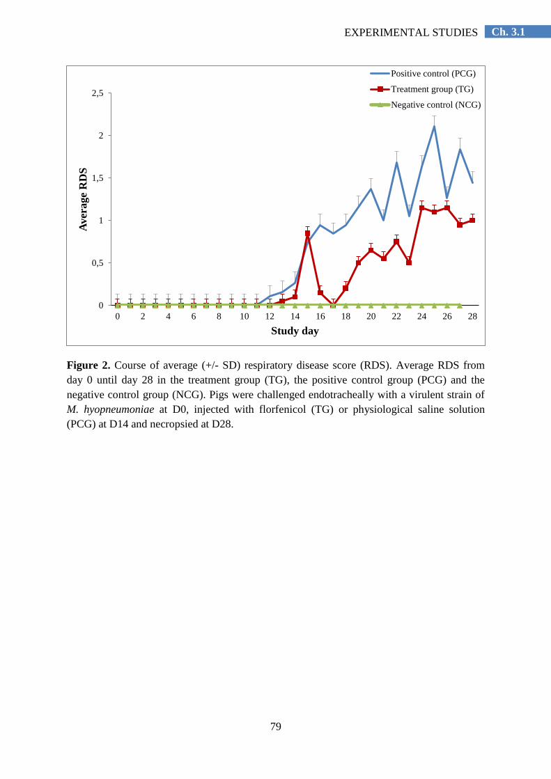
79
Ch. 3.1 EXPERIMENTAL STUDIES
Figure 2. Course of average (+/- SD) respiratory disease score (RDS). Average RDS from
day 0 until day 28 in the treatment group (TG), the positive control group (PCG) and the
negative control group (NCG). Pigs were challenged endotracheally with a virulent strain of
M. hyopneumoniae at D0, injected with florfenicol (TG) or physiological saline solution
(PCG) at D14 and necropsied at D28.
0
0,5
1
1,5
2
2,5
0 2 4 6 8 10 12 14 16 18 20 22 24 26 28
Aver
age
RD
S
Study day
Positive control (PCG)
Treatment group (TG)
Negative control (NCG)

80
Ch. 3.1 EXPERIMENTAL STUDIES
Table 1. Results (average ± standard deviation) of the clinical, pathological, performance,
infection and serological parameters in the treatment group (TG) and the positive control
group (PCG). Pigs were challenged endotracheally with a virulent strain of M.
hyopneumoniae at D0, injected with florfenicol (TG) or physiological saline solution (PCG)
at D14, and necropsied at D28.
TG
n = 20
PCG
n = 20 P value
Rectal Temperature (°C) 39.80 ± 0.30 39.86 ± 0.24 0.503
RDS D0 to D28
8.69 ± 7.90 17.16 ± 12.35 0.024
RDS D14 to D28 8.72 ± 7.94 17.10 ± 12.26 0.015
ADG D0 to D28 (g/day)
220 ± 88 212 ± 94 0.768
ADG D14 to D28 (g/day) 341 ± 120 275 ± 127 0.100
FCR 1.69 2.03 -
Mortality Rate (%) 0.00 10.00 0.232
Macroscopic LLS
4.51 ± 3.87 5.94 ± 3.47 0.232
Microscopic LLS 3.85 ± 0.65 3.72 ± 0.67 0.540
Percentage Air Analysis 21.94 ± 6.28 22.20 ± 5.31 0.892
IF
0.92 ± 0.73 0.95 ± 0.65 0.892
qPCR testing: Log copies of M.
hyopneumoniae/ml (mean±sd) 4.91 + 5.96 4.74 + 4.92 0.952
% of pigs serologically positive for
M. hyopneumoniae 14.3% 26.3% 0.290
Rectal Temperature (average from D0 to D28); RDS (average Respiratory Disease Score; ADG (Average Daily Weight
Gain); FCR (Feed Conversion Ratio: no statistical analysis, since FCR has been calculated at pen level, with only one
observation); LLS (Lung Lesion Score); Microscopic LLS (peribronchiolar and perivascular lymphohistiocytic infiltration
and nodule formation); IF (Immunofluorescence); qPCR (quantitative PCR: number of M.hyopneumoniae-organisms); per
cent of pigs serologically positive for M. hyopneumoniae at D28.
81
Ch. 3.1 EXPERIMENTAL STUDIES
Discusion
The present study assessed the efficacy of a florfenicol formulation administered
intramuscularly for the treatment of respiratory disease caused by an experimental M.
hyopneumoniae infection.
According to the RDS, onset of clinical disease took place between 12 and 15 days
after endotracheal inoculation and the decision to start the treatment was made on D14. The
results showed that the treatment was efficacious only for improving clinical respiratory
symptoms. The RDS is indeed an important indicator of the severity of M. hyopneumoniae
infections (Vicca et al., 2003), and hence to assess the clinical effectiveness of the treatment.
The injection of florfenicol significantly decreased the RDS in TG, especially on days 16 and
17. However, this score increased again on day 18, i.e. four days post-treatment.
Also, at necropsy (D28 or 29) only a numerical benefit was observed for the
macroscopic and the histopathological lung lesions. The percentage of air in the lung tissue
and the qPCR results were very similar for the TG and PCG (P>0.05). This suggests that the
studied parameters may be delayed shortly after treatment, but 14 days later, no significant
differences can be found between the groups. Liu et al. (2003) reported a long duration of the
therapeutic plasma level of florfenicol in pigs experimentally infected with Actinobacillus
pleuropneumoniae. Our findings suggest that a single injection with this antibiotic only
partially controls M. hyopneumoniae infection during a period of approximately four days.
All infected pigs were positive by nPCR in the BAL fluid, indicating that the challenge
infection was successful and that the treatment did not eliminate M. hyopneumoniae from the
lungs. The latter confirms previous reports, stating that antimicrobial treatment does not
eliminate the pathogen from the lung tissue nor heal existing lesions (Thacker, 2006).
Antimicrobial agents may delay infection, but cannot avoid colonization, nor assure total
elimination of M. hyopneumoniae.
However, M. hyopneumoniae could not be isolated from the lungs because of
overgrowth of other bacterial species, particularly Bordetella bronchiseptica, Haemophilus
parasuis and Streptococcus suis which were isolated in high numbers. All animals were
serologically negative for PRRSV (D28).
The performance parameters (ADG, FCR) and mortality were numerically improved in
the treated group. More animals would have been needed to obtain statistically significant
results, but the tendencies indicate that an economical improvement could be obtained when
florfenicol is injected.

82
Ch. 3.1 EXPERIMENTAL STUDIES
The present study showed that the tested florfenicol formulation, when administered
intramuscularly as a single dose of 30 mg/kg bodyweight, was effective in reducing the
clinical respiratory symptoms in pigs experimentally infected with a highly virulent M.
hyopneumoniae strain. Despite these findings, optimizing housing and management practices,
as well as vaccination should still be the key processes used in the control of M.
hyopneumoniae infections.
Conflict of interest statement
The study was funded by MSD Animal Health, manufacturer of the antimicrobial used
in this study. J. Thiry and E. Thomas are employees of MSD Animal Health.
Acknowledgements
This research was financially supported by MSD Animal Health. The authors thank
Hanne Vereecke for technical assistance in the laboratory work and Eva Zschiesche for the
statistical analyses.

83
Ch. 3.1 EXPERIMENTAL STUDIES
References
Dewulf, J., Catry, B., Timmerman, T., Opsomer, G., de Kruif, A. and Maes, D. (2007). Tetracycline-resistance
in lactose-positive enteric coliforms originating from Belgian fattening pigs: degree of resistance, multiple
resistance and risk factors. Preventive Veterinary Medicine 78, 339-351.
Friis, N.F. (1975). Some recommendations concerning primary isolation of Mycoplasma suipneumoniae and
Mycoplasma flocculare a survey. Nordisk Veterinaermedicin 27, 337-339.
Halbur, P., Paul, P., Meng, X., Lum, M., Andrews, J. and Rathje, J. (1996). Comparative pathogenicity of nine
US Porcine Reproductive and Respiratory Syndrome Virus (PRRSV) isolates in a five-week-old cesarean-
derived, colostrum-deprived pig model. Journal of Veterinary Diagnostic Investigation 8, 11-20.
Hannan, P., Bhogal, B. and Fish, J. (1982). Tylosin tartrate and tiamutilin effects on experimental piglet
pneumonia induced with pneumonic pig lung homogenate containing mycoplasmas, bacteria and viruses.
Research in Veterinary Science 33, 76-88.
Kobisch, M., Tillon, J.P., Vannier, P., Magueur, S. and Morvan, P. (1978). Pneumonie enzootique à
Mycoplasma suipneumoniae chez le porc: diagnostic rapide et recherche d‟anticorps. Récueil de Médecine
Vétérinaire 154, 847-852.
Liu, J., Fung, K.F., Chen, Z., Zeng, Z. and Zhang, J. (2003). Pharmacokinetics of florfenicol in healthy pigs and
in pigs experimentally infected with Actinobacillus pleuropneumoniae. Antimicrobial Agents and
Chemotherapy 47, 820-823.
Lobell, R.D., Varma, K.J., Johnson, J.C., Sams, R.A., Gerken, D.F. and Ashcraft, S.M. (1994).
Pharmacokinetics of florfenicol following intravenous and intramuscular doses to cattle. Journal of
Veterinary Pharmacology and Therapeutics 17, 253-258.
Maes, D., Segales, J., Meyns, T., Sibila, M., Pieters, M. and Haesebrouck, F. (2008). Control of Mycoplasma
hyopneumoniae infections in pigs. Veterinary Microbiology 126, 297-309.
Marois, C., Dory, D., Fablet, C., Madec, F. and Kobisch, M. (2010). Development of a quantitative Real-Time
TaqMan PCR assay for determination of the minimal dose of Mycoplasma hyopneumoniae strain 116
required to induce pneumonia in SPF pigs. Journal of Applied Microbiology 108, 1523-1533.
Morris, C., Gardner, I., Hietala, S., Carpenter, T., Anderson, R. and Parker, K. (1995). Seroepidemiologic study
of natural transmission of Mycoplasma hyopneumoniae in a swine herd. Preventive Veterinary Medicine 21,
323-337.
Quinn, P., Carter, M., Markey B. and Carter, G. (1994). Bacterial Pathogens: Microscopy, culture and
identification. In: Quinn, P.J. (Ed.). Clinical Veterinary Microbiology. Mosby, Edinburgh, UK, pp 21–66.
Shin, S.J., Kang, S.G., Nabin, R., Kang, M.L. and Yoo, H.S. (2005). Evaluation of the antimicrobial activity of
florfenicol against bacteria isolated from bovine and porcine respiratory disease. Veterinary Microbiology
106, 73-77.
Stärk, K.D., Nicolet, J. and Frey, J. (1998). Detection of Mycoplasma hyopneumoniae by air sampling with a
nested PCR assay. Applied and Environmental Microbiology 64, 543-548.
Thacker, E.L., 2006. Mycoplasmal diseases. In: Straw, B.E., Zimmerman, J.J., D‟Allaire, S., Taylor, D.J. (Eds.).
Diseases of Swine. Blacwell Publishing Ltd., Oxford, UK, pp. 701–717.
Vicca, J., Stakenborg, T., Maes, D., Butaye, P., Peeters, J., de Kruif, A. and Haesebrouck, F. (2003). Evaluation
of virulence of Mycoplasma hyopneumoniae field isolates. Veterinary Microbiology 97, 177-190.

Ch. 3.2 EXPERIMENTAL STUDIES
3.2. Efficacy of early Mycoplasma hyopneumoniae vaccination against
mixed respiratory disease in older fattening pigs
R. del Pozo Sacristána, A. Sierens
a, S.B. Marchioro
b, F. Vangroenweghe
c, J. Jourquin
c, G.
Labarquec, F. Haesebrouck
b, D. Maes
a
aUnit of Porcine Health Management, Department of Reproduction, Obstetrics and Herd
Health, Faculty of Veterinary Medicine, Ghent University, Salisburylaan 133, 9820,
Merelbeke, Belgium
bDepartment of Pathology, Bacteriology and Avian Diseases, Faculty of Veterinary
Medicine, Ghent University, Salisburylaan 133, 9820, Merelbeke, Belgium
cElanco Animal Health, Plantin en Moretuslei 1A, 2018, Antwerpen, Belgium
The Veterinary Record (2014), volume 174, issue 8, p. 197.

86
Ch. 3.2 EXPERIMENTAL STUDIES
Abstract
The present field study investigated the efficacy of early Mycoplasma hyopneumoniae
(M. hyopneumoniae) vaccination in a farrow-to-finish pig herd with respiratory disease late in
the fattening period due to combined infections with M. hyopneumoniae and viral pathogens.
Five-hundred-and-forty piglets were randomly divided into three groups of 180 piglets each:
two groups were vaccinated (Stellamune Once®) at either 7 (V1) or 21 days of age (V2), and
a third group was left non-vaccinated (NV). The three treatment groups were housed in
different pens within the same compartment during the nursery period, and were housed in
different but identical compartments during the fattening period. The efficacy was evaluated
using performance and pneumonia lesions. The average daily weight gain during the
fattening period was 19 (V1) and 18 g/day (V2) higher in both vaccinated groups when
compared with the NV group. However, the difference was not statistically significant
(P>0.05). The prevalence of pneumonia was significantly lower in both vaccinated groups
(V1:71.5 and V2:67.1 per cent) when compared with the NV group (80.2 per cent) (P<0.05).
There were no significant differences between the two vaccination groups. In conclusion, in
the present herd with respiratory disease during the second half of the fattening period caused
by M. hyopneumoniae and viral infections, prevalence of pneumonia lesions were
significantly reduced and growth losses numerically (not statistically significant) decreased
by both vaccination schedules.
Keywords: Mycoplasma hyopneumoniae; pig; early vaccination; one-shot
87
Ch. 3.2 EXPERIMENTAL STUDIES
Introduction
Mycoplasma hyopneumoniae (M. hyopneumoniae) is the causative agent of enzootic
pneumonia in pigs and is one of the primary agents involved in the porcine respiratory
disease complex (PRDC). This disease is distributed worldwide and leads to a significant
reduction of economical profit in pig production, mainly due to decreased growth, higher
feed conversion ratios and higher costs for treatments (Maes et al., 2008; Thacker and Minion
2012). Control of M. hyopneumoniae infections can be accomplished by improving
management and housing practices, by the use of antimicrobials and by vaccination (Maes et
al., 2008). Vaccination is frequently practiced worldwide. The major advantages of
vaccination include a reduction of the losses of average daily gain (ADG; 2-8 per cent), feed
conversion ratio (2-5 per cent) and mortality (Maes et al., 2008). Additionally, shorter time to
reach slaughter weight, reduced clinical signs and lung lesions and lower treatments costs are
observed (Maes et al., 2008).
Different vaccination strategies can be implemented, depending on the type of herd, the
production system and management practices, the infection pattern and the preferences of the
pig producer. Currently, one-shot vaccination is more often practiced than two-shots
vaccination, mainly because it requires less labor, it confers similar results than two-shots
vaccination (Roof et al., 2001, 2002; Alexopoulos et al., 2004; Lillie et al., 2004; Greiner et
al., 2011), and it can be implemented more easily in routine management practices on the
farm. Because M. hyopneumoniae infections can take place in piglets already during the first
weeks of life (Calsamiglia and Pijoan 2000; Ruiz et al., 2003; Fano et al., 2007; Sibila et al.,
2007; Nathues et al., 2010; Villarreal et al., 2010, 2011b; Vranckx et al., 2012a; Fablet et al.,
2012a), and because vaccination is most likely effective if active immunity can be established
before the exposure to the pathogen, vaccination is commonly applied in suckling piglets.
When compared with weaned piglets, suckling piglets are less infected with pathogens, such
as porcine reproductive and respiratory syndrome virus (PRRSV) and porcine circovirus type
2 (PCV-2), which may cause problems after weaning and interfere with the establishment of
a protective immune response to M. hyopneumoniae vaccination (Maes et al., 2008). Early
vaccination has also the advantage that immunity is induced at a young age. This may be
important as the onset of infection may vary between herds and also within a herd among
successive batches (Sibila et al., 2004b, 2007; Fano et al., 2007; Segalés et al., 2012). Apart
from inducing early protection, it is also important that early vaccinated pigs remain

88
Ch. 3.2 EXPERIMENTAL STUDIES
protected until the end of the fattening period, as in most pig herds, the highest infection
levels of M. hyopneumoniae occur during the grow-finishing period (Sibila et al., 2004c).
Several studies have assessed the efficacy of vaccination at seven days of age against
M. hyopneumoniae challenge infection under experimental conditions. This early vaccination
at seven days of age has been demonstrated effective in reducing lung lesions and/or clinical
signs in several challenge infections either at two (Reynolds et al., 2006), four (Reynolds et
al., 2009, Villarreal et al., 2011a), six (Meyns et al., 2006), eight (Villarreal et al., 2011a) or
nineteen (Kim et al., 2011) weeks post-infection. However, the duration of the effect of one-
shot early vaccination under field conditions with mixed respiratory disease is less clear.
Mixed respiratory disease is commonly identified as PRDC and results from infections with
several primary and secondary respiratory pathogens, such as PRRSV, PCV-2, swine
influenza virus (SIV), M. hyopneumoniae, Pasteurella multocida, Bordetella bronchiseptica,
Actinobacillus pleuropneumoniae and Streptococcus suis (Opriessnig et al., 2011). As the
disease course and lung lesions can be largely influenced by viral respiratory pathogens such
as PRRSV (Thacker et al., 1999), PCV-2 (Opriessnig et al., 2004) and swine influenza virus
(Thacker et al., 2001), it is important to investigate whether M. hyopneumoniae vaccination is
also beneficial under such circumstances. The objective of this study was to investigate the
efficacy of a one-shot vaccination applied at either one or three weeks of age in a Belgian
farrow-to-finish pig herd with mixed respiratory disease late in the fattening period and
including infections with M. hyopneumoniae and viral pathogens.

89
Ch. 3.2 EXPERIMENTAL STUDIES
Materials and methods
Herd description and study population
The study was conducted between April and October 2011 in a Belgian farrow-to-finish
pig herd comprising 1000 commercial hybrid sows (Topigs 20) that operated a four-week
batch production system. Semen from Piétrain boars was used for artificial insemination. The
sows were vaccinated against swine influenza virus (Gripovac 3®, Merial), Atrophic Rhinitis
(Rhiniffa–T®, Merial), Escherichia coli (E. coli) (Neocolipor®, Merial), porcine parvovirus
and Erysipelothrix rhusiopathiae, combined according to the manufacturer´s instructions
(Parvoruvax®, Merial). The piglets received an iron injection (Uniferon®, Pharmacosmos)
(intramuscular, 1 ml/pig) and toltrazuril per os (Baycox®, Bayer; 20 mg/kg), both according
to the manufacturer´s instructions, at three days of age, and they were surgically castrated
during the first week of life. Toltrazuril was administered as preventive treatment for
coccidiosis. At weaning (three weeks of age), the pigs were moved to a nursery unit where
they stayed until 10 weeks of age. Thereafter, they were transferred to a fattening unit, in
which they were kept until slaughter weight (approximately 28 weeks of age). Colistin in-
feed medication (Promycine 400®, VMD) was prophylactically administered (according to
the manufacturer´s instructions) after weaning for seven days to prevent problems with post-
weaning E. coli infections. All pigs received anthelmintic treatment at 10 weeks of age via
the feed with flubendazole (Flubenol 5 %®, Elanco Animal Health) for five days.
At weaning, all piglets included in the experiment were allocated into one compartment
of the nursery (n=576 pigs). This compartment consisted of 32 pens (18 pigs per pen) with a
partially slatted floor and there was mechanical channel ventilation. At 10 weeks of age, all
study piglets were moved to six identical compartments of a fattening unit. The three
different treatment groups were housed in different compartments (two compartments for
each treatment group). Each compartment consisted of eight pens (12 pigs per pen) with a
fully slatted floor and there was mechanical door ventilation. The nursery unit and the
fattening unit were equipped with an automatic ventilation system controlled by computer.
During the entire experiment, water and feed (meal) were supplied ad libitum.
Before the start of the study, clinical signs of respiratory disease, namely coughing and
sneezing, were evident in nursery piglets from six-to-seven weeks of age onwards. Limited
sampling using nasal swabs in 20 two-week-old piglets and broncho-alveolar lavage (BAL)
fluids in 20 six-week-old piglets and 10 ten-week-old piglets showed that 0/20, 0/20 and 5/10
piglets, respectively, were positive for M. hyopneumoniae by nested polymerase chain

90
Ch. 3.2 EXPERIMENTAL STUDIES
reaction (PCR) (nPCR) (Stärk et al., 1998). Blood samples taken from 20 randomly selected
pigs at 19 weeks of age revealed that 75 per cent of the pigs were serologically positive for
M. hyopneumoniae and 100 per cent for PRRSV using a blocking Enzyme-Linked
Immunosorbent Assay (ELISA) (IDEI, Mycoplasma hyopneumoniae EIA kit, Oxoid, UK) for
M. hyopneumoniae and the HerdChek PRRS X3 (IDEXX) for PRRSV. Gross examination of
the lungs from fattening pigs (n=826) slaughtered during 12 months before the start of the
study indicated an average pneumonia prevalence of 35 per cent.
Experimental design
In total, 540 pigs originating from one batch of sows were selected for this study.
Although the original group comprised 250 sows, only the offspring of 60 sows was included
in the trial. These 60 sows were selected at random from the group of 250 sows to obtain a
representative sample of sows (all parities represented) and piglets. The pigs were
individually ear-tagged and randomly allocated to one of the three treatment groups. Within
each litter (n=60), an equal number of pigs (n=3) was allocated to each treatment group
(blocked randomization). Therefore, in total, each treatment group consisted of 180 pigs. One
group was vaccinated intramuscularly at 7 days of age with two ml of a commercial
inactivated M. hyopneumoniae vaccine (Stellamune Once®, Elanco Animal Health)
(designated group V1), a second group was vaccinated intramuscularly at 21 days of age with
the same vaccine (designated group V2), a third group was left unvaccinated (designated
group NV). Non-vaccinated animals were not injected with a placebo. Upon weaning, piglets
were moved to a nursery unit, where they were partly re-grouped according to pen-size,
weight and sex. Pigs of the different treatment groups were housed in different pens within
the same compartment. The stocking density in the nursery unit was 0.27 m2 per pig. Pens
were separated by solid pen partitions. As the capacity of the nursery unit (576) was slightly
higher than the number of study pigs (540), two pens (36 pigs) were not included in the trial.
Twelve of these pigs had been vaccinated at one week, twelve at three weeks, and twelve had
not been vaccinated.
The pigs were moved to a fattening unit at approximately 70 days of age. At that time,
they were partly re-grouped according to pen size and weight. Barrow and gilts were housed
in different pens, and pigs of different treatment groups were housed in different
compartments (two compartments for each treatment group). The 36 extra pigs were divided
in three pens, one in each treatment group. They were not included in the study as the
approval for the ethical committee before the study included 540 pigs. The stocking density
91
Ch. 3.2 EXPERIMENTAL STUDIES
in the fattening unit was 0.75 m2 per pig. Fattening pigs of neighbouring pens could have
direct nose-to-nose contact. All other factors that could possibly influence the outcome
parameters e.g. environment and management factors, sex ratio, and feed composition were
the same for the three groups. The person responsible for assessing the efficacy parameters
was unaware of the allocation of the groups. All animals were housed and reared in
accordance with EU animal welfare regulations and in accordance with EU experimental
animal legislation (Directive 86/609/EEC). The study was approved by the ethical committee
for animal experiments of the Faculty of Veterinary Medicine, Ghent University
(EC2011/068).
Parameters of comparison
Performance parameters
Pigs were individually weighed at one week of age (7 days), at weaning (21 days), at
the end of the nursery period (74 days) and prior to slaughter (200 days) to determine the
average daily gain (ADG). The ADG (g/pig/day) was computed for the different production
stages (lactation, nursery and fattening period) as the difference between starting and final
weights divided by the number of days during that period.
Clinical and pathological parameters
All pigs that died during the study were necropsied to assess the possible cause of
death. Where appropriate, necropsied pigs were processed for further laboratory
examinations. When the dead animals were suspected to be infected with M. hyopneumoniae,
nPCR testing (Stärk et al., 1998) was performed on the lungs. DNA was extracted using the
DNA easy kit (Quiagen, Blood and tissue kit, Belgium).
When individual signs of respiratory disease (coughing, sneezing, dyspnea, tachypnea)
were observed by the farmer and confirmed by the veterinarian, individual treatment against
respiratory disease was allowed. Concomitant antimicrobial treatment of study pigs
(parenterally or orally) was only allowed with antimicrobials ineffective against M.
hyopneumoniae, such as β-lactam antibiotics (penicillins and cephalosporins). For parenteral
treatment, ceftiofur (Excenel RTU®, Pfizer Animal Health) or amoxicillin (Duphamox Long
Acting®, Pfizer Animal Health) could be used, for group treatment, amoxicillin applied via
the drinking water (Amoxicilline 70 %®, Kela Laboratoria) could be used. The total number

92
Ch. 3.2 EXPERIMENTAL STUDIES
of treatment days against respiratory disease (based on individual or group treatment) was
recorded in the different treatment groups.
At slaughter (201-214 days of age), the prevalence of Mycoplasma-like pneumonia
lesions and adhesive pleurisy and the extent of macroscopically visible pneumonia were
recorded by the first author in a blinded manner. Mycoplasma-like pneumonia lesions were
defined as red to purplish consolidated areas on the cranio-ventral portions of the lungs in a
lobular pattern. Adhesive pleurisy was defined as fibrotic adherence between the visceral and
parietal membranes of the pleural sac. Due to practical reasons, only the visceral membrane
was evaluated for the detection of pleurisy. The extent of macroscopically visible pneumonia
was quantified based on a score described by Morrison et al. (1985). Briefly, this lung lesion
score expresses the total area (percentage) of the lung tissue affected by pneumonia. Each
lobe represents a percentage of the total lung surface area: apical (10 per cent), cardiac (7 per
cent), accessory (6 per cent) and diaphragmatic (30 per cent). An average score was
calculated for each group based on the lung lesion score of the individual animals.
Detection of M. hyopneumoniae by nested PCR (nPCR)
Nasal swabs, tracheo-bronchial swabs (Marois et al., 2007; Fablet et al., 2010) and
broncho-alveolar lavage (BAL) fluids (Villarreal et al., 2011b) were collected from
approximately 30 randomly selected animals per group that were serially sampled at different
ages (Table 1). Nasal swabs (Copan®, Italy) were collected at weaning (21 days), halfway
through the nursery period (49 days), at the end of the nursery period (74 days), halfway
through the fattening period (136 days) and prior to slaughter (200 days). Tracheo-bronchial
swabs (Euromedis®, Neuilly-sous-Clermont, France) were collected at the same time points
as the ones for nasal swabs, but not at 200 days of age. BAL fluids were collected at 49, 74
and 136 days of age. DNA was extracted with the DNA easy kit (Quiagen, Blood and tissue
kit, Belgium) and a nPCR was performed (Stärk et al., 1998).
Serological examinations
Blood samples were collected from the same 30 animals per group (Table 1) at 7, 21,
74, 136, and 200 days of age. Serological examination to detect antibodies to M.
hyopneumoniae was carried out using a blocking ELISA (IDEI, Mycoplasma hyopneumoniae
EIA kit, Oxoid, UK). Sera with optical density (OD) values <50 per cent of the ODbuffer-control
were considered as positive. Sera with OD values ≥50 per cent of the ODbuffer-control were
considered as negative. Fifteen blood samples were randomly selected from the 30 blood

93
Ch. 3.2 EXPERIMENTAL STUDIES
samples collected at 74, 136, and 200 days of age and additionally tested for antibodies to
PRRSV (HerdChek PRRS X3, IDEXX), PCV-2 (Ingezim Circovirus IgG/IgM, Ingenasa),
subtypes H1N1, H1N2, and H3N2 of swine influenza virus (SIV) (standard
haemagglutination-inhibition test), and serotype 2 (Swinecheck APP 2, Biovet) and serotypes
1, 9 or 11 (Swinecheck APP 1,9,11, Biovet) of Actinobacillus pleuropneumoniae.
Carcase quality
At slaughter, carcase quality, including the percentage of meat, back fat thickness and
muscle thickness of the Longissimus dorsi, was automatically recorded by the abattoir
services.
Statistical analysis
The number of animals in each treatment group (180) allowed to assess a difference of
20 g (sd=70) in ADG with 95 per cent certainty and 80 per cent statistical power (Win
Episcope 2.0, CLIVE, Edinburgh, UK). The pig was considered as statistical unit. To correct
for the effects of hierarchical data, pen and compartment were included as random effects.
Analysis of variance (ANOVA) models were used to analyse the continuous variables
(weight, number of treatment days against respiratory disease, carcase quality parameters).
Some other continuous variables (ADG and lung lesion scores) did not fulfill the criteria of
normality and homogeneity of variances, and they were analysed using a non-parametric
Kruskal-Wallis ANOVA. Mortality and prevalence of pneumonia and pleurisy were analysed
using logistic regression analysis. Differences were considered as statistically significant
when P-values were lower than 0.05 (two-sided test). Statistical analyses were performed
using the software package SPSS version 20.0 (SPSS Institute Inc., Illinois, USA). Results
are presented as mean ± sd unless stated otherwise.
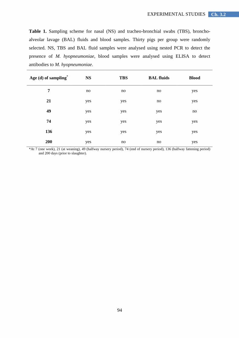
94
Ch. 3.2 EXPERIMENTAL STUDIES
Table 1. Sampling scheme for nasal (NS) and tracheo-bronchial swabs (TBS), broncho-
alveolar lavage (BAL) fluids and blood samples. Thirty pigs per group were randomly
selected. NS, TBS and BAL fluid samples were analysed using nested PCR to detect the
presence of M. hyopneumoniae, blood samples were analysed using ELISA to detect
antibodies to M. hyopneumoniae.
Age (d) of sampling* NS TBS BAL fluids Blood
7 no no no yes
21 yes yes no yes
49 yes yes yes no
74 yes yes yes yes
136 yes yes yes yes
200 yes no no yes
*At 7 (one week), 21 (at weaning), 49 (halfway nursery period), 74 (end of nursery period), 136 (halfway fattening period)
and 200 days (prior to slaughter).
95
Ch. 3.2 EXPERIMENTAL STUDIES
Results
Performance parameters
The weights of the pigs at 7, 21, 74, and 200 days of age are presented in Table 2. On
average, pigs of the V1 and V2 groups gained 2.3 kg (group V2) to 2.4 kg (group V1) more
than pigs in the NV group between 7 and 200 days of age. The ADG during the lactation
period (7 to 21 days), the nursery period (21 to 74 days), the fattening period (74 to 200
days), and during the entire study period (7 to 200 days) are presented in Table 3. Over the
entire fattening period, the ADG in the V1 and V2 group was 19 and 18 g/pig/day,
respectively, higher than in the NV group. These differences were not statistically significant
(P>0.05).
Clinical and pathological parameters
None of the pigs died between the first and second vaccination. In total, 15 pigs (2.8 per
cent) died from 21 days of age until slaughter. Four of them died following either sampling or
weighing (1 in the V1 group, 3 in the NV group). The other dead animals occurred as follows
in the three treatments groups: V1 (n=4; 2.2 per cent), V2 (n=1; 0.6 per cent), and NV (n=6;
3.3 per cent) (P>0.05). Pneumonia was the major post-mortem finding in five out of 15
necropsied pigs (1 in the V2 group, 4 in the NV group). Mycoplasma hyopneumoniae was not
detected by nPCR in any of these five lungs.
Sporadic non-productive coughing was observed at approximately 151 days of age.
Pigs showing clinical respiratory disease signs were treated parenterally with long-acting
amoxicillin or ceftiofur (V1=13; V2=8; NV=9). At 161 days of age, respiratory disease was
observed in many pigs of the three groups. The three groups were more or less similarly
affected. Therefore, all pigs of the study were treated for five days with amoxicillin via the
drinking water. The total numbers of days (±sd) of additional antibiotic treatment were 5.06 ±
0.28 (V1), 5.02 ± 0.13 (V2) and 5.03 ± 0.26 (NV) (P>0.05).
At slaughter, the prevalence of Mycoplasma-like pneumonia lesions and pleurisy and
the extent of macroscopically visible pneumonia were recorded in 505 pigs (V1: 165; V2:
173; NV: 167). Because of practical reasons (pigs with lost ear tags and/or lungs that did not
reach the examination stand at the slaughterhouse), a few plucks could not be scored in the
slaughterhouse (10, 6 and 4 in V1, V2 and NV, respectively).
The percentage of pigs with pneumonia was significantly lower in both vaccinated
groups V1 (118/165 pigs; 71.5 per cent) and V2 (116/173 pigs; 67.1 per cent) when

96
Ch. 3.2 EXPERIMENTAL STUDIES
compared with the NV group (134/167 pigs; 80.2 per cent) pigs (P<0.05). The prevalence of
pleurisy was also lower in both vaccinated groups (V1, 7.6 per cent; V2, 11.6 per cent) when
compared with NV group (12.3 per cent), however it was not statistically significant
(P>0.05). The average pneumonia scores in the V1, V2, and NV group were 13.0, 9.8, and
14.4, respectively (P<0.05) (Table 2). In accordance with this score, 51 per cent of the pigs
presented mild pneumonia lesions characterized by an extent lower than 10 per cent of the
overall lung surface, which is indicative of a recent (late in fattening period) and mild
respiratory disease.
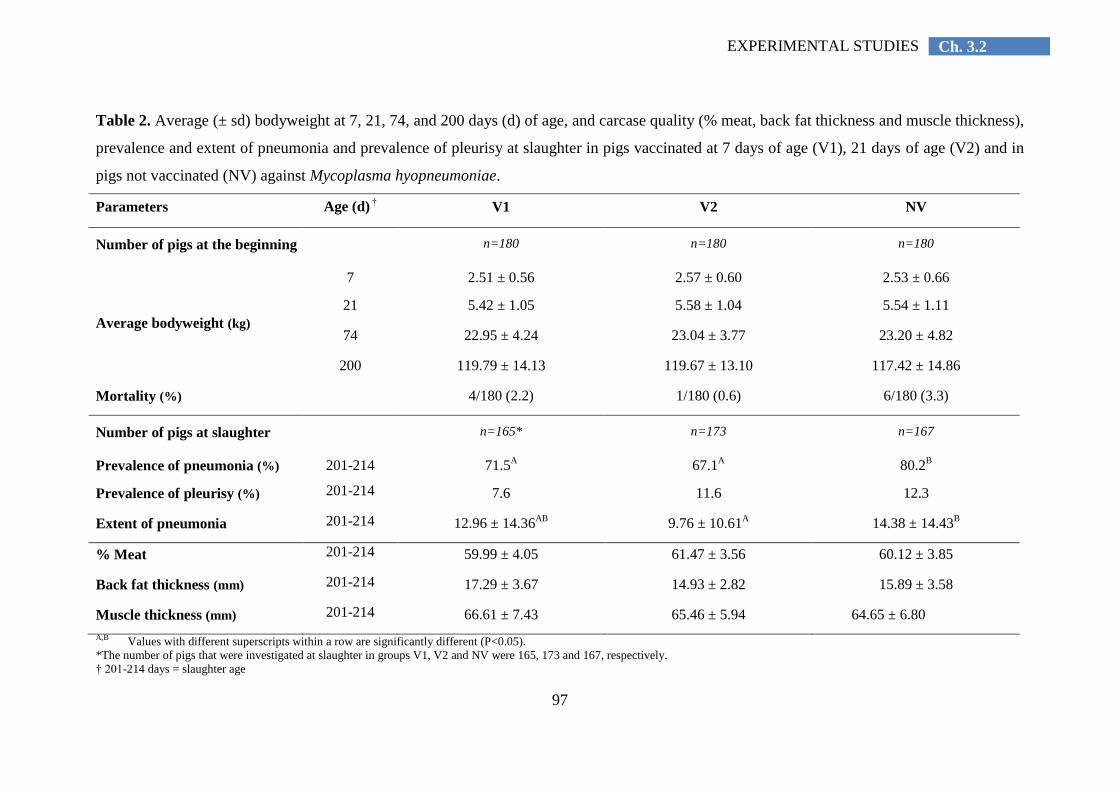
97
Ch. 3.2 EXPERIMENTAL STUDIES
Table 2. Average (± sd) bodyweight at 7, 21, 74, and 200 days (d) of age, and carcase quality (% meat, back fat thickness and muscle thickness),
prevalence and extent of pneumonia and prevalence of pleurisy at slaughter in pigs vaccinated at 7 days of age (V1), 21 days of age (V2) and in
pigs not vaccinated (NV) against Mycoplasma hyopneumoniae.
Parameters Age (d) † V1 V2 NV
Number of pigs at the beginning n=180 n=180 n=180
Average bodyweight (kg)
7 2.51 ± 0.56 2.57 ± 0.60 2.53 ± 0.66
21 5.42 ± 1.05 5.58 ± 1.04 5.54 ± 1.11
74 22.95 ± 4.24 23.04 ± 3.77 23.20 ± 4.82
200 119.79 ± 14.13 119.67 ± 13.10 117.42 ± 14.86
Mortality (%) 4/180 (2.2) 1/180 (0.6) 6/180 (3.3)
Number of pigs at slaughter n=165* n=173 n=167
Prevalence of pneumonia (%) 201-214 71.5A 67.1
A 80.2
B
Prevalence of pleurisy (%) 201-214 7.6 11.6 12.3
Extent of pneumonia 201-214 12.96 ± 14.36AB
9.76 ± 10.61A 14.38 ± 14.43
B
% Meat 201-214 59.99 ± 4.05 61.47 ± 3.56 60.12 ± 3.85
Back fat thickness (mm) 201-214 17.29 ± 3.67 14.93 ± 2.82 15.89 ± 3.58
Muscle thickness (mm) 201-214 66.61 ± 7.43 65.46 ± 5.94 64.65 ± 6.80
A,B Values with different superscripts within a row are significantly different (P<0.05).
*The number of pigs that were investigated at slaughter in groups V1, V2 and NV were 165, 173 and 167, respectively.
† 201-214 days = slaughter age
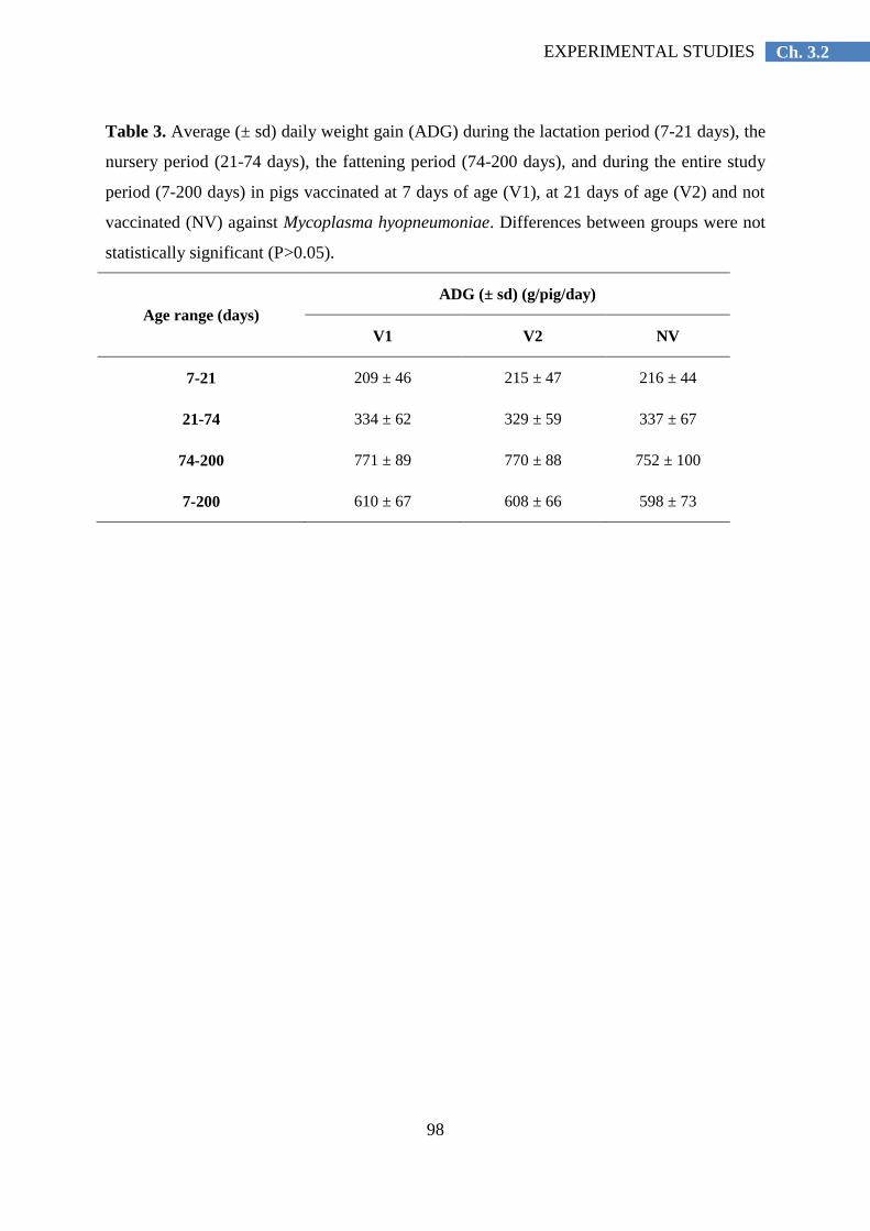
98
Ch. 3.2 EXPERIMENTAL STUDIES
Table 3. Average (± sd) daily weight gain (ADG) during the lactation period (7-21 days), the
nursery period (21-74 days), the fattening period (74-200 days), and during the entire study
period (7-200 days) in pigs vaccinated at 7 days of age (V1), at 21 days of age (V2) and not
vaccinated (NV) against Mycoplasma hyopneumoniae. Differences between groups were not
statistically significant (P>0.05).
Age range (days)
ADG (± sd) (g/pig/day)
V1 V2 NV
7-21 209 ± 46 215 ± 47 216 ± 44
21-74 334 ± 62 329 ± 59 337 ± 67
74-200 771 ± 89 770 ± 88 752 ± 100
7-200 610 ± 67 608 ± 66 598 ± 73

99
Ch. 3.2 EXPERIMENTAL STUDIES
Detection of M. hyopneumoniae by nested PCR (nPCR)
The numbers of nPCR-positive nasal swabs, tracheo-bronchial swabs and BAL fluids at
21, 49, 74, 136, and 200 days of age are presented in Table 4. All samples tested at 21 and 49
days of age were negative for M. hyopneumoniae. At 74 days of age, the percentage of M.
hyopneumoniae-positive tracheo-bronchial swabs and BAL fluids in the non-vaccinated (NV)
group was 7.7 per cent (2/26 pigs) and 4.0 per cent (1/25 pigs), respectively, whereas all
samples collected at that age in the vaccinated groups remained negative. At 136 days of age,
the percentage of M. hyopneumoniae-positive BAL fluids was 24.1 per cent (7/29 pigs), 20.0
per cent (5/25 pigs), and 30.4 per cent (7/23 pigs) in the V1, V2, and NV group, respectively.
At 200 days of age, the percentage of M. hyopneumoniae-positive nasal swabs was 14.3 per
cent (4/28 pigs), 8.7 per cent (2/23 pigs) and 23.1 per cent (6/26 pigs) in the V1, V2, and NV
group, respectively.
Serological examinations
The number of pigs seropositive for M. hyopneumoniae at 7, 21, 74, 136, and 200 days
of age is presented in Table 5. Briefly, the percentage of seropositive pigs remained low
during lactation (7 days: 13 per cent; 21 days: 14 per cent), decreased during the nursery
period (74 days: 2 per cent) and increased during the fattening period (136 days: 5 per cent;
200 days: 21 per cent).
The number of pigs seropositive for PRRS(V), PCV-2 (IgG and IgM), subtypes H1N1,
H1N2 and H3N2 of SIV, serotypes 1, 9 or 11 and serotype 2 of A. pleuropneumoniae at 74,
136, and 200 days of age is presented in Table 6.
Carcase quality
The results of the carcase quality are included in Table 2. There were no significant
differences between the different groups (P>0.05).

100
Ch. 3.2 EXPERIMENTAL STUDIES
Table 4. Results of nested polymerase chain reaction (nPCR) assays on nasal swabs (NS), tracheo-bronchial swabs (TBS) and broncho-alveolar
lavage fluids (BALF) at 21, 49, 74, 136, and 200 days (d) of age in pigs vaccinated at 7 days of age (V1), vaccinated at 21 days of age (V2) and
not vaccinated (NV) against Mycoplasma hyopneumoniae.
Age (d) of
pigs at
sampling
Number (%) of nPCR-positive pigs/total number of pigs sampled
V1 V2 NV
NS TBS BALF NS TBS BALF NS TBS BALF
21 0/30
(0.0%)
0/30
(0.0%) NA*
0/30
(0.0%)
0/30
(0.0%) NA
0/30
(0.0%)
0/30
(0.0%) NA
49 0/30
(0.0%)
0/30
(0.0%)
0/30
(0.0%)
0/30
(0.0%)
0/30
(0.0%)
0/30
(0.0%)
0/30
(0.0%)
0/30
(0.0%)
0/30
(0.0%)
74 0/29
(0.0%)
0/29
(0.0%)
0/29
(0.0%)
0/28
(0.0%)
0/28
(0.0%)
0/28
(0.0%)
0/26
(0.0%)
2/26
(7.7%)
1/25
(4.0%)
136 0/29
(0.0%)
4/29
(13.8%)
7/29
(24.1%)
0/26
(0.0%)
0/26
(0.0%)
5/25
(20.0%)
4/25
(16.0%)
7/25
(28.0%)
7/23
(30.4%)
200 4/28
(14.3%) NA NA
2/23
(8.7%) NA NA
6/26
(23.1%) NA NA
*NA: not applicable

101
Ch. 3.2 EXPERIMENTAL STUDIES
Table 5. Number (%) of pigs with serum antibodies to Mycoplasma hyopneumoniae at 7, 21, 74, 136, and 200 days (d) of age in pigs vaccinated
at 7 days of age (V1), vaccinated at 21 days of age (V2) and not vaccinated (NV) against Mycoplasma hyopneumoniae.
Age (d) of pigs at
sampling
Number (%) of seropositive pigs for M. hyopneumoniae/total number of pigs sampled
V1 V2 NV
7 5/30
(16.7%)
3/30
(10.0%)
4/30
(13.3%)
21 3/29
(10.3%)
5/29
(17.2%)
4/30
(13.3%)
74 2/29
(6.9%)
0/27
(0.0%)
0/30
(0.0%)
136 0/27
(0.0%)
2/24
(8.3%)
2/27
(7.4%)
200 8/26
(30.8%)
2/23
(8.7%)
6/27
(22.2%)
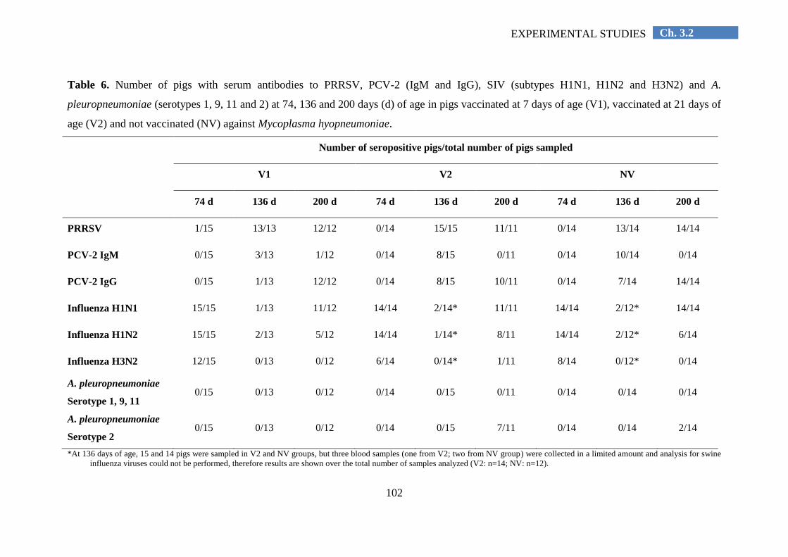
102
Ch. 3.2 EXPERIMENTAL STUDIES
Table 6. Number of pigs with serum antibodies to PRRSV, PCV-2 (IgM and IgG), SIV (subtypes H1N1, H1N2 and H3N2) and A.
pleuropneumoniae (serotypes 1, 9, 11 and 2) at 74, 136 and 200 days (d) of age in pigs vaccinated at 7 days of age (V1), vaccinated at 21 days of
age (V2) and not vaccinated (NV) against Mycoplasma hyopneumoniae.
Number of seropositive pigs/total number of pigs sampled
V1 V2 NV
74 d 136 d 200 d 74 d 136 d 200 d 74 d 136 d 200 d
PRRSV 1/15 13/13 12/12 0/14 15/15 11/11 0/14 13/14 14/14
PCV-2 IgM 0/15 3/13 1/12 0/14 8/15 0/11 0/14 10/14 0/14
PCV-2 IgG 0/15 1/13 12/12 0/14 8/15 10/11 0/14 7/14 14/14
Influenza H1N1 15/15 1/13 11/12 14/14 2/14* 11/11 14/14 2/12* 14/14
Influenza H1N2 15/15 2/13 5/12 14/14 1/14* 8/11 14/14 2/12* 6/14
Influenza H3N2 12/15 0/13 0/12 6/14 0/14* 1/11 8/14 0/12* 0/14
A. pleuropneumoniae
Serotype 1, 9, 11 0/15 0/13 0/12 0/14 0/15 0/11 0/14 0/14 0/14
A. pleuropneumoniae
Serotype 2 0/15 0/13 0/12 0/14 0/15 7/11 0/14 0/14 2/14
*At 136 days of age, 15 and 14 pigs were sampled in V2 and NV groups, but three blood samples (one from V2; two from NV group) were collected in a limited amount and analysis for swine
influenza viruses could not be performed, therefore results are shown over the total number of samples analyzed (V2: n=14; NV: n=12).
103
Ch. 3.2 EXPERIMENTAL STUDIES
Discussion
The present study demonstrated that a single vaccination against M. hyopneumoniae
applied at either 7 or 21 days of age reduced the prevalence and extent of pneumonia lesions
in a pig herd with clinical respiratory disease during the second half of the fattening period.
Numerical, but not statistically significant improvements of performance and reduction of the
prevalence of pleurisy lesions were observed.
The clinical respiratory disease signs occurred first in a few pigs at approximately 150
days of age, and one-to-two weeks later, more pronounced clinical signs were observed in
many pigs. Therefore, initially only individual treatments were applied, whereas in the
second stage, a group treatment with antimicrobials was initiated. Based on the diagnostic
testing, it is clear that infections with many different pathogens had occurred from
approximately halfway through the fattening period and onwards. At slaughter age, almost all
pigs were seropositive for PRRSV, PCV-2 and H1N1, and approximately half of them for
H1N2. Infections with M. hyopneumoniae were also involved, but the precise role of M.
hyopneumoniae infections in the present outbreak is difficult to assess. At day 136, 20-30 per
cent of the pigs were positive based on nPCR testing of BAL fluid, but the serological
prevalence at slaughter age was rather low namely approximately 10-30 per cent. Andreasen
et al. (2001) also found a low seroprevalence of M. hyopneumoniae close to slaughter in pigs
with high percentage of pneumonia at slaughter. This may be due to the long and variable
time (3-to-11 weeks) needed for seroconversion after infection with this pathogen (Morris et
al., 1995). A. pleuropneumoniae had likely not played a major role as most of the animals
remained seronegative at slaughter age, the mortality remained rather low and no typical
lesions were found in the dead animals at postmortem examination. The severity of the
clinical signs warranted an antimicrobial treatment, but in general, the signs were not very
severe and a short group treatment of five days was sufficient. However, there was a very
high percentage of pigs with pneumonia lesions (80 per cent in the NV group) at slaughter
age. This high percentage can likely be explained by the fact that multiple viral infections and
M. hyopneumoniae were involved, and that the infections mainly took place during the
second half the fattening period, allowing insufficient time for the lesions to be healed at
slaughter age (Blanchard et al., 1992). Pneumonia lesions at slaughter are not pathognomonic
for infections with M. hyopneumoniae. Apart from M. hyopneumoniae, it is well known that
infections with viruses and in particular swine influenza virus may cause similar pneumonia
lesions and may be involved in PRDC (Thacker et al., 2001; Sibila et al., 2009).

104
Ch. 3.2 EXPERIMENTAL STUDIES
The respiratory problems in the present batch were different from those in the previous
batches in this herd. They occurred later in the present batch, M. hyopneumoniae infections
were less prevalent and mainly occurred from halfway the fattening period and onwards
(serology and PCR on swabs and BAL fluid), and there were complicating viral infections
(serology). In previous batches, respiratory problems mainly occurred in the nursery and were
clearly related to M. hyopneumoniae infections. This confirms that the infection pattern of M.
hyopneumoniae is not constant over time within the same herd, and that important variations
may occur between successive batches (Fano et al., 2007; Sibila et al., 2007). Previous
studies showed a seasonal variation of M. hyopneumoniae infections, i.e. higher prevalence
and severity of lung lesions (Straw et al., 1986), higher seroprevalence to M. hyopneumoniae
(Maes et al., 2000), and higher probability of being M. hyopneumoniae-PCR positive
(Segalés et al., 2012) during winter period. The absence of a constant infection pattern, i.e.
infection in younger or older pigs depending on the batch, is one of the major reasons why
vaccination of piglets during the first weeks of life is commonly practiced worldwide. Using
this strategy, piglets are immunized before infection, irrespective of whether infection takes
place in the nursery, growing or fattening unit.
Both vaccination schedules (V1 and V2) led to a numerical, but not statistically
significant, increase of the ADG during the fattening period of 19 and 18 g/pig/day,
respectively, when compared with the NV pigs. The fact that the improvement was not
statistically significant may be due to the high standard variations (88-100 g/day), the fact
that the respiratory problems occurred only late in the fattening period, and that different viral
infections were involved in the outbreak. Similar improvements in ADG following M.
hyopneumoniae vaccination were obtained in a meta-analysis of 28 published field trials
(Jensen et al., 2002). In that study, the ADG in pigs vaccinated against M. hyopneumoniae
was on average 21 g higher when compared with non-vaccinated pigs. An ADG improvement
of 20 g is associated with an important economic profit (Maes et al., 2003). Additionally,
Pagot et al. (2007) described a negative association between growth during fattening period
and prevalence of pneumonia at slaughter. Our study results corroborate with these
observations: in a population with high prevalence of pneumonia (67 to 80 per cent),
vaccination results in an increase of 2.5 per cent of ADG, when compared with non-
vaccinated pigs.
Vaccination also significantly reduced the percentage of slaughter pigs with pneumonia
lesions, which is in agreement with previous studies using challenge infections more than five
105
Ch. 3.2 EXPERIMENTAL STUDIES
months after vaccination (Reynolds et al., 2009). Our data showed a low prevalence of
pleurisy (7-12 per cent) in the slaughter pigs of this herd, when compared with prevalence
figures (i.e. 15 to 60 per cent) obtained in several epidemiological studies carried out recently
in Europe (Fraile et al., 2010; Meyns et al., 2011; Fablet et al., 2012b; Merialdi et al., 2012).
This low prevalence of pleurisy may indicate a low impact on respiratory disease in this
study.
In addition, the percentage of pigs that tested positive with PCR was lower and positive
pigs were detected at a later stage in the vaccinated groups than in the control group,
illustrating that vaccination had decreased the infection level of M. hyopneumoniae in this
herd during the experiment. By using qPCR, Vranckx et al. (2012b) recently showed that
vaccination also significantly reduced the number of M. hyopneumoniae organisms in the
lungs of experimentally infected pigs. The fact that M. hyopneumoniae DNA could still be
detected by PCR confirms previous results from previous experimental and field transmission
studies with different M. hyopneumoniae vaccines (Meyns et al., 2006; Villarreal et al.,
2011b). These studies showed that vaccination does not prevent infection, and suggested that
vaccination alone is not able to eliminate M. hyopneumoniae from infected pig herds.
More M. hyopneumoniae-positive pigs were found using nPCR in both tracheo-
bronchial swabs and BAL fluids than in nasal swabs. This confirms previous reports (Kurth
et al., 2002; Sibila et al., 2004a, 2007; Marois et al., 2007; Fablet et al., 2010) and is
consistent with the fact that not the nose, but the trachea and bronchi are the main
multiplication sites of M. hyopneumoniae (Blanchard et al., 1992). In total, 90 pigs were
restrained five times for collecting blood and BAL fluid, tracheo-bronchial and nasal swabs
samples. These sampling procedures likely caused stress to the animals and may have
increased their susceptibility to respiratory disease and affected growth. The number of
sampled pigs was the same in three groups. To keep a sufficient number of animals per group
in the study, the sampled pigs were also included for the data analysis.
Assuming that the detected antibodies at 7 and 21 days of age in all three groups are
maternally-derived antibodies, the present study further confirms that vaccination of piglets
against M. hyopneumoniae either in the first or third week of age in the presence of
maternally-derived serum antibodies is efficacious to control M. hyopneumoniae infections
(Martelli et al., 2006; Ritzmann et al., 2006; Reynolds et al., 2009). The low percentages
(<10 per cent) of pigs seropositive for M. hyopneumoniae at 74 and 136 days of age in both
vaccinated groups further confirms that one-shot vaccination does not lead to significant
106
Ch. 3.2 EXPERIMENTAL STUDIES
increases in serum antibodies, and that serum antibody concentrations are not correlated with
protection against M. hyopneumoniae (Djordjevic et al., 1997; Thacker et al., 1998). It also
implies that testing for the presence of serum antibodies is not reliable to evaluate whether
pigs have been vaccinated properly in herds endemically infected with M. hyopneumoniae.
In conclusion, the present study demonstrated that vaccination against M.
hyopneumoniae at 7 or 21 days of age significantly reduced pneumonia lesions in a herd with
clinical respiratory problems during the second half of the fattening period due to combined
M. hyopneumoniae and viral infections. A numerical, but not statistically significant
reduction of growth losses was also observed.
Acknowledgements
This research was partially supported by Elanco Animal Health. The authors are
grateful to the herd owner for his collaboration, as well as to Hanne Vereecke and Annelies
Michiels for their support in the laboratory.

107
Ch. 3.2 EXPERIMENTAL STUDIES
References
Alexopoulos, C., Kritas, S.K., Papatsas, I., Papatsiros, V.G., Tassis, P.D. and Kyriakis, S.C. (2004). Efficacy of
one and two shot vaccines for the control of enzootic pneumonia (EP) in a pig unit suffering from respiratory
syndrome due to EP, PRRS and PMWS. In: Proceedings of the 18th
International Pig Veterinary Society
Congress. Hamburg, Germany, June 27 to July 1, p 449.
Andreasen, M., Mousing, J. and Krogsgaard Thomsen, L. (2001). No simple association between time elapsed
from seroconversion until slaughter and the extent of lung lesions in Danish swine. Preventive Veterinary
Medicine 52, 147-161.
Blanchard, B., Vena, M.M., Cavalier, A., Le Lannic, J., Gouranton, J. and Kobisch, M. (1992). Electron
microscopic observation of the respiratory tract of SPF piglets inoculated with Mycoplasma hyopneumoniae.
Veterinary Microbiology 30, 329-341.
Calsamiglia, M. and Pijoan, C. (2000). Colonisation state and colostral immunity to Mycoplasma
hyopneumoniae of different parity sows. The Veterinary Record 146, 530-532.
Djordjevic, S.P., Eamens, G.J., Romalis, L.F., Nicholls, P.J., Taylor, V. and Chin, J. (1997). Serum and mucosal
antibody responses and protection in pigs vaccinated against Mycoplasma hyopneumoniae with vaccines
containing a denatured membrane antigen pool and adjuvant. Australian Veterinary Journal 75, 504-511.
Fablet, C., Marois, C., Kobisch, M., Madec, F. and Rose, N. (2010). Estimation of the sensitivity of four
sampling methods for Mycoplasma hyopneumoniae detection in live pigs using a Bayesian approach.
Veterinary Microbiology 143, 238-245.
Fablet, C., Marois-Créhan, C., Simon, G., Grasland, B., Jestin, A., Kobisch, M., Madec, F. and Rose, N.
(2012a). Infectious agents associated with respiratory diseases in 125 farrow-to-finish pig herds: A cross-
sectional study. Veterinary Microbiology 157, 152-163.
Fablet, C., Marois, C., Dorenlor, V., Eono, F., Eveno, E., Jolly, J.P., Le Devendec, L., Kobisch, M., Madec, F.
and Rose, N. (2012b). Bacterial pathogens associated with lung lesions in slaughter pigs from 125 herds.
Research in Veterinary Science 93, 627–630.
Fano, E., Pijoan, C., Dee, S. and Deen, J. (2007). Effect of Mycoplasma hyopneumoniae colonization at weaning
on disease severity in growing pigs. Canadian Journal of Veterinary Research 71, 195-200.
Fraile, L., Alegre, A, López-Jiménez, R., Nofrarías, M. and Segalés, J. (2010). Risk factors associated with
pleuritis and cranio-ventral pulmonary consolidation in slaughter-aged pigs. Veterinary Journal 184, 326–
333.
Greiner, L., Connor, J.F. and Lowe, J.F. (2011). Comparison of Mycoplasma hyopneumoniae vaccination. In:
Proceedings of the 42nd
Annual Meeting of the American Association of Swine Veterinarians, Phoenix,
Arizona, March 5 to 8, p 245-248.
Jensen, C.S., Ersbøll, A.K. and Nielsen, J.P. (2002). A meta-analysis comparing the effect of vaccines against
Mycoplasma hyopneumoniae on daily weight gain in pigs. Preventive Veterinary Medicine 54, 265-278.
Kim, D., Kim, C.H., Han, K., Seo, H.W., Oh, Y., Park, C., Kang, I. and Chae, C. (2011). Comparative efficacy
of commercial Mycoplasma hyopneumoniae and porcine circovirus 2 (PCV2) vaccines in pigs
experimentally infected with M. hyopneumoniae and PCV2. Vaccine 29, 3206-3212.
Kurth, K.T., Hsu, T., Snook, E.R., Thacker, E.L., Thacker, B.J. and Minion, F.C. (2002). Use of a Mycoplasma
hyopneumoniae nested polymerase chain reaction test to determine the optimal sampling sites in swine.
Journal of Veterinary Diagnostic Investigation 14, 463-469.
Lillie, K., Ritzmann, M. and Heinritzi, K. (2004). Field study on the effect of the early single dose Mycoplasma
hyopneumoniae vaccination (Stellamune® One) in a naturally infected herd. In: Proceedings of the 18
th
International Pig Veterinary Society Congress, Hamburg, Germany, June 27 to July 1, p 414.
Maes, D., Deluyker, H., Verdonck, M., Castryck, F., Miry, C., Vrijens, B. and de Kruif, A. (2000). Herd factors
associated with the seroprevalences of four major respiratory pathogens in slaughter pigs from farrow-to-
finish pig herds. Veterinary Research 31, 313-327.
Maes, D., Verbeke, W., Vicca, J., Verdonck, M. and de Kruif, A. (2003). Benefit to cost of vaccination against
Mycoplasma hyopneumoniae in pig herds under Belgian market conditions from 1996 to 2000. Livestock
Production Science 83, 85–93.
Maes, D., Segalés, J., Meyns, T., Sibila, M., Pieters, M. and Haesebrouck, F. (2008). Control of Mycoplasma
hyopneumoniae infections in pigs. Veterinary Microbiology 126, 297-309.

108
Ch. 3.2 EXPERIMENTAL STUDIES
Marois, C., Le Carrou, J., Kobisch, M. and Gautier-Bouchardon, A.V. (2007). Isolation of Mycoplasma
hyopneumoniae from different sampling sites in experimentally infected and contact SPF piglets. Veterinary
Microbiology 120, 96-104.
Martelli, P., Terreni, M., Guazzetti, S. and Cavirani, S. (2006). Antibody response to Mycoplasma
hyopneumoniae infection in vaccinated pigs with or without maternal antibodies induced by sow
vaccination. Journal of Veterinary Medicine, Series B 53, 229-233.
Merialdi, G., Dottori, M., Bonilauri, P., Luppi, A, Gozio, S., Pozzi, P., Spaggiari, B. and Martelli, P. (2012).
Survey of pleuritis and pulmonary lesions in pigs at abattoir with a focus on the extent of the condition and
herd risk factors. Veterinary Journal 193, 234–239.
Meyns, T., Dewulf, J., de Kruif, A., Calus, D., Haesebrouck, F. and Maes, D. (2006). Comparison of
transmission of Mycoplasma hyopneumoniae in vaccinated and non-vaccinated populations. Vaccine 24,
7081-7086.
Meyns, T., Van Steelant, J., Rolly, E., Dewulf, J., Haesebrouck, F. and Maes, D. (2011). A cross-sectional study
of risk factors associated with pulmonary lesions in pigs at slaughter. The Veterinary Journal 187, 388–392.
Morris, C.R., Gardner, I.A., Hietala, S.K., Carpenter, T.E., Anderson, R.J. and Parker, K.M. (1995).
Seroepidemiology study of natural transmission of Mycoplasma hyopneumoniae in a swine herd. Preventive
Veterinary Medicine 21, 323–337.
Morrison, R.B., Hilley, H.D. and Leman, A.D. (1985). Comparison of methods for assessing the prevalence and
extent of pneumonia in market weight swine. Canadian Veterinary Journal 26, 381-384.
Nathues, H., Kubiak, R., Tegeler, R. and Grosse Beilage, E. (2010). Occurrence of Mycoplasma hyopneumoniae
infections in suckling and nursery pigs in a region of high pig density. The Veterinary Record 166, 194-198.
Opriessnig, T., Thacker, E.L., Yu, S., Fenaux, M., Meng, X. and Halbur P (2004). Experimental reproduction of
postweaning multisystemic wasting syndrome in pigs by dual infection with Mycoplasma hyopneumoniae
and porcine circovirus type 2. Veterinary Pathology 41, 624-640.
Opriessnig, T., Gimenez-Lirola, L.G. and Halbur, P.G. (2011). Polymicrobial respiratory disease in pigs. Animal
Health Research Reviews 12, 133-148.
Pagot, E., Pommier, P. and Keïta, A. (2007). Relationship between growth during the fattening period and lung
lesions at slaughter in swine. Revue Médecine Vétérinaire, 158, 253-259.
Reynolds, S., Cooper, J., Andrews, S.J., Salt, J.S. and Peters, A.R. (2006). Stellamune One, administered to pigs
at approximately one week of age, reduces the severity of lung lesions associated with M. hyopneumoniae
from as early as 2 weeks post vaccination. In: Proceedings of the 19th
International Pig Veterinary Society
Congress, Copenhagen, Denmark, July 16 to 19, p 230.
Reynolds, S.C., St Aubin, L.B., Sabbadini, L.G., Kula, J., Vogelaar, J., Runnels, P. and Peters, A.R. (2009).
Reduced lung lesions in pigs challenged 25 weeks after the administration of a single dose of Mycoplasma
hyopneumoniae vaccine at approximately 1 week of age. The Veterinary Journal 181, 312-320.
Ritzmann, M., Lillie, K., Palzer, A. and Heinritzi, K. (2006). Influence of maternal antibodies on Mycoplasma
hyopneumoniae vaccines. In: Proceedings of the Allen D. Leman Swine Conference, Saint Paul, Minnesota,
September 23 to 26,.
Roof, M., Burkhart, K. and Zuckermann, F.A. (2001). Evaluation of the immune response and efficacy of 1 and
2 dose commercial Mycoplasma hyopneumoniae bacterins. Proceedings of the 32nd
Annual Meeting of the
American Association of Swine Veterinarians, Nashville, Tennessee, February 27 to 27, pp 163-167.
Roof, M., Burkhart, K. and Zuckermann, F. (2002). Pig efficacy study comparing Mycoplasma vaccines used at
one and 2 doses. In: Proceedings of the 17th
International Pig Veterinary Society Congress, Ames, Iowa,
June 2 to 5, p 395.
Ruiz, A.R., Utrera, V. and Pijoan, C. (2003). Effect of Mycoplasma hyopneumoniae sows vaccination on piglet
colonization at weaning. Journal of Swine Health and Production 11, 131-135.
Segalés, J., Valero, O., Espinal, A., López-Soria, S., Nofrarías, M., Calsamiglia, M. and Sibila, M. (2012).
Exploratory study on the influence of climatological parameters on Mycoplasma hyopneumoniae infection
dynamics. International Journal of Biometeorology 56, 1167-1171.
Sibila, M., Calsamiglia, M., Nofrarías, M., López-Soria, S., Espinal, A., Segalés, J., Riera, P., Llopart, D. and
Artigas, C. (2004a). Correlation between localization of M. hyopneumoniae in respiratory airways,
macroscopic and microscopic lung lesions in a longitudinal study. In: Proceedings of the 18th
International
Pig Veterinary Society Congress, Hamburg, Germany, June 27 to July 1, p 194.

109
Ch. 3.2 EXPERIMENTAL STUDIES
Sibila, M., Calsamiglia, M., Nofrarías, M., López-Soria, S., Espinal, A., Segalés, J., Riera, P., Llopart, D. and
Artigas, C. (2004b). Longitudinal study of Mycoplasma hyopneumoniae infection in naturally infected pigs.
In: Proceedings of the 18th
International Pig Veterinary Society Congress, Hamburg, Germany, June 27 to
July 1, p 169.
Sibila, M., Calsamiglia, M., Vidal, D., Badiella, L., Aldaz, A. and Jensen, J.C. (2004c). Dynamics of
Mycoplasma hyopneumoniae infection in 12 farms with different production systems. Canadian Journal of
Veterinary Research 68, 12–18.
Sibila, M., Nofrarías, M., López-Soria, S., Segalés, J., Riera, P., Llopart, D. and Calsamiglia, M. (2007)
Exploratory field study on Mycoplasma hyopneumoniae infection in suckling pigs. Veterinary Microbiology
121, 352-356.
Sibila, M., Pieters, M., Molitor, T., Haesebrouck, F. and Segalés, J. (2009). Current perspectives on the
epidemiology of Mycoplasma hyopneumoniae infection. The Veterinary Journal 181, 221-231.
Stärk, K.D., Nicolet, J. and Frey, J. (1998). Detection of Mycoplasma hyopneumoniae by air sampling with a
nested PCR assay. Applied and Environmental Microbiology 64, 543-548.
Straw, B., Backström, L. and Leman, A. (1986). Evaluation of swine at slaughter. I. The mechanics of
examination, and epidemiologic considerations. Compendium on continuing education for practicing
veterinarian 8, 541-548.
Thacker, E.L., Thacker, B.J., Boettcher, T.B. and Jayappa, H. (1998). Comparison of antibody production,
lymphocyte stimulation, and protection induced by four commercial Mycoplasma hyopneumoniae bacterins.
Journal of Swine Health and Production 6, 107-112.
Thacker, E.L., Halbur, P., Ross, R., Thanawongnuwech, R. and Thacker B.J. (1999). Mycoplasma
hyopneumoniae potentiation of porcine reproductive and respiratory syndrome virus-induced pneumonia.
Journal of Clinical Microbiology 37, 620-627.
Thacker, E.L., Thacker, B.J. and Janke, B.H. (2001). Interaction between Mycoplasma hyopneumoniae and
swine influenza virus. Journal of Clinical Microbiology 39, 2525-2530.
Thacker, E.L. and Minion, F.C. (2012). Mycoplasmosis. In: Zimmerman, J.J., Karriker, L.A., Ramírez, A.,
Schwartz, K.J., Stevenson, G.W. (Eds.). Diseases of Swine. Wiley-Blackwell, Ames, Iowa, USA, pp. 779–
797.
Villarreal, I., Vranckx, K., Duchateau, L., Pasmans, F., Haesebrouck, F., Jensen, J.C., Nanjiani, I.A. and Maes,
D. (2010). Early Mycoplasma hyopneumoniae infections in European suckling pigs in herds with respiratory
problems: detection rate and risk factors. Veterinarni Medicina 55, 318-324.
Villarreal, I., Maes, D., Vranckx, K., Calus, D., Pasmans, F. and Haesebrouck, F. (2011a). Effect of vaccination
of pigs against experimental infection with high and low virulence Mycoplasma hyopneumoniae strains.
Vaccine 29, 1731-1735.
Villarreal, I., Meyns, T., Dewulf, J., Vranckx, K., Calus, D., Pasmans, F., Haesebrouck, F. and Maes, D.
(2011b). The effect of vaccination on the transmission of Mycoplasma hyopneumoniae in pigs under field
conditions. The Veterinary Journal 188, 48-52.
Vranckx, K., Maes, D., Del Pozo Sacristán, R., Pasmans, F. and Haesebrouck, F. (2012a). A longitudinal study
of the diversity and dynamics of Mycoplasma hyopneumoniae infections in pig herds. Veterinary
Microbiology 156, 315-321.
Vranckx, K., Maes, D., Marchioro, S.B., Villarreal, I., Chiers, K., Pasmans, F. and Haesebrouck, F. (2012b).
Vaccination reduces macrophage infiltration in bronchus-associated lymphoid tissue in pigs infected with a
highly virulent Mycoplasma hyopneumoniae strain. BMC Veterinary Research 8, 24.


Ch. 3.3 EXPERIMENTAL STUDIES
3.3. Efficacy of in-feed medication with chlortetracycline in a farrow-to-
finish herd against a clinical outbreak of respiratory disease in
fattening pigs
R. Del Pozo Sacristána, A. López Rodríguez
a, A. Sierens
a, K. Vranckx
b, F. Boyen
b, A.
Dereuc, F. Haesebrouck
b, D. Maes
a
aUnit of Porcine Health Management, Department of Reproduction, Obstetrics and Herd
Health, Faculty of Veterinary Medicine, Ghent University, Salisburylaan 133, 9820,
Merelbeke, Belgium
bDepartment of Pathology, Bacteriology and Avian Diseases, Faculty of Veterinary
Medicine, Ghent University, Salisburylaan 133, 9820, Merelbeke, Belgium
cPfizer Animal Health International Operations, Av du Dr. Lannelongue 23-25, F-75668
Paris Cedex 14, France
The Veterinary Record (2012), volume 171, issue 25, p. 645

112
Ch. 3.3 EXPERIMENTAL STUDIES
Abstract
The efficacy of chlortetracycline in-feed medication to treat pigs with clinical
respiratory disease was investigated in a farrow-to-finish pig herd infected with Mycoplasma
hyopneumoniae and with clinical respiratory disease in growing pigs. In total, 533 pigs were
included. The animals were vaccinated against M. hyopneumoniae and porcine circovirus type
2 at weaning. At onset of clinical respiratory disease, they were randomly allocated to one of
the following treatment groups: chlortetracycline 1 (CTC1) (two consecutive weeks, 500
ppm), chlortetracycline 2 (CTC2) (two non-consecutive weeks, with a non-medicated week-
interval in between, 500 ppm) or tylosin (T) (three consecutive weeks, 100 ppm).
Performance (daily weight gain, feed conversion ratio), pneumonia lesions at slaughter and
clinical parameters (respiratory disease score) were assessed. Only numeric differences in
favour of the CTC2 group were obtained for the performance and the clinical parameters. The
prevalence of pneumonia lesions was 20.5AB
, 13.1A and 23.0
B per cent (P<0.05) for the
CTC1, CTC2 and T groups, respectively. The study demonstrated that chlortetracycline, when
administered at onset of clinical respiratory disease via the feed at a dose of 500 ppm during
two alternative weeks, was able to decrease the prevalence of pneumonia lesions, and
numerically reduce performance losses and clinical signs.
Keywords: Pig; Mycoplasma hyopneumoniae; Treatment; Chlortetracycline

113
Ch. 3.3 EXPERIMENTAL STUDIES
Introduction
The porcine respiratory disease complex (PRDC) causes major economic losses to the
swine industry. One of the main pathogens of PRDC is Mycoplasma hyopneumoniae (Dee,
1996), which together with porcine reproductive and respiratory syndrome virus (PRRSV)
and porcine circovirus type 2 (PCV-2), are considered to play an important role in this
complex (Opriessnig et al., 2004; Thacker, 2006; Sibila et al., 2009). Moreover, M.
hyopneumoniae is also the primary etiological agent of enzootic pneumonia, a chronic
respiratory disease in swine. The organism attaches to cilia and colonizes the mucosal surface
of the ciliated epithelium of the trachea, bronchi and bronchioles. This adhesion to the cilia on
the epithelial surface provokes a disruption in the mucosal clearance system and predisposes
to concurrent infections with other respiratory pathogens (Maes et al., 2008), such
Actinobacillus pleuropneumoniae, Haemophilus parasuis, Pasteurella multocida and
Streptoccocus suis (Opriessnig et al., 2011). Additionally, the organism modulates the
immune system of the respiratory tract (Thacker et al., 2006). Mycoplasmal pneumonia is
characterised by chronic non-productive coughing, reduced growth rate and higher feed
conversion ratio in grow-finishing pigs. This decreased performance of the pigs, as well as the
increased use of medication to treat and control respiratory disease, imply additional
economic losses to pig producers (Maes et al., 2008).
Control of M. hyopneumoniae infections can be achieved by different strategies, such as
improvements in management and housing conditions, vaccination and antimicrobial
treatment. Vaccination has been demonstrated in previous field studies to significantly reduce
performance losses and the severity of clinical signs and lung lesions (Maes et al., 1998,
1999, Jensen et al., 2002). However, vaccination is not able to prevent colonization of the
respiratory tract with M. hyopneumoniae, to significantly reduce the transmission of M.
hyopneumoniae, nor to eliminate M. hyopneumoniae from infected herds (Meyns et al., 2006,
Villarreal et al., 2011). Consequently, respiratory disease problems caused by M.
hyopneumoniae infections are only partially alleviated by vaccination, and in some herds,
clinical problems may still be present. In that case, pigs need to be medicated with
antimicrobials active against M. hyopneumoniae such as tetracyclines, macrolides,
pleuromutilins, lincosamides, fluoroquinolones, aminocyclitols, aminoglycosides and
florfenicol (Vicca, 2005).
The present study compared the efficacy of in-feed medication with chlortetracycline
and tylosin phosphate against a clinical outbreak of respiratory disease in fattening pigs.

114
Ch. 3.3 EXPERIMENTAL STUDIES
Chlortetracycline is a broad spectrum bacteriostatic antimicrobial. It inhibits bacterial protein
synthesis, by irreversible binding to receptors of the 30S bacterial ribosome (Prescott et al.,
2000). It is active against many Gram-positive and Gram-negative bacteria, chlamydia,
rickettsia, mycoplasmas and some protozoa. Tetracyclines, as amphoteric molecules, diffuse
readily through biological barriers. Oral bioavailability of tetracyclines is very low when
administered as in-water or in-feed medication (Pijpers et al., 1991; Mason et al., 2009), due
to the fact that bivalent cations decrease the absorption and activity by chelating tetracycline
(Luthman and Jacobsson, 1983).
Tylosin belongs to the macrolides. These are bacteriostatic antibiotics inhibiting
bacterial protein synthesis by binding on the 50S ribosomes (Weisblum, 1998). They are
active against many Gram-positive and selected Gram-negative bacteria, mycoplasmas and
anaerobes (Prescott et al., 2000). From a pharmacokinetic point of view, they present good
absorption and tissue distribution, and accumulate principally in lysozomes of phagocytes
(Scorneux and Shryock, 1998).
Both chlortetracycline and tylosin have already been shown to be efficacious to treat
and control respiratory disease due to M. hyopneumoniae under experimental conditions
(Hannan et al., 1982; Ueda et al., 1994; Thacker et al., 2006; Vicca et al., 2005), but only a
few field studies are reported in the literature (Kunesh, 1981; Ganter et al., 1995). The present
study included a large number of pigs, measured different clinical, pathological and
performance parameters, and was conducted in a herd practising vaccination against M.
hyopneumoniae and PCV-2.
115
Ch. 3.3 EXPERIMENTAL STUDIES
Material and Methods
Study herd
The study was conducted in a single-site farrow-to-finish Belgian pig herd, operating a
one-week batch production system for the sows. There were approximately 270 commercial
hybrid sows (Topigs 20), and semen from Piétrain boars was used for the inseminations. They
were vaccinated against PRRSV (Ingelvac PRRS MLV®, Boehringer Ingelheim; mass
vaccination twice per year), Atrophic Rhinitis (Porcilis AR-T®, MSD), Escherichia coli
(Porcilis coli®, MSD), Porcine Parvovirus and Erysipelothrix rhusiopathiae, both combined
according to the manufacturer´s instructions (Parvoruvax®, Merial). The breeding gilts were
purchased from a breeding herd, transferred to the quarantine facilities of the farm and kept
there during six weeks. During this quarantine period, all gilts were vaccinated against
PRRS(V) (Ingelvac PRRS MLV®, Boehringer Ingelheim; two weeks after arrival), M.
hyopneumoniae and PCV-2 (both combined according to the manufacturer´s instructions;
Comboflex®, Boheringer Ingelheim), Athrophic rhinitis (Porcilis AR-T®, MSD), Escherichia
coli (Porcilis coli®, MSD), Porcine Parvovirus and Erysipelothrix rhusiopathiae
(Parvoruvax®, Merial).
The piglets received an iron injection (Ferraject®, Eurovet) during the first week of life.
Approximately one third (31 per cent) of the males included in this study were surgically
castrated, the remainder of the males were vaccinated against boar taint (Improvac®, Pfizer)
when they were 10 and 18 weeks old. At three weeks of age, the piglets were weaned and
vaccinated with a one-shot commercial vaccine against M. hyopneumoniae (Mycoflex®,
Boehringer Ingelheim) and PCV-2 (Circoflex®, Boehringer Ingelheim).
At weaning, the piglets were moved to a nursery unit until 10 weeks of age, the were
separated by sex and partly re-grouped according to weight. The nursery unit consisted of
three compartments with four pens each and one compartment with two pens (partial slatted
floor, channel ventilation, central heating with delta tubes on the walls and in the channels, 40
pigs/pen). At 10 weeks of age, they were transferred to a fattening unit and kept separated by
treatment group and sex until slaughter weight (approximately six months of age). The
fattening unit consisted of four compartments. Three of them had similar housing
characteristics (fully slatted floor, mechanical ceiling ventilation, eight pens, 15 pigs/pen).
The other one was slightly different (partially slatted floor, mechanical door ventilation, 12
pens, 15 pigs/pen). As the pen size was different between the nursery and fattening unit, the
116
Ch. 3.3 EXPERIMENTAL STUDIES
pigs were re-grouped at 10 weeks but mixing was minimized as much as possible. During the
entire experiment, water and commercial feed (meal) were supplied ad libitum to the pigs.
Description of clinical problems in the herd and serological data before the study
Chronic respiratory disease was commonly observed in this herd from 14 weeks of age
onwards. Approximately one month before the onset of the trial, serological and pathological
evaluations were carried out to establish the major pathogen(s) involved in the respiratory
problems. Blood samples were taken from 15 randomly selected pigs per age group (8, 12, 16
and 20 weeks of age) to detect possible antibodies against M. hyopneumoniae (DAKO Mh
ELISA, Dako Citomation, Denmark), PRRSV (HerdCheck PRRS ELISA, IDEXX, Idexx
Laboratories, Westbrook, USA), PCV-2 (IgG and IgM, Ingezim PCV2 ELISA, Ingenasa,
Madrid, Spain), Swine Influenza virus (SIV) (H1N1, H1N2, H3N2, standard
haemagglutination-inhibition test) and Actinobacillus pleuropneumoniae (A.
pleuropneumoniae) (App Serotypes 1, 9 or 11 and Serotype 2; Actinobacillus
pleuropneumoniae 1, 9, 11 Antibody Test Kit ELISA and Actinobacillus pleuropneumoniae 2
Antibody Test Kit ELISA, Biovet Inc. CA). At 8 and 12 weeks of age, all pigs were
serologically negative for M. hyopneumoniae, whereas at 16 and 20 weeks of age, all pigs had
antibodies against M. hyopneumoniae. All pigs were seropositive for PRRSV. For PCV-2,
none of the animals were positive for IgM antibodies before 12 weeks of age. At this time
point, most of the animals became positive. The number of positive animals for anti-PCV-2
IgM antibodies decreased in the following weeks. The presence of PCV-2 IgG was only
detected from week 12 onwards and at week 16, most of the tested animals were positive. The
first seropositive pigs against SIV (H3N2) and A. pleuropneumoniae serotypes 1, 9 or 11 were
found at 12 weeks of age. No antibodies against other SIV subtypes were found. Most of the
pigs tested at 20 weeks were seropositive against A. pleuropneumoniae serotype 2, but not
earlier. Slaughterhouse examination on 100 fattening pigs showed that 53 per cent of them
had Mycoplasma-like lung lesions.
Study population and experimental design
In total, 533 fattening pigs were included. At the start of the fattening period (10 weeks
of age), they were randomly assigned at pen level to one of the three treatment groups. Four
weeks later (14 weeks of age), at the onset of disease, in-feed medication was administered
(D1 or beginning of treatment). The three treatment groups were: CTC1 (500 ppm
chlortetracycline chlorhydrate in-feed medication, during two consecutive weeks; Aurofac

117
Ch. 3.3 EXPERIMENTAL STUDIES
100 mg/g Granular®, 100 mg/g, Pfizer Animal Health; n=180), CTC2 (500 ppm
chlortetracycline in feed, one week medication, one week no medication and again one week
medication; n=180) and T (100 ppm tylosin phosphate in-feed medication, during three
weeks; Tylan 250 Vet Premix®, 250 mg/g, Eli Lilly and Company Limited; n=173). The
treatment durations complied with the label directions of both products (chlortetracycline: at
least one week; tylosin: three weeks) to treat and control porcine respiratory disease caused by
pathogens susceptible to these products. Animals housed within the same pen were included
into the same treatment group. All other factors that could possibly influence the outcome
parameters, e.g. environment and management factors, stocking densities, sex distribution,
castration versus vaccination against boar taint, feed composition (apart from the medication)
were the same for the three groups. The investigator responsible of the trial was blinded for
the allocation of the treatment groups. Concomitant antimicrobial treatment of study pigs (via
parenteral, drinking water of feed) was only allowed for antimicrobials ineffective against M.
hyopneumoniae such as β-lactam antibiotics (penicillins and cephalosporins). A pooled
medicated feed sample was taken at the beginning of the trial from each treatment group to
determine the concentration of the two antimicrobials (one sample for CTC1 and CTC2, one
sample for T). The measured concentrations were very close to the recommended dose: CTC
508 mg chlortetracycline/kg, T 108 mg tylosin phosphate/kg.
The study was approved by the ethical committee for animal experiments of the faculty
of veterinary medicine, Ghent University (EC2010/156).
Parameters of comparison
Performance parameters
To calculate average daily weight gain (ADG; g/pig/day) and feed conversion ratio
(FCR), the individual bodyweight (BW) of all pigs was measured at three different time
points: at 14 weeks (D0; onset of the clinical signs just before treatment), 17 weeks (D24;
shortly after treatment) and 26 weeks (D84; end of fattening period). ADG was calculated
during the treatment period (BW at D24 – BW at D0/ 25 days), the post-treatment period (BW
at D84 – BW at D24/ 60 days) and for the overall period (BW at D84 – BW at D0/ 85 days).
The feed intake was recorded at pen level during the treatment period. The FCR was
calculated by dividing the feed intake per pen by the difference between final and starting
BW. The number of dead pigs was recorded, as well as the weight and the age of each dead
pig. Data from dead pigs were included in the analysis in order to calculate the mortality rate.
118
Ch. 3.3 EXPERIMENTAL STUDIES
To establish the possible cause of death, post-mortem examination of a representative number
of dead pigs (9 pigs out of 12) and additional laboratory examinations (bacteriological culture
and PCR testing) were done. When Haemophilus parasuis was suspected to be cause of death
and was not isolated by bacteriological culture, PCR testing was performed on lung samples,
in order to detect the presence of DNA from this bacteria (Oliveira and others 2001). DNA
was extracted using a DNA kit manufactured by Qiagen (Qiamp DNA mini kit, The
Netherlands).
Days of additional treatment for each pig
The total number of days that a pig was treated individually against respiratory disease
was recorded. Since concomitant antimicrobial treatment was allowed only with antibiotics
ineffective against M. hyopneumoniae, pigs with clinical respiratory symptoms were treated
parenterally either with ceftiofur (Excenel RTU®, Pfizer Animal Health) or amoxicillin
(Duphamox Long Acting®, Pfizer Animal Health). Concomitant antimicrobial treatment via
drinking water or feed was allowed, however it was not required.
Lung lesions
The prevalence of Mycoplasma-like pneumonia lesions, fissures and pleuritis was
recorded at slaughter from 488 pigs. Mycoplasma-like pneumonia lesions (active lesions)
were defined as red to purplish consolidated areas on the cranial-ventral parts of the apical,
cardiac, accessory and diaphragmatic lobes. Fissures (recovering lesions) were defined as red
to purplish interlobular scar retractions with the same location as Mycoplasma-like lesions.
Pleuritis was defined as fibrotic adherence between the visceral and pleural membranes of the
pleural sac. The severity of Mycoplasma-like pneumonia lesions (lung lesion score) was
assessed for each individual pig. The lung lesion score was based on the total percentage of
affected lung tissue, with the different lung lobes representing the following percentage of the
total lung surface area: apical (10 per cent), cardiac (7 per cent), accessory (6 per cent) and
diaphragmatic (30 per cent) (Morrison et al., 1985). An average score was calculated for each
treatment group based on the lung lesion score of the individual animals.
Respiratory disease score (RDS)
The severity of respiratory symptoms was assessed every two days during the first week
of the treatment and twice per week during the remainder of the trial until slaughter age. To
this end, the pigs were moved up and the number of pigs coughing per pen during 10 minutes
was counted in all pens. A RDS was calculated by dividing the number of pigs per pen that

119
Ch. 3.3 EXPERIMENTAL STUDIES
coughed during 10 min, by the total number of pigs in that pen, multiplied by 100 (Mateusen
et al., 2002). The RDS was summarised for three periods: pre-treatment (D-7/D0), treatment
(D1/D22) and post-treatment (D22/D84) period.
Isolation of M. hyopneumoniae and MIC testing
In order to cultivate and isolate M. hyopneumoniae, affected lungs showing
Mycoplasma-like lesions were collected at slaughter and transported to the laboratory, where
they were processed according to previous reports (Friis, 1975). Isolates were identified as M.
hyopneumoniae using a species specific PCR (Vranckx et al., 2011). Minimum inhibitory
concentration (MIC) testing of tylosin phosphate, chlortetracycline and oxytetracycline
against M. hyopneumoniae was performed as described by Vicca et al. (2004). MIC was
defined as the lowest concentration of the antimicrobial that completely inhibited visible
growth. The M. hyopneumoniae strain ATCC 25634 (J-strain) was used as control strain. For
the susceptibility testing, tylosin phosphate, chlortetracycline and oxytetracycline were tested
for concentrations ranging from 0.015 µg/ml to 16 µg/ml.
Presence of M. hyopneumoniae in broncho-alveolar lavage (BAL) fluid and bacteriology
The BAL fluid was collected from 30 randomly selected pigs in each group just prior to
the beginning of the treatment (14 weeks; D0) and after treatment (17 weeks; D25).
Therefore, the animals were anaesthetised by administrating intramuscularly 0.22 ml/kg of a
mixture of xylazine (Xyl-M 2 %®, VMD) and zolazepam + tiletamine (Zoletil 100®, Virbac).
The recovered BAL fluid was divided into three aliquots.
The first aliquot was used for detection of M. hyopneumoniae-organisms. DNA was
extracted using a DNA easy Kit manufactured by Qiagen (Blood and Tissue kit, Belgium) and
a quantitative PCR (qPCR) test was performed on the extract (Marois et al., 2010). The qPCR
analysis was carried out by CFX96 real-time PCR detection system (Bio-Rad), using a ten-
fold dilution series of M. hyopneumoniae DNA in order to transform the threshold values to
the number of organisms, considering values below the last dilution as negative. The second
aliquot of the BAL fluid was used for bacteriological culture to detect the presence of main
respiratory pathogens. It was inoculated on Columbia agar and Columbia CNA agar plates,
both supplemented with 5 per cent sheep blood (Oxoid, Hampshire, UK). The Columbia agar
plates were streaked with a Staphylococcus pseudintermedius strain to support growth of
NAD-dependent bacterial species, such as Actinobacillus pleuropneumoniae biotype 1 and
Haemophilus parasuis. Plates were incubated overnight in a 5 per cent CO2-enriched

120
Ch. 3.3 EXPERIMENTAL STUDIES
environment at 35 °C and identification of isolated bacteria was performed (Quinn et al.,
1994). A disk diffusion sensitivity testing was performed on isolated pathogens following the
standards of Clinical and Laboratory Standards Institute (CLSI, 2008). Chlortetracycline disks
were obtained from AlPharma-Pfizer and tylosin phosphate disks from Rosco (Neo-sensitabs,
Denmark). Reading of the inhibition zones (in mm) was performed as recommended by the
manufacturers based on clinical breakpoints, namely, diameter zones ≥ 23 mm (susceptible)
and < 19 mm (resistant) for chlortetracycline, whereas diameter zones ≥ 26 mm (susceptible)
and < 22 mm (resistant) for tylosin phosphate. The third aliquot was frozen (-80 °C) and kept
as reserve.
Serology
Blood samples were taken from 45 randomly selected pigs per group (the same 30 pigs
from which BAL fluid was collected plus 15 other pigs) at two time-points, namely before
treatment (D0) and 35 days later. The sera were analysed to detect antibodies against M.
hyopneumoniae, PRRSV, PCV-2 (IgG and IgM), SIV (H1N1, H1N2, H3N2) and A.
pleuropneumoniae (serotypes 1, 9 or 11; serotype 2) as described previously in the clinical
outbreak section.
Statistical analysis
The number of 144 animals in each treatment group permitted to assess a difference of
20 g (sd=60) in ADG and 3 points (sd=9) in lung lesion score with 95 per cent certainty and
80 per cent statistical power (Win Episcope 2.0, CLIVE, Edinburgh, UK). The ADG and lung
lesion score were considered as major parameters.
The ADG, lung lesion score and qPCR were measured at the individual level, FCR and
RDS at pen level. Analysis of variance (ANOVA) models were used to analyse the
continuous variables ADG, FCR, qPCR (treatment effect) and DAT. In these models,
treatment, compartment and sex were included as fixed factor and pen as random variable. In
case of significant differences, pairwise comparisons between the different treatment groups
were made using Scheffé‟s test. Data that did not fulfil the criteria of normality and
homogeneity of variances (ADG) were analysed using non-parametric Kruskal-Wallis
ANOVA. Lung lesion scores were compared between groups using a non-parametric
Wilcoxon test. RDS and qPCR (time and group-effect) data were analysed using repeated
measures ANOVA. Mortality rate, the percentage of pigs showing Mycoplasma-like lesions,
fissures and pleuritis and the percentage of pigs showing M. hyopneumoniae antibodies and

121
Ch. 3.3 EXPERIMENTAL STUDIES
that were PCR positive were analysed using logistic regression analysis, with treatment group
and pen as fixed factors. Differences were considered as statistically significant when P-
values were lower than 0.05 (two-sided test). Statistical analyses were performed using the
software package SPSS version 18.0 (SPSS Institute Inc., Illinois, USA). Results are
presented as mean ± sd unless stated otherwise.
122
Ch. 3.3 EXPERIMENTAL STUDIES
Results
Performance parameters
The results of ADG, FCR and mortality are presented in Table 1. The overall ADG
(g/pig/day) for CTC1, CTC2 and T groups were 659 ± 147, 656 ± 145 and 638 ± 255,
respectively. The overall FCR for CTC1, CTC2 and T groups were 2.59 ± 0.17, 2.32 ± 0.17
and 2.57 ± 0.22, respectively. There were no significant differences between groups in ADG
(P=0.567) and FCR (P=0.511).
Mortality rate was 1.1, 1.1 and 4.6 per cent for the CTC1, CTC2 and T groups,
respectively (P=0.076). In total, 12 pigs died during the trial (2/12 CTC, 2/12 CTC2 and 8/12
T) and nine out of 12 were necropsied. Only pigs that died during the clinical outbreak and
the following two months (n=9) were necropsied. The three pigs (one in each group) that died
during the last month of the fattening period were not further investigated, as the cause of
death was not considered to be related to the respiratory disease outbreak. A peak in mortality
was registered during treatment period (first two weeks), affecting five pigs from group T
(5/12). All of them had suffered from clinical respiratory disease; pathological findings
showed pneumonia (5/5), fibrinous pleurisy (3/5) and meningitis (4/5), and M.
hyopneumoniae was detected by PCR in the lungs (4/5). Streptococcus suis (1/5), Pasteurella
multocida (1/5) and Bordetella bronchiseptica (2/5) were isolated from the lungs. P.
multocida and B. bronchiseptica were isolated from the pig in which M. hyopneumoniae
DNA was not detected. This pig showed pneumonia but no meningitis.
The four pigs that died during the two months after the treatment were distributed as
follows: CTC1 (1/4), CTC2 (1/4) and T (2/4). All of them had suffered from clinical
respiratory disease. Pathological findings showed pneumonia (4/4) and meningitis (2/4), and
M. hyopneumoniae DNA was not detected by PCR in the lungs. H. parasuis was detected
(PCR) in the lungs of one pig affected by fibrinous pleurisy (CTC1), as well as A.
pleuropneumoniae biotype 1 (serotype 2) and a moderate viral load of PCV-2 (PCR; 2.7x10⁶
organisms/gram) in the lungs of a wasted pig affected by purulent bronchopneumonia
(CTC2). S. suis was isolated from the lungs and brain of two wasting pigs affected by
bronchopneumonia and meningitis (T). Trueperella pyogenes (previously Arcanobacterium
pyogenes) was isolated from the lungs of one wasting pig affected by bronchopneumonia and
meningitis (T). All these bacteria isolated from necropsied animals were susceptible towards
chlortetracycline and tylosin, based on the described clinical breakpoints.

123
Ch. 3.3 EXPERIMENTAL STUDIES
Days of additional treatment of each pig
The average number of days of additional treatment per pig were 2.18 ± 1.74, 1.65 ±
1.11 and 2.54 ± 2.10 for CTC1, CTC2 and T groups (P=0.297), respectively (Table 1). The
use of ceftiofur/amoxicillin between groups was similar.
Table 1. Performance parameters (mean ± sd) (average daily weight gain ADG, feed
conversion ratio FCR, mortality and days of additional treatment (DAT) in the different
treatment groups: CTC1, CTC2 and T. Treatment started at D1 (when pigs were 14 weeks
old).
Period
CTC1 (n=180)
CTC2 (n=180)
Tylosin (n=173)
P-value
ADG (g/pig/day)
Treatment (D1-22) 542 ± 195 536 ± 183 517 ± 315 0.657
Post-Treatment (D23-84) 704 ± 234 694 ± 339 726 ± 187 0.284
Overall (D1-84) 659 ± 147 656 ± 145 638 ± 255 0.567
FCR Treatment 2.59 ± 0.17 2.32 ± 0.17 2.57 ± 0.22 0.511
Mortality* Overall 2/180 (1.1) 2/180 (1.1) 8/173 (4.6) 0.076
DAT Overall 2.2 ± 1.7 1.7 ± 1.1 2.5 ± 2.1 0.297
*Mortality: number (per cent) of dead pigs
CTC1: chlortetracycline in feed (500 ppm) during two consecutive weeks; CTC2: chlortetracycline in feed (500 ppm) every
other week during three weeks; Tylosin: tylosin phosphate in feed (100 ppm) during three consecutive weeks.

124
Ch. 3.3 EXPERIMENTAL STUDIES
Lung lesions
The results of the lung lesions are shown in Table 2. The prevalence of Mycoplasma-
like lesions was lower (P=0.046) and the prevalence of recovering lesions (fissures) was
higher (P=0.031) in the CTC2 compared to the T group. The lung lesion scores, namely 15.0
± 11.4 (CTC1), 13.4 ± 10.9 (CTC2) and 14.9 ± 12.3 (T), were not significantly different
between the groups (P=0.355). There were no significant differences in the prevalence of
pleuritis (P=0.135).
Respiratory disease score (RDS)
For all groups, the average RDS during the pre-treatment (12.92 ± 6.73) and treatment
(12.97 ± 5.50) period was very similar, but it was significantly lower during the post-
treatment period (6.88 ± 2.86). There were no significant differences between the groups
(Table 3). The interaction terms treatment x compartment (P=0.981) and treatment x sex
(P=0.807) were not significant.
Table 2. Percentage of slaughter pigs in the three groups with M. hyopneumoniae-like lesions
(Mhyo), fissures and pleuritis, and severity of lung lesions expressed as lung lesion score
(mean ± sd).
CTC1
(n=166)
CTC2
(n=161)
Tylosin
(n=161) P-value
Percentage Mhyo-like
lesions 20.5
ab 13.1
a 23.0
b 0.046
Percentage fissures 72.9ab
78.1a 66.5
b 0.031
Percentage pleuritis 21.7 25.5 18.0 0.133
Lung lesion score 15.0 ± 11.4 13.4 ± 10.9 14.9 ± 12.3 0.355
a,b Values with different superscripts within a row are significantly different (P<0.05)
Treatment groups: CTC1: chlortetracycline in feed (500 ppm) during two consecutive weeks; CTC2: chlortetracycline in feed
(500 ppm) every other week during three weeks; Tylosin: tylosin phosphate in feed (100 ppm) during three consecutive
weeks.

125
Ch. 3.3 EXPERIMENTAL STUDIES
Table 3. The average respiratory disease score (RDS) of the pigs during pre-treatment,
treatment, post-treatment period in the three groups. RDS was based on the percentage of pigs
coughing per pen during 10 minutes,15 pigs were present in each pen.
CTC1
(n=180)
CTC2
(n=180)
Tylosin
(n=173) P-value
Pre-treatment (D-7-D0) 13.14 ± 5.34a 13.06 ± 7.53
a 12.57 ± 7.33
a 0.976
Treatment (D1-22) 12.94 ± 4.16a 12.11 ± 6.16
a 13.86 ± 6.19
a 0.747
Post-treatment (D23-84) 6.93 ± 1.96b 6.99 ± 2.32
b 6.73 ± 4.30
b 0.961
a,b Values with different superscripts within a column are significantly different (P<0.05)
Treatment groups: CTC1: chlortetracycline in feed (500 ppm) during two consecutive weeks; CTC2: chlortetracycline in feed
(500 ppm) every other week during three weeks; Tylosin: tylosin phosphate in feed (100 ppm) during three consecutive
weeks.
Isolation of M. hyopneumoniae and MIC testing
The MIC of tylosin phosphate for the M. hyopneumoniae isolate obtained from the
lungs of a slaughter pig was 0.12 µg/ml, of chlortetracycline 8 µg/ml and of oxytetracycline
0.5 µg/ml. For the J-strain, the following MIC values were obtained: tylosin phosphate 0.12
µg/ml, chlortetracycline > 8 µg/ml and oxytetracycline 1 µg/ml.
Presence of M. hyopneumoniae in broncho-alveolar lavage (BAL) fluid and bacteriology
The number of M. hyopneumoniae-organisms tested by qPCR in the BAL fluid was not
significantly different between the groups (P=0.411). No time-effect was demonstrated on the
load of M. hyopneumoniae-organisms in BAL fluid (P=0.732). Also, the interaction term
between sampling time and treatment group was not significant (P=0.538) (Table 4).
S. suis was isolated from BAL fluid of four different pigs. Two out of these four
isolates, belonging both to the T group and isolated from BAL fluid on sampled pigs, were
resistant to tylosin phosphate, but not to chlortetracycline.
Serology
The percentage of seropositive pigs for M. hyopneumoniae before treatment was 60, 67
and 86 per cent in the CTC1, CTC2 and T groups, respectively. After treatment, the
percentage of seropositive animals was 73, 80 and 86 per cent in the CTC1, CTC2 and T
groups, respectively (Table 4). The serological results for the other respiratory pathogens are
shown in Table 4.
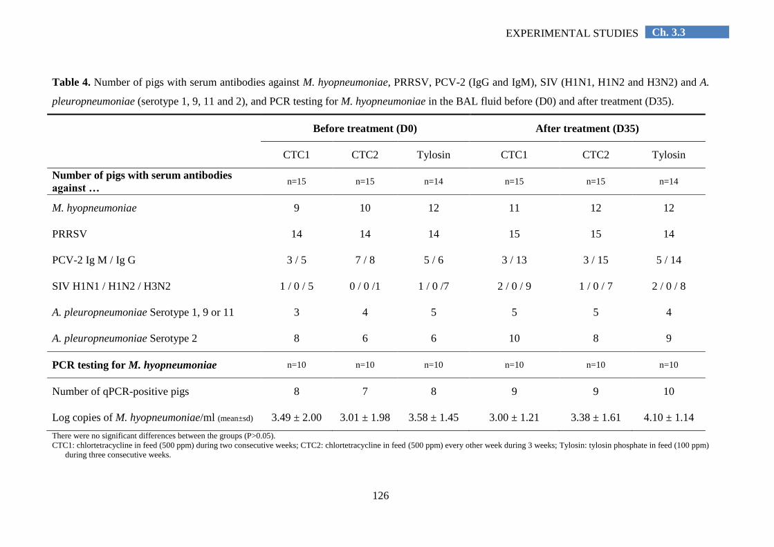
126
Ch. 3.3 EXPERIMENTAL STUDIES
Table 4. Number of pigs with serum antibodies against M. hyopneumoniae, PRRSV, PCV-2 (IgG and IgM), SIV (H1N1, H1N2 and H3N2) and A.
pleuropneumoniae (serotype 1, 9, 11 and 2), and PCR testing for M. hyopneumoniae in the BAL fluid before (D0) and after treatment (D35).
Before treatment (D0) After treatment (D35)
CTC1 CTC2 Tylosin CTC1 CTC2 Tylosin
Number of pigs with serum antibodies
against … n=15 n=15 n=14 n=15 n=15 n=14
M. hyopneumoniae 9 10 12 11 12 12
PRRSV 14 14 14 15 15 14
PCV-2 Ig M / Ig G 3 / 5 7 / 8 5 / 6 3 / 13 3 / 15 5 / 14
SIV H1N1 / H1N2 / H3N2 1 / 0 / 5 0 / 0 /1 1 / 0 /7 2 / 0 / 9 1 / 0 / 7 2 / 0 / 8
A. pleuropneumoniae Serotype 1, 9 or 11 3 4 5 5 5 4
A. pleuropneumoniae Serotype 2 8 6 6 10 8 9
PCR testing for M. hyopneumoniae n=10 n=10 n=10 n=10 n=10 n=10
Number of qPCR-positive pigs 8 7 8 9 9 10
Log copies of M. hyopneumoniae/ml (mean±sd) 3.49 ± 2.00 3.01 ± 1.98 3.58 ± 1.45 3.00 ± 1.21 3.38 ± 1.61 4.10 ± 1.14
There were no significant differences between the groups (P>0.05).
CTC1: chlortetracycline in feed (500 ppm) during two consecutive weeks; CTC2: chlortetracycline in feed (500 ppm) every other week during 3 weeks; Tylosin: tylosin phosphate in feed (100 ppm)
during three consecutive weeks.

127
Ch. 3.3 EXPERIMENTAL STUDIES
Discussion
The present study demonstrated that chlortetracycline, when administered in the feed
during two alternative weeks starting at the onset of clinical respiratory disease, was able to
decrease the prevalence of pneumonia lesions, and to numerically but not significantly reduce
performance losses and clinical signs in a herd infected with M. hyopneumoniae. The
treatment was compared with tylosin phosphate medication during three weeks in the feed.
The tylosin treated group was considered as a positive control group, as the efficacy of this
antibiotic to treat and control respiratory disease due to M. hyopneumoniae infections had
been shown in previous studies. Kunesh (1981) reported an improvement of ADG after
intramuscular administration of tylosin (4 mg/kg), and Vicca et al. (2005) demonstrated that
pigs treated in-feed with tylosin (100 ppm during 21 days) had less pneumonia lesions and
clinical signs following an experimental M. hyopneumoniae infection. It is likely that more
pronounced effects of chlortetracycline medication would have been obtained when a
negative (non-treated) control was used. A negative control group was not included because
not treating clinically diseased pigs when efficacious products are available is ethically not
acceptable, and also the pig producer would not have allowed to leave diseased pigs untreated
(Dohoo et al. 2009).
The infection level for M. hyopneumoniae was high in the present herd, as evidenced by
the high prevalence of slaughter pigs with pneumonia lesions and the fact that M.
hyopneumoniae was detected in many necropsied pigs. The necropsy findings and the
serological and bacteriological analyses also showed that other pathogens were involved in
the respiratory problem. The number of pigs with serum antibodies against PCV-2 (IgG), SIV
H3N2 and A. pleuropneumoniae increased during the treatment period, and also bacterial
pathogens such as S. suis, B. bronchiseptica, P. multocida and T. pyogenes were found in the
lungs of necropsied pigs. PRDC is a complex disease and it may be difficult to understand and
interpret field outbreaks of respiratory disease, because of the multiple interactions of
different pathogens with environmental conditions (Opriessnig et al., 2011). Polymicrobial
co-infections have been widely described in the literature being focused on the nature of the
pathogens (virus vs bacteria), the role (primary vs opportunistic) and the mode of action
(additive vs synergic) of these pathogens, and the sequencing of infection (concurrent vs
simultaneous). All these factors have a crucial role on the nature of the mixed infections and,
therefore, have implications on the large variability of this PRDC on field conditions. Co-
infection of pigs with M. hyopneumoniae in conjunction with PRRSV (Thacker et al., 1999),
128
Ch. 3.3 EXPERIMENTAL STUDIES
PCV-2 (Opriessnig et al., 2004) and SIV (Thacker et al., 2001) showed potentiation and
increased severity of the respiratory disease. However, during the respiratory outbreak of the
present herd, no clinical signs and mortality associated to these pathogens (PRRSV, PCV-2,
SIV) were seen, nor DNA was detected by PCR in dead animals. Only in one necropsied pig,
a moderate PCV-2 viral load in the lungs was detected. Co-infection of pigs with M.
hyopneumoniae in conjunction with bacteria, such P. multocida (Ciprián et al., 1988) and A.
pleuropneumoniae (Marois et al., 2009) is also characterised by an increased severity of
respiratory disease and enhancement of lesions, respectively. P. multocida was isolated from
the lungs of only one dead pig, which was negative by PCR for M. hyopneumoniae. A.
pleuropneumoniae was isolated in only one necropsied pig, nevertheless the prevalence of
pleuritis at slaughter was lower than 22 per cent. Therefore, the presence of these bacteria
could suggest an opportunistic infection. Analysis of the stable climate and the ventilation
pattern could not elucidate major deficiencies in housing conditions or management practices
(e.g. stocking density, management, deworming). Since the size of the nursery and the
fattening unit was not the same, all-in/all-out management was not strictly followed, but only
a limited number of pigs were mixed. This could also have contributed to respiratory disease
by transmission and spread of pathogens. However, the statistical interaction terms (treatment
x compartment and treatment x sex) were calculated for each parameter and no significant
differences were assessed. The study illustrated that clinical problems of respiratory disease,
requiring antimicrobial treatment, are still possible in herds without major deficiencies in
housing and management conditions, and that practice vaccination against two major
pathogens (M. hyopneumoniae and porcine PCV-2) involved in the PRDC.
The fact that chlortetracycline treatment performed similar or even better than a known
efficacious treatment is in line with previous studies showing the efficacy of chlortetracycline.
Ganter et al. (1995) reported that chlortetracycline-medicated feed (800 ppm, during 21 days)
was able to reduce clinical signs of respiratory disease under field conditions, and Thacker et
al. (2006) found that in-feed medication with chlortetracycline (500 ppm, during 14 days) was
effective against an experimental M. hyopneumoniae infection. However, the medication
schemes were definitively not completely efficacious. The RDS in all groups remained very
similar during the treatment period as before onset of treatment, a considerable number of
pigs had pneumonia lesions at slaughter, and M. hyopneumoniae could still be detected by
PCR in the lung tissue. The latter was not unexpected and confirmed previous reports
indicating that similar to vaccination, antimicrobial medication is not able to eliminate M.
hyopneumoniae from the respiratory tract (Thacker et al., 2006). It is not known why there

129
Ch. 3.3 EXPERIMENTAL STUDIES
was no significant decrease in RDS after medication had started. It may be due to the fact that
other pathogens e.g. viruses or bacteria not susceptible to the antimicrobials, were involved in
the respiratory problem. The high mortality in the T group, nearly significantly different from
the other groups, may be due to the presence of concurrent other bacterial pathogens such as
P. multocida, A. pleuropneumoniae, which are less susceptible to tylosin (Barigazzi et al.,
1994) or S. suis strains with acquired resistance to tylosin. Indeed two S. suis strains isolated
from BAL fluid showed resistance to this antimicrobial.
Based on the MIC testing performed by Vicca et al. (2004), the M. hyopneumoniae
strain collected at slaughter did not show acquired resistance to tylosin and oxytetracycline.
However, a high MIC value of chlortetracycline was obtained, but MIC testing of
chlortetracycline is not reliable as the antimicrobial is not stable during the cultivation
procedure. When chlortetracycline is incorporated in broth medium and rehydrated, it is
quickly degraded: activity reduces to 1/3 after 12 h, to less than 10 per cent after 24 h and to
about 2 per cent after 72 h (Pommier, 2006). Growth of M. hyopneumoniae on the other hand
takes several days. Williams (1978) found a lower in vitro susceptibility of chlortetracycline
against M. hyopneumoniae compared to oxytretracycline and doxycycline. Inamoto et al.
(1994) also observed higher MIC values of chlortetracycline than of oxytetracycline against
M. hyopneumoniae. Other studies also showed high MIC values (≥ 40 µg/ml) (Yamamoto and
Koshimizu, 1984) or a high variation in the MIC values (1.6 – 25 µg/ml) of chlortetracycline
(Etheridge et al., 1979). Our finding that the M. hyopneumoniae isolate did not show acquired
resistance to oxytetracycline strongly indicates that acquired resistance was also not present
against chlortetracycline, since there exists cross-resistance between both antibiotics (CLSI,
2008).
There were only small and non-significant differences between the CTC1 and CTC2
group, but for most parameters (e.g. prevalence and severity of pneumonia lesions), the CTC2
group performed slightly better. An exact explanation is not available, but it may be due to the
fact the pigs of the CTC2 had a better possibility to generate an active immune response
during the non-medicated week in between the two medicated weeks. Walter et al. (2000)
stated that metaphylactic pulse medication might permit a sufficient natural exposure to the
pathogen and more effective stimulation of active immunity against M. hyopneumoniae when
compared to continuous medication. Continuous medication may lead to animals that might
remain immunological naïve and potentially susceptible to subsequent re-exposure to M.
hyopneumoniae when the medication is withdrawn.
130
Ch. 3.3 EXPERIMENTAL STUDIES
Chlortetracycline hydrochloride, when administered via the feed at the dose of 500 ppm
during two alternative weeks at onset of clinical outbreak of respiratory disease, decreased the
prevalence of M. hyopneumoniae-like lesions, and numerically reduced performance losses
and clinical signs compared with tylosin phosphate at the dose of 100 ppm administered
during three weeks. Vaccination and medication may significantly reduce but do not prevent
infections with M. hyopneumoniae, implying that optimal housing and management practices
remain important to keep the infection pressure at acceptable levels.
Acknowledgements
This study was partially financed by Pfizer Animal Health. The authors want to thank
Hanne Vereecke for the technical support in the laboratory and the pig producer for
collaboration.

131
Ch. 3.3 EXPERIMENTAL STUDIES
REFERENCES
Barigazzi, G., Candotti, P., Foni, E., Martinelli, L. and Raffo, A. (1996). In vitro susceptibility of 108 isolated
Actinobacillus pleuropneumoniae strains to 17 antimicrobial agents from pig lungs in Italy in 1994-95. In
Proceedings of the 14th
International Pig Veterinary Society Congress, Bologna, Italy. Reading, July 7 to 10,
p 207.
Ciprián, A., Pijoan, C., Cruz, T., Camacho, J., Tórtora, J., Colmenares, G., López-Revilla, R. and De La Garza,
M. (1988). Mycoplasma hyopneumoniae increases the susceptibility of pigs to experimental Pasteurella
multocida pneumonia. Canadian Journal of Veterinary Research 52, 434–438.
Clinical And Laboratory Standards Institute (CLSI) (2008). Performance standards for antimicrobial disk and
dilution susceptibility tests for bacteria isolated from animals; approved standard. 3rd edn. M31-A3.
Dee, S.A. (1996). The porcine respiratory disease complex. Are subpopulations important? Swine Health
Production 4, 147-149.
Dohoo, I., Martin, W. and Stryhn, H. (2009). Controlled studies. In: Dohoo, I., Martin, W. and Stryhn, H. (Eds.)
Veterinary Epidemiologic Research. VER Inc., Prince Edward Island, Canada, pp 213-241.
Etheridge, J.R., Lloyd, L.C. and Cottew, G.S. (1979). Resistance of Mycoplasma hyopneumoniae to
chlortetracycline. Australian Veterinary Journal 55, 40.
Friis, N.F. (1975). Some recommendations concerning primary isolation of Mycoplasma suipneumoniae and
Mycoplasma flocculare: a survey. Nordisk Veterinaermedicin 27, 337-339.
Ganter, M., Dudziak, D. and Delbeck, F. (1995). Treatment of swine with chronic pneumonia with
chlortetracycline-medicated feed. Deutsche Tierärztliche Wochenschrift 102, 44-49.
Hannan, P., Bhogal, B. and Fish, J. (1982). Tylosin tartrate and tiamutilin effects on experimental piglet
pneumonia induced with pneumonic pig lung homogenate containing mycoplasmas, bacteria and viruses.
Research in Veterinary Science 33, 76-88.
Inamoto, T., Takahashi, H., Yamamoto, K., Nakai, Y. and Ogioto, K. (1994). Antibiotic susceptibility of
Mycoplasma hyopneumoniae isolated from swine. The Journal of Veterinary Medical Science 56, 393-394.
Jensen, C. S., Ersbøll, A. K. and Nielsen, J. P. (2002). A meta-analysis comparing the effect of vaccines against
Mycoplasma hyopeumoniae on daily weight gain in pigs. Preventive Veterinary Medicine 54, 265-278.
Kunesh, J. (1981). A comparison of two antibiotics in treating Mycoplasma pneumoniae in swine. Veterinary
Medicine, Small Animal Clinics 76, 871-872.
Luthman, J. and Jacobsson, S.O. (1983). The availability of tetracyclines in calves. Nordisk Veterinaer Medicin
35, 292-299.
Maes, D., Deluyker, H., Verdonck, M., Castryck, F., Miry, C., Lein, A., Vrijens, B. and de Kruif, A. (1998).
Effect of vaccination against Mycoplasma hyopneumoniae in pig herds with a continuous production system.
Journal of Veterinary Medicine Series B 45, 495-505.
Maes, D., Deluyker, H., Verdonck, M., Castryck, F., Miry, C., Vrijens, B., Verbeke, W., Viaene, J. and de
Kruif, A. (1999). Effect of vaccination against Mycoplasma hyopneumoniae in pig herds with an all-in/all-
out production system. Vaccine 17, 1024-1034.
Maes, D., Segalés, J., Meyns, T., Sibila, M., Pieters, M. and Haesebrouck, F. (2008). Control of Mycoplasma
hyopeumoniae infections in pigs. Veterinary Microbiology 126, 297-309.
Marois, C., Gottschalk, M., Morvan, H., Fablet, C., Madec, F. and Kobisch, M. (2009). Experimental infection
of SPF pigs with Actinobacillus pleuropneumoniae serotype 9 alone or in association with Mycoplasma
hyopneumoniae. Veterinary Microbiology 135, 283–291.
Marois, C., Dory, D., Fablet, C., Madec, F. and Kobisch, M. (2010). Development of a quantitative Real-Time
TaqMan PCR assay for determination of the minimal dose of Mycoplasma hyopneumoniae strain 116
required to induce pneumonia in SPF pigs. Journal of Applied Microbiology 108, 1523-1533.
Mason, S.E., Baynes, R.E., Almond, G.W., Riviere, J.E. and Scheidt, A.B. (2009). Pharmacology of tetracycline
water medication in swine. Journal of Animal Science 87, 3179-3186.
Mateusen, B., Maes, D., Van Goubergen, M., Verdonck, M. and de Kruif, A. (2002). Effectiveness of treatment
with lincomycin hydrochloride and/or vaccination against Mycoplasma hyopneumoniae for controlling
chronic respiratory disease in a herd of pigs. The Veterinary Record 151, 135–140.

132
Ch. 3.3 EXPERIMENTAL STUDIES
Meyns, T., Dewulf, J., de Kruif, A., Calus, D., Haesebrouck, F. and Maes, D. (2006). Comparison of
transmission of Mycoplasma hyopneumoniae in vaccinated and non-vaccinated populations. Vaccine 24,
7081–7086.
Morrison, R. B., Hilley, H. D. and Leman, A. D. (1985). Comparison of methods for assessing the prevalence
and extent of pneumonia in market weight swine. Canadian Veterinary Journal 26, 381–384.
Oliveira, S., Galina, L. and Pijoan, C. (2001). Development of a PCR test to diagnose Haemophilus parasuis
infections. Journal of Veterinary Diagnostic Investigation 13, 495-501.
Opriessnig, T., Thacker, E.L., Yu, S., Fenaux, M., Meng, X.J. and Halbur, P.G. (2004). Experimental
reproduction of postweaning multisystemic wasting syndrome in pigs by dual infection with Mycoplasma
hyopneumoniae and porcine circovirus type 2. Veterinary Pathology 41, 624-640.
Opriessnig, T., Gimenez-Lirola, L.G. and Halbur, P.G. (2011). Polymicrobial respiratory disease in pigs. Animal
Health Research Reviews 12, 133-148.
Pijpers, A., Schoevers, E.J., Van Gogh, H., Van Leenhoed, L.A.M.G., Visser, I.J.R., Van Miert, A.S.J.P.A.M.
and Verheijden, J.H.M. (1991). The influence of disease on feed and water consumption and on
pharmacokinetics of orally administered oxytetracycline in pigs. Journal of Animal Science 69, 2947-2954.
Pommier, P. (2006). In vitro, are all the tetracyclines equivalent? In: Proceedings of the 19th
International Pig
Veterinary Society Congress, Copenhagen, Denmark. Reading, July, 16 to 19, p 411.
Prescott, J.F. (2000). Tetracyclines. In: Prescott, J.F., Baggot, J.D., Walker, R.D. (Eds). Antimicrobial Therapy
in Veterinary Medicine. Iowa State University Press, Ames, USA, pp. 275-289.
Quinn, P., Carter, M., Markey B. and Carter, G. (1994). Bacterial Pathogens: Microscopy, culture and
identification. In: Quinn, P.J. (Ed.). Clinical Veterinary Microbiology. Mosby, Edinburgh, UK, pp 21–66.
Scorneaux, B. and Shryock, T.R. (1998). Intracellular accumulation, subcellular distribution and efflux of
tilmicosin in swine phagocytes. Journal of Veterinary Pharmacology and Therapeutics 21, 257-268.
Sibila, M., Pieters, M., Molitor, T., Maes, D., Haesebrock, F. and Segales, J. (2009). Current perspectives on the
diagnosis and epidemiology of Mycoplasma hyopneumoniae infection. The Veterinary Journal 181, 221–
231.
Thacker, E.L., Halbur, P.G., Ross, R.F., Thanawongnuwech, R. and Thacker, B.J. (1999). Mycoplasma
hyopneumoniae potentiation of porcine reproductive and respiratory syndrome virus-induced pneumonia.
Journal of Clinical Microbiology 37, 620–627.
Thacker, E.L., Thacker, B.J. and Janke, B.H. (2001) Interaction between Mycoplasma hyopneumoniae and
swine influenza virus. Journal of Clinical Microbiology 39, 2525–2530
Thacker E. L., Thacker B. J. and Wolff, T. (2006). Efficacy of a chlortetracycline feed additive in reducing
pneumonia and clinical signs induced by experimental Mycoplasma hyopneumoniae challenge. Journal of
Swine Health and Production 14, 140-144.
Thacker, E. (2006). Mycoplasmal diseases. In: Straw, B.E., Zimmerman, J.J., D´Allaire, S. and Taylor, D.J.
(Eds). Diseases of Swine. Blackwell Publishing Ltd., Oxford, UK, pp 701–717.
Ueda, Y., Ohtsuki, S., Narukawa, N. and Takeda, K. (1994). Effect of terdecamycin on experimentally induced
Mycoplasma hyopneumoniae-infection in pigs. Journal of Veterinary Medicine Series B 41, 283-290.
Vicca, J., Stakenborg, T., Maes, D. Butaye, P., Peeters, J., de Kruif, A. and Haesebrouck, F. (2004). In vitro
susceptibilities of Mycoplasma hyopneumoniae field isolates. Antimicrobial Agents and Chemotherapy 48,
4470-4472.
Vicca, J. (2005). Virulence and antimicrobial susceptibility of Mycoplasma hyopneumoniae isolates from pigs.
Faculty of Veterinary Medicine. Ghent University. Thesis. pp 27-58.
Vicca, J., Maes, D., Jonker, L., de Kruif, A. and Haesebrouck, F. (2005). Efficacy of in-feed medication with
tylosin for the treatment and control on Mycoplasma hyopneumoniae infections. The Veterinary Record, 156,
606-610.
Villarreal, I., Meyns, T., Dewulf, J., Vranckx, K., Calus, D., Pasmans, F., Haesebrouck, F. and Maes, D. (2011).
The effect of vaccination on the transmission of Mycoplasma hyopneumoniae in pigs under field conditions.
The Veterinary Journal 188, 48-52.
Vranckx, K., Maes, D., Calus, D., Villarreal, I., Pasmans, F. and Haesebrouck, F. (2011). Multiple locus
variable number of tandem repeats analysis is a suitable tool for the differentiation of Mycoplasma
hyopneumoniae strains without cultivation. Journal of Clinical. Microbiology 49, 2020-2023.

133
Ch. 3.3 EXPERIMENTAL STUDIES
Walter D., Holck, J.T., Sornsen, S., Hagen, C. and Harris I.T. (2000). The effect of a metaphylactic pulse dosing
in-feed antimicrobial strategy on finishing pig health and performance. Swine Health and Production 8, 65-
71.
Weisblum, B. (1998). Macrolide resistance. Drug Resistance Updates 1, 29-41.
Williams, P.P. (1978). In Vitro Susceptibility of Mycoplasma hyopneumoniae and Mycoplasma hyorhinis to
Fifty-One Antimicrobial Agents. Antimicrobial Agents and Chemotherapy 14, 210-213.
Yamamoto, K. and Koshimizu, K. (1984). In vitro susceptibility of Mycoplasma hyopneumoniae to antibiotics.
In: Proceedings of the 8th
International Pig Veterinary Society Congress, Ghent, Belgium. August 27 to 31, p
116.


Ch. 4 GENERAL DISCUSSION
Chapter 4. GENERAL DISCUSSION
General discussion

137
Ch. 4 GENERAL DISCUSSION
GENERAL DISCUSSION
Respiratory disease in pigs is one of the most important problems in modern pig
production worldwide. It mostly results from infections with primary and opportunistic
infectious agents, both viral and bacterial pathogens. Adverse environmental and
management conditions may exacerbate this mixed respiratory disease resulting in the PRDC.
M. hyopneumoniae is considered as one of the main pathogens involved in the PRDC.
Infections with M. hyopneumoniae are present in almost all countries with an intensive swine
production, and they lead to major economic losses due to the reduced growth, increased
mortality and feed conversion, costs for antimicrobials and vaccination and increased time to
market weight (Maes et al., 2008).
The control of M. hyopneumoniae infections can be accomplished by optimization of
management practices and housing conditions, antimicrobial treatment and vaccination. The
present thesis investigated the treatment and control of M. hyopneumoniae infections. First,
the efficacy of a new florfenicol formulation to treat pigs by means of a single intramuscular
injection against experimental M. hyopneumoniae infection was investigated (chapter 3.1).
Secondly, the efficacy of a one-shot vaccination against M. hyopneumoniae applied at either
one or three weeks of age in a Belgian farrow-to-finish pig herd with mixed respiratory
disease late in the fattening period and including infections with M. hyopneumoniae and viral
pathogens was studied (chapter 3.2). Thirdly, the efficacy of in-feed medication with
chlortetracycline and tylosin phosphate against a clinical outbreak of respiratory disease in
fattening pigs that were vaccinated against M. hyopneumoniae was compared (chapter 3.3).
The present chapter will provide a general discussion of the findings obtained in the
different studies. We will successively focus on (1) the major parameters used in this thesis to
assess efficacy, (2) the different antimicrobial treatments (chapter 3.1 and 3.3), and (3) the
different vaccination strategies (chapter 3.2 and 3.3) applied in this thesis. Finally, (4) general
conclusions and (5) perspectives for further research are provided.
Assessment of efficacy under in vivo (experimental, field) conditions
This thesis includes the assessment of efficacy of antimicrobial treatment and
vaccination against M. hyopneumoniae infections in different in vivo studies, conducted
either under experimental or field conditions.

138
Ch. 4 GENERAL DISCUSSION
The importance of the measured efficacy parameters may depend whether a study is
conducted under experimental or field conditions. Therefore, a table with the main
advantages and disadvantages of field and experimental studies is first shown (Table 1).
Table 1. Comparison between in vivo studies, both laboratory and field trials.
Laboratory experiment Field trial
Housing conditions controlled real
Animals homogeneous heterogeneous
Management controlled real
Disease model real
Infection dose standard for all animals unknown
Time of infection standard for all animals unpredictable
Concurrent diseases avoided present
Variability of the outcome low high
External validity of the study limited high
Different parameters can be measured to assess efficacy of treatment and control
measures against M. hyopneumoniae infections. The most commonly used parameters are
clinical signs, lung lesions and in case of field studies also performance data.
Coughing is the main clinical sign of respiratory disease. Clinical examination of pigs
affected by respiratory disease does not provide a specific aetiological diagnosis. Non-
productive coughing is not a pathognomonic sign of M. hyopneumoniae infection, but it is
commonly associated with this pathogen. The assessment of the RDS in laboratory
experiments may be useful to evaluate the severity of the challenge infection, to determine
the moment of the intervention (mainly treatment) and to assess the efficacy of this
intervention. Regarding field experiments, it can be used in the diagnosis of enzootic
pneumonia at onset of respiratory disease (Nathues et al., 2012). A quantification of the
severity of coughing, for instance by using a RDS, may indeed be an important indicator.
Scoring the respiratory symptoms by means of a RDS may be valuable to select an
appropriate antimicrobial treatment or vaccination strategy (type of product, route of
administration, duration of treatment) required to treat and control respiratory disease (e.g.
acute coughing present in a few pigs from the same pen should be treated at individual or at
pen level), but also in the assessment of the clinical effectiveness of the treatment/vaccination
established.

139
Ch. 4 GENERAL DISCUSSION
Lung lesions, such as pneumonia and adhesive pleurisy, are common findings in
slaughter pigs reared under field conditions and both have been associated with respiratory
disease (Fraile et al., 2010; Meyns et al., 2011; Fablet et al., 2012b; Merialdi et al., 2012).
Slaughter examinations (prevalence of pneumonia and pleurisy lesions) are an excellent
method to monitor the respiratory health in a herd and to evaluate the efficacy of
treatments/vaccinations applied under field conditions. However, this technique has some
limitations. The lack of diagnostic specificity and the subjectivity of the visual assessment are
major disadvantages (Sibila et al., 2009). Subclinical infections may not be detected and
lesions occurring early in the fattening period may be healed by the time of slaughter (Regula
et al., 2000). In most experimental studies, only a limited number of animals are available
due to practical and economic reasons. The current EU regulations on experimental animals
also indicate to limit the number of laboratory animals as much as possible because of animal
welfare reasons. However, a low number of animals can lead to not finding possible
differences between groups. This disadvantage may partially be overcome by estimating the
extent of lung lesions in each animal, as well as by evaluating the extent and severity of
microscopic lung lesions (e.g. percentage of air in lung tissue, severity of peribronchiolar and
perivascular lymphohistiocytic infiltration and nodule formation).
Preventing the performance losses is the main goal when treatment/vaccination against
M. hyopneumoniae infections are established under field conditions. Although performance
parameters are also commonly measured under experimental conditions, usually, no
significant differences are obtained due to the limited number of animals used. Challenge
infection models, are only able to provide a general tendency regarding the differences
between treated and non-treated/vaccinated and non-vaccinated animals. When looking for
biological, relevant and statistically significant differences, a much larger number of animals
reared under commercial conditions should be included.
Serological examinations are mainly used in both experimental and field trials as a
descriptive parameter to better understand the dynamics of the infection in the studied
population. However, nowadays this parameter is not a good indicator to assess efficacy of
current commercial vaccines against M. hyopneumoniae, since vaccination does not always
induce seroconversion and the presence of antibodies induced after vaccination is not
correlated with protection from colonization and disease (Djordjevic et al., 1997).
Nevertheless, serological examinations can also provide information regarding other
pathogens circulating within the population and causing disease.

140
Ch. 4 GENERAL DISCUSSION
In vivo trials, especially when conducted under field conditions, may be largely
influenced by environmental conditions. To provide standard climatic environment and
management conditions to the animals will minimize the effect of this variability on the
outcome of the experiments. Control systems are already available on the market to control
and prevent adverse conditions. The installation of sensors capable to measure variation in
temperature and ventilation, as well as feed and water intake, may be useful to understand
respiratory disease course and to detect failures in antimicrobial treatment, due to deficient
administration/intake, respectively. This early detection would favour an early prevention or
correction of these treatment failures. Application of computerized systems may help also to
reduce the variation due to subjective measurements. A smart monitoring system has been
recently developed by the University of Leuven, the “Pig Cough Monitor” (Exadaktylos et
al., 2008). This is based on the detection of sounds produced by pigs, recognition of coughing
and therefore, monitoring of frequency of this coughing. Similar applied technology based on
visual detection may be also useful for recognizing macroscopic lung lesions at slaughter, and
therefore for calculation of the extent of pulmonary lesions in slaughter pigs.
Antimicrobial treatment and control of M. hyopneumoniae infections
It is generally accepted that optimization of the housing and management conditions
should be the first to be accomplished for the prevention and control of enzootic pneumonia.
However, even in herds with optimal management and housing conditions, clinical outbreaks
of M. hyopneumoniae can still occur (Vicca et al., 2002). Thus, antimicrobial treatment
remains an important tool to control respiratory disease caused by this bacterium.
Additionally, under field conditions, infections with M. hyopneumoniae are commonly
accompanied by other bacterial infections (Sorensen et al., 1997; Maes et al., 1999; Maes et
al., 2000) that may increase the severity of the disease and the lesions. Antimicrobial
treatment may be effective also against bacterial respiratory pathogens other than M.
hyopneumoniae, and it can be implemented faster and in a more flexible way compared to
vaccination. However, antimicrobial treatment is able to reduce the number of M.
hyopneumoniae organism from the lung tissue, but complete elimination of this pathogen
cannot be achieved (Vicca et al., 2005; Thacker and Minion, 2012).
The in vitro activity of all three antibiotics tested in this thesis, florfenicol (chapter 2.1),
chlortetracycline and tylosin (chapter 2.3), has been widely investigated. It has been shown
that M. hyopneumoniae is intrinsically susceptible to all three antimicrobials (Prieb and
Schwarz, 2003; Vicca et al., 2004). However, in vivo activity seems to vary, likely due to the
141
Ch. 4 GENERAL DISCUSSION
location of M. hyopneumoniae at the cilia, where therapeutic levels of antimicrobials may not
be easily reached (Vicca, 2005). An antimicrobial may only be effective against M.
hyopneumoniae infection when it is able to achieve significant levels within the mucous and
fluids of the respiratory tract (Thacker and Minion, 2012). Therefore, in vivo studies are still
needed to evaluate the efficacy of antimicrobials against this pathogen.
Our first study (3.1) showed that florfenicol applied once intramuscularly at the onset
of disease was effective in controlling the acute disease caused by an experimental challenge
with M. hyopneumoniae organisms. It is well known that observations from experimental
challenges must be interpreted with caution before generalization to field conditions. The
amount of M. hyopneumoniae organisms inoculated during experimental challenge (7 mL of
inoculum containing 107 CCU/mL) is high, but the infection takes place only once. Under
field conditions, the infection dose is not known, but pigs are exposed for several days and
weeks to the infection. Additionally, under experimental conditions, the inoculum is directly
placed in the trachea, whereas in case of natural infections, the first contact surface is the
nasal mucosa. Clinical and acute outbreaks of respiratory disease are characterized by the
lack of appetite in affected animals, and consequently by a reduction of the feed and water
intake. Therefore oral medication is less appropriate in controlling acute outbreaks, and
individual (parenteral) treatment, i.e. intramuscular injection, would be required.
Immediately after intramuscular injection, florfenicol presents a high initial blood
concentration as well as a high tissue distribution (bioavailability approximately 100 per cent)
(Liu et al., 2003). This rapid distribution (mean distribution half-life: 0.15 h) ensures a quick
initial response, making this antimicrobial suitable for the treatment of acute outbreaks of M.
hyopneumoniae infections. Florfenicol has a high in vitro antimicrobial activity against M.
hyopneumoniae (Hannan et al., 2000; Wilhelm et al., 2012). Florfenicol is eliminated slowly
(mean elimination half-life: 14h), enabling therapeutic plasma levels up to 53h (Liu et al.,
2003). This guarantees a long acting effect, which makes it particularly convenient to treat
nursery and fattening pigs. Additionally, the therapeutic plasma level remains above the MIC
of important other respiratory bacteria (A. pleuropneumoniae, P. multocida, H. parasuis) for
at least 48h. This ensures a prolonged protection, which may enable the immune system to
control secondary bacterial infections (Voorspoels et al., 1999).
Scarce data is available in the literature regarding the clinical efficacy of florfenicol in
pigs (Dowling, 2013). A recent field study in a pig herd infected with M. hyopneumoniae
(Ciprián et al., 2012) showed that in-feed medication with florfenicol during 35 days
significantly improved performance. However, such a prolonged antimicrobial therapy may

142
Ch. 4 GENERAL DISCUSSION
select resistant bacteria. Thus, if a single injection at onset of disease is effective to treat M.
hyopneumoniae in pig herds, this could significantly reduce the antimicrobial use in pig herds
in comparison with oral treatments which are usually established for longer periods of time.
The florfenicol formulation used in chapter 3.1 differs from the reference veterinary
medicinal product by the higher concentration of active substance (450 mg/mL), the single
administration but also by a different therapeutic indication. Metaphylaxis is considered
superior to prophylaxis in terms of a rational and minimized use of antimicrobials (Catry et
al., 2008). A metaphylactic medication (single injection of florfenicol) established at onset of
disease and characterized by a higher concentration of active substance may guarantee
low/very low risk of mutant selection bacteria (Drlica, 2003; Burch, 2012), while providing
clinical efficacy. Altough this metaphylactic strategy has been already demonstrated
efficacious to treat bovine respiratory disease (Catry et al., 2008), no reports are available
regarding porcine respiratory disease.
The treatment significantly decreased the RDS in treated pigs, especially two days post-
treatment. However, the clinical symptoms increased again four days later. Also, at necropsy
only a numerical benefit was observed for the macroscopic and the histopathological lung
lesions. Our findings suggested that a single injection with this antibiotic only partially
controls M. hyopneumoniae infection during a period of approximately four days.
The performance losses and mortality were numerically improved in the treated group.
Including more animals might result in statistically significant improvement, but the
tendencies indicate that an economical improvement might be obtained when florfenicol is
injected.
All infected pigs were positive by nPCR in the BAL fluid, indicating that the challenge
infection was successful and that the treatment did not eliminate M. hyopneumoniae from the
lungs. The latter confirms previous reports, stating that antimicrobial treatment does not
eliminate the pathogen from the lung tissue nor heal existing lesions (Thacker and Minion,
2012). Antimicrobial agents may delay infection, but cannot avoid colonization, nor assure
total elimination of M. hyopneumoniae.
Our third study (3.3) showed that chlortetracycline administered via the feed during
two consecutive weeks at onset of clinical outbreak of respiratory disease was effective in
reducing the prevalence of pneumonia, the performance losses and the severity of clinical
signs in a herd infected with M. hyopneumoniae and that practiced vaccination against M.
hyopneumoniae. The chronic respiratory disease observed in this problem herd was
associated with a high infection level of M. hyopneumoniae that was evidenced by the high
143
Ch. 4 GENERAL DISCUSSION
prevalence of pneumonia at slaughter and the large proportion of M. hyopneumoniae PCR-
positive pigs at necropsy. Also other respiratory pathogens were involved in the respiratory
problems. Despite all the pigs were vaccinated against M. hyopneumoniae, sporadic acute
outbreaks of respiratory disease prior to the study were observed at 14 weeks of age.
Consequently, the medication can be considered as a metaphylactic treatment at group level.
Clinically diseased animals, as well as subclinically infected animals were treated. From the
viewpoint of prudent use of antimicrobials, strategic or preventive medication during periods
at risk should be minimized and priority should be given to vaccination. However, as shown
in the present herd, even in vaccinated pigs, clinical symptoms/outbreaks may occur
(Mateusen et al., 2002) and antimicrobial medication may be necessary.
Both chlortetracycline and tylosin medications administered in chapter 3.3 complied
with the label directions of both products (chlortetracycline: between 7 to 14 days; tylosin: at
least 21 days). However, two different treatments strategies were investigated for
chlortetracycline medication: CTC1 (chlortetracycline in feed, two consecutive weeks) and
CTC2 (two non-consecutive weeks, with a non-medicated week-interval in between). All
three metaphylactic medications established at onset of disease and characterized by a high
concentration of active substance may guarantee low/very low risk of mutant selection
bacteria (Drlica, 2003; Burch, 2012), while providing clinical efficacy. However, this
hypothesis has not yet been proven with M. hyopneumoniae. Additionally, both
chlortetracycline medications (two weeks) shortened the duration of treatment when
compared with tylosin medication (tylosin in feed, three consecutive weeks), hence reducing
the risk of selection mutant bacteria. Although chlortetracycline has been proven efficacious
in the control of porcine respiratory disease, no reports are available comparing the efficacy
of this antimicrobial agent administered during two consecutives or alternating weeks.
Both chlortetracycline (Thacker et al., 2006) and tylosin (Hannan et al., 1982; Ueda et
al., 1994; Vicca et al., 2005) have already been shown to be efficacious to treat and control
respiratory disease due to M. hyopneumoniae under experimental conditions, but only a few
field studies are reported in the literature (Kunesh 1981; Ganter et al., 1995). In chapter 3.3,
the efficacy of in-feed medication with chlortetracycline and tylosin phosphate against a
clinical outbreak of respiratory disease in fattening pigs was compared. The study included a
large number of pigs, measured different clinical, pathological and performance parameters,
and was conducted in a herd practising vaccination against M. hyopneumoniae and PCV-2.
Chlortetracycline, administered by ad libitum intake of medicated feed, has been shown
to quickly (after 2h) reach peak serum concentrations. It has a good diffusion in lung tissue

144
Ch. 4 GENERAL DISCUSSION
(Kilroy et al., 1990; Del Castillo et al., 1998; Li et al., 2008). These two features ensure a
quick and targeted initial response, making this antimicrobial suitable for the treatment of
outbreaks of M. hyopneumoniae infections. Tetracyclines have been demonstrated to have a
good in vitro activity against M. hyopneumoniae (Hannan et al., 2000). Inamoto et al. (1994)
reported acquired antimicrobial resistance to tetracyclines occurring in M. hyopneumoniae
field strains isolated in Japan. Generally speaking, results of in vitro susceptibility testing of
M. hyopneumoniae for chlortetracycline should be interpreted with caution, since MIC testing
of chlortetracycline is not reliable as the antimicrobial is not stable during the cultivation
procedure, i.e. it quickly degrades (activity reduced to 2 per cent after 72h) (Pommier et al.,
2006) and M. hyopneumoniae grows slowly (more than 72h). Chlortetracycline is classified
as a short-acting tetracycline due to its short half-life of elimination (4h) (Del Castillo et al.,
1998). This quick elimination justifies the use of in-feed medication in order to maintain
therapeutic levels in diseased animals for a sufficiently long time. Additionally,
chlortetracycline has also been shown to be effective and is recommended for the treatment
of other respiratory bacteria, such as P. multocida and H. parasuis (Burch, 2012).
Tylosin phosphate, administered via the feed, has been shown to present a good
absorption and tissue distribution. Tylosin can be detected in plasma already 15 min after oral
administration and the peak serum concentration are reached fast (after 4h) (summary of
products characteristics). It presents a good lipid solubility, which favors its wide distribution
among all tissues except for the brain (Guiguère, 2013). Tylosin is concentrated in the lungs
and accumulated principally in lysozymes of macrophages (Scorneux and Shryock 1998).
These characteristics ensure a quick and targeted initial response against respiratory bacterial
infections. Tylosin has a good in vitro activity against M. hyopneumoniae (Hannan et al.,
2000). However, Vicca et al. (2004) reported acquired antimicrobial resistance in one out of
21 M. hyopneumoniae field strains isolated in Belgium. The elimination half-life of tylosin in
pigs is similar to chlortetracycline (approximately 4h) (Giguère, 2013). This quick
elimination justifies the use of in-feed medication in order to maintain therapeutic levels in
diseased animals for a sufficiently long time. Additionally, tylosin seems to be effective for
the treatment of other respiratory bacteria (A. pleuropneumoniae, P. multocida, H. parasuis),
although MICs are intrinsically high for these Pasteurellaceae (Burch, 2012).
The bioavailability of antimicrobials after oral administration can be influenced by
several factors, such as the pH of the stomach and gastric emptying. Tetracyclines present a
low systemic bioavailability after oral administration, especially in non-fasting pigs (Del
Castillo et al., 2013). The presence of mycotoxins may also alter the bioavailability of
145
Ch. 4 GENERAL DISCUSSION
antimicrobials administered per os (Goossens et al., 2012b). Recent in vitro studies showed
that mycotoxins can damage intestinal epithelial cells resulting in increased passage of drugs
(Goossens et al., 2012a). This was confirmed under in vivo conditions, where administration
of feed contaminated with deoxynivalenol and T-2 toxin resulted in significant increased
plasma concentrations of doxycycline (Goossens et al., 2012b) and chlortetracycline
(Goossens et al., 2013), respectively.
The tylosin treated group was considered as a positive control group, as the efficacy of
this antibiotic to treat and control respiratory disease due to M. hyopneumoniae infections had
been shown in previous studies (Kunesh, 1981; Vicca et al., 2005). A negative control group
was not included because not treating clinically diseased pigs when efficacious products are
available is ethically not acceptable, and also the pig producer would not have allowed to
leave diseased pigs untreated (Dohoo et al., 2009). It is likely that more pronounced effects of
chlortetracycline medication would have been obtained if a negative (non-treated) control had
been used.
The efficacy of chlortetracycline treatment was in line with previous field (Ganter et
al., 1995) and experimental (Thacker et al., 2006) studies. However, it is clear that the
medication schemes were definitively not completely efficacious. The RDS in all groups
remained very similar during the treatment period and a considerable number of pigs had
pneumonia lesions at slaughter. Despite antimicrobial medication, M. hyopneumoniae could
still be detected by PCR in the lung tissue. It is not known why there was no significant
decrease in RDS after medication had started. Other pathogens, e.g. viruses or bacteria not
susceptible to the antimicrobials may have been involved in the respiratory problem. The
high mortality in the treated pigs may be due to the presence of concurrent other bacterial
pathogens such as P. multocida, A. pleuropneumoniae, which present intrinsically high MICs
for tylosin (Barigazzi et al., 1994) or S. suis strains with acquired resistance to this
antimicrobial. Indeed two S. suis strains isolated from BAL fluid showed resistance to
tylosin.
Based on the MIC testing procedure used by Vicca et al. (2004), the M. hyopneumoniae
strain collected at slaughter did not show acquired resistance to tylosin and oxytetracycline.
Although a high MIC value of chlortetracycline was obtained, our finding that the M.
hyopneumoniae isolate did not show acquired resistance to oxytetracycline strongly indicates
that acquired resistance was also not present against chlortetracycline, since cross-resistance
exists between both antibiotics (CLSI, 2008).

146
Ch. 4 GENERAL DISCUSSION
There were only small and non-significant differences between the CTC1 and CTC2
groups, but for most parameters (e.g. prevalence and severity of pneumonia lesions), the
CTC2 group performed slightly better. An exact explanation is not available, but it may be
due to the fact the pigs of the CTC2 group had a better possibility to generate an active
immune response during the non-medicated week in between the two medicated weeks.
Walter et al., (2000) stated that metaphylactic pulse medication might permit a sufficient
natural exposure to the pathogen and more effective stimulation of active immunity against
M. hyopneumoniae when compared to continuous medication. These authors suggested that
continuous medication might lead to animals that remain potentially susceptible to
subsequent re-exposure to M. hyopneumoniae when the medication is withdrawn.
Analysis of the stable climate and the ventilation pattern could not elucidate major
deficiencies in housing conditions or management practices (e.g. stocking density,
management, deworming). This illustrated that clinical problems of respiratory disease,
requiring antimicrobial treatment, are still possible in herds without major deficiencies in
housing and management conditions, even if vaccination is practiced against two major
pathogens (M. hyopneumoniae and porcine PCV-2) involved in the PRDC.
A general observation from both chapters 3.1 and 3.3, is that treatment and control of
outbreaks of respiratory disease represents a very complex decision-making process,
including lots of variables. When there is an acute clinical outbreak of respiratory disease,
injectable treatment is necessary in those animals in which water or feed intake is reduced
and therefore, are not capable to consume antimicrobials orally. This parenteral
administration ensures a fast response against acute outbreaks where urgency of treatment is
required. When long-acting formulations are used, the labor of repeated injections is reduced.
On the other hand, oral medication is especially suited for endemic or chronic diseases, in
which the appetite of the affected animals is not reduced. The presence of subclinically
infected animals may pose a threat to the herd health. The administration at group level
enables the treatment of the clinically and subclinically infected animals.
Vaccination against M. hyopneumoniae infections
Commercial vaccines are used worldwide to control M. hyopneumoniae infections, and
in many countries vaccination is applied in more than 70 per cent of the pig herds (Maes et
al., 2008). Major advantages of vaccination include a reduction of performance losses,
severity and extent of clinical signs and lung lesions, mortality, time to reach slaughter
weight, and treatments costs (Maes et al., 2008).

147
Ch. 4 GENERAL DISCUSSION
However, the protection conferred by commercial vaccines against M. hyopneumoniae
is often incomplete. Several studies demonstrated that the current vaccines are not able to
prevent colonization (Thacker et al., 1998), to significantly reduce transmission under
experimental conditions (Meyns et al., 2006), and to prevent infection under field conditions
(Villarreal et al., 2011). Therefore, these studies suggested that vaccination alone is not able
to eliminate M. hyopneumoniae from infected pig herds. Nevertheless, recent studies have
demonstrated that vaccination may reduce the number of M. hyopneumoniae organisms in the
respiratory tract of experimentally infected pigs (Meyns et al., 2006; Vranckx et al., 2012b),
and therefore, may decrease the infection level in a herd (Sibila et al., 2007). This further
confirms that maximum beneficial effects of vaccination are reached several months after the
initiation of vaccination (Haesebrouck et al., 2004).
Different vaccination strategies can be implemented to control M. hyopneumoniae
infections at herd level. Currently, one-shot vaccination is more often practiced than two-shot
vaccination, mainly because it requires less labor, and it confers similar results as two-shot
vaccination (Roof et al., 2001, 2002; Alexopoulos et al., 2004; Lillie et al., 2004; Greiner et
al., 2011). Early vaccination (<4 weeks of age) of piglets is commonly practiced in Europe
(Haesebrouck et al., 2004).
Our second study (3.2) showed that a single vaccination against M. hyopneumoniae
applied at either 7 or 21 days of age reduced the prevalence and extent of pneumonia lesions
in a pig herd with clinical respiratory disease during the second half of the fattening period.
It is well known that vaccination is most likely effective if active immunity can be
established before the exposure to the pathogen. It has been demonstrated that M.
hyopneumoniae infections can take place in piglets already during the first weeks of life
(Sibila et al., 2007; Nathues et al., 2010; Villarreal et al., 2010, 2011; Vranckx et al., 2012a;
Fablet et al., 2012a). Therefore, an early vaccination has the advantage of inducing immunity
before pigs become infected, not only with M. hyopneumoniae, but also with other pathogens
that can interfere with immune response (Maes et al., 2008). The clinical course of the
disease in this problem herd was characterized by a low infection level of M. hyopneumoniae.
In previous batches before the start of the experiment, M. hyopneumoniae was evidenced in
nursery piglets (6-7 weeks of age) by the clinical signs, the detection of M. hyopneumoniae
PCR-positive pigs at 10 weeks of age and the presence of pneumonia at slaughter. Also other
respiratory pathogens were involved in the clinical respiratory signs, but mainly during the
fattening period. Consequently, this motivated the early vaccination of suckling piglets in
order to control the respiratory disease in this herd.
148
Ch. 4 GENERAL DISCUSSION
However, the respiratory problems were different from those in the previous batches in
this herd. They occurred later in the present batch, M. hyopneumoniae infections were less
prevalent and mainly occurred from halfway the fattening period onwards, and there were
complicating viral infections. This confirms that the infection pattern of M. hyopneumoniae is
not constant over time within the same herd, and that important variations may occur between
successive batches (Fano et al., 2007; Sibila et al., 2007). Other studies showed also a
seasonal variation of M. hyopneumoniae infections (Straw et al., 1986; Maes et al., 2000;
Segalés et al., 2012). The absence of a constant infection pattern i.e. infection in younger or
older pigs depending on the batch, is one of the major reasons why vaccination of piglets
during the first weeks of life is commonly practiced worldwide. Using this strategy, piglets
are immunized before infection, irrespective of whether infection takes place in the nursery,
growing or fattening unit.
Several studies have demonstrated the efficacy of vaccination at seven days of age after
M. hyopneumoniae challenge infection under experimental conditions in reducing lung
lesions and/or clinical signs. Apart from inducing early protection, it is also important that
early vaccinated pigs remain protected until the end of the fattening period, as in most pig
herds, the highest infection levels of M. hyopneumoniae occur during the grow-finishing
period (Sibila et al., 2004). In this case, the question remains as to whether the effect of one-
shot early vaccination under field conditions with mixed respiratory disease will last until the
end of the fattening period.
Despite vaccination against M. hyopneumoniae, there was a very high percentage of
pigs with pneumonia lesions at slaughter age (80 and 67-71 per cent in non-vaccinated and
vaccinated pigs, respectively). This may likely be due to the involvement of not only M.
hyopneumoniae, but also multiple viral infections. The infections mainly took place during
the second half the fattening period, allowing insufficient time for the lesions to be healed at
slaughter age (Blanchard et al., 1992). Pneumonia lesions at slaughter are not pathognomonic
for infections with M. hyopneumoniae. Apart from M. hyopneumoniae, swine influenza virus
may cause similar pneumonia lesions (Thacker et al., 2001; Sibila et al., 2009). Clinical signs
of respiratory disease were only present in the second half of the fattening period, while
expected in nursery pigs. Additionally, because of the large number of efficacy parameters
included in this experiment, it was decided to exclude the respiratory disease scoring from the
efficacy assessment in order to reduce the complexity of the study design.
Both vaccination schedules led to a numerical reduction of growth losses (18-19
g/pig/day) during the fattening period. The fact that the improvement was not statistically
149
Ch. 4 GENERAL DISCUSSION
significant may be due to the high standard variations, the occurrence of respiratory problems
only late in the fattening period, and the different viral infections that were involved in the
outbreak. However, similar improvements in performance (21 g/pig/day) following M.
hyopneumoniae vaccination were obtained in a meta-analysis of 28 published field trials
(Jensen et al., 2002).
Vaccination reduced the prevalence of nPCR-positive pigs, and therefore decreased the
infection level of M. hyopneumoniae in this herd. However, M. hyopneumoniae DNA could
still be detected by PCR. This further confirms that vaccination alone is not able to eliminate
M. hyopneumoniae from infected pig herds (Meyns et al., 2006; Villarreal et al., 2011b).
It was also further confirmed that vaccination in the presence of maternally-derived
serum antibodies is efficacious to control M. hyopneumoniae infections (Martelli et al., 2006;
Ritzmann et al., 2006; Reynolds et al., 2009). However, one-shot vaccination did not lead to
significant increases in serum antibodies, confirming that serum antibody concentrations
measured using the currently available ELISAs are not correlated with protection against M.
hyopneumoniae (Djordjevic et al., 1997; Thacker et al., 1998). This implies that testing for
the presence of serum antibodies is not reliable to evaluate whether pigs have been vaccinated
properly in herds endemically infected with M. hyopneumoniae.
A general observation from both field studies (chapters 3.2 and 3.3) is that different
vaccination schemes and commercial bacterins can be used to control M. hyopneumoniae
infections. In chapter 3.3, despite vaccination at 3 weeks of age, clinical outbreaks of M.
hyopneumoniae were observed, making antimicrobial treatment necessary. Vaccination
against M. hyopneumoniae (Mycoflex®) was routinely done on this farm. In chapter 3.2, both
vaccination schedules (Stellamune Once®) were able to reduce the prevalence of pneumonia;
however, other viral pathogens were involved in this respiratory disease. Different
commercial bacterins were used in both field studies. However, both of them have been
widely tested and proven efficacious against M. hyopneumoniae infections. It is generally
accepted that optimization of the housing and management conditions should be the first to
accomplish for the prevention and control of enzootic pneumonia. However, in the herds
from both field studies, analysis of stable climate and ventilation pattern could not elucidate
major deficiencies in housing and management conditions. Improvement of housing and
ventilation remains as milestone in the control of respiratory disease in intensive swine
production systems, and therefore, further research on this topic can guarantee a better
understanding of the control of the PRDC.
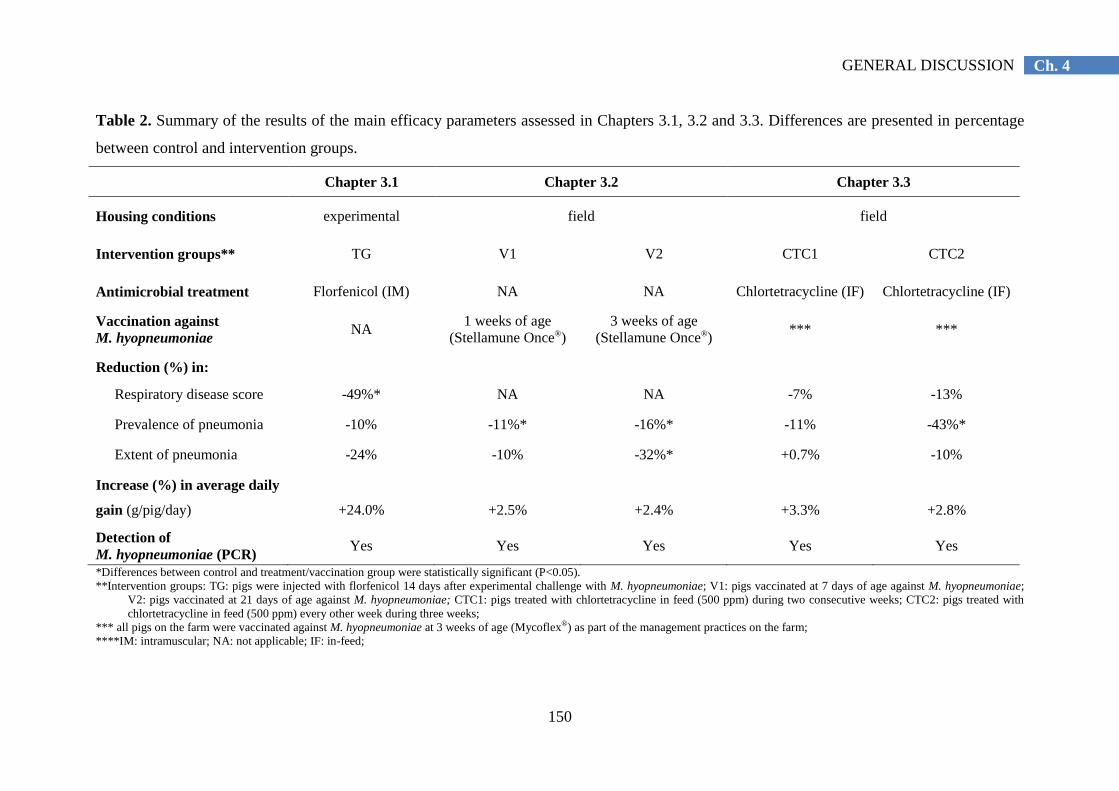
150
Ch. 4 GENERAL DISCUSSION
Table 2. Summary of the results of the main efficacy parameters assessed in Chapters 3.1, 3.2 and 3.3. Differences are presented in percentage
between control and intervention groups.
Chapter 3.1 Chapter 3.2 Chapter 3.3
Housing conditions experimental field field
Intervention groups** TG V1 V2 CTC1 CTC2
Antimicrobial treatment Florfenicol (IM) NA NA Chlortetracycline (IF) Chlortetracycline (IF)
Vaccination against
M. hyopneumoniae NA
1 weeks of age
(Stellamune Once®)
3 weeks of age
(Stellamune Once®) *** ***
Reduction (%) in:
Respiratory disease score -49%* NA NA -7% -13%
Prevalence of pneumonia -10% -11%* -16%* -11% -43%*
Extent of pneumonia -24% -10% -32%* +0.7% -10%
Increase (%) in average daily
gain (g/pig/day)
+24.0%
+2.5%
+2.4%
+3.3%
+2.8%
Detection of
M. hyopneumoniae (PCR) Yes Yes Yes Yes Yes
*Differences between control and treatment/vaccination group were statistically significant (P<0.05).
**Intervention groups: TG: pigs were injected with florfenicol 14 days after experimental challenge with M. hyopneumoniae; V1: pigs vaccinated at 7 days of age against M. hyopneumoniae;
V2: pigs vaccinated at 21 days of age against M. hyopneumoniae; CTC1: pigs treated with chlortetracycline in feed (500 ppm) during two consecutive weeks; CTC2: pigs treated with
chlortetracycline in feed (500 ppm) every other week during three weeks;
*** all pigs on the farm were vaccinated against M. hyopneumoniae at 3 weeks of age (Mycoflex®) as part of the management practices on the farm;
****IM: intramuscular; NA: not applicable; IF: in-feed;
151
Ch. 4 GENERAL DISCUSSION
General conclusions
From the studies included in this thesis (Table 2), it was concluded that both
antimicrobial medication and vaccination against M. hyopneumoniae were able to reduce
clinical signs and extent and prevalence of pneumonia lesions, as well as to improve
performance in pigs infected with M. hyopneumoniae. However, M. hyopneumoniae was not
eliminated from infected animals by these control strategies.
More specifically, it can be concluded that:
The tested florfenicol formulation, when administered intramuscularly as a single
dose of 30 mg/kg bodyweight, was effective in reducing the clinical respiratory
symptoms in pigs experimentally infected with a highly virulent M. hyopneumoniae
strain. A temporary reduction of clinical signs (4 days) was suggested after
treatment. This metaphylactic medication characterized by a higher concentration of
active substance may significantly reduce the antimicrobial use in pig herds.
Vaccination against M. hyopneumoniae at either 7 or 21 days of age significantly
reduced pneumonia lesions and numerically decreased growth losses in a herd with
clinical respiratory problems during the second half of the fattening period due to
combined M. hyopneumoniae and viral infections.
Chlortetracycline hydrochloride, when administered via the feed at a dose of 500
ppm during two alternative weeks at onset of clinical outbreak of respiratory disease,
decreased the prevalence of M. hyopneumoniae-like lesions, and numerically
reduced performance losses and clinical signs compared with tylosin phosphate at a
dose of 100 ppm administered during three weeks. This metaphylactic medication
may significantly reduce the antimicrobial use in pig herds.
Clinical outbreaks of respiratory tract disease requiring antimicrobial medication
may still occur in herds without major deficiencies in housing and management
conditions, despite vaccination against two major pathogens (M. hyopneumoniae and
porcine PCV-2) involved in PRDC.
Vaccination and antimicrobial medication may reduce but do not prevent infections
with M. hyopneumoniae.

152
Ch. 4 GENERAL DISCUSSION
Further research
Future research on antimicrobials should be focused on in vitro and in vivo efficacy.
The creation of a Mycoplasmotheque including different M. hyopneumoniae strains, followed
by testing their antimicrobial susceptibility (MIC) to the main antimicrobials used in practice
may lead to a better understanding of the in vitro efficacy. A subsequent correlation of this in
vitro efficacy with in vivo models should be investigated. The association between
antimicrobial susceptibility (MIC values) and quantification of the reduction of M.
hyopneumoniae shedding (by qPCR) after antimicrobial treatment might be studied. This
standardization would facilitate the registration of new products according to the EU laws,
the establishment of correct guidelines of prudent use of antimicrobials against M.
hyopneumoniae, as well as a reliable monitoring of the antimicrobial resistance against this
pathogen. A better understanding of the pharmacokinetics may be achieved by the
quantification of the antimicrobial concentration in plasma and at the site of infection. Since
M. hyopneumoniae is a mucosal pathogen which mainly adheres to the cilia of the epithelial
cells from the respiratory tract, the concentration of antimicrobial in BAL fluid and its
correlation with therapeutic levels and MIC values might be used as indicators of the
expected in vivo efficacy.
Early vaccination has been demonstrated to be efficacious against M. hyopneumoniae
infections occurring late in the fattening period. So far, there are no reports investigating the
effect of different early vaccination schemes in herds with infections occurring shortly after
weaning. In addition, vaccination is often applied at weaning, as piglets are handled anyway
at that time. However, weaning is also causing stress to the piglets. The effect of the stress
due to weaning on efficacy of vaccination warrants further studies.
Apart from treatment and vaccination, the prevention or control of M. hyopneumoniae
infections in pig herds by optimizing housing conditions and management practices remains
important. Further studies are guaranteed in this field.
Further research to develop and validate technical devices that guarantee standard and
controlled conditions along the experiments is required. Application of computerized systems
able to recognize coughing of live pigs and to monitor the frequency of this coughing, as well
as to recognize macroscopic lung lesions at slaughter and calculate the extent of pulmonary
lesions may limit variability due to subjective measurements.
153
Ch. 4 GENERAL DISCUSSION
REFERENCES
Alexopoulos, C., Kritas, S.K., Papatsas, I., Papatsiros, V.G., Tassis, P.D. and Kyriakis, S.C. (2004). Efficacy of
one and two shot vaccines for the control of enzootic pneumonia (EP) in a pig unit suffering from respiratory
syndrome due to EP, PRRS and PMWS. Proceedings of the 18th
International Pig Veterinary Society
Congress. Hamburg, Germany, June 27 to July 1, p 449.
Barigazzi, G., Candotti, P., Foni, E., Martinelli, L. and Raffo, A. (1996). In vitro susceptibility of 108 isolated
Actinobacillus pleuropneumoniae strains to 17 antimicrobial agents from pig lungs in Italy in 1994-95. In:
Proceedings of the 14th
International Pig Veterinary Society Congress, Bologna, Italy. Reading, July 7 to 10,
p 207.
Blanchard, B., Vena, M.M., Cavalier, A, Le Lannic, J., Gouranton, J. and Kobisch, M. (1992). Electron
microscopic observation of the respiratory tract of SPF piglets inoculated with Mycoplasma hyopneumoniae.
Veterinary microbiology 30, 329–341.
Burch, D.G.S. (2012). Examination of the pharmacokinetic/pharmacodynamic (PK/PD) relationships of orally
administered antimicrobials and their correlation with the therapy of various bacterial and mycoplasmal
infections in pigs. Royal College of Veterinary Surgeons, London. Thesis.
Catry, B., Duchateau, L., Van de Ven, J., Laevens, H., Opsomer, G., Haesebrouck, F. and de Kruif, A. (2008).
Efficacy of metaphylactic florfenicol therapy during natural outbreaks of bovine respiratory disease. Journal
of Veterinary Pharmacology Therapy 31, 479-487.
Ciprián, A., Palacios, J.M., Quintanar, D., Batista, L., Colmenares, G., Cruz, T., Romero, A., Schnitzlein, W.
and Mendoza, S. (2012). Florfenicol feed supplemented decrease the clinical effects of Mycoplasma
hyopneumoniae experimental infection in swine in México. Research in Veterinary Science 92, 191–196.
Clinical And Laboratory Standards Institute (CLSI) (2008). Performance standards for antimicrobial disk and
dilution susceptibility tests for bacteria isolated from animals; approved standard. 3rd edn. M31-A3.
Del Castillo J.R.E., Elsener, J. and Martineau, G.P. (1998). Pharmacokinetic modeling of in-feed tetracycline in
pigs using a meta-analytic compartmental approach. Swine Health Production 5, 189-202.
Del Castillo, J.R.E. (2013). Tetracyclines. In: Giguère, S., Prescott, J.F. and Dowling, P.M. (Eds). Antimicrobial
Therapy in Veterinary Medicine. John Wiley & Sons, pp. 257-268.
Djordjevic, S.P., Eamens, G.J., Romalis, L.F., Nicholls, P.J., Taylor, V. and Chin, J. (1997). Serum and mucosal
antibody responses and protection in pigs vaccinated against Mycoplasma hyopneumoniae with vaccines
containing a denatured membrane antigen pool and adjuvant. Australian Veterinary Journal 75, 504-511.
Dohoo, I., Martin, W. and Stryhn, H. (2009). Controlled studies. In: Dohoo, I., Martin, W. and Stryhn, H. (Eds.)
Veterinary Epidemiologic Research. VER Inc., Prince Edward Island, Canada, pp 213-241.
Dowling, P.M. (2013). Chloramphenicol, Thiamphenicol, and Florfenicol. In: Giguère, S., Prescott, J.F. and
Dowling, P.M. (Eds). Antimicrobial Therapy in Veterinary Medicine. John Wiley & Sons, pp. 269-277.
Drlica, K. (2003). The mutant selection window and antimicrobial resistance. Journal of Antimicrobial
Chemotherapy 52, 11-17.
Exadaktylos, V., Silva, M., Aerts, J.M., Taylor, C.J. and Berckmans, D. (2008). Real-time recognition of sick
pig cough sounds. Computers and Electronics in Agriculture 63, 207–214.
Fablet, C., Marois-Créhan, C., Simon, G., Grasland, B., Jestin, A., Kobisch, M., Madec, F. & Rose, N. (2012a)
Infectious agents associated with respiratory diseases in 125 farrow-to-finish pig herds: A cross-sectional
study. Veterinary Microbiology 157, 152-163.
Fablet, C., Marois, C., Dorenlor, V., Eono, F., Eveno, E., Jolly, J.P., Le Devendec, L., Kobisch, M., Madec, F.
and Rose, N. (2012b). Bacterial pathogens associated with lung lesions in slaughter pigs from 125 herds.
Research in Veterinary Science 93, 627–630.
Fano, E., Pijoan, C., Dee, S. and Deen, J. (2007). Effect of Mycoplasma hyopneumoniae colonization at weaning
on disease severity in growing pigs. Canadian Journal of Veterinary Research 71, 195–200.
Fraile, L., Alegre, A., López-Jiménez, R., Nofrarías, M. and Segalés, J. (2010). Risk factors associated with
pleuritis and cranio-ventral pulmonary consolidation in slaughter-aged pigs. Veterinary journal 184, 326–
333.
Ganter, M., Dudziak, D. and Delbeck, F. (1995). Treatment of swine with chronic pneumonia with
chlortetracycline-medicated feed. Deutsche Tierärztliche Wochenschrift 102, 44-49.
154
Ch. 4 GENERAL DISCUSSION
Goossens, J., Pasmans, F., Verbrugghe, E., Vandenbroucke, V., De Baere, S., Meyer, E., Haesebrouck, F., De
Backer, P. and Croubels, S. (2012a). Porcine intestinal epithelial barrier disruption by the Fusarium
mycotoxins deoxynivalenol and T-2 toxin promotes transepithelial passage of doxycycline and
paromomycin. BMC Veterinary Research 17, 245.
Goossens, J., Vandenbroucke, V., Pasmans, F., De Baere, S., Devreese, M., Osselaere, A., Verbrugghe, E.,
Haesebrouck, F., De Saeger, S., Eeckhout, M., Audenaert, K., Haesaert, G., De Backer, P. and Croubels, S.
(2012b). Influence of mycotoxins and a mycotoxin adsorbing agent on the oral bioavailability of commonly
used antibiotics in pigs. Toxins 4, 281-295.
Goossens, J., Devreese, M., Pasmans, F., Osselaere, A., De Baere, S., Verbrugghe, E., Haesebrouck, F., De
Backer P. and Croubels, S. (2013). Chronic exposure to the mycotoxin T-2 promotes oral absorption of
chlortetracycline in pigs. Journal of Veterinary Pharmacology Therapy 36, 621-624.
Greiner, L., Connor, J.F. and Lowe, J.F. (2011). Comparison of Mycoplasma hyopneumoniae vaccination. In:
Proceedings of the 42nd
Annual Meeting of the American Association of Swine Veterinarians, Phoenix,
Arizona, March 5 to 8, p 245-248.
Guiguere, S. (2013). Macrolides, Azalides, and Ketolides. In: Giguère, S., Prescott, J.F. and Dowling, P.M.
(Eds). Antimicrobial Therapy in Veterinary Medicine. John Wiley & Sons, pp. 211-232.
Hannan, P.C., Bhogal, B.S. and Fish, J.P. (1982). Tylosin tartrate and tiamutilin effects on experimental piglet
pneumonia induced with pneumonic pig lung homogenate containing mycoplasmas, bacteria and viruses.
Research in Veterinary Science 33, 76–88.
Hannan, P.C.T. (2000). Guidelines and recommendations for antimicrobial minimum inhibitory concentration
testing against veterinary mycoplasma species. Veterinary Research 31, 373-395.
Haesebrouck, F., Pasmans, F., Chiers, K., Maes, D., Ducatelle, R. and Decostere, A. (2004). Efficacy of
vaccines against bacterial diseases in swine: what can we expect? Veterinary microbiology 100, 255–268.
Inamoto, T., Takahashi, H., Yamamoto, K., Nakai, Y. and Ogimoto, K. (1994). Antibiotic susceptibility of
Mycoplasma hyopneumoniae isolated from swine. The Journal of Veterinary Medical Science 56, 393-394.
Jensen, C.S., Ersbøll, A.K. and Nielsen, J.P. (2002). A meta-analysis comparing the effect of vaccines against
Mycoplasma hyopneumoniae on daily weight gain in pigs. Preventive veterinary medicine 54, 265–278.
Kilroy, C.R., Hall, W.F., Bane, D.P., Bevill, R.F. and Koritz, G.D. (1990). Chlortetracycline in swine -
bioavailability and pharmacokinetics in fasted and fed pigs. Journal of Veterinary Pharmacology Therapy
13, 49-58.
Kunesh, J. (1981). A comparison of two antibodies in treating mycoplasma pneumonia in swine. Veterinary
Medicine. Small Animal Clinics 76, 871-872.
Li, J., Petit-Jetté, C.E., Gohore, B.D., Fenneteau, F., Del Castillo, J.R. and Nekka, F. (2008). Assessing
pharmacokinetic variability directly induced by drug intake behaviour through development of a feeding
behaviour-pharmacokinetic model. Journal of Theoretical Biology 251, 468-479.
Lillie, K., Ritzmann, M. and Heinritzi, K. (2004). Field study on the effect of the early single dose Mycoplasma
hyopneumoniae vaccination (Stellamune® One) in a naturally infected herd. In: Proceedings of the 18
th
International Pig Veterinary Society Congress, Hamburg, Germany, June 27 to July 1, p. 414.
Liu, J., Fung, K.F., Chen, Z., Zeng, Z. and Zhang, J. (2003). Pharmacokinetics of florfenicol in healthy pigs and
in pigs experimentally infected with Actinobacillus pleuropneumoniae. Antimicrobial Agents and
Chemotherapy 47, 820-823.
Maes, D., Deluyker, H., Verdonck, M., Castryck, F., Miry, C., Vrijens, B., Verbeke, W., Viaene, J. and de
Kruif, A. (1999). Effect of vaccination against Mycoplasma hyopneumoniae in pig herds with an all-in/all-
out production system. Vaccine 17, 1024-1034.
Maes, D., Deluyker, H., Verdonck, M., Castryck, F., Miry, C., Vrijens, B. and de Kruif, A. (2000). Herd factors
associated with the seroprevalences of four major respiratory pathogens in slaughter pigs from farrow-to-
finish pig herds. Veterinary Research 31, 313-327.
Maes, D., Segales, J., Meyns, T., Sibila, M., Pieters, M. and Haesebrouck, F. (2008). Control of Mycoplasma
hyopneumoniae infections in pigs. Veterinary Microbiology 126, 297–309.
Martelli, P., Terreni, M., Guazzetti, S. and Cavirani, S. (2006). Antibody response to Mycoplasma
hyopneumoniae infection in vaccinated pigs with or without maternal antibodies induced by sow
vaccination. Journal of veterinary medicine. B, Infectious diseases and veterinary public health 53, 229–233.

155
Ch. 4 GENERAL DISCUSSION
Mateusen, B., Maes, D., Van Goubergen, M., Verdonck, M. and de Kruif, A. (2002). Effectiveness of treatment
with lincomycin hydrochloride and/or vaccination against Mycoplasma hyopneumoniae for controlling
chronic respiratory disease in a herd of pigs. The Veterinary Record 151, 135-140.
Merialdi, G., Dottori, M., Bonilauri, P., Luppi, A., Gozio, S., Pozzi, P., Spaggiari, B. and Martelli, P. (2012).
Survey of pleuritis and pulmonary lesions in pigs at abattoir with a focus on the extent of the condition and
herd risk factors. Veterinary journal 193, 234–239.
Meyns, T., Dewulf, J., de Kruif, A., Calus, D., Haesebrouck, F. and Maes, D. (2006) Comparison of
transmission of Mycoplasma hyopneumoniae in vaccinated and non-vaccinated populations. Vaccine 24,
7081-7086.
Meyns, T., Van Steelant, J., Rolly, E., Dewulf, J., Haesebrouck, F. and Maes, D. (2011). A cross-sectional study
of risk factors associated with pulmonary lesions in pigs at slaughter. Veterinary journal 187, 388–392.
Nathues, H., Kubiak, R., Tegeler, R. and Grosse Beilage, E. (2010). Occurrence of Mycoplasma hyopneumoniae
infections in suckling and nursery pigs in a region of high pig density. The Veterinary Record 166, 194-198.
Nathues, H., Spergser, J., Rosengarten, R., Kreienbrock, L. and Grosse Beilage E. (2012). Value of the clinical
examination in diagnosing enzootic pneumonia in fattening pigs. The Veterinary Journal 193, 443-447.
Regula, G., Lichtensteiger, C.A., Mateus-Pinilla, N.E., Scherba, G., Miller, G.Y. and Weigel, R.M. (2000).
Comparison of serologic testing and slaughter evaluation for assessing the effects of subclinical infection on
growth in pigs. Journal of American Veterinary Medicine Association 217, 888-895.
Pommier, P. (2006). In vitro, are all the tetracyclines equivalent? In: Proceedings of the 19th
International Pig
Veterinary Society Congress, Copenhagen, Denmark. Reading, July, 16 to 19, p 411.
Priebe, S. and Schwarz, S. (2003). In vitro activities of florfenicol against bovine and porcine respiratory tract
pathogens. Antimicrobial Agents and Chemotherapy 47, 2703-2705.
Reynolds, S.C., St Aubin, L.B., Sabbadini, L.G., Kula, J., Vogelaar, J., Runnels, P. and Peters, A.R. (2009).
Reduced lung lesions in pigs challenged 25 weeks after the administration of a single dose of Mycoplasma
hyopneumoniae vaccine at approximately 1 week of age. The Veterinary Journal 181, 312-320.
Ritzmann, M., Lillie, K., Palzer, A. and Heinritzi, K. (2006). Influence of maternal antibodies on Mycoplasma
hyopneumoniae vaccines. In: Proceedings of the Allen D. Leman Swine Conference, Saint Paul, Minnesota,
September 23 to 26.
Roof, M., Burkhart, K. and Zuckermann, F.A. (2001). Evaluation of the immune response and efficacy of 1 and
2 dose commercial Mycoplasma hyopneumoniae bacterins. In: Proceedings of the 32nd
Annual Meeting of
the American Association of Swine Veterinarians, Nashville, Tennessee, February 27 to 27, pp. 163-167.
Roof, M., Burkhart, K. and Zuckermann, F. (2002). Pig efficacy study comparing Mycoplasma vaccines used at
one and 2 doses. In: Proceedings of the 17th
International Pig Veterinary Society Congress, Ames, Iowa,
June 2 to 5, p 395.
Scorneaux, B. and Shryock, T.R. (1998). Intracellular accumulation, subcellular distribution and efflux of
tilmicosin in swine phagocytes. Journal of Veterinary Pharmacology and Therapeutics 21, 257-268.
Segalés, J., Valero, O., Espinal, A., López-Soria, S., Nofrarías, M., Calsamiglia, M. and Sibila, M. (2012).
Exploratory study on the influence of climatological parameters on Mycoplasma hyopneumoniae infection
dynamics. International journal of biometeorology 56, 1167–1171.
Sibila, M., Calsamiglia, M., Vidal, D., Badiella, L., Aldaz, A. & Jensen, J.C. (2004). Dynamics of Mycoplasma
hyopneumoniae infection in 12 farms with different production systems. Canadian Journal of Veterinary
Research, 68, 12–18.
Sibila, M, Nofrarías, M., López-Soria, S., Segalés, J., Riera, P., Llopart, D., and Calsamiglia, M. (2007).
Exploratory field study on Mycoplasma hyopneumoniae infection in suckling pigs. Veterinary microbiology
121, 352–356.
Sibila, M., Pieters, M., Molitor, T., Maes, D., Haesebrouck, F. and Segalés, J. (2009). Current perspectives on
the diagnosis and epidemiology of Mycoplasma hyopneumoniae infection. The Veterinary journal 181, 221–
231.
Sorensen, V., Ahrens, P., Barfod, K., Feenstra, A.A., Feld, N.C., Friis, N.F., Bille-Hansen, V., Jensen, N.E. and
Pedersen, M.W. (1997). Mycoplasma hyopneumoniae infection in pigs: duration of the disease and
evaluation of four diagnostic assays. Veterinary Microbiology 54, 23–34.
Straw, B.E., Backstrom, L. and Leman, A.D. (1986). Examination of swine at slaughter. Part II. Findings at
slaughter and their significance. Compendium on Continuing Education for the Practicing Veterinarian 8,
106–112.
156
Ch. 4 GENERAL DISCUSSION
Thacker, E.L., Thacker, B.J., Boettcher, T.B. and Jayappa, H. (1998).Comparison of antibody production,
lymphocyte stimulation, and protection induced by four commercial Mycoplasma hyopneumoniae bacterins.
Swine Health Production 6, 107–112.
Thacker, E.L., Thacker, B.J. and Janke, B.H. (2001). Interaction between Mycoplasma hyopneumoniae and
swine influenza virus. Journal of Clinical Microbiology 39, 2525-2530.
Thacker, E., Thacker, B., and Wolff, T. (2006). Efficacy of a chlortetracycline feed additive in reducing
Mycoplasma hyopneumoniae challenge. Journal of Swine Health and Production 14, 140–144.
Thacker, E.L. and Minion, F.C. (2012). Mycoplasmosis. In: Zimmerman, J.J., Karriker, L.A., Ramírez, A.,
Schwartz, K.J., Stevenson, G.W. (Eds.). Diseases of Swine. Wiley-Blackwell, Ames, Iowa, USA, pp. 779–
797.
Ueda, Y., Ohtsuki, S., Narukawa, N. and Takeda, K. (1994). Effect of terdecamycin on experimentally induced
Mycoplasma hyopneumoniae-infection in pigs. Journal of Veterinary Medicine Series B 41, 283-290.
Vicca, J., Maes, D., Thermote, L., Peeters, J., Haesebrouck, F. and de Kruif, A. (2002). Patterns of Mycoplasma
hyopneumoniae infections in Belgian farrow-to-finish pig herds with diverging disease-course. Journal of
veterinary medicine. B, Infectious diseases and veterinary public health 49, 349–353.
Vicca, J., Stakenborg, T., Maes, D., Butaye, P., Peeters, J., and de Kruif, A. (2004). In vitro susceptibilities of
Mycoplasma hyopneumoniae field isolates 48, 4470–4472.
Vicca, J. (2005). Virulence and antimicrobial susceptibility of Mycoplasma hyopneumoniae isolates from pigs.
Faculty of Veterinary Medicine. Ghent University. Thesis.
Vicca, J., Maes, D., Jonker, L., de Kruif, A. and Haesebrouck, F. (2005). Efficacy of in-feed medication with
tylosin for the treatment and control of Mycoplasma hyopneumoniae infections. The Veterinary Record 156,
606-610.
Villarreal, I., Vranckx, K., Duchateau, L., Pasmans, F., Haesebrouck, F., Jensen, J.C., Nanjiani, I.A. and Maes,
D. (2010). Early Mycoplasma hyopneumoniae infections in European suckling pigs in herds with respiratory
problems: detection rate and risk factors. Veterinarni Medicina 55, 318-324.
Villarreal, I., Meyns, T., Dewulf, J., Vranckx, K., Calus, D., Pasmans, F., Haesebrouck, F. and Maes, D. (2011).
The effect of vaccination on the transmission of Mycoplasma hyopneumoniae in pigs under field conditions.
The Veterinary Journal 188, 48-52.
Voorspoels, J., D'Haese, E., De Craene, B.A., Vervaet, C., De Riemaecker, D., Deprez, P., Nelis, H. and
Remon, J.P. (1999). Pharmacokinetics of florfenicol after treatment of pigs with single oral or intramuscular
doses or with medicated feed for three days. Veterinary Record 145, 397-399.
Vranckx, K., Maes, D., Del Pozo Sacristán, R., Pasmans, F. and Haesebrouck, F. (2012a). A longitudinal study
of the diversity and dynamics of Mycoplasma hyopneumoniae infections in pig herds. Veterinary
Microbiology 156, 315-321.
Vranckx, K., Maes, D., Marchioro, S.B., Villarreal, I., Chiers, K., Pasmans, F. and Haesebrouck, F. (2012b).
Vaccination reduces macrophage infiltration in bronchus-associated lymphoid tissue in pigs infected with a
highly virulent Mycoplasma hyopneumoniae strain. BMC Veterinary Research 8, 24.
Walter, D., Dvm, J.T.H., Sornsen, S., Hagen, C. and Harris, I.T. (2000). The effect of a metaphylactic pulse
dosing in-feed antimicrobial strategy on finishing pig health and performance. Swine Health and Production
8, 65–71.
Wilhelm, C., Gautier-Bouchardon, A.V., Boyen, F., Spergser, J. and Thomas, E. (2012). Determination of the in
vitro activity of florfenicol against Mycoplasma hyopneumoniae. In: Proceedings 22nd
IPVS south Korea, p
702.

SUMMARY
SUMMARY
Summary


159
SUMMARY
SUMMARY
Mycoplasma hyopneumoniae is the primary agent of enzootic pneumonia, a chronic
respiratory disease in pigs resulting from mixed respiratory infections with M.
hyopneumoniae and other bacterial pathogens. M. hyopneumoniae infections lead to major
economic losses due to the reduced growth, increased mortality and feed conversion, costs
for antimicrobials and immunoprophylaxis and increased time to market.
The control of M. hyopneumoniae infections can be accomplished by three different
strategies, namely optimization of housing and management practices, antimicrobial
treatment and vaccination. Both, antimicrobial treatment and vaccination have been shown to
reduce infections with M. hyopneumoniae, but not to eliminate M. hyopneumoniae from
infected pigs. Despite vaccination, clinical outbreaks of respiratory disease due to M.
hyopneumoniae infections may still occur. Nevertheless, there are limited studies available
describing the in vivo efficacy of antimicrobials under experimental M. hyopneumoniae
induced infections. Similarly, scarce data is available regarding the efficacy of different
treatment and vaccination strategies against M. hyopneumoniae in commercial herds infected
with this bacterium and other pathogens involved in Porcine Respiratory Disease Complex
(PRDC). These current control measures are beneficial from an economic point of view.
Answers to these questions are necessary for a development of optimal treatment and control
strategies for this disease.
The general aim of this thesis was to obtain better insights regarding different treatment
and control strategies against M. hyopneumoniae infections.
In the first study (Chapter 3.1), the efficacy of a single intramuscular injection of
florfenicol to treat clinical respiratory disease following experimental M. hyopneumoniae
infection was investigated. M. hyopneumoniae-free piglets were allocated to three groups,
namely, a treatment (TG) and positive control group (PCG), which were both inoculated
endotracheally with a highly virulent isolate of M. hyopneumoniae, and a negative control
group. At onset of clinical disease, TG received a single injection of a new florfenicol
formulation (30 mg/kg). All pigs were euthanized 4 weeks post-infection. Clinical symptoms
were significantly reduced in TG in comparison with PCG (P<0.05). Average daily weight
gain, feed conversion ratio, mortality and lung lesions were improved in TG compared to
PCG, but the differences were not statistically significant. The results showed that, following
an experimental M. hyopneumoniae infection, a single injection with the tested florfenicol

160
SUMMARY
significantly decreased clinical symptoms and numerically improved severity of lung lesions
and performance when compared to a non-treated control group.
In the second study (Chapter 3.2), the efficacy of early M. hyopneumoniae vaccination
in a farrow-to-finish pig herd with respiratory disease late in the fattening period due to
combined infections with M. hyopneumoniae and viral pathogens was investigated. Five-
hundred-and-forty piglets were randomly divided into three groups of 180 piglets each: two
groups were vaccinated (Stellamune Once®) at either 7 (V1) or 21 days of age (V2), and a
third group was left non-vaccinated (NV). The three treatment groups were housed in
different pens within the same compartment during the nursery period, and were housed in
different but identical compartments during the fattening period. The efficacy was evaluated
using performance and pneumonia lesions. The average daily weight gain during the
fattening period was 19 (V1) and 18 g/day (V2) higher in the vaccinated groups when
compared with the NV group. However, the difference was not statistically significant
(P>0.05). The prevalence of pneumonia at slaughter was significantly lower in both
vaccinated groups (V1:71.5 and V2:67.1 per cent) when compared with the NV group (80.2
per cent) (P<0.05). There were no significant differences between the two vaccination
groups. In this herd with respiratory disease during the second half of the fattening period
caused by M. hyopneumoniae and viral infections, prevalence of pneumonia lesions was
significantly reduced and growth losses numerically (not statistically significant) decreased
by both vaccination schedules.
In the third study (Chapter 3.3), the efficacy of chlortetracycline in-feed medication to
treat pigs with clinical respiratory disease was investigated in a farrow-to-finish pig herd
infected with M. hyopneumoniae and with clinical respiratory disease in growing pigs. In
total, 533 pigs were included. The animals were vaccinated against M. hyopneumoniae and
PCV-2 at weaning. At onset of clinical respiratory disease, they were randomly allocated to
one of the following treatment groups: chlortetracycline 1 (CTC1) (two consecutive weeks,
500 ppm), chlortetracycline 2 (CTC2) (two non-consecutive weeks, with a non-medicated
week-interval in between, 500 ppm) or tylosin (T) (three consecutive weeks, 100 ppm).
Performance (average daily gain, feed conversion ratio), pneumonia lesions at slaughter and
clinical parameters (respiratory disease score) were assessed. Only numeric differences in
favour of the CTC2 group were obtained for the performance and the clinical parameters. The
prevalence of pneumonia lesions was 20.5, 13.1 and 23.0 per cent (P<0.05) for the CTC1,
CTC2 and T groups, respectively. The study demonstrated that chlortetracycline, when
administered at onset of clinical respiratory disease via the feed at a dose of 500 ppm during

161
SUMMARY
two alternative weeks, was able to decrease the prevalence of pneumonia lesions, and
numerically reduce performance losses and clinical signs.
From the studies included in this thesis, it was concluded that both antimicrobial
medication and vaccination against M. hyopneumoniae were able to reduce clinical signs and
extent and prevalence of pneumonia lesions, as well as to improve performance in pigs
infected with M. hyopneumoniae. Although M. hyopneumoniae was not eliminated from
infected animals by these control strategies, metaphylactic medication and vaccination may
contribute to the reduction of antimicrobial use in pig herds.
Future research on antimicrobials could focus on in vitro and in vivo efficacy, e.g. to
better understand the association between antimicrobial susceptibility (MIC values) and
quantification of the reduction of M. hyopneumoniae shedding (by qPCR) after antimicrobial
treatment. In addition, the quantification of antimicrobial concentration in plasma and at the
site of infection and its correlation with MIC values might be used as indicators of the
expected in vivo efficacy. Interesting further research on vaccination against M.
hyopneumoniae could address the effect of different early vaccination schemes in herds with
infections occurring shortly after weaning. The effect of stress at vaccination, for example at
weaning, on efficacy of vaccination warrants further studies. Development and validation of
technical devices that guarantee standard and controlled conditions along the experiments
may be required. Future studies might focus on the application of computerized systems to
recognize coughing of live pigs as well as on macroscopic lung lesions at slaughter in order
to reduce variability of subjective measurements.


SAMENVATTING
SAMENVATTING
Samenvatting


165
SAMENVATTING
SAMENVATTING
Mycoplasma hyopneumoniae is het causaal agens van een chronische respiratoire ziekte
bij varkens genaamd enzoötische pneumonie. Deze ziekte ontstaat door menginfecties van M.
hyopneumoniae en andere bacteriële pathogenen. Infectie met M. hyopneumoniae leidt tot
grote economische verliezen, verminderde groei, een hoger sterftecijfer, hogere
voederconversie, meer kosten aan profylactische en antimicrobiële middelen en de varkens
hebben meer tijd nodig om slachtgewicht te bereiken.
Drie strategieën om M. hyopneumoniae-infecties onder controle te houden worden
aangeraden: optimaliseren van huisvesting en management, antimicrobiële medicatie en
vaccinatie. Deze twee laatste strategieën zijn in staat om infecties met M. hyopneumoniae te
reduceren, maar niet te elimineren. Niettegenstaande vaccinatie wordt uitgevoerd, kunnen er
nog steeds infecties met M. hyopneumoniae voorkomen. Er is slechts weinig onderzoek
beschikbaar dat de werkzaamheid van antimicrobiële middelen tegen experimenteel
geïnduceerde M. hyopneumoniae-infecties beschrijft. Eveneens is er weinig kennis omtrent
de werkzaamheid van behandelings- en vaccinatiestrategieën tegen M. hyopneumoniae in
commerciële varkensbedrijven waar M. hyopneumoniae en andere pathogenen betrokken in
het Porcine Respiratoir Ziekte Complex (PRDC) een rol spelen. Deze huidige
controlemaatregelen zijn interessant vanuit economisch oogpunt, bijgevolg zijn antwoorden
op deze vragen noodzakelijk voor de ontwikkeling van een optimale behandelings- en
controlestrategie voor deze ziekte.
Het doel van deze doctoraatsthesis was om een beter inzicht te verkrijgen wat
verschillende behandelings- en controlestrategieën tegen M. hyopneumoniae betreft.
In de eerste studie (hoofdstuk 3.1) werd de werkzaamheid van een éénmalige
intramusculaire injectie met florfenicol voor de behandeling van een experimentele M.
hyopneumoniae infectie nagegaan. Biggen, vrij van M. hyopneumoniae, werden aan drie
groepen toegewezen: een behandelingsgroep (BH), een positieve controlegroep (PCG) en een
negatieve controlegroep. De BH en PCG werden beiden endotracheaal geïnoculeerd met een
hoogvirulent isolaat van M. hyopneumoniae. Bij de aanvang van de klinische symptomen
kreeg de BH een enkelvoudige dosis van een nieuwe florfenicol-formulatie (30 mg/kg). Alle
biggen werden vier weken na infectie geëuthanaseerd. De klinische symptomen waren
significant lager in de BH in vergelijking met de PCG (P<0.05). De dagelijkse groei, de
voederconversie, het sterftecijfer en de longletsels in de BH werden positief beïnvloed in
vergelijking met de PCG, maar de verschillen waren niet statistisch significant. Uit deze
166
SAMENVATTING
resultaten kon worden besloten dat een injectie met de geteste florfenicol-formulatie na een
experimentele M. hyopneumoniae infectie de klinische symptomen significant deed afnemen
en dat een numerieke verbetering kon gezien worden in de ernst van longlesies en productie
in vergelijking met de niet behandelde groep.
In de tweede studie (hoofdstuk 3.2) werd de werkzaamheid van een vroege M.
hyopneumoniae vaccinatie onderzocht in een gesloten bedrijf met respiratoire ziektetekenen
die optraden laat in de afmestperiode te wijten aan een combinatie van M. hyopneumoniae en
virale pathogenen. Vijfhonderd veertig biggen werden at random verdeeld over drie groepen
van 180 biggen: twee groepen werden gevaccineerd (Stellamune Once®) op respectievelijk 7
(V1) of 21 (V2) dagen leeftijd en een derde groep werd niet gevaccineerd (NV). In de
biggen-batterij werden de biggen gehuisvest in verschillende hokken binnen dezelfde stal en
tijdens de afmestperiode in verschillende maar gelijkaardige compartimenten. De
werkzaamheid werd beoordeeld door middel van productieparameters en pneumonielesies.
De dagelijkse groei tijdens de afmestperiode was 19 (V1) en 18 g/dag (V2) hoger in de
gevaccineerde groepen in vergelijking met de NV groep, hoewel het verschil niet statistisch
significant was (P>0.05). De prevalentie van pneumonie aan de slachtlijn was significant
lager in beide gevaccineerde groepen (V1: 71.5 en V2: 67.1 per cent) wanneer vergeleken
werd met de NV groep (80.2 per cent) (P<0.05). Tussen de twee gevaccineerde groepen
werden er geen significante verschillen gezien. In dit bedrijf met late respiratoire symptomen
te wijten aan een combinatie van M. hyopneumoniae en virale infecties werd dus bij
toepassing van beide vaccinatieschema‟s een statistisch significante verbetering gezien van
de pneumonie prevalentie en een numeriek (niet statistisch significant) betere groei.
In de derde studie (hoofdstuk 3.3) werd de werkzaamheid nagegaan van
chloortetracycline toegediend in het voeder om varkens in een gesloten bedrijf geïnfecteerd
met M. hyopneumoniae en met klinische respiratoire ziekte te behandelen. Vijfhonderd
drieëndertig varkens werden opgenomen in de studie. De varkens werden gevaccineerd tegen
M. hyopneumoniae en PCV-2 bij het spenen. Bij aanvang van de klinische respiratoire
symptomen werden de dieren at random verdeeld tot een van de volgende
behandelingsgroepen: chloortetracycline 1 (CTC1) (twee opeenvolgende weken, 500 ppm),
chloortetracycline 2 (CTC2) (twee niet-opeenvolgende weken, met een niet gemedicineerde
week als interval tussen de twee weken, 500 ppm) en tylosine (T) (drie opeenvolgende
weken, 100 ppm). Productieparameters (dagelijkse groei, voeder conversie ratio), pneumonie
lesies bij slacht en klinische parameters (respiratoire ziekte score) werden beoordeeld. Voor
de productie- en klinische parameters werden enkel numerieke verschillen ten gunste van de
167
SAMENVATTING
CTC2-groep verkregen. De pneumonie prevalentie was 20.5, 13.1 en 23.0 per cent (P<0.05)
voor respectievelijk de CTC1, CTC2 en de T-groep. Deze studie toonde aan dat wanneer
chloortetracycline toegediend via het voeder in een dosis van 500 ppm tijdens twee niet-
opeenvolgende weken bij de aanvang van de klinische respiratoire symptomen in staat was
om de pneumonie prevalentie te verminderen en om de productie parameters en klinische
symptomen numeriek te verbeteren.
Uit de studies die in deze doctoraatsthesis werden beschreven, kon besloten worden dat
zowel antimicrobiële middelen als vaccinatie tegen M. hyopneumoniae in staat waren om de
klinische symptomen en de uitgebreidheid en prevalentie van pneumonie-letsels te reduceren
en de productieresultaten van varkens geïnfecteerd met M. hyopneumoniae te optimaliseren.
Alhoewel deze controlemaatregelen niet in staat waren om M. hyopneumoniae uit de
geïnfecteerde dieren te elimineren, kunnen metafylactische medicatie en vaccinatie wel
bijdragen tot het reduceren van gebruik van antimicrobiële middelen in de varkensbedrijven.
Verder onderzoek i.v.m. antimicrobiële middelen zou zich kunnen toespitsen op de in
vitro en in vivo werkzaamheid, bv. om de associatie tussen antimicrobiële gevoeligheid
(MIC-waarden) en de kwantificering van de reductie van M. hyopneumoniae na
antimicrobiële behandeling (d.m.v. qPCR) na te gaan. Bovendien kan de kwantificering van
de antimicrobiële concentratie in het plasma en t.h.v. de infectiehaard gecorreleerd met de
MIC-waarde aangewend worden als indicator voor de verwachte in vivo werkzaamheid.
Verder onderzoek aangaande vaccinatie tegen M. hyopneumoniae zou het effect van
verschillende vroege vaccinatieschema‟s kunnen nagaan in bedrijven waar de biggen vroeg
na het spenen geïnfecteerd worden. Er is verder onderzoek nodig naar het effect van stress,
bv. tijdens het spenen, op de werkzaamheid van de vaccinatie. Ontwikkeling en validatie van
technische apparatuur waarbij klinische symptomen en longletsels op een objectieve en
gestandaardiseerde manier gemeten worden, kunnen hun nut hebben. Verder onderzoek kan
zich aldus toespitsen op het gebruik van geautomatiseerde systemen voor het registreren van
hoesten bij varkens en het herkennen van macroscopische longlesies aan de slachtlijn, om zo
de variabiliteit van subjectieve metingen te verminderen.


CURRICULUM VITAE
CURRICULUM VITAE
Curriculum vitae


171
CURRICULUM VITAE
Rubén del Pozo Sacristán was born on June 4th 1981, in Segovia (Spain). He finalized
his secondary studies in Biomedical Sciences in 1999 in the College of H.H. Maristas Ntra.
Señora de la Fuencisla, Segovia. He continued his academic formation at the Faculty of
Veterinary Medicine of León (Spain), where he obtained his DVM diploma in 2007.
Immediately after graduation and during two years, he was working as swine
practitioner in a pig farming integration, Progatecsa, in Valladolid (Spain).
He started an internship program for the European College of Porcine Health
Management in October 2009 at the Unit of Porcine Health Management, Department of
Reproduction, Obstetrics and Herd Health, Faculty of Veterinary Medicine, Ghent
University. From October 2010 onwards, he followed a residency program for the same
college at the same department. His interest in research led him to combine this residency
with different research projects that resulted in a PhD project entitled “Treatment and control
of Mycoplasma hyopneumoniae infections”.
Rubén is first author and co-author of several articles published in international peer
reviewed journals. His experimental work has been presented during different European and
international congresses.


BIBLIOGRAPHY
BIBLIOGRAPHY
Bibliography

175
BIBLIOGRAPHY
Publications in peer-reviewed journals
López Rodríguez, A., Dewulf, J., Meyns, T., Del Pozo Sacristán, R., Chapat, L., Vila, T., Joisel, F.,
Goubier, A., Maes, D. (2014). Effect of sow vaccination against porcine circovirus type 2 on
virological profiles in porcine circovirus diseases affected and non-affected herds. The Canadian
Veterinary Journal. Submitted.
Marchioro, S.B., Del Pozo Sácristan, R., Michiels, A., Haesebrouck, F., Conceição, F.R.,
Dellagostin, O.A. and Maes, D. (2014). Immune responses and efficacy of a chimeric protein
vaccine containing Mycoplasma hyopneumoniae antigens and LTB against experimental M.
hyopneumoniae infection in pigs. Vaccine. In press.
Sahaa, D., Del Pozo Sacristán, R., Van Renne, N., Huang, L., Decaluwe, R., Michiels, A., López
Rodríguez, A., Rodríguez, M.J., García Durán, M., Declerk, I., Maes, D., Nauwynck, H.J. (2014).
Evidence of leakage of anti-PCV2 antibodies from sows to fetuses through the placental barrier:
impact on diagnosis of intra-uterine PCV2 infection. Virologica Sinica 29, 1-3.
Del Pozo Sacristán, R., Michiels, A., Martens, M., Haesebrouck, F., Maes, D. (2014). Efficacy of
vaccination against Actinobacillus pleuropneumoniae in two Belgian farrow-to-finish pig herds
with history of chronic pleuritis. The Veterinary Record 174, 302.
Del Pozo Sacristán, R., Sierens, A., Marchioro, S.B., Vangroenweghe, F., Jourquin, J., Labarque, G.,
Haesebrouck, F., Maes, D. (2014). Efficacy of early Mycoplasma hyopneumoniae vaccination
against mixed respiratory disease in older fattening pigs. The Veterinary Record 174, 197.
Marchioro, S.B., Maes, D., Flahou, B., Pasmans, F., Del Pozo Sacristán, R., Vranckx, K.,
Melkebeek, V., Cox, E., Wuyts, N., Haesebrouck, F. (2013). Local and systemic immune
responses in pigs intramuscularly injected with an inactivated Mycoplasma hyopneumoniae
vaccine. Vaccine 31, 1305-1311.
Del Pozo Sacristán, R., López Rodríguez, A., Sierens, A., Vranckx, K., Boyen, F., Dereu, A.,
Haesebrouck, F., Maes D. (2012). Efficacy of in-feed medication with chlortetracycline in a
farrow-to-finish herd against a clinical outbreak of respiratory disease in fattening pigs. The
Veterinary Record 171, 645.
Del Pozo Sacristán, R., Thiry, J., Vranckx, K., López Rodríguez, A., Chiers, K., Haesebrouck, F.,
Thomas, E., Maes, D. (2012). Efficacy of florfenicol injection in the treatment of Mycoplasma
hyopneumoniae induced respiratory disease in pigs. Veterinary Journal 194, 420 – 422.
Vranckx, K., Maes, D., Del Pozo Sacristán, R., Pasmans, F., Haesebrouck F. (2012). A longitudinal
study of the diversity and dynamics of Mycoplasma hyopneumoniae infections in pig herds.
Veterinary Microbiology 156, 315–321.
Maes, D., Steyaert, M., Vanderhaeghe, C., López Rodríguez, A., De Jong, E., Del Pozo Sacristán, R.,
Vangroenweghe, F., Dewulf, J. (2011). Comparison of oral versus parenteral iron supplementation
on the health and productivity of piglets. The Veterinary Record 168, 188-193.

176
BIBLIOGRAPHY
Proceedings of national/international conferences
Michiels, A.,Vranckx, K, Del Pozo Sacristán, R., Haesebrouck, F. and Maes, D. Association between
diversity of Mycoplasma hyopneumoniae strains in pig herds and lung lesions at slaughter. Poster.
In: Proceedings of the 23rd
International Pig Veterinary Society Congress, 8-11th June 2014,
Cancún, Mexico, p. 493.
Michiels, A., Piepers, S., Del Pozo Sacristán, R., Ulens, T., Van Ransbeeck, N., Sierens, A.,
Haesebrouck, F., Demeyer, P. and Maes, D. Influence of particulate matter (PM10) and NH3 on
production parameters and lung lesions of fattening pigs. Poster. In: Proceedings of the 23rd
International Pig Veterinary Society Congress, 8-11th June 2014, Cancún, Mexico, p. 492.
Michiels, A.,Vranckx, K., Del Pozo Sacristán, R., Haesebrouck, F. and Maes, D. Association
between diversity of Mycoplasma hyopneumoniae strains in pig herds and lung lesions at
slaughter. Poster. In: Proceedings of the 20th International Organization for Mycoplasmology
Congress, 1-6th June 2014, Blumenau, Brazil.
Arsenakis, J., Panzavolta, L., Michiels, A., Del Pozo Sacristán, R., Boyen, F., Haesebrouck, F. and
Maes, D. Possible influence of weaning stress on efficacy of Mycoplasma hyopneumoniae
vaccination against experimental challenge infection in pigs. Oral presentation. In: Proceedings of
the 6th European Symposium of Porcine Health Management, 7-9
th May 2014, Sorrento, Italy,
p.70.
Del Pozo Sacristán, R., Sierens, A., Marchioro, S.B., Vangroenweghe, F., Jourquin, J., Labarque, G.,
Haesebrouck, F. and Maes, D. Efficacy of early Mycoplasma hyopneumoniae vaccination against
mixed respiratory disease in older fattening pigs. Poster. In: Proceedings of the 6th European
Symposium of Porcine Health Management, 7-9th
May 2014, Sorrento, Italy, p.131.
Michiels, A., Piepers, S., Del Pozo Sacristán, R., Sierens, A., Demeyer, P., Haesebrouck, F. and
Maes D. Influence of particulate matter (PM10) and NH3 on Enzootic pneumonia and production
of fattening pigs. Poster. In: Proceedings of the 6th European Symposium of Porcine Health
Management, 7-9th May 2014, Sorrento, Italy, p.132.
Michiels, A., Piepers, S., Del Pozo Sacristán, R., Sierens, A., Demeyer, P., Haesebrouck, F. and
Maes, D. Effect of dust and air quality on animal health. Oral presentation. International Pig
Veterinary Society Congress Belgian Branch, 27th November 2013, Tervuren, Belgium.
Marchioro, S.B., Michiels, A., Del Pozo Sacristán, R., Conceição, F., Dellagostin, O., Haesebrouck,
F., Maes, D. A chimeric protein vaccine containing Mycoplasma hyopneumoniae antigens:
protective efficacy against enzootic pneumonia in pigs. Oral presentation. In: Proceedings of the
European Mycoplasma Meeting, 6-7th July 2013, Dubrovnic, Croatia, p. 21.
Marchioro, S.B.; Maes, D., Flahou, B., Pasmans, F., Del Pozo Sacristán, R., Vranckx, K.,
Melkebeek, V., Cox, E., Wuyts, N., Haesebrouck, F. Local and systemic immune responses in pigs
intramuscularly injected with an inactivated Mycoplasma hyopneumoniae vaccine. Poster. In:
Proceedings of the European Mycoplasma Meeting, 6-7th July 2013, Dubrovnic, Croatia, p. 35.
Del Pozo Sacristán, R., Michiels, A., Martens, M., Haesebrouck, F., Maes, D. Efficacy of
vaccination against Actinobacillus pleuropneumoniae on pleuritis lesions in slaughter pigs and
their technical and economic performance in Belgium. Oral presentation. In: Proceedings of the 5th
European Symposium of Porcine Health Management, 22-24th May 2013, Edinburgh, UK, p.69.
Marchioro, S.B., Maes, D., Flahou, B., Pasmans, F., Del Pozo Sacristán, R., Vranckx, K.,
Melkebeek, V., Cox, E., Wuyts, N., Haesebrouck, F. Local and systemic immune responses in pigs
177
BIBLIOGRAPHY
intramuscularly injected with an inactivated Mycoplasma hyopneumoniae vaccine. Poster. In:
Proceedings of the 5th European Symposium of Porcine Health Management, 22-24
th May 2013,
Edinburgh, UK, p.196.
Michiels, A., Del Pozo Sacristán, R., Sierens, A., Demeyer, P., Vanransbeeck, N., Ulens, T., López
Rodriguez, A., Maes, D. Effect of hygiene and disinfection procedures on health and productivity
of fattening pigs. Oral presentation. In: Proceedings of the International Pig Veterinary Society
Congress Belgian Branch, 23rd
November, 2012, Ghent, Belgium.
Marchioro, S.B., Maes, D., Melkebeek, V., Pasmans, F., Del Pozo Sacristán, R., Cox, E., Flahou, B.,
Haesebrouck, F. Evaluation of cytokine production after vaccination with an inactivated
Mycoplasma hyopneumoniae vaccine. Poster. In: Proceedings of the 19th International
Organization for Mycoplasmology Congress, 15-20th July 2012, Toulouse, France, p. 106.
Del Pozo Sacristán, R., Thiry, J., Vranckx, K., López Rodríguez, A., Chiers, K., Haesebrouck, F.,
Thomas, E., Maes, D. Efficacy of florfenicol injection in the treatment of Mycoplasma
hyopneumoniae induced respiratory disease in pigs. Poster. In: Proceedings of the 22nd
International Pig Veterinary Society Congress, 10-13th
June 2012, Jeju, South Korea, p. 713.
Del Pozo Sacristán, R., López Rodríguez, A., Sierens, A., Vranckx, K., Dereu, A., Haesebrouck, F.,
Maes, D. Efficacy of chlortetracycline against a clinical outbreak of respiratory disease in fattening
pigs. Poster. In: Proceedings of the 22nd
International Pig Veterinary Society Congress, 10-13th
June 2012, Jeju, South Korea, p. 782.
López Rodríguez, A., Dewulf, J., Caij, B., Meyns, T., Del Pozo Sacristán, R., Chapat, L., Vila, T.,
Joisel, F., Goubier, A., Maes, D. Effect of sow vaccination against PCV2 on virological profiles in
PCV2 affected and non-affected herds. Poster. In: Proceedings of the 22nd
International Pig
Veterinary Society Congress, 10-13th June 2012, Jeju, South Korea, p. 860.
Michiels, A., Del Pozo Sacristán, R., Sierens, A., Demeyer, P., Vanransbeeck, N., Ulens, T., López
Rodríguez, A., Maes, D. Effect of hygiene and disinfection procedures on health and productivity
of fattening pigs. Poster. In: Proceedings of the 22nd
International Pig Veterinary Society Congress,
10-13th June 2012, Jeju, South Korea, p. 561.
Del Pozo Sacristán, R., Thiry, J., Vranckx, K., López Rodríguez, A., Chiers, K., Haesebrouck, F.,
Thomas, E., Maes, D. Efficacy of florfenicol injection in the treatment of Mycoplasma
hyopneumoniae induced respiratory disease in pigs. Poster. In: Proceedings of the 4th European
Symposium of Porcine Health Management, 25-27th April 2012, Brugge, Belgium, p.182.
Del Pozo Sacristán, R., Sierens, A., López Rodríguez, A., Vranckx, K., Dereu, A., Haesebrouck, F.,
Maes, D. Efficacy of chlortetracycline against a clinical outbreak of respiratory disease in fattening
pigs. Poster. In: Proceedings of the International Pig Veterinary Society Congress Belgian Branch,
18th November, 2011, Ghent, Belgium.
López Rodríguez, A., Caij A.B., Dewulf, J., Meyns, T., Del Pozo Sacristán, R., Charreyre, C., Joisel,
F., Benkhelil, A., Maes, D. PCV2 virological profiles in PCVD affected and non-affected herds.
Oral presentation. In: Proceedings of the International Pig Veterinary Society Congress Belgian
Branch, 18th November, 2011, Ghent, Belgium.
López Rodríguez, A., Caij, B., Dewulf, J., Meyns, T., Del Pozo Sacristán, R., Charreyre, C., Joisel,
F., Benkhelil, A., Maes, D. PCV2 virological profiles in PCVD affected and non-affected herds.
Poster. In: Proceedings of the 6th International symposium on emerging and re-emerging diseases,
12-15th June 2011, Barcelona, Spain, p. 105.
Del Pozo Sacristán, R., López Rodríguez, A., Sierens, A., Vranckx, K., Dereu, A., Haesebrouck, F.,
Maes, D. Efficacy of chlortetracycline against a clinical outbreak of respiratory disease in fattening

178
BIBLIOGRAPHY
pigs. Oral presentation. In: Proceedings of the 3rd
European Symposium of Porcine Health
Management, 25-27th May 2011, Espoo, Finland, p. 89.
Steyaert, M., Dewulf, J., Vangroenweghe, F., De Jong, E., Vanderhaeghe, C., López, A., Del Pozo
Sacristán, R., Maes, D. Effect of peroral iron supplementation in piglets on health and production.
Poster. In: Proceedings of the 21st International Pig Veterinary Society Congress, 18-21
st July
2010, Vancouver, Canada, p. 1057.

ACKNOWLEDGEMENTS
ACKNOWLEDGEMENTS
Acknowledgements


181
ACKNOWLEDGEMENTS
ACKNOWLEDGEMENTS
As Cicero said, “Gratitude is not only the greatest of virtues, but the parent of all
others”. Nowadays, very few things are free. Smiling and gratitude are both still for free, and
the cost-to-revenue ratio of both attitudes outweighs the effort required. Therefore, there is
not a better way of ending up this thesis than being grateful to all of you who in one or
another way have contributed to this work.
My first thanks go to my promoters: Prof. Maes and Prof. Haesebrouck. By definition,
leader is a person responsible for inspiring, guiding and leading a group of people on a path
for a common cause. Boss is a person who is in charge of the work place. The difference
between boss and leader is clear. Both of you perfectly match in the leader definition. It is
difficult, very difficult, to work hard in a lazy environment, but it is even more difficult to be
lazy having such hard workers as leaders. Therefore, part of the work presented in here today
belongs to you.
Dominiek, I only can find words of appreciation for you. I cannot forget the first and
only job interview that we had in a bar in Alcalá de Henares during a congress. You trusted
on me even when our communication during that first meeting was in a vestigial mix of
French and English. All your motivation and extra patience towards me has served as the
main light to sign the correct way. If I would have to choose a memory from these years, it
would be your human touch towards all people working with you, without exceptions.
Prof. Haesebrouck, I want to thank you publicly for your hard work and dedication.
Your commitment to this project is second to none. Your erudition, experience, vision and
constructive criticism have contributed to the final character of this project. Your guidance,
suggestions and revisions of papers have provided a high valuable input.
I want also to acknowledge the members of the guidance committee. Prof. Croubels,
Prof. Deprez, thank you very much for reading my thesis and sharing your knowledge and
experience with me. The final stage of this project would not have been achieved without the
excellent contribution of each member of the exam committee. Prof. Claerebout, Prof.
Martineau, Prof. Butaye, Prof. Chiers, Dr. Deley, Dr. Miry, many thanks.
A special thank to all pharmaceutical companies that have supported all my research
during this period: MSD (Julien Thiry, Emmanuel Thomas, Sonja Agten, Marc Martens),
Elanco (Geoffrey Labarque, Frederic Vangroenweghe, Jan Jourquin), Zoetis (André Dereu),
ECO Animal Health (John Tasker, Marc Roozen), Merial (Alexandra Richard-Mazet,

182
ACKNOWLEDGEMENTS
Anthony Pfefferkon, Tom Meyns), Hipra (Ricard March, Marta Noguera). All practitioners,
farmers and pigs involved in these studies deserve also my gratitude.
Alfonso, you were my “First Aid Kit” at UGent when I arrived. You were the first filter
(and more strict) in the evolution from my practitioner view into science. I will never forget
the first time that we used a portable folding ladder as necropsy/surgery table on your
mother-in-law´s garden. You always told me that “in this life, everyone should plant a tree,
have a child and write a book”. Now I tell you that this is not very difficult, but what is really
complicate is “to drink that tree, to raise that child and to find someone that had read that
book”. Así que cuídame de los cachorros porque tu tesis ya me la he leído yo!!!
Annelies Michiels, your name deserves a special mention and a big place after mine in
this thesis. All the pigs weighed, sampled and slaughtered together are maybe not well
represented in this thesis. However, you have to take into account that during my last 2 years
at the Faculty, your emotional support was as important as (I would say even more than) the
rest of the practical issues for the evolving of this thesis. As I have told you several times,
you should be proud of yourself about how you deal with pigs and how big (and fast) your
experience on this topic has become. You snare pigs better that a lot of vets that I know!!! I
hope that all the time we spent discussing about pig production has helped you to better
understand pigs. For sure, this has been really valuable for me, not only for my PhD but also
for the Residency and for preparing my exam for the ECPHM. Therefore, I cannot be
sufficiently grateful to you by all the good memories that I will take with me.
Lotte, I am very glad to have shared with you so many hours sampling pigs. Thank you
very much for your enthusiasm in all our daily work. Even though you knew that a dirty job
was waiting to be done, a smile and a happy reaction was always coming from your side. And
yes, indeed, we still love Science, as you always remind us. Lot of success with your next
steps!!! Verduras!
I cannot forget about all the people that have been part of the room 31 from our
department. Vanessa and Benedicte, thank you for accepting me at arrival and for all the calm
provided during my first days at the office. Calm that was lost when Alfonso moved into here
(Jenne is a good witness of this). Annelies Sierens, it was nice to meet you and to share our
(fighting-) office for discussion about cyclism. At least we both now that we will never agree
about who is better cyclist: Cancellara or Contador. I hope you and your family will be very
happy doing what you really like. Luca, it was nice to be with you during your first year of
Internship. I hope you find your place in France. Ioannis, I am aware about how difficult may
be to be adapted to Belgian conditions, so keep on trying and you will succeed!!! This is also

183
ACKNOWLEDGEMENTS
the embarrassing moment in which I should apologize to our cleaning ladies for all the mess
that we use to accumulate in our office after clinical trials.
Hanne, thank you very much for all your help and patience with me, especially during
those trials in which I bother you so much with GLP. Katleen many thanks for sharing with
me all your experience in the lab. Take care of Billie!! Iris Villarreal, it was a pleasure to
meet such multi-cultural girl. I am grateful to all your guidance during my first year. Silvana,
muito obrigado por sua ajuda no laboratório. Eu sei que eu nunca teria entendido o qPCR sem
suas explicações. Foi um prazer trabalhar com você e formar uma equipe tão bem
complementado: campo-laboratório. Muito obrigado pela preparação de feijoada e caipirinhas
tam fabulosas. Se você decidir voltar para a Europa (ou onde eu vivo), você sabe que sempre
vai ter uma casa. Filip Boyen, many thanks for sharing with me all your knowledge. I really
appreciate your point in several parts of this thesis.
Also is time to show gratitude to the rest of the people from the Unit of Porcine not yet
mentioned: Caroline, Emily, An, Josine, Ellen, Liesbet, Ilse, Ruben, Janne, Tommy. I cannot
imagine a better group to discuss about every different topic of swine medicine. It was a kind
of dream to have so many experienced colleagues nearby when many of my non-
mycoplasmal questions were arisen. All beers that we enjoyed during conferences were also a
strong support. Close related with this team is the Epi-team: Jeroen, Benedicte, Merel, Loes,
Ilyas, many thanks for sharing your knowledge on population medicine. It really helps to
focus pig medicine with other perspective.
To all people from rohh department: Ria, thanks for handle always with a smile all our
stinking overalls; Marnik, Willy, Dirk, all tonnes of pig feed were less loud with your help, I
(and my back) will always remember your help. Els, Steven: many thanks for all your logistic
support and your team-building skills. Nadine, Leila, Sandra, you all are the (economic)
engineer of our department: legal, economic and routine issues would not be correctly done
without your support. Jan, Jef, Boudewijn, thank you very much for all the rehearsals and
concerts that we enjoyed together. Sebastiaan, Cyril, Miel, thank you for all “Faculty clubs”
in het boerderetje that we shared. Science is not useful if you do not share it with colleagues.
Sonia, Nerea, Osvaldo, you will have to deal with the “noisy” impression that the previous
Spaniards of our department (Alfonso and I) left at the Di08. Prof. Opsomer, I really
appreciate your efforts to organize and enjoy football matches with the students. Your
invitation to play with all of you, even knowing that we were far away from the level of the
current Spanish National Team, was appreciated. Prof. Van Soom and Prof. de Kruif many
thanks for all your experience and leadership as heads of our department.

184
ACKNOWLEDGEMENTS
To all my friends that have shared with me the daily life in Belgium during these years.
Thank all of you for being a second family: Amparo, Conchita, Joana, Laurinha, Pili, Nuria,
Alicia, Lobo, Juliana, Prince (y sus cachorros), Silke, Carlos, Adri, Miguel, Follonero,
Imagine, Paco, ConÉ, Gonzalo, Luigi, Viviana, Tijana, María, Ana, Michelle, Yiyus,
Fernando, David, María, Julián, Paco, Julia, Arturo, Diego, Joao, Pepe, Johnny, Glenn,
Kenny. Also it is time to apologize to people that I have forgotten to mention.
Once, I heard that memories are made not only by places but also by the people present
at a specific moment of time in these places. These five years here in Merelbeke (by
extension in Belgium) were a wonderful time and all of you are responsible of this. I cannot
find a better word to thank you all:
DANKUWEL!!!
At this point, I must not forget my past coming from Spain. Prof. Tejerina, José Carlos
García, as guiders of my first steps as veterinarian, I cannot overlook our experiences in
Centrotec and all doors that you opened to me. Many thanks also to all Spanish
practitioners/colleagues that motivated me to love pigs. My gratitude to all members of
PigChamp for taking care of me during that productive stage in November 2013. A special
thank to Progatecsa (Javier Llamazares, Javier Rabanal, Chema Ordoñez, Juanma Martínez
and Marcial Marcos). You offered me your unconditional support during my first steps as
swine practitioner. Therefore, part of my success is also yours.
Thanks to H. Agustin Montero, for his example as teacher, for his love and dedication
to his job. Agustín, now I realize that your role on this story was to light a flame on me,
which continuously remembered me how fortunate I am to have the possibility to learn and
the opportunity to share knowledge with the rest. Continúa sonriendo como tú lo hacías.
Because in this life not everything is work, a special mention is required to
MADRONA. To “Los del Corral” [Nando (79), Lele, Santines, Morenín (80), Josito, Kikines,
Hugo, Davicín, Julito (81), Germán, Jaime, Javi (82), Luismi y el Indio-cabrón], because of
all these 25 years together; because of all vermouths together; because of everything…
Nowadays is called “teamworking”, but I am sure that this is simply friendship (al final he
perdido la factura de la nevera azul, así que os la perdonaré). A las chicas del Corral, que
aunque no os dejemos ser de la peña, siempre os tenemos presentes (Laura, Leti, Sara, Virgi,
Ana Pastor, Valle, Hermanas Conde…). Charanga Alioli, I am very pleased to be re-enrolled
into “Charangueo” with all of you. Thanks for doing music funny and not mandatory. Lara,
Sara y Elena, thanks for your help on that slaughter visit; sure that you will never forget it.
GRACIAS
ACKNOWLEDGEMENTS
A huge step into my spiritual maturation was due to all years I have been living in
León, mainly in the C.M.U. San Isidoro. Pneb and Miguel were my first roommates. If I was
able to get a Vet diploma, it was in part because of you (y nuestra puerta al infierno, la 201).
Carlitos, Paquito, Pelayo, Pablo, Nachín, Pedro, Claver, Leandro, Penkas, Potes, Colibrí,
“Abuelo Manuel”, Cabrero, Puma, Cochino, Lomillos, Sagui, Ulibarri, Dani Frasco y José
Luis Zidane… tenías razón, al final lo que uno recuerda de esos maravillosos años es sólo lo
bueno (lo malo se borra).
Miguel, I do not have space enough to thank all your support, moments and experiences
that we lived during these 15 years of friendship. First León, then Madrid, Valladolid, Gent
… and now Brazil, I am sure that this will not be a difficulty to follow quite similar
philosophy of life. “No matter where you go, there you are”.
Last but not least, to my family. No lo recuerdo bien, pero si alguien es el culpable de
mi amor por los animales, y en especial por los cerdos, esos son mis abuelos. Abuelo
Lorenzo, abuela Vitorina, abuelo Mariano y abuela Olimpia, el hecho de tener animales en
casa y el hecho de compartir cada año la tradicional matanza en familia ha causado un efecto
imborrable en mi infancia y estoy seguro que seguirá guiando mi futuro. Todo eso os lo debo
a los cuatro. Vuestro entusiasmo y dedicación por cuidar la vida familiar es admirable y es
algo que siempre llevaré conmigo. Allá donde estéis, no os preocupéis, porque sigo vuestras
enseñanzas. Y recordad: “el que pa´lante no mira, atrás se queda”. A todos mis tíos y
primos. We all are lucky for belonging to a this diverse and huge family. Thank you very
much for taking care of our parents while both of us we were far away.
To my sister. Marina. Eres de las pocas personas a las que miro a la cara y sé que están
pensando. Ahora me doy cuenta de que todo lo que compartimos cuando éramos niños, me ha
servido para entender la vida de otra forma. Siempre hay varias perspectivas para todo, la
tuya, la mía y la de Papá y Mamá. Ninguna es menos buena que las demás, pero todas son
válidas.
To my parents. A mis padres. Mamá, Papá, porque todo lo que soy y todo lo que no soy
os lo debo a vosotros. Nunca os podré demostrar todo lo agradecido que os estoy por todo lo
que me habéis dado en esta vida. Consejos, apoyo moral, sabiduría, y el conocimiento
necesario para saber decidir por mi solo. Gracias por apoyarme cuando decidí estudiar
Veterinaria. Espero que este día lo recordéis siempre y que estéis orgullosos de mí. Aunque
sea una persona que no expresa sus sentimientos constantemente, quiero que sepáis que me
acuerdo de vosotros cada día y que os llevaré en el corazón durante toda mi vida.


Experiencia es esa cosa maravillosa que te permite reconocer un
error cuando lo cometes de nuevo
Franklin P. Jones
Experience enables you to recognize a mistake when you make it
again
Franklin P. Jones

Department of Reproduction, Obstetrics and Herd Health Department of Pathology, Bacteriology and Avian Diseases
Faculty of Veterinary Medicine Ghent University