a disconnection account of gerstmann syndrome - inserm-cea
TRANSCRIPT

A Disconnection Account of GerstmannSyndrome: Functional Neuroanatomy
EvidenceElena Rusconi, PhD,1,2 Philippe Pinel, PhD,1,3,4 Evelyn Eger, MD,1,3,4 Denis LeBihan, MD, PhD,3,4
Bertrand Thirion, PhD,3 Stanislas Dehaene, PhD,1,3,4 and Andreas Kleinschmidt, MD, PhD1,3,4
Objective: To examine the functional neuroanatomy that could account for pure Gerstmann syndrome, which is the selectiveassociation of acalculia, finger agnosia, left-right disorientation, and agraphia.Methods: We used structural and functional neuroimaging at high spatial resolution in healthy subjects to seek a shared corticalsubstrate of the Grundstorung posited by Gerstmann, ie, a common functional denominator accounting for this clinical tetrad.We construed a functional activation paradigm that mirrors each of the four clinical deficits in Gerstmann syndrome anddetermined cortical activation patterns. We then applied fiber tracking to diffusion tensor images and used cortical activationfoci in the four functional domains as seed regions.Results: None of the subjects showed parietal overlap of cortical activation patterns from the four cognitive domains. In everysubject, however, the parietal activation patterns across all four domains consistently connected to a small region of subcorticalparietal white matter at a location that is congruent with the lesion in a well-documented case of pure Gerstmann syndrome.Interpretation: Our functional neuroimaging findings are not in agreement with Gerstmann’s postulate of damage to a com-mon cognitive function underpinning clinical semiology. Our evidence from intact functional neuroanatomy suggests that pureforms of Gerstmann’s tetrad do not arise from lesion to a shared cortical substrate but from intraparietal disconnection afterdamage to a focal region of subcortical white matter.
Ann Neurol 2009;66:654–662
In a series of works starting in the 1920s, Josef Gerst-mann described a tetrad of acalculia, finger agnosia,left-right disorientation, and agraphia that he had ob-served in a few patients.1–3 He claimed that this asso-ciation constituted a syndrome, resulted from damageto the left parietal lobe and expressed a Grundstorung,ie, damage to a common functional denominator thatis essential across these four cognitive faculties. Despiteheavy attack from cohort-based neuropsychologicalstudies, the first two of Gerstmann’s three claims wereconfirmed by several well-documented modern era sin-gle cases with pure Gerstmann syndrome.1,4,5 Concep-tually, even contemporary reports still interpret Gerst-mann’s syndrome in terms of a Grundstorung althoughthe flavor of the putative critical cognitive function haschanged.1 Yet to date there is no direct evidence for afunctional association of the four domains that fail inGerstmann syndrome. While intraoperative electro-
stimulation has found a close spatial proximity of thefour domains it failed to identify a single site wheredisruptive stimulation would elicit all four symptomsand also showed that many other cognitive functionsare readily disrupted at these parietal sites.6,7 We there-fore pursued an alternative hypothesis according towhich the selective association of the four symptomsthat has been reported in pure cases might rather bedue to a circumscribed subcortical lesion yielding a dis-connection syndrome.8–11
We tested this hypothesis by performing in-depthfunctional and structural neuroimaging in a series ofhealthy subjects. To avoid spurious overlap from blur-ring and intersubject brain variability we conducted ourexperiment at high spatial resolution and sensitivity andanalyzed our findings as a series of single cases. In a firststep, we recorded brain activation in response to exper-imental and control conditions that were construed to
From 1Institut Nationale de la Sante et de la Recherche Medicale(INSERM), Unite 562, Gif-sur-Yvette, France; 2Institute of Cogni-tive Neuroscience, University College London, London, UK; 3Com-missariat a l’Energie Atomique (CEA), Direction des Sciences duVivant (DSV), Institut d’Imagerie BioMedicale (I2BM), NeuroSpin,Gif-sur-Yvette, France; and 4Universite Paris-Sud, Orsay, France.
Address correspondence to Dr Andreas Kleinschmidt, INSERMUnit 562, F91191 Gif-Sur-Yvette, France; E-mail: [email protected]
Additional Supporting Information may be found in the online ver-sion of this article.
Potential conflict of interest: Nothing to report.
Received Nov 27, 2008, and in revised form Apr 16, 2009. Acceptedfor publication May 22, 2009. Published online, in Wiley Inter-Science (www.interscience.wiley.com). DOI: 10.1002/ana.21776
654 © 2009 American Neurological Association

identify cortical candidate regions where damage wouldputatively result in one of the four symptoms of Gerst-mann’s tetrad, and we then tested for overlap of thesefour cortical patterns. In a second step, based on diffu-sion tensor imaging data, we used the functional resultsto define seeding points for tracking the white-matterconnections of the activated cortical zones, and we againexamined whether there was an area of overlap betweenthese four groups of white-matter tracts.
Subjects and MethodsParticipants and Imaging ParametersFive neurologically healthy right-handed volunteers (1 fe-male, 4 male, mean age 21 years) gave written informed con-sent to participate in the study, which was approved by thelocal ethics committee. This sample size is adequate for thepurpose as well as the means employed in this study, whichinvolved in-depth analyses in a series of single subjects ratherthan relying on group averaging.12 Functional magnetic res-onance imaging (fMRI) images were acquired on a SiemensTrio 3T whole-body scanner with a gradient-echo echo-planar imaging sequence (TE � 30ms, flip angle � 78°,voxel size � 1.5 � 1.5 � 2.0mm3, and no gap); 32 axialslices were acquired every 3sec, covering the parietal lobe, itsunderlying white matter, and most of prefrontal cortex. Eachtask sequence consisted of 94 scan volumes for the left/rightorientation and finger gnosis tasks, 100 scan volumes for thewriting task, and 103 scan volumes for the calculation task.Anatomical images were acquired as 176 T1-weighted sagit-tal slices (voxel size � 1.0 � 1.0 � 1.0mm3, TE � 4.18ms,TR � 2.3sec, flip angle � 9°, inversion time � 900ms, andfield-of-view [FOV] � 256mm). Diffusion-weighted imag-ing (DWI) used a spin-echo echo-planar technique with bfactor � 700sec/mm2 (TE � 81ms and TR � 14sec), and41 diffusion gradient encoding orientations. Sixty interleavedtransversal slices covering the whole brain were acquired(voxel size � 1.9 � 1.9 � 2.0mm3, and FOV � 240mm).
Functional Activation ParadigmEach of the four tasks—calculation, left-right orientation,finger gnosis, and writing—was studied by contrasting cere-bral activations in a related experimental condition withthose in a control task that was approximately matched interms of sensory input, motor output, and nonspecific cog-nitive aspects (for more details see Fig 1 and the SupportingInformation). Briefly, the calculation task involved addingand subtracting sequentially presented numbers interspersedwith “�” or “�” symbols. The result was then to be re-ported by key press when it occurred within a sequence ofnumbers presented after the calculation epoch. In the controltask, numbers were replaced by letters and the task was toreport whether or not the letter shown in red at the end ofeach trial had also been shown in the preceding series. Toprobe left–right orientation, subjects reported on which sideof a manikin drawing, which could be in front or back view,a dot was superimposed. Using the same stimuli, this condi-tion was contrasted with one where subjects reportedwhether the dot was left or right in their field of view, what-ever the orientation of the manikin. For finger gnosis, sub-
jects reported by button press those out of a sequence ofpictures of a hand where the ring finger was extended. In thecontrol task with the same stimuli, they reported wheneverthe palm was visible, regardless of the identity of extendedand flexed fingers. Finally, to assess graphia, subjects wrotedown dictated pseudowords, then this was contrasted withlistening to the same words while they only hatched ontopaper.
Prior to scanning, subjects underwent a training sessionwith each task until they felt confident. During scanning, werepeated each task in two separate sessions, yielding a total ofeight sessions per subject. Each scanning session includedthree blocks of the experimental condition and three of thecontrol condition, and these six blocks were separated by fivebaseline epochs of rest, during which participants were askedto fixate on a white central dot on a dark background for20sec. Experimental and control conditions were done in acounterbalanced alternating order (ABABAB or BABABA).Each session was preceded by an instruction display describ-ing the two alternating tasks. Once subjects had studied theinstruction they pressed a key to start the session. Image ac-quisition and presentation of behavioral stimuli were syn-chronized by this latter key press. Using E-Prime 1.1.3 soft-ware on a personal computer, stimuli were video-projectedonto a screen at which subjects looked via a mirror attachedto the head coil.
Functional Imaging Data AnalysisFunctional images were processed with Statistical ParametricMapping software (SPM5; http://www.fil.ion.ucl.ac.uk/spm/software/spm5). Following motion correction, normalizationto the Montreal Neurological Institute (MNI) template, andslight spatial smoothing (3mm kernel), the effects of experi-mental and control conditions were modeled as epochs con-volved with a canonical hemodynamic response function.Contrasts were generated for each subject for the four do-mains and in addition separately for repeated sessions of thesame task, by testing for greater activation during each ex-perimental task than during its control. Because one of thesessions probing left–right orientation failed for technicalreasons in two subjects, these sessions were not considered inthe analysis. The cerebral activations in the four domainswere analyzed by determining in each subject for each of thedomains, the areas showing a significant activation in exper-imental compared to control conditions (p � 0.001, uncor-rected, at voxel level; p � 0.05, corrected, at cluster level,corresponding to a 30-voxel cluster extent). We then per-formed for each subject a conjunction analysis across thefour domains (testing conjunction null hypothesis contrast-wise at p � 0.01, uncorrected, at voxel level). In the contextof our study, the left parietal lobe was considered the appro-priate reference volume to correct for multiple comparisons.We hence applied a small volume correction to voxels in-cluded in a mask of the left parietal lobe (from WFU-Pick-Atlas; http://fmri.wfubmc.edu/cms/software). We consideredclusters as significant when they survived a corrected p-valueof �0.05 at the voxel level (family-wise error correction).
Structural Imaging Data AnalysisDiffusion tensor images were preprocessed and corrected fordistortion using BrainVISA software (http://brainvisa.info)13
Rusconi et al: Disconnection Account of Gerstmann Syndrome 655

and visualized using Anatomist software (http://anato-mist.info).14 White matter tractography was performed fromnative diffusion-tensor images using BrainVISA. To track fi-bers from each domain-related individual cortical activationpattern, we extracted seed regions from spatially normalizedstatistical maps using an automatic procedure (see Support-ing Fig 1). Only regions above a threshold of T � 1.96(p � 0.05, uncorrected) were considered, to avoid selectionof seed regions from noise. The selection of regions was sub-sequently constrained to the left parietal lobe by maskingwith a template (defined in WFU-PickAtlas) and unnormal-ized by applying the individual inverted SPM5 normalizationmatrix in order to match the native structural image. Fiberswere tracked using a deterministic protocol of the FACTtype15 with anisotropy threshold � 0.2, maximum angle �45°, and tracking step � 1mm.
For each subject, we computed the Boolean intersection offiber bundle images connected to the cortical activation zonesof the four domains. In analogy with the conjunction analysison the fMRI data and to ensure that for each fiber tract only
robust signals would be considered, individual fiber overlapanalysis was performed on the four thresholded voxel-wisebundles images at a fiber density value of 30. This fiber den-sity value is determined by the tractography algorithm we usedand expressed in arbitrary units. As a threshold it selects lessthan the upper 10% of voxels included into reconstructedtracts. To investigate whether the site of tract overlap was re-liable across subjects, we then computed the sum of the nor-malized intersections (MNI space). Finally, we simulated theimpact of a “virtual” lesion at this site by backtracking fibersfrom the individual parietal white matter site where the fibersthat were connected with the four domain-related cortical ac-tivations overlapped. The seed region for this analysis was re-stricted to the overlap found between subjects (to avoid track-ing from isolated voxels that were not reproducible over thesample of subjects). We then tracked fibers from this subcor-tical white-matter seed region using the same parameters asmentioned above. We report the projection of the resultingtracking onto the gray-/white-matter boundary under the cor-tical surface. To assess whether this disconnection pattern was
Fig 1. Experimental paradigm addressing the four domains affected in Gerstmann syndrome. Each domain was assessed by contrast-ing activations in an experimental condition (left side of each quadrant) with those in a control condition (right side) with therelated instructions indicated below. In all domains, sensory input was identical or at least matched (calculation) for experimentaland control conditions. Where applicable, the number of target occurrences and thus behavioral responses (indicated by hand sym-bols) were also matched. For the writing and hatching conditions we show examples from the graphic production by one subjectduring scanning. For more details, see Subjects and Methods as well as Supporting Information.
656 Annals of Neurology Vol 66 No 5 November 2009

specific to the precise location of the seed region, we per-formed additional tracking from three other control seed re-gions, using spheres in close proximity to the locus ofGerstmann-related fiber overlap (1cm more anterior, 1cmmore posterior, and 1cm more medial, respectively).
ResultsBehaviorFor three of four experimental and control tasks wecould record and analyze accuracy and reaction time(RT) data that are summarized in the Table. In gen-eral, either of these parameters or both indicatedgreater difficulty for the experimental than for the con-trol condition of each domain. This greater task diffi-culty in the experimental conditions comes from thefact that we construed experimental and control con-ditions to share many demands as closely as possible,eg, processing sensory stimuli and generating responses,but that the experimental conditions always containedone further element that was unmatched by the controltask and that was specific for one of the four domainsthat fail in Gerstmann syndrome. For writing andhatching as conditions in the fourth domain, we couldnot obtain quantitative data but observed that partici-pants complied with the instructions (see representativeillustration of performance from one subject duringscanning in Fig 1).
Cortical Activation Patterns Mapped byFunctional ImagingTo mirror each of the four elements of Gerstmann syn-drome we compared activations between experimentalconditions and their respective control conditions. Inevery subject and session, these domain-related con-trasts revealed greater activation in the experimentalconditions, usually in several regions of the parietal andfrontal lobes and with variable degrees of lateralizationto the left hemisphere. Activation in the writing do-main was most strongly lateralized to the left hemi-
sphere and only two out of five subjects showed anyright parietal activation across all four domains. We re-frain from describing these activation patterns for eachdomain in detail because they are beyond the scope ofinterest of our experiment. For illustration, thedomain-related findings in one of the subjects are dis-played for both left and right hemispheres in Support-ing Fig 2. More importantly and presumably due tothe use of a powerful block design, we found the acti-vation maps at high spatial resolution to be very reli-able within individual subjects across the two sessionswith the same conditions (see axial sections in Support-ing Fig 2 for an example). The within-domain robust-ness and reliability were crucial properties that permit-ted to assess with confidence the degree of similarityacross domains in the following.
The first aim of our experiment was to test for cor-tical overlap of activations elicited in each of the fourdomains, calculation, left–right orientation, finger gno-sis, and writing. Unthresholded activation maps in pa-rietal cortex from the individual subjects and condi-tions are displayed in Fig 2 to illustrate the degree ofsubject-related and domain-related response patterndifferences. We tested overlap by computing in eachindividual subject conjunctions across domains, whichcan be thought of as a logical AND operation. Theresulting maps are presented in Fig 2 for all participat-ing subjects but are visualized at threshold levels of sta-tistical significance that do not correct for the multiplecomparisons in the brain volume covered by imaging.To assess the statistical significance of these maps wehence dealt with the multiple comparisons issue in thefollowing, voluntarily permissive manner. As Gerst-mann syndrome has been related to dominant parietallobe damage, we corrected the significance levels of ac-tivation foci only for the volume within a mask of theleft parietal lobe. Applying a significance threshold ofp � 0.05, corrected, we could not identify any signif-
Table. Behavioral Findings for Each Experimental and Control Task in Three Domains Yielding QuantifiableResults
Task Accuracy Reaction Time
Calculation vs. control Experimental task: 74% correct (SE �5.68); control task: 98% correct (SE �1.60); T(4) � 4.09, MSE � 5.97, p �0.015
Experimental task: 1055ms (SE � 48);control task: 1011ms (SE � 32); T(4) � 1
L/R orientation vs. control Experimental task: 98% correct (SE �0.56); control task: 98% correct (SE �0.47); T(4) � 1
Experimental task: 607ms (SE � 44);control task: 499ms (SE � 37); T(4) � 6.57,MSE � 16, p � 0.003
Finger gnosis vs. control Experimental task: 89% correct (SE �1.60); control task: 99% correct (SE �0.37); T(4) � 7.01, MSE � 1.38, p �0.02
Experimental task: 814ms (SE � 59);control task: 506ms (SE � 50); T(4) � 5.97,MSE � 52, p � 0.004
Mean accuracy and reaction times for correct responses, SE in parentheses. L/R, left/right; MSE � ; SE � standard error; T(4) �.
Rusconi et al: Disconnection Account of Gerstmann Syndrome 657

icant parietal conjunction of domain-related activationsin any of the subjects. We therefore conclude from thefunctional neuroimaging data that our experiment didnot provide evidence in favor of a specific overlap inparietal activations across all four domains that are af-fected in Gerstmann syndrome.
Fiber Tracking from Diffusion Tensor ImagingThe automatic procedure for defining seed regions ex-tracted comparable sets of cortical zones for the fourdomains (Fig 3A; see also Supporting Fig 1). Trackingstarting from these left parietal activation zones identi-fied extensive associated fiber bundles. These domain-
Fig 2. Overview of the functional activation findings in all subjects of the case series. Left-hand column shows surface renderings(3D conjunction maps; using Caret software, http://brainvis.wustl.edu/wiki/index.php/Caret:About27) of the left hemispheres in allsingle subjects with superimposed maps of conjunction across the four domains (visualized at a voxelwise p � 0.01, uncorrected).None of the conjunction findings displayed here attained statistical significance when correcting for the volume of the left parietallobe. Middle panels show details of axial sections from activation maps covering the left parietal lobe with corresponding anatomicalimages from each subject shown in the right-hand column. To visualize for each subject the spatial relation between the left pari-etal maxima in the activation maps for calculation, orientation, finger and writing tasks, respectively, we depicted on axial planesthe statistical parametric map from each domain-related contrast as an isocline of positive T-values. The use of individually ad-justed color scaling for the different contrasts permits to illustrate activation patterns as threshold-free landscapes, from lowest (blue)to highest T values (red). Slices were selected such that they included for each domain (column) the response peak closest to the sub-threshold anterior intraparietal conjunction sites depicted in the left-hand column. This led to slight z-axis offsets across domainsbetween the selected slices and therefore response peak coordinates are reported along with T values below each panel.
658 Annals of Neurology Vol 66 No 5 November 2009

related fiber bundles were intermixed and variable be-tween individuals (Fig 3B, C) but nonetheless alsoshowed a systematic focal overlap (indicated by a whitecircle in Fig 3B for the four domains in a single subjectand by red dots in three-dimensional renderings forthree subjects in the Fig 3C). Automatic extraction offiber bundle overlap across domains confirmed that inall subjects at least one region of white matter, andsometimes but not systematically also more posteriorregions of white matter, were contacted by fiber bun-dles associated with the four different domain-relatedcortical activations (Fig 4A). The location of reliableoverlap relative to anatomical landmarks as the intrapa-rietal sulcus was strikingly similar across subjects. Thisobservation prompted us to probe the degree of spatialreliability between subjects after image normalizationinto standard stereotactic space. The sum of individualnormalized overlap confirmed two closely neighboring
sites of maximal intersubject reliability (Fig 4B, coor-dinates –35, –38, 35, and –35, –45, 33, for x, y, andz, respectively). Due to fine-grained intersubject vari-ability, however, and thus retrospectively justifying ourreluctance to use group averaging procedures in thisexperiment, results from maximally three out of fivesubjects overlapped at any given stereotactic coordi-nates. Nonetheless, the individual results of all subjectsfall within the range of the more extensive subcorticallesion in the most recent case report of pure Gerst-mann syndrome (Fig 4C).1
Next, we asked which pattern of parietal deafferen-tation or deefferentation a lesion at this white matterlocation would produce. We simulated the effects ofsuch a lesion by backtracking fibers from a virtual sub-cortical lesion locus to cortical areas. The bulk of thedisconnection effect was produced for cortex of thefundus and walls of the anterior intraparietal sulcus,
Fig 3. Example of fiber tracking from one subject (subject 1). (A) Functional seed regions used to track fibers related to the fourdomains, respectively. They are projected onto the subject’s left parietal white matter surface with colors representing T-value iso-clines. The location of the zoomed detail is indicated on the 3D rendering of the subject’s head (right). (B) Axial slice indicatingthe cross-sectional position of fibers tracts related to the calculation (red), left-right orientation (blue), finger gnosis (green), andwriting domain (orange). White circles highlight a white matter region contacted by fibers related to activation patterns from allfour domains. Left is on reader’s left. The entire four tracings are displayed on a 3D rendering of the subject’s head on the right.(C) Results of the tracking procedure for three of the subjects (same color code as in B). Arrows indicate the region of overlap be-tween domains (red dots).
Rusconi et al: Disconnection Account of Gerstmann Syndrome 659
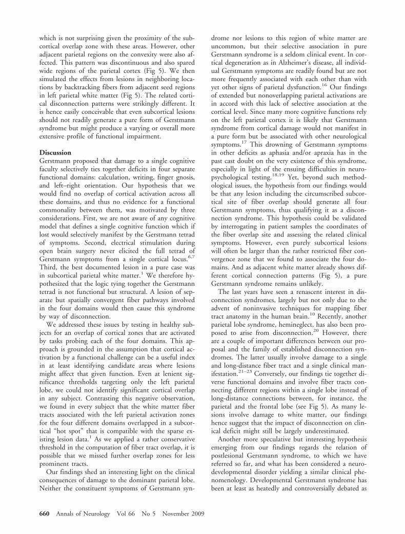
which is not surprising given the proximity of the sub-cortical overlap zone with these areas. However, otheradjacent parietal regions on the convexity were also af-fected. This pattern was discontinuous and also sparedwide regions of the parietal cortex (Fig 5). We thensimulated the effects from lesions in neighboring loca-tions by backtracking fibers from adjacent seed regionsin left parietal white matter (Fig 5). The related corti-cal disconnection patterns were strikingly different. Itis hence easily conceivable that even subcortical lesionsshould not readily generate a pure form of Gerstmannsyndrome but might produce a varying or overall moreextensive profile of functional impairment.
DiscussionGerstmann proposed that damage to a single cognitivefaculty selectively ties together deficits in four separatefunctional domains: calculation, writing, finger gnosis,and left–right orientation. Our hypothesis that wewould find no overlap of cortical activation across allthese domains, and thus no evidence for a functionalcommonality between them, was motivated by threeconsiderations. First, we are not aware of any cognitivemodel that defines a single cognitive function which iflost would selectively manifest by the Gerstmann tetradof symptoms. Second, electrical stimulation duringopen brain surgery never elicited the full tetrad ofGerstmann symptoms from a single cortical locus.6,7
Third, the best documented lesion in a pure case wasin subcortical parietal white matter.1 We therefore hy-pothesized that the logic tying together the Gerstmanntetrad is not functional but structural. A lesion of sep-arate but spatially convergent fiber pathways involvedin the four domains would then cause this syndromeby way of disconnection.
We addressed these issues by testing in healthy sub-jects for an overlap of cortical zones that are activatedby tasks probing each of the four domains. This ap-proach is grounded in the assumption that cortical ac-tivation by a functional challenge can be a useful indexin at least identifying candidate areas where lesionsmight affect that given function. Even at lenient sig-nificance thresholds targeting only the left parietallobe, we could not identify significant cortical overlapin any subject. Contrasting this negative observation,we found in every subject that the white matter fibertracts associated with the left parietal activation zonesfor the four different domains overlapped in a subcor-tical “hot spot” that is compatible with the sparse ex-isting lesion data.1 As we applied a rather conservativethreshold in the computation of fiber tract overlap, it ispossible that we missed further overlap zones for lessprominent tracts.
Our findings shed an interesting light on the clinicalconsequences of damage to the dominant parietal lobe.Neither the constituent symptoms of Gerstmann syn-
drome nor lesions to this region of white matter areuncommon, but their selective association in pureGerstmann syndrome is a seldom clinical event. In cor-tical degeneration as in Alzheimer’s disease, all individ-ual Gerstmann symptoms are readily found but are notmore frequently associated with each other than withyet other signs of parietal dysfunction.16 Our findingsof extended but nonoverlapping parietal activations arein accord with this lack of selective association at thecortical level. Since many more cognitive functions relyon the left parietal cortex it is likely that Gerstmannsyndrome from cortical damage would not manifest ina pure form but be associated with other neurologicalsymptoms.17 This drowning of Gerstmann symptomsin other deficits as aphasia and/or apraxia has in thepast cast doubt on the very existence of this syndrome,especially in light of the ensuing difficulties in neuro-psychological testing.18,19 Yet, beyond such method-ological issues, the hypothesis from our findings wouldbe that any lesion including the circumscribed subcor-tical site of fiber overlap should generate all fourGerstmann symptoms, thus qualifying it as a discon-nection syndrome. This hypothesis could be validatedby interrogating in patient samples the coordinates ofthe fiber overlap site and assessing the related clinicalsymptoms. However, even purely subcortical lesionswill often be larger than the rather restricted fiber con-vergence zone that we found to associate the four do-mains. And as adjacent white matter already shows dif-ferent cortical connection patterns (Fig 5), a pureGerstmann syndrome remains unlikely.
The last years have seen a renascent interest in dis-connection syndromes, largely but not only due to theadvent of noninvasive techniques for mapping fibertract anatomy in the human brain.10 Recently, anotherparietal lobe syndrome, hemineglect, has also been pro-posed to arise from disconnection.20 However, thereare a couple of important differences between our pro-posal and the family of established disconnection syn-dromes. The latter usually involve damage to a singleand long-distance fiber tract and a single clinical man-ifestation.21–23 Conversely, our findings tie together di-verse functional domains and involve fiber tracts con-necting different regions within a single lobe instead oflong-distance connections between, for instance, theparietal and the frontal lobe (see Fig 5). As many le-sions involve damage to white matter, our findingshence suggest that the impact of disconnection on clin-ical deficit might still be largely underestimated.
Another more speculative but interesting hypothesisemerging from our findings regards the relation ofpostlesional Gerstmann syndrome, to which we havereferred so far, and what has been considered a neuro-developmental disorder yielding a similar clinical phe-nomenology. Developmental Gerstmann syndrome hasbeen at least as heatedly and controversially debated as
660 Annals of Neurology Vol 66 No 5 November 2009

Fig 4. (A) Overlap of fibers traced for the four domains in the left parietal region of single subjects. (B) Intersection of the fivesubjects’ fiber overlap zones after spatial normalization into standard stereotactic space (left a coronal view; right two axial views).(C) Lesion localization in a patient with pure Gerstmann syndrome illustrated on a T2-weighted axial MRI section (from Mayer etal.1). Note spatial congruence of lesion extent with the distribution of probability with which fibers that are related to all fourfunctional domains overlap across subjects. Left is on reader’s left.
Fig 5. Fiber tracking from seed regions in left parietal white matter. To illustrate putative cortical disconnection effects, we createdvirtual lesions by taking different subcortical white matter loci as seed regions for fiber tracking. The zone of fiber convergenceacross the domain-related cortical activations is illustrated in red on the left-hand axial (top) and coronal MRI sections (bottom)and adjacent control regions are numbered. Right-hand panels present the related fiber tracking results (blue) with a three-dimensional reconstruction for the seed region of interest (middle column) and renderings of the left hemisphere white-matter sur-face (tilted view from above, in front and the side) for all seed regions. Small displacements in seed regions yield strikingly differentcortical disconnection patterns.
Rusconi et al: Disconnection Account of Gerstmann Syndrome 661

the postlesional variant.24 In recent years, diffusiontensor imaging has associated several other neurodevel-opmental disorders such as dyslexia and persistent stut-tering with white-matter alterations.25,26 In each case,fiber tract disorganization was located in white matterunderlying cortical areas that are critical for the im-paired functions. Again, our results could thereforeserve as a starting point for investigating the relationbetween structural white-matter integrity and cognitivefunction in the four domains that are compromised indevelopmental Gerstmann syndrome.
This work was supported by the Volkswagen Foundation (E.E.,S.D., A.K.); and by the European Commission Experienced Re-searcher Fellowship (MRTN-CT-2003-504927) (E.R.) and Reinte-gration Grant (MERG-CT-046511) (E.R.).
We thank Laurent Laribiere for help with the stimula-tion setup, Cyril Poupon, and the developers of Brain-Visa and Anatomist software.
References1. Mayer E, Martory MD, Pegna AJ, et al. A pure case of Gerst-
mann syndrome with a subangular lesion. Brain 1999;122:1007–1020.
2. Gerstmann J. Fingeragnosie. Eine umschriebene Storung derOrientierung am eigenen Korper. Wien Klin Wochenschr1924;40:1010–1012. [German] Finger agnosia. A circumscriptdisturbance of orientation with respect to the own body.
3. Gerstmann J. Syndrome of finger agnosia, disorientation forright and left, agraphia and acalculia. Arch Neurol Psychiatry1940;44:398–407.
4. Benton AL. Gerstmann’s syndrome. Arch Neurol 1992;49:445–447.
5. Roeltgen DP, Sevush S, Heilman KM. Pure Gerstmann’s syn-drome from a focal lesion. Arch Neurol 1983;40:46–47.
6. Morris HH, Luders H, Lesser RP, et al. Transient neuropsy-chological abnormalities (including Gerstmann’s syndrome)during cortical stimulation. Neurology 1984;34:877–883.
7. Roux FE, Boetto S, Sacko O, et al. Writing, calculating, andfinger recognition in the region of the angular gyrus: a corticalstimulation study of Gerstmann syndrome. J Neurosurg 2003;99:716–727.
8. Geschwind N. Disconnexion syndromes in animals and men.Part I. Brain 1965;88:237–294.
9. Geschwind N. Disconnexion syndromes in animals and men.Part II. Brain 1965;88:585–644.
10. Catani M, fftyche DH. The rises and falls of disconnection syn-drome. Brain 2005;12:2224–2239.
11. Catani M, Mesulam M. What is a disconnection syndrome?Cortex 2008;44;8:911–913.
12. Friston KJ, Holmes AP, Worsley KJ. How many subjects con-stitute a study? Neuroimage 1999;10:1–5.
13. Cointepas Y, Mangin J-F, Garnero L, et al. BrainVISA: soft-ware platform for visualization and analysis of multi-modalitybrain data. Neuroimage 2001;6:S98.
14. Riviere D, Mangin JF, Papadopulos-Orfanos D, et al. Auto-matic recognition of cortical sulci of the human brain using acongregation of neural networks. Med Image Anal 2002;6:77–92.
15. Mori S, Crain BJ, Chacko VP, van Zijl PC. Three-dimensionaltracking of axonal projections in the brain by magnetic reso-nance imaging. Ann Neurol 1999;45:265–269.
16. Wingard EM, Barrett AM, Crucian GP, et al. The Gerstmannsyndrome in Alzheimer disease. J Neurol Neurosurg Psychiatry2002;72:403–405.
17. Simon O, Mangin JF, Cohen L, et al. Topographical layout ofhand, eye, calculation and language-related areas in the humanparietal lobe. Neuron 2002;33:475–487.
18. Critchley M. The enigma of Gerstmann’s syndrome. Brain1966;89:183–199.
19. Kinsbourne M, Warrington E. A study of finger agnosia. Brain1962;85:47–66.
20. Thiebaut de Schotten M, Urbanski M, et al. Direct evidencefor a parietal-frontal pathway subserving spatial awareness inhumans. Science 2005;309:2226–2228.
21. Epelbaum S, Pinel P, Gaillard R, et al. Pure alexia as a discon-nection syndrome: new diffusion imaging evidence for an oldconcept. Cortex 2008;44:962–974.
22. Catani M, Mesulam M. The arcuate fasciculus and the discon-nection theme in language and aphasia: history and currentstate. Cortex 2008;44:953–961.
23. Heilman KM, Watson RT. The disconnection apraxias. Cortex2008;44:975–982.
24. Miller CJ, Hynd GW. What ever happened to developmentalGerstmann’s syndrome? Links to other pediatric, genetic, andneurodevelopmental syndromes. J Child Neurol 2004;19:282–289.
25. Klingberg T, Hedehus M, Temple E, et al. Microstructure oftemporo-parietal white matter as a basis for reading ability: ev-idence from diffusion tensor magnetic resonance imaging. Neu-ron 2000;25:257–259.
26. Sommer M, Koch MA, Paulus W, et al. Disconnection ofspeech-relevant brain areas in persistent developmental stutter-ing. Lancet 2002;359:2085–2086.
27. Van Essen DC, Dickinson J, Harwell J, et al. An integratedsoftware system for surface-based analyses of cerebral cortex.J Am Med Inform Assoc 2001;8:443–459.
662 Annals of Neurology Vol 66 No 5 November 2009