a general statistical analysis for fmri data
DESCRIPTION
A general statistical analysis for fMRI data. Keith Worsley 12 , Chuanhong Liao 1 , John Aston 13 , Jean-Baptiste Poline 4 , Gary Duncan 5 , Vali Petre 2 , Alan Evans 2 1 Department of Mathematics and Statistics, McGill University, 2 Brain Imaging Centre, Montreal Neurological Institute, - PowerPoint PPT PresentationTRANSCRIPT

A general statistical analysis for fMRI data
Keith Worsley12, Chuanhong Liao1, John Aston13,
Jean-Baptiste Poline4, Gary Duncan5, Vali Petre2, Alan Evans2
1Department of Mathematics and Statistics, McGill University,2Brain Imaging Centre, Montreal Neurological Institute,
3Imperial College, London,4Service Hospitalier Frédéric Joliot, CEA, Orsay,
5Centre de Recherche en Sciences Neurologiques, Université de Montréal
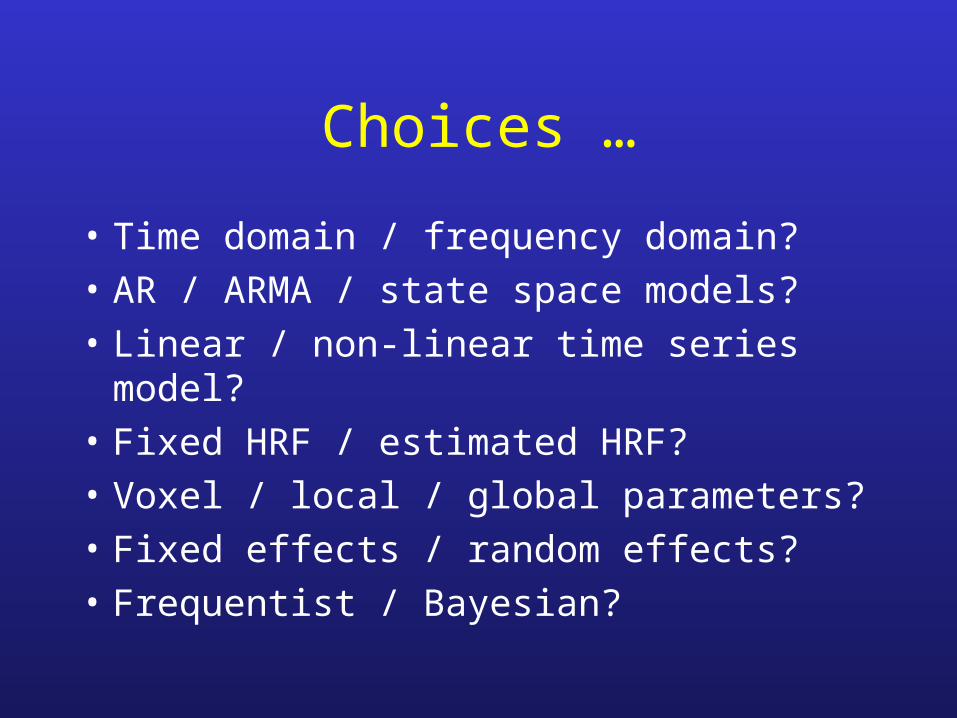
Choices …
• Time domain / frequency domain?
• AR / ARMA / state space models?
• Linear / non-linear time series model?
• Fixed HRF / estimated HRF?
• Voxel / local / global parameters?
• Fixed effects / random effects?
• Frequentist / Bayesian?

More importantly ...
• Fast execution / slow execution?
• Matlab / C?
• Script (batch) / GUI?
• Lazy / hard working … ?
• Why not just use SPM?
• Develop new ideas ...
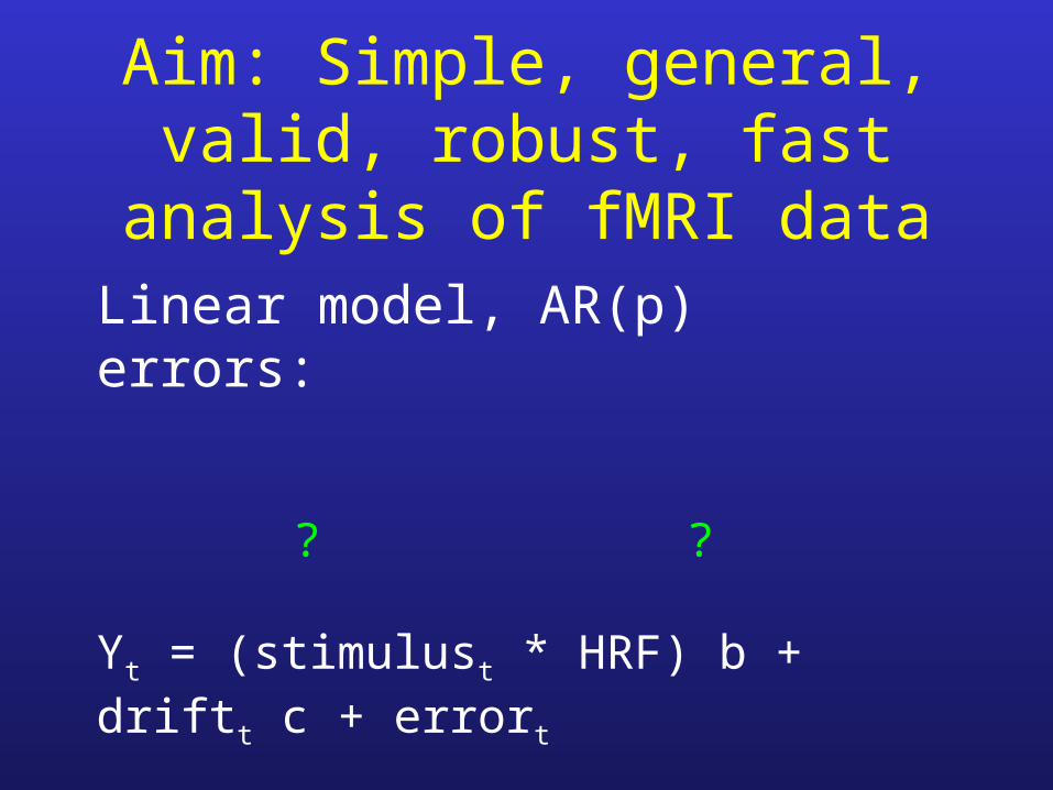
Aim: Simple, general, valid, robust, fast analysis of fMRI data
Linear model, AR(p) errors:
? ? Yt = (stimulust * HRF) b + driftt c + errort
unknown parameters ? ? ? errort = a1 errort-1 + … + ap errort-p + s WNt

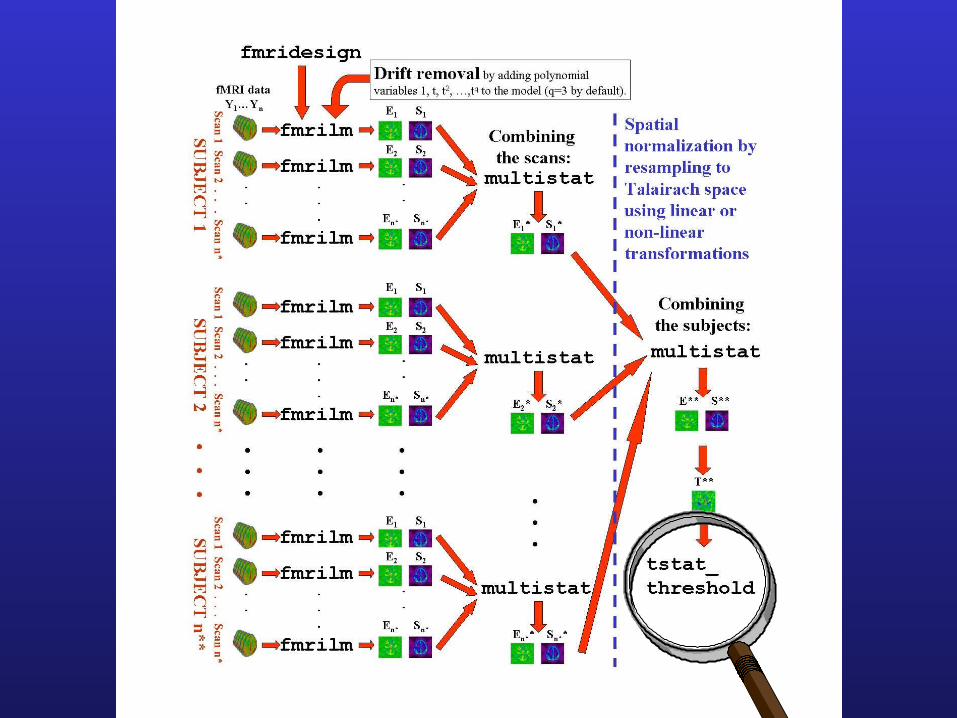
MATLAB: reads MINC or analyze format (www/math.mcgill.ca/keith/fmristat)
• FMRIDESIGN: Sets up stimulus, convolves it with the HRF and its derivatives (for estimating delay).
• FMRILM: Fits model, estimates effects (contrasts in the magnitudes, b), standard errors, T and F statistics.
• MULTISTAT: Combines effects from separate scans/sessions/subjects in a hierarchical fixed / random effects analysis.
• TSTAT_THRESHOLD: Uses random field theory / Bonferroni to find thresholds for corrected P-values for peaks and clusters of T and F maps.

0 50 100 150 200 250 300 350 400-1
0
1
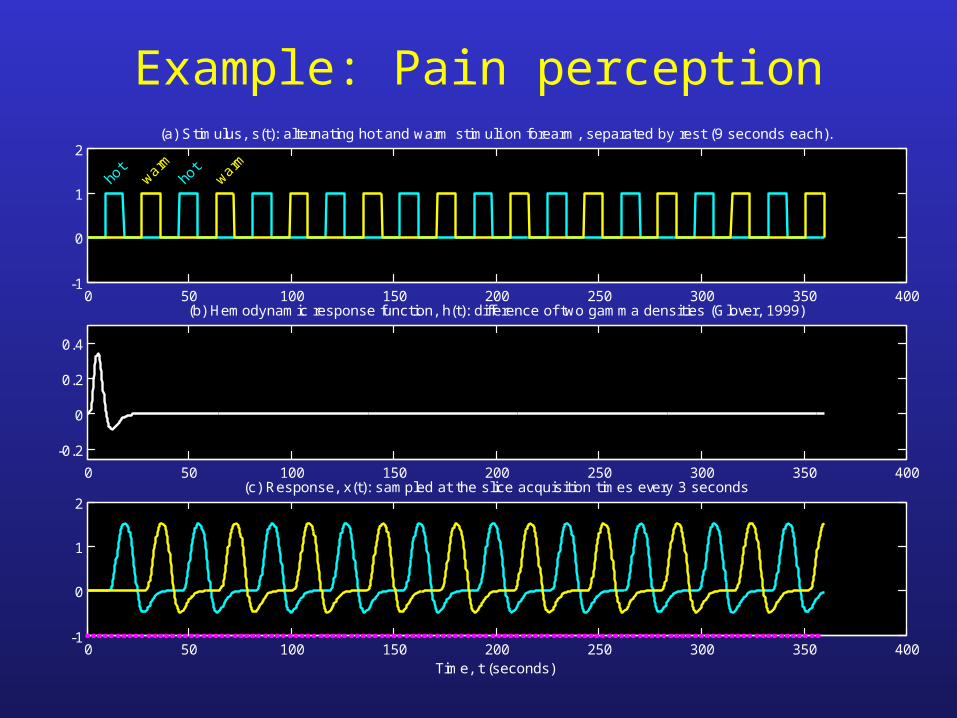
2(a) Stimulus, s(t): alternating hot and warm stimuli on forearm, separated by rest (9 seconds each).
hot
warmho
twarm
0 50 100 150 200 250 300 350 400
-0.2
0
0.2
0.4
(b) Hemodynamic response function, h(t): difference of two gamma densities (Glover, 1999)
0 50 100 150 200 250 300 350 400-1
0
1
2(c) Response, x(t): sampled at the slice acquisition times every 3 seconds
Time, t (seconds)
Example: Pain perception

-0.1
0
0.1
0.2
0.3
0.4
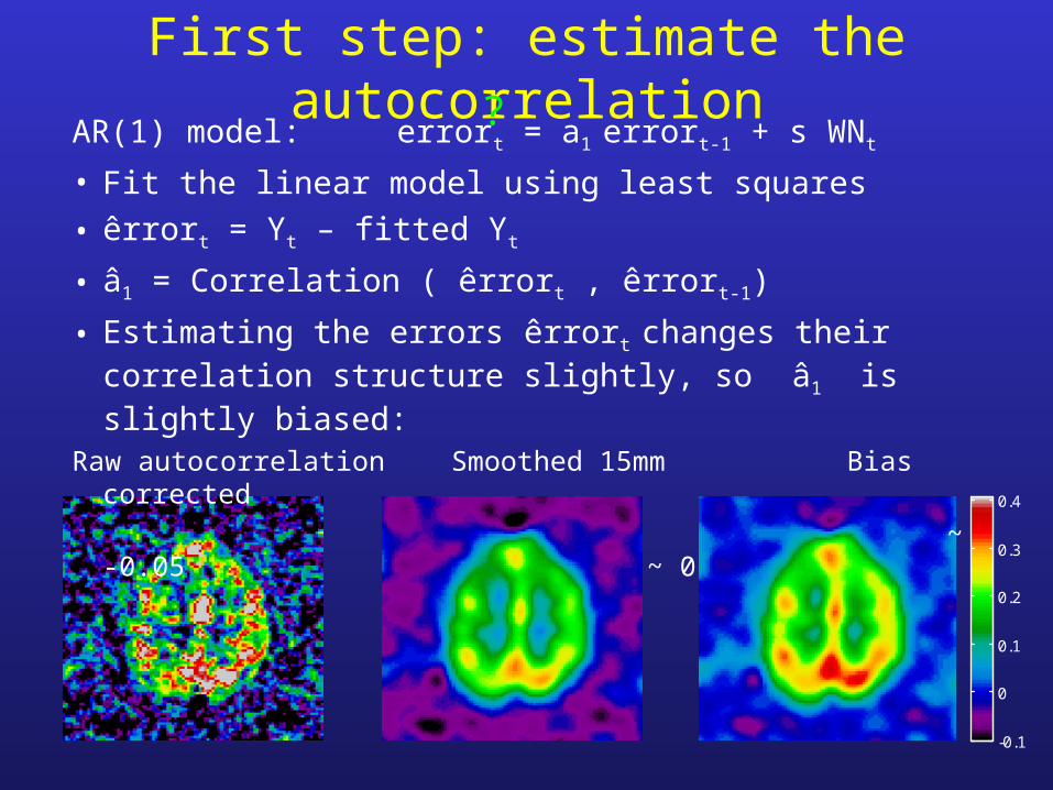
First step: estimate the autocorrelationAR(1) model: errort = a1 errort-1 + s WNt
• Fit the linear model using least squares
• êrrort = Yt – fitted Yt
• â1 = Correlation ( êrrort , êrrort-1)
• Estimating the errors êrrort changes their correlation structure slightly, so â1 is slightly biased:
Raw autocorrelation Smoothed 15mm Bias corrected
~ -0.05 ~ 0
?

-6
-4
-2
0
2
4
6
0
0.5
1
1.5
2
2.5
-6
-4
-2
0
2
4
6
Second step: refit the linear modelPre-whiten: Yt
* = Yt – â1 Yt-1, then fit using least squares:
Effect: hot – warm Sd of effect
T statistic = Effect / Sd
T > 4.86 (P < 0.05, corrected)
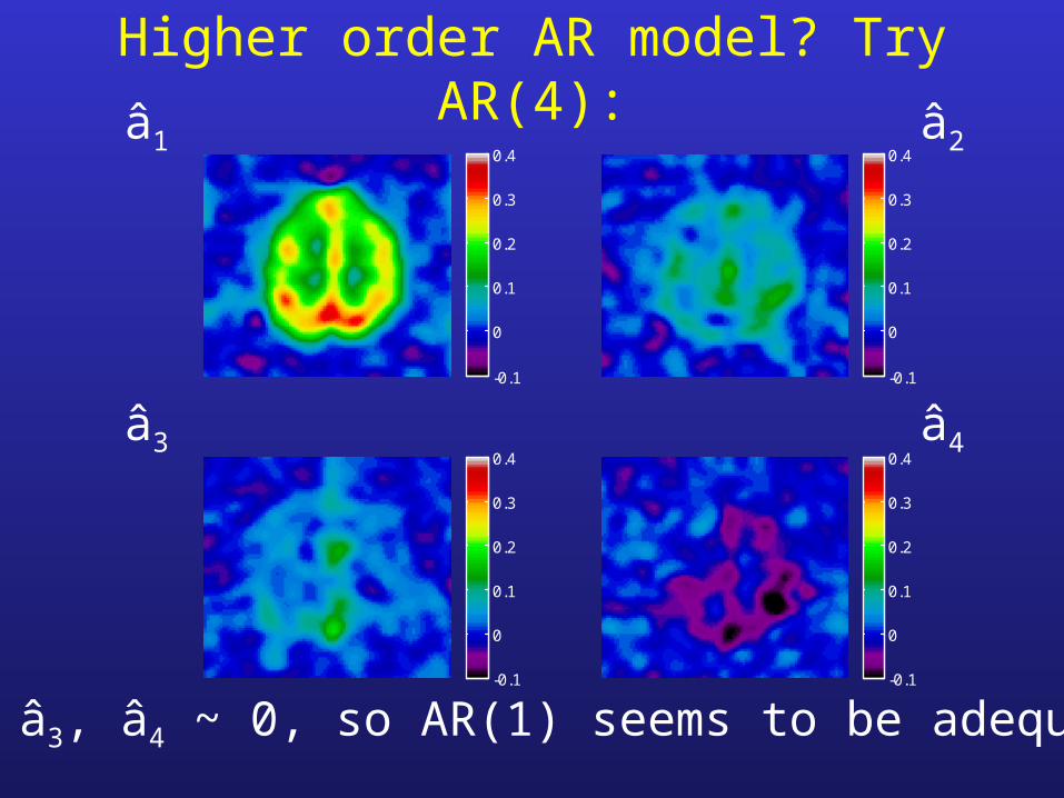
Higher order AR model? Try AR(4):
-0.1
0
0.1
0.2
0.3
0.4
-0.1
0
0.1
0.2
0.3
0.4
-0.1
0
0.1
0.2
0.3
0.4
-0.1
0
0.1
0.2
0.3
0.4
â1 â2
â3 â4
â2, â3, â4 ~ 0, so AR(1) seems to be adequate

-6
-4
-2
0
2
4
6
-6
-4
-2
0
2
4
6
-6
-4
-2
0
2
4
6
-6
-4
-2
0
2
4
6
… has no effect on the T statistics:AR(1) AR(2)
AR(4) But using zero correlation …
biases T up ~12% more false positives

-5
0
5
0
0.5
1
1.5
2
2.5
-5
0
5
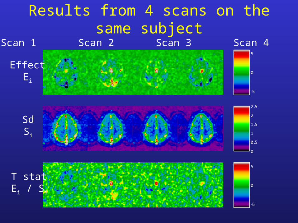
Results from 4 scans on the same subject
Scan 1 Scan 2 Scan 3 Scan 4
EffectEi
SdSi
T statEi / Si

MULTISTAT: combines effects from different scans/sessions/subjects:
• Ei = effect for scan/session/subject i
• Si = standard error of effect
• Mixed effects model:
Ei = covariatesi c + fi + ri
Random effect,due to variability from scan to scan,unknown sd
‘Fixed effects’ error,due to variabilitywithin the same scan,known sd Si
Usually 1, but could add group,treatment, age,sex, ...
}from
FMRILM
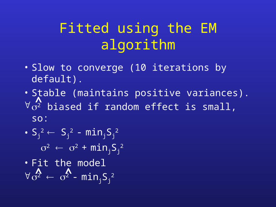
Fitted using the EM algorithm
• Slow to converge (10 iterations by default).
• Stable (maintains positive variances).2 biased if random effect is small, so:
• Sj2 Sj
2 - minjSj2
2 2 + minjSj2
• Fit the model2 2 - minjSj
2^ ^
^

-5
0
5
0
0.5
1
1.5
2
2.5
-5
0
5
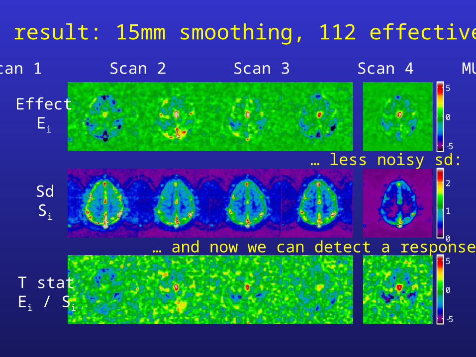
Scan 1 Scan 2 Scan 3 Scan 4 MULTISTAT
EffectEi
SdSi
T statEi / Si
Problem: 4 scans, 3 df for random effects sd ...
… and no response is detected:
… very noisy sd:

• Basic idea: increase df by spatial smoothing (local pooling) of the sd.
• Can’t smooth the random effects sd directly, - too much anatomical structure.
• Instead,
random effects sd
fixed effects sd
which removes the anatomical structure before smoothing.
Solution: Spatial regularization of the sd
sd = smooth fixed effects sd )

0
1
2
3
4
0
1
2
3
Random effects sd(3 df)
Fixed effects sd(448 df)
Random effects sdFixed effects sd
Smooth 15mm
Regularized sd(112 df)
Fixed effects sd
Over scans Over subjects

Effective df
dfratio = dfrandom ( 2 ( FWHMratio / FWHMdata )2 + 1 )3/2
dfeff = 1 / ( 1 / dfratio + 1 / dffixed )
e.g. dfrandom = 3, dffixed = 112, FWHMdata = 6mm:
FWHMratio (mm) 0 5 10 15 20 infinite
dfeff 3 11 45 112 192 448
variability bias compromise!
-5
0
5
0
1
2
-5
0
5
Scan 1 Scan 2 Scan 3 Scan 4 MULTISTAT
EffectEi
SdSi
T statEi / Si
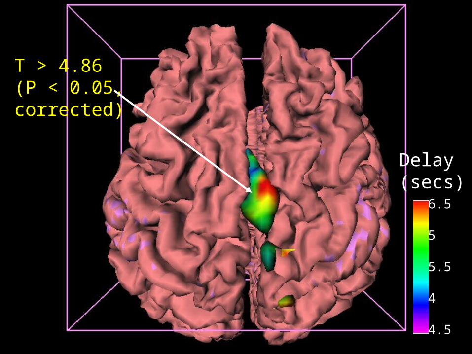
Final result: 15mm smoothing, 112 effective df …
… less noisy sd:
… and now we can detect a response:

T>4.86T > 4.86 (P < 0.05, corrected)

Conclusion
• Largest portion of variance comes from the last stage i.e. combining over subjects:
sdscan2 sdsess
2 sdsubj2
nscan nsess nsubj nsess nsubj nsubj
• If you want to optimize total scanner time, take more subjects.
• What you do at early stages doesn’t matter very much!
+ +

0 5 10 15 20 25-0.2
0
0.2
0.4
0.6
Delay = 5.4 seconds, log scale shift = 0 (reference hrf, h0)
Delay = 4.0 seconds, log scale shift = -0.3
Delay = 7.3 seconds, log scale shift = +0.3
t (seconds)
P.S. Estimating the delay of the response• Delays or latency in the neuronal response are modeled as a
temporal scale shift in the reference HRF:
• Fast voxel-wise delay estimator is found by adding the derivative of the reference HRF with respect to the log scale shift as an extra term to the linear model.
• Bias correction using the second derivative.• Shrunk to the reference delay by a factor of 1/(1+1/T2), T is the T statistic for the magnitude.

-5
0
5
-5
0
5
0
2
4
6
8
10
0
1
2
3
4
5
Delay of the hot stimulusT stat for magnitude = 0 T stat for delay = 5.4 secs
Delay (secs) Sd of delay (secs)

-5
0
5
-5
0
5
0
5
10
0
1
2
3
4
5
-5
0
5
-5
0
5
0
5
10
0
1
2
3
4
5
Varying the delay of the reference HRF
Ref.delay= 4.0
Ref.delay= 7.3
-5
0
5
-5
0
5
0
5
10
0
1
2
3
4
5
T stat for mag T stat for delay Delay Sd of delay
Ref.delay= 5.4
>4.86 ~0 ~5.4s >4.86 ~0 ~5.4s 0.6s0.6s
~5.4s~5.4s
~5.4s~5.4s
Delay(secs)
6.5
5
5.5
4
4.5
T > 4.86 (P < 0.05, corrected)

Delay(secs)
6.5
5
5.5
4
4.5
T > 4.86 (P < 0.05, corrected)

References
• http:/www.math.mcgill.ca/keith/fmristat
• Worsley et al. (2000). A general statistical analysis for fMRI data. NeuroImage, 11:S648, and submitted.
• Liao et al. (2001). Estimating the delay of the fMRI response. NeuroImage, 13:S185 (Poster #185, Tuesday morning), and submitted.