a histochemical study of glycogen in tissues of the alsino ... · pdf filewith the use of a...
TRANSCRIPT
Atlanta University CenterDigitalCommons@Robert W. Woodruff Library, AtlantaUniversity Center
ETD Collection for AUC Robert W. Woodruff Library
6-1-1949
A histochemical study of glycogen in tissues of thealsino mouse by use of the freezing techniqueGeneral Houston RichardsonAtlanta University
Follow this and additional works at: http://digitalcommons.auctr.edu/dissertations
Part of the Biology Commons
This Thesis is brought to you for free and open access by DigitalCommons@Robert W. Woodruff Library, Atlanta University Center. It has beenaccepted for inclusion in ETD Collection for AUC Robert W. Woodruff Library by an authorized administrator of DigitalCommons@Robert W.Woodruff Library, Atlanta University Center. For more information, please contact [email protected].
Recommended CitationRichardson, General Houston, "A histochemical study of glycogen in tissues of the alsino mouse by use of the freezing technique"(1949). ETD Collection for AUC Robert W. Woodruff Library. Paper 2318.

A HISTOCHEMIGAL STUDY OF GLYCOGEN IN TISSUES OF THE
ALBINO MOUSE BY USE OF THE FREEZING TECHNIQUE
A THESIS
SUBMITTED TO TIE FACULTY OF ATLANTA UNIVERSITY IN PARTIAL FULFILLMENT
OF THE REQUIREMENTS FOR THE DEGREE OF MASTER OF SCIENCE
0
BY
GENERAL HOUSTON RICHARDSON
DEPARTMENT OF BIOLOGY
ATLANTA, GEORGIA
JUNE, I9h9
"■
TABLE OF CONTENTS
Page
I. IMlHQDUGTIGIf 1
II. RMimi OF LITiSRATUtilS 2
III. M"MtI.aLS AtTD iifl-md) 4
IV. HBSULTS 7
V. DISCUSSION 9
VI. SUiiuiKX 11
VII. LITiiHATUfijJi GIT&J 12
iii
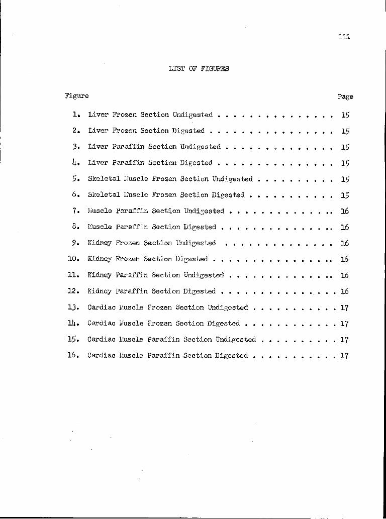
LIST OF FIGURES
Figure Page
1» Liver Frozen Section Undigested 15
2. Liver Frozen Section Digested ..... 15
3. Liver Paraffin Section Undigested 15
h» Liver paraffin Section Digested ....... 15
5. Skeletal Muscle Frozen Section Undigested .... 15
6. Skeletal Lfuscle Frozen Section Digested 15
7. luuscle Paraffin Section Undigested 16
8. Lfuscle Paraffin Section Digested 16
9. Kidney Frozen Section Undigested 16
10. Kidney Frozen Section Digested 16
11. Kidney Paraffin Section Undigested 16
12. Kidney paraffin Section Digested 16
13* Cardiac Muscle Frozen Section Undigested 17
lit. Cardiac Kuscle Frozen Section Digested 17
15. Cardiac Muscle Paraffin Section Undigested . 17
16. Cardiac Muscle Paraffin Section Digested 17
HISTOCHEHCAL STUD! OF GLYCOGEN IN TISSUES OF THE
ALBINO MOUSE BY USE OF THE FREEZING TECHNIQUE
INTRODUCTION
This investigation was designed to study the glycogen content in the
tissues of the albino mouse by the use of the freezing technique. Animals
used in such a study should be provided with the same kind of diet for an
accurate determination of the qualitative and quantitative distribution of
glycogen in the various tissues. Functional differences in tissues should
be indicated by the histochemical pictures of glycogen. Mann (f28) has
shown thet under wide varietion of bodily activity the cytological picture
of the liver is an ever changing one due to the function of the liver in
the maintenance of blood sugar level. Accordingly, the basic activities
in which other tissues participate rcay alter the distribution of substances
within their cells.
Preparation of the tissues for study and also interpretation of results
are of the utmost importance due to the occurrence of artifacts. Chemical
fixatives and polar solvents hamper accurate histochemical demonstration
of soluble components of cells and tissues by distortion, displacement, ex
traction and slow diffusion (Mencini '48). Glycogen is easily soluble in
water, therefore aqueous fixatives would not be satisfactory nor reliable.
Gersh in 1932 introduced the Altmann Technique for fixation by drying
while freezing. This introduced a method suitable for the accurate locali
zation of substances either norael to the organism, or introduced into the
body experimentally. By the use of this technique accurate localization of
intravitals with the production of permanent preparation was possible.
1
2
With the use of a non-polar solvent (Mancini 'AS), a histochemical study
of glycogen in tissues of the albino mouse was undertaken to determine its
distribution qualitatively,
REVIEW OF LITERATURE
Glycogen was discovered by Claude Bernard (1857) in cells of the liver.
Other tissues such as the placenta and many embyronic tissues were studied
by Bernard (1859), for their glycogen distribution. Plaques of cells in
the amniotic membrane of the calf by the use of iodine reaction revealed
amorphous and round granules of a brown staining material comparable to
cells of the liver. The liver cells contained amorphous and round granules
of glycogen.
Schiff seid, during the year 1857, that he could identify granules of
glycogen in unstained liver. He claimed that these granules were plentiful
in livers which contained an abundance of glycogen and scarce in livers con
taining little glycogen. The glycogen granules were distinguishable from
fat droplets and disappeared by amylase digestion. In cartilage and muscle
Rouget (1859), stated that the glycogen existed not as granules but as a
"plasma!1. According to Bock and Hoffman (1872), livers containing no glycogen
should show granules described by Schiff. Staining with a solution of iodine
in potassium iodide a diffuse brown staining network could be seen between
colorless granules of Schiff. Salivary digestion would cause the iodine re
action to disappear.
Sections fixed in alcohol (Kyser 1879), (Heidenhain 1883), have the
glycogen existing in the form of granules and flakes regularly located in
one side of the cell. Other workers including Kulz (1881), pointed out
that granularity localized at one end of the cell was an artifact due to
the alcohol fixation.
3
Ehrlich (1883), thought that if granules of glycogen were preformed
they should appear in dried preparation. With iodine, a homogenous brown
staining of cytoplasm of a dried smear of liver was observed and the nucleus
contained no brown color. Lewis (1921) found glycogen in a diffuse con
dition in cells of Fundulus embryo growing in tissue culture fluid. $y
using the freezing technique Gersh's («38) results confirmed those of
Ehrilich.
Giarke ('05), observed glistening balls in fresh cartilage cells which
corresponded to the location of glycogen upon fixation. He referred to the
regularity of glycogan granules in the kidney tubles of the cat as related
to glistening balls in the cartilage cells. Arndt ('24), noted a small
amount of glycogen deposited extracellularly in the liver. Bartelmez and
Bensley («32), mentioned orientation of glycogen granules in uterine
epithelium. They were uncertain whether the granules were preformed or re
sulted from plasraolysis. In the process of staining by iodine vapor, Lewis
(«21), observed that the type of cells in the tissue culture, which showed a
diffuse stsining previously, would at tines suddenly develop blebs or
granules of brown staining material upon the death of the cell. Lazarow
(«42) stated that iodide solution is a strong protein coagulator and that
these changes may well be the result of the iodine coagulstor. They do not
necessarily account for the vacuolar appearence seen in certain fresh or
dried cells.
Kinoshita (»33), reported that the best fixatives before staining
tissues to show glycogen were: (l) alcohol-ether containing magnesium
sulphate; (2) alcohol-ether made alkaline; and (3) formalin with magnesium
sulphate. These gave excellent inflitration. Excellent results were
h .
obtained ?/hen alkaline alcohol-ether was injected into the portal vein in
situ followed by sectioning of pieces of the liver with the capsule removed.
By using the freezing drying technique and Gersh's apparatus, Kaneini
(fi4.8) found glycogen diffusely distributed in mammalian liver, cartilage
cells, fatty tissue, epithelial cells of the vagina, in the epithelium
lining the cavity and the glandular tissue of the uterus, kidney tubules
of experimental and human diabetes, in biopsis of human skin from the belly,
scrotum, arm and face, in the syncytium of the placenta and in the vesicular
cells of the pjlycogenic organs of the chicken. Good results were obtained
with frozen sections. From evidence cited the existence of glycogen as a
substance diffusely distributed in cells of the liver is substantiated.
MATERIALS AM) METHOD
The albino mice used for this study were kept in a combination wood
and screen wire cage in a well ventilated laboratory. The diet consisted
of whole grain oats, scratch chicken feed xvhich contained sunflower seeds,
corn and wheat, cheese, green cabbage, turnip greens and plenty of fresh
water everyday. Every two days a few drops of cod liver oil were placed
on the food. Every effort was made to maintain a balanced diet. The cage
was brushed and scrubbed daily.
Paraffin and frozen sections of cardiac muscle, liver, kidney and
skeletal muscle tissues were used. In preparation of the paraffin sections,
the mouse was killed by a blow on the head with a small hammer. The animal
was. quickly dissected, the tissues dropped into an alcohol-ether fixative
saturated with magnesium sulphate (Kinoshita f33) and allowed to remain for
twenty-four hours. The tissues were placed in absolute alcohol for two
hours after being removed from the fixative. They were then infiltrated
and cleared with cederwood oil in an embedding oven at 56° c. . The
tissues remained in the oven until no more bubbles were given off.
Paraffin infiltration, embedding and subsequent sectioning followed.
Sections were fixed to slides with a mixture of alcohol, fresh egg albumin
and a few drops of glycerin. The sections ?rere flattened on the slides by
using the alcohol mixture as one would water. They were then allowed to
dry and the paraffin was removed by placing the slides in xylol. The histo-
chemical technique involved the use of iodine dissolved in mineral oil,
Lilly' M-32 Petronol, (Mancini «2^). Sections were placed in a saturated
solution of iodine-mineral oil for ten minutes, differentiated for five
minutes in an iodine saturated solution of absolute alcoholj washed in
absolute alcohol five minutes, and then cleared for ten minutes in oil of
thyme. These sections were mounted in Canada balsam.
For the preparation of frozen sections the animal was killed by placing
~.t on a block of dry ice (carbon dioxide) under a small glass which per
mitted no air to enter. The animal would succumb within one minute or less.
Dissection was performed immediately while the animal was still on the ice
block. The cardiac muscle, liver, kidney and skeletal muscle tissues Yjere
removed and dropped on the ice block where they became frozen through almost
instantly, depending on the size of the piece. The temperature of the
tissue was below -30 C. after thirty minutes. Each tissue was transferred
separately to the plate of a carbon dioxide gas freezing chamber. Here it
was kept frozen and embedded in a syrupy solution of gum acacia. The gum
acacia syrup was added to the freezing plate around the tissues a little at
a time with continued freezing until the tissue was completely covered with
a solid frozen mass. Sectioning was done with a Minot Rotery microtome. In
6
order to get satisfactory freezing on the plats the bottom of the carbon
dioxide chamber was raised above the outlet. The best staining method
was the same as the above for the paraffin sections.
Other staining methods were used with paraffin and frozen sections.
Methods included the use of Ehrlich's haematoxylin and Best's Carmine.
Sections were stained in Ehrlich's haematoxylin for five to ten
minutes and blued in tap water substitute made up as follows:
Tap xvater substitute
Potassium bicarbonate »——■— 2 grams
Magnesium sulphate —— 20 grams
Distilled wator uo to — 1000 C G
total 1000 C C
This solution ivas saturated with camphor.
The sections were then passed to a two per cent solution of potassium
iodide saturated with iodine (a Lugol solution) and left for five minutes.
They were removed, tissues wiped around and dehydrated in absolute alcohol
saturated in iodine, cleared in thyme oil and mounted in Canada basalm.
In using Best's Carmine stain, sections were stained first in Ehrlich's
haematoxylin as for Lugol's solution but differentiated in acid alcohol.
After staining for five minutes in Best's Carmine the sections were
differentiated in:
Absolute alcohol —■—• ■ 80 parts
Methyl alcohol ——— itO parts .
Distilled water —-————— 100 parts
until no more red came out (three to five minutes), washed in 80 per cent
alcohol and cleared in clove oil. These sections were also mounted in
Canada balsam.
Slides were prepared as controls for all tissues studied. These slides
had saliva added to them and ware set aside until ths glycogen was dissolved
7
by the diestass in the saliva. They were placed in warm water for fifteen
minutes or more. The slides were stained as usual by all the methods re
ferred to previously. Comparison between an undigested slide and a digested
one assisted in properly identifying the glycogen. (procedure of the late
Dr. B. R. G. ixussell, Iroperial Cancer Research Laboratory)
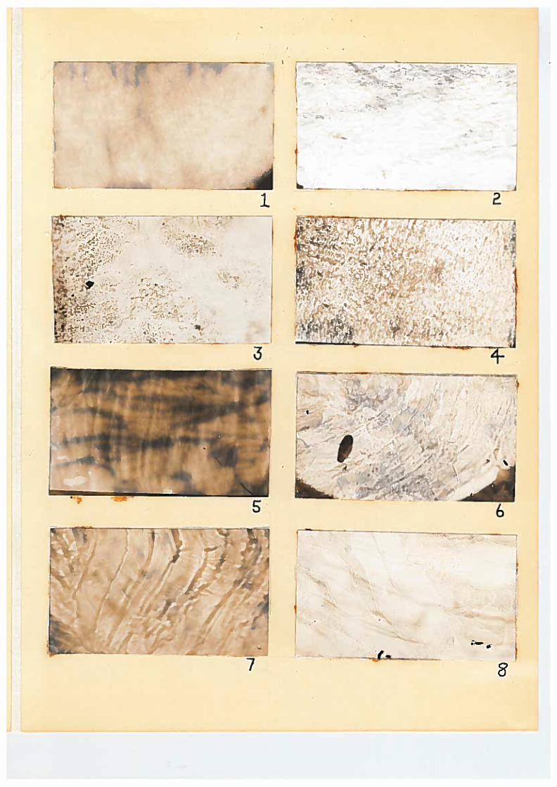
RESULTS
By using the carbon dioxide block for special preparation of the frozen
sections, the glycogen appeared in a diffuse condition in sections of the
liver of the albino mouse. Figure 1 represents tho condition that the
glycogen assumed in the frozen section. The control which is seen in figure
2 is the result of salivary digestion. Although there is a very small amount
of glycogen present in the control, it appears largely as granules in the
interstices between the cells.
Paraffin sections of the liver (fig. 3) show granular fiocculations of
glycojjen throughout the tissue. In the control (fig. 4) most of the
flocculacions have disappeared leaving a diiainished amount of scattered
glyeo.fjen granules. A striking contrast can be seen in diffuse and granulated
«";lyco;,jsn by the comparison of figures 1 and 4.
Frozen section of the skeletal muscle (fig. 5) caused the glycogen to
appear homogenous in and between the muscle fibers. However, there are areas
of varying intensity. The homogeneity might result frua the slight amount
of contration observed in the striated muscle tissue. In figure 6 salivary
digestion has left only a siT.aH amount of glycogen granules between the muscle
fibers.
In the paraffin section of the skeletal -muscle (fig. 7) there is less
glycogen present than in the frozen section. Mevertheless, the glycogen
8
present is preponderantly interi'ibrillar, appearing as irre/juiar clonus.
The control (fij. 3) indicates a disappearance oi' the jlycojen cluups which
dominated as seen in figure 7.
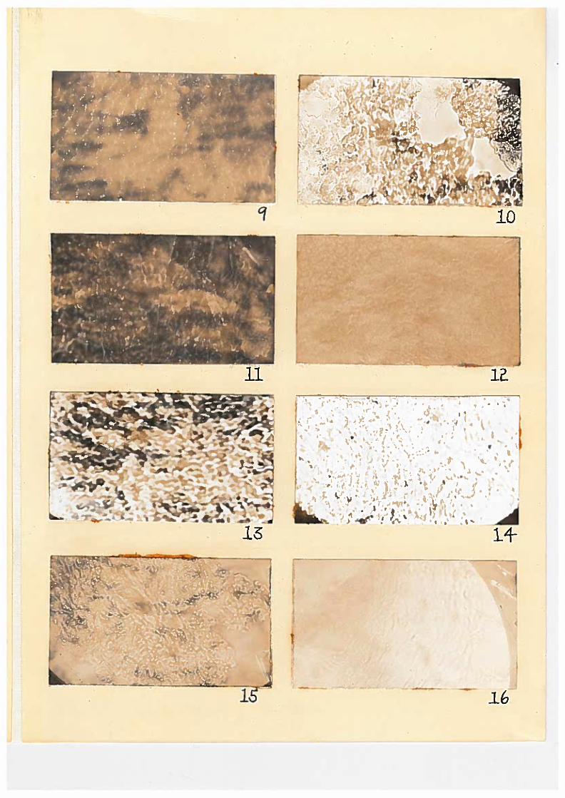
In a longitudinal frozen section oi" Kidney tubules, (fig.1 9) a lar^e
amount of diffuse giycogen appears.. 1'here can be seen light lines re
presenting spaces between the tubmles. The control (fig. 10) has much less
glyeogen present. The glycotjen present in the control, which is a cross
section of the tubules, is not in the intertubular spaces but within the
tubules themselves.
Paraffin sections of the Kidney (fig. 11) reveal the glyeogen is being-
deposited between the kidney tubules and in the ltuien. In the control
granulated reianants left after salivary digestion are intratubular (fig, 12).
In frozen sections of the heart (fig. 13) there is a mixture of diffuse
and granulated glyeogen. Many clearly defined glyeogen-free spaces exist in
the cardiac tissue. Apparently the heart is well supplied with jlyeogen but
there is not as xaucn present as one would ordinarily assume. The control
(fig. 1U) reveals a lainimutii amount of slyco^en as compared to the test
frozen section. The glycoyen in this instance appears somewhat flakey
between the muscle fibers.
Glyeogen in paraffin sections of cardiac tissue (fij. 15) shows intra
and extra fibrous granulation. In the control (fig. 16} a s<uall amount of
interfibrous granulated gLyoogen is present. Frozen sections revealed more
glycogan in heart muscle than paraffin sections although the saue staining
technique was used for both. Frozen and paraffin sections curried through
staining methoua other than the one outlined using mineral oil as a non-
polar solvent vrere not satisfactory. In staining vdbh Ehrlich's haeniatoxylin
9
and subsequently Lugol's solution, nuclex of one cells were outstanding
but the gijcogen was larval/ obscure. The haemato.,ylin apparently masked
the true color of the glycogen. This condition might have been due to
the presence of artifacts (iiancini «4S). The results with haematoaylin and
Best'3 Carmine were not reliable. The sections revealed indication of
glycogen stained red but a lar^e amount of the red color was not, even in
the section intra or exbra-cellulurljr. The Best's Gamine itself produced
an unusual amount of artifacts.
Salivary digestion was not one hunared per cent complete in any ©f the
controls studied.
DISCUSSION
A comparison of frozen sections with paraffin sections in a histo-
chemical study of glycogen in tissues of the albino mouse substantiated
ideas of granular and diffuse distribution.
Frozen sections stained in a non-polar solvent show more glycogen in
liver, skeletal muscle, kidney and cardiac muscle tissues of the white mouse
than did the paraffin sections. When diffuse glycogen is seen in paraffin
sections there is present a large number of granules. More diffuse glycogen
is seen in frozen sections, however, both types of sections reveal some
granules. The larger-amount of granular glycogen in the paraffin sections
is the product of fixation plus the effects produced by heat during preparation.
Glycogen as it appeared in the liver is diffuse in this study, thus
giving support to the findings of Ehrlich (1883) Otersh ('38) and Mancini(«48).
Granulations of glycogen in the paraffin sections of the liver are artifacts.
Laaarow («42) pointed out that the glycogen which appears diffuse in the liver
cells is composed of sutamicroscropic particles. Me liberated the particles
10
from the liver cells by fragmentation. Grapaic analysis and, ;,iultiple
correlation by Fern and Lorraine ('40} confirm that -ji/cogen is definitely
associated with fin appreciable ciaount of water when it is laid down in the
liver. Presumably this applies also to the deposition of glycogen in
muscles, kcoride, kanson, arid Scott ('41' gave evidence to show that the
apparent ratio of glycogen to water varies with an increase in the content
of non jlyeogen solids of the liver.
According to observation reported here glycogen in the frozen sections
of skeletal muscle appear homogenous in and between the muscle fibers. This
observation conflicts with those of soue of the other workers. The study
of Rojas and Hesta's ('38) reveals that giyco^en in the iauscle fibers was
found in the form of siiiall granules in the isotropic disc immediately con-
■ tiguous to the Z bandj no glycogen was found in the band. Gendre ('38)
working vdth fetal and adult rnuscle frora the rat and Guinea pig fixed in
Bouin-iillen solution and stained with Lugol aniline blue, said glycogen
was in granular concentration in short longitudinal rods in the '4 disc of
the muscle. Glycogen also appeared external to the muscle fiber in granular
form.■ By using the freezing-drying technique Mancini ('48) demonstrated
that glyaogen is present only inaide the muscle fiber. Explanation for
observations reported here of homogenous glycogen in skeletal muscle may
be due to the slight amount of contraction observed in the fibers. It is
questionable whether granular glycogen changes to homogenous glycogen during
contraction.
The method used here for iaaking frozen sections by use of the carbon
dioxide block and subsequently staining with iodine in uineral oil reveals
much granular and some diffuse ^iyco^en in cardiac tissue. The condition
11
of the glycogen shown, could not have chunked appreciably from its original
state due to bhs rapid freezing which began in situ and ended with complete
separation of the tissue. Blatherwick et al. (f35), employing largely
procedures of other workers concluded that muscles which had been frozen
after the hind leg had been severed from the body, contained more lactic
acid and less glycogen than muscle that had been frozen in situ. According
to Steiner ('35), the freezing of muscle cannot be relied upon to yield
absolute values for ;;lycogen. He observed that the contraction elicited
hy freezing results in a sin~.ll loss of glycvgea, a loss that is reflected
in a rise in both fermentable sugar and hexoseiaonophos:>hate. To detect
this however a chemical -in-jlysis would be required. The frozen sections
for showing glycogen present in tissues is definitely superior to the
paraffin sections.
1. ii comparison of frozen sections with paraffin sections in studying
glycogen histocnemically in tissues was made.
2. Frozen sections are superior to paraffin sections for staining to
show the presence of glycogen.
3. Granular and diffuse glycogen was found in cardiac muscle, skeletal
muscle, kidney and liver tissues of the albino mouse.
4. Ideas of granular and diffuse distribution of glycogen in tissues have
been substantiated.
12
LITERATURE CITED
Arndt, H. J. 1922; Vergleichend-histologiche Beitrage Zur Kenntnis des
Leberglykogen.
Bartelmez, G. «., and Bensley, C. M. 1932 In Special Cytology, edited byE. V. Comdry, 3: 1525 Paul B. Koeber, Inc. New York.
Bernard, Claude 185? Sur le mechanisms phyoiologique de la formation dusucre dans le fole. Corapt. rend. Acad. d. So., vol. isii, p. 578.
Bernard, Claude 1858 Memoire sur une noiivelle fonction du placenta.
Brovm-Sequard, J. de Physiol., 2:-31.
Blatherwick, K. H., Bradshaw, P. J.,Swing, M. E., Larson, li. ¥., andSawyer, S. D., 1935. The Determination of Tissue Carbohydrates. J.
Biol. Cnem., y. 537.
Bock, C, and Hoffmann, F. A, 1872 Veber d.?js microchemische verbalten der
Leberzellen. Virchows Arch. f. Path. Anat., %6t 201.
Shrlich, P. 1883 Appears as an Appendix in the Article by F. F. Frerichs.Uber den Plotzlichen Tod undiiber das coma bei Dibetes (diabetische
dutoxication) Ztsclir. f. Klin, led., 6:35.
Fern, 1". 0., an.d Lorraine, F. H. 19U0 Deposition of Glycogen with water in
ths livers of cats. J. Biol. Chen. 136:87.
Gendre, ii. 1938 Aspects et localisation du glycoglne dans le tissuinusculaire strie'. Bull. Histol. Appl. Physiol, et Path. 15 (e) s 265.
Gersh, I. 1932 The Altraan technique for drying while freezing. Anat. Rec,
Gersh, I. 1938 Results quoted in Maximow, A. A., and Bloom, ¥. Textbook ofHistology. W. B. Saunders Company, Philadelphia.
Guyer, M. F. Animal iticrology 19U7, The University of Chicago Press.
Chicago, Illinois.
Hoidenhain, R. 1883 In L. Hermann's Handbuch der Physiolgie, 5 Lepzig,
F. C. I. Vogel 1883. ■ "
Kinoshita, Hyojun, 1928 On the Staining of glycogen. Trans. Japanese
Path. Soc. 18: 211. . . ■
Kulz, R. 1886 Zur quantitativen Bestinramung des Glykogens. Ztschr f.
Biol. 22: 161.
13
Lazarow, A. 19h2 Praticulate glycogen. Anat. Rec, 8^:31.
Lee, Bollos 192C Microtomist's Yadenecun. P. Blakiston's Son and Co.
1012 Walnut St. Philadelphia, Pa.
Lev/is, M. H. 1921 The Presence of glycogen in the cells of embryos of
Fundulus heteroclitus studied, in tissue culture. Bio. Bull. lj.l:2
Kann, F. C. 1928 The Cytology of the liver and its functional significance.
Special Cytology 1:336.
McBride, J. J., Mason, l,i. G., and Scott, S. L., 19kl The Storage of themajor liver components j emphasizing; the relationship of glycogen
to water in the liver and the hydration of glycogen. J. Biol. Chem.
139s 9h3.
Rojas, P. y. Resta, L. S. Una nueva observation ou sobere el glycoceno
muscular (Observation on muscle glycogen) Rev. Soc. Argentina Biol.
Ui (5): 350 1938.
Rouget, C. I8p9 Be la substance an^flacee amorphe dans les tissue des
embryons des vertebres. Compt. rend. Acad. d Sc. UO* 1018.
Schiff, *>L. 1857 liitteilung. arch. f. physiol. Heillcund. Is 263.
Steiner, A. i»roc. Soc. Exptl. Biol. led. 32: 968 1935.
14
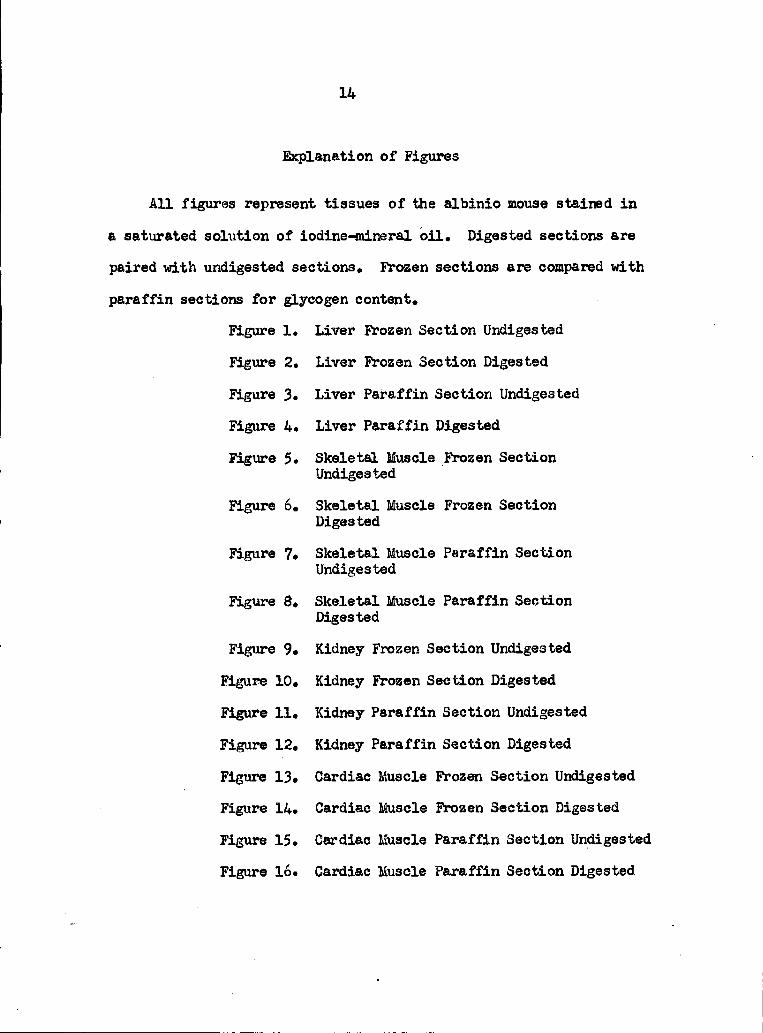
Explanation of Figures
All figures represent tissues of the albinio mouse stained in
a saturated solution of iodine-mineral oil. Digested sections are
paired with undigested sections. Frozen sections are compared with
paraffin sections for glycogen content.
Figure 1. Liver Frozen Section Undigested
Figure 2. Liver Frozen Section Digested
Figure 3. Liver Paraffin Section Undigested
Figure 4. Liver Paraffin Digested
Figure 5. Skeletal Muscle Frozen Section
Undigested
Figure 6. Skeletal Muscle Frozen Section
Digested
Figure 7. Skeletal Muscle Paraffin Section
Undigested
Figure 8. Skeletal Muscle Paraffin Section
Digested
Figure 9. Kidney Frozen Section Undigested
Figure 10. Kidney Frozen Section Digested
Figure 11. Kidney Paraffin Section Undigested
Figure 12. Kidney Paraffin Section Digested
Figure 13. Cardiac Muscle Frozen Section Undigested
Figure 14. Cardiac Muscle Frozen Section Digested
Figure 15. Cardiac Muscle Paraffin Section Undigested
Figure 16. Cardiac Muscle Paraffin Section Digested