a more biologically plausible learning rule for neural networks
TRANSCRIPT

Proc. Nati. Acad. Sci. USAVol. 88, pp. 4433-4437, May 1991Neurobiology
A more biologically plausible learning rule for neural networks(reinforcement learning/coordinate transformation/posterior parietal cortex/sensorimotor integration/Hebbian synapses)
PIETRO MAZZONIt*, RICHARD A. ANDERSENt§, AND MICHAEL I. JORDANttDepartment of Brain and Cognitive Sciences, and tHarvard-MIT Division of Health Sciences and Technology, Massachusetts Institute of Technology,Cambridge, MA 02139
Communicated by Francis Crick, February 21, 1991 (received for review October 19, 1990)
ABSTRACT Many recent studies have used artificial neuralnetwork algorithms to model how the brain might processinformation. However, back-propagation learning, the methodthat is generally used to train these networks, is distinctly"unbiological." We describe here a more biologically plausiblelearning rule, using reinforcement learning, which we haveapplied to the problem of how area 7a in the posterior parietalcortex ofmonkeys might represent visual space in head-centeredcoordinates. The network behaves similarly to networks trainedby using back-propagation and to neurons recorded in area 7a.These results show that a neural network does not require backpropagation to acquire biologically interesting properties.
Recently neural network models have been used to modeland predict certain aspects of brain function. A criticism ofsuch models, however, has been their reliance on backpropagation, a learning algorithm that has been considered"unbiological" because it requires passage of informationbackward through synapses and along axons and because ituses error signals that must be precise and different for eachneuron in the network. Attempts to implement more biolog-ically realistic forms of back-propagation still require unre-alistic conditions, such as symmetrical feedback pathwaysthat are identical in every way, including strength of theindividual synaptic connections (1, 2). Crick has suggestedthat "what is really required is a brain-like algorithm whichproduces results of the same general character as back-propagation" (3).
In our laboratory, we have been refining our neural net-work models to bring them more in line with what we knowof nervous system function. This paper describes the appli-cation of a variant of the associative reward-penalty (AR-P)learning rule of Barto and colleagues (4-6) to the training ofa multilayer neural network in a biologically relevant super-vised learning task. We used this network to model theprocess of coordinate transformation, believed to be com-puted by the posterior parietal cortex (for review, see ref. 7)and found that units in the middle layer of the networkdevelop response properties similar to those of area 7aneurons. These properties are also similar to those obtainedwith a previous model due to Zipser and Andersen (8), whichrelied on back-propagation learning. The AR-P rule has theadvantage of possessing several more physiological corre-lates, such as a feedback system that transmits performanceinformation along explicit and plausible pathways, Hebb-likesynapses that correlate pre- and postsynaptic activity, and asingle scalar performance evaluation that is computed fromthe overall output of the network and is sent to all connec-tions in the network in the form of a reinforcement signal.
MODELNeurons in area 7a appear to compute head-centered loca-tions of visual stimuli by combining retinal and eye-position
information (7, 9). A feature ofthe responses ofthese neuronsthat may be crucial for this computation is an approximatelyplanar modulation by eye position of the response to a visualstimulus (10). In other words, if one records from an area 7aneuron in an awake monkey while a spot of light is presentedat a fixed location on its retina, then as the animal looks invarious directions, the neuronal firing rate varies approxi-mately linearly with changes in the horizontal and/or verticalangle of gaze. A plot of this modulation of visual response byeye position is termed the "spatial gain field." Andersen andcolleagues (7-10) hypothesized that an ensemble of neuronswith this response property, each with its own slope, direc-tion, and range of planar eye position sensitivity, couldencode a distributed representation of craniotopic locations.Zipser and Andersen (8) set up a three-layer network totransform the coordinates from a retinotopic frame to acraniotopic one, using retinal-stimulus location and eye po-sition as input signals and the resulting head-centered loca-tion as the training signal. After training this network byback-propagation, the units in the middle layer (so-called"hidden" units) displayed planar gain fields remarkablysimilar to those of area 7a neurons. This result suggested thatsome fundamental computational feature embodied by thenetwork may be shared by area 7a neurons in their repre-sentation of head-centered space.As properties ofthe hidden units in the Zipser and Andersen
model suggested a possible connection between that modeland area 7a, it was natural to ask how crucial back-propagationis for the development of these properties. We addressed thisquestion by training a neural network with an architecturesimilar to the Zipser and Andersen model but using the morebiologically plausible AR-P learning algorithm. Our presentnetwork has a three-layer, fully connected, feed-forward ar-chitecture (Fig. la). The input layer consists ofa visual and aneye position group of units, which were modeled according tocharacteristics of area 7a neurons established in previousstudies (Fig. 1 b-c; ref. 8). The hidden and output layersconsist of binary stochastic elements (Fig. id), which producean output of 1 with a probability given by the logistic functionof the summed weighted inputs and an output of 0 otherwise.The output layer encodes the craniotopic location that is thevector sum of the retinal and eye position inputs and iscomposed of one of two alternative formats (Fig. 1 e-f), oneanalogous to the monotonic eye position representation andthe other to the retinal gaussian format.We modified the supervised learning procedure for AR-P
networks, introduced by Barto and Jordan (6), to train ournetwork. Every unit in the network receives a scalar rein-forcement signal r (Fig. la), the value of which depends onhow close the current network output is to the desired output.[Specifically, r assumes a value between 0 and 1, with 0indicating maximum error in the output (i.e., every unit thatshould be firing is not and vice versa) and 1 corresponding to
Abbreviation: AR-P, associative reward-penalty.§To whom correspondence should be addressed.
4433
The publication costs of this article were defrayed in part by page chargepayment. This article must therefore be hereby marked "advertisement"in accordance with 18 U.S.C. §1734 solely to indicate this fact.

4434 Neurobiology: Mazzoni et al.
d Evaluator ---
output 2output 1
or~ "1' I IF0
or
head x (+ step) 4
head x (- step) 4
head y (+ step) 1head x
head y (- step)--pb-
r
r
DDrnDoooEE0Z
X[DLIE EEDZ ElEDlEE eye x (+ slope): EE0 0ELL EDElMJIfllfeyex(-slope)$ D0 0 0-HEELILI *EEE 2 eye y (+ slope)0000ED 00ELE0EEEJ1*lE eyey(-slope)
retina x
e
* ~~~~~~~~~~~~~~~~~~~~~~~~~~~I,,... , ..,1...r- 5-
4
0
head-centered position
d
b4-3
r- i
retinal position
fr.
"00
head-centered x
C
eye position
FIG. 1. (a) Network structure. Retinal input is encoded by 64 units with gaussian receptive fields (b), while eye position is represented by32 units with linear activation functions (c). In the retinal input, each unit has an output between 0 and 1, a l/e width of 15° and a receptivefield peak 100 apart from that of its horizontal and vertical neighbors. In the eye-position input, the output of each unit, between- 0 and 1, is a
linear function of horizontal or vertical orbital angle, with random slope and intercept. These input formats reproduce properties of certain area
7a neurons that respond only to visual stimuli or to changes in eye position. The shading of each unit is proportional to its activity, with blackrepresenting maximum activity. The hidden and output layers are composed of binary stochastic elements (d), which produce an output of 1with probability (prob) p equal to the logistic function of the sum of the weighted inputs (si = Zito w,1xj), and zero with probability 1 - p. Thejth unit in the network provides input x; to the ith unit via the connection wij; m is the number of inputs to the units, and b is a bias. The networkused from two to eight hidden units. The output units encode head-centered locations according to one of two output formats. In the"binary-monotonic" format (e), each unit produces an output of 1 or 0, depending on whether the encoded location is to the right or to the left(or, for some units, above or below) a certain reference point. For example, a typical output layer consisted of four sets of three units, givingan output of 1 when the x (or y) craniotopic coordinate is > (or <) -40, 0, or +40 degrees. This format is analogous to the eye-position inputformat, in that four groups of units encode an increase in horizontal or vertical position angle by increasing or decreasing their activationmonotonically. Another format we used is the "binary-gaussian" one (f), in which four units give an output of 1 when the spatial position iswithin 1000 of their receptive field centers, which are located at (±60, ±60)0. This format is analogous to that ofthe retinal input, in that a positionangle is encoded topographically by units with overlapping receptive fields.
optimal performance (no error in the computed head-centered position).] The weights of all the connections arethen adjusted, after each pattern presentation, in such a wayas to maximize the value of this reinforcement. If we let xidenote the output of the ith unit in the network, pi denote itsprobability of firing, and wy denote the connection weight forits input from the jth unit (Fig. ld), the equation for updatingthe weights is
Awij = pr(xi - pi)xj + Ap(1- r)(1 -xi-p)xj,
forcement signal, presynaptic activity, and postsynaptic ac-
tivity. Thus, for instance, a correct response (large r) willstrengthen connections that were active during the response,and an incorrect response (small r) will weaken activesynapses. The value of r is a function of the average outputerror and is computed as r = 1 - E, with
E={ K -XkK} 1/n [2][1]
where p and A are constants. The first term in this sumcomputes the reward portion of the learning rule, whereas thesecond term is the penalty portion. Ignoring for the momentthe constant terms and the stochastic component, this equa-tion changes the synaptic weights by correlating the rein-
where k indexes the K output units in the network, x4 is thedesired output of the kth unit in the output layer, Xk is itsactual output, and n is a constants.
IA bias bi on each unit is also adjusted (as described in ref. 11) by the
a
r
Proc. Natl. Acad Sci. USA 88 (1991)
Proc. Natl. Acad. Sci. USA 88 (1991) 4435
RESULTS
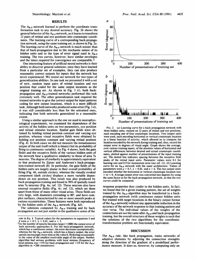
The AR-P network learned to perform the coordinate trans-formation task to any desired accuracy. Fig. 2b shows thegeneral behavior of the AR-P network, as it learns to transform12 pairs of retinal and eye positions into craniotopic coordi-nates. The learning curve of a corresponding back-propaga-tion network, using the same training set, is shown in Fig. 2a.The learning curve of the AR-P network is much noisier thanthat of back-propagation due to the stochastic nature of itshidden units and to the type of error signal used in AR-Ptraining. The two curves, however, have similar envelopesand the times required for convergence are comparable. 11One interesting feature of artificial neural networks is their
ability to discover general solutions; once they have learnedfrom a particular set of examples, they can also producereasonably correct outputs for inputs that the network hasnever experienced. We tested our network for two types ofgeneralization abilities. In one task we presented it with a setof new, random input pairs of retinal location and eyeposition that coded for the same output locations as theoriginal training set. As shown in Fig. 3 (i), both back-propagation and ARp-trained networks performed this taskextremely well. The other generalization task required thetrained networks to give the correct output for input patternscoding for new output locations, which is a more difficulttask. Although both networks produced some error (Fig. 3 ii),it was still considerably less than for the untrained ones,indicating that both networks generalized to a reasonableextent.
Using a similar approach to the one we used in neurophys-iological experiments, we examined the dependence of theactivity of the hidden units on two parameters, eye positionand retinal stimulus location. Spatial gain fields were ob-tained by holding retinal position constant and varying eyeposition, whereas visual receptive fields were obtained byholding eye position constant and varying retinal position(Fig. 4). In both cases we did not measure the instantaneousoutput of the unit itself (which is binary) but its probability offiring (a continuous variable). As Fig. 4 shows, both the gainfields and the receptive fields of the various hidden units ofthe network bear a qualitative similarity to those of area 7aneurons. The degree of similarity is approximately equivalentto that produced by Zipser and Andersen's back-propaga-tion-trained network (8). In particular, the gain fields of thehidden units are largely planar in their overall probability offiring (Fig. 4b, outside circles), whereas the visually evokedcomponent (dark circles) displays a more variable depen-dence on eye position. This result was also produced byback-propagation training and found in 78% of spatially tunedarea 7a neurons (Fig. 4a; ref. 12). These neurons also haveunusual receptive fields (Fig. 4c; ref. 12), which set themapart from those of many other visual areas. These fields arevery large, with diameters extending to 800, and have com-plex surfaces, characterized by one or more smooth peaks atvarious eccentricities. These features were both reproducedby the hidden units of the AR-P network (Fig. 4d).The solutions computed by AR-P training and by back
propagation are not just similar in the qualitative sense of the
rule in Eq. 1. Typical values for the parameters in equations 1 and2 were p = 0.5, A = 0.01, and n = 3.
As the number of epochs becomes large (>1000) the output error ofboth networks approaches 0. For the back-propagation network,which has a continuous output, the error decreases asymptotically,whereas for the AR-P network, which has a binary output, the errorspends increasingly more time at the value 0, flickering occasionallyto the value of the smallest resolvable angle of the output. Neitheralgorithm had serious problems with local minima (frequency oflocal minima was =5% for back propagation and <1% for the AR-Palgorithm in =200 simulations).
a
60In
ci200
0 40-
ro
0'.4'.4
X 20
b60-
U)'4)
40
0S.4W1 201
100 200 300Number of presentations of training set
400
1
MIa4|.- I1 IAAI-flI1IB Al1--1M-- AMUlbeA i'"'"'i i I Al -
M M I I t ,I ,vI0 100 200 300
Number of presentations of training set
IA
400
FIG. 2. (a) Learning curve for a back propagation network withthree hidden units, trained on 12 pairs of retinal and eye positions,each encoding one of four craniotopic locations. Two output unitswere used, each encoding horizontal or vertical craniotopic locationas a linear function of activity, which allowed us to compute thelocation encoded by the network during training and, thus, record theoutput error in degrees of visual angle. Graph shows the average,over various training inputs, of the absolute values of horizontal andvertical differences between desired and actual craniotopic coordi-nates, plotted against number of presentations of the input trainingset. The dotted line indicates spacing between the receptive fieldpeaks of the retinal input units. Parameter values were 0.1 forlearning rate and 0.9 for momentum term (see ref. 11). (b) Learningcurve for an AR-P network with the same architecture. Values ofparameters were p = 0.5, A = 0.01, and n = 6. The two output unitsencoded whether the horizontal or vertical craniotopic location was> or < 0. Average output error was converted into degrees by usingthe same factor as for the back-propagation network, so that the twocurves could be compared.
response properties they confer to the hidden units. In fact,we found that for a given training pattern, the set of weightstrained by the AR-P algorithm may be transferred to a back-propagation network (with continuous output hidden unitsbut trained with target locations in the binary output formatofthe AR-P network) without any appreciable reduction in theaccuracy of the network response to that training pattern andvice versa. The individual values of the weights of theconnections are not the same after AR-P and back-propagationtraining, but the overall structure ofthese weights is such thatthe solutions of the two algorithms for the coordinate-transformation problem are functionally equivalent.
DISCUSSIONThe AR-P rule, like back propagation, trains networks ofadaptive elements by adjusting the connection strengthsalong the direction of the gradient of a predefined perfor-mance measure. It does so, however, by computing only an
Neurobiology: Mazzoni et al.

4436 Neurobiology: Mazzoni et al.
30-
2O0
0
Backprop. AR-P Backprop. ARPi) New input pairs, ii) New input pairs,same output locations new output locations
FIG. 3. Output error produced by back-propagation and AR-Pnetworks described in Fig. 2 before and after training with 40 inputpairs, when presented with (i) 40 new, random inputs coding for thesame output locations as in the training set, and (ii) 40 random inputscoding for 40 new, random output locations. Error was computed foreach network as described in Fig. 2.
estimate of this gradient (6, 13). Units trained by the AR-P ruledo not have the detailed information about the error vectorand the state of other units that is necessary to compute theexact gradient and which back-propagation units obtainthrough nonbiological pathways. Due to the random noise intheir output, however, AR-P units can "jitter" their activityduring learning so as to get an estimate of how variations inactivity affect the reinforcement they receive, which, in turn,
a
b
allows them to estimate the direction in weight space alongwhich to change their weights to increase reinforcement.Although this method allows ARP-trained units to properlyadjust their weights using only locally available information,it is more random in its search for a solution than back-propagation, as reflected in the fluctuations in the learningcurve in Fig. 2b. The precise computation through back-propagation of the performance gradient tells the algorithmthe exact manner in which to change the weights so that theerror is monotonically decreased, resulting in the smoothcurve of Fig. 2a.An important element of the AR-P model, which aligns it
with many neurobiological models of learning, is the rein-forcement signal. As in any supervised learning scheme, thissignal is computed by comparing the activities of output unitsto desired activities. After these errors are averaged, how-ever, the feedback system transmits only a single value to allthe network connections and is not assumed to provide theseconnections with separate information about the activities ofindividual output units. The fact that in AR-P training a singlevalue is valid for all the connection weights implies that onlyone projection is necessary from the reinforcement comput-ing region to area 7a. The existence of signals originating froma small cluster of neurons distributed to entire cortical areasand that possibly carry information about reward has beensuggested by anatomical as well as experimental studies (e.g.,see ref. 14). In contrast, back propagation requires as feed-back an error vector the components of which must course tothe appropriate output units and from there to individualhidden units along specified pathways, either retrogradelyalong axons or through complicated feedback loops withcompletely symmetrical connection strengths (1, 2).
C
111
OH
-4-
I-.
L-
FIG. 4. (a) Spatial gain fields recorded from four area 7a neurons (i-iv) (8). Outside thin circles represent overall activity elicited by a visualstimulus. Each circle corresponds to one of nine eye positions, spaced 20° apart. The dark circles represent the visual contribution to theresponse, while the annulus is the eye-position contribution. (b) Gain fields of four hidden units in AR P networks trained on various sets of fourpairs of retinal and eye positions. (c) Receptive fields of four area 7a neurons (8). The response to a visual stimulus is plotted against the retinallocation ofthe stimulus. (d) Receptive fields offour hidden units in AR-P networks trained on various sets offour pairs of retinal and eye positions.Radius of sampling circle is 400 for the plot in c and d.
*s 0 0 li*** 'Lit0 * 'Lv+0
*8+®® * *a
i ii 'itt 'LIV
® ® * * **' ® . '*X
Proc. Natl. Acad. Sci. USA 88 (1991)
rx
....:;;i

Proc. Natl. Acad. Sci. USA 88 (1991) 4437
Another "biological" feature of learning by AR-P units isthe use of information locally available to the synapse thestrength of which is being adjusted. The AR-P learning rule(Eq. 1) is a sum of two terms, each containing the following:(i) the reinforcement signal r (and the corresponding penaltyvalue, 1 - r); (ii) information regarding the current state ofthe unit (xi - pi); and, (iii) the input (xj) from each unitconnecting to this unit. We have already discussed r. Thevariable xi is the output of the unit (0 or 1), and pi is theprobability that this output will be 1 given the current netinput, which depends on the weights of the unit. The quantitypi could be interpreted as the rate at which the unit will fire,given the present input. These two values, as well as xj, aredirectly available at the connection between the input, or"presynaptic," unit and the given ("postsynaptic") unit.With back-propagation, on the other hand, changes instrength at one connection require information about theactivities and error signals for all units in the following layers.The AR-P rule, therefore, embodies a fundamental feature ofHebbian learning-that is, the proportionality of a change insynaptic strength to both presynaptic and postsynaptic sig-nals (ref. 15; for reviews, see refs. 16 and 17). Indeed, theconnections in an AR-P network fit a modern definition ofHebbian synapses introduced by Brown and coworkers, inthat they embody "a time-dependent, highly local andstrongly interactive mechanism to increase synaptic efficacyas a function of the conjunction or correlation between pre-and postsynaptic activity" (16). Hebbian learning remainsone of the more plausible mechanisms for synaptic strengthmodification, both on theoretical (18) and experimentalgrounds (e.g., refs. 15, 16, and 19). The reinforcement signalin the AR-P rule does not alter the Hebbian character of thealgorithm; indeed, it has been suggested that such "globalcontrol signals . .. may enable the induction or consolidationof changes at synapses that have met the . . . criteria for aHebbian modification ... and thus control Hebbian plastic-ity in a large population of activated synapses" (16).The last feature that adds some biological flavor to the AR-P
unit is the probabilistic nature of its output. The unpredict-ability of the exact firing rate produced by a neuron for anygiven presentation ofa certain input has long been recognizedas a feature of nerve cells. In fact, this stochastic aspect ofactivity is one of the reasons neurophysiologists usuallypresent data as summed histograms of several trials (20). Thisis a feature not included in the deterministic units of back-propagation networks. In the AR-P network, moreover, thenoise of the units is an essential component of the learningprocess, as it produces the variability in the output necessaryto direct the search for a solution in an environment thatprovides limited feedback information. Similarly, the noise inneuronal activity may play an important role in biologicallearning.We have not examined in our study the issue of how the
AR-P algorithm behaves for networks with considerably largernumbers of hidden units and training locations. We expectlearning to be significantly slower for such networks (6). It ispossible, however, that the algorithm could be modified toaddress the scaling issue, for example, by embodying morespecificity-perhaps of a topographic nature-in the rein-forcement signal. In this sense one could view our use of asingle scalar feedback signal as a worst-case scenario thatdoes not exclude more specialized signals that may be usedby biological systems.
Overall, we have shown that a number of features of theAR-P algorithm bring it closer than back-propagation to what
is known about biological learning. The fact that a morebiologically plausible algorithm produces hidden unit re-sponse properties like those of area 7a neurons supports thevalidity of neural-network models as tools for studying thecomputations by populations ofneurons in cortical areas. Wemust emphasize, however, that the focus of our interest atthis point is not in how literally AR-P networks reproduceindividual neurophysiological processes; it is rather the factthat the AR-P rule, back-propagation, and perhaps othersupervised learning algorithms may form a family of trainingprocedures that yield similar functional representations whenapplied to parallel networks and that some of these algo-rithms can do so by using mechanisms not excluded and,perhaps, suggested by neurophysiological evidence.
We thank Sabrina J. Goodman for helpful discussion and formaking available several computer programs. This work was sup-ported by grants from the Office of Naval Research (N00014-89-J-1236) and the National Institutes of Health (EY05522) to R.A.A., bya Medical Scientist Training Program Grant (National Institutes ofHealth 5T32GM07753-10) to P.M., and by a grant from the SiemensCorporation to M.I.J.
1. Zipser, D. & Rumelhart, D. E. (1990) in Computational Neu-roscience, ed. Schwartz, E. L. (MIT Press, Cambridge, MA),pp. 192-200.
2. Parker, D. B. (1985) Technical Report TR-47 (Center for Com-putational Research in Economics and Management Science,MIT, Cambridge, MA).
3. Crick, F. H. C. (1989) Nature (London) 337, 129-132.4. Barto, A. G. (1985) Hum. Neurobiol. 4, 229-256.5. Barto, A. G. (1989) in The Computing Neuron, eds. Durbin,
R. M., Miall, R. C. & Mitchison, G. J. (Addison-Wesley,Reading, MA) pp. 73-98.
6. Barto, A. G. & Jordan, M. I. (1987) Proc. IEEE Int. Conf. onNeural Networks 2, 629-636.
7. Andersen, R. A. (1989) Annu. Rev. Neurosci. 12, 377-403.8. Zipser, D. & Andersen, R. A. (1988) Nature (London) 331,
679-684.9. Andersen, R. A. (1988) in Neurobiology of Neocortex, eds.
Rakic, P. & Singer, W. (Wiley, New York), pp. 285-295.10. Andersen, R. A., Essick, G. K. & Siegel, R. M. (1985) Science
230, 456-458.11. Rumelhart, D. E., Hinton, G. E. & Williams, R. J. (1986) in
Parallel Distributed Processing: Explorations in the Micro-structure of Cognition, eds. Rumelhart, D. E., McClelland,J. L. & PDP Research Group (MIT Press, Cambridge, MA),Vol. 1, pp. 318-362.
12. Andersen, R. A. & Zipser, D. (1988) Can. J. Physiol. Phar-macol. 66, 488-501.
13. Williams, R. J. (1987) Proc. IEEE Int. Conf. on Neural Net-works 2, 601-608.
14. Richardson, R. T., Mitchell, S. J., Baker, F. H. & Delong,M. R. (1988) in Cellular Mechanisms of Conditioning andBehavioral Plasticity, eds. Woody, C. D., Alkon, D. L. &McGaugh, J. L. (Plenum, New York), pp. 161-173.
15. Kelso, S. R., Ganong, A. H. & Brown, T. H. (1986) Proc.NatI. Acad. Sci. USA 83, 5326-5330.
16. Brown, T. H., Kairiss, E. W. & Keenan, C. L. (1990) Annu.Rev. Neurosci. 13, 475-511.
17. Squire, L. R. (1987) Memory and Brain (Oxford Univ. Press,Oxford, U.K.).
18. Linsker, R. (1986) Proc. NatI. Acad. Sci. USA 83, 7508-7512.19. Sejnowski, T. J., Chattarji, S. & Stanton, P. K. (1989) in The
Computing Neuron, eds. Durbin, R. M., Miall, R. C. & Mitch-ison, G. J. (Addison-Wesley, Reading, MA) pp. 105-124.
20. Sejnowski, T. J. (1981) in Parallel Models ofAssociative Mem-ory, eds. Hinton, G. E. & Anderson, J. A. (Erlbaum, Hillsdale,NJ), pp. 189-212.
Neurobiology: Mazzoni et al.