a new method of dissociating cells
TRANSCRIPT
A New Method of Dissociating Cells.By
Edwin S. Goodrich, F.R.S.
With 17 Test-figures.
IT is often desirable to ascertain and to demonstrate theshape of isolated cells of animal tissues, not only in the courseof research, but also and particularly when teaching students.For these purposes a simple method easy to apply and certainin its results would be very useful.
More and more our modern text-books of histology seem to beillustrated not by figures of complete cells as they occur innature, but either by photographs of sections showing onlyportions of cells or by diagrammatic reconstructions of cells.These often convey to the student a very inadequate idea of thestructure of tissues and the cells of which they are made up.Even Schneider's excellent 'Lehrbuch' (1902), one of the bestbooks on the subject, contains but few figures of whole isolatedcells.
The method here described seems to fulfil the necessaryrequirements; moreover, the ingredients used are easily ob-tained and are quite cheap. It consists in immersing smallpieces of tissue, or whole small animals such as H y d r a , in asaturated solution of boric acid (H3B03) in normal salt solution,to which a trace of Lugol's solution of iodine has been added.
Boric acid is not very soluble, and at ordinary temperature ittakes several days to obtain a saturated solution. A convenientmethod is to have two bottles of 0-75 per cent, solution of NaClto which an excess of boric acid has been added; the bottle notin use can be constantly refilled either with the acid or with thesolvent as required. Just before use sufficient of the iodineshould be added to give the mixture a pale yellow colour—about2 drops of Lugol's solution to 25 c.c. The use of this reagent forpreserving delicate protoplasmic processes or membranes and
246 EDWIN S. GOODEICH
cilia with little or no distortion has already been described ina previous work (Goodrich, 1919).
After the specimen has been thoroughly impregnated, theiodized solution may be replaced by pure saturated solution ofboric acid in which it may remain for days or weeks. The bestresults seem to be reached on the second or third day of immer-sion ; but the time varies somewhat according to the nature ofthe tissue. After some days the cells may slowly deteriorate;some, however, seem to undergo little change even after a month.
The dissociation may be combined with staining when desired.Solutions of various appropriate stains which mix with the boricacid without precipitation will be found useful. Such arehaemalum, carmalum, and aniline dyes such as toluidin blue,methylene blue, &c. A drop of the stain may be introducedunder the cover-glass, or the stain may be added to the boricacid solution with the specimen. A little dilute glycerine addedunder the cover-glass will prevent the preparation from dryingup and enable it to be kept for some days. For marine Inverte-brates the boric acid solution should be made up with sea-water.
The method, of course, has its limitations. It does not dissolvecertain membranes, such as the connective tissue membraneswhich surround most organs in the Metazoa. Although theintercellular substance may be readily dissolved the coveringmembranes may prevent the cells from separating. The methodworks best with epithelia having a free surface. In such casesthe cells may fall off, or be readily pipetted off and placed on aslide; in other cases they may have to be separated by tearingwith needles on the slide. In this paper no attempt will be madeto give a detailed description of histological details, but a fewinstances will be given of the general results obtained by treat-ment of the tissues of certain common animals, more especiallythose used in teaching.
Hydra.—Both Hydra fusca and H y d r a v i r id i s havebeen treated. The whole animal when moderately extended canbe plunged into the iodized solution. It usually dies withoutexcessive contraction. After one or two days the specimen maybe placed on a slide, if necessary teased with needles, covered,the cover-glass tapped to help the cells to separate. The musculo-
DISSOCIATING CELLS 247
epithelial cells have been well described by Schneider (1890),Gelei (1924), Eoskin (1922), and others.
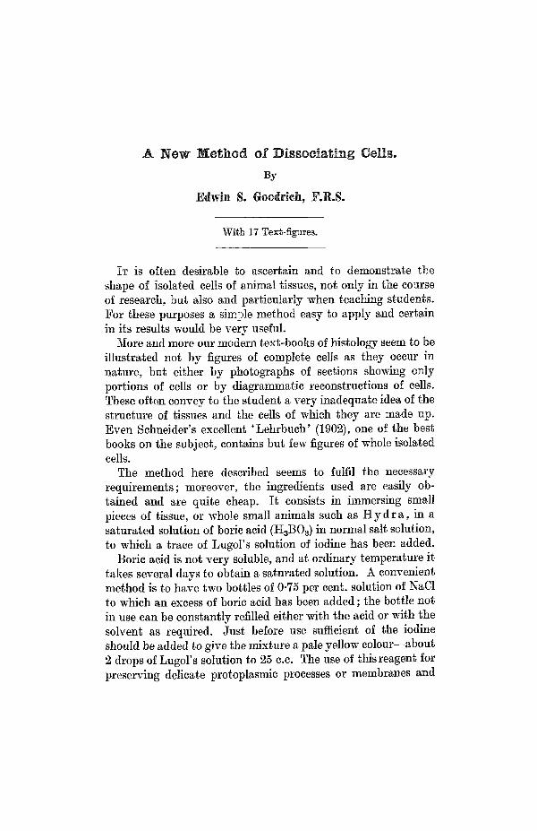
Both ectodermal and endodermal muscnlo-epithelial cells aresomewhat elongated transversely to the long axis of the animal,and their contractile fibrils are disposed in two layers, one oneach side of the intervening mesogloea. The fibrils cross at rightangles, those of the ectoderm running longitudinally and those
TEXT-FIG. 1.
Two ectodermal cells of H y d r a v i r i d i s seen from inner surface,showing muscular processes, pr; c, cell outline; n, nucleus.
of the endoderm running transversely (circularly). But it willbe noticed that whereas each endoderm cell has only one basalfibril, each ectoderm cell may have several (from 1 to 5 or 6)set at intervals along its base (Text-figs. 1 and 2). For theseectodermal fibrils, set closely in parallel lines, are more numerousthan the cells (Text-fig. 2). They do not radiate from the baseof the cell, as figured by Schneider (1890), and the appearanceof occasional branching seems to be due merely to overlapping.
The endodermal musculo-epithelial cells, on the other hand,have only one basal contractile fibril (Text-figs. 3 and 4).
The contractile fibres are covered with an irregular layer ofcytoplasm; delicate processes along each side and extending intothe mesogloea can generally be seen. The endodermal cilia,though very slender, usually show clearly. Irregular amoeboidprocesses project from the nutritive endodermal cells, which
248 EDWIN S. GOODBICH
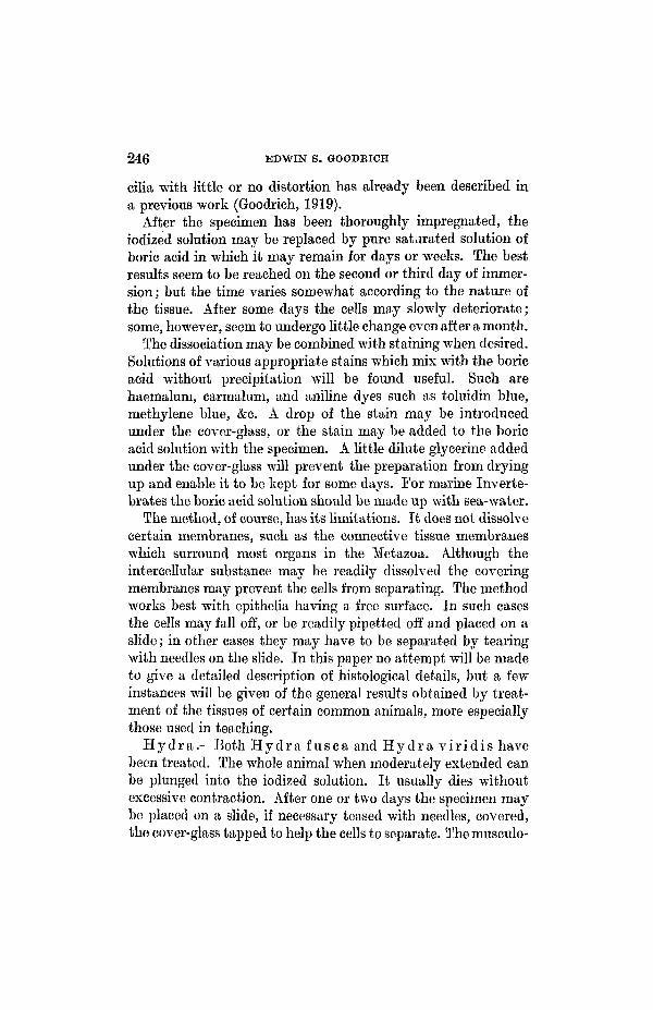
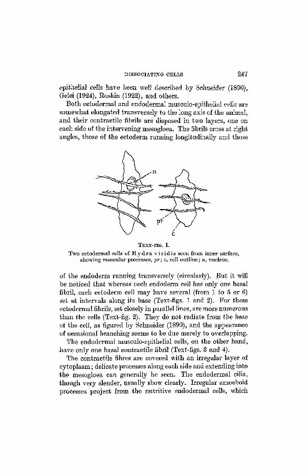
3OjJL
pr
TBXT-FIG. 2.Hydra vir idis , inner view of ectoderm, showing longitudinal
muscular processes, pr, and cell outlines, c. Preparation madewith Bichromate of Potash.
TEXT-FIG. 3.
Hydra fusca. Endodermal musculo-epithelial cells.
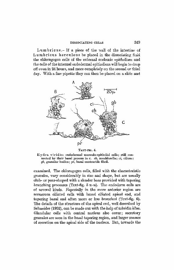
contain various food vacuoles and other granules. In H y d r av i r id i s the zoochlorellae occupy chiefly the more basal region,while in the more apical region are many colourless sphericalbodies of uniform granular appearance—possibly stored foodmaterial (Text-fig. 4). The glandular cells are usually conspi-cuous. Delicate branching cells are probably nervous.
DISSOCIATING CELLS 249
Lumbricus.—If a piece of the wall of the intestine ofL u m b r i c u s he reu leus be placed in the dissociating fluidthe chloragogen cells of the external coelomic epithelium andthe cells of the internal endodermal epithelium will begin to dropoff even in 24 hours, and more completely on the second or thirdday. With a fine pipette they can then be placed on a slide and
TEXT-FIG. 4.
H y d r a v i r i d i s : endodermal musculo-epithelial cells; still con-nected by their basal process in c. ch, zoochlorella; ci, cilium;gb, granular bodies; pr, basal contractile fibril.
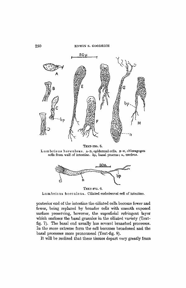
examined. The chloragogen cells, filled with the characteristicgranules, vary considerably in size and shape, but are usuallyclub- or pear-shaped with a slender base provided with taperingbranching processes (Text-fig. 5 E-H). The endoderm cells areof several kinds. Especially in the more anterior region arenumerous ciliated cells with broad ciliated apical end, andtapering basal end often more or less branched (Text-fig. 6).The details of the structure of the apical end, well described bySchneider (1902), can be made out with the help of toluidin blue.Glandular cells with central nucleus also occur; secretorygranules are seen in the basal tapering region, and larger massesof secretion on the apical side of the nucleus. But, towards the
250 EDWIN S. GOODKICH
TEXT-FIG. 5.Lumbricus herculeus. A-D, epidermal cells, E-H, chloragogen
cells from wall of intestine, bp, basal process; n, nucleus.
50H
CI n
TEXT-FIG. 6.
Lumbricus herculeus. Ciliated endodermal cell of intestine.
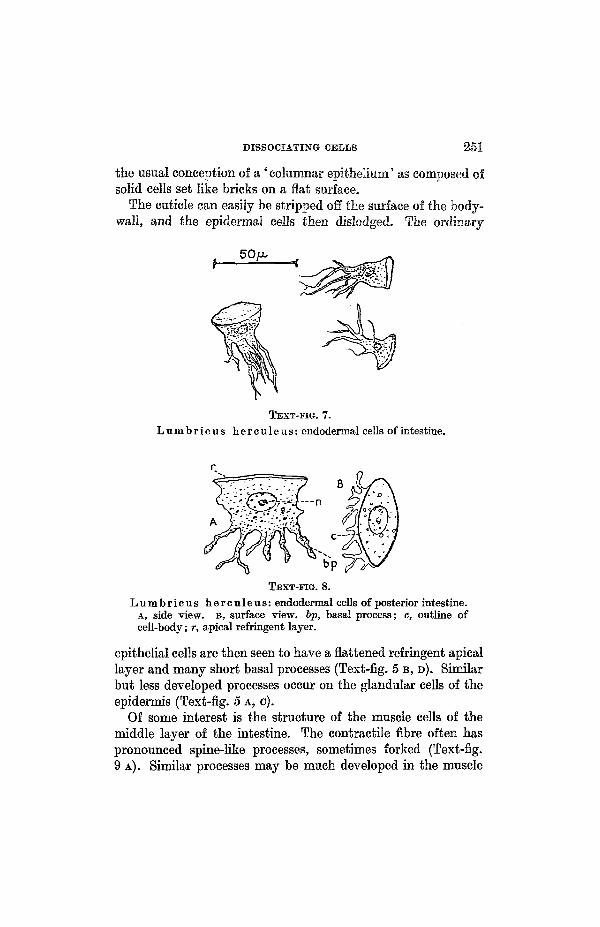
posterior end of the intestine the ciliated cells become fewer andfewer, being replaced by broader cells with smooth exposedsurface preserving, however, the superficial refringent layerwhich encloses the basal granules in the ciliated variety (Text-fig. 7). The basal end usually has several branched processes.In the more extreme form the cell becomes broadened and thebasal processes more pronounced (Text-fig. 8).
It will be realized that these tissues depart very greatly from
DISSOCIATING CELLS 251
the usual conception of a 'columnar epithelium' as composed ofsolid cells set like bricks on a flat surface.
The cuticle can easily be stripped off the surface of the body-wall, and the epidermal cells then dislodged. The ordinary
TEXT-FIG. 7.
Lumbricus herculeus: endodermal cells of intestine.
Lumbricus hereuleus: endodermal cells of posterior intestine.A, side view, B, surface view, bp, basal process; c, outline ofcell-body; r, apical refringent layer.
epithelial cells are then seen to have a flattened refringent apicallayer and many short basal processes (Text-fig. 5 B, D). Similarbut less developed processes occur on the glandular cells of theepidermis (Text-fig. 5 A, C).
Of some interest is the structure of the muscle cells of themiddle layer of the intestine. The contractile fibre often haspronounced spine-like processes, sometimes forked (Text-fig.9 A). Similar processes may be much developed in the muscle

252 EDWIN S. GOODRICH
cells of the larger blood-vessels and especially those of the hearts.Here they tend to form on either side a spreading extension withfringed edge and outstanding rounded lobes (Text-fig. 9 B).These extensions seem to belong to a siieath of refringentmaterial enclosing the true muscle fibre, and it frequently showsfine closely set folds transverse to the main axis of the fibre, and
TEXT-FIG. 9.
Lumbricus herculeus. Portions of muscular fibres of wall ofintestine, A, and of heart, B and C; showing processes and bilateralfringes of sheath of contractile fibril.
giving it the deceptive appearance of a striated muscle (Text-fig. 9 c, B).
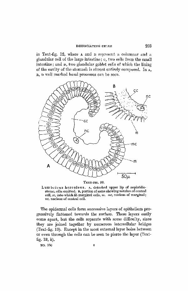
Unfortunately, the convoluted canal of the nephridium affordsan instance of the failure of the boric-acid method owing to thepresence of closely investing insoluble membranes. The freelyexposed cells of the open nephridiostome can, however, be dealtwith. Text-fig. 10 A is an outline sketch of the upper lip whichmay be made to separate off entire by tapping the cover-glass.It shows the shape of the large central cell and the surroundingmarginal cells. The way in which the marginals fit into thenotched margin of the central cell is seen in Text-fig. 10 B.
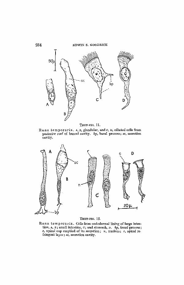
E a n a.—From Bana t e m p o r a r i a a few examples may betaken of easily dissociated cells. Such are the ciliated and theglandular cells lining the roof of the buccal cavity (Text-fig. 11).While the basal end of these cells is usually simple, some of themoften have divergent processes (Text-fig, l i e ) .
Typical cells of the endodermal lining of the gut are shown
DISSOCIATING CELLS 258
in Text-fig. 12, where A and B represent a columnar and aglandular cell of the large intestine; c, two cells from the smallintestine; and D, two glandular goblet cells of which the liningof the cavity of the stomach is almost entirely composed. In A,B, D well marked basal processes can be seen.
Bcc
TEXT-FIG. 10.Lumbricus herculeus. A, detached upper lip of nephridio-
stome, cilia omitted, B, portion of same showing notches of centralcell, cc, into which fit marginal cells, m. me, nucleus of marginal;nc, nucleus of central cell.
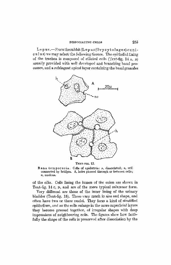
The epidermal cells form successive layers of epithelium pro-gressively flattened towards the surface. These layers easilycome apart, but the cells separate with some difficulty, sincethey are joined together by numerous intercellular bridges(Text-fig. 13). Except in the most external layer holes betweenor even through the cells can be seen to pierce the layer (Text-fig. 13, h).
NO. 330 S
254 EDWIN S. GOODRICH
50fl
TEXT-FIG. 11.
Rana temporar ia . A, B, glandular, andc, D, ciliated cells fromposterior roof of buccal cavity, bp, basal process; sc, secretioncavity.
"SC
20 H*h
TBXT-FIG. 12.
Rana temporar ia . Cells from endodermal lining of large intes-tine, A, B; small intestine, c; and stomach, D. bp, basal process;c, apical cup emptied of its secretion; n, nucleus; r, apical re-fringent layer; sc, secretion cavity.
DISSOCIATING CELLS 255
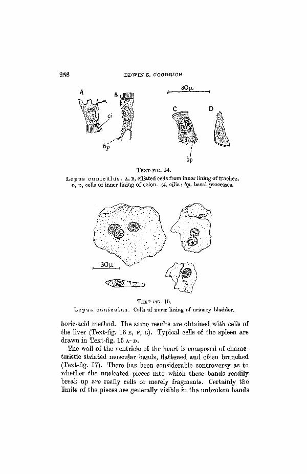
Lepus.—From the rabbit (Lep us (Or ycy to lagus )c un i -c u 1 u s) we may select the following tissues. The epithelial liningof the trachea is composed of ciliated cells (Text-fig. 14 A, B)usually provided with well developed and branching basal pro-cesses, and a refringent apical layer containing the basal granules
TEXT-FIG. 13.Rana temporar ia . Cells of epidermis: A, dissociated; B, still
connected by bridges, h, holes pierced through or between cells;n, nucleus.
of the cilia. Cells lining the lumen of the colon are shown inText-fig. 14 c, D, and are of the more typical columnar form.
Very different are those of the inner lining of the urinarybladder (Text-fig. 15). These vary much in size and shape, andoften have two or three nuclei. They form a kind of stratifiedepithelium, and as the cells enlarge in the more superficial layersthey become pressed together, of irregular shapes with deepimpressions of neighbouring cells. The figures show how faith-fully the shape of the cells is preserved after dissociation by the
256 EDWIN S. GOODRICH
30 [x
TEXT-FIG. 14.Lepus cuniculus. A, B, ciliated cells from inner lining of trachea.
c, D, cells of inner lining of colon, ci, cilia; bp, basal processes.
TEXT-FIG. 15.
Lepus cuniculus. Cells of inner lining of urinary bladder.
boric-acid method. The same results are obtained with cells ofthe liver (Text-fig. 16 B, F, G). Typical cells of the spleen aredrawn in Text-fig. 16 A-D.
The wall of the ventricle of the heart is composed of charac-teristic striated muscular bands, flattened and often branched(Text-fig. 17). There has been considerable controversy as towhether the nucleated pieces into which these bands readilybreak up are really cells or merely fragments. Certainly thelimits of the pieces are generally visible in the unbroken bands

DISSOCIATING CELLS 257
TEXT-ITG. 16.
Lepus cuniculus. A-D, cells of spleen, E, F, O, cells of liver.
TEXT-HG. 17.
Lepus cuniculus . Muscle cells of ventricle of heart.A, B, in outline; c, with contractile fibrils.
though even after prolonged treatment two or more may adhereowing probably to the minute fibrils being continuous fromsegment to segment (Text-fig. 17 B, C).
No mention has so far been made of the nervous system, atissue peculiarly ill adapted to dissociation owing to the tangle
258 EDWIN S. GOODRICH
of nerve fibres holding the cells together. Nevertheless, goodresults have been obtained by treating pieces of the spinal cordor brain with the fluid, and then teasing them on a slide. Butthe results are possibly no better than those reached by otherwell-known methods usually recommended.
In conclusion it may be said that the boric-acid method heredescribed has the merit of being very easy to apply to suitabletissues. It is possible that, if combined with other reagents andappropriate stains, it may help to reveal fine histological detailsand thus help in the study of histological problems.
SUMMAEY.
A method for the dissociation of the cells of Metazoan tissuesis described consisting in their treatment with a saturatedsolution of boric acid in normal saline to which a trace of Lugol'ssolution of iodine has been added. Examples of the resultsobtained are described and figured.
LIST OF REFERENCES.
Gelei, J . v., 1924.—"Cytologieder Hydragrisea", 'Z. f. Zellen u. Gewebel',!•
Goodrich, E. S., 1919.—"Pseudopodia of Leucocytes", 'Quart. Journ.Micr. Sci.', 64.
Roskin, Gr. v., 1922.—"Epithel-Muskelzellen v. Hydra", 'Anat. Anz.', 56.Schneider, K. C, 1890.—"Histol. v. Hydrafusea", 'Arch. mikr. Anat.', 35.
1902.—'Lehrbuch d. vergl. Histol. der Tiere.' Jena.