a new radiological measurement method used to evaluate the
TRANSCRIPT

RESEARCH ARTICLE Open Access
A new radiological measurement methodused to evaluate the modifiedtransconjunctival orbital fat decompressionsurgeryBei Li1*†, Li Feng2†, Huamin Tang2, Liuzhi Zeng1 and Wei Lin2
Abstract
Purpose: A new radiological method was used to evaluate the plastic effect of modified transconjunctival orbitalfat decompression surgery in patients with inactive thyroid-associated ophthalmopathy.
Methods: In this study, 10 inactive patients (14 eyes) with moderate to severe thyroid-associated ophthalmopathywere selected. The patients underwent modified transconjunctival orbital fat decompression surgery. According tothe results of a spiral CT scan before and 6months after the surgery, the INFINITT system workstation was used tomeasure the eyeball protrusion value. According to the results obtained by the PHLIPS IntelliSpace Portal ellipticalarea and line segment measurement tools, the standard elliptical vertebral volume formula was used to calculatethe muscular cone inner volume. Changes in eyeball protrusion and the inner volume of the muscular cone beforeand after surgery were examined. Statistical analysis of the correlation between the two parameters was performed.
Results: Radiological measurement results confirmed that removing the orbital fat in the muscle cone duringsurgery was effective for alleviating eyeball protrusion in patients with thyroid-associated ophthalmopathy (P <0.05). This surgery caused an obvious change in the muscle cone inner volume (P < 0.05). And there was significantcorrelation between changes in eyeball protrusion and muscle cone inner volume (r = 0.797, P = 0.0006, P < 0.05).
Conclusion: The radiological assessment method used in this study is simple and easy to implement. For inactivepatients with moderate to severe thyroid-associated ophthalmopathy who just want to improve their appearance,the modified orbital fat decompression surgery is worth considering.
Keywords: Radiological, Fat decompression, Thyroid-associated ophthalmopathy
© The Author(s). 2021 Open Access This article is licensed under a Creative Commons Attribution 4.0 International License,which permits use, sharing, adaptation, distribution and reproduction in any medium or format, as long as you giveappropriate credit to the original author(s) and the source, provide a link to the Creative Commons licence, and indicate ifchanges were made. The images or other third party material in this article are included in the article's Creative Commonslicence, unless indicated otherwise in a credit line to the material. If material is not included in the article's Creative Commonslicence and your intended use is not permitted by statutory regulation or exceeds the permitted use, you will need to obtainpermission directly from the copyright holder. To view a copy of this licence, visit http://creativecommons.org/licenses/by/4.0/.The Creative Commons Public Domain Dedication waiver (http://creativecommons.org/publicdomain/zero/1.0/) applies to thedata made available in this article, unless otherwise stated in a credit line to the data.
* Correspondence: [email protected]: Modified retroconjunctival orbital decompression surgery caneffectively improve the appearance of patients with inactive thyroid-associated ophthalmopathy. The effect can be evaluated by a newradiological measurement method.†Bei Li and Li Feng contributed equally to this work.1Department of Ophthalmology, Chengdu First People’s Hospital, No.18Wanxiang North Road, Chengdu 610041, Sichuan Province, ChinaFull list of author information is available at the end of the article
Li et al. BMC Ophthalmology (2021) 21:176 https://doi.org/10.1186/s12886-021-01911-9

BackgroundThyroid-associated ophthalmopathy (TAO), also knownas Graves’ ophthalmopathy (GO) or thyroid eye disease(TED), is an autoimmune disease that accounts for thehighest incidence of orbital diseases in adults. In March2016, the European Thyroid Association/European Groupon Graves’ Orbitopathy (EUGOGO) published guidelinesfor the management of GO. Based on the guidelines, sur-gery is an effective treatment for moderate to severe in-active patients, patients with active severe exposed ocularsurface inflammation, and active patients with oppressiveoptic neuropathy [1]. However, more inactive patients re-quire surgery because of their appearance [2]. A study hadshown that orbital fat decompression surgery can signifi-cantly improve the overall quality of life with a low com-plication rate [3].In most of the literature, during orbital fat decompres-
sion surgery, not only the orbital fat inside the musclecone, but also the orbital fat outside the muscle cone isremoved [4–7]. However, based on the anatomy of theorbit, most of the orbital fat outside the muscle cone islocated in the front of the orbit, and the most directforce that causes exophthalmos comes from the fat inthe muscle cone behind the eyeball. In our experience,removing the orbital fat outside the muscle cone had noeffect on improving the exophthalmos and might affectthe position of the eyeball. Some surgeons only removedthe fat behind the ball during the surgery without theneed to disturb the anterior orbital fat pads, andachieved obvious surgical effect [2].In order to explore the relationship between changes
in eyeball protrusion and changes in cone volume, weselected moderate to severe inactive patients who under-went the modified transconjunctival orbital fat decom-pression surgery at the department of ophthalmology inChengdu First People’s Hospital. We conducted a
retrospective study using the results of preoperative andpostoperative computed tomography (CT) examinations.During the study we used a new radiological measure-ment method to acquire data.
Materials and methodsCase source and inclusion criteriaThis study was approved by the Ethics Committee ofChengdu First People’s Hospital (certificate approvalnumber:2021-WZ-001). The study adhered to the tenetsof the Declaration of Helsinki. The patients’ partial ap-pearance photos and orbital CT scan results in this art-icle had been approved by the patients. The writteninformed consent—for both study participation and pub-lication of identifying information/images in an onlineopen-access publication was obtained from the patients.A copy of the written informed consent is available forreview by the Editor of this journal.The cases included 14 eyes of 10 TAO patients (mean
age: 41.8 ± 9.7 years; sex: 5 males and 5 females; meancourse of disease: 22.4 ± 7.2 months) who underwent or-bital fat decompression surgery in 2019. Case inclusioncriteria: all patients met the criteria for moderate to se-vere patients in the inactive phase based on the guide-lines formulated by EUGOGO [1], and their thyroidfunction and exophthalmos had been stable for morethan half a year. Case exclusion criteria: patients had suf-fered from other orbital diseases or received any previ-ous orbital surgery, and according to the results of thepreoperative CT scan, the extraocular muscles werethickened significantly.
Surgical techniqueBased on the surgical technique for deep orbital fat re-section [8] from “Chinese Ophthalmology”, first, we ax-ially cut the lateral canthus with surgical scissors, cut off
Fig. 1 The position of cutting fat. It’s in the muscle cone and between the inferior rectus and the external rectus
Li et al. BMC Ophthalmology (2021) 21:176 Page 2 of 8

Fig. 2 Method for measuring eyeball protrusion. The length of the line segment pointed by the arrow is the value of eyeball protrusion
Fig. 3 Method for measuring the muscle cone inner volume. On the a the line pointed to by the arrow is the sagittal axis. On the b the linepointed to by the arrow is the coronal axis. On the c the area of the ellipse pointed to by the arrow is the muscle cone bottom area (S), and thelength of the line segment pointed to by the arrow is the muscle cone height (h)
Li et al. BMC Ophthalmology (2021) 21:176 Page 3 of 8

the lower branch of the lateral canthal ligament, andfreed the lower eyelid. Next, we cut the bulbar conjunc-tiva along the inferior fornix and entered the surgicalgap between the orbital periosteum and extraocularmuscles. Then, we entered the muscle cone from thegap between the inferior rectus muscle and the lateralrectus muscle by incising the intermuscular septum, andremoved the fat tissue in the muscle cone carefully withhemostatic forceps. However, we did not remove theorbital fat outside the muscle cone. Finally, the bulbarconjunctiva and lateral canthus were sutured (Fig. 1). Allsurgeries were performed by Dr. Bei Li.
Method of spiral CT scanningAll patients underwent non-enhanced spiral CT scans(PHILIPS, Netherlands) before and 6months after sur-gery at the department of radiology in Chengdu FirstPeople’s Hospital. Each patient was scanned in a supineposition with both eyes fixed on a target. The Frankfurtline was used as the baseline, and a spiral volume scanwas performed (scan parameters: layer thickness 1 mm,layer spacing 1 mm, pitch 0.6, matrix 512 × 512, windowwidth 300 HU and window position 35 HU). The
obtained images were all transmitted to the picture ar-chiving and communication systems (PACS, PHLIPS,Netherlands) for the measurement of eyeball protrusionand muscle cone inner volume.
Method for measuring eyeball protrusionMulti-planner reformation (MPR) was performed onthe spiral CT image data, and axial, sagittal and cor-onal images of the orbit were obtained simultaneously(Fig. 2). Using the sagittal image as the benchmark(Fig. 2A), we determined the most prominent layer of theeyeball and used the corresponding axial image as thestandard layer to measure eyeball protrusion (Fig. 2B). Onthe standard level of the axial position, we connected theleft and right highest points of the outer edge of the orbitsas a reference line, passed the furthest point of the frontprojection of the eyeball, drew a perpendicular to the ref-erence line, and then measured the distance of the per-pendicular as eyeball protrusion value. Three skilledradiologists (Li Feng, Huamin Tang and Wei Lin) inde-pendently completed the measurement of each data andobtained the mean. They were masked and double blind.
Table 1 Demographic and clinical characteristics of studied patients
Case Eyes Age(years) Gender Course of disease (months) Steroid pulse CAS score Optic neuropathy Diplopia
1 right 28 female 14 yes 1 no no
2 right 38 male 18 yes 0 no no
3 left 39 female 20 yes 0 no no
4 both 50 male 16 yes 0 no no
5 both 55 female 30 yes 1 no no
6 left 30 female 16 no 0 no no
7 both 35 female 22 yes 0 no no
8 right 45 male 22 yes 0 no no
9 left 56 male 35 no 1 no no
10 both 42 male 31 yes 0 no no
Fig. 4 Preoperative and postoperative appearance photos of a subset of patients. The arrows point to the surgical eyes
Li et al. BMC Ophthalmology (2021) 21:176 Page 4 of 8

Method for measuring the muscle cone inner volume (Fig. 3)Method for determining the standard measurement positionWe used the “tangential coronal position” as the stand-ard measurement position. First, on the axial image ofthe orbit with the optic nerve displayed (Fig. 3A), we ad-justed the sagittal axis to overlap with the optic nervelong axis to obtain the reference sagittal position (Fig.3B). Second, on the reference sagittal image, we adjustedthe coronal axis to be tangential to the position wherethe optic nerve exited the eyeball to obtain the“tangential coronal position” for measurement (Fig. 3C).
Method for calculating the muscle cone inner volumeBecause the muscle cone anatomy was similar to anelliptical cone, we used the standard elliptical vertebralbody volume formula (V=Sh/3, V: vertebral body
volume, S: cone base area, h: vertebral height) to calcu-late the muscle cone inner volume.First, on the “tangential coronal position”, using the ellip-
tical area measurement tool, with the optic nerve as themeasurement center, we manually outlined and adjustedthe elliptical edge to be tangential to the inner edges of thefour extraocular muscles. The resulting elliptical area wasthe muscle cone bottom area (S) (Fig. 3C). Then, on thereference transection with the optic nerve displayed, weused the line measurement tool to measure the distancefrom the beginning of the intraorbital segment of the opticnerve to the beginning of the optic canal. The resulting dis-tance was the muscle cone height (h) (Fig. 3D). Threeskilled radiologists (Li Feng, Huamin Tang and Wei Lin) in-dependently completed the measurement of each data andobtained the mean. They were masked and double blind.
Table 2 Preoperative and postoperative eyeball protrusion (x ± s, n = 3)
Eyes Preoperative protrusion (mm) Postoperative protrusion (mm)
1 20.82 ± 0.35 18.26 ± 0.31
2 20.08 ± 0.16 16.32 ± 0.15
3 21.77 ± 0.26 17.94 ± 0.17
4 20.46 ± 0.24 17.02 ± 0.05
5 18.68 ± 0.36 15.31 ± 0.21
6 21.58 ± 0.12 18.87 ± 0.25
7 23.36 ± 0.31 20.41 ± 0.16
8 23.08 ± 0.11 20.27 ± 0.19
9 19.76 ± 0.32 16.84 ± 0.33
10 18.32 ± 0.25 16.13 ± 0.28
11 19.85 ± 0.22 17.15 ± 0.23
12 19.81 ± 0.30 16.85 ± 0.32
13 20.31 ± 0.16 16.99 ± 0.22
14 19.59 ± 0.27 16.55 ± 0.09
P < 0.05 Preoperative protrusion vs Postoperative protrusion (paired t - test)
Fig. 5 Preoperative protrusion vs Postoperative protrusion, t = 24.47, P<0.0001. (paired t- tests)
Li et al. BMC Ophthalmology (2021) 21:176 Page 5 of 8

Statistical methodsGraphPad Prism 9.0.0 (USA, GraphPad Software) wasused for statistical analysis. Based on the results of anormality distribution test, all data were normally dis-tributed. Paired t-tests and Pearson product-momentcorrelation were used. Paired t-tests was used to com-pare the changes of eyeball protrusion and muscle coneinner volume between preoperative and postoperative.Pearson product-moment correlation was used toanalyze the correlation between the two changes. Thecorrelation coefficient was represented byr. The resultswere considered statistically significant if P < 0.05.
ResultsDemographic and clinical characteristics of studiedpatients (Table 1)
Preoperative and postoperative appearance photos of asubset of patients (Fig. 4)
Results of eyeball protrusion measurements (Table 2, Fig. 5)
Results of muscle cone inner volume measurements(Table 3, Fig. 6)
Correlation analysis results of differences in protrusionand volume (Table 4, Fig. 7)
DiscussionIn recent years of research, some scholars measured theeyeball protrusion of TAO patients on the images ob-tained by CT scan, and they determined the most for-ward measurement position of the eyeballs by the thethickest position of the lens [9, 10]. For TAO patients
without myopathy, the CT slice of the thickest lens cor-responds to the apex of the cornea, which is the mostprotruding position of the eyeball. However, since theapex of the cornea and interzygomatic line cannot be in-cluded in the same plane on a two-dimensional (2D) CTscan, the level of the measured CT slice may not corres-pond to the area of maximal proptosis [11]. For TAOpatients with varying degrees of restrictive strabismus,the apex of the cornea corresponding to the CT slicemay not be the most protruding position of the eyeball.Therefore, the method of determining the measurementposition of the eyeball protrusion through the thickestposition of the lens is limited. In our study, we used themost prominent position of eyeballs in the sagittal pos-ition to determine the corresponding axial position formeasurement. The method is more intuitive and easy.And it can avoid the measurement difficulty and errorcaused by the deviation of the eyeball position.The fascial tissue between the extraocular muscles
can’t be clearly displayed on the images obtained by CT.So there is no software can directly measure the innervolume of the muscle cone. In the previous literature,the use of software can only measure the retrobulbar or-bital volume [7]. In our study, to measure the musclecone inner volume, we simulated the muscle cone as anelliptical cone. In the “tangential coronal position”, weused measurement tools of the system workstation toobtain the required data and calculate the muscle coneinner volume. This method is effective and feasible.The muscular cone is an independent and relatively
closed structure surrounded by extraocular muscles andfascia in the orbit. An increase and decrease in intraco-nal fat mass will most directly affect the increase and de-crease in eyeball protrusion. Most of the extracone fat is
Table 3 Preoperative and postoperative muscle cone inner volume (x ± s, n = 3)
Eyes Preoperative volume (mm3) Postoperative volume (mm3)
1 6258.95 ± 55.11 4407.76 ± 63.28
2 6222.78 ± 80.14 4019.14 ± 35.22
3 7609.94 ± 39.26 5685.99 ± 41.65
4 7321.41 ± 40.35 5425.61 ± 65.15
5 6377.56 ± 33.16 4653.23 ± 77.01
6 7839.59 ± 52.51 6275.37 ± 56.23
7 8192.59 ± 48.67 6512.58 ± 64.27
8 7558.99 ± 32.07 6149.72 ± 49.37
9 6044.08 ± 45.80 4451.24 ± 50.03
10 5474.37 ± 81.25 4458.18 ± 36.25
11 6251.20 ± 29.78 4717.31 ± 46.18
12 5997.75 ± 57.20 4182.40 ± 67.74
13 7398.49 ± 42.25 5484.63 ± 58.45
14 7422.71 ± 63.31 5519.61 ± 85.31
P < 0.05 Preoperative protrusion vs Postoperative volume (paired t - test)
Li et al. BMC Ophthalmology (2021) 21:176 Page 6 of 8
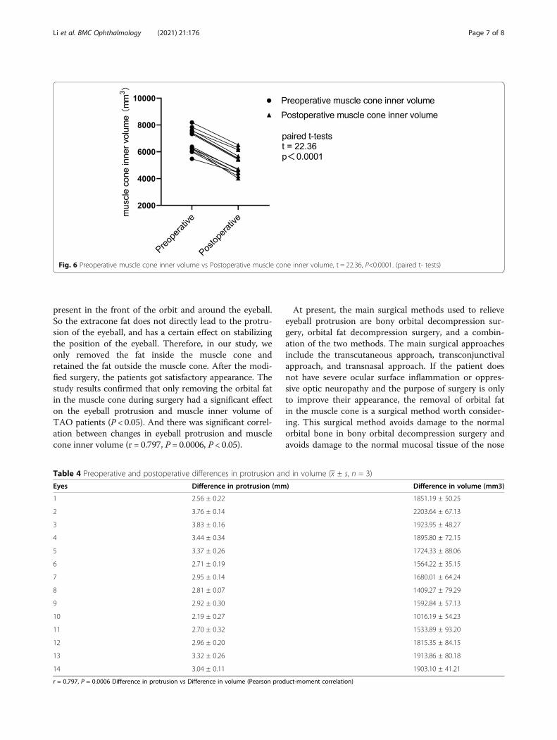
present in the front of the orbit and around the eyeball.So the extracone fat does not directly lead to the protru-sion of the eyeball, and has a certain effect on stabilizingthe position of the eyeball. Therefore, in our study, weonly removed the fat inside the muscle cone andretained the fat outside the muscle cone. After the modi-fied surgery, the patients got satisfactory appearance. Thestudy results confirmed that only removing the orbital fatin the muscle cone during surgery had a significant effecton the eyeball protrusion and muscle inner volume ofTAO patients (P < 0.05). And there was significant correl-ation between changes in eyeball protrusion and musclecone inner volume (r = 0.797, P = 0.0006, P < 0.05).
At present, the main surgical methods used to relieveeyeball protrusion are bony orbital decompression sur-gery, orbital fat decompression surgery, and a combin-ation of the two methods. The main surgical approachesinclude the transcutaneous approach, transconjunctivalapproach, and transnasal approach. If the patient doesnot have severe ocular surface inflammation or oppres-sive optic neuropathy and the purpose of surgery is onlyto improve their appearance, the removal of orbital fatin the muscle cone is a surgical method worth consider-ing. This surgical method avoids damage to the normalorbital bone in bony orbital decompression surgery andavoids damage to the normal mucosal tissue of the nose
Fig. 6 Preoperative muscle cone inner volume vs Postoperative muscle cone inner volume, t = 22.36, P<0.0001. (paired t- tests)
Table 4 Preoperative and postoperative differences in protrusion and in volume (x ± s, n = 3)
Eyes Difference in protrusion (mm) Difference in volume (mm3)
1 2.56 ± 0.22 1851.19 ± 50.25
2 3.76 ± 0.14 2203.64 ± 67.13
3 3.83 ± 0.16 1923.95 ± 48.27
4 3.44 ± 0.34 1895.80 ± 72.15
5 3.37 ± 0.26 1724.33 ± 88.06
6 2.71 ± 0.19 1564.22 ± 35.15
7 2.95 ± 0.14 1680.01 ± 64.24
8 2.81 ± 0.07 1409.27 ± 79.29
9 2.92 ± 0.30 1592.84 ± 57.13
10 2.19 ± 0.27 1016.19 ± 54.23
11 2.70 ± 0.32 1533.89 ± 93.20
12 2.96 ± 0.20 1815.35 ± 84.15
13 3.32 ± 0.26 1913.86 ± 80.18
14 3.04 ± 0.11 1903.10 ± 41.21
r = 0.797, P = 0.0006 Difference in protrusion vs Difference in volume (Pearson product-moment correlation)
Li et al. BMC Ophthalmology (2021) 21:176 Page 7 of 8

in transnasal orbital decompression surgery. The radio-logical measurement method used in our research caneffectively evaluate the effect of this surgery.
AcknowledgementsThanks to Dachuan Huang, MD, National Hospital of Singapore, for his helpduring the paper writing.
Authors’ contributionsBL completed the design of the study, the data collection, analysis andinterpretation, and the writing of the manuscript. LF and HMT participated inthe design of the study. LF, HMT, WL and LZZ participated in the datacollection of the study. All authors read and approved the final manuscript.
FundingNot applicable.
Availability of data and materialsThe datasets during and/or analysed during the current study available fromthe corresponding author on reasonable request.
Declarations
Ethics approval and consent to participateThis study was approved by the ethics committee of Chengdu First People'sHospital.The research adhered to the tenets of the Declaration of Helsinki.
Consent for publicationWritten informed consent was obtained from the patients for publication ofany accompanying images. A copy of the written consent is available forreview by the Editor of this journal.
Competing interestsThe authors declare that they have no competing interests. No meetingpresentation, financial support and proprietary interest statement.
Author details1Department of Ophthalmology, Chengdu First People’s Hospital, No.18Wanxiang North Road, Chengdu 610041, Sichuan Province, China.2Department of Radiology, Chengdu First People’s Hospital, Chengdu,Sichuan Province, China.
Received: 27 January 2021 Accepted: 16 March 2021
References1. Bartalena L, Baldeschi L, Boboridis K, Eckstein A, Kahaly GJ, Marcocci C, et al.
The 2016 European thyroid association/European group on Graves'
Orbitopathy guidelines for the Management of Graves' Orbitopathy [J]. EurThyroid J. 2016;5(1):9–26. https://doi.org/10.1159/000443828.
2. Al-Sharif E, Alsuhaibani AH. Fat-removal orbital decompression for thyroidassociated orbitopathy: the right procedure for the right patient. Saudi JOphthalmol. 2017;31(3):156–61. https://doi.org/10.1016/j.sjopt.2017.05.017.
3. Cheng AM, Wei YH, Tighe S, Sheha H, Liao SL. Long-term outcomes oforbital fat decompression in Graves' orbitopathy. Br J Ophthalmol. 2018;102(1):69–73. https://doi.org/10.1136/bjophthalmol-2016-309888.
4. Prat MC, Braunstein AL, Dagi Glass LR, Kazim M. Orbital fat decompressionfor thyroid eye disease: retrospective case review and criteria for optimalcase selection. Ophthalmic Plast Reconstr Surg. 2015;31(3):215–8. https://doi.org/10.1097/IOP.0000000000000260.
5. Lee KH, Jang SY, Lee SY, Yoon JS. Graded decompression of orbital fat andwall in patients with Graves' Orbitopathy. Korean J Ophthalmol. 2014;28(1):1–11. https://doi.org/10.3341/kjo.2014.28.1.1.
6. Li EY, Kwok TY, Cheng AC, Wong AC, Yuen HK. Fat-removal orbitaldecompression for disfiguring proptosis associated with Graves'ophthalmopathy: safety, efficacy and predictability of outcomes. IntOphthalmol. 2015;35(3):325–9.
7. Liao S, Huang S. Correlation of retrobulbar volume change with resect-edorbital fat volume and Proptosis reduction after fatty decompression forgraves. Ophthalmopathy Am J Ophthalmol. 2011;151(3):465–9. https://doi.org/10.1016/j.ajo.2010.08.042.
8. Li F. Chinese ophthalmology (2). Beijing: People's Medical Publishing House;2005. p. 1103.
9. Bingham CM, Sivak-Callcott JA, Gurka MJ, Nguyen J, Hogg JP, Feldon SE,et al. Axial globe position measurement: a prospective multi-center studyby the international thyroid eye disease society. Ophthal Plast ReconstrSurg. 2016;32(2):106–12.
10. Pereira TS, Kuniyoshi CH, Leite CA, Gebrim EMMS, Monteiro MLR, PieroniGonçalves AC. A comparative study of clinical vs. digital Exophthalmometrymeasurement methods. J Ophthalmol. 2020;2020:1397410.
11. Huh J, Park SJ, Lee JK. Measurement of proptosis using computedtomography based three-dimensional reconstruction software in patientswith graves’ orbitopathy. Sci Rep. 2020;10:14554.
Publisher’s NoteSpringer Nature remains neutral with regard to jurisdictional claims inpublished maps and institutional affiliations.
Fig. 7 Difference in protrusion vs Difference in volume, r = 0.797, P = 0.0006. (Pearson product-moment correlation)
Li et al. BMC Ophthalmology (2021) 21:176 Page 8 of 8