a novel mitochondrial sphingomyelinase in zebrafish cells ... · 1 a novel mitochondrial...
TRANSCRIPT

1
A NOVEL MITOCHONDRIAL SPHINGOMYELINASE IN ZEBRAFISH CELLS
Takeshi Yabu, Akio Shimuzu, and Michiaki Yamashita* National Research Institute of Fisheries Science, Yokohama, Kanagawa, 236-8648, Japan
Running Title: Mitochondrial sphingomyelinase Keywords: cDNA cloning, cell growth, ceramide, mitochondria, mitochondrial sphingomyelinase, neutral sphingomyelinase 2 sphingomyelin, zebrafish. *To whom correspondence should be addressed: National Research Institute of Fisheries Science, Yokohama, Kanagawa, 236-8648, Japan. Tel: +81-045-788-7665; Fax: +81-045-788-5001; E-mail: [email protected] Sphingolipids are important signaling molecules in many biological processes, but little is known regarding their physiological roles in the mitochondrion. We focused on the biochemical characters of a novel sphingomyelinase (SMase) and its function in mitochondrial ceramide generation in zebrafish embryonic (ZE) cells. The cloned SMase cDNA encoded a polypeptide of 545 amino acid residues (putative molecular weight, 61.3 K) containing a mitochondrial localization signal (MLS) and a predicted transmembrane domain (TD). The mature endogenous enzyme was predicted to have a molecular weight of 57 K, and MALDI-TOF-MS analysis indicated that the N-terminal-amino acid residue of the mature enzyme was Ala-36. The purified enzyme optimally hydrolyzed [14C]sphingomyelin in the presence of 10 mM Mg2+ at pH 7.5. In HEK293 cells that overexpressed SMase cDNA, the enzyme was localized to the mitochondrial fraction, whereas mutant proteins lacking MLS or both the MLS and the TD were absent from the mitochondrial fraction. Endogenous SMase protein co-localized with a mitochondrial cytostaining marker. Using a protease protection assay, we found that SMase was distributed throughout the intermembrane space and/or the inner membrane of the mitochondrion. Furthermore, the overexpression of SMase in HEK293 cells induced ceramide generation, and sphingomyelin hydrolysis in the mitochondrial fraction. Antisense phosphorothioate oligonucleotide-induced knockdown repressed ceramide generation, and sphingomyelin hydrolysis in the mitochondrial fraction in ZE cells. These observations indicate that SMase catalyzes the hydrolysis of sphingomyelin and generates ceramide in mitochondria in fish cells.
Sphingomyelinase (SMase, sphingomyelin phosphodiesterase, EC3.1.4.12) hydrolyzes sphingomyelin and produces ceramide and phosphocholine. Ceramide plays an important role as a signaling molecule in cell proliferation, apoptosis, cell cycle arrest, differentiation, and the stress response in animal cells (1−5). To date, three distinct classes of acid, neutral, and alkaline SMases have been identified according to optimum pH, cation dependency, amino acid sequence, and subcellular localization (3). The Mg2+-dependent neutral SMases have emerged as major candidates in the mediation of ceramide-induced cell signaling (6). Recent research has identified at least three distinct neutral SMases in human and mouse, designated as neutral SMase 1, SMase 2, and SMase 3 (7−9). Neutral SMase 1 was the first SMase identified in human and mouse. Although mammalian enzymes exhibited Mg2+-dependent neutral SMase activity in vitro (9), no significant biological functions in sphingomyelin and ceramide metabolism were identified in SMase 1-overexpressing cells (10) or neutral SMase 1 knockout mice (11). In zebrafish embryos, Mg2+-dependent neutral SMase 1 produced ceramide and caused thalidomide-induced vascular defects (12). In addition, SMase 1 was found to mediate heat-induced ceramide generation and apoptosis (13). A neutral SMase 2 gene has also been identified based on its similarity to Bacillus cereus SMase DNA sequences (7). This gene encodes a membrane-bound protein expressed in the brain and liver that has two highly hydrophobic segments near the N-terminal region, both of which are thought to function as transmembrane domains (TDs). Unlike neutral SMase 1, neutral SMase 2 possesses Mg2+-dependent neutral SMase activity in vivo in MCF-7 cells (14). When overexpressed in the confluent phase of MCF-7 cells, mouse neutral SMase 2 was palmitoylated via
http://www.jbc.org/cgi/doi/10.1074/jbc.M109.004580The latest version is at JBC Papers in Press. Published on May 8, 2009 as Manuscript M109.004580
Copyright 2009 by The American Society for Biochemistry and Molecular Biology, Inc.
by guest on July 4, 2018http://w
ww
.jbc.org/D
ownloaded from
2
thioester bonds and localized in the inner leaflet of the plasma membrane (15). In MCF-7 cells stably expressing neutral SMase 2, the enzyme inhibited cell growth and was required for cells to undergo confluence-induced cell cycle arrest (16). Interestingly, neutral SMase 2 was isolated as a confluent 3Y1 cell-associated 1 (cca1) gene in rat 3Y1 cells (17). Neutral SMase 2 has been implicated in signal transduction events in cell growth and the cellular response to cytokines (18,19), oxidative stress (20), and amyloid ß-peptide (21). Stoffel et al. (22) demonstrated that gene-targeted mice deficient for neutral SMase 2 developed a novel form of dwarfism and had delayed puberty as part of a hypothalamus-induced pituitary hormone deficiency. Strikingly, positional cloning of the recessive mutation fragilitas ossium in mice identified a deletion in the gene that encodes neutral SMase 2, leading to the complete loss of neutral SMase activity (23). The mutant fragilitas ossium mice develop severe osteogenesis and dentinogenesis imperfecta, with no collagen defect. Thus, mouse neutral SMase 2 is essential for late embryonic and postnatal development. Mitochondria contain small amounts of a variety of sphingolipids, including ceramide and sphingomyelin (24–26), which may be derived from the endoplasmic reticulum via intimate membrane contacts or produced in response to apoptosis. For mitochondria isolated from HL-60 cells, treatment with ceramide inhibited the mitochondrial respiratory chain complex III (27). Birbes et al. (28) found that the selective hydrolysis of a mitochondrial pool of sphingomyelin induced apoptosis. They transfected MCF-7 cells with Bacillus cereus SMase targeted to various subcellular organelles, but observed cytochrome c release and apoptosis induction only when the enzyme was targeted to the mitochondria. Ceramide activated the mitochondrial protein phosphatase 2A, which dephosphorylated Bcl-2 and led to apoptosis (29). In MCF-7 cells, mitochondrial ceramide generation in response to TNF-a induced Bax translocation to mitochondria and subsequent cytochrome c release and apoptosis (30). The permeability of the mitochondrial outer membrane correlates directly with the level of ceramide in the membrane (31). The concentration of ceramide at which significant channel formation occurs is consistent with the level of mitochondrial ceramide that occurs during the induction phase of apoptosis (31). In isolated mitochondria, ceramide can also form membrane
channels large enough to release cytochrome c and other small proteins (32). Ceramide-metabolizing enzymes, such as a bovine liver ceramide synthase (33) and human ceramidase (34), are localized to the mitochondrion. These observations suggest the existence of a mitochondrial pool of sphingomyelin and the function of a sphingomyelin-specific metabolic pathway in mitochondria. However, no SMase has been identified in mitochondria. We identified and examined the biochemical properties of a novel SMase localized to the zebrafish mitochondrion. The enzyme was cloned from a cDNA library of embryonic zebrafish cells. It was found to regulate mitochondrial ceramide levels. EXPERIMENTAL PROCEDURES
Reagents—The C6-7-nitro-2-1,3-benzoxadiazol-4-yl (C6-NBD) sphingomyelin was purchased from Matreya (Pleasant Gap, PA, USA). The [N-methyl-14C]sphingomyelin (2 GBq/mmol), L-3-phosphatidyl [N-methyl-14C]choline, 1,2-dipalmitoyl (2.11 GBq/mmol), [1-O-octadecyl-3H]lyso-platelet activating factor (5.99 TBq/mmol), protein G Sepharose, polyvinylidine fluoride (PVDF) membrane, and ECL™ Western Blotting Detection kit were purchased from GE Healthcare (Piscataway, NJ, USA). Anti-actin monoclonal antibody, anti-FLAG M2 monoclonal antibody, anti-HSP60 monoclonal antibody, anti-catalase monoclonal antibody, anti-cytochrome c polyclonal antibody, and p3XFLAG-CMV™-14 expression vector were purchased from Sigma-Aldrich (St Louis, MO, USA). Rabbit anti-cadherin polyclonal antibody and goat anti-aldolase polyclonal antibody were purchased from Santa Cruz Biotechnology (Santa Cruz, CA, USA). Anti-KDEL mouse monoclonal antibody and anti-58K Golgi protein mouse monoclonal antibody were purchased from Abcam (Cambridge, MA, USA). Cell culture reagents, the ThermoScript RT-PCR System, Xpress™ System Synthetic Oligonucleotides, goat anti-rabbit and anti-mouse IgG Alexa Fluor 488-labeled antibody, goat anti-mouse IgG Alexa Fluor 594-labeled antibody, and MitoTracker Red were purchased from Invitrogen (Carlsbad, CA, USA). The Premix Taq (ExTaq™ Version 2) and PrimeSTAR™HS DNA polymerase were purchased from Takara Biomedical (Shiga, Japan). Cell Culture—A zebrafish embryonic (ZE) cell line was cultured in Leibovitz’s L-15 medium (Invitrogen) supplemented with 2% fetal calf serum (FCS; JRH
by guest on July 4, 2018http://w
ww
.jbc.org/D
ownloaded from

3
Biosciences, Lenexa, KS, USA) at 28.5°C (13). Human embryonic kidney (HEK) 293 cells were obtained from the Health Science Research Resources Bank (Osaka, Japan). HEK293 cells were cultured in RPMI1640 (Invitrogen) supplemented with 10% FCS at 37°C in a humidified incubator in 5% CO2. Cloning of Mitochondrial Neutral SMase and Neutral SMase 2—The amino acid sequence of human neutral SMase 2 (GenBank accession number: AJ250460) was used to search the zebrafish EST database of GenBank. Zebrafish EST clones for mitochondrial neutral SMase (GenBank accession numbers: EH455427, EH443470, and DT055903) and zebrafish genomic sequences (GenBank accession numbers: EH455427, EH443470, and DT055903) for neutral SMase 2 (GenBank accession number: NW_001512806) homologous to human neutral SMase 2 were obtained from the National Center for Biotechnology Information (NCBI) EST database. Total RNA from zebrafish ZE cells was extracted using TRIZOL reagent (Invitrogen), according to the manufacturer’s protocol. For first -strand cDNA synthesis, 5 µg of RNA was reverse-transcribed in a 20-µL reaction volume using the ThermoScript RT-PCR System (Invitrogen), according to the manufacturer’s protocol. The reverse transcription (RT) products were diluted by 10 times with distilled water and stored at –20°C until use. PCR primers for mitochondrial SMase and neutral SMase 2 were synthesized. The following primers were used to amplify mitochondrial SMase: upstream, 5′-GAGTA ACTCA GTAGG GTGTT GAGGA CACGG-3′; downstream, 5′-AGCTG ATCAG AGGTG GGGTT GTATT GATCT-3′ (PCR product, 1820 bp). The following primers were used to amplify neutral SMase 2: upstream, 5′-AAGGT GAGCC AGAAA TGGTC TTGCA CACC-3′; downstream, 5′-AAGGT GAGCC AGAAA TGGTC TTGCA CACC-3′ (PCR product, 2083 bp). PCR was carried out in a total reaction volume of 50 µL using PrimeSTAR™HS DNA polymerase. The reaction conditions were 96ºC for 2 min; 30 cycles of denaturation at 98ºC for 10 s, annealing at 60ºC for 5 s, and polymerization at 72ºC for 2 min; and a final extension at 72ºC for 5 min. The PCR products were subcloned into the pGEM-T Easy vector, and the cloned nucleotide sequences were determined using a DNA sequencer (ABI 3100; Applied Biosystems, Foster City, CA, USA) with a BigDye Terminator Cycle Sequencing Kit (Applied Biosystems). Sequence Alignment—The proteins, including
zebrafish mitochondrial neutral SMase (AB361066), zebrafish neutral SMase 2 (AB361067), human neutral SMase 2 (AJ250460), and mouse neutral SMase 2 (AJ250461), were subjected to sequence alignment using the DNASTAR program (DNASTAR, Madison, WI, USA). Preparation of Rabbit Polyclonal Antibody against Mitochondrial SMase—To generate a polyclonal antibody for zebrafish mitochondrial SMase, the recombinant protein was purified as an antigen. The cDNA sequence of mitochondrial neutral SMase was amplified by PCR using the sense primer 5′-CATAT GCCAC TGCAA GCAAT ACGCC GACCG-3′ and the antisense primer 5′-GGATC CTCAG TTCTG CTCTG AATCC AGAGA-3′, each of which contained a BamH I and a Nde I site; the amplified DNA was subcloned into the pGEM-T Easy plasmid vector using the TA-Cloning method (Promega, Madison, WI, USA). The cloned nucleotide sequence was confirmed by sequencing and then subcloned into both the Nde I and BamH I sites in the multiple cloning site of the pET-16b vector (Novagen, Madison, WI, USA) to fuse to a His-tag sequence at the N-terminus of the mitochondrial SMase ORF. The construct was designated pETZMTSMase. Mitochondrial SMase was expressed in Escherichia coli BL21(DE3)pLysE cells (Novagen) transformed with pETZMTSMase. The cells were inoculated to 200 mL of Luria-Bertani (LB) broth and grown overnight at 30°C in a shaker at 200 rpm. The culture was transferred to 2000 mL of fresh LB broth supplemented with 100 µg/mL ampicillin in a 5-L flask, and the above incubation conditions were continued until turbidity at 600 nm reached 0.8. IPTG was added to a final concentration of 0.5 mM, and the culture was grown for a further 4 h to induce the expression of the transgene products. Bacterial cells were collected by centrifugation at 4000 × g for 15 min. The N-terminal His-tagged mitochondrial SMase was purified from the bacterial extract by affinity chromatography using a His Trap HP column (GE Healthcare), according to the manufacturer’s protocol. The bacterial cells were suspended in lysis buffer (50 mM Tris-HCl [pH 7.5], 10 % glycerol, 1× proteinase inhibitor cocktail, 5 mM MgCl2, 60 mM imidazole, 0.1% Triton X-100, 1 mM EDTA, and 1 mg/mL lysozyme) by passing through a 22-gauge needle. All subsequent procedures were carried out at 4°C. The supernatant was collected by centrifuging the lysate at 10,000 × g for 30 min and was then dialyzed with wash buffer (50 mM Tris-HCl [pH 7.5], 300 mM
by guest on July 4, 2018http://w
ww
.jbc.org/D
ownloaded from

4
NaCl, 60 mM imidazole, 0.1% Triton X-100, and 1 mM EDTA). The dialyzed sample was loaded onto a 5-mL His Trap HP column (GE Healthcare) that had been equilibrated in wash buffer. After sample loading, the column was washed with 100 mL of wash buffer, followed by a 0−100% linear gradient of elution buffer (50 mM Tris-HCl [pH 7.5], 300 mM NaCl, 800 mM imidazole, 0.1% Triton X-100, and 1 mM EDTA). The flow rate was 1 mL/min, and 2-mL fractions were collected. The His Trap HP fractions with neutral SMase activity against C6-NBD-sphingomyelin were pooled and dialyzed with gel filtration buffer (25 mM Tris-HCl [pH 7.5], 150 mM NaCl, 5 mM MgCl2, 0.1% Triton X-100, and 1 mM EDTA) and then loaded onto a Sephacryl S-100 column (HR 16/60, GE Healthcare) equilibrated in gel filtration buffer. The column was eluted at 1 mL/min with 150 mL of gel filtration buffer. The fractions were collected into test tubes and analyzed by SDS-PAGE. The recombinant protein was used to immunize rabbits, and the obtained antiserum was affinity-purified using a protein G Sepharose column (GE Healthcare) coupled to the same antigen. Purification of Mitochondrial SMase from ZE Cells—To purify endogenous mitochondrial SMase, protein G Sepharose coupled to rabbit anti-mitochondrial SMase antibody was used for affinity chromatography. ZE cells (5 × 10⁷) were collected and centrifuged at 1,000 × g for 15 min. The cells were suspended in lysis buffer (20 mM Tris-HCl [pH 7.5], 150 mM NaCl, 1% Triton X-100, 1% NP-40, 1 mM dithiothreitol [DTT], 1 mM EDTA, 1 mM EGTA, and 1× protease inhibitor cocktail) using a Dounce homogenizer. All subsequent procedures were carried out at 4ºC. The supernatant was collected and centrifuged at 10,000 × g for 30 min and loaded onto a protein G Sepharose column coupled to an anti-mitochondrial neutral SMase antibody column equilibrated with lysis buffer at a flow rate of 10 mL/h. The column was washed with two volumes of lysis buffer, followed by wash buffer (20 mM Tris-HCl [pH 7.5], 300 mM NaCl, 0.1% Triton X-100, 0.1% Nonidet P-40, 1 mM EDTA, and 1 mM EGTA). The enzyme was eluted with 5 mL of elution buffer (20 mM Tris-HCl [pH 7.5], 800 mM NaCl, 0.1% Triton X-100, 1 mM EDTA, and 1 mM EGTA). Fractions (1.5 mL) were collected in test tubes and dialyzed in 20 mM Tris-HCl (pH 7.5), 0.1% Triton X-100, and 150 mM NaCl and then subjected to SDS-PAGE and assay of sphingomyelin hydrolyzing assay.
Protein Identification by Mass Spectrometry—N-terminal identification of the mature enzyme was performed using matrix-assisted laser desorption ionization time-of-flight mass spectrometry (MALDI-TOF-MS). The protein derived from Coomassie-stained polyacrylamide gels was digested using an in-gel digestion procedure with Staphylococcus aureus V8 protease (Wako Pure Chemical, Osaka, Japan) according to previously described methods (35). For TOF-MS, mass measurements were made after peak smoothing and internal calibration (Autoflex III, Bruker Daltonics, Bremen, Germany). The N terminus of the mature enzyme was identified based on the deduced amino acid sequence of the cDNA. SMase Assay—For characterization of purified enzyme, sphingomyelin hydrolyzing activity was determined using a radiolabeled substrate in a mixed micelle assay system, as previously described (36). In the SMase assay, the reaction mixture contained an enzyme preparation that was pH-adjusted by the addition of the following buffers at a final concentration of 100 mM (reaction volume, 100 µL): sodium acetate (pH 4 and 5), PIPES (pH 6 and 7), Tris (pH 7.5, 8, 8.5, and 9), 5 nmol of [14C]sphingomyelin (100,000 dpm), 5 mM DTT, 0.1% Triton X-100, and 5 mM MgCl2. Typically, [14C]sphingomyelin and bovine brain sphingomyelin were placed in a glass tube and dried under nitrogen. A mixed micelle solution was prepared by sonicating the tube for 5 min in a bath sonicator and vortexing for 5 min at room temperature. The mixture was incubated for 30 min at 37°C, and the enzymatic reactions were quenched by the addition of 0.2 mL of water and 1.5 mL of chloroform/methanol (2:1, by volume). After vortexing and two-phase separation by centrifugation, 0.2 mL of the upper aqueous phase was removed and added to 2 mL of scintillation solution for radioactivity counting. The reaction was linear with incubation times of up to 3 h. The amount of enzyme added to the reaction mixtures was chosen such that < 10% of the substrate was hydrolyzed. An appropriate blank, containing denatured enzyme, was run with each reaction and subtracted from the experimental samples. To examine the effects of metals ions on the activity of the purified enzyme, aliquots of the enzyme were incubated in SMase buffer with or without EDTA in the presence or absence of magnesium. The phosphatidylcholine-hydrolyzing activity of the purified enzyme was measured similarly in a mixed micelle solution, with the [14C]sphingomyelin
by guest on July 4, 2018http://w
ww
.jbc.org/D
ownloaded from

5
replaced by 10 nmol [14C]phosphatidylcholine (100,000 dpm). Hydrolyzing activity against lyso-platelet activating factor was determined similarly, as previously described (13). The enzyme was added to 100 µL of a mixed micelle system containing 10 nmol [3H]lyso-PAF (200,000 dpm) in 100 mM Tris-HCl [pH 7.5], 5 mM DTT, and 5 mM MgCl2, and the mixture was incubated at 37°C for 30 min. The lipids were extracted (36) and separated by TLC in a solvent system consisting of chloroform/methanol/15 mM CaCl2(aq) (60:35:8, v/v/v). To determine the monoalkylglycerol content, we exposed the TLC plate to imaging film, and the radioactivity of the positive spots scraped from the TLC plate was established using liquid scintillation counting. For the assay of neutral SMase activity in the cellular lysates, synthesized fluorescent substrate, i. e., C6-NBD-sphingomyelin, was used as a substrate. The cells were lysed by passing them through a 27-gauge needle in a lysis buffer (10 mM Tris-HCl [pH 7.5], 1 mM EDTA, 0.1% Triton X-100, and 1× protease inhibitor cocktail). The lysate was centrifuged at 10,000 × g for 15 min at 4°C. The supernatant was used as an enzyme source. Supernatant protein (60 µg) or the mitochondrial fraction (20 µg) was mixed in reaction buffer (100 mM Tris-HCl [pH 7.5], 10 mM MgCl2, 5 mM DTT, 50 µM C6-NBD-sphingomyelin, and 0.1% Triton X-100) and incubated at 37°C for 1 h. The reaction was quenched by the addition of 900 µL of water and 2 mL of chloroform/methanol (2:1, v/v), mixed well, and then centrifuged. The lower phase was collected, and the solvent was evaporated. Aliquots were applied to TLC plates. The solvent system used to separate C6-NBD-ceramide and C6-NBD-sphingomyelin was chloroform/methanol/12 mM MgCl2 in water (65:25:4, v/v/v). C6-NBD-ceramide contents were measured with a fluorescent spectrophotometer (475 nm excitation, 525 nm emission). Immunofluorescence and Confocal Microscopy—ZE cells were cultured on a coverslip and then fixed with 4% paraformaldehyde in PBS for 15 min. After rinsing in PBS, the cells were permeabilized with 0.1% Triton X-100 in PBS for 10 min at room temperature. After treatment with PBS containing 3% FCS for 15 min, the fixed cells were incubated with anti-zebrafish mitochondrial SMase polyclonal antibody (1:1000) and anti-HSP60 monoclonal antibody (1:100) or anti-KDEL monoclonal antibody (1:250) in blocking buffer at
room temperature for 1 h. The cells were washed with PBS for 10 min three times, and incubated with goat anti-mouse IgG Alexa Fluor 488-labeled antibody (1:250) and goat anti-rabitt IgG Alexa Fluor 594-labeled antibody (1:250) for 30 min. Finally, cells were washed with PBS for 10 min three times, and then nuclei were counterstained with To-Pro3. Confocal images were obtained with a laser-scanning confocal microscope (LSM 510, Carl Zeiss, Wetzlar, Germany). For mitochondrial staining with MitoTracker Red, the cells were incubated with a mitochondrial fluorescent probe, MitoTracker Red CMXRos (Molecular Probes, Eugene, OR, USA) at a concentration of 100 nM for 15 min to stain the mitochondria before fixation. The cells were stained with anti-mitochondrial SMase antibody and To-Pro3. Fluorescence signals were collected by single-line excitation at 648 nm (blue), 594 nm (red) and 488 nm (green). For immunocytochemical observation of the zebrafish SMase with FLAG tag in HEK293 cells, the transfected cells were incubated with 25 nM MitoTracker Red and fixed with 4% paraformaldehyde in PBS. After treatment with PBS containing 3% FCS for 15 min, the fixed cells were incubated with anti-FLAG monoclonal antibody (1:250) in blocking buffer at room temperature for 1 h. The cells were washed with PBS for 10 min three times, and incubated with goat anti-mouse IgG Alexa Fluor 488-labeled antibody (1:250) for 30 min. Finally, cells were washed with PBS for 10 min three times, and then nuclei were counterstained with To-Pro3. Signals for zebrafish SMase with FLAG-tag (green color image) and signals for mitochondrial marker (red color image) were observed with a fluorescence microscope (Edge Scientific Instruments, R400). Raw data images were cropped in Adobe Photoshop CS3 (Adobe Systems, San Jose, CA, USA) for publication. Preparation of the Mitochondrial Fraction—Adherent cells were scraped into a subcellular fractionation lysis buffer, 20 mM Tris-HCl [pH 7.4], 150 mM NaCl, 250 mM sucrose, and 1× protease inhibitor cocktail, and then homogenized for 50 strokes using a Dounce homogenizer. The lysates were centrifuged at 1,300 × g for 15 min to remove nuclei and unbroken cells. The supernatant was collected and centrifuged again at 5,600 × g for 30 min to obtain the secondary pellet (the mitochondrial fraction). The supernatant fraction was centrifuged at 100,000 × g for 1 h at 4°C, and the supernatant was used as the cytosolic fraction. The tertiary pellet was used as the microsomal fraction. After determining the
by guest on July 4, 2018http://w
ww
.jbc.org/D
ownloaded from

6
protein concentration, the mitochondrial, cytosolic, and microsomal fractions were stored at –80ºC until use for Western blotting to measure ceramide and sphingomyelin content and to perform the SMase assay against C6-NBD-sphingomyelin. The mitochondrion of ZE cells was isolated using a density gradient with sucrose via ultracentrifugation according to previously described methods (37). To separate the mitochondrion from the lysosomes and peroxisomes by density gradient, the mitochondrial fraction prepared according to the methods described above was washed with subcellular fractionation lysis buffer. The resulting pellet was suspended in 1 mL 20 mM Tris-HCl [pH 7.4] containing 250 mM sucrose, 25 mM KCl, and 5 mM MgCl2. This fraction was layered above a continuous sucrose gradient (0.8–1.6 M sucrose containing 20 mM Tris-HCl [pH 7.4]), and centrifuged in the 28SA1 rotor of the HIMAC ultracentrifuge (Hitachi, Ibaragi, Japan) at 82,500 × g for 200 min at 4°C. After ultracentrifugation, the contents of the tube were collected from the top in 1-mL aliquots using a Pasteur pipette. The aliquot fractions were stored at –80ºC until use. Marker Enzymes Assay—Subcellular fractions were characterized by measuring organelle-specific marker enzyme activities. Cytochrome c oxidase activity (mitochondrial marker) was assayed with a Cytochrome c Reductase Assay Kit (Sigma-Aldrich) as described previously (38). A cytochrome c oxidase activity (endoplasmic reticulum marker) was measured with the Cytochrome c Oxidase Assay Kit (Sigma-Aldrich) according to the method as previously reported (38). Acid phosphatase activity (lysosomal marker) was measured with 15 µg of protein for each fraction as described previously (39). Protease Protection Assay—Freshly prepared mitochondrial fraction was incubated at 0ºC for 30 min in a reaction buffer (20 mM HEPES-NaOH [pH 7.4], 150 mM NaCl, and 250 mM sucrose with or without 1 mg/mL proteinase K in the presence or absence of 0.1% Triton X-100) according to previously described methods (40). The residual proteins were precipitated with trichloroacetic acid and used for Western blotting. Western Blotting—The proteins extracted from whole cells and the mitochondrial and cytosolic fractions were separated by SDS-PAGE and electroblotted onto a PVDF membrane according to Yabu et al. (41). Anti-mitochondrial SMase polyclonal, anti-neutral SMase 1 polyclonal (13), anti-cytochrome c polyclonal, anti-cadherin
polyclonal, anti-KDEL monoclonal, anti-58K golgi protein monoclonal, anti-cathepsin L polyclonal (42), anti-catalase monoclonal, anti-HSP60 monoclonal, and anti-FLAG monoclonal antibodies were used as the primary antibody. Following the addition of the secondary antibody, signals were detected using an ECL™ Western blotting detection kit according to the manufacturer’s protocol. Construction of Mitochondrial SMase Variants—SMase fusion constructs, alanine substitution mutants, and FLAG-tag-containing mutants were created by PCR using zebrafish mitochondrial sphingomyelinase cDNA, p3 X FLAG-CMV™-14 expression vector, pET-16b vector for the recombinant protein, or their derivatives as the templates, and appropriate combinations of the forward and reverse oligonucleotide primers. All constructs were confirmed by nucleotide sequencing. Ceramide Measurement—Lipids in the whole cell or the mitochondrial fraction were extracted according to Bligh and Dyer (43), and ceramide contents were measured with Escherichia coli diacylglycerol kinase according to previously described methods (5). The solvent system used to separate ceramide-1-phosphate was chloroform/acetone/methanol/acetic acid/water (10:4:3:2:1, v/v/v). Ceramide contents were measured using a STORM 860 analyzer system (Molecular Dynamics, Tokyo, Japan). Sphingomyelin Measurement—Lipids in whole cells and the mitochondrial fraction were extracted according to Bligh and Dyer (43), and developed using a solvent system consisting of chloroform, methanol and water (60:35:8, v/v/v) on a plastic TLC plate with sphingomyelin as a standard. Spots corresponding to sphingomyelin were scraped, and the lipids were extracted according to Bligh and Dyer (43). Inorganic phosphate in the extract was measured using the ammonium molybdate/ascorbic acid method to determine sphingomyelin contents (44). Transfection of Mitochondrial SMase Gene into HEK239 cells—HEK293 cells were cultured at a density of 5 × 10⁵ cells per 60-mm dish in 4 mL of RPMI1640 medium supplemented with 10% FCS. At approximately 85% confluence, each dish of cells was transiently transfected with 4 µg of SMase fusion constructs, alanine substitution mutants, or FLAG-tag-containing mutants using FuGENE 6 Transfection Reagent (Roche, Basel, Switzerland), according to the manufacturer’s protocol. At 48 h following transfection, the cells were washed twice in
by guest on July 4, 2018http://w
ww
.jbc.org/D
ownloaded from

7
PBS, homogenized in the lysis buffer, and neutral SMase activity was measured. Generation of Stable HEK293 Transfectants—HEK293 cells were cultured in RPMI1640 medium containing 10% FCS, 5 U/mL penicillin, and 50 µg/mL streptomycin. To obtain stable transfectants, 5 × 10⁶ HEK293 cells were transfected with 4 µg of SMase fusion constructs, alanine substitution mutants or FLAG-tag-containing mutants vector using FuGENE 6 Transfection Reagent (Roche), according to the manufacturer’s protocol, and then the cells were selected in the presence of 0.8 mg/mL geneticin (Invitrogen). The overexpression of wild-type mitochondrial SMase and its alanine substitution mutant was established in three independent cell lines for each construct. Gene Knockdown of Mitochondrial SMase—The following phosphorothiate oligonucleotides were synthesized to block the translation of mitochondrial SMase: antisense mitochondrial SMase, 5′-GAAAG GAGAC TCTCT TAAAG ACATA-3′; sense mitochondrial SMase, 5′-TATGT CTTTA AGAGA GTCTC CTTTC-3′. The cells were incubated with sense or antisense mitochondrial SMase oligonucleotide (0−20 µM) at a concentration 5 × 10⁵ cells/mL in Leibovitz’s L-15 medium supplemented with 5% FCS for 48 h. Statistical Analysis—The results are expressed as the mean ± standard deviation. Differences among groups were analyzed using one-way analysis of variance (ANOVA) followed by Bonferroni’s post-hoc t-test. Comparisons between two experimental groups were based on two-tailed t-tests. P < 0.01 was deemed statistically significant.
RESULTS Cloning of Novel SMase—A zebrafish cDNA homologous to human neutral SMase 2 was isolated. This SMase cDNA had a 1635-bp ORF that encoded a protein of 545 amino acids with a predicted molecular weight of 61.300 (Fig. 1A). The deduced amino acid sequence of the zebrafish SMase showed 51% identity to that of the known human and mouse neutral SMase 2 (Fig. 1B). The three amino acid residues of the putative magnesium-binding site, the substrate-binding asparagine site, and the histidine residue of the active site on the zebrafish SMase (Gln-258, Asp-404, and His-529, respectively; 43−45), which are critical for the enzymatic regulation of mammalian neutral SMase 2, were completely conserved (Fig. 1B). The mammalian neutral SMase 2
had a single collagenous domain between the membrane-anchoring domain and the catalytic domain (7), whereas the zebrafish SMase had no collagenous domain. Additionally, mouse neutral SMase 2 was palmitoylated on four Cys residues, i.e., Cys-53, Cys-54, Cys-395, and Cys-396, creating two Cys clusters that bound to the inner leaflet of the plasma membrane (15). The zebrafish SMase had no Cys cluster structure or palmitoylation, but the Cys-53 and Cys-294 residues were conserved. Based on a phylogenetic analysis, the zebrafish SMase was classified into a cluster with neutral SMase 2 (Fig. 1C). The isolated SMase gene was located on the zebrafish chromosome 16 (Table 1). Based on an analysis of gene synteny between the zebrafish and human genomes, an ortholog of the zebrafish SMase gene was found on chromosome 8 of the human genome. In contrast, the neutral SMase 2 was located on chromosome 25 of the zebrafish genome and was orthologous to the human neutral SMase 2 gene located on chromosome 16. Therefore, the isolated zebrafish SMase is a novel SMase distinct from the mammalian neutral SMase 2 and zebrafish neutral SMase 2 (Table 1). We also isolated another cDNA clone that encoded neutral zebrafish SMase 2 and possessed significant homology with the human neutral SMase 2, SMPD3 (7). The zebrafish neutral SMase 2 cDNA contained a predicted ORF encoding a 684-amino acid protein (predicted molecular weight, 76.000; Fig. 1B). The deduced amino acid sequence showed 55% identity to human and mouse neutral SMase 2, belonged to the neutral SMase 2 cluster in the phylogenetic tree (Fig. 1C), and showed partial conservation of the putative palmitoylation sites (Cys-53, Cys-421, and Cys-422). When the secondary structure of SMase was predicted using the SMART program (48), a single signal peptide sequence and a hydrophobic transmembrane domain were identified in the N-terminal region (Fig. 1A and B), suggesting that the SMase protein was membrane bound. Based on the presence of a long signal peptide sequence in the first 35 amino acid residues and similarity to the mitochondrial targeting sequence, the SMase appeared to be a mitochondrial protein. Characterization of the Purified Novel SMase from ZE Cells—To determine the enzymatic activity and substrate specificity of the SMase, the active enzyme was purified from ZE cells using antibody-affinity chromatography. SDS-PAGE of the purified enzyme
by guest on July 4, 2018http://w
ww
.jbc.org/D
ownloaded from

8
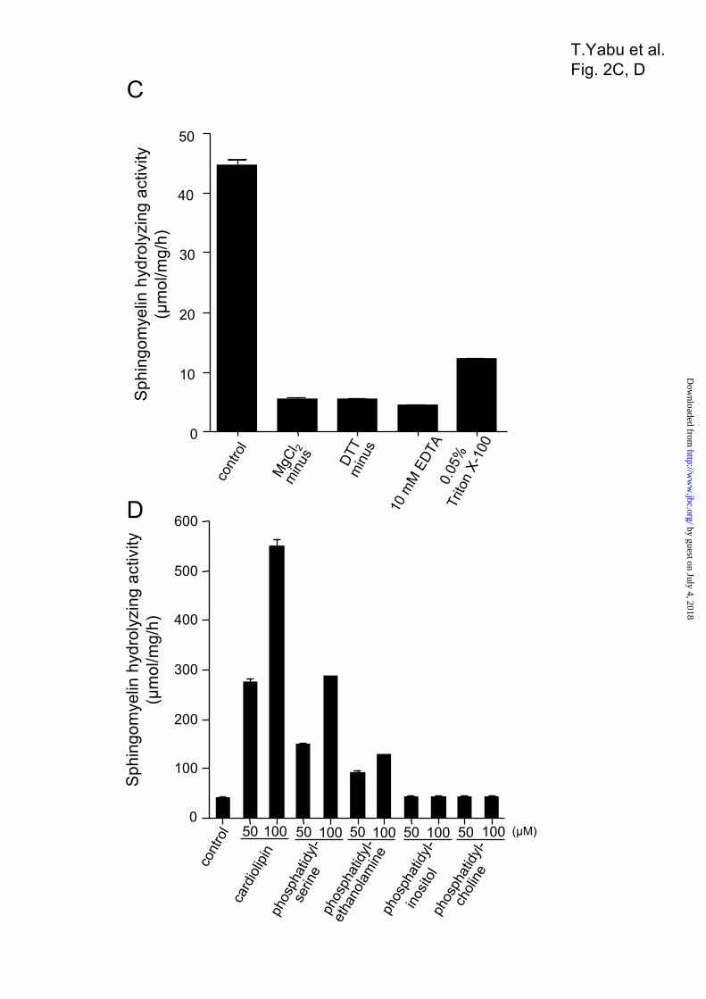
revealed a single band with an estimated molecular weight of 57.000 (Fig. 2A). The protein of excised gel sections was reduced and S-carboxymethylated, and then digested with Staphylococcus V8 protease. MALDI-TOF-MS analysis of the mixture of peptides detected an ion that corresponds to the amino acid sequence AVCISTTLE, (M + H) m/z 995.4 (observed), 995.14 (calculated), containing the S-carboxymethylated Cys residue. Thus, the Ala-36 residue was identified as the N-terminal amino acid of the mature enzyme (Fig. 1A). The protein showed high activity toward the substrate [14C]sphingomyelin, whereas it showed no activity against the phospholipid [14C]phosphatidylcholine and [3H]lyso-PAF (Table 2). The sphingomyelin hydrolyzing activity was optimal at pH 7.5, with half-maximal activity at pH 6.5 (Fig. 2B); the activity was absolutely dependent upon the presence of magnesium ions (Fig. 2C). Finally, several kinds of lipids derived from the mitochondrial and other membranes were tested for modulation of the activity of the purified SMase. The activity against sphingomyelin was induced 12-fold in the presence of 100 µM cardiolipin, 6-fold in the presence of 100 µM phosphatidylserine, and 3-fold in the presence of 100 µM phosphatidylethanolamine, whereas the activity was not influenced by phosphatidylcholine or phosphatidylinositol (Fig. 2D). Subcellular Localization of Novel SMase—The subcellular localization of the isolated SMase in ZE cells was examined by centrifugal fractionation. According to Western blotting, the SMase was mainly detected in the mitochondrial fraction (lane 2 in Fig. 3A) when using HSP60 as a mitochondrion marker, aldolase as a cytosol marker, and cadherin and neutral SMase 1 (13) as cell-membrane markers, whereas SMase was not detected in the cytosolic (lane 3 in Fig. 3A) or microsomal fractions (lane 4 in Fig. 3A). To confirm the subcellular localization of the SMase in the mitochondrion, the cells were fractionated using a sucrose-density gradient by ultracentrifuge (37). The separated proteins in each fraction in the sucrose-density gradient were applied to Western blotting, and the neutral SMase was detected with specific polyclonal antibody. When we used HSP60 and cytochrome c as mitochondrial markers, KDEL protein as an endoplasmic reticulum marker, 58K protein as a Golgi marker, cathepsin L as a lysosomal marker and catalase as a peroxisomal marker, the neutral SMase was mainly detected in the fraction No. 4–6 rich in the mitochondrion (Fig. 3B), but not in the lysosomal fractions (Fig. 3B, fraction
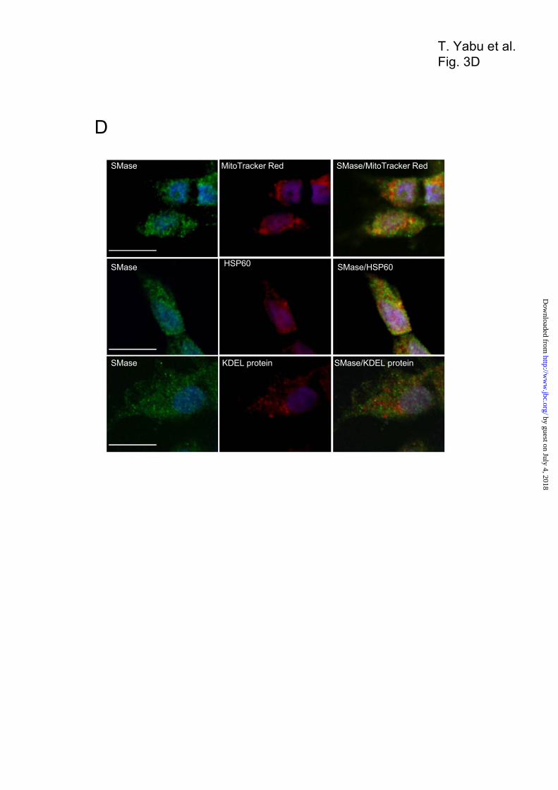
No. 1 and 2), endoplasmic reticulum fractions (Fig. 3B, fraction No. 1 and 2), Golgi fractions (Fig. 3B, fraction No. 1 and 2), and peroxisomal fractions (Fig. 3B, fraction No. 7 and 8). In addition, higher neutral SMase activity was also detected in the fraction No. 4–6 rich in the mitochondrial fractions containing cytochrome c oxidase activity, but not in the endoplasmic reticulum showing cytochrome c reductase activity and the lysosomal fractions showing acid phosphatase activity (Fig. 3C). The ZE cells were co-stained with the anti-SMase antibody together with the specific mitochondrial probe MitoTracker Red, anti-HSP60 antibody (mitochondrial marker), or anti-KDEL antibody (endoplasmic reticulum marker) by confocal microscopy. Immunocytochemical localization of the SMase with that of MitoTracker Red and that of HSP60 showed that the SMase exhibited major signals in the mitochondria (Fig. 3D). Consequently, staining with the antibody against endoplasmic reticulum maker, i. e., KDEL protein, the SMase was not localized in the endoplasmic reticulum (Fig. 3D). Therefore, the SMase was located in the mitochondria. To establish the topology of enzyme localization, we performed a protease protection assay. Mitochondrial fractions prepared from ZE cells were treated with proteinase K to digest peripheral proteins and then subjected to Western blotting with antibodies against mitochondrial SMase and cytochrome c, as an intermembrane space marker, or against HSP60, as a matrix marker (40). The expressed proteins, as well as the endogenous SMase, were recovered from mitochondria (Fig. 3E, lane 1), but not from the post-mitochondrial supernatant (data not shown). The endogenous SMase recovered from mitochondria was unaffected by the presence of proteinase K (Fig. 3E, lane 2). Upon disruption of the outer membrane, the endogenous SMase was completely degraded by proteinase K, whereas HSP60 protein remained undigested (Fig. 3E, lane 3). As a control, all proteins were digested by proteinase K when mitochondrial membranes were solubilized in Triton X-100 (Fig. 3E, lane 4). These results indicate that this SMase is bound to both the intermembrane space and/or the inner membrane in the mitochondrion. Identification of the Mitochondrial Localization Signal (MLS) with the First 35 Residues—Based on the structural features of zebrafish SMase, a putative MLS was identified within the first 35 residues of the N terminus, a putative hydrophobic transmembrane
by guest on July 4, 2018http://w
ww
.jbc.org/D
ownloaded from

9
domain was predicted within Ser-64 to Ala-86, and a putative catalytic center site was identified at His-529 (Fig. 1A). To confirm the presence of these features, we constructed various SMase-FLAG fusion proteins: a histidine to alanine substitution at the catalytic site (H529A mutant), a deletion mutant lacking the N-terminal MLS (MLS deletion mutant), a deletion mutant lacking the transmembrane domain (TM deletion mutant), and a deletion mutant lacking both the MLS and the transmembrane domain (MLS-TM deletion mutant, Fig. 4A). These constructs were transiently expressed in HEK293 cells, and the FLAG tag of the expressed proteins was detected via Western blotting (Fig. 4B). We examined the intracellular distribution of these mutants and the wild-type protein in HEK293 cells via subcellular fractionation. When expressed in HEK293 cells, the wild-type, H529A mutant, and TM mutant proteins localized correctly to the mitochondrial fraction (Fig. 4C and Table 3), whereas the MLS deletion and the MLS-TM deletion mutants were not detected in the mitochondrial fraction, but were instead found in the cytosolic fraction. To further clarify the protein localization, HEK293 cells transfected with the wild-type and the MLS deletion were stained with both anti-FLAG antibody and MitoTracker Red. The SMase with FLAG tag was co-localized with the mitochondrial probe, whereas the MLS deletion mutant was not co-localized with the mitochondrial probe (Fig. 4D). These findings indicate that first 35 residues of the N-terminal sequence represent an MLS essential for localization in mitochondria.These findings indicate that first 35 residues of the N-terminal sequence represent an MLS essential for localization in mitochondria. Ceramide and Sphingomyelin Levels and Neutral SMase Activity in Transient Transfectants—We used HEK293 cells transiently transfected with the wild-type or H529A mutant constructs to examine whether SMase is involved in ceramide generation. When the intercellular ceramide content was measured using a diacylglycerol kinase assay, the wild-type transfectants showed significantly higher ceramide levels than cells transfected with the H529A mutant or mock vector; in H529A mutants, peak ceramide levels (twofold) occurred 48 h after transfection (Fig. 5A). At 24, 48, and 72 h post-transfection, cells harboring wild-type SMase showed higher Mg2+-dependent neutral SMase activity than cells harboring the H529A mutant or mock vector (Fig. 5B). In wild-type transfectants, a
decrease in cellular sphingomyelin levels was accompanied by an increase in cellular ceramide levels at 48 h post-transfection (Fig. 5C). These changes in ceramide and sphingomyelin levels in association with neutral SMase activity in SMase-overexpressing cells indicate a significant role for this enzyme in ceramide metabolism. Ceramide and Sphingomyelin Levels and Neutral SMase Activity in Mitochondria—To examine SMase-mediated ceramide metabolism in mitochondria, we established three wild-type and three H529A mutant cell lines and confirmed that the transfected HEK293 cells overexpressed the transgene products (Fig. 6A). When Mg2+-dependent neutral SMase activity was measured in the mitochondrial fraction using C6-7-NBD-4-yl-sphingomyelin as a substrate, neutral SMase activity was 25 times higher in the wild-type transfectants than in the H529A mutants (Fig. 6, B and C). In addition, the mitochondrial ceramide content increased by 1.5−1.8 times in the wild-type transfectants compared to the H529A mutants (Fig. 6D). This increase in mitochondrial ceramide was accompanied by a decrease in mitochondrial sphingomyelin (Fig. 6E). These changes in mitochondrial ceramide and sphingomyelin levels in association with increased enzyme activity indicate a significant role for SMase in sphingomyelin metabolism. Effects of SMase Knockdown on ZE Cells—To confirm whether SMase regulates ceramide generation, a phosphorothioate oligonucleotide was used to repress mitochondrial SMase levels. Anti-mitochondrial SMase antibodies detected a 57-K protein band (Fig. 7A). At 48 h after antisense oligonucleotide treatment, both the protein band and the activity level of Mg2+-dependent neutral SMase in the mitochondrial fraction decreased in a dose-dependent manner (Fig. 7B). Treatment with 20 µM of the antisense oligonucleotide reduced the basal activity of Mg2+-dependent neutral SMase from 1.32 to 0.34 nmol/mg/h (Fig. 7B). The antisense oligonucleotide treatment repressed basal endogenous ceramide content in mitochondrial fractions (Fig. 7C), whereas it induced an increase in the sphingomyelin content (Fig. 7D). In contrast, treatment with control sense oligonucleotides had no effect. Thus, antisense oligonucleotide-induced enzyme deficiency affected ceramide metabolism.
by guest on July 4, 2018http://w
ww
.jbc.org/D
ownloaded from

10
DISCUSSION A novel mitochondrial SMase was cloned, and the active endogenous enzyme was purified from ZE cells. The purified enzyme showed specific activity against [14C]sphingomyelin in the presence of 10 mM Mg2+ at pH 7.5. Cytoimmunostaining revealed that the enzyme was localized to the mitochondrion, and subcellular fractionation indicated that the enzyme was distributed in the mitochondrial fraction. A protease protection assay revealed that the enzyme was distributed in the intermembrane space and/or the inner membrane of the mitochondrion. The overexpression of wild-type and mutant proteins showed that an MLS was required for translocation of the enzyme to the mitochondrion. Mutants lacking this MLS were instead directed to the cytoplasm. Cytostaining for zebrafish SMase in ZE cells indicated that the enzyme was co-localized with MitoTracker Red and HSP60 in the mitochondrion. The transient and stable overexpression of mitochondrial SMase cDNA in HEK293 cells resulted in enhanced neutral SMase activity and increased ceramide levels paralleled by reduced sphingomyelin levels. Antisense oligonucleotide-induced SMase deficiency confirmed that SMase was required for mitochondrial ceramide generation. Therefore, the identified zebrafish SMase is a novel mitochondrial SMase that regulates mitochondrial ceramide production. The activity and substrate specificity of this novel enzyme are similar to those of the known mammalian neutral SMase 2s (7,16). The purified zebrafish SMase showed specific hydrolyzing activity against sphingomyelin, producing ceramide, whereas it showed no activity against phosphatidylcholine. In addition, the enzyme regulated the metabolism of sphingomyelin and ceramide in cells. When SMase was overexpressed in cultured cells, we observed decreases in sphingomyelin and corresponding increases in ceramide levels in the mitochondrial fraction. Thus, the zebrafish enzyme is a typical Mg2+-dependent neutral SMase that accelerates the catabolism of sphingomyelin in the mitochondrion. The previously purified bovine brain neutral SMase, the partially purified rat brain neutral SMase, yeast Isc1p, and mouse neutral SMase 2 showed dependence upon anionic phospholipids such as phosphatidylserine for in vitro activity (14,49−51). Interestingly, site-directed mutagenesis in Isc1p indicated the importance of positively charged C-terminal amino acid residues in protein−anionic phospholipid interaction (52). The zebrafish SMase
described here was also activated by cardiolipin and phosphatidylserine, similar to mammalian SMase 2 (7). Cardiolipin and phosphatidylserine are highly enriched in the outer and inner membranes of the mitochondrion. Therefore, cardiolipin and phosphatidylserine may be a candidate for the activator of mitochondrial SMase. Molecular phylogenetic analysis based on amino acid sequences showed that the zebrafish mitochondrial SMase falls into a cluster consisting of human, mouse, and zebrafish neutral SMase 2s. Both zebrafish mitochondrial SMase and SMase 2 share common structural features, such as a magnesium-binding site, a substrate-binding site, and an active center His residue (Fig. 1B). We found that the mitochondrial SMase also possesses an MLS and a transmembrane domain as a stop-transfer sequence in the N-terminal region. This MLS was also conserved in other SMase 2 enzymes, suggesting that all known SMase 2s and zebrafish mitochondrial SMase may be mitochondrial enzymes. In eukaryotes, the MLS has been identified in the majority of pre-sequences cleaved by mitochondrial processing peptidases. This signal sequence is characterized by an overall positive charge, a predicted ability to form an amphiphilic a-helix, and the presence of an Arg residue at the –2 or –3 position from the cleavage site (53). Four cleavage site motifs have been identified (54): xRx↓x[S/x] (R-2 motif), xRx[Y/x]↓[S/A/x]x (R-3 motif), xRx↓[F/L/I] xx[S/T/G]xxxx (R-10 motif), and xx↓x[S/x] (R-none motif). The identified zebrafish mitochondrial SMase was consistent with the R-3 motif (i.e., DRLI↓AV). Hoffman et al. (7) firstly identified that mammalian neutral SMase 2 was localized in the Golgi in a number of cell lines by immunofluorescent staining. Tani and Hannun (15) reported that mouse neutral SMase 2 was palmitoylated at four Cys residues (Cys-53, Cys-54, Cys-395, and Cys-396) that were localized to the inner leaflet of the cell membrane. Zebrafish neutral SMase 2 also showed three conserved Cys residues (Cys-53, Cys-421, and Cys-422), but zebrafish mitochondrial SMase showed no palmitoylated Cys cluster. This work suggest that in contrast to zebrafish mitochondrial SMase, neutral SMase 2 might be localized to the cell membrane as a result of palmitoylation. Further studies of the intracellular localization of SMase 2 are required to examine whether SMase 2 and mitochondrial SMase are co-localized in the mitochondrion.
by guest on July 4, 2018http://w
ww
.jbc.org/D
ownloaded from

11
Based on an analysis of gene synteny between zebrafish and human genomes, the mitochondrial SMase gene is located on zebrafish chromosome 16. The ortholog of the mitochondrial SMase gene is found on chromosome 8 of the human genome and is expressed in human breast tumors (55). In contrast, the neutral SMase 2 gene is located on zebrafish chromosome 25 and was orthologous to the neutral SMase 2 gene located on human chromosome 16. Therefore, in terms of gene synteny, the isolated zebrafish mitochondrial SMase is distinct from other neutral SMase 2s. At least five SMases, i.e., acidic SMase, neutral SMase 1, SMase 2, SMase 3, and mitochondrial SMase, have been characterized in mammalian and other animal cells, and all of these enzymes were expressed in ZE cells (unpublished). Thus, ZE cells represent an important model for characterizing the precise roles of mitochondrial and other types of SMases, as well as the mechanism of ceramide
signaling. Our results indicate that mitochondrial SMase has a specific function in mitochondrial ceramide generation. Previous studies have demonstrated that ceramidase (34), ceramide synthase (33), dihydroceramide synthase (56), glycosyltransferase (57), yeast inositol sphingolipid phospholipase C (58), and sphingosine kinase (59) are localized to the mitochondrion. Therefore, a set of enzymes, including mitochondrial SMase and other ceramide-related enzymes, may regulate sphingolipid metabolism in the mitochondrion and regulate cell growth and apoptosis in response to mitochondrial functions.
by guest on July 4, 2018http://w
ww
.jbc.org/D
ownloaded from

12
REFERENCES
1. Hannun, Y. A., and Luberto, C. (2000) Trends Cell Biol. 10(2), 73−80
2. Levade, T., Malagarie-Cazenave, S., Gouaze, V., Segui, B., Tardy, C., Betito, S., Andrieu-Abadie, N.,
and Cuvillier, O. (2002) Neurochem. Res. 27(7−8), 601−607
3. Marchesini, N., and Hannun, Y. A. (2004) Biochem. Cell Biol. 82(1), 27−44
4. Obeid, L. M., Linardic, C. M., Karolak, L. A., and Hannun, Y. A. (1993) Science 259(5102),
1769−1771
5. Okazaki, T., Bell, R. M., and Hannun, Y. A. (1989) J. Biol. Chem. 264(32), 19076−19080
6. Clarke, C. J., Snook, C. F., Tani, M., Matmati, N., Marchesini, N., and Hannun, Y. A. (2006)
Biochemistry 45(38), 11247−11256
7. Hofmann, K., Tomiuk, S., Wolff, G., and Stoffel, W. (2000) Proc. Natl. Acad. Sci. USA 97(11),
5895−5900
8. Krut, O., Wiegmann, K., Kashkar, H., Yazdanpanah, B., and Kronke, M. (2006) J. Biol. Chem.
281(19), 13784−13793
9. Tomiuk, S., Hofmann, K., Nix, M., Zumbansen, M., and Stoffel, W. (1998) Proc. Natl. Acad. Sci.
USA 95(7), 3638−3643
10. Sawai, H., Domae, N., Nagan, N., and Hannun, Y. A. (1999) J. Biol. Chem. 274(53), 38131−38139
11. Zumbansen, M., and Stoffel, W. (2002) Mol. Cell. Biol. 22(11), 3633−3638
12. Yabu, T., Tomimoto, H., Taguchi, Y., Yamaoka, S., Igarashi, Y., and Okazaki, T. (2005) Blood
106(1), 125−134
13. Yabu, T., Imamura, S., Yamashita, M., and Okazaki, T. (2008) J. Biol. Chem. 283 (44), 29971-29982
14. Marchesini, N., Luberto, C., and Hannun, Y. A. (2003) J. Biol. Chem. 278(16), 13775−13783
15. Tani, M., and Hannun, Y. A. (2007) J. Biol. Chem. 282(13), 10047−10056
16. Marchesini, N., Osta, W., Bielawski, J., Luberto, C., Obeid, L. M., and Hannun, Y. A. (2004) J. Biol.
Chem. 279(24), 25101−25111
17. Hayashi, Y., Kiyono, T., Fujita, M., and Ishibashi, M. (1997) J. Biol. Chem. 272(29), 18082−18086
18. Clarke, C. J., Truong, T. G., and Hannun, Y. A. (2007) J. Biol. Chem. 282(2), 1384−1396
by guest on July 4, 2018http://w
ww
.jbc.org/D
ownloaded from

13
19. Karakashian, A. A., Giltiay, N. V., Smith, G. M., and Nikolova-Karakashian, M. N. (2004) FASEB J.
18(9), 968−970
20. Levy, M., Castillo, S. S., and Goldkorn, T. (2006) Biochem. Biophys. Res. Commun. 344(3), 900−905
21. Jana, A., and Pahan, K. (2004) J. Biol. Chem. 279(49), 51451−51459
22. Stoffel, W., Jenke, B., Block, B., Zumbansen, M., and Koebke, J. (2005) Proc. Natl. Acad. Sci. USA
102(12), 4554−4559
23. Aubin, I., Adams, C. P., Opsahl, S., Septier, D., Bishop, C. E., Auge, N., Salvayre, R.,
Negre-Salvayre, A., Goldberg, M., Guenet, J. L., and Poirier, C. (2005) Nat. Genet. 37(8), 803−805
24. Tepper, A. D., de Vries, E., van Blitterswijk, W. J., and Borst, J. (1999) J. Clin. Invest. 103(7),
971−978
25. Ardail, D., Popa, I., Alcantara, K., Pons, A., Zanetta, J. P., Louisot, P., Thomas, L., and Portoukalian,
J. (2001) FEBS Lett. 488(3), 160−164
26. Colbeau, A., Nachbaur, J., and Vignais, P. M. (1971) Biochim. Biophys. Acta 249(2), 462−492
27. Gudz, T. I., Tserng, K. Y., and Hoppel, C. L. (1997) J. Biol. Chem. 272(39), 24154−24158
28. Birbes, H., El Bawab, S., Hannun, Y. A., and Obeid, L. M. (2001) FASEB J. 15(14), 2669−2679
29. Ruvolo, P. P., Deng, X., Ito, T., Carr, B. K., and May, W. S. (1999) J. Biol. Chem. 274(29),
20296−20300
30. Birbes, H., Luberto, C., Hsu, Y. T., El Bawab, S., Hannun, Y. A., and Obeid, L. M. (2005) Biochem.
J. 386(Pt 3), 445−451
31. Siskind, L. J., Kolesnick, R. N., and Colombini, M. (2006) Mitochondrion 6(3), 118−125
32. Siskind, L. J., Kolesnick, R. N., and Colombini, M. (2002) J. Biol. Chem. 277(30), 26796−26803
33. Shimeno, H., Soeda, S., Sakamoto, M., Kouchi, T., Kowakame, T., and Kihara, T. (1998) Lipids
33(6), 601−605
34. El Bawab, S., Roddy, P., Qian, T., Bielawska, A., Lemasters, J. J., and Hannun, Y. A. (2000) J. Biol.
Chem. 275(28), 21508−21513
35. Thongboonkerd, V., Luengpailin, J., Cao, J., Pierce, W. M., Cai, J., Klein, J. B., and Doyle, R. J.
(2002) J. Biol. Chem. 277(19), 16599−16605
by guest on July 4, 2018http://w
ww
.jbc.org/D
ownloaded from
14
36. Okazaki, T., Bielawska, A., Domae, N., Bell, R. M., and Hannun, Y. A. (1994) J. Biol. Chem. 269(6),
4070−4077
37. Kalra, J., and Brosnan, J. T. (1974) J. Biol. Chem. 249(10), 3255−3260
38. Shiao, Young-Ji., Lupo, G., and Vance. (1995) J. Biol. Chem. 270(19), 11190−1119
39. Sugihara, Y., Kawabe, H., Tanaka, H., Fujimoto, S., and Ohara A. (1981) J. Biol. Chem. 256(20),
10664−10670
40. Otera, H., Ohsakaya, S., Nagaura, Z., Ishihara, N., and Mihara, K. (2005) EMBO J. 24(7), 1375−1386
41. Yabu, T., Kishi, S., Okazaki, T., and Yamashita, M. (2001) Biochem. J. 360(Pt 1), 39−47
42. Yamashita, M., and Konagaya, S. (1991) J. Agric. Food Chem. 39, 1402-1405
43. Bligh, E. G., and Dyer, W. J. (1959) Can. J. Biochem. Physiol. 37(8), 911−917
44. Sawai, H., Okazaki, T., Yamamoto, H., Okano, H., Takeda, Y., Tashima, M., Sawada, H., Okuma,
M., Ishikura, H., Umehara, H., and Okazaki, T. (1995) J. Biol. Chem. 270(45), 27326−27331
45. Matsuo, Y., Yamada, A., Tsukamoto, K., Tamura, H., Ikezawa, H., Nakamura, H., and Nishikawa, K.
(1996) Protein Sci. 5(12), 2459−2467
46. Fujii, S., Ogata, K., Inoue, B., Inoue, S., Murakami, M., Iwama, S., Katsumura, S., Tomita, M.,
Tamura, H., Tsukamoto, K., Ikezawa, H., and Ikeda, K. (1999) J. Biochem. 126(1), 90−97
47. Fujii, S., Inoue, B., Yamamoto, H., Ogata, K., Shinki, T., Inoue, S., Tomita, M., Tamura, H.,
Tsukamoto, K., Ikezawa, H., and Ikeda, K. (1998) J. Biochem. 124(6), 1178−1187
48. Letunic, I., Copley, R. R., Pils, B., Pinkert, S., Schultz, J., and Bork, P. (2006) Nucleic Acids Res.
34(Database issue), D257−260
49. Bernardo, K., Krut, O., Wiegmann, K., Kreder, D., Micheli, M., Schafer, R., Sickman, A., Schmidt,
W. E., Schroder, J. M., Meyer, H. E., Sandhoff, K., and Kronke, M. (2000) J. Biol. Chem. 275(11),
7641−7647
50. Liu, B., Hassler, D. F., Smith, G. K., Weaver, K., and Hannun, Y. A. (1998) J. Biol. Chem. 273(51),
34472−34479
51. Sawai, H., Okamoto, Y., Luberto, C., Mao, C., Bielawska, A., Domae, N., and Hannun, Y. A. (2000)
J. Biol. Chem. 275(50), 39793−39798
by guest on July 4, 2018http://w
ww
.jbc.org/D
ownloaded from
15
52. Okamoto, Y., Vaena de Avalos, S., and Hannun, Y. A. (2003) Biochemistry 42(25), 7855−7862
53. Gakh, O., Cavadini, P., and Isaya, G. (2002) Biochim. Biophys. Acta 1592(1), 63−77
54. Gavel, Y., and von Heijne, G. (1990) Protein Eng. 4(1), 33−37
55. Dias Neto, E., Correa, R. G., Verjovski-Almeida, S., Briones, M. R., Nagai, M. A., da Silva, W., Jr.,
Zago, M. A., Bordin, S., Costa, F. F., Goldman, G. H., Carvalho, A. F., Matsukuma, A., Baia, G. S.,
Simpson, D. H., Brunstein, A., de Oliveira, P. S., Bucher, P., Jongeneel, C. V., O'’Hare, M. J., Soares,
F., Brentani, R. R., Reis, L. F., de Souza, S. J., and Simpson, A. J. (2000) Proc. Natl. Acad. Sci. USA
97(7), 3491−3496
56. Bionda, C., Portoukalian, J., Schmitt, D., Rodriguez-Lafrasse, C., and Ardail, D. (2004) Biochem. J.
382(Pt 2), 527−533
57.Ardail, D., Popa, I., Bodennec, J., Louisot, P., Schmitt, D., and Portoukalian, J. (2003) Biochem. J.
371(Pt 3), 1013−1019
58. Kitagaki, H., Cowart, L. A., Matmati, N., Vaena de Avalos, S., Novgorodov, S. A., Zeidan, Y. H.,
Bielawski, J., Obeid, L. M., and Hannun, Y. A. (2007) Biochim. Biophys. Acta 1768(11), 2849−2861
59. Liu, H., Toman, R. E., Goparaju, S. K., Maceyka, M., Nava, V. E., Sankala, H., Payne, S. G., Bektas,
M., Ishii, I., Chun, J., Milstien, S., and Spiegel, S. (2003) J. Biol. Chem. 278(41), 40330−40336
by guest on July 4, 2018http://w
ww
.jbc.org/D
ownloaded from

16
FOOTNOTES
This work was supported in part by grants from the Japan Science and Technology Corporation, the
Japan Society for Promotion of Science, and the Ministry of Agriculture, Forestry, and Fisheries of Japan.
Abbreviations used: [14C]phosphatidylcholine, L-3-phosphatidyl[N-methyl-14C]choline,1,2-dipalmitoyl;
[14C]sphingomyelin, [N-methyl-14C]sphingomyelin; C6-NBD-ceramide, C6-7-nitro-2-1,3-
benzoxadiazol-4-yl-ceramide; C6-NBD-sphingomyelin,
6-7-nitro-2-1,3-benzoxadiazol-4-yl-sphingomyelin; DTT, dithiothreitol; [3H]lyso-PAF,
[1-O-octadecyl-3H]lyso-platelet activating factor; FCS, fetal calf serum; HEK293 cells, human embryonic
kidney cells; IPTG, isopropyl-1-thio-β-D-galactopyranoside; LB, Luria-Bertani broth; MLS, mitochondrial
localization signal; PMSF, phenylmethylsulfonyl fluoride; PVDF, polyvinylidene fluoride; SMase,
sphingomyelinase; TM, transmembrane domain; ZE cells, zebrafish embryonic cells
by guest on July 4, 2018http://w
ww
.jbc.org/D
ownloaded from

17
FIGURE LEGENDS
Figure 1. Amino acid sequence of zebrafish mitochondrial SMase and neutral SMase 2, and the
phylogenetic relationship among vertebrate neutral SMases. (A) Deduced amino acid sequence of
zebrafish mitochondrial SMase. Amino acid positions are shown on the right. The putative mitochondrial
localization signal (MLS) identified by the SMART program is underlined. The N-terminal amino acid
sequence from the purified mature enzyme, as determined by MALDI-TOF-MS, is boxed. The putative
transmembrane domain identified by the SMART program is also double underlined. The putative
Mg2+-complexing glutamine residue (∆), the asparagine residue involved in substrate binding (#), and the
catalytic base histidine residue (*) are shown. (B) Amino acid sequence alignment for zebrafish
mitochondrial SMase, zebrafish neutral SMase 2, human neutral SMase 2, and mouse neutral SMase 2.
Proteins with significant amino acid sequence homology were identified using a FASTA search of the
GenBank database. The sequences of neutral SMases were aligned using the deduced amino acid
sequences of homologous proteins from zebrafish mitochondrial SMase, zebrafish neutral SMase 2, human
neutral SMase 2, and mouse neural SMase 2. The putative MLS is boxed (I). The putative transmembrane
domain is also boxed (II). (C) A phylogenetic tree based on the amino acid sequences of the vertebrate
neutral SMases. The phylogenetic analysis was performed using the neighbor-joining method in Clustal X.
Numbers on the internal branches denote the bootstrap percentages of 1000 replicates. The scale indicates
the evolutionary distance of one amino acid substitution per site. The amino acid sequences used in the
analysis were obtained from the National Center for Biotechnology Information (NCBI) protein database
with the following accession numbers: Bacillus cereus SMase (X12854), human neutral SMase 1 and 2
(NM_009213 and AJ250460, respectively), mouse neutral SMase 1 and 2 (NM_009213 and AJ250461,
respectively), zebrafish neutral SMase 1 and 2 (AB196165 and AB361067, respectively), and zebrafish
mitochondrial SMase (AB361066).
Figure 2. Purification and characterization of the mitochondrial SMase from a zebrafish embryonic
cell line. (A) Polyacrylamide gel electrophoresis (PAGE) of purified enzyme. The arrowhead indicates the
by guest on July 4, 2018http://w
ww
.jbc.org/D
ownloaded from

18
molecular weight of a 57-K protein. SDS-PAGE (10% gel) was performed after reduction of the sample.
The gel was stained with Coomassie Brilliant Blue R-250. (B) The pH dependence of neutral SMase
activity. The sphingomyelin hydrolyzing activity of the purified enzyme was measured at 37ºC for 30 min
at various pHs, with an estimated optimum pH of 7.5. The pH was adjusted by the addition of the following
buffers at a final concentration of 100 mM: acetate (pH 4 and 5), PIPES (pH 6, 6.5, and 7), and Tris (pH 7.5,
8, 8.5, and 9). (C) The effect of Mg2+ ions on neutral sphingomyelin hydrolyzing activity. The basal assay
mixture contained 100 mM Tris-HCl (pH 7.5), 5 mM MgCl2, 0.1% Triton X-100, and 5 mM dithiothreitol
(DTT). Effects of Mg2+ ion on the activity were measured in the presence of 10 mM EDTA. (D) The effect
of lipids on mitochondrial SMase activity. The sphingomyelin hydrolyzing activity of the purified enzyme
was determined under standard conditions in the presence of [14C]sphingomyelin under standard conditions
and the indicated phospholipids (50 or 100 µM). The basal enzyme activity (control) was 45 ± 3
µmol/mg/h.
Figure 3. Subcellular localization and distribution of SMase. Whole lysate of ZE cells (lane 1) was
fractionated into the mitochondrial fraction (lane 2), cytosolic fraction (lane 3), and microsomal fraction
(lane 4) via ultracentrifugation. These fractions were analyzed by Western blotting using antibodies against
zebrafish mitochondrial SMase, HSP60 (a mitochondrial marker), aldolase (a cytosolic marker), and neutral
SMase 1 and cadherin (cell-membrane markers). The mitochondrial fraction was separated into 8 fractions
using the sucrose-gradient method via ultracentrifugation. The separated proteins in each fraction were
subjected to Western blotting using antibodies against zebrafish mitochondrial SMase, HSP60 and
cytochrome c (a mitochondrial marker), KDEL protein (an endoplasmic reticulum marker), 58K protein (a
Golgi marker), cathepsin L (a lysosomal marker), and catalase (peroxisomal marker), and neutral SMase
activities in each fraction were determined. The lysosomal marker, endoplasmic reticulum marker, and
Golgi marker, mitochondrial marker or peroxisomal marker were detected in the fraction No. 1 and 2,
fraction No. 4–6, and fraction No. 7 and 8, respectively. (C) Specific activities of marker enzymes in
subcellular fractionation of ZE cells. The C6-NBD-sphingomyelin hydrolyzing activities of the SMase,
mitochondrial cytochrome c oxidase, endoplasmic reticulum cytochorome c reductase, and lysosomal acid
by guest on July 4, 2018http://w
ww
.jbc.org/D
ownloaded from

19
phosphatase were measured in each fraction by subcellular fractionation. Values and bars indicate the mean
± standard deviation of three independent experiments. (D) ZE cells were fixed and permeabilized with
0.1% Triton-X100. The cells were incubated with 100 nM MitoTracker Red, and then fixed with 4%
paraformaldehyde in PBS. The cells were co-stained with anti-zebrafish SMase antibody and an antibody
against either HSP60 protein (a mitochondria marker) or KDEL protein (an ER marker) and stained with
fluorescent secondary antibodies. Signals for SMase (green color image) and signals for subcellular
markers such as mitochondria and ER (red color image) were observed. The overlay images indicate the
SMase and the subcellular marker were co-localized either in the sample place or adjacent to one another as
described in the Experimental Procedures. Scale bar = 10 µm. (E) The distribution of SMase in the
mitochondrion. Mitochondrial fractions were obtained from a zebrafish embryonic cell line and incubated
at 0ºC for 30 min in the absence (lane 1) or presence (lanes 2–4) of proteinase K, and under swelling
condition (20 mM HEPES-NaOH [pH 7.4], 10 mM sodium orthovanadate, 20 mM NaF, 250 mM sucrose,
2 mM CaCl2) (lanes 3 and 4) or in the presence (lane 4) of Triton X-100. The samples were subjected to
Western blotting with anti-mitochondrial SMase, anti-cytochrome c, and anti-HSP60 antibodies.
Figure 4. Mitochondrial localization of SMase variants. (A) Schematic representation of SMase
constructs. (B) HEK293 cells were transiently transfected with their constructs alone. Lane 1, control; lane
2, mock; lane 3, wild-type; lane 4, H529A mutant; lane 5, MLS deletion mutant; lane 6, TM deletion
mutant; lane 7, MLS-TM deletion mutant. At 48 h after transfection, the expressed proteins were detected
using anti-FLAG antibody or anti-actin via Western blotting as described in the Experimental Procedures.
(C) HEK293 cells expressing the indicated constructs were fractionated into mitochondrial (lane 1−7) and
cytosolic (lane 8−14) fractions, and each fraction was analyzed by Western blotting using antibodies against
FLAG, SMase, cytochrome c, and aldolase. Lanes 1 and 8, control; lanes 2 and 9, mock; lanes 3 and 10,
wild-type; lanes 4 and 11, H529A mutant; lanes 5 and 2, MLS deletion mutant; lanes 6 and 13, TM deletion
mutant; lanes 7 and 14, MLS-TM deletion mutant. (D) Mitochondrial localization of the wild-type construct.
The HEK293 cells transfected with wild-type of SMase and MLS deletion mutant were incubated with 25
nM MitoTracker Red and fixed with 4% paraformaldehyde in PBS. The cells were co-stained with
by guest on July 4, 2018http://w
ww
.jbc.org/D
ownloaded from
20
anti-FLAG antibody. Signals for zebrafish SMase with FLAG-tag (green color image) and signals for
mitochondrial marker (red color image) and the overlay images (merge) were observed as described in the
Experimental Procedures.
Figure 5. Ceramide and sphingomyelin levels in association with neutral SMase activity in SMase
transfectants. Cellular lipids were extracted at the indicated times after transfection. The levels of ceramide
(A) and sphingomyelin (C) were quantified using the diacylglycerol kinase assay and phosphate
measurement after TLC separation, respectively. The cells were lysed, and C6-NBD-sphingomyelin
hydrolyzing activity (B) was determined as described in the Experimental Procedures. Values and bars
indicate the mean ± standard deviation of three independent experiments.
Figure 6. Increase in neutral SMase activity with increased ceramide and decreased sphingomyelin
levels in the mitochondrial fraction of SMase transfectants. HEK293 cells were stably transfected with
the wild-type or H529A mutant construct. Three wild-type and three H529A mutant lines were established
as described in the Experimental Procedures. The expressed proteins in each group of three lines (A) were
detected via Western blotting using anti-FLAG antibody as described in the Experimental Procedures. (B)
Neutral SMase activity against C6-NBD-sphingomyelin in the mitochondrial fraction was demonstrated via
TLC separation and visualized by UV irradiation at 254 nm. (C) Increase in neutral SMase activity in
wild-type cell lines. The cells were lysed, and C6-NBD-sphingomyelin hydrolyzing activity was
determined as described in the Experimental Procedures. (D) Increasing ceramide content in the
mitochondrial fraction isolated from three wild-type lines. (E) Decreasing sphingomyelin content in the
mitochondrial fraction isolated from three wild-type lines. Cellular lipids in the mitochondrial fraction were
extracted, and the levels of ceramide and sphingomyelin were quantified using the diacylglycerol kinase
assay and phosphate measurement, respectively. Values and bars indicate the mean ± standard deviation of
three independent experiments. Different letters denote a statistical difference between wild-type and
H529A mutant cells (P < 0.01).
by guest on July 4, 2018http://w
ww
.jbc.org/D
ownloaded from

21
Figure 7. Antisense oligonucleotides against mitochondrial SMase protein inhibit ceramide
generation. Zebrafish embryonic (ZE) cells were pre-treated with 0−20 µM antisense or 20 µM sense
oligonucleotides against mitochondrial SMase for 48 h. (A) The expressed proteins in
oligonucleotide-treated cells were detected with anti-mitochondrial SMase or anti-actin antibodies by
Western blotting, as described in the Experimental Procedures. (B) C6-NBD-sphingomyelin hydrolyzing
activity, (C) ceramide content, and (D) sphingomyelin content in the mitochondrial fractions. Values and
bars indicate the mean ± standard deviation of three independent experiments. *P < 0.01 versus sense
oligonucleotide-treated cells.
by guest on July 4, 2018http://w
ww
.jbc.org/D
ownloaded from

22
Table 1 Zebrafish genes neighboring the mitochondrial SMase genes and the neutral SMase 2 genes. Zebrafish genes in the chromosome containing the mitochondrial Human homolog SMase gene and the neutral SMase 2 gene (localization) (chromosomal localization) Chr.16 Myomesin family, member 3 MYOM3, NM_152372 (61.68 Mb) (Chr. 1, 24.25 Mb)
LIM homeobox 3 LHX3, NM_178138 (62.69 Mb) (Chr. 3, 138.22 Mb)
SAC1 suppressor of actin mutations 1-like SACM1L, NM_014016 (62.73 Mb) (Chr. 3, 45.7 Mb)
Mitochondrial sphingomyelinase ENSG00000204791 (62.38 Mb) (Chr. 8, 145.177 Mb)
N-methyl D-aspartate-associated protein 1 N-methyl D-aspartate-associated protein 1 GRINA, NM_001009184 (62.85 Mb) (62.85 Mb) (Chr. 8, 145.136 Mb)
Phosphatidylserine synthase 1 PTDSS1, NM_014754 (63.07 Mb) (Chr. 8, 97.343 Mb) Chr.25 1-O-acylceramide synthase precursor LYPLA3, NM_012320 (16.85 Mb) (Chr. 16, 66.83 Mb)
DNA-directed RNA polymerase II 33-kDa polypeptide POLR2C, NM_032940 (18.56 Mb) (Chr. 16, 56.05 Mb)
Ubiquinone biosynthesis protein COQ9, mitochondrial precursor COQ9, NM_020312 (18.57 Mb) (Chr. 16, 56.03 Mb)
Cytokine induced apoptosis inhibitor 1 CIAPIN1, NM_020313 (18.59 Mb) (Chr. 16, 56.02 Mb)
Protein arginine N-methyltransferase 7 PRMT7, NM_019023 (18.60 Mb) (Chr. 16, 66.90 Mb)
Neutral sphingomyelinase 2 SMPD3, NM_018667 (19.61 Mb) (Chr. 16, 66.94 Mb)
Olfactomedin 4 OLFM4, NM_006418 (19.73 Mb) (Chr. 13, 52.5 Mb)
NDRG family member 4 NDRG4, NM_022910 (19.99 Mb) (Chr. 16, 57.05 Mb)
Mps one binder kinase activator-like 2 MOB2, VSP_012298 (20.04 Mb) (Chr. 11, 1.44 Mb)
Potassium inwardly rectifying channel, subfamily J, member 11 KCNJ11, NM_000525 (20.07 Mb) (Chr. 11, 17.36 Mb)
ATP-binding cassette, subfamily C (CFTR/MRP), member 8 ABCC8, NM_000352 (20.09 Mb) (Chr. 11, 17.37 Mb)
Usher syndrome 1C USH1C, NM_005709 (20.23 Mb) (Chr. 11, 17.47 Mb)
by guest on July 4, 2018http://w
ww
.jbc.org/D
ownloaded from

23
Table 2
Substrate specificity of the cloned purified enzyme.
Substrate Activitya (µmol/mg/h)
[14C]Sphingomyelin 45 ± 2.76b
[14C]Phosphatidylcholine 0.003 ± 0.0003b
[3H]Lyso-PAF 0.024 ± 0.0012c
aMean ± standard deviation (n = 3). bActivity was determined in the presence of 0.1% Triton X-100. cActivity was determined in the absence of Triton X-100.
by guest on July 4, 2018http://w
ww
.jbc.org/D
ownloaded from

24
Table 3
Subcellular distribution of various SMase constructs in HEK293-transfected cells. Construct Distribution Wild type Mitochondrial fraction H529A mutant Mitochondrial fraction MLS deletion mutant Cytosolic fraction TM deletion mutant Mitochondrial fraction MLS-TM deletion mutant Cytosolic fraction Various SMase constructs (Fig. 4) were transiently expressed in HEK293 cells.
by guest on July 4, 2018http://w
ww
.jbc.org/D
ownloaded from

MSLRESPFPN GFLEGLHAVG WGLIFPCFWF LDRLIAVCIS TTLERMWRLE 50
QECYLHPLKV VFGSILFFIL FVISTPFALL GFILWAPLQA IRRPFSYHKQ 100
EQSTPMENRN ARWEEMGKIS FGFLTANVCL LPDGIARFNN LGHTQKRALI 150
IGKSIVQGVT RPRIRIFVDS PSSCGTVTPS SSLIPQPNAS SYGSVDASGE 200
LPDAIEVNEI TPKPNCNQNS NHQKHPPRSL RTLLRDADIP MEVSALFPPS 250
VDIVCLQEVF DKRAARKLTQ ALGPLYGHVL YDVGVYACHP AGSCSSFKFF 300
NSGLFLASRF PIMEAEYRCF PNGRGEDALA AKGLLTVKVD IGLQKGEKRM 350
VGFINCTHLH APEGDGAIRF EQLNMLSKWT SEFQTLNRRD DELPLFDVLC 400
GDFNFDNCSP DDRLEQSHSV FDEYTDPCRA AAGKEKPWVI GTLLEQPTLY 450
DENMRTPDNL QRTLESEDLR KDFISPPVAL DGVPLVYPEA DQPWIGRRID 500
YLLYREKSGH RTEVEELTYV TQLAGLTDHI PVGFRLSVSL DSEQN 545
T. Yabu et al. Fig. 1A
A
∆
#
*
by guest on July 4, 2018http://w
ww
.jbc.org/D
ownloaded from

T. Yabu et al.Fig. 1B, C
B
100
0.05
100
100100
Zebrafish neutral SMase 1
Human neutral SMase 1
Mouse neutral SMase 1
Zebrafish neutral SMase 2
Human neutral SMase 2
Mouse neutral SMase 2
Mitochondrial SMase
100
Bacillus cereus SMase
C
1111
71717171
139139134134
200205204230
230275264262
242345317315
281411387385
351479457452
414549522520
484619592590
545684655655
Mitochondrial SMaseZebrafish nSMase 2Human nSMase 2Mouse nSMase 2
Mitochondrial SMaseZebrafish nSMase 2Human nSMase 2Mouse nSMase 2
Mitochondrial SMaseZebrafish nSMase 2Human nSMase 2Mouse nSMase 2
Mitochondrial SMaseZebrafish nSMase 2Human nSMase 2Mouse nSMase 2
Mitochondrial SMaseZebrafish nSMase 2Human nSMase 2Mouse nSMase 2
Mitochondrial SMaseZebrafish nSMase 2Human nSMase 2Mouse nSMase 2
Mitochondrial SMaseZebrafish nSMase 2Human nSMase 2Mouse nSMase 2
Mitochondrial SMaseZebrafish nSMase 2Human nSMase 2Mouse nSMase 2
Mitochondrial SMaseZebrafish nSMase 2Human nSMase 2Mouse nSMase 2
Mitochondrial SMaseZebrafish nSMase 2Human nSMase 2Mouse nSMase 2
#
∆
by guest on July 4, 2018http://w
ww
.jbc.org/D
ownloaded from

T. Yabu et al.Fig. 2A, B
97.4 K-
69 K-
55 K-
39.5 K-
29 K-
A
B
4 5 6 7 8 90
10
20
30
40
50
pH
Sph
ingo
mye
lin h
ydro
lyzi
ng a
ctiv
ity(µ
mol
/mg/
h)
by guest on July 4, 2018http://w
ww
.jbc.org/D
ownloaded from
50 100
phos
phat
idyl-
serin
eph
osph
atidy
l-
etha
nolam
ine
(µM)
D
T.Yabu et al.Fig. 2C, D
cont
rol
MgC
l 2m
inus DT
Tm
inus
0.05
%
Trito
n X-
100
10 m
M E
DTA
C
0
10
20
30
40
50
cont
rol
0
100
200
300
400
500
600
50 100 50 100 50 100 50 100
phos
phat
idyl-
inosit
ol ph
osph
atidy
l-ch
oline
card
iolip
in
Sph
ingo
mye
lin h
ydro
lyzi
ng a
ctiv
ity(µ
mol
/mg/
h)
Sph
ingo
mye
lin h
ydro
lyzi
ng a
ctiv
ity(µ
mol
/mg/
h)
by guest on July 4, 2018http://w
ww
.jbc.org/D
ownloaded from

T. Yabu et al.Fig. 3A
204 K-131.2 K-
89.1 K-
41.7 K-
31.4 K-
17 K-
SMase
SMase 1
HSP60
aldolase
cadherin
1 2 3 4
A
by guest on July 4, 2018http://w
ww
.jbc.org/D
ownloaded from

T. Yabu et al.Fig. 3A and B
131.2 K-
89.1 K-
41.7 K-
31.4 K-
58K Golgi protein
204 K-
17 K-
SMase
cathepsin L
HSP60
catalase1 2 3 4 5 6 7 8
B
KDEL (GRP94)
KDEL (GRP78)
cytochorome c
by guest on July 4, 2018http://w
ww
.jbc.org/D
ownloaded from

C T. Yabu et al.Fig. 3C
C6-
NB
D-s
phin
gom
yelin
hydr
olyz
ing
activ
ity
Cyt
ochr
ome
cox
idas
e ac
tivity
Cyt
ochr
ome
cre
duct
ase
activ
ityA
cid
phos
phat
asea
ctiv
ity
1 2 3 4 5 6 7 80
1
2
3
4
5
Fraction No.
1 2 3 4 5 6 7 8012
34567
Fraction No.
1 2 3 4 5 6 7 80.00
0.05
0.10
0.15
Fraction No.
1 2 3 4 5 6 7 80.0
0.2
0.4
0.6
0.8
1.0
Fraction No.
by guest on July 4, 2018http://w
ww
.jbc.org/D
ownloaded from
T. Yabu et al.Fig. 3D
D
HSP60
KDEL protein
MitoTracker RedSMase
SMase
SMase
SMase/HSP60
SMase/KDEL protein
SMase/MitoTracker Red
by guest on July 4, 2018http://w
ww
.jbc.org/D
ownloaded from

T.Yabu et al.Fig. 3E
E
SMase
cytochrome c
HSP60
Triton X-100
swelling
proteinase K
- - - +
+ + ++ +- -
-
1 2 3 4
by guest on July 4, 2018http://w
ww
.jbc.org/D
ownloaded from

SMase
actin
H529
A529
wild-type
H529A
MLS deletion
TM deletion
MLS-TM deletion
mitochondrial localization signal:transmembrane domain:
FLAG-tag:
T. Yabu et al.Fig. 4A-C
A
B
1 2 3 4 5 6 7
1 35 64 86
SMase
cytochorome c
aldolase
C
mitochondrial fraction cytosolic fraction
8 9 10 11 12 13 141 2 3 4 5 6 7
by guest on July 4, 2018http://w
ww
.jbc.org/D
ownloaded from

T. Yabu et al.Fig. 4D
D
Wild-type SMase MLS deletionA
nti-F
LAG
Mito
track
er R
edM
erge
by guest on July 4, 2018http://w
ww
.jbc.org/D
ownloaded from
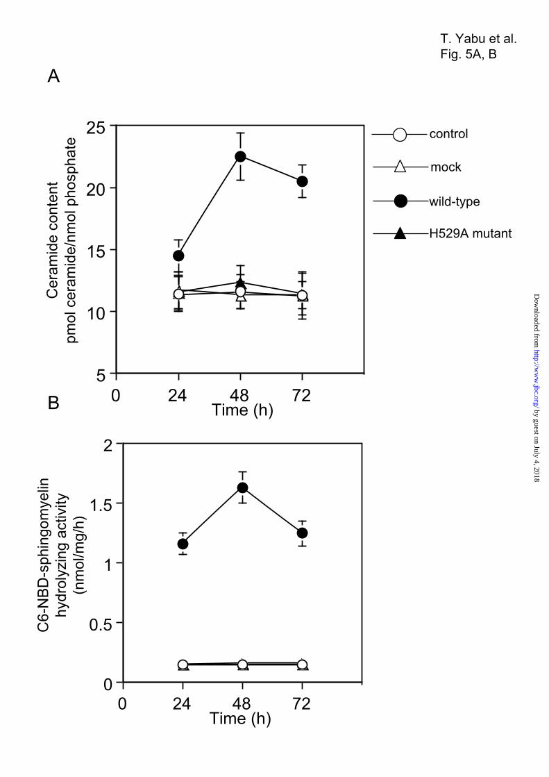
5
10
15
20
25
0 24 48 72 96
control
mock
wild-type
H529A mutant
Cer
amid
e co
nten
tpm
ol c
eram
ide/
nmol
pho
spha
te
0
0.5
1
1.5
2
0 24 48 72 96
C6-
NBD
-sph
ingo
mye
linhy
drol
yzin
g ac
tivity
(nm
ol/m
g/h)
Time (h)
Time (h)
T. Yabu et al.Fig. 5A, B
A
B by guest on July 4, 2018http://w
ww
.jbc.org/D
ownloaded from

Sph
ingo
mye
lin c
onte
nt(n
omol
sph
ingo
mye
lin/n
mol
pho
spha
te)
Time (h)
T. Yabu et al.Fig. 5C
C
0
0.1
0.2
0.3
0.4
0 24 48 72 96 by guest on July 4, 2018http://w
ww
.jbc.org/D
ownloaded from

SMase
actin
wild-type H529A mutant1 2 3 1 2 3
C6-NBD-ceramide
C6-NBD-sphingomyelin
Origin
1 2 31 23 controlwild-type H529A mutant
T. Yabu et al.Fig. 6A, B
A
B
by guest on July 4, 2018http://w
ww
.jbc.org/D
ownloaded from

Mito
chon
dria
l cer
amid
e co
nten
t(p
mol
cer
amid
e/nm
ol p
hosp
hate
)
control 1 2 3 1 2 3wild-type H529A mutant
Mito
chon
dria
l neu
tral
SMas
e ac
tivity
(nm
ol/m
g/h)
T. Yabu et al.Fig. 6C, DC
D
0
0.5
1.0
1.5
2.0
2.5
control 1 2 3 1 2 3wild-type H529A mutant
0
5
10
15
20
aa a
b b b
b b b
aa
a
by guest on July 4, 2018http://w
ww
.jbc.org/D
ownloaded from

control 1 2 3 1 2 3wild-type H529A mutant
Mito
chon
dria
l sph
ingo
mye
lin c
onte
nt(n
mol
sph
ingo
mye
lin/n
mol
pho
spha
te)
T. Yabu et al.Fig. 6E
E
0
0.025
0.050
0.075
0.100
0.125
aa a
b b b
by guest on July 4, 2018http://w
ww
.jbc.org/D
ownloaded from

SMase
actin
control20 µMsense 5 10 20
antisense (µM)
control 20 µMsense
5 10 20antisense (µM)
T. Yabu et al.Fig. 7A, B
A
B
0
0.5
1.0
1.5
*
**
C6-
NBD
-sph
ingo
mye
linhy
drol
yzin
g ac
tivity
(nm
ol/m
g/h)
by guest on July 4, 2018http://w
ww
.jbc.org/D
ownloaded from

Mito
chon
dria
l cer
amid
e co
nten
t(p
mol
cer
amid
e/nm
ol p
hosp
hate
)
5 10 20antisense (µM)
5 10 20antisense (µM)
Mito
chon
dria
l sph
ingo
mye
lin c
onte
nt(n
mol
sph
ingo
mye
lin/n
mol
pho
spha
te)
T. Yabu et al.Fig. 7C, D
C
D
0
3
6
9
12
0
0.01
0.02
0.03
0.04
0.05
**
* *
control 20 µMsense
control 20 µMsense
by guest on July 4, 2018http://w
ww
.jbc.org/D
ownloaded from
Takeshi Yabu, Akio Shimizu and Michiaki YamashitaA novel mitochondrial sphingomyelinase in zebrafish cells
published online May 8, 2009J. Biol. Chem.
10.1074/jbc.M109.004580Access the most updated version of this article at doi:
Alerts:
When a correction for this article is posted•
When this article is cited•
to choose from all of JBC's e-mail alertsClick here
by guest on July 4, 2018http://w
ww
.jbc.org/D
ownloaded from