a novel source for mesenchymal stem cells jayanti tokas 1, deepika gupta 1, divya pasrija 1, rubina...
TRANSCRIPT

A novel source for Mesenchymal stem cells
Jayanti Tokas1, Deepika Gupta1, Divya Pasrija1, Rubina Begum1, Shalini Jain2 and Hariom Yadav2
1Department of Biotechnology, JMIT, Radaur, Haryana, India
2NIDDK, National Institutes of Health, Bethesda, MD 20892, USA
Corresponding: [email protected]

FETAL STEM CELLS
Sources Specific fetal tissues Fetal circulation, placenta, Amniotic fluid Umbilical cord
Potentiality varies from pluripotent to multipotent.
Fetal stem cells Hemopoietic stem cells (HSC) Mesenchymal stem cells (MSC)
First-trimester blood richer source of HSC and MSC Greater proliferative capacity wider differentiation ability
Can be clonally derived.
FETAL STEM CELLS
AMNIOTIC FLUID
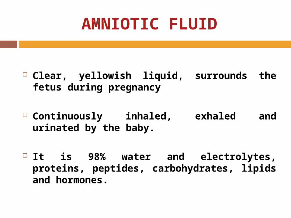
Clear, yellowish liquid, surrounds the fetus during pregnancy
Continuously inhaled, exhaled and urinated by the baby.
It is 98% water and electrolytes, proteins, peptides, carbohydrates, lipids and hormones.

Protects the developing baby against blows to the mother's abdomen
Allows easier fetal movement
Promotes muscular/skeletal development
Protect the fetus from heat loss.
Plays a significant defensive role
AMNIOTIC FLUID
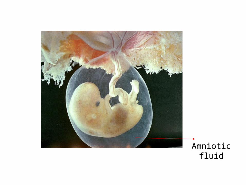
Amniotic fluid
ROLE IN FETAL DEVELOPMENT
Contains nutritional components such as proteins, glucose, triglyceride and cholesterol
proteins and peptides possess potent bioactivity
Growth factors in AF are transported throughout fetal body
Helps cellular movement, organs development, cellular growth and proliferation.

AMNIOTIC FLUID STEM CELLS
Potentiality varies from pluripotent to multipotent
Second and third trimester amniotic fluid is major source
Allow scientists to sidestep the controversy over destroying embryos for research
Can be used for tissue repair and engineering organs
Mesenchymal stem cells quantity is high in amniotic fluid
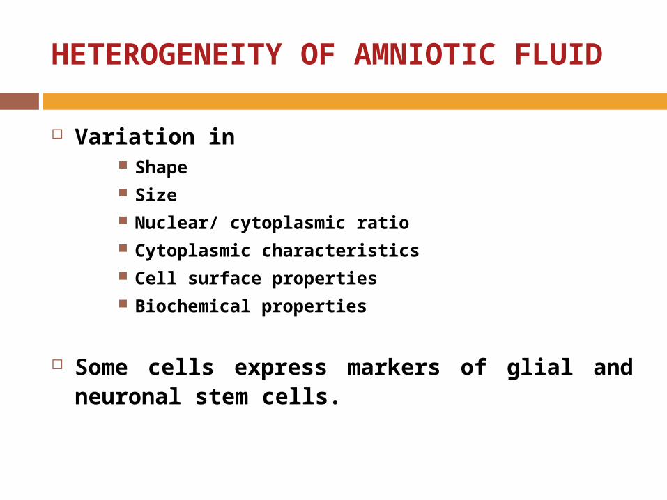
HETEROGENEITY OF AMNIOTIC FLUID
Variation in Shape Size Nuclear/ cytoplasmic ratio Cytoplasmic characteristics Cell surface properties Biochemical properties
Some cells express markers of glial and neuronal stem cells.
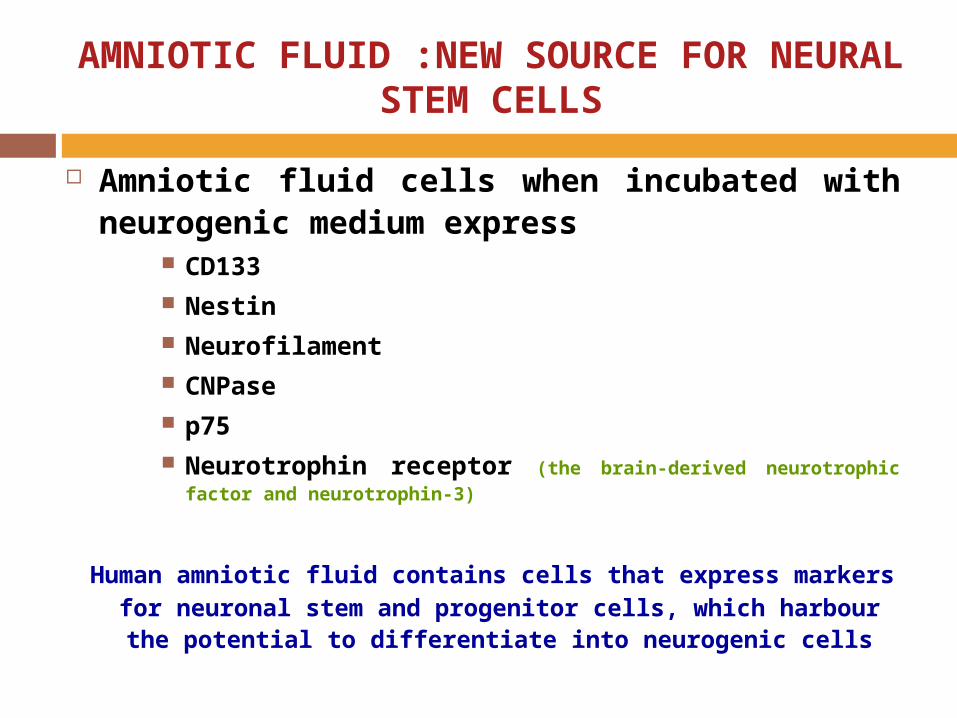
AMNIOTIC FLUID :NEW SOURCE FOR NEURAL STEM CELLS
Amniotic fluid cells when incubated with neurogenic medium express
CD133 Nestin Neurofilament CNPase p75 Neurotrophin receptor (the brain-derived neurotrophic factor and
neurotrophin-3)
Human amniotic fluid contains cells that express markers for neuronal stem and progenitor cells, which harbour the potential to differentiate
into neurogenic cells
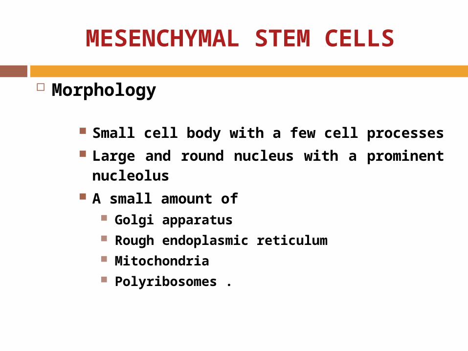
MESENCHYMAL STEM CELLS
Morphology
Small cell body with a few cell processes Large and round nucleus with a prominent
nucleolus A small amount of
Golgi apparatus Rough endoplasmic reticulum Mitochondria Polyribosomes .
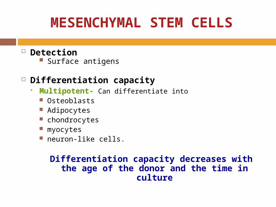
Detection Surface antigens
Differentiation capacity Multipotent- Can differentiate into
Osteoblasts Adipocytes chondrocytes myocytes neuron-like cells.
Differentiation capacity decreases with the age of the donor and the time in culture
MESENCHYMAL STEM CELLS

MESENCHYMAL STEM CELLS AND ITS DIFFERENTIATION CAPACITY
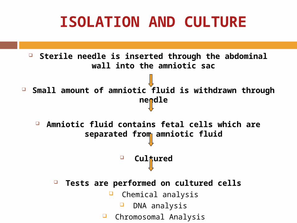
ISOLATION AND CULTURE
Sterile needle is inserted through the abdominal wall into the amniotic sac
Small amount of amniotic fluid is withdrawn through needle
Amniotic fluid contains fetal cells which are separated from amniotic fluid
Cultured
Tests are performed on cultured cells Chemical analysis
DNA analysis Chromosomal Analysis

MESENCHYMAL STEM CELLS : FIRST TRIMESTER
CD291
CD441
SH21
SH31
SH41
prolyl-4-hydroxylase
Actin
CD452
CD342
CD142
CD682
Vwf2
HLA-DR2
Fibronectin
Laminin
Vimentin
In undifferentiated state, fetal blood, liver and bone marrow MSCs express

MSCs differentiate into Adipocytes Osteocytes Chondrocytes.
Provide novel targets for in utero cellular and gene therapy.
MESENCHYMAL STEM CELLS : FIRST TRIMESTER
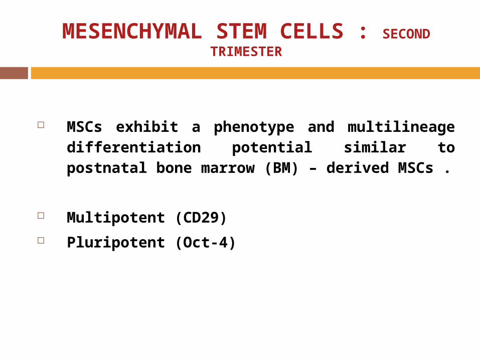
MSCs exhibit a phenotype and multilineage differentiation potential similar to postnatal bone marrow (BM) – derived MSCs .
Multipotent (CD29)
Pluripotent (Oct-4)
MESENCHYMAL STEM CELLS : SECOND TRIMESTER

Express Oct-4.
Samples can be collected in larger amount than the second trimester, with a much lower risk of uterine contamination.
High renewal capacity and therapeutic applications.
Share same properties and differentiation capabilities as human embryonic stem cells .
Differ from embryonic stem cells in promising ways :
1. Do not produce teratomas when transplanted into animals.
2. Low antigenicity is an advantage for cell transplantation or cell replacement therapy.
MESENCHYMAL STEM CELLS : THIRD TRIMESTER

PRESERVATION
Vitrification
Amniotic stem cell banks

VITRIFICATION
AMNIOTIC STEM CELL BANKS
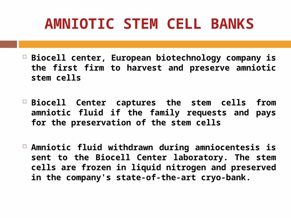
Biocell center, European biotechnology company is the first firm to harvest and preserve amniotic stem cells
Biocell Center captures the stem cells from amniotic fluid if the family requests and pays for the preservation of the stem cells
Amniotic fluid withdrawn during amniocentesis is sent to the Biocell Center laboratory. The stem cells are frozen in liquid nitrogen and preserved in the company's state-of-the-art cryo-bank.

“H.R. 1892—National Amniotic and Placental Stem Cell Bank of 2007
The bill shows the importance of amniotic stem cells bank
which would
Establish a National Amniotic and Placental Stem Cell Bank for the purpose of obtaining, storing, and making available for research and treatment human stem cells derived from amniotic fluid or placenta .
Will maintain a collection of at least 100,000 samples of stem cells in order to ensure genetic diversity
Will obtain stem cells only if informed consent is provided

REGENERATIVE MEDICINE
As regenerative medicine amniotic fluid stem cells has many applications .
Cultured MSCs have been transplanted in children with osteogenesis imperfecta (OI), a disease causing bone fractures and fragility.
Reduced bone fractures and increased bone density were reported to be found when MSCs were engrafted into the defective bone

MYOCARDIAL INFARCTION
MSCs can be engrafted at the damaged
site(s), attenuate pathologic remodeling of
the heart tissue and reduce scar size,
leading to improved post-MI cardiac
function
ETHICAL ASPECTS
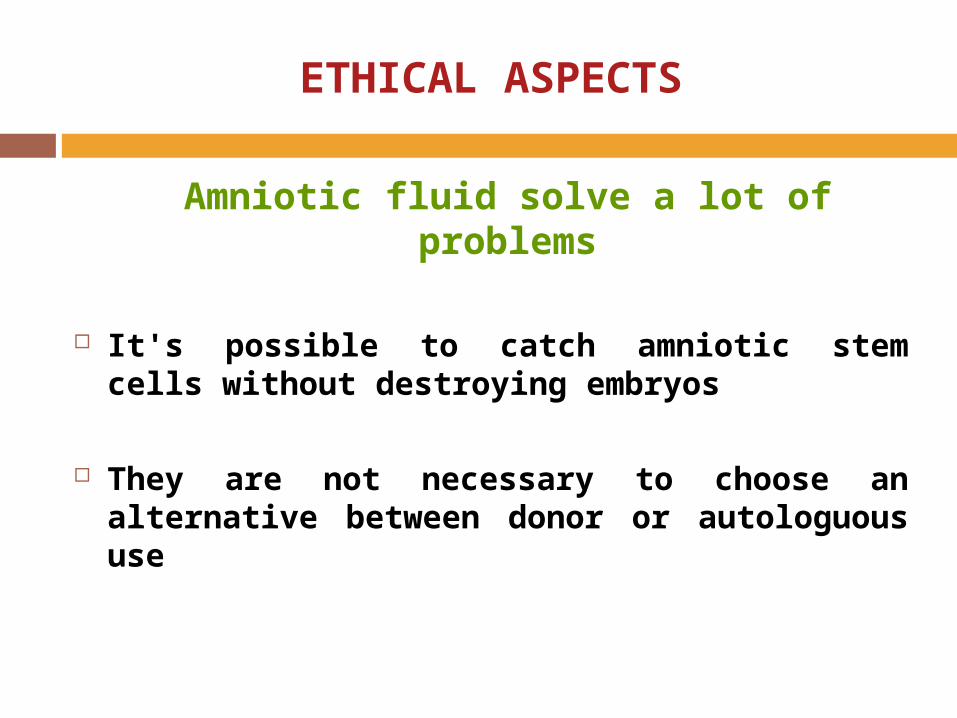
Amniotic fluid solve a lot of problems
It's possible to catch amniotic stem cells without destroying embryos
They are not necessary to choose an alternative between donor or autologuous use

NEXT STEPS TO DO
For the future it is of great importance to obtain more insights into the spectrum of cells contained in human amniotic fluid.
Using RT-PCR the expression of a wide variety of genes, known as markers for stem cells, progenitor cells and differentiated cell types, should be analysed.
Amniotic fluid contains Oct-4 positive cells, a major issue for the future is to investigate the differentiation potential of the Oct-4 positive amniotic fluid cells
It is essential to think about strategies to isolate/enrich Oct-4 positive cells out of amniotic fluid.

“It is an ocean full of pearls
and we need to search
these pearls only for our
benefit”.
