a pre- and post-surgery fmri study - meteore service
TRANSCRIPT

Late plasticity for language in a child's non-dominant hemisphereA pre- and post-surgery fMRI study
Lucie Hertz-Pannier,1,2 Catherine Chiron,1,3 Isabelle JambaqueÂ,3,5 Virginie Renaux-Kieffer,3,5
Pierre-FrancËois Van de Moortele,1 Olivier Delalande,4 Martine Fohlen,4 Francis Brunelle2 andDenis Le Bihan1
1Department of Medical Research, Service Hospitalier
Frederic Joliot, CEA and Institut Federatif de Recherche
49, 2Pediatric Radiology Department, Necker-Enfants
Malades Hospital, 3Neuropediatric Department, Saint
Vincent de Paul Hospital, 4Neurosurgery Department,
Rothschild Foundation Hospital, Paris and 5U29,
INSERM, Marseille, France
Correspondence to: Dr Lucie Hertz-Pannier, Pediatric
Radiology Department, Necker-Enfants Malades Hospital,
149 rue de Sevres, 75015 Paris, France
E-mail: [email protected]
SummaryThe ability of the right hemisphere to sustain the acqui-sition or the recovery of language after extensive dam-age to the left hemisphere has been essentially relatedto the age at the time of injury. Better language abilitiesare acquired when the insult occurs in early childhood(perinatal insults) compared with later occurrence.However, while previous studies have described theneuropsychological pattern of language development intypical cases, the neural bases of such plasticity remainunexplored. Non-invasive functional MRI (fMRI) is aunique tool to assess the neural correlates of brain plas-ticity through repeated studies, but the technique hasnot been widely used in children because of methodo-logical limitations. Plasticity of language was studied ina boy who developed intractable epilepsy related toRasmussen's syndrome of the left hemisphere at age5 years 6 months, after normal language acquisition.The ®rst fMRI study at age 6 years 10 months showedleft lateralization of language networks during a word¯uency task. After left hemispherotomy at age 9 years,
the child experienced profound aphasia and alexia, withrapid recovery of receptive language but slower andincomplete recovery of expressive language and reading.Postoperative fMRI at age 10 years 6 months showed ashift of language-related networks to the right duringexpressive and receptive tasks. Right activation wasseen mainly in regions that could not be detected preo-peratively, but mirrored those previously found in theleft hemisphere (inferior frontal, temporal and parietalcortex), suggesting reorganization in a pre-existingbilateral network. In addition, neuropsychological dataof this case support the hypothesis of innately morebilateral distribution of receptive than expressive lan-guage. This ®rst serial fMRI study illustrates the greatplasticity of the child's brain and the ability of the righthemisphere to take over some expressive language func-tions, even at a relatively late age. It also suggests alimit for removal of the dominant hemisphere beyondthe age of 6 years, a classical limit for the criticalperiod of language acquisition.
Keywords: language; dominance; plasticity; functional MRI; child
Abbreviations: BA = Brodmann area; fMRI = functional MRI; SPM = Statistical Parametric Mapping
IntroductionThe ability of the non-dominant hemisphere to sustain
language functions has been studied in various clinical
situations. In patients with early lesions of the left hemi-
sphere, acquired either prenatally or in early childhood (such
as large infarcts or extended angiomatosis in Sturge±Weber
disease), the right hemisphere can support nearly normal
development of language, provided there is no associated
epilepsy (Isaacs et al., 1996; Muter et al., 1997), but with
subtle differences compared with non-lesioned children
(Vargha-Khadem et al., 1994). In patients with lesions
occurring after language acquisition, language recovery
highly depends on individual pre-lesional lateralization,
ã Oxford University Press 2002
Brain (2002), 125, 361±372

which is dif®cult to assess in clinical practice. In adults, the
functional role of the right hemisphere in aphasia recovery
might be limited compared with that of peri-lesional struc-
tures in the left hemisphere (Weiller et al., 1995; Karbe et al.,
1998b; Samson et al., 1999). The best situation for studying
plasticity of the right hemisphere for language is a situation of
late onset lesion, involving the left hemisphere in right-
handers previously tested for language and resulting in
isolated right hemisphere.
Rasmussen's syndrome has a unique pathology, which
ful®ls these conditions. Partial seizures typically appear
between 4 and 13 years of age in normally developed
children, and are refractory to medical treatment. Cortical
atrophy, hemiplegia and mental deterioration appear progres-
sively, and complete disconnection of the hemisphere using
either hemispherectomy or hemispherotomy (i.e. complete
hemispheric disconnection without removing the hemi-
sphere) is needed to control epilepsy (Andermann et al.,
1993; Vining et al., 1997; Delalande et al., 2000). But
surgeons are often reluctant to disconnect the dominant
hemisphere after 6 years of age, a limit proposed as the end of
the critical period for language acquisition (Woods and
Teuber, 1978; Woods and Carey, 1979; Marcotte and Morere,
1990). However, recent reports clearly demonstrate the
capacity for the right hemisphere to acquire language later.
One boy with Sturge±Weber disease de novo developed a
well-structured language from 9 years of age, following left
hemispherectomy at the age of 8 years 6 months (Vargha-
Khadem et al., 1997). Six right-handed children with
Rasmussen's disease recovered in a year their previous
capacities for receptive language after complete aphasia
immediately following left hemispherectomy performed
between 7 and 14 years (Boatman et al., 1999).
Functional imaging such as PET and functional MRI
(fMRI) provides a unique means of identifying the neuro-
anatomical correlates sustaining such a plasticity of the non-
dominant hemisphere. Because of the lack of ionizing
radiations fMRI is largely preferred to PET in children.
Both techniques have been used to localize the different brain
areas activated in various productive and receptive language
functions in adult volunteers (Petersen et al., 1988; Frith et al.,
1991; Mazoyer et al., 1993; Rueckert et al., 1994;
Frackowiak, 1995; Binder et al., 1997) and in patients
(Weiller et al., 1995; Binder et al., 1996; Benson et al., 1999;
Lehericy et al., 2000). Clinical applications of fMRI have
been developed mainly to assess non-invasively language
dominance before epilepsy surgery, showing an excellent
ability to lateralize language networks when compared with
the intra-carotid Amytal procedure (IAP, Wada test) in adults
and children (Binder et al., 1996; Hertz-Pannier et al., 1997,
2001; Benson et al., 1999; Lehericy et al., 2000).
Imaging studies of post-lesional plasticity of language
networks are scarce. In adults recovering from aphasia
following left infarct, the areas activated during expressive
tasks involve right hemisphere regions homologous to those
usually found on the left side, leading to the hypothesis of
`reorganization of cerebral activity in a pre-existing bilateral
language network' (Weiller et al., 1995; Thulborn et al.,
1999). Similarly, in children with early-acquired large left-
sided lesions, language tasks activate a right-sided perisyl-
vian network, resulting from early specialization of the right
hemisphere for language (Hertz-Pannier et al., 1999; Muller
et al., 1999). However, none of these studies was performed
in the ideal situation required for testing a `naive' non-
dominant hemisphere after language acquisition.
We report on serial pre- and post-surgical language testing,
including neuropsychological and fMRI studies, of a child
suffering from Rasmussen's syndrome who underwent com-
plete disconnection of the left hemisphere at 9 years of age
after previously normal and complete language development.
Table 1 Neuropsychological data/fMRI timing
Before surgery After surgery
Age 6 years 9 months 7 years 8 months 9 years 3 months 10 years 6 monthsTime/surgery ±2 years 3 months ±1 year 4 months +3 months +1 year 6 monthsfMRI Study 1 Failure NT Study 2Attention/behaviour Impulsivity Familiarity Apathy Slowness
Mild attention de®cit Marked attention de®citIQ 118 96 NT 64Performance IQ 128 102 67 80Verbal IQ 107 91 NT 48Oral production Normal Hypospontaneity Few words Short sentencesWord repetition m m NT mNon-word repetition m m NT ±2 SDDigit span m m NT ±2 SDVerbal ¯uency +1 SD m NT ±2 SDNaming m ±1 SD NT ±3 SDSemantic comprehension m m ±3 SD ±3 SDSyntactic comprehension m ±1 SD ±3 SD ±5 SDReading m m NT ±3 SD
IQ = intelligence quotient; m = mean for that age group; SD = standard deviation; NT = not testable.
362 L. Hertz-Pannier et al.

Case reportPatient historyThe ®rst seizures were experienced at 5.5 years in this
previously normal boy, who was ambidextrous and had a
family history of left-handedness (heterozygous twin). His
epilepsy consisted of partial motor seizures of the right limbs
and right epilepsia partialis continua, which were refractory
to all antiepileptic drugs including high doses of corticoster-
oids. He was diagnosed as having Rasmussen's syndrome.
The boy was evaluated for the ®rst time at 6 years 9 months
(Table 1), when on 3 mg/kg/day of hydrocortisone. At that
time, he presented with slight right hemiparesis and neglect,
and some degree of behavioural disorder. However, he still
had normal schooling; he could read, write and count
normally and obtained high IQ scores (Wechsler, 1991).
Dichotic listening showed a clear right ear advantage,
suggesting a relative predominance of the left hemisphere
for language. The procedure consisted of a non-selective
free recall of 36 sets of different monosyllabic digit pairs
simultaneously delivered to each ear, after calibration of
stimuli intensity during monaural presentation (100%
good responses, i.e. no left ear extinction) and a practice
run. The child was instructed to report heard digit pairs
as accurately as possible. Because of an attentional
de®cit, he reported only one digit of each pair, with
66% correct responses for the digits delivered to the right
ear versus 34% for those delivered to the left ear. Normal
control children exhibit equivalent performances for both
ears during such non-selective dichotic listening, probably
due to a ceiling effect during this easy task (Duvelleroy-
Hommet et al., 1995). A forced listening condition
(where children are instructed to recall what was heard
in one given ear) is usually needed to reveal right ear
advantage in right-handed children (88% correct responses
versus 11% for the left ear) (Duvelleroy-Hommet et al.,
1995). Right ear advantage is also observed in 65% of
normal left-handed children aged 8±9 years (Hugdahl and
Andersson, 1989).
MRI was normal. Single photon emission computed
tomography using 133Xe, performed on a day when the
child had no seizure discharge or myoclonus, disclosed left
hemisphere hypoperfusion at rest (±2.2 SD), which was
relatively more pronounced in frontal than posterior cortical
areas (Fig. 1).
The ®rst fMRI, performed at age 6 years 10 months,
showed left lateralization of language networks during covert
semantic word generation (Fig. 2, Table 2). Because right
motor de®cit was not complete, the child underwent subpial
trans-section of the left motor strip at age 7 years 2 months,
followed by resection of the left paracentral lobule at age
7 years 4 months, but both procedures failed to control
seizures. At age 7 years 8 months, IQ scores had decreased by
a mean of 22 points, mostly due to his behavioural disorders,
but verbal abilities were still in the normal range (Table 1)
(Chevrie-Muller et al., 1981). An attempt to perform a second
fMRI failed because of lack of co-operation and the presence
of movement artefacts. The child was able to perform the
tasks at pre-fMRI testing.
At age 9 years, the child had several seizures a day with
speech arrest and falls, could no longer walk due to complete
right hemiplegia and had deteriorated at the cognitive level.
However, no formal neuropsychological testing was per-
Fig. 1 Interictal 133Xe single photon emission computed tomography before left hemisphere disconnection. Absolute ¯ow maps showhypoperfusion in the left hemisphere compared with the right hemisphere, more pronounced in frontal regions (left hemisphere is on theleft, right hemisphere on the right).
Language plasticity after left hemispherotomy 363

Fig. 2 Pre-surgery fMRI (Study 1). SPM map of word generation compared with rest, overlaid on the SPM template. White lines indicatethe extent of slice acquisition. Height threshold P < 0.01 (uncorrected, at a voxel-level), with corrected extent threshold of P < 0.227 percluster. Left-sided activated network involves inferior frontal gyrus, supplementary sensory-motor area and supramarginal gyrus (P < 0.07in each cluster, Z > 3.28). Note that the activation in the right supramarginal gyrus does not reach cluster-level signi®cance (P = 0.227,Z = 4.26) (see Table 2).
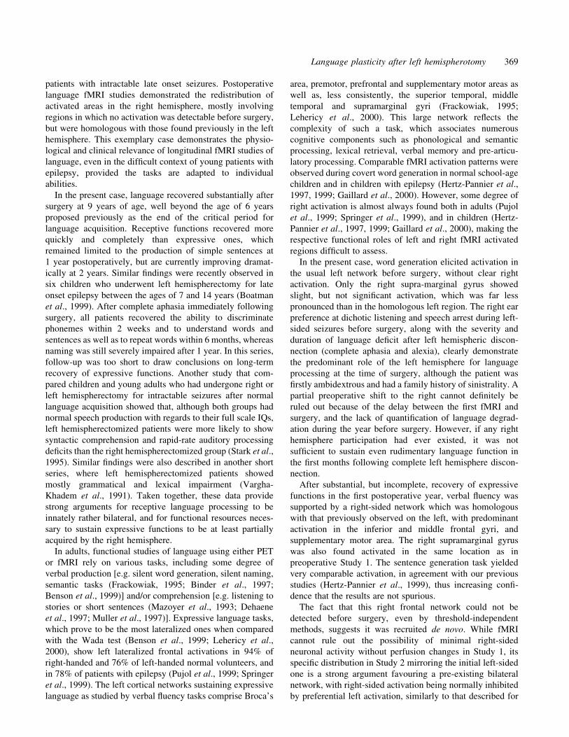
Fig. 3 Activation-related effect in left and right frontal regions in Study 1. The plots display verbal ¯uency minus rest contrast of theparameter estimates (relative size and standard error) in a typical activated voxel located in the left inferior gyrus (±52, 16, 25,Zmax = 3.92), and its right homologue (52, 16, 25). The magnitude of the activation-related effect is negligible (even slightly negative) onthe right, with huge variability (error bar), and is ~16 times greater on the left. Similar results were found in all tested frontal lobe voxels.
364 L. Hertz-Pannier et al.

formed. MRI disclosed peri-insular atrophy. Left hemispher-
otomy was performed, which consisted of extra-thalamic
deep white matter disconnection of the whole hemisphere
along with complete callosotomy and section of the anterior
and posterior commissures, without removing the discon-
nected hemisphere (Delalande et al., 2000). This procedure
results in a normally perfused, but totally isolated left
hemisphere, i.e. with no single connection left with both the
right hemisphere and the descending pathways of the pons.
Seizures immediately disappeared and treatment was discon-
tinued at age 10 years. Neuropathological examination of a
frontal fragment con®rmed the diagnosis of Rasmussen's
syndrome. Despite persistent right hemiplegia, the child
could walk again. However, persistent and complete mutism
with complete loss of reading and counting immediately
followed surgery. Three months after surgery, while the
patient had begun to recover, his semantic comprehension
scored at a level of 6 years (Lege and Dagne, 1976) (Table 1),
whereas he was only beginning to say a few words and failed
in most oral verbal tasks (de Agostini et al., 1998). He could
count up to six, but could not read. He presented with severe
attention de®cit. Six months after surgery, his lexical stock
had improved and he could recognize a few letters.
At age 10 years, the child was admitted in specialized
schooling and bene®ted from orthophonic rehabilitation. The
last evaluation was performed 6 months later, i.e. 1.5 years
after surgery (Table 1). Attention was still ¯uctuating and
Wechsler Intelligence Scale for Children sub-scores were
strongly dissociated with a low verbal IQ. However, the child
had substantially recovered as he could construct short
sentences, name images, repeat words, read some words and
perform simple additions. He was able to perform a third
fMRI, which showed a right-sided activated network for the
three language tasks tested (word generation, sentence
generation and story listening) (Fig. 4A±C and Table 2), in
locations that were not detectable on the ®rst study. One year
later, when he had progressively recovered spontaneous and
intelligible ¯uent speech, although with persistent syntactic
de®cit according to his teachers and carers, he refused to
submit to another fMRI study.
Table 2 Longitudinal fMRI data
Study 1 (P = 0.01 uncorrected) Study 2 (P = 0.001 uncorrected)
Word generation Word generation Sentence generation Listening to sentences
Area P Zmax Area P Zmax Area P Zmax Area P Zmax
L SMA <10±4 4.99L IFG+ preCG <10±4 4.36L SMG 0.002 3.28 L SMG =10±3 4.80 L SMG <10±4 4.93
0.074* 3.85L Insula <10±4 4.65 L Insula <10±4 4.66L Fr <10±4 4.09 L Fr <10±4 4.26L Occ <10±4 4.59 L Occ 0.001 4.26R SMA <10±4 7.19 R SMA <10±4 4.78R Cing <10±4 6.82 R Cing <10±4 6.98R IFG <10±4 6.65 R IFG <10±4 5.39R IFS =10±3 5.84 R IFS <10±4 5.56R Insula <10±4 7.28 R Insula <10±4 6.05R DLPFC <10±4 9.96 R DLPFC <10±4 5.97R preCG <10±4 inf R preCG <10±4 7.38R caudate <10±4 5.01 R caudate <10±4 5.12R Fr <10±4 4.26 R Fr <10±4 4.74R postCS 0.01 3.92 R postCS <10±4 4.07
R SMG 0.227* 4.26 R SMG <10±4 5.87 R SMG 0.03 3.92R AG <10±4 4.63
R STS/STG <10±4 5.96 R STS/STG <10±4 5.03 R STS/STG <10±4 6.53R ITG 0.05 4.09 R MTG <10±4 4.99R Occ <10±4 4.64 R Occ 0.03 4.23L cerebell 0.003 4.99
P values are given for each cluster; Zmax corresponds to Z score of maximum local voxel; L = left; R = right; inf = in®nite;SMA = supplementary motor area [Brodmann area (BA) 6]; IFG = inferior frontal gyrus (BA 44, 45); IFS = posterior part of the inferiorfrontal sulcus (junction between BA 8, 6 and 44); preCG = precentral gyrus, primary motor cortex (BA 4); postCS = postcentral sulcus(BA 7); SMG = supramarginal gyrus (BA 40); AG = angular gyrus (BA 39); Cing = cingulum (BA 24/32); DLPFC = dorsolateralprefrontal cortex (BA 9/46); Fr = frontal pole and mesial cortex (BA 10/11); STS = superior temporal sulcus (BA 22); STG = superiortemporal gyrus (BA 41/42/22); MTG = middle temporal gyrus (BA 21); ITG = inferior temporal gyrus (BA 37); Occ = occipital cortex(BA 17/18). *Not signi®cant.
Language plasticity after left hemispherotomy 365

366 L. Hertz-Pannier et al.

fMRIData acquisition and task designAll fMRI procedures were performed after obtaining parental
consent and child assent according to the ethical committee of
Bicetre Hospital, on a 3T Bruker spectrometer, using
gradient-echo echo-planar sequences and block paradigms.
The child was trained carefully before the fMRI procedure
and tested to ensure good understanding and performance of
the tasks. Because of the long timespan between the
examinations (3 years), the acquisition technique and para-
digms differed slightly.
Study 1 (pre-surgical study). Covert word generation to
concrete categories (animals, colours) was tested during two
activation periods lasting 60 s each, interleaved with three
rest periods of the same duration during which the child was
asked to stop thinking of words and listen to the scanner
noise. This paradigm was repeated twice during the acqui-
sition of a total of 100 gradient-echo echo-planar images [TR
(repetition time) = 6 s, TE (echo time) = 35 ms, voxel
size = 3.7 3 3.7 3 5 mm3, 10 axial slices].
Study 2 (post-surgical study). Three language tasks were
studied according to our ongoing protocol of expressive
and receptive language (Hertz-Pannier et al., 1999). As in
all fMRI language studies, all tasks were performed
covertly to avoid head movements. (i) Word generation to
concrete categories (similar to Study 1, using animals,
colours, ®rst names, food) during four activation blocks
of 27 s duration each, interleaved with rest periods of the
same duration, for a total of 3 min 54 s (TR = 3.5 s,
67 repetitions). (ii) Sentence generation from a concrete
noun with the same experimental design as in the word
generation paradigm (four activation and four rest
periods). This task was chosen because it requires less
attentional and working memory demands than covert
Fig. 4 fMRI after left hemisphere disconnection (Study 2). SPM maps of three language tasks compared with rest, overlaid on thenormalized patient's brain surface rendering. Note left hemisphere atrophy and brain resection. Height threshold P < 0.001 (uncorrected ata voxel level), with corrected extent threshold of P < 0.05 per cluster. See Table 2 for statistics. (A) Word generation compared with rest.Right-lateralized activated network involves mostly frontal regions (inferior and middle frontal gyri, precentral gyrus, supplementarymotor area) and a few superior and inferior temporal regions, as well as the supramarginal gyrus. Activation can be also observed on theleft in the supramarginal gyrus, and bilaterally in occipital cortex and frontal poles. (B) Sentence generation compared with rest. Duringthis task, the activated network is very comparable to the previous one, although less extended, with additional activation in the rightangular gyrus. (C) Listening to sentences compared with rest. In this purely receptive task, activated network is restricted to right-sidedsuperior and middle temporal regions.
Language plasticity after left hemispherotomy 367

word production, and is therefore more amenable to
impaired children, while showing a good lateralization
power (Muller et al., 1997). A concrete word was
presented through the intercom every 7 s with four
nouns presented in each activation period. The child was
asked to make a simple sentence with the presented noun
(such as subjectÐverbÐobject). No words were presented
during the rest periods, during which the child was asked
to listen to the scanner noise. (iii) Listening attentively to
simple sentences (one sentence lasting a maximum of
2.5 s every 5 s, six sentences per activation block)
presented through the scanner intercom without any
explicit verbal production, compared with rest (similar
to previous tasks), for a total duration of 4 min 15 s
(TR = 5 s, 51 repetitions).
To avoid interference from the scanner noise and to ensure
good comprehension of the auditory stimuli, we used a
modi®ed echo planar imaging sequence in which the slice
acquisition gradients (loud beeps) are grouped over a 1.4 s
interval to leave a silent interval within each repetition time,
while maintaining proper RF (radio frequency) excitation
(van de Moortele et al., 1998). The duration of silent intervals
was adapted to the length of the stimuli (2.1 s for presentation
of simple words within a TR = 3.5 s and 3.6 s for the
presentation of the sentences within a TR = 5 s). Twenty-two
axial slices (voxel size = 3.7 3 3.7 3 5 mm3) were acquired.
Data analysisData from both studies were analysed using statistical
parametric mapping (SPM) (SPM99, Wellcome Institute
for Cognitive Neuroscience, University College, London,
UK). After motion correction, linear normalization of the
images in the Talairach space, and spatial and temporal
smoothing, data were analysed using voxel by voxel
uncorrected threshold of P = 0.01 in Study 1 and
P = 0.001 in Study 2. In both studies, we selected only
the clusters with a probability of activation of P = 0.05
for each cluster (extent threshold). One major question
pertaining to the mechanisms of brain plasticity during
language recovery in this patient was whether we could
detect a posteriori in Study 1, some activation (even
below standard statistical thresholds) in the right frontal
regions that were found activated in Study 2. Therefore,
we re-analysed Study 1 at very low thresholds (P = 0.1
uncorrected) and used a threshold-independent approach.
This involved comparing the verbal ¯uency minus rest
contrast of the parameter estimates (relative size and
standard error) in the 17 left frontal voxels that reached
maximum Z scores versus their right homologues (Fig. 3).
For anatomical display, data from Study 1 were displayed
on the SPM template because we could not acquire good
anatomical data in this early study (standard MRI at 1.5 T was
normal at that time). Data from Study 2 were displayed on the
normalized surface rendering of the patient's brain.
ResultsIn Study 1, the SPM-activated regions during word gener-
ation were restricted to the left hemisphere (Fig. 2 and Table
2). They involved the left inferior frontal gyrus [IFG,
Brodmann area (BA) 44, 45] and the lateral precentral
gyrus (preCG, BA 4/6), the supramarginal gyrus (SMG,
BA 40) and the supplementary motor area (SMA, BA 6). The
only activation found on the right side, in the right
supramarginal gyrus, did not reach cluster signi®cance
(P = 0.227, Zmax = 4.26). No activation could be detected in
the right frontal regions, even at very low thresholds. In
addition, the threshold-independent approach con®rmed that
the magnitude of the activation-related effect was negligible
on the right, with huge variability, and that it was four to 16
times greater on the left. A typical left and right IFG voxel
comparison is shown in Fig. 3. In this early study, temporal
regions and cerebellum were not studied due to the limited
number of slices.
In Study 2, activated regions were strongly lateralized to
the right (Fig. 4A±C). They differed slightly according to the
tasks. (i) During word generation, activated areas involved
mostly right regions homologous to those found preopera-
tively on the left, namely the right inferior frontal gyrus
(BA 44), anterior insula and precentral gyrus (BA 4/6), the
right supramarginal gyrus (BA 40) and the right supplemen-
tary sensory-motor area (BA 6). Additional activation was
found in the right dorsolateral prefrontal cortex (DLPFC,
BA 46) and cingulum (BA 24/32), the right frontal pole, the
right caudate and putamen, the right superior temporal sulcus
and gyrus (STS, STG, BA 41/42/22), inferior temporal gyrus
(ITG, BA 37), the right post-central sulcus (postCS, BA 7),
the occipital cortex and the left cerebellum. Surprisingly,
some activation was also found on the left side, in the anterior
insula and frontal pole, the supramarginal gyrus and occipital
cortex. (ii) During the sentence generation task, the activated
network was strongly comparable with the previous one, with
additional activation in the right angular gyrus (AG, BA 39),
but no activation in the right inferior temporal gyrus and in
the cerebellum. (iii) Finally, during sentence listening,
activation was strictly restricted to the right superior temporal
region, including primary and secondary auditory cortices
(STG, BA 41, 42), as well as the superior temporal sulcus
(STS, BA 22) and middle temporal gyrus (MTG, BA 21).
DiscussionIn this serial fMRI study of language plasticity, we could
compare for the ®rst time pre- and postoperative data of
language networks in a child who had had normal language
development before undergoing complete disconnection of
the dominant left hemisphere for severe epilepsy at age
9 years. The isolated right hemisphere was shown able to
sustain late plasticity changes for language, thus indicating
that it would be reasonable to consider surgical disconnection
of the dominant hemisphere at least until 9 years of age in
368 L. Hertz-Pannier et al.
patients with intractable late onset seizures. Postoperative
language fMRI studies demonstrated the redistribution of
activated areas in the right hemisphere, mostly involving
regions in which no activation was detectable before surgery,
but were homologous with those found previously in the left
hemisphere. This exemplary case demonstrates the physio-
logical and clinical relevance of longitudinal fMRI studies of
language, even in the dif®cult context of young patients with
epilepsy, provided the tasks are adapted to individual
abilities.
In the present case, language recovered substantially after
surgery at 9 years of age, well beyond the age of 6 years
proposed previously as the end of the critical period for
language acquisition. Receptive functions recovered more
quickly and completely than expressive ones, which
remained limited to the production of simple sentences at
1 year postoperatively, but are currently improving dramat-
ically at 2 years. Similar ®ndings were recently observed in
six children who underwent left hemispherectomy for late
onset epilepsy between the ages of 7 and 14 years (Boatman
et al., 1999). After complete aphasia immediately following
surgery, all patients recovered the ability to discriminate
phonemes within 2 weeks and to understand words and
sentences as well as to repeat words within 6 months, whereas
naming was still severely impaired after 1 year. In this series,
follow-up was too short to draw conclusions on long-term
recovery of expressive functions. Another study that com-
pared children and young adults who had undergone right or
left hemispherectomy for intractable seizures after normal
language acquisition showed that, although both groups had
normal speech production with regards to their full scale IQs,
left hemispherectomized patients were more likely to show
syntactic comprehension and rapid-rate auditory processing
de®cits than the right hemispherectomized group (Stark et al.,
1995). Similar ®ndings were also described in another short
series, where left hemispherectomized patients showed
mostly grammatical and lexical impairment (Vargha-
Khadem et al., 1991). Taken together, these data provide
strong arguments for receptive language processing to be
innately rather bilateral, and for functional resources neces-
sary to sustain expressive functions to be at least partially
acquired by the right hemisphere.
In adults, functional studies of language using either PET
or fMRI rely on various tasks, including some degree of
verbal production [e.g. silent word generation, silent naming,
semantic tasks (Frackowiak, 1995; Binder et al., 1997;
Benson et al., 1999)] and/or comprehension [e.g. listening to
stories or short sentences (Mazoyer et al., 1993; Dehaene
et al., 1997; Muller et al., 1997)]. Expressive language tasks,
which prove to be the most lateralized ones when compared
with the Wada test (Benson et al., 1999; Lehericy et al.,
2000), show left lateralized frontal activations in 94% of
right-handed and 76% of left-handed normal volunteers, and
in 78% of patients with epilepsy (Pujol et al., 1999; Springer
et al., 1999). The left cortical networks sustaining expressive
language as studied by verbal ¯uency tasks comprise Broca's
area, premotor, prefrontal and supplementary motor areas as
well as, less consistently, the superior temporal, middle
temporal and supramarginal gyri (Frackowiak, 1995;
Lehericy et al., 2000). This large network re¯ects the
complexity of such a task, which associates numerous
cognitive components such as phonological and semantic
processing, lexical retrieval, verbal memory and pre-articu-
latory processing. Comparable fMRI activation patterns were
observed during covert word generation in normal school-age
children and in children with epilepsy (Hertz-Pannier et al.,
1997, 1999; Gaillard et al., 2000). However, some degree of
right activation is almost always found both in adults (Pujol
et al., 1999; Springer et al., 1999), and in children (Hertz-
Pannier et al., 1997, 1999; Gaillard et al., 2000), making the
respective functional roles of left and right fMRI activated
regions dif®cult to assess.
In the present case, word generation elicited activation in
the usual left network before surgery, without clear right
activation. Only the right supra-marginal gyrus showed
slight, but not signi®cant activation, which was far less
pronounced than in the homologous left region. The right ear
preference at dichotic listening and speech arrest during left-
sided seizures before surgery, along with the severity and
duration of language de®cit after left hemispheric discon-
nection (complete aphasia and alexia), clearly demonstrate
the predominant role of the left hemisphere for language
processing at the time of surgery, although the patient was
®rstly ambidextrous and had a family history of sinistrality. A
partial preoperative shift to the right cannot de®nitely be
ruled out because of the delay between the ®rst fMRI and
surgery, and the lack of quanti®cation of language degrad-
ation during the year before surgery. However, if any right
hemisphere participation had ever existed, it was not
suf®cient to sustain even rudimentary language function in
the ®rst months following complete left hemisphere discon-
nection.
After substantial, but incomplete, recovery of expressive
functions in the ®rst postoperative year, verbal ¯uency was
supported by a right-sided network which was homologous
with that previously observed on the left, with predominant
activation in the inferior and middle frontal gyri, and
supplementary motor area. The right supramarginal gyrus
was also found activated in the same location as in
preoperative Study 1. The sentence generation task yielded
very comparable activation, in agreement with our previous
studies (Hertz-Pannier et al., 1999), thus increasing con®-
dence that the results are not spurious.
The fact that this right frontal network could not be
detected before surgery, even by threshold-independent
methods, suggests it was recruited de novo. While fMRI
cannot rule out the possibility of minimal right-sided
neuronal activity without perfusion changes in Study 1, its
speci®c distribution in Study 2 mirroring the initial left-sided
one is a strong argument favouring a pre-existing bilateral
network, with right-sided activation being normally inhibited
by preferential left activation, similarly to that described for
Language plasticity after left hemispherotomy 369

syllable discrimination (Boatman et al., 1998). The hypoth-
esis of collateral inhibition of trans-callosal activity being
responsible for functional brain asymmetries during expres-
sive language processing has been documented using PET
(Karbe et al., 1998a). In the case of complete hemispheric
disconnection, such an inhibition would be suppressed,
leading to expression of pre-existing contralateral networks,
analogous to the recovery of motor functions in similar cases
(Holloway et al., 2000). This hypothesis has also been raised
to explain right hemisphere participation in functional
recovery after stroke (Weiller et al., 1995).
Various methodological issues must be discussed in
relation to this study. The choice of the task is a key point
when studying young or debilitated children using fMRI,
because task complexity limits feasibility. All tasks must be
performed silently in the scanner to avoid head movements.
Thus, special care was taken to train the child in selected
tasks. These were tested by a neuropsychologist and practised
overtly before fMRI to ensure good comprehension, to check
the feasibility and to improve performance (Price and Friston,
1999). Word generation and sentence generation have
demonstrated good feasibility in children, even with mild
cognitive impairment, and excellent correlation with the
Wada test (Hertz-Pannier et al., 1997; Hertz-Pannier et al.,
2001). Word generation is known to explore semantic
¯uency, which proved to be normal before surgery (Table
1). Performance of this task does not necessarily re¯ect the
level of daily propositional speech and may be impaired in
non-aphasic patients with verbal memory de®cits. At the time
of postoperative fMRI, our patient showed global language
impairment, but was able to perform the verbal ¯uency task
(Table 1). Similarly, he could generate short sentences
overtly at a rate of one sentence per 7 s, both during
neuropsychological evaluation and immediately before fMRI.
As for the receptive task, while the child showed impairment
in both semantic and syntactic comprehension at neuropsych-
ology, he easily understood the simple active non-reversible
sentences presented during fMRI.
Interictal 133Xe single photon emission computed tomo-
graphy performed at the same time as preoperative fMRI
Study 1 showed a hypo-functioning left hemisphere at rest,
more pronounced in frontal regions, according to the clinical
motor de®cit, and in agreement with the literature (Yacubian
et al., 1997). This left hypoperfusion, however, did not
preclude the expression of activation-related haemodynamic
changes in expected frontal locations, as demonstrated by
fMRI Study 1. No perfusion abnormality was found in the
right hemisphere.
In longitudinal studies, one would ideally wish to obtain
similar sensitivities for the detection of activation. In the
present case, the sensitivity of detection of activation cannot
be strictly compared between both studies because of
differences in data acquisition and task design (these explain
differences in statistical power in addition to the obvious
differences in cognitive abilities). However, the key issue was
to demonstrate that no activation could be detected in the
right hemisphere before surgery. Since one might question
the statistical power of Study 1 owing to the limited number
of slices and time points, we used a lower uncorrected
threshold than in Study 2 (P = 0.01 versus P = 0.001).
Moreover, the possibility that a threshold effect might have
obscured right activations was investigated by a threshold-
independent approach and proved negative. Finally, the
Talairach coordinates of activated regions of both studies
cannot be strictly compared despite normalization of all
images, for two reasons: (i) in Study 1, the limited number of
slices and lack of good anatomical images may have
hampered the accuracy of spatial normalization; and (ii) in
Study 2, the large anatomical changes secondary to both
serial surgical procedures and left hemisphere atrophy
resulted in a mid-line shift to the left and may have induced
some distortion of normalized images.
Finally, two points need to be mentioned. The right-sided
post-surgical activation is more extended and pronounced
than the left-sided pre-surgical one. Beside differences in
statistical power, which preclude rigorous comparison of the
extent of activation between both studiesÐthe child could
have been more cooperative during the second than the ®rst
study. Another speculative hypothesis could relate to recent
plasticity and learning effects that require more widespread
networks, as suggested by fMRI studies conducted in normal
children compared with adults (Hertz-Pannier et al., 1997;
Gaillard et al., 2000), as well as in low-pro®ciency adult
volunteers using a foreign language (Dehaene et al., 1997). In
longitudinal developmental studies, the use of comparable
tasks over time remains a problematic issue because of
variability of performance either due to evolving cognitive
abilities and strategies in normal children and/or cognitive
consequences of treatment in patients. More surprising is the
post-surgical activation of a few left regions, including the
supramarginal gyrus, anterior insula and frontal cortex, and
occipital cortex. Such an observation has not been reported
previously and is not fully understood. Indeed, all hemi-
spheric connections were cut during hemispherotomy, which
included complete callosotomy, section of anterior and
posterior commissures and peri-thalamic disconnection, and
resulted in complete clinical seizure control. There were no
residual head movement artefacts after image registration in
either contrast (such artefacts usually present as mirror
images in positive and negative contrasts, in the white matter,
at the edges of the brain, the ventricles or the resection
cavity). In addition, motion-related artefacts would be
unlikely to create such regional and reproducible effects.
Indeed, left-sided activation was very consistent during both
word ¯uency and sentence generation (which are very close at
a cognitive level) and involved cortical regions close to those
normally activated by these tasks, but was not found during
the listening task. Because the vascular supply to the left
hemisphere was maintained, one may wonder whether some
degree of regional vascular reactivity could be preserved
without neuronal activity. Another hypothesis relates to
residual activity of deafferented neurones in a disconnected
370 L. Hertz-Pannier et al.

hemisphere, by analogy with epilepsy patients in which infra-
clinical seizures can be recorded in disconnected megalen-
cephalic hemispheres (unpublished personal data). But the
mechanism by which this activity could be synchronized with
contralateral activity remains unknown.
AcknowledgementsWe wish to thank the patient and his family for their
patience during serial tests. We also wish to thank
Professor Jacques Motte, MD, who referred the patient,
Professor Olivier Dulac, MD, who reviewed the manu-
script, Jean-Baptiste Poline, PhD, for advice in statistics,
Jean-Francois Mangin, PhD, for assistance in anatomical
reconstruction and analysis, and Ghislaine Dehaene-
Lambertz, MD, PhD, for assistance in neuropsychological
analysis and fruitful discussions. This work was supported
in part by the Institut Electricite SanteÂ, Paris, France.
References
Andermann F, Freeman J, Vigevano F. Surgically remediable
diffuse hemispheric syndromes. In: Engel J Jr, editor. Surgical
treatment of the epilepsies. New York: Raven Press; 1993. p. 87±
101.
Benson RR, FitzGerald DB, LeSueur LL, Kennedy DN, Kwong
KK, Buchbinder BR, et al. Language dominance determined by
whole brain functional MRI in patients with brain lesions.
Neurology 1999; 52: 798±809.
Binder JR, Swanson SJ, Hammeke TA, Morris GL, Mueller WM,
Fischer M, et al. Determination of language dominance using
functional MRI: a comparison with the Wada test. Neurology 1996;
46: 978±84.
Binder JR, Frost JA, Hammeke TA, Cox RW, Rao SM, Prieto T.
Human brain language areas identi®ed by functional magnetic
resonance imaging. J Neurosci 1997; 17: 353±62.
Boatman D, Hart J Jr, Lesser RP, Honeycutt N, Anderson NB,
Miglioretti D, et al. Right hemisphere speech perception revealed
by amobarbital injection and electrical interference. Neurology
1998; 51: 458±64.
Boatman D, Freeman J, Vining E, Pulsifer M, Miglioretti D,
Minahan R, et al. Language recovery after left hemispherectomy
in children with late-onset seizures. Ann Neurol 1999; 46: 579±
86.
Chevrie-Muller C, Simon A, Decante P. Epreuves pour l'examen du
langage. Paris: Editions Centre de Psychologie AppliqueÂe (ECPA);
1981.
de Agostini M, Metz-Lutz MN, van Hout A, Chavance M, Deloche
G, Pavao-Martins I, et al. Oral language evaluation battery of
aphasic children (ELOLA): a French standardization (4±12 years).
Rev Neuropsychol 1998; 3: 319±68.
Dehaene S, Dupoux E, Mehler J, Cohen L, Paulesu E, Perani D,
et al. Anatomical variability in the cortical representation of ®rst
and second language. Neuroreport 1997; 8: 3809±15.
Delalande O, Fohlen M, Jalin C, Pinard JM. From hemispherectomy
to hemispherotomy In: LuÈdeus H, editor. Surgery of epilepsy, 2nd
ed. Philadelphia: Lippincott Williams and Wilkins; 2000. p. 741±6.
Duvelleroy-Hommet C, Gillet P, Billard C, Loisel ML, Barthez
MA, Santini JJ, et al. Study of unilateral hemispheric performance
in children with developmental dysphasia. Neuropsychologia 1995;
33: 823±34.
Frackowiak RSJ. An European activation study of verbal ¯uency:
results from a multicentre PET experiment. EU concerted action on
functional imaging (Essen, Groeningen, Leuven, Liege, London,
Lyon, Milano, Orsay, Stockholm). J Cereb Blood Flow Metab 1995;
15 Suppl 1: S52.
Frith CD, Friston KJ, Liddle PF, Frackowiak RS. A PET study of
word ®nding. Neuropsychologia 1991; 29: 1137±48.
Gaillard WD, Hertz-Pannier L, Mott SH, Barnett AS, Le Bihan D,
Theodore WH. Functional anatomy of cognitive development:
fMRI of verbal ¯uency in children and adults. Neurology 2000; 54:
180±5.
Hertz-Pannier L, Gaillard W, Mott S, Cuenod C, Bookheimer S,
Weinstein S, et al. Non invasive assessment of language dominance
in children and adolescents with functional MRI: a preliminary
study. Neurology 1997; 48: 1003±12.
Hertz-Pannier L, Chiron C, van de Moortele PF, Bourgeois M,
Fohlen M, Dulac O, et al. Multi-task fMRI presurgical language
mapping in children with cognitive impairment. Neuroimage 1999;
9: S691.
Hertz-Pannier L, Chiron C, Vera P, Van de Morteele P, Kaminska
A, Bourgeois M, et al. Functional imaging in the work-up of
childhood epilepsy. Child's Nerv Syst. In press 2001.
Holloway V, Gadian DG, Vargha-Khadem F, Porter DA, Boyd
SG, Connelly A. The reorganization of sensorimotor function in
children after hemispherectomy. A functional MRI and
somatosensory evoked potential study. Brain 2000; 123: 2432±
44.
Hugdahl K, Andersson B. Dichotic listening in 126 left-handed
children: ear advantages, familial sinistrality and sex differences.
Neuropsychologia 1989; 27: 999±1006.
Isaacs E, Christie D, Vargha-Khadem F, Mishkin M. Effects of
hemispheric side of injury, age at injury and presence of seizure
disorder on functional ear and hand asymmetries in hemiplegic
children. Neuropsychologia 1996; 34: 127±37.
Karbe H, Herholz K, Halber M, Heiss WD. Collateral inhibition of
transcallosal activity facilitates functional brain asymmetry. J Cereb
Blood Flow Metab 1998a; 18: 1157±61.
Karbe H, Thiel A, Weber-Luxenburger G, Herholz K, Kessler J,
Heiss WD. Brain plasticity in poststroke aphasia: what is the
contribution of the right hemisphere? Brain Lang 1998b; 64: 215±
30.
Lege Y, Dagne P. Test de vocabulaire en images. Paris: Editions
Centre de Psychologie AppliqueÂe (EPCA); 1976.
Lehericy S, Cohen L, Bazin B, Samson S, Giacomini E, Rougetet R,
et al. Functional MR evaluation of temporal and frontal language
dominance compared with the Wada test. Neurology 2000; 54:
1625±33.
Language plasticity after left hemispherotomy 371

Marcotte AC, Morere DA. Speech lateralization in deaf
populations: evidence for a developmental critical period. Brain
Lang 1990; 39: 134±52.
Mazoyer B, Tzourio N, Frak V, Syrota A, Murayama N, Levrier O,
et al. The cortical representation of speech. J Cogn Neurosci 1993;
5: 467±79.
Muller RA, Rothermel RD, Behen ME, Muzik O, Mangner TJ,
Chugani HT. Receptive and expressive language activations for
sentences: a PET study. Neuroreport 1997; 8: 3767±70.
Muller RA, Rothermel RD, Behen ME, Muzik O, Chakraborty PK,
Chugani HT. Language organization in patients with early and late
left-hemisphere lesion: a PET study. Neuropsychologia 1999; 37:
545±57.
Muter V, Taylor S, Vargha-Khadem F. A longitudinal study of early
intellectual development in hemiplegic children. Neuropsychologia
1997; 35: 289±98.
Petersen SE, Fox PT, Posner M, Mintun MI, Raichle ME. Positron
emission tomographic studies of the cortical anatomy of single-
word processing. Nature 1988; 331: 585±9.
Price CJ, Friston KJ. Scanning patients with tasks they can perform.
[Review]. Hum Brain Mapp 1999; 8: 102±8.
Pujol J, Deus J, Losilla JM, Capdevila A. Cerebral lateralization of
language in normal left-handed people studied by functional MRI.
Neurology 1999; 52: 1038±43.
Rueckert L, Appollonio I, Grafman J, Jezzard P, Johnson R Jr, Le
Bihan D, et al. Magnetic resonance imaging functional activation of
left frontal cortex during covert word production. J Neuroimaging
1994; 4: 67±70.
Samson Y, Belin P, Zilbovicius M, Remy P, van Eeckhout P,
Rancurel G. Mechanisms of aphasia recovery and brain imaging.
[Review]. Rev Neurol (Paris) [French] 1999; 155: 725±30.
Springer JA, Binder JR, Hammeke TA, Swanson SJ, Frost JA,
Bellgowan PS, et al. Language dominance in neurologically normal
and epilepsy subjects: a functional MRI study. Brain 1999; 122:
2033±46.
Stark RE, Bleile K, Brandt J, Freeman J, Vining EP. Speech-
language outcomes of hemispherectomy in children and young
adults. Brain Lang 1995; 51: 406±21.
Thulborn KR, Carpenter PA, Just MA. Plasticity of language-
related brain function during recovery from stroke. Stroke 1999; 30:
749±54.
van de Moortele PF, Le Clec'H G, Dehaene S, Le Bihan D.
Improving auditory comprehension in fMRI: insertion of silent
intervals in multi-slice EPI. Neuroimage 1998; 7: S554.
Vargha-Khadem F, Isaacs EB, Papaleloudi H, Polkey CE, Wilson J.
Development of language in six hemispherectomized patients.
Brain 1991; 114: 473±95.
Vargha-Khadem F, Isaacs E, Muter V. A review of cognitive
outcome after unilateral lesions sustained during childhood.
[Review]. J Child Neurol 1994; 9 Suppl 2: 67±73.
Vargha-Khadem F, Carr LJ, Isaacs E, Brett E, Adams C, Mishkin
M. Onset of speech after left hemispherectomy in a nine-year-old
boy. Brain 1997; 120: 159±82.
Vining EP, Freeman JM, Pillas DJ, Uematsu S, Carson BS, Brandt
J, et al. Why would you remove half a brain? The outcome of 58
children after hemispherectomy: the Johns Hopkins experience:
1968 to 1996. Pediatrics 1997; 100: 163±71.
Wechsler D. Wechsler Intelligence Scale for Children. San Antonio:
The Psychological Corporation; 1991.
Weiller C, Isensee C, Rijntjes M, Huber W, Muller S, Bier D, et al.
Recovery from Wernicke's aphasia: a positron emission
tomographic study. Ann Neurol 1995; 37: 723±32.
Woods BT, Carey S. Language de®cits after apparent clinical
recovery from childhood aphasia. Ann Neurol 1979; 6: 405±9.
Woods BT, Teuber HL. Changing patterns of childhood aphasia.
Ann Neurol 1978; 3: 273±80.
Yacubian EM, Marie SK, Valerio RM, Jorge CL, Yamaga L,
Buchpiguel CA. Neuroimaging ®ndings in Rasmussen's syndrome.
J Neuroimaging 1997; 7: 16±22.
Received April 30, 2001. Revised August 6, 2001.
Accepted September 10, 2001.
372 L. Hertz-Pannier et al.