a private investigation: radiologic-pathologic … · a private investigation:...
TRANSCRIPT

ARASH BEDAYAT,MD LARRY ZHENG,MD BYRON Y. CHEN,MD MORRIS HAYIM,MD LACEY J. MCINTOSH,DO,MPH STACI M. GAGNE,MD HAO S. LO,MD
A Private Investigation: Radiologic-Pathologic
Correlation of Testicular Tumors

Disclosure
None of the authors have conflicts of interest to disclose

Learning Objectives
1. Review sonographic findings of seminoma and nonseminomatous tumors of the testis, as well as less common tumors including lymphoma, epidermoid cyst and gonadal stromal tumor.
2. Direct comparison of sonographic findings with gross and histologic pathology findings.
3. Discuss pearls and pitfalls in accurately diagnosing testicular tumors.

Testicular Tumors
Demographics
• 1% of all solid tumors in males.
• Most common male solid tumor malignancy between 15-35 years.
• Most common are germ cell tumors (95%) followed by sex cord-stromal tumors.
Risk factors
• Cryptorchidism
• History of prior testicular malignancy
• Age (20-34) and ethnicity (Whites)
• Infertility
• Intersex syndrome
• HIV infection
• Family history

Classification
Germ-cell tumors
Seminoma
Nonseminomatous germ cell tumor (GCT) Pure or mixed malignant GCT
(polyembryonal)
Embryonal cell
Teratoma
Yolk sac (endodermal sinus tumor)
Choriocarcinoma
Non Germ-cell tumors
Leydig (interstitial cell)
Sertoli (andoblastoma)
Metastasis
Lymphoma
Epidermoid cyst
Paratesticular tumors
Mimicks/pitfalls

Germ Cell Tumors

Seminoma
Demographics
Most common single cell-type tumor and most common tumor in undescended testis
Age 40-50
1-3% bilateral
Increased hCG
25% metastasis at presentation
Good prognosis
Spermatocytic subtype: older age group, no symptoms, no tumor marker, no metastasis
Imaging, pathology and treatment
Well-defined, hypoechoic, solid
mass
Small tumors (<1.5 cm) avascular; larger tumors hypervascular
May have cystic component
Calcifications may be present
Treatment: radiotherapy ± chemotherapy Unless spermatocytic subtype,
treatment is orchiectomy

Imaging: Enlarged left testicle with numerous heterogeneous and hypoechoic nodules and masses with hyperemic intervening parenchyma between the nodules and masses Pathology: seminoma
Seminoma

Embryonal Cell Carcinoma
Demographics
Pure:
Rare, represents 2-3% all testicular tumors
Mixed:
Common, present in 87% mixed germ cell tumors
3rd and 4th decades
Often small at presentation
Aggressive
Imaging and pathology
Heterogeneous, mostly solid mass
Poorly defined margins
May demonstrate necrosis
+/- coarse calcifications
Can invade tunica albuginea and cause abnormal testicular contours
Anaplastic epithelial cells

Pure GCT, Embryonal Cell Carcinoma Predominant
Imaging: Ill-defined hypoechoic intratesticular mass with coarse and fine calcifications (white arrow) resulting in abnormal contour of the testicle (yellow arrow) Pathology: Embryonal cell carcinoma, pure (green arrow)

Teratoma
Demographics
4-9% all testicular tumors
Pure: Very young children (<2
years)
Mixed: Young adults (3rd and 4th
decade)
Present as painless testicular mass
Imaging, pathology and treatment Well-defined anechoic/complex
heterogeneous cystic intratesticular mass
Cystic areas, calcification, and/or fibrosis can suggest teratoma
May contain mucinous or sebaceous material, hair follicles
Treatment:
Varies depending on stage
Surgical chemotherapy

Imaging: 2 year old patient with asymmetrically enlarged testicle with painless, firm, heterogeneously hypoechoic testicular mass demonstrating intermittent vascular flow Pathology (image not available): Malignant GCT, nonseminoma (60% immature teratoma, 40% yolk sac tumor)
Mixed GCT, Teratoma Predominant

Yolk Sac Tumor (Endodermal Sinus Tumor)
Demographics
Common
80% childhood testicular tumors
<2 years
Pure: Rare in adults
Mixed: Present in 44% adult cases
AFP elevated >90%
Imaging, pathology and treatment Nonspecific imaging features
May only have testicular enlargement without discrete mass
Totipotential germ cells
Treatment:
Varies depending on stage
Often confined to testis at time of orchiectomy
If serum AFP is not elevated, orchiectomy may be curative

Imaging: Asymmetrically enlarged testicle with complex solid and cystic intratesticular mass with vascularity to the solid components in background of microlithiasis Pathology: Malignant mixed GCT, nonseminomatous (40% yolk sac tumor, 30% embryonal cell carcinoma, 30% immature teratoma with rare syncytiotrophonlasts)
Mixed GCT, Yolk Sac Tumor Predominant

Choriocarcinoma
Demographics
Rare
Pure:
Represents <1% testicular tumors
Mixed:
Present in 8% mixed germ cell tumors
Often present with widespread, early metastases
Lung, liver, GI tract, brain
HCG elevated in 10%
Imaging and treatment
Heterogeneous solid intratesticular mass
Commonly with hemorrhage and focal necrosis
Calcification and cystic necrosis also common
Metastases also hemorrhagic
Treatment:
Worst prognosis
Death usually within 1 year of diagnosis (pure)
5 year survival rate of 48% (mixed)

Imaging: Heterogenously hypoechoic mass containing coarse and punctate calcifications (white arrow) with increased vascularity Pathology: Malignant mixed GCT nonseminomatous (40% yolk sac tumor, 30% embryonal carcinoma, 20% immature teratoma, and 10% choriocarcinoma)
Mixed GCT, containing Choriocarcinoma

Non-Germ Cell Tumors

Sertoli Cell Tumor
Demographics
<1% of testicular tumors
Mean size: 3.5 cm, majority benign; malignant > 5 cm
Mean age 45 years; up to 20% occur in childhood
May produce estrogen/Müllerian inhibiting factor
Association with Peutz-Jegher or Carney syndromes in younger ages.
Some bilateral
Presentation: slowly enlarging testicular mass
Imaging and treatment
Solid hypoechoic mass with cystic component +/- punctate calcifications.
Large calcifications associated with syndromes
Internal or perinodular flow
Treatment: orchiectomy

Imaging: Small, heterogeneous, hypoechoic, solid lesion involving the lateral aspect of the right testicle with increased color Doppler flow Pathology: Sertoli cell tumor
Sertoli Cell Tumor
Lymphoma
Demographics Imaging, pathology and treatment
5% of testicular tumors
Most common testicular malignancy in >60 years
Median age: 66 - 68 years
Most common bilateral testicular neoplasm
Presents as firm painless mass
Constitutional symptoms uncommon. If present, strongly suggests systemic disease
Hypoechoic mass with increased vascularity
Hydrocele in ∼40% of cases
Involves epididymis and spermatic cord in 1/2 of cases
Majority are diffuse large B-cell lymphoma
Treatment: orchiectomy + chemotherapy

Imaging: Hypoechoic focal intratesticular masses with high vascularity and associated hydrocele Pathology: lymphoma
Lymphoma

Epidermoid Cyst
Demographics
1% of all testicular tumors
0.5-10.5 cm in diameter
Most common in 2nd-4th decade
No malignant transformation
Imaging, pathology and treatment
Well-circumscribed
encapsulated round mass
Alternating hypo and hyperechoic rings (onion skin appearance) or echogenic center (bull’s eye or target appearance)
No blood flow
Keratinizing squamous epithelium within a fibrous wall
Treatment: local excision

Imaging: Well-circumscribed predominantly hypoechoic lesion with an echogenic rim and lamellated periphery with heterogeneous internal echotexture in the medial aspect of the left testicle abutting the mediastinum Pathology: epidermoid cyst
Epidermoid Cyst

Paratesticular Masses
3-5th decade
Usually slow-growing
Most are benign • Adenomatoid, most common (30%)
• Papillary cystadenomas
• Leiomyomas
Malignant masses, extremely rare in adults • Adenocarcinomas
• Sarcomas
Rhabdomyosarcomas
Leiomyosarcoma
Liposarcoma
LM

Adenomatoid Tumor Demographics
Benign solid tumor of
epididymis
Most common solid mass of epididymal tail
> 3rd decade
98% asymptomatic
Can slowly enlarge over time
Imaging and treatment
Solid round or oval mass
Most often in epididymal tail (4x more common)
Mostly iso- or hypoechoic
Rarely cystic
Typically hypovascular
Treatment: benign, although most are surgically excised to confirm diagnosis

Scrotal Liposarcoma
Nonspecific imaging appearance on US. If can identify fat, helpful
Often contain calcification
CT and MR more specific for recognition of fatty tissue
Treatment: excision including inguinal lymph nodes
Additional treatment depends on stage and histologic profile
Solid, bulky lipomatous malignant tumor
2nd most common soft tissue tumor in adults, 10-16% incidence
Lipoma of spermatic cord
~7% paratesticular sarcomas
Middle aged and elderly
Up to 1/4 recur, 1/10 metastasize
Round cell type: poorly differentiated and highly metastatic
Demographics Imaging and treatment
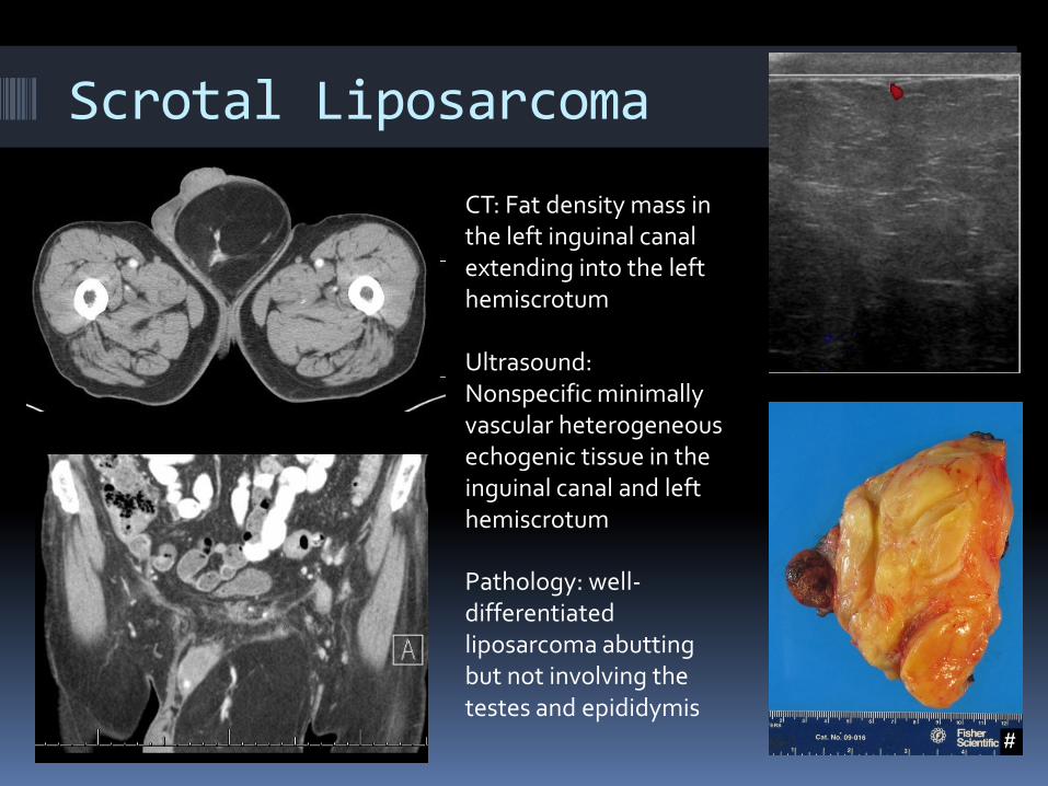
CT: Fat density mass in the left inguinal canal extending into the left hemiscrotum Ultrasound: Nonspecific minimally vascular heterogeneous echogenic tissue in the inguinal canal and left hemiscrotum
Scrotal Liposarcoma
Pathology: well-differentiated liposarcoma abutting but not involving the testes and epididymis

Mimics/Pitfalls
Testicular Paratesticular
Infarct
Rete testis cyst
Hematoma
Abscess
Paratesticular cystic lesions can rarely mimic solid tumors Spermatocele
Complicated epididymal cyst
Tubular ectasia of rete testis
Tunica albuginea cyst
Hematocele
Pyocele
Complicated hydrocele

Heterogeneously hypoechoic solid and cystic lesion of the testis without definite blood flow to the solid component Pathology: Small circumscribed infarct without evidence of malignancy
Testicular Tumor Mimic: Subacute Testicular Infarct

Imaging: Several small cystic lesions in the periphery of the testis, consistent with cystic dilation of the rete testis
Testicular Tumor Mimic: Cystic Dilation of Rete Testis

Testicular Tumor Mimic: Testicular Hematoma
Imaging: Avascular , heterogeneous parenchymal echogenicity of testis in a patient with history of trauma
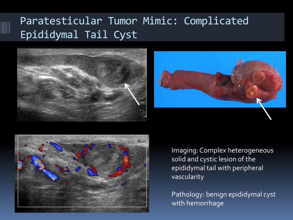
Imaging: Complex heterogeneous solid and cystic lesion of the epididymal tail with peripheral vascularity Pathology: benign epididymal cyst with hemorrhage
Paratesticular Tumor Mimic: Complicated Epididymal Tail Cyst
References Siegel R, Ward E, Brawley O, Jemal A. “Cancer statistics, 2011: The impact of eliminating socioeconomic and racial
disparities on premature cancer deaths.” A Cancer Journal for Clinicians, 2011; 61:212.
Woodward PJ, Sohaey R, O’Donoghue MJ, Green DE. From the archived of the AFIP: Tumors and tumorlike lesions of the testis: Radiologic-pathologic correlation. Radiographics 2002; 22:189-216.
Rypens F, Garel L, Franc-Guimond J, et al. Paratesticular rhabdomyosarcoma presenting as thickening of the tunica vaginalis. Pediatr Radiol 2009;39:1010–12
Hashimoto H, Enjoii M. Liposarcoma: a clinicopathologic subtyping of 52 cases. Acta Pathol Jpn 1982;32:933
Sogani PC, Grabstald H, Withmore WJ. Spermatic cord sarcoma in adults. J Urol 1978;120:301.
Patel NG, Rajagopalan A, Shrotri NS. Scrotal liposarcoma – a rare extratesticular tumour. JRSM Short Rep. Dec 2011; 2(12): 93. http://www.ncbi.nlm.nih.gov/pmc/articles/PMC3265830/
Rypens F, Garel L, Franc-Guimond J, et al. Paratesticular rhabdomyosarcoma presenting as thickening of the tunica vaginalis. Pediatr Radiol 2009;39:1010–12
Hashimoto H, Enjoii M. Liposarcoma: a clinicopathologic subtyping of 52 cases. Acta Pathol Jpn 1982;32:933
Sogani PC, Grabstald .H, Withmore WJ. Spermatic cord sarcoma in adults. J Urol 1978;120:301.
Statdx https://my.statdx.com/
Radiopaedia http://radiopaedia.org/