a prospective, comparative study of effect of …
TRANSCRIPT

1
A PROSPECTIVE, COMPARATIVE STUDY OF EFFECT OF
ROFLUMILAST IN CHRONIC OBSTRUCTIVE PULMONARY DISEASE
AND ITS EFFICACY IN REDUCING ACUTE EXACERBATIONS.
Dissertation submitted to
THE TAMIL NADU DR. MGR MEDICAL UNIVERSITY
CHENNAI
In partial fulfilment of the regulations for the award of the degree of
M.D. PHARMACOLOGY
Branch VI
GOVT. KILPAUK MEDICAL COLLEGE AND HOSPITAL
CHENNAI – 10
MAY 2018
2
CERTIFICATE
This is to certify that this dissertation titled “A PROSPECTIVE, COMPARATIVE
STUDY OF EFFECT OF ROFLUMILAST IN CHRONIC OBSTRUCTIVE
PULMONARY DISEASE AND ITS EFFICACY IN REDUCING
EXACERBATIONS” is the bonafide original work done by Dr.D.Thamizh Vani.,
Post graduate in Pharmacology, under my overall supervision in the Department of
Pharmacology, Govt. Kilpauk Medical College and Hospital, Chennai, in partial
fulfilment of the regulations of The Tamil Nadu Dr.M.G.R. Medical University for the
award of M.D Degree in Pharmacology (Branch VI).
Dr.RAMACHANDRA BHAT,M.D., Dr.VASANTHAMANI,M.D.,D.G.O
Professor & HOD The Dean
Department of Pharmacology Govt. Kilpauk Medical College and
Govt. Kilpauk Medical College and Hospital
Hospital Chennai – 10.
Chennai – 10.

3
CERTIFICATE
This is to certify that this dissertation titled “A PROSPECTIVE, COMPARATIVE
STUDY OF EFFECT OF ROFLUMILAST IN CHRONIC OBSTRUCTIVE
LUNG DISEASE AND ITS EFFICACY IN REDUCING EXACERBATIONS” is
the bonafide original work done by Dr.D.Thamizh Vani., Post graduate in
Pharmacology, under my overall supervision and guidance in the Department of
Pharmacology, Govt. Kilpauk Medical College and Hospital, Chennai, in partial
fulfilment of the regulations of The Tamil Nadu Dr.M.G.R. Medical University for the
award of M.D Degree in Pharmacology (Branch VI).
Dr.MALAR SIVARAMAN, M.D.
Professor
Department of Pharmacology
Govt. Kilpauk Medical College and Hospital
Chennai – 600010.

4
DECLARATION
I solemnly declare that this dissertation titled “A PROSPECTIVE,
COMPARATIVE STUDY OF EFFECT OF ROFLUMILAST IN CHRONIC
OBSTRUCTIVE LUNG DISEASE AND ITS EFFICACY IN REDUCING
EXACERBATIONS”, is the bonafide work done by me at the Department of
Pharmacology, Govt. Kilpauk Medical College and Hospital, Chennai, under the
supervision of Dr. RAMACHANDRA BHAT, M.D., Professor and HOD of
Pharmacology, and guidance of DR.MALAR SIVARAMAN, M.D., Professor,
Department of Pharmacology and DR.NALINI JAYANTHI, M.D., Superintendant,
Department of Thoracic Medicine, Govt. Thiruvoteeswarar TB and Chest Hospital,
Chennai. This dissertation is submitted to The Tamil Nadu Dr.M.G.R. Medical
University, Chennai in partial fulfilment of the University regulations for the award of
Degree of M.D.Pharmacology (Branch VI) examinations to be held in May 2018.
Place : Chennai
Date :
Dr.D.Thamizh Vani

5
ACKNOWLEDGEMENT
I would like to express my humble gratitude to Dr.Vasanthamani,M.D., D.G.O, Dean,
Government Kilpauk Medical College and Hospital for giving me permission to carry
out my dissertation work.
I would like to express my sincere gratitude to Dr.RAMACHANDRA BHAT, M.D.,
Professor and HOD, Department of Pharmacology, Govt. Kilpauk Medical College
and Hospital, for introducing me to the world of medical research and riveting in me a
strong foundation in ethics in medical research.
I am deeply grateful for the efficient support and guidance of Dr.MALAR
SIVARAMAN, M.D., Professor, Department of Pharmacology, Govt. Kilpauk
Medical College and Hospital, for her continued guidance, commitment, and
dedication during the entire course of this endeavour.
I am also grateful to Dr.NALINI JAYANTHI, M.D., Superintendent, Govt.
Thiruvoteeswarar TB and Chest Hospital, Otteri, Chennai, for her enthusiasm and
willingness to co guide this dissertation.
I extend my heartfelt gratitude to Dr.ARUNA.T, M.D., Professor, Department of
Pharmacology, Govt. Kilpauk Medical College and Hospital, who provided insightful
inputs into the study and kept me focussed throughout the study period.
I also thank Dr.Jeyaponmari, M.D, Dr.Sasikala, M.D, Dr.Rajesh Kumar, M.D,
Dr.Keerthana Brattiya M.D, Assistant Professors, Department of Pharmacology, Govt.

6
Kilpauk Medical College and Hospital, and my fellow post graduates for their help
and their valuable support.
This acknowledgement would be incomplete if I did not thank my family for their
blessings and good wishes.

7
TABLE OF CONTENTS
S No. Contents PAGE No.
1 INTRODUCTION 10
2 REVIEW OF LITERATURE 14
3 AIM AND OBJECTIVES 52
4 MATERIALS AND METHODS 53
5 RESULTS 63
6 DISCUSSION 78
7 CONCLUSION 82
8 BIBLIOGRAPHY 83
9
ANNEXURES
Institute Ethics Committee Clearance
certificate
Case report form
Patient Information sheet
Consent form
Plagiarism Assessment Report
91
92
94
95
96

8
LIST OF ABBREVIATIONS
COPD - Chronic Obstructive Pulmonary Disease
DALY - Daily affected life years
TGF- - Transforming growth factor beta
1AT - Alpha one Antitrypsin
MMP - Matrix metalloproteinase
IL - Interleukin
TNF - - Tumour necrosis factor alpha
CD-8 - Cluster of differentiation 8
IP -10 - Inducible protein-10
mTOR - mammalian target of rapamycin
Ig - Immunoglobulin
FEV1 - Forced Expiratory Volume in one second
FVC - Forced vital capacity
PEF - Peak expiratory flow rate
PI - Protease inhibitor
SNP - Single nucleotide polymorphism
GOLD - Global initiative of lung disease
pCO2 - partial pressure of carbon dioxide
PDE4 - Phosphodiesterase 4
IV - Intravenous
CT - Computed tomography
LVRS - Lung Volume Reduction Surgery
DLCO - Diffusion Capacity of lung for Carbon monoxide
NIPPV - Non invasive positive pressure ventilation

9
PEEP - Positive end expiratory pressure
cAMP - cyclic Adenosine monophosphate
IAD - Internal airflow distribution
CYP - Cytochrome P
PFT - Pulmonary function test
BD - Bronchodilator
HHIP - Hedgehog interacting protein

10
INTRODUCTION

11
Introduction
Chronic obstructive pulmonary disease (COPD) is a type of obstructive lung disease
which is characterised by poor air flow for a long term. It lasts for years and can be
present lifelong. The disease makes it hard for the person to breathe. It is progressive
in nature i.e. gets worse over time [1]. COPD affects about 329 million people every
year which is nearly 5% of the global population [27,28]. Prevalence of COPD in
India accounts to about 30 million people [2]. It occurs in people above 40 years old
i.e. it is diagnosed in middle aged or older adults. Both males and females are
commonly affected. It is one of the major causes of disability in the world [3]. COPD
is the cause of about 2.9 million deaths every year and this number is progressing
every year [2]. It forms the third leading cause of death in the world. Low and middle
income countries contribute to the burden of deaths due to COPD. In India, mortality
due to COPD occurs in 102.3/100,000. In the world COPD contributes to 6740,000
DALYs out of 27,756,000 [2]. The disease thus significantly affects health related
Quality of life in the world [3].
Smoking is the most common cause and risk factor which lead to development of
COPD [4]. Likelihood of developing COPD increases with the overall exposure of
smoke. Bidi smokers were at higher risk of developing COPD than those who smoked
cigarettes [4]. Other types of smoke like marijuana, cigar, water pipe smoke are also
risk factors. Cooking fuel, kerosene, biomass fuel, firewood also contributed to the
development of disease. Poorly ventilated cooking fires leads to indoor air pollution
and is the common cause of disease in developing countries. Second hand smoke is

12
the cause of COPD in about 20% of cases [5]. Second hand smoke is also called
environmental tobacco smoke. It is a combination of two forms of smoke that is
formed due to burning of tobacco – the smoke exhaled by a smoker and the smoke
from lighted end of a cigarette, cigar, pipe or tobacco. Intense and prolonged exposure
to fumes, dust, chemicals in workplace also increase risk of COPD in smokers and
non smokers [5]. During pregnancy, if women smoke, may increase the risk of COPD
in the child. Exposure to these irritants for a long time causes an inflammatory
response in the lungs which results in narrowing of airways and in breakdown of lung
tissue. People who live in large cities have a higher rate of developing COPD as
compared to people living in rural areas [7]. Genetic factor plays a small role in
development of COPD. Alpha 1- antitrypsin deficiency is the only clearly inherited
risk factor. This contributes to about 1 – 5% of cases [6].
Acute exacerbation of COPD is defined as increased shortness of breath, cough, and
increased production of sputum in a patient diagnosed with COPD. There is sudden
worsening of symptoms [8]. It is triggered by infection, environmental pollutants, and
cold temperature. Those with severe disease have more frequent exacerbations and
lung function deteriorates at a faster rate [9].
Diagnosis of COPD is done using Spirometer in persons presenting with the clinical
symptoms. Spirometry determines the severity of airflow limitation [12,13].
The current treatment modalities available for COPD are inhaled bronchodilators
which is the primary medication. The two major types are beta2 agonists and
anticholinergics. Both long acting and short acting forms are available [15]. In mild

13
disease, short acting form is recommended whereas long acting form is used in severe
disease and in maintenance therapy [16]. They reduce shortness of breath and exercise
limitation and result in an improved quality of life. If these drugs are ineffective, then
corticosteroids are added [17]. Methylxanthines are used as a second line agent if not
controlled by other measures [18]. Supplemental oxygen is recommended in patients
with low oxygen level at rest. Medications are given with a metered dose inhaler with
a spacer or via a nebuliser [19]. Reducing risk factors like stopping smoking is a must.
Pulmonary rehabilitation which is a program of exercise, disease management and
counseling, may improve quality of life [20]. Though these measures may reduce the
duration of symptoms, improve exercise capacity, reduce risk of exacerbation, they do
not change the progression of underlying disease and do not reduce the rate of hospital
admissions [21].
The prognosis of persons affected with COPD is bad as the disease gets worse over
time and can lead to death [10]. The number of years living with disability due to
COPD is increasing in the world. It can also lead to many comorbid conditions such
as cor pulmonale and end stage lung disease leading to respiratory failure [22,23].
Other complications of the disease include pneumonia, polycythemia and
pneumothorax [40,41]. The effects of COPD extend beyond the lungs. Multiple
comorbidities may occur with COPD which includes cardiovascular disease, diabetes
mellitus, osteoporosis, depression, and pneumonia [80]. Increased use of medications
and hospitalisation is needed in acute exacerbation. Also in COPD, airflow reduction
does not improve significantly with bronchodilators, in contrast to asthma [11,14].

14
Therefore, there is a need for new drug to decrease disease progression, reduce
exacerbations and to improve the quality of life in patients with COPD.
Drug Roflumilast is selected in this study for COPD and to reduce acute exacerbations
for the following reasons. Roflumilast is a selective, long acting inhibitor of
Phosphodiesterase-4 (PDE-4) which leads to accumulation of cAMP (cyclic adenosine
monophosphate) [24]. It has anti inflammatory property and has gained approval for
use in severe COPD for preventing exacerbations. It works by decreasing swelling in
the lungs and reducing irritation [25]. Also due to its property of changing the internal
airflow distribution, it improves efficacy of Steroids and B2 agonists as well [26,27].
Therefore, in this study comparison of effect of standard treatment of COPD with
Roflumilast as add on therapy to standard is done.

15
REVIEW OF
LITERATURE

16
REVIEW OF LITERATURE
In the past, COPD was referred to as “chronic airflow obstruction” and “chronic
obstructive lung disease.” Dr. William Briscoe is thought to be the first person to use
the term COPD at the 9thAspen Emphysema Conference in 1965. It was also during
the 1960's when the term FEV1 was first used to measure expiratory flow. The history
of COPD started a long time ago. In 1821, René Laënnec, the doctor who invented the
stethoscope, discovered emphysema as a part of COPD. Because smoking during the
early 1800s was not common, Laënnec identified environmental and genetic factors as
the primary causes of COPD. While Laënnec is correct in identifying environmental
and genetic factors as causes of COPD, it is well-known today that smoking is one of
the leading causes of COPD [86].
Years later in 1846, John Hutchinson invented the spirometer, and Robert Tiffeneau, a
respiratory medicine pioneer, built on Hutchinson’s invention about 100 years later.
Tiffeneau created a complete diagnostic instrument for COPD, and the spirometer,
which measure vital lung capacity, is still an essential device in diagnosing COPD
today [87].
Chronic obstructive pulmonary disease (COPD) is defined as a state of disease which
is characterized by airflow limitation that is not reversed fully. COPD includes
emphysema and chronic bronchitis. Emphysema is an anatomically defined condition
characterized by destruction and enlargement of the lung alveoli while chronic
bronchitis is a clinical condition with chronic cough and sputum production [29]. It

17
also comprises of small airways disease, where the small bronchioles are narrowed.
COPD is said to be present only if chronic airflow obstruction occurs. Chronic
bronchitis without chronic airflow obstruction is not included within COPD. COPD
affects >10 million people and is the third leading cause of death in the United States
[30].
PATHOGENESIS
The major physiologic change in COPD is airflow limitation which can result from
both small airway obstruction and emphysema. Small airways may become narrow
due to hyperplasia of cells and accumulation of mucus. This then leads to fibrosis
formation. Airway fibrosis occurs due to activation of transforming growth factor
(TGF- ) whereas parenchymal inflammation and emphysema is due to lack of TGF-
[12]. Four interrelated events contribute to the dominant paradigm of the
pathogenesis of emphysema. They are: (1) Chronic exposure to cigarette smoke leads
to inflammatory and immune cell recruitment within the terminal air spaces of the
lung. (2) Elastolytic and other proteinases are released by inflammatory cells. This
leads to damage of the extracellular matrix of the lung. (3) Structural cell death of
endothelial and epithelial cells occurs directly through oxidant-induced cigarette
smoke damage and senescence as well as indirectly through proteolytic loss of matrix
attachment. (4) Air space enlargement occurs due to ineffective repair of elastin and
other extracellular matrix components which results in pulmonary emphysema [34].

18
THE ELASTASE: ANTIELASTASE HYPOTHESIS
Elastin is the principal and highly stable component of elastic fibers which makes up
the extracellular matrix. This is critical to the integrity of the lung. The
elastase:antielastase hypothesis was proposed in the mid1960s [33,34]. This
hypothesis states that elastin-degrading enzymes along with their inhibitors determine
the susceptibility of the lung to destruction. This results in air space enlargement. This
hypothesis was based on the clinical observation that patients with genetic deficiency
in 1 antitrypsin ( 1AT) which is the inhibitor of serine proteinase neutrophil elastase,
were at increased risk of emphysema. It was also found that instillation of elastases,
which included neutrophil elastase, into animals for experimental purpose results in
emphysema [30]. The elastase:antielastase hypothesis remains a prevailing
mechanism for the development of emphysema [33,34].
Fig 1. Pathogenesis of emphysema. Upon long-term exposure to cigarette smoke, inflammatorycells are recruited to the lung; they release proteinases in excess, this leads to air spacedestruction and enlargement

19
INFLAMMATION AND EXTRACELLULAR MATRIX PROTEOLYSIS
Upon exposure to oxidants from cigarette smoke, macrophages and epithelial cells are
activated, and proteinases and chemokines are produced. This attracts other
inflammatory and immune cells [33]. One mechanism of macrophage activation is
oxidant-induced inactivation of histone deacetylase-2 is, which shifts the balance
towards acetylated or loose chromatin, thereby leading to exposure of nuclear factor-
B sites. This results in transcription of matrix metalloproteinases, proinflammatory
cytokines such as interleukin 8 (IL-8), and tumor necrosis factor (TNF- ) and leads
to neutrophil recruitment. Due to cigarette smoke CD8+ T cells are also recruited.
They release interferon-inducible protein-10 (IP-10, CXCL-7). This in turn leads to
macrophage production of elastase which is matrix metalloproteinase-12 (MMP-12).
Matrix metalloproteinases and serine proteinases, most importantly neutrophil
elastase, function together. Their work is to degrade the inhibitor of the other, leading
to lung destruction. Proteolytic cleavage products of elastin also function as a
macrophage chemokine, increasing the destructive positive feedback loop [33,34].
Autoimmune mechanisms may be involved in promoting the progression of disease.
In patients, particularly those with advanced disease, B cells and lymphoid follicles
are present. It has been detected that IgG autoantibodies with avidity for pulmonary
epithelium have the potential to mediate cytotoxicity. Macrophage phagocytosis and
loss of cilia induced by cigarette smoke in the airway epithelium, predispose to
bacterial infection and neutrophilia. There remains an exuberant inflammatory
response, long after smoking cessation, in end-stage lung disease. This suggests that
mechanisms of cigarette smoke–induced inflammation that initiate the disease differ
20
from mechanisms that sustain inflammation after smoking cessation. Cigarette smoke
oxidant-mediated structural cell death occurs via a variety of mechanisms [35]. One
of them is rt801 inhibition of mammalian target of rapamycin (mTOR), which leads to
cell death as well as inflammation and proteolysis. Uptake of apoptotic cells by
macrophages results in production of growth factors and dampens inflammation. This
causes promotion of lung repair. This uptake of apoptotic cells by macrophages is
impaired by cigarette smoking, thereby limiting lung repair. The ability of the adult
lung to repair damaged alveoli appears limited. The process of septation that is
responsible for alveologenesis during lung development is very unlikely to be
reinitiated [36,37].
PATHOLOGY
Cigarette smoke exposure may affect the large airways, small airways i.e airways
which are 2 mm in diameter, and alveoli. Changes which occur in large airways are
the reason for cough and production of sputum, while changes in small airways and
alveoli are found responsible for physiologic alterations. Both emphysema and small
airway pathology are present in most persons with COPD. However, they do not
appear to be related to each other, and their relative contributions to obstruction seem
to vary from one person to another [38,39].
21
LARGE AIRWAY
Cigarette smoking often results in mucus gland enlargement and goblet cell
hyperplasia, leading to cough and mucus production. These symptoms define chronic
bronchitis, though these abnormalities are not related to airflow limitation. Goblet
cells increase in number and in extent throughout the bronchial tree. Bronchi also
undergo squamous metaplasia. This predisposes to carcinogenesis and disrupts
mucociliary clearance. Patients may have smooth-muscle hypertrophy and bronchial
hyperreactivity, but they are not as prominent as seen in asthma. This is the ultimate
cause leading to airflow limitation. Purulent sputum of upper respiratory tract
infections has been associated with neutrophil influx [42].
SMALL AIRWAYS
In COPD, the major site of increased resistance is the airways which are 2 mm
diameter. Characteristic cellular changes in them include goblet cell metaplasia, and
surfactant-secreting Clara cells (club cells or bronchiolar exocrine cells) replaced by
mucus-secreting cells. Smooth-muscle hypertrophy may also be seen. These
abnormalities may lead to luminal narrowing by fibrosis, excess mucus, edema, and
cellular infiltration. This leads to reduced surfactant production and may increase
surface tension at the air-tissue interface. This predisposes to airway narrowing or
collapse. Respiratory bronchiolitis with mononuclear inflammatory cells collecting in
distal airway tissues may lead to proteolytic destruction of elastic fibers in the
respiratory bronchioles and alveolar ducts. Narrowing and drop-out of small airways
precede the onset of emphysematous destruction [14].
22
LUNG PARENCHYMA
Emphysema is characterized by destruction of air spaces, where gas exchange occurs
i.e., the respiratory bronchioles, alveolar ducts, and alveoli. Their walls become
perforated and obliterated later with small distinct air spaces coalescing into abnormal
and much larger air spaces. Accumulation of macrophages occurs in respiratory
bronchioles of essentially all young smokers. Bronchoalveolar lavage fluid which is
taken from individuals who smoke contains roughly five times as many macrophages
as compared to lavage from nonsmokers. In smokers’ lavage fluid, macrophages
comprise >95% of the total cell count, and neutrophils, account for 1–2% of the cells.
These are nearly absent in nonsmokers’ lavage. T lymphocytes, particularly CD8+
cells, are also increased in the alveolar space of smokers. Emphysema is classified
into distinct pathologic types, of which the most important ones are centriacinar and
panacinar [33]. The type most frequently associated with cigarette smoking is
Centriacinar emphysema. Centriacinar type is characterized by enlarged air spaces. It
is often focally seen and is usually most prominent in the upper lobes and superior
segments of lower lobes. Panacinar emphysema is characterized by abnormal large
air spaces which are evenly distributed within and across acinar units. Panacinar type
of emphysema is usually observed in patients with 1AT deficiency, which has a
predilection for the lower lobes [79].
PATHOPHYSIOLOGY
The most typical finding in COPD is persistent reduction in forced expiratory flow
rates. Increases in the residual volume and the residual volume/total lung capacity

23
ratio, nonuniform distribution of ventilation, and ventilation-perfusion mismatching
are also seen [37].
AIRFLOW OBSTRUCTION
Airflow limitation is nothing but obstruction to airflow, is determined by spirometry
[6]. In spirometry forced expiratory maneuvers are involved. This is assessed after the
subject has inhaled to total lung capacity. Key parameters which are obtained from
spirometry include the volume of air exhaled within the first second of the forced
expiratory maneuver, called (FEV1) and the total volume of air exhaled during the
entire spirometric maneuver which is forced vital capacity [FVC]. Patients with
obstruction of airflow which is related to COPD have a chronically reduced ratio of
FEV1/FVC. In contrast to asthma, the reduced FEV1 in COPD seldom shows large
responses to inhaled bronchodilators, although improvements up to 15% are common.
Airflow during forced exhalation is the result of the balance between the promoted
flow which is due to elastic recoil of the lungs and the limited flow which is due to
resistance of the airways. In lungs affected by COPD, maximal expiratory flow
diminishes as the lungs empty because progressively less elastic recoil is provided by
lung parenchyma. The cross-sectional area of the airways falls, which raises the
resistance to airflow. The abnormality in airflow is only evident at lung volumes at or
below the functional residual capacity which is closer to residual volume in the early
stages of COPD.
24
HYPERINFLATION
Lung volumes are routinely assessed in pulmonary function testing. In COPD “air
trapping” is seen very often [43,44]. There is increased residual volume and increased
ratio of residual volume to total lung capacity. In the late stages of COPD, during tidal
breathing hyperinflation of the thorax occurs. This preserves the maximum expiratory
airflow, because as lung volume increases, elastic recoil pressure increases, and
airways enlarge so that airway resistance decreases. Despite compensating for airway
obstruction, hyperinflation can push the diaphragm into a flattened position. This
causes a number of adverse effects. Positive abdominal pressure during inspiration is
not applied as effectively to the chest wall due to decrease in zone of apposition
between the diaphragm and the abdominal wall. This hinders rib cage movement and
impairs inspiration. Also, the muscle fibers of the flattened diaphragm are shorter than
those of a more normally curved diaphragm, so they are less capable of generating
inspiratory pressures than normal. The flattened diaphragm also leads to increased
radius of curvature(r). Therefore diaphragm must generate greater tension (t) to
develop the transpulmonary pressure (p) which is required to produce tidal breathing.
This follows from Laplace’s law, p = 2t/r. Also, due to distension of the thoracic cage
beyond its normal resting volume, during tidal breathing the inspiratory muscles must
do work to overcome the resistance of the thoracic cage to further inflation [33,34].

25
GAS EXCHANGE
The partial pressure of oxygen in arterial blood Pao2 usually remains near normal
until the FEV1 is decreased to ~50% of predicted. At rest even much lower FEV1
values can be associated with a normal Pao2. An elevation of arterial level of carbon
dioxide (Paco2) is not expected until the FEV1 is <25% of predicted. Pulmonary
hypertension severe enough to cause cor pulmonale and right ventricular failure due to
COPD typically occurs in individuals who have marked decreases in FEV1 (<25% of
predicted) and chronic hypoxemia (Pao2 <55 mmHg) [46]. Nonuniform ventilation
and ventilation-perfusion mismatching are characteristic of COPD. Physiologic
studies are consistent with the finding that multiple parenchymal compartments have
different rates of ventilation due to regional differences in compliance and airway
resistance [47,48]. Reduction in Pao2 that occurs in COPD is accounted by
ventilation-perfusion mismatch. Therefore, the effectiveness of inspired oxygen in
treating hypoxemia due to COPD can be explained.
RISK FACTORS
CIGARETTE SMOKING
By 1964, the Advisory Committee to the Surgeon General of the United States had
concluded that cigarette smoking was the major risk factor for COPD and also the
reason for mortality [11,14]. Subsequent longitudinal studies have shown accelerated
decline in FEV1 in a dose-response relationship to the intensity of cigarette smoking
[49]. It is typically expressed as pack-years which is defined as average number of
26
packs of cigarettes smoked per day multiplied by the total number of years of smoking
[1]. Higher prevalence rates of COPD with increasing age is accounted by the dose-
response relationship between reduced pulmonary function and cigarette smoking
intensity. Higher prevalence of COPD seen among males is due to the higher rate of
smoking among males. However, as the gender gap in smoking rates has diminished
in the past 50 years the prevalence of COPD among females is increasing. Although
pack-years of cigarette smoking is the most significant predictor of FEV1, only 15%
of the variability in FEV1 is explained by pack-years [50]. This finding suggests that
there are additional environmental and/or genetic factors, which contribute to the
impact of smoking on the development of airflow obstruction.
AIRWAY RESPONSIVENESS AND COPD
One of the defining features of asthma is the tendency for increased
bronchoconstriction in response to a variety of exogenous stimuli, which includes
methacholine and histamine. However, this feature of airway hyperresponsiveness is
also shared by many patients with COPD. There is considerable overlap between
persons with asthma and those with COPD in airway responsiveness, airflow
obstruction, and pulmonary symptoms. Therefore, this has led to the formulation of
the Dutch hypothesis [34]. The hypothesis suggests that asthma, chronic bronchitis,
and emphysema are variations of the same basic disease, which is modulated by
environmental and genetic factors. Asthma is viewed as largely an allergic
phenomenon, whereas COPD results from smoking-related inflammation and damage.

27
Also, the interactions between these postulated genetic factors and environmental risk
factors must be taken into account. Longitudinal studies that compared airway
responsiveness at the beginning of the study to subsequent decline in pulmonary
function have demonstrated that increased airway responsiveness is clearly a
significant predictor of subsequent decline in pulmonary function [34].
RESPIRATORY INFECTIONS
The decline in pulmonary function due to the impact of adult respiratory tract
infections, are not well defined. Significant reductions in pulmonary functions are not
typically seen following an episode of bronchitis or pneumonia [69]. Due to a lack of
adequate longitudinal data, the impact of the effects of childhood respiratory illnesses
on the subsequent development of COPD has been difficult to assess. Respiratory
infections are important causes of exacerbations of COPD. Though this association is
present, it is yet to be proven.
OCCUPATIONAL EXPOSURES
Exposure to dust and fumes at work has resulted in increased respiratory symptoms
and airflow obstruction. Specific occupational exposures, such as coal mining, gold
mining, and cotton textile dust, have also been suggested as risk factors for chronic
airflow obstruction. Among coal miners, coal mine dust exposure was a significant
risk factor for emphysema in both smokers and non-smokers [71]. Compared to the

28
effect of cigarette smoking, the magnitude of risk of COPD due to occupational
exposures is substantially less important [72].
AMBIENT AIR POLLUTION
Due to increased pollution in the urban settings increased respiratory symptoms have
been reported in those living in urban areas [41]. The relationship of air pollution to
chronic airflow obstruction disease still remains to be proved. Prolonged exposure to
smoke produced by biomass combustion which is a common mode of cooking in
some countries, also appears to be a significant risk factor for COPD among women in
those countries [45].
PASSIVE, OR SECOND-HAND, SMOKING EXPOSURE
Exposure of children to maternal smoking results in significantly reduced lung
growth. In utero, tobacco smoke exposure also contributes to significant reductions in
postnatal pulmonary function [52,53]. Although passive smoke exposure has been
associated with reductions in pulmonary function, the importance of this risk factor in
the development of the severe pulmonary function reductions in COPD remains
uncertain.

29
GENETIC CONSIDERATIONS
A proven genetic risk factor for COPD is severe 1AT deficiency [6]. Increasing
evidence of other genetic determinants also exist.
1ANTITRYPSIN DEFICIENCY
Many variants of the locus of protease inhibitor (PI or SERPINA1) which encodes
1AT have been described [11]. The common allele that is associated with normal
1AT levels is M allele. The S allele is associated with slightly reduced 1AT levels,
and the Z allele is associated with markedly reduced 1AT levels. The S allele and Z
allele also occur with frequencies of >1% in most white populations. Inheritation of
null allele, is seen in rare individuals which lead to the absence of any 1AT
production. This occurs through a heterogeneous collection of mutations. Individuals
with two Z alleles or one Z and one null allele are referred to as PiZ. This is the most
common form of severe 1AT deficiency. Approximately only 1% of COPD patients
are found to have severe 1AT deficiency as a contributing cause of COPD. These
patients demonstrate that genetic factors can have a profound influence on the
susceptibility for developing COPD. PiZ individuals often develop early-onset COPD.
Approximately 1 in 3000 individuals in the United States inherits severe 1AT
deficiency, but only a small minority of these individuals has been identified [6]. The
clinical laboratory test used most frequently to screen for 1AT deficiency is
measurement of the immunologic level of 1AT in serum. Cigarette smokers with

30
severe 1AT deficiency are more likely to develop COPD at early ages. Other factors
which appear to increase the risk of COPD in PiZ subjects are asthma and male
subjects. Specific treatment in the form of 1AT augmentation therapy is available for
severe 1AT deficiency as a weekly IV infusion. Recent studies have suggested that
PiMZ subjects are also at slightly increased risk for the development of airflow
obstruction [7,12]. It still remains unclear whether all PiMZ subjects are at slightly
increased risk for COPD or if only a subset of PiMZ subjects are at an increased risk
for COPD due to other genetic or environmental factors. Studies of pulmonary
function measurements performed have suggested that genetic factors other than PI
type also have influence in variation of pulmonary function. A well-powered
association study comprising 8300 patients and 7 separate cohorts found that a minor
allele single nucleotide polymorphism (SNP) of MMP12 (rs2276109) associated with
decreased MMP12 expression has a positive effect on lung function in children with
asthma and in adult smokers [83]. Recent genome-wide association studies have
identified several COPD susceptibility loci, including a region near the hedgehog
interacting protein (HHIP) gene on chromosome 4, a cluster of genes on chromosome
15 (including components of the nicotinic acetylcholine receptor), and a region within
a gene of unknown function (FAM13A) [34]. A regulatory SNP upstream from the
HHIP gene has been identified as one potential functional variant; the specific genetic
determinants in the other genomic regions are yet to be definitely identified.
31
NATURAL HISTORY
COPD due to cigarette smoking depends on the intensity of smoking exposure, the
timing of smoking exposure, and the baseline lung function of the individual. Most
individuals follow a steady increase in pulmonary function during childhood and
adolescence which is followed by a gradual decline with aging. The risk of eventual
mortality due to COPD is associated with declined levels of FEV1 [55].
The rate of decline in pulmonary function can be modified by changing environmental
exposures (i.e., quitting smoking). Smoking cessation at an earlier age provided a
more beneficial effect than smoking cessation after marked reductions in pulmonary
function have already developed. Genetic factors contribute to the level of pulmonary
function achieved during growth and to the rate of decline in response to smoking and
potentially to other environmental factors as well.
CLINICAL PRESENTATION
HISTORY
COPD most commonly presents as cough, sputum production, and exertional dyspnea.
Many patients have such symptoms for months or years before seeking medical
attention [57]. Onset of the disease is attributed to an acute illness or exacerbation by
many patients though the development of airflow obstruction is gradual. Symptoms
are always present prior to the acute exacerbation. A careful history elicits it. The
development of exertional dyspnea, which is described as an increased effort to
32
breathe, air hunger, or gasping, and a feeling of heaviness, can be insidious. History
should be focused on typical physical activities and how the patient’s ability to
perform them has changed. Activities which involve significant arm work, particularly
at the level of shoulder or above it, are particularly difficult for patients with COPD.
Activities that allow the patient to use accessory muscles of respiration and to brace
arms are better tolerated. Examples of such activities include pushing a shopping cart
or walking on a treadmill. The principal feature as COPD advances is worsening of
dyspnea on exertion. This is accompanied by an increasing intrusion on the ability of
the individual to perform vocational or avocational activities. Patients are breathless in
the most advanced stages. Therefore, they can only perform simple activities of daily
living. Worsening airflow obstruction is accompanied by an increased frequency of
exacerbations. Resting hypoxemia is developed in many patients and they require
institution of supplemental oxygen.
PHYSICAL FINDINGS
Patients usually have an entirely normal physical examination in the early stages of
COPD. Signs of active smoking may be seen in current smokers. This includes an
odor of smoke or nicotine staining of fingernails. In patients with more severe disease,
the finding obtained by physical examination is, a prolonged expiratory phase and
expiratory wheezing. Signs of hyperinflation is also seen which include a barrel chest
and enlarged lung volumes with poor diaphragmatic excursion as assessed by
percussion. Use of accessory muscles of respiration is exhibited by patients with

33
severe airflow obstruction. Patients sit in the characteristic “tripod” position, as the
actions of sternocleidomastoid, scalene, and intercostal muscles are facilitated.
Patients may develop cyanosis which is visible in the lips and nail beds. Patients with
predominant emphysema are termed “pink puffers”. These patients are thin and
noncyanotic at rest. They have prominent use of accessory muscles. Patients with
chronic bronchitis are very likely to be heavy and cyanotic and they are termed “blue
bloaters” [33,34]. Current evidence demonstrates that most patients have elements of
both bronchitis and emphysema and that the two entities cannot be differentiated by
physical examination. Advanced disease may be accompanied by cachexia, with
significant weight loss, bitemporal wasting, and diffuse loss of subcutaneous adipose
tissue. These signs are associated with both inadequate oral intake due to disease and
elevated levels of inflammatory cytokines such as TNF- . If wasting is seen in COPD,
it is considered as a poor prognostic factor. In some patients with advanced disease,
Hoover’s sign is seen in which there is paradoxical inward movement of the rib cage
with inspiration instead of outward as is normal. This implies a flat, but functioning
diaphragm. This is the result of alteration of the vector of diaphragmatic contraction
on the rib cage due to chronic hyperinflation [48]. An overt complication of COPD is
cor pulmonale which shows signs of right heart failure. It is now relatively infrequent
due to the advent of supplemental oxygen therapy. Clubbing of the digits is not a sign
of COPD. Presence of clubbing should alert the clinician to initiate an investigation
for causes of clubbing.
A substantial proportion of COPD patients have extra-pulmonary symptoms and
signs. Common manifestations include skeletal muscle weakness, osteoporosis,

34
cardiac arrhythmias, weakness, ischemic heart disease, stroke, depression, and cancer.
The presence of these extra-pulmonary pulmonary manifestations of COPD increases
morbidity and mortality. Peripheral skeletal muscle dysfunction is an established
systemic feature of COPD [52].
LABORATORY FINDINGS
The hallmark of COPD is airflow obstruction. Pulmonary function testing shows a
reduction in FEV1 and FEV1/FVC with airflow obstruction. With worsening disease
severity, lung volumes may increase, resulting in an increase in total lung capacity,
functional residual capacity, and residual volume. The diffusing capacity may be
reduced in patients with emphysema. This reflects the lung parenchymal destruction,
which is characteristic of the disease. An important prognostic factor in COPD is the
degree of airflow obstruction. This is also the basis for the Global Initiative for Lung
Disease (GOLD) severity classification [56].
GOLD classification –
This classification is used to describe the severity of the obstruction or airflow
limitation. The worse a person's airflow limitation is, the lower their FEV1. As COPD
progresses, FEV1 tends to decline. GOLD staging uses four categories of severity for
COPD, based on the value of FEV1:

35
Stage I Mild COPD FEV1/FVC<0.70FEV1 80% normal
Stage II Moderate COPD FEV1/FVC<0.70FEV1 50-79% normal
Stage III Severe COPD FEV1/FVC<0.70FEV1 30-49% normal
Stage IV Very Severe COPD FEV1/FVC<0.70FEV1 <30% normal, or
<50% normal with
chronic respiratory
failure present
More recently it has been shown that a multifactorial index incorporating airflow
obstruction, exercise performance, dyspnoea, and body mass index is a better
predictor of mortality rate than pulmonary function alone [57]. In 2011, the GOLD
added an additional classification system which incorporated symptoms and
exacerbation history. Resting or Exertional hypoxemia may be demonstrated by
arterial blood gases and oximetry. The arterial blood gas is an important component of
the evaluation of patients presenting with symptoms of an exacerbation. Arterial blood
gases also provide additional information about alveolar ventilation and acid-base
status by measuring arterial Pco2 and pH. In acute state the change in pH with Pco2 is
0.08 units/10 mmHg and in the chronic state it is 0.03 units/10 mmHg. An elevated
hematocrit and signs of right ventricular hypertrophy, suggests the presence of chronic
hypoxemia. Classification of the type of COPD is assisted by radiographic studies.
Presence of emphysema is suggested by obvious bullae, paucity of parenchymal
markings, or hyperlucency. Increased lung volumes and flattening of the diaphragm

36
suggest hyperinflation. For establishing the presence or absence of emphysema in
living subjects, the current definitive test is Computed tomography (CT) scan. From a
practical perspective, the CT scan currently does little to influence therapy of COPD
except in individuals considering surgical therapy for their disease and as screening
for lung cancer. In all subjects with COPD or asthma with chronic airflow obstruction,
testing of 1AT deficiency has been suggested by recent guidelines. For subjects with
low 1AT levels, the definitive diagnosis of 1AT deficiency requires protease
inhibitor (PI) type determination. This is typically performed by isoelectric focusing
of serum, which reflects the genotype at the PI locus for the common alleles and many
of the rare PI alleles as well. For the common PI alleles (M, S, and Z), molecular
genotyping of DNA can be performed.
Fig 2. Chest computed tomography scan of a patient with COPD. There are reducedparenchymal markings in the right lung (left side of figure) as compared to the leftlung, representing emphysematous destruction of the lung, and mediastinal shift to theleft, indicative of hyperinflation.

37
TREATMENT
Early Therapies in COPD Treatment
By the mid-20th century, a better understanding of the disease and advancements in
medicine started with the use of antibiotics, mucus thinners like potassium iodide and
also ephedrine and theophylline [88].
COPD Treatment in the 60s
During the 1960s, the use of short-acting beta-2 agonists named isoproterenol, as an
inhaled therapy, was first used as a COPD treatment. These treatments relax the
muscles that line the lungs, allowing for increased airflow within minutes. A group of
researchers at the University of Colorado Medical Center in Denver did one of the
first trials of oxygen therapy in the mid-1960s. Over time, oxygen therapy developed
further and is a common treatment for COPD [88].
STABLE PHASE COPD
To influence the natural history of patients with COPD, only three interventions which
are smoking cessation, oxygen therapy given for chronically hypoxemic patients, and
lung volume reduction surgery which is done in selected patients diagnosed with
emphysema, have been demonstrated [59]. Currently there is only suggestive, but not
definitive, evidence that the use of inhaled glucocorticoids may alter mortality rate but
it may not alter lung function. All other current therapies which are given for COPD
are directed at improving symptoms and decreasing the frequency and severity of

38
exacerbations. An assessment of symptoms, potential risks, costs, and benefits of
therapy should be involved in the institution of these therapies. This is followed by
assessment of response to therapy. Then a decision should be made whether treatment
can be continued or not.
PHARMACOTHERAPY
Bronchodilators
In general, for symptomatic benefit in patients with COPD, bronchodilators are used.
For medication delivery to COPD patients, the inhaled route is preferred rather than
the use of parenteral medication delivery, because the incidence of side effects is
lower.
Anticholinergic Agents
Ipratropium bromide produces acute improvement in FEV1 and improves symptoms.
Tiotropium, which is a long-acting anticholinergic, has been shown to reduce
exacerbations and improve symptoms. Studies have failed to demonstrate that both
ipratropium and tiotropium have influenced the rate of decline in FEV1 [60]. In a
large randomized clinical trial, in tiotropium-treated patients there was a trend toward
reduced mortality rate which approached, but did not reach, statistical significance
[61]. A trial of inhaled anticholinergics is recommended in symptomatic patients with
COPD, whose side effects are minor. Recent retrospective analyses in the COPD
39
population have raised the possibility that anticholinergic use is associated with
increased cardiovascular events [69].
Beta Agonists
These drugs have provided symptomatic benefit in COPD. The main side effects are
tremor and tachycardia. Inhaled long-acting agonists, such as salmeterol or
formoterol, have benefits which can be compared to ipratropium bromide. Their use is
more convenient than short-acting agents. An incremental benefit has been
demonstrated by the addition of a agonist to inhaled anticholinergic therapy [63]. A
recent study in asthma suggests that those patients, particularly African Americans,
using a long-acting agonist without concomitant inhaled corticosteroids have an
increased risk of deaths resulting from respiratory causes [48]. The applicability of
these data to patients with COPD is unclear and requires further investigation.
Inhaled Glucocorticoids
A number of well-designed randomized trials have not demonstrated an apparent
benefit from the regular use of inhaled glucocorticoids on the rate of decline of lung
function. Patients who were studied included those who were found to have mild to
severe airflow obstruction and also included current and ex-smokers [49]. Use of
inhaled glucocorticoids has been associated with increased rates of oropharyngeal
candidiasis and an increased rate of loss of bone density [64]. Available data suggest
that inhaled glucocorticoids have reduced frequency of exacerbations by ~25%. The
impact of inhaled corticosteroids on mortality rates in COPD is controversial. A meta-
analysis and several retrospective studies have suggested a benefit in mortality [47]. In

40
patients with frequent exacerbations, which is defined as two or more per year, a trial
of inhaled glucocorticoids should be considered. It should also be given to patients
who demonstrate a significant amount of acute reversibility in response to inhaled
bronchodilators.
Oral Glucocorticoids
The chronic use of oral glucocorticoids for COPD is not recommended because it is
associated with significant side effects such as osteoporosis, weight gain, cataracts,
glucose intolerance, and increased risk of infection. A recent study demonstrated that
patients tapered off chronic low dose prednisone (~10 mg/d) did not experience any
adverse effect on the frequency of exacerbations, health-related quality of life, or lung
function [61,62].
Theophylline
Theophylline, a methylxanthine, produces modest improvements in expiratory flow
rates and vital capacity and a slight improvement in arterial oxygen and carbon
dioxide levels in patients with moderate to severe COPD. Its bronchodilator property
is due to increase in cAMP levels which favour bronchial relaxation. Nausea is a
common side effect of Theophylline. Tachycardia and tremor have also been reported.
Cardiovascular and central nervous system toxicities have also been reported.
Therefore, monitoring of blood theophylline levels is required [18,36]. The selective
phosphodiesterase 4 (PDE4) inhibitor roflumilast has been demonstrated to reduce
exacerbation frequency in COPD patients with chronic bronchitis and a prior history
of exacerbations [50,51].

41
Antibiotics
There are strong data evidences implicating bacterial infection as a precipitant of a
substantial portion of exacerbations. Early trials of prophylactic or suppressive
antibiotics, given either seasonally or year round, failed to show a positive impact on
exacerbation occurrence [65]. More recently, a randomized clinical trial of
azithromycin, chosen for both its anti-inflammatory and antimicrobial properties,
administered daily to subjects with a history of exacerbation in the past 6 months has
demonstrated a reduced frequency of exacerbation and a longer time to first
exacerbation [84].
Oxygen
Supplemental O2 is the only pharmacologic therapy demonstrated to decrease
mortality rates in patients with COPD. Significant impact on mortality rate has been
demonstrated for patients with resting hypoxemia. Resting hypoxia means - resting
O2 saturation 88% or <90% along with signs of right heart failure or pulmonary
hypertension. Patients who meet these criteria should be on continual oxygen
supplementation because the mortality benefit is proportional to the number of hours
per day oxygen has been used. Various delivery systems are available which include
portable systems that patients may carry to allow mobility outside the home. Patients
with exertional hypoxemia or nocturnal hypoxemia are commonly prescribed with
supplemental oxygen. The benefits of such therapy are not well substantiated,
although the rationale for supplemental O2 is physiologically sound [23,73].

42
Other Agents
N-acetyl cysteine has been used in patients with COPD as it possesses both mucolytic
and antioxidant properties. A prospective trial did not find any benefit with respect to
decline in lung function or prevention of exacerbations [17]. Specific treatment in the
form of IV 1AT augmentation therapy is available for individuals with severe 1AT
deficiency. As 1AT is a blood derived product, despite sterilization procedures,
Hepatitis B vaccination is recommended prior to starting augmented therapy.
Although biochemical efficacy of 1AT augmentation therapy has been shown, a
randomized controlled trial of 1AT augmentation therapy has failed to establish the
efficacy of augmentation therapy in reducing decline of pulmonary function [18].
Eligibility for 1AT augmentation therapy requires a serum 1AT level <11 M
which is approximately 50 mg/dL. This is mostly recommended for PiZ type of AT
deficiency. Other rare types associated with severe deficiency (e.g., null-null) are also
eligible for therapy. Because only a fraction of individuals with severe 1AT
deficiency will develop COPD, 1AT augmentation therapy is not recommended for
severely 1AT-deficient persons with normal pulmonary function and a normal chest
CT scan [34].
Inducible nitric oxide synthase inhibitors – NO is increased in COPD as a result of
increased production of iNOS in airways. Several selective nitric oxide synthase
inhibitors are now under development.
Cytokine modifiers – Cytokines play an important role in perpetuating and amplifying
inflammation in COPD. Anti cytokines may be beneficial as therapy.

43
Chemokine receptor antagonists – Chemokines are involved in COPD and play an
important role in recruitment of anti inflammatory cells. CXCR2 antagonists prevent
neutrophil and monocyte chemotaxis, and have been effective in animal models of
COPD.
Soluble epoxide hydrolase inhibitors - They ease inflammation in animals that are
exposed to tobacco smoke.
Selective glucocorticoid receptor modulators - These help the steroids used now to treat
COPD work better, and cause fewer unpleasant side effects.
New to the history of COPD treatment is stem cell therapy. In stem cell therapy, the
cells are extracted from the patient through blood or bone marrow, separated in our
onsite lab and then reintroduced to the patient intravenously. Because stem cell
therapy works to promote healing from within, many patients report experiencing an
improved quality of life after treatment.
Identification of novel therapeutic targets
It is important to identify the genetic factors that determine why only 10–20% of
smokers develop COPD [86,87]. Identification of genes that predispose to the
development of COPD in smokers may identify novel therapeutic targets. Powerful
techniques such as high density DNA arrays (gene chips) are able to identify multiple
polymorphisms; differential display may identify the expression of novel genes and
the proteomics of novel proteins expressed.

44
Pharmacotherapy for Smoking Cessation
Middle aged smokers who were able to successfully stop smoking experienced a
significant improvement in the rate of decline in pulmonary function. The pulmonary
function returned to annual changes which was similar to that of non smoking
patients. Thus, all patients with COPD should be educated about the benefits of
quitting and strongly urged to quit smoking. It has been demonstrated by an emerging
body of evidence that when pharmacotherapy is combined with traditional supportive
approaches, the chances of successful smoking cessation is considerably enhanced. To
the problem there are three principal pharmacologic approaches. They are1)
bupropion given orally as tablet; 2) nicotine replacement therapy which is available as
gum, transdermal patch, lozenge, inhaler, and nasal spray; and 3)varenicline, a
nicotinic acid receptor agonist/antagonist. Current recommendations are that all adult,
nonpregnant smokers who considering quitting should be offered pharmacotherapy, in
the absence of any contraindication to treatment [34,35].
General Medical Care
Patients with COPD should receive the influenza vaccine annually. Polyvalent
pneumococcal vaccine is also recommended, although definitive proof of efficacy in
the population is not known. Similar recommendations and limitations of evidence
also exist for vaccination for Bordetella pertussis [66].

45
NONPHARMACOLOGIC THERAPIES
Pulmonary Rehabilitation
Pulmonary rehabilitation refers to a treatment program that incorporates
cardiovascular conditioning and education. In COPD, pulmonary rehabilitation has
been demonstrated to improve health-related quality of life, dyspnea, and exercise
capacity. It has also been shown to reduce rates of hospitalization over a 6- to 12-
month period [42,43].
Lung Volume Reduction Surgery (LVRS)
In the 1950s, Surgery was first introduced to reduce the volume of lung in patients
with emphysema with minimal success. It was then reintroduced in the 1990s. Patients
who are excluded from surgery are those with a significant pleural disease, extreme
deconditioning, congestive heart failure, pulmonary artery systolic pressure >45
mmHg, or other severe comorbid conditions. Patients with an FEV1 <20% of
predicted and either diffusely distributed emphysema on CT scan or those patients
with diffusing capacity of lung for carbon monoxide (DlCO) <20% of predicted have
an increased mortality rate after the procedure [67,68]. Thus these patients are not
candidates for LVRS. LVRS offers both a symptomatic benefit and mortality benefit
in certain patients with emphysema, has been demonstrated by The National
Emphysema Treatment trial [85]. The anatomic distribution of emphysema and post-
rehabilitation exercise capacity are important characteristics for prognosis of disease.
Those patients who are most likely to benefit from LVRS are those with upper lobe–
predominant emphysema and a low post-rehabilitation exercise capacity [73].

46
Lung Transplantation
COPD is currently the second leading indication for lung transplantation. Current
recommendations for lung transplantation are that candidates should have severe
disability despite maximal medical therapy and should be free of comorbid conditions
such as liver, renal, or cardiac disease. The presence of pulmonary hypertension and
the anatomic distribution of emphysema are not contraindications to lung
transplantation, which is in contrast to LVRS [47].
EXACERBATIONS OF COPD
A prominent feature of the natural history of COPD is exacerbations. Exacerbations
are characterised by episodes of increased cough and dyspnea and change in the
amount and character of sputum. Other signs of illness, which may accompany the
exacerbations, are fever, sore throat and myalgias [14]. The frequency of
exacerbations increases as airflow obstruction increases. In patients with moderate to
severe airflow obstruction (GOLD stage III or IV), one to three episodes per year are
seen on an average. A strong predictor of future exacerbations is a history of prior
exacerbations. Increased risk of COPD exacerbations has been associated with an
elevated ratio of the diameter of the pulmonary artery to aorta on chest CT recently
[74]. The approach to a patient experiencing an exacerbation includes an assessment
of both, acute and chronic components of the severity of the patient’s illness; an
attempt to identify the reason for precipitation of the exacerbation; and the institution
of therapy.
47
Precipitating Causes and Strategies to Reduce Frequency of Exacerbations
The final common pathway of airway inflammation is the result of a variety of
stimuli. Increased symptoms are the characteristics of COPD exacerbations. Studies
have reported that acquiring a new strain of bacteria is associated with increased near-
term risk of exacerbation [86]. Also bacterial infection/superinfection is involved in
over 50% of exacerbations. Approximately one-third of COPD exacerbations are due
to viral respiratory infections [62,63]. No specific precipitant can be identified in a
significant minority of instances which contribute to 20–35% [44]. The role of
pharmacotherapy in reducing frequency of exacerbation is less well studied. It has
been suggested that chronic use of oral glucocorticoids are not recommended for this
purpose from previous studies [38,39]. In most analyses it has been suggested that
inhaled glucocorticoids has reduced the frequency of exacerbations by 25–30%.
Consideration of use of inhaled glucocorticoids in patients with frequent exacerbations
should be done. Similar magnitudes of reduction have been reported after
administration of anticholinergic and long-acting -agonist therapy [40]. In vaccine,
the influenza vaccine has been shown to reduce exacerbation rates in patients with
COPD [42]. As outlined above, on daily administration of azithromycin to subjects
with COPD and to those who have an exacerbation history reduction of frequency of
exacerbation is seen.
Patient Assessment
The severity of the exacerbation and the severity of preexisting COPD should be
established. If either of these two components is severe, it is more likely that the

48
patient will require hospital admission. In the history quantification of the degree of
dyspnea should be included by asking about breathlessness during activities of daily
living and assessing typical activities for the patient. The patient should be elicited
about fever; change in character of sputum; any ill contacts. The history should also
include associated symptoms such as nausea, vomiting, diarrhea, myalgias, and chills.
Important information can be acquired by inquiring about the frequency and severity
of prior exacerbations. The degree of distress of the patient can be assessed by
incorporation of physical examination. Specific attention should be focused on
tachycardia, tachypnea, use of accessory muscles, signs of perioral or peripheral
cyanosis, the ability to speak in complete sentences, and the patient’s mental status.
The presence or absence of focal findings, degree of air movement, presence or
absence of wheezing can be established by chest examination. An asymmetry
observed in the chest examination suggests large airway obstruction or pneumothorax.
These conditions can mimic an exacerbation [78]. The presence or absence of
paradoxical motion of the abdominal wall also can simulate as an exacerbation. Chest
x-ray should be done in patients with severe underlying COPD, who are in moderate
or severe distress, or those with focal findings. The most frequent findings in this
clinical situation are pneumonia and congestive heart failure. An arterial blood-gas
measurement should be done in patients with a history of hypercarbia advanced
COPD, or those in significant distress. Hypercarbia, defined as a PCO2 >45 mmHg, is
an important implications for treatment. Measurement of pulmonary function has not
been demonstrated to be helpful in the diagnosis or management of exacerbations of
COPD which is in contrast to its utility in the management of exacerbations of asthma

49
[79]. Definitive guidelines regarding inpatient treatment of exacerbations has not been
defined. Admission to the hospital should be done in patients with respiratory acidosis
and hypercarbia, significant hypoxemia, or severe underlying disease or those whose
living situation is not conducive to careful observation and the delivery of prescribed
treatment [33,34].
ACUTE EXACERBATIONS
Bronchodilators
Typically, patients are treated with an inhaled agonist, often with the addition of an
anticholinergic agent. These may be administered separately or together, and the
frequency of administration depends on the severity of the exacerbation. Patients are
often treated initially with nebulized therapy, as such treatment is often easier to
administer in older patients or to those in respiratory distress. It has been shown,
however, that conversion to metered-dose inhalers is effective when accompanied by
education and training of patients and staff. This approach has significant economic
benefits and also allows an easier transition to outpatient care. Consideration to
addition of methylxanthines such as theophylline to this regimen can be done,
although convincing proof of its therapeutic efficacy is lacking. If added, serum levels
should be monitored in an attempt to minimize toxicity [36,37].

50
Antibiotics
Patients with COPD are frequently colonized with potential respiratory pathogens. It
is often difficult to identify a specific species of bacteria to be responsible for a
particular clinical event. Bacteria which are frequently implicated in COPD
exacerbations include Streptococcus pneumoniae, Haemophilus influenzae, and
Moraxella catarrhalis. In addition, Mycoplasma pneumoniae or Chlamydia
pneumoniae are found in 5–10% of exacerbations. The choice of antibiotic should be
based on local patterns of antibiotic susceptibility of the above pathogens as well as
the patient’s clinical condition. Most practitioners treat patients with moderate or
severe exacerbations with antibiotics, even in the absence of data implicating a
specific pathogen [80].
Glucocorticoids
The use of glucocorticoids in exacerbations of COPD has been demonstrated to reduce
the length of stay, hasten recovery, and reduce the chance of subsequent exacerbation
or relapse for a period of up to 6 months. One study demonstrated that 2 weeks of
glucocorticoid therapy produced benefit indistinguishable from 8 weeks of therapy
[82]. The GOLD guidelines recommend 30–40 mg of oral prednisolone or its
equivalent for a period of 10–14 days. Hyperglycemia, particularly in patients with
preexisting diagnosis of diabetes, is the most frequently reported acute complication
of glucocorticoid treatment [68].

51
Oxygen
Supplemental O2 should be supplied to keep arterial saturations 90%. Previous
studies have demonstrated that administration of supplemental O2 in patients with
acute and chronic hypercarbia, does not reduce minute ventilation [73]. Though, in
some patients, a modest increase in arterial PCO2 has been reported. This is due to
altering ventilation-perfusion relationships within the lung [20]. This should not deter
practitioners from providing the oxygen needed to correct hypoxemia.
Mechanical Ventilatory Support
The initiation of non invasive positive pressure ventilation (NIPPV) in patients with
respiratory failure, defined as PaCO2 >45 mmHg, results in a significant reduction in
mortality rate, need for intubation, complications of therapy, and hospital length of
stay. Contraindications to NIPPV include cardiovascular instability, impaired mental
status or inability to cooperate, copious secretions or the inability to clear secretions,
craniofacial abnormalities or trauma precluding effective fitting of mask, extreme
obesity, or significant burns [33]. Invasive i.e conventional mechanical ventilation via
an endotracheal tube is indicated for patients with severe respiratory distress despite
initial therapy, life-threatening hypoxemia, severe hypercarbia and/or acidosis,
markedly impaired mental status, respiratory arrest, hemodynamic instability, or other
complications. The goal of mechanical ventilation is to correct the aforementioned
conditions. The factors to be considered during mechanical ventilatory support are the
need to provide sufficient expiratory time in patients with severe airflow obstruction
and the presence of auto-PEEP (positive end-expiratory pressure). The mortality rate

52
of patients requiring mechanical ventilatory support contributes to 17–30% for that
particular hospitalization [34]. For patients age >65 admitted to the intensive care unit
for treatment, the mortality rate doubles over the next year to 60%, regardless of
whether mechanical ventilation was required [68].
PHARMACOLOGICAL PROFILE OF ROFLUMILAST
Roflumilast, Phosphodiesterase 4 (PDE4) inhibitor, prevents breakdown of cAMP.
Cyclic AMP regulates many cellular functions, including relaxation of smooth muscle
and reduction in immune and inflammatory activity of specific cells.
Inhibition of PDE4 results in inhibiting release of cytokines and chemokines, which
in turn results in decrease in immune cell migration and activation.
Roflumilast reduce inflammation in smaller airways leading to reduction in
hyperinflation and a change in internal airflow distribution (IAD) [62].
The change in IAD enhances the deposition of Long acting B2 agonists / Inhalational
corticosteroid therapy, which are commonly used for COPD, leading to clinical
improvements. Roflumilast has also been shown to reduce allergen-induced
inflammation and also stabilizes lipopolysaccharide-induced systemic inflammation
[63].
Roflumilast is available in a once-daily oral dosage form (500 µg tablets) with a
bioavailability of approximately 80%. Maximum plasma concentrations of roflumilast
are achieved in ~1 hour (range: 0.5–2 hours) after a single dose, and maximum

53
concentrations of the active N-oxide metabolite are achieved in ~8 hours (range: 4–13
hours). Roflumilast is a highly protein bound drug ( 97%) and its active metabolite
also has the same property. Metabolism occurs by Phase I which includes enzymes
cytochrome P450 (CYP) and by Phase II conjugation. Half-life is approximately 17
hours. Roflumilast is three times more potent than its metabolite, but the metabolite
has approximately ten times greater exposure (plasma area under the curve) than the
active drug [32]. Patients with hepatic dysfunction may have impaired elimination,
although dose adjustments are not necessary. No dosage adjustments are required for
renal impairment. However, roflumilast should not be coadministered with strong
inhibitors of CYP3A4 or dual inhibitors of CYP3A4 and CYP1A2 (eg, erythromycin,
ketoconazole, fluvoxamine, enoxacin, cimetidine, or rifampicin). The macrolide
azithromycin, which is commonly used in patients with COPD, is only a weak
inhibitor of CYP3A4 and is expected to interact with roflumilast to a much lesser
degree than erythromycin [64,65].
Fig 3 : Roflumilast oral tablet

54
ROLE OF SPIROMETRY IN COPD
Pulmonary function tests (PFTs) are most commonly measured using Spirometry
instrument. Spirometry measures lung function, specifically the amount (volume)
and/or speed (flow) of air that can be inhaled and exhaled. It is also helpful in
assessing breathing patterns that leads to identification of conditions such
as asthma, COPD, cystic fibrosis, and pulmonary fibrosis [57,58].
Indications
Spirometry is indicated for the following reasons:
to diagnose or manage asthma
to distinguish respiratory from cardiac disease as the cause of breathlessness
to measure bronchial responsiveness in patients suspected of having asthma
to diagnose and differentiate between obstructive lung disease and restrictive lung
disease
to follow the natural history of disease in respiratory conditions
to assess of impairment from occupational asthma
to identify those at risk from pulmonary barotrauma while scuba diving
to conduct pre-operative risk assessment before anaesthesia or cardiothoracic
surgery
to measure response to treatment of conditions which is detected by spirometry
to diagnose vocal cord dysfunction [57,58].

55
PROCEDURE
The basic forced volume vital capacity (FVC) test varies slightly depending on the
equipment used.
Generally, the patient is asked to take the deepest breath they can, and then exhale into
the sensor as hard as possible, for as long as possible, preferably at least 6 seconds.
When assessing possible upper airway obstruction, it is sometimes directly followed
by a rapid inhalation.
Soft nose clips may be used to prevent air escaping through the nose when the test is
being performed. To prevent the spread of microorganisms, filter mouthpieces may be
used [59,60].
Bronchial challenge test is used to determine bronchial hyperresponsiveness to
inhalation of cold/dry air, rigorous exercise, or with agents such
as methacholine or histamine. Spirometry can also be a part of it.
A bronchodilator is administered before performing another round of tests for
comparison, to assess the reversibility of a particular condition. This is referred to as
a reversibility test, or a post bronchodilator test (Post BD). This test is used to
differentiate asthma from COPD [68].
Forced vital capacity (FVC)
Forced vital capacity (FVC) is the volume of air that is forcibly blown out after full
inspiration. It is measured in litres. FVC is the most basic manoeuvre in spirometry
tests.
56
Forced expiratory volume in 1 second (FEV1)
FEV1 is the volume of air that is forcibly blown out in one second, after full
inspiration. Values between 80% and 120% of the average value are considered
normal [66].
FEV1/FVC ratio (FEV1%)
FEV1/FVC (FEV1%) is the ratio of FEV1 to FVC. In healthy adults the normal value
is approximately 70–85%. This declines with age. In obstructive diseases such as
asthma, COPD, chronic bronchitis, emphysema, FEV1 is diminished because of
increased airway resistance to expiratory flow. FVC may be decreased but not in the
same proportion as FEV1. This is due to the premature closure of airway in
expiration.. FEV1 is more affected because of the increased airway resistance. A
reduced value (<80%, often ~45%) is seen in these conditions. In restrictive diseases,
such as pulmonary fibrosis, both FEV1 and FVC are reduced proportionally. The
value may be normal or even increased as a result of decreased lung compliance.
FEV1% predicted is another derived value of FEV1%. This is defined as FEV1% of
the patient divided by the average FEV1% in the population [62].
Peak expiratory flow
Peak expiratory flow (PEF) is the maximal flow (or speed) achieved during the
maximally forced expiration initiated at full inspiration, measured in litres per minute
or per second [59].

57
AIM & OBJECTIVE

58
AIMS AND OBJECTIVES
Aim
To study the efficacy of Roflumilast as add on therapy in Chronic Obstructive
Pulmonary Disease and its ability in reducing the exacerbations in Chronic
Obstructive Pulmonary Disease.
Objective
1. To determine whether Roflumilast improves lung function whose
parameters are assessed using spirometry and decreases exacerbation
frequency over a period of 6 months in patients with Chronic Obstructive
Pulmonary Disease.
2. To determine whether Roflumilast increases quality of life using Quality of
life Questionairre.

59
MATERIALS AND
METHODS

60
MATERIALS AND METHODS
This was a prospective, comparative, randomized, open label study conducted at Govt
Thiruvoteeswarar TB and chest hospital, Otteri, Chennai which belongs to Govt.
Kilpauk Medical College between March 2016 and January 2017.
The study procedure required screening for patients with Chronic Obstructive
Pulmonary Disease, who satisfy the inclusion criteria, their subsequent recruitment,
randomization and grouping. This was followed by a 6 month treatment regimen and
analysis of post therapy investigations which assessed the efficacy of the treatment
given.
Approval from The Institute Ethics Committee was obtained prior to commencement
of the study. The conduct of the study was along the guidelines laid down by ICMR
on the conduct of biomedical research.
Sample Size
The sample size was calculated using the following formula:
2
n = 2(Za+Z1– )
2
where n is the required sample size
Z , Z is a constant (set by convention according to the accepted error).

61
is the standard deviation (estimated) and the difference in effect of two
interventions which is required (estimated effect size).
This gives the number of sample per arm in a controlled clinical trial.
Let us assume we will accept a p<0.05 as acceptable and a study with 80% power;
using the above tables, we get the following values: Z , is 1.96 and Z1- , is 0.8416. The
standard deviation based on data would be approximately 0.35. For , the effect size
would be 15% (i.e., 0.15).
A minimum sample size of 100 (50 per group) was required to have 80% chance
(alpha error of 0.05) of detecting an improvement in lung function tests in the
experimental group. Therefore a sample size of 100 was arrived at.
Screening
The study was done at Govt. Thiruvoteeswarar Tuberculosis and Chest Hospital,
Otteri, Chennai which is a unit of Govt. Kilpauk Medical College. The study
procedure required screening for patients with Chronic Obstructive Pulmonary
Disease, from those who attended outpatient department of the hospital. COPD was
diagnosed in the patients based on the GOLD criteria. Patients (old and newly
diagnosed) who satisfied the inclusion criteria and those who were willing to take part
in study were recruited. Diagnosed patients of COPD who were on treatment with
Theophylline group of drugs were excluded, as Theophylline group is also a
phosphodiesterase inhibitor Patients were subjected to spirometry test by which their
lung function was recorded and persistent airflow limitation was noted. Those patients

62
with FEV1/FVC value of < 0.7, and value after post bronchodilator therapy with
<12% change were chosen for the study. In post bronchodilation testing after 30
minutes inhalation of salbutamol and Hydocortisone, there should not be reversal of
obstruction or less than 12% change should be seen. This differentiates bronchial
asthma from COPD as reversibility is a feature of bronchial asthma.
Inclusion criteria
A patient was considered suitable for the study if he/ she satisfied the following
inclusion criteria:
1. Patients diagnosed with COPD of any grade based on Gold criteria with H/O at
least one exacerbation within last 1 year.
2. Patients of either sex, aged above 30 years
Exclusion criteria
Patients with the following criteria were excluded from the study:
1. Patients with co morbid conditions like diabetes, hypertension, tuberculosis, and
cardiovascular diseases.
2. Patients diagnosed with asthma,
3. Patients of COPD on treatment with theophylline group of drugs

63
Recruitment
Patients fulfilling the inclusion criteria and those who were chosen for the study were
briefed about the study. Written informed consent was obtained from those willing to
participate in the study. A detailed history and clinical examination was performed on
the participants.
Randomization
A simple randomization method was adopted. Patients diagnosed with COPD and
those who were willing to participate were randomised into 2 groups, i.e, Roflumilast
drug therapy group and the standard treatment group.
Patients were administered drugs based on the group they belonged to for 6 months.
The two groups, the drugs administered, dose and regimen are given below.
Group 1
Standard treatment
Group 2
Standard along with Roflumilast
n=50 n=50
Oral Salmeterol 250ug and inhalational
Steroid Fluticasone50 ug twice daily 12
hours apart for 6 months.
Oral Roflumilast 500ug along with
Salmeterol and inhalational Steroid once
daily for 6 months.

64
Grouping
The study design envisaged a randomisation of the study population into 2 groups.
Group 1 consisted of patients who received standard treatment for COPD comprising
of Inhalational steroids and Beta2 agonists.
Group 2 patients in addition to standard treatment, also received Roflumilast 500
microgram once daily.
The study period was 6 months for each patient.
Drugs used in the study
1. Salmeterol 250ug was given orally and steroid Fluticasone 50ug was given by
inhalational route twice daily 12 hours apart.
2. Tablet Roflumilast 500ug, Phosphodiesterase 4 inhibitor was taken once daily
in the morning after food.
All supplements were administered for a period of 6 months.
Assessment of Participants
A clinical assessment of General examination, vital signs and systemic examination
was performed on every patient recruited after obtaining a detailed history. The
findings were duly noted in the case report forms (CRF).

65
Procedure
After recruitment of patients and before initiation of therapy, lung function test is
performed using spirometry. It measures lung function, specifically the amount
(volume) and/or speed (flow) of air that can be inhaled and exhaled and is helpful in
assessing breathing patterns. The patient was asked to take the deepest breath they
can, and then exhale into the sensor as hard as possible, for as long as possible,
preferably at least 6 seconds.
Lung function tests such as FEV1 (Forced expiratory volume in 1 second), PEFR
(Peak expiratory flow rate) and FVC (Forced vital capacity) were determined. Patients
with value of FEV1/FVC <0.7, were recruited in the study.
Group 1 patients received salmeterol 250ug and inhalational steroid 50 ug twice daily
12 hours apart for 6 months.
Group 2 patients received oral Roflumilast 500ug along with salmeterol and
inhalational steroid once daily for 6 months.
In both the groups, patients were checked weekly for exacerbations, and were
enquired about occurrence of any subjective symptoms, side effects and were
examined for any external signs such as rashes, urticaria, and other hypersensitivity
features.
If exacerbation occurred, the median time to exacerbation was noted and proportion of
participants experiencing exacerbations was noted and compared between the two
groups.

66
Lung function tests readings were done every month, i.e. in the beginning after
recruitment, then at the end of 1, 2, 3, 4, 5, and 6 months and changes in improvement
or decrement were noted.
Fig 4 – Spirometry machine

67
Fig 5 – Report of a patient given by Spirometry

68
Patients were also questioned about their improvement in quality of life with the help
of a questionnaire at the end of every month and were also enquired about any
subjective side effects experienced due to intake of drug. They were also examined for
any external manifestations which may have been obtained due to the ingestion of
drug such as rashes, symptoms of hypersensitivity reactions.
The COPD questionnaire to assess Quality of Life
Almostevery day
Severaldays aweek
A few daysa month
Only withlung orrespiratoryinfections
Not at all
1. Over the last 4weeks, I havecoughed
2. Over the last 4 weeks, Ihave brought up phlegm(sputum)3. Over the last 4 weeks, Ihave had shortness ofbreath4. Over the last 4 weeks, Ihave had episodes ofwheezing

69
When do you feel breathlessness? YES NO
Sitting or lying still
Washing yourself or dressing
Walking in the house
Walking outside on level ground
Walking up a flight of stairs
Walking uphill
Playing sports or active games (tennis, cricketetc)
For the first two questions, scores were given from 0 to 4 based on severity from mild
to severe, respectively.
For the third questionnaire, ‘yes’ was given 1 score and ‘no’ was given 0 score.
This questionnaire was asked to the patient after recruitment, and then every month
during follow up. The values at the end of 6 months were compared to the values at
the beginning. The score given was as follows:
0 -10: mildly disabled
11-20: moderately disabled
21-30: severely disabled
Based on above score, the quality of life index of patients of both groups was
compared.

70
STATISTICAL ANALYSIS
Continuous variables were to be described as means along with their standard
deviations while discrete variables were expressed as frequencies and percentages.
Within each group, mean change in lung function tests i.e. FVC and FEV1 from
baseline to post therapy was assessed using repeated measures ANOVA. A two way
ANOVA was employed to assess the significance of changes in parameters between
the two groups.
IBM SPSS version 22 was used for statistical analysis [76].
A descriptive analysis was undertaken for the analysis of Adverse Drug Reactions that
occurred during the study period.

71
RESULTS
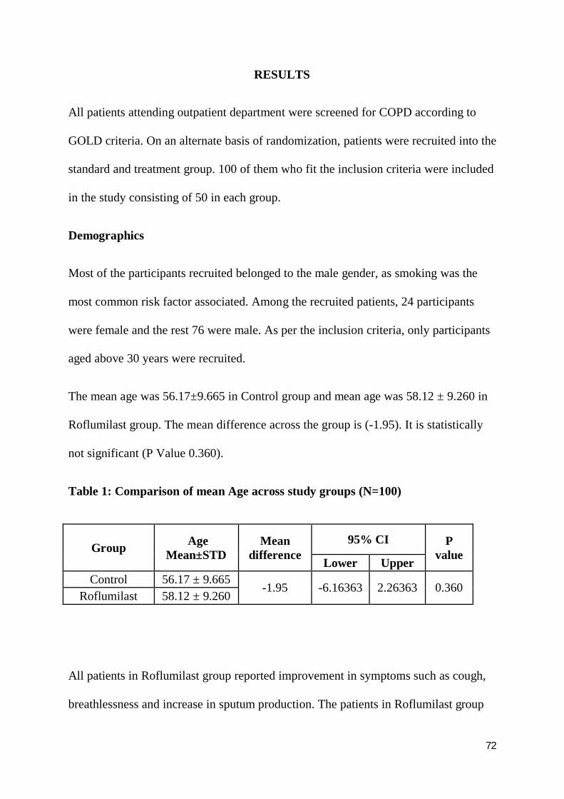
72
RESULTS
All patients attending outpatient department were screened for COPD according to
GOLD criteria. On an alternate basis of randomization, patients were recruited into the
standard and treatment group. 100 of them who fit the inclusion criteria were included
in the study consisting of 50 in each group.
Demographics
Most of the participants recruited belonged to the male gender, as smoking was the
most common risk factor associated. Among the recruited patients, 24 participants
were female and the rest 76 were male. As per the inclusion criteria, only participants
aged above 30 years were recruited.
The mean age was 56.17±9.665 in Control group and mean age was 58.12 ± 9.260 in
Roflumilast group. The mean difference across the group is (-1.95). It is statistically
not significant (P Value 0.360).
Table 1: Comparison of mean Age across study groups (N=100)
Group AgeMean±STD
Meandifference
95% CI PvalueLower Upper
Control 56.17 ± 9.665-1.95 -6.16363 2.26363 0.360
Roflumilast 58.12 ± 9.260
All patients in Roflumilast group reported improvement in symptoms such as cough,
breathlessness and increase in sputum production. The patients in Roflumilast group
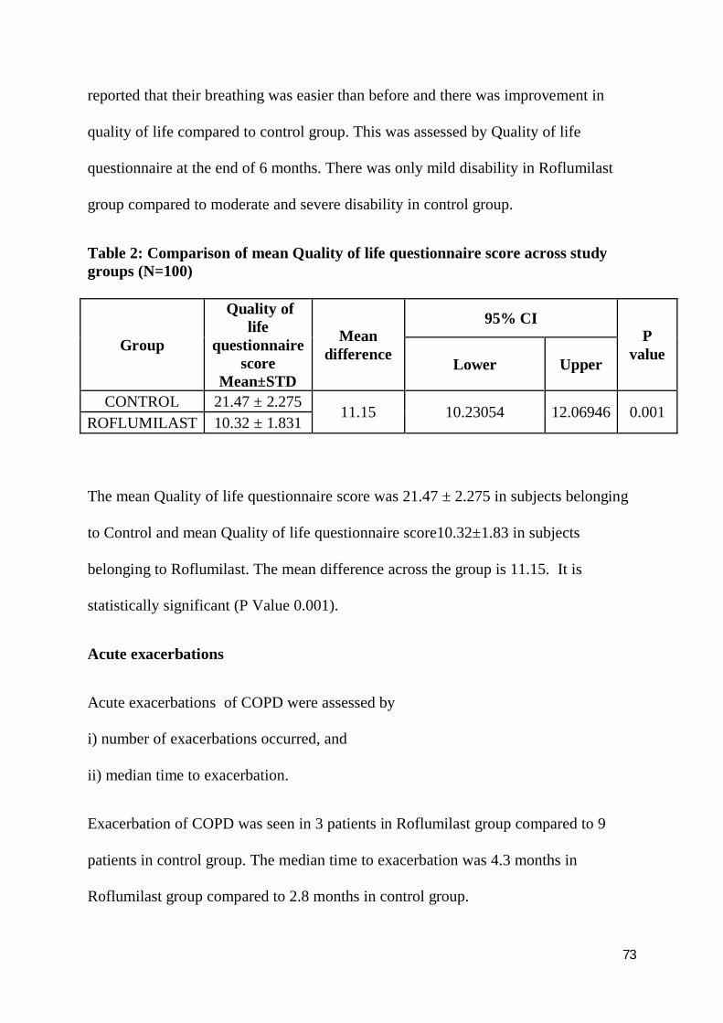
73
reported that their breathing was easier than before and there was improvement in
quality of life compared to control group. This was assessed by Quality of life
questionnaire at the end of 6 months. There was only mild disability in Roflumilast
group compared to moderate and severe disability in control group.
Table 2: Comparison of mean Quality of life questionnaire score across studygroups (N=100)
Group
Quality oflife
questionnairescore
Mean±STD
Meandifference
95% CIP
valueLower Upper
CONTROL 21.47 ± 2.27511.15 10.23054 12.06946 0.001
ROFLUMILAST 10.32 ± 1.831
The mean Quality of life questionnaire score was 21.47 ± 2.275 in subjects belonging
to Control and mean Quality of life questionnaire score10.32±1.83 in subjects
belonging to Roflumilast. The mean difference across the group is 11.15. It is
statistically significant (P Value 0.001).
Acute exacerbations
Acute exacerbations of COPD were assessed by
i) number of exacerbations occurred, and
ii) median time to exacerbation.
Exacerbation of COPD was seen in 3 patients in Roflumilast group compared to 9
patients in control group. The median time to exacerbation was 4.3 months in
Roflumilast group compared to 2.8 months in control group.
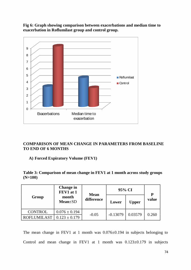
74
Fig 6: Graph showing comparison between exacerbations and median time toexacerbation in Roflumilast group and control group.
COMPARISON OF MEAN CHANGE IN PARAMETERS FROM BASELINETO END OF 6 MONTHS
A) Forced Expiratory Volume (FEV1)
Table 3: Comparison of mean change in FEV1 at 1 month across study groups(N=100)
Group
Change inFEV1 at 1
monthMean±SD
Meandifference
95% CIP
valueLower Upper
CONTROL 0.076 ± 0.194-0.05 -0.13079 0.03579 0.260
ROFLUMILAST 0.123 ± 0.179
The mean change in FEV1 at 1 month was 0.076±0.194 in subjects belonging to
Control and mean change in FEV1 at 1 month was 0.123±0.179 in subjects
0
1
2
3
4
5
6
7
8
9
Exacerbations Median time toexacerbation
Roflumilast
Control

75
belonging to Roflumilast. The mean difference across the group is (-0.05). It is
statistically not significant (P Value 0.260).
Table 4: Comparison of mean Change in FEV1 at 2 months across studygroups (N=100)
Group
Change inFEV1 at 2
monthsMean±SD
Meandifference
95% CI PvalueLower Upper
CONTROL 0.05 ± 0.148-0.13 -0.21537 -0.03963 0.005
ROFLUMILAST 0.177 ± 0.236
The mean change in FEV1 at 2 months was 0.05±0.148 in subjects belonging to
control and mean change in FEV1 at 2 months was 0.177±0.236 in subjects belonging
to Roflumilast group. The mean difference across the group is (-0.13). It is statistically
significant (P Value 0.005).
Table 5: Comparison of mean Change in FEV1 at 3 months across studygroups (N=100)
Group
Change inFEV1 at 3
monthsMean±SD
Meandifference
95% CI PvalueLower Upper
CONTROL 0.053 ± 0.196-0.19 -0.29303 -0.09347 0.001
ROFLUMILAST 0.246 ± 0.248
The mean change in FEV1 at 3 months was 0.053±0.196 in subjects belonging to
Control and mean change in FEV1 at 3 months was 0.246±0.248 in subjects belonging

76
to Roflumilast group. The mean difference across the group is (-0.19). It is statistically
significant (P Value 0.001).
Table 6: Comparison of mean change in FEV1 at 4 months across study groups(N=100)
Group
Change inFEV1 at 4
monthsMean±SD
Meandifference
95% CI PvalueLower Upper
CONTROL 0.078 ± 0.197-0.21 -0.30114 -0.10986 0.001
ROFLUMILAST 0.283 ± 0.230
The mean change in FEV1 at 4 months was 0.078±0.197 in subjects with Control
and mean change in FEV1 at 4 months was 0.283±0.230 in subjects belonging to
Roflumilast group. The mean difference across the group is (-0.21). It is statistically
significant (P Value 0.001).
Table 7: Comparison of mean Change in FEV1 at 5 months across study groups(N=100)
Group
Change inFEV1 at 5
monthsMean±SD
Meandifference
95% CI PvalueLower Upper
CONTROL 0.103 ± 0.214-0.20 -0.30179 -0.10421 0.001
ROFLUMILAST 0.306 ± 0.228
The mean change in FEV1 at 5 months was 0.103±0.214 in subjects belonging to
Control and mean change in FEV1 at 5 months was 0.306±0.228 in subjects
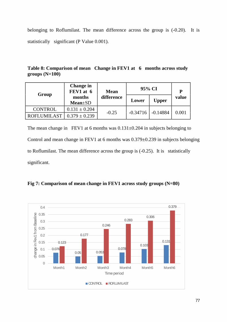
77
belonging to Roflumilast. The mean difference across the group is (-0.20). It is
statistically significant (P Value 0.001).
Table 8: Comparison of mean Change in FEV1 at 6 months across studygroups (N=100)
Group
Change inFEV1 at 6
monthsMean±SD
Meandifference
95% CI PvalueLower Upper
CONTROL 0.131 ± 0.204-0.25 -0.34716 -0.14884 0.001
ROFLUMILAST 0.379 ± 0.239
The mean change in FEV1 at 6 months was 0.131±0.204 in subjects belonging to
Control and mean change in FEV1 at 6 months was 0.379±0.239 in subjects belonging
to Roflumilast. The mean difference across the group is (-0.25). It is statistically
significant.
Fig 7: Comparison of mean change in FEV1 across study groups (N=80)
0.0760.05 0.053
0.0780.103
0.1310.123
0.177
0.2460.283
0.306
0.379
0
0.05
0.1
0.15
0.2
0.25
0.3
0.35
0.4
Month1 Month2 Month3 Month4 Month5 Month6
chan
ge in
Fev
1 fr
om B
asel
ine
Time period
CONTROL ROFLUMILAST

78
B) Forced vital Capacity (FVC)
Table: 9 Comparison of mean Change in FVC at 1 month across study groups(N=100)
Group
Change inFVC at 1
monthMean±STD
Meandifference
95% CI PvalueLower Upper
CONTROL -0.02 ± 0.285-0.08 -0.18886 0.03586 0.179
ROFLUMILAST 0.054 ± 0.213
The mean change in FVC at 1 month was -0.02±0.285 in subjects belonging to
Control and mean change in FVC at 1 month was 0.054±0.213 in subjects belonging
to Roflumilast. The mean difference across the group is (-0.08). It is statistically
not significant (P Value 0.179).
Table 10: Comparison of mean Change in FVC at 2 months across study groups(N=100)
Group
Change inFVC at 2months
Mean±STD
Meandifference
95% CI PvalueLower Upper
CONTROL -0.02 ± 0.304-0.11 -0.25955 0.03355 0.129
ROFLUMILAST 0.089 ± 0.351
The mean change in FVC at 2 months was -0.02±0.394 in subjects belonging to
Control and mean change in FVC at 2 months was 0.089±0.351 in subjects belonging
to Roflumilast group. The mean difference across the group is (-0.11). It is
statistically not significant (P Value 0.129).

79
Table11: Comparison of mean Change in FVC at 3 months across study groups
(N=100)
Group
Change inFVC at 3months
Mean±STD
Meandifference
95% CI PvalueLower Upper
CONTROL -0.00 ± 0.354-0.17 -0.31404 -0.03246 0.017
ROFLUMILAST 0.173 ± 0.273
The mean change in FVC at 3 months was -0.00±0.354 in subjects belonging to
control and mean change in FVC at 3 months was 0.173±0.273 in subjects belonging
to Roflumilast group. The mean difference across the group is (-0.17). It is
statistically significant (P Value 0.017).
Table 12: Comparison of mean Change in FVC at 4 months across study groups(N=100)
Group
Change inFVC at 4months
Mean±STD
Meandifference
95% CI PvalueLower Upper
CONTROL -0.02 ± 0.343-0.23 -0.37270 -0.08930 0.002
ROFLUMILAST 0.207 ± 0.291
The mean change in FVC at 4 months was -0.02±0.343 in subjects belonging to
Control and mean change in FVC at 4 months was 0.207±0.291 in subjects
belonging to Roflumilast group. The mean difference across the group is (-0.23). It is
statistically significant (P Value 0.002).

80
Table 13: Comparison of mean Change in FVC at 5 months across study groups
(N=100)
Group
Change inFVC at 5months
Mean±STD
Meandifference
95% CI PvalueLower Upper
CONTROL -0.02 ± 0.338-0.22 -0.35289 -0.08161 0.002
ROFLUMILAST 0.192 ± 0.266
The mean change in FVC at 5 months was -0.02±0.338 in subjects belonging to
Control and mean change in FVC at 5 months was 0.192±0.266 in subjects belonging
to Roflumilast group. The mean difference across the group is (-0.22). It is
statistically significant (P Value 0.002).
Table 14: Comparison of mean Change in FVC at 6 months across study groups(N=100)
Group
Change inFVC at 6months
Mean±STD
Meandifference
95% CI PvalueLower Upper
CONTROL 0.02 ± 0.3450.29 -0.42205 -0.15195 0.001
ROFLUMILAST 0.257 ± 0.253
The mean change in FVC at 6 months was 0.02±0.345 in subjects belonging to
Control and mean change in FVC at 6 months was 0.257±0.253 in subjects
belonging to Roflumilast group. The mean difference across the group is (-0.29). It is
statistically significant (P Value 0.001).

81
Fig 8: Comparison of mean change in FVC across study groups (N=100)
C) % FEV1 (FV1/FVC)
Table 15: Comparison of mean Change in FEV1/FVC at 1 month across studygroups (N=100)
Group
Change inFEV1/FVC at
1 monthMean±STD
Meandifference
95% CI PvalueLower Upper
CONTROL 0.061 ± 0.1300.01 -0.04023 0.05973 0.699
ROFLUMILAST 0.052 ± 0.090
The mean change in FEV1/FVC at 1 month was 0.061±0.130 in subjects belonging to
Control and mean change in FEV1/FVC at 1 month was 0.052±0.090 in subjects
belonging to Roflumilast group. The mean difference across the group is (0.01). It is
statistically not significant (P Value 0.699).
-0.02 -0.02
0
-0.02 -0.02 -0.02
0.054
0.089
0.173
0.2070.192
0.257
-0.05
0
0.05
0.1
0.15
0.2
0.25
0.3
Month1 Month2 Month3 Month4 Month5 Month6
chan
ge in
Fvc
from
Bas
elin
e
Time period
CONTROL ROFLUMILAST

82
Table 16: Comparison of mean Change in FEV1/FVC at 2 months across studygroups (N=100)
Group
Change inFEV1/FVC at
2 monthsMean±STD
Meandifference
95% CI PvalueLower Upper
CONTROL 0.046 ± 0.128-0.03 -0.08881 0.02081 0.221
ROFLUMILAST 0.080 ± 0.116
The mean change in FEV1/FVC at 2 months was 0.046±0.128in subjects belonging to
control group and mean change in FEV1/FVC at 2 months was 0.080±0.116 in
subjects belonging to Roflumilast group. The mean difference across the group is (-
0.03). It is statistically not significant (P Value 0.221).
Table 17: Comparison of mean Change in FEV1/FVC at 3 months across studygroups (N=100)
Group
Change inFEV1/FVC at
3 monthsMean±STD
Meandifference
95% CI PvalueLower Upper
CONTROL 0.044 ± 0.109-0.04 -0.09639 0.01039 0.113
ROFLUMILAST 0.087 ± 0.129
The mean change in FEV1/FVC at 3 months was 0.044±0.109 in subjects belonging
to Control and mean Change in FEV1/FVC at 3 months was 0.087±0.129 in subjects
belonging to Roflumilast group. The mean difference across the group is (-0.04). It is
statistically not significant (P Value 0.113).
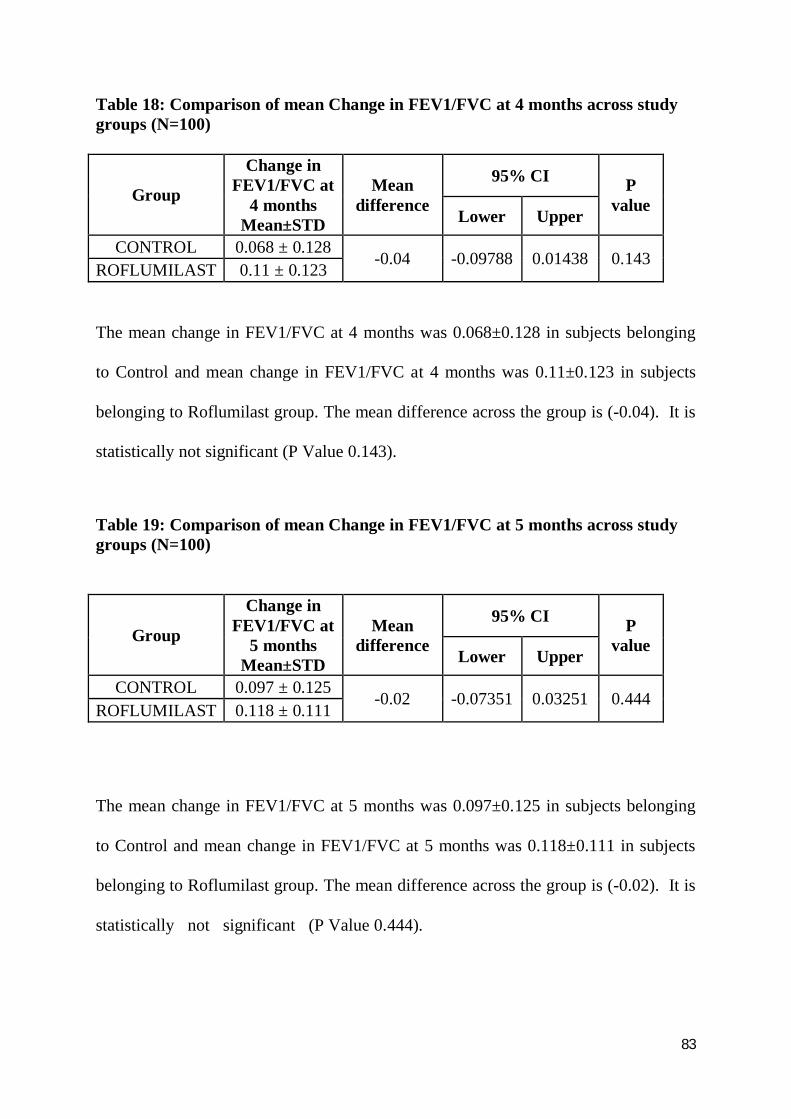
83
Table 18: Comparison of mean Change in FEV1/FVC at 4 months across studygroups (N=100)
Group
Change inFEV1/FVC at
4 monthsMean±STD
Meandifference
95% CI PvalueLower Upper
CONTROL 0.068 ± 0.128-0.04 -0.09788 0.01438 0.143
ROFLUMILAST 0.11 ± 0.123
The mean change in FEV1/FVC at 4 months was 0.068±0.128 in subjects belonging
to Control and mean change in FEV1/FVC at 4 months was 0.11±0.123 in subjects
belonging to Roflumilast group. The mean difference across the group is (-0.04). It is
statistically not significant (P Value 0.143).
Table 19: Comparison of mean Change in FEV1/FVC at 5 months across studygroups (N=100)
Group
Change inFEV1/FVC at
5 monthsMean±STD
Meandifference
95% CI PvalueLower Upper
CONTROL 0.097 ± 0.125-0.02 -0.07351 0.03251 0.444
ROFLUMILAST 0.118 ± 0.111
The mean change in FEV1/FVC at 5 months was 0.097±0.125 in subjects belonging
to Control and mean change in FEV1/FVC at 5 months was 0.118±0.111 in subjects
belonging to Roflumilast group. The mean difference across the group is (-0.02). It is
statistically not significant (P Value 0.444).

84
Table 20: Comparison of mean Change in FEV1/FVC at 6 months across studygroups (N=100)
GroupChange in
FEV1/FVC at 6monthsMean±STD
Meandifference
95% CI PvalueLower Upper
CONTROL 0.101 ± 0.120-0.04 -
0.09127 0.01077 0.120ROFLUMILAST 0.142 ± 0.108
The mean change in FEV1/FVC at 6 months was 0.101±0.120 in subjects belonging
to Control and mean change in FEV1/FVC at 6 months was 0.142±0.108 in subjects
belonging to Roflumilast group. The mean difference across the group is (-0.04). It is
statistically not significant (P Value 0.120).
Fig 9: Comparison of mean Change in FEV1/FVC across study groups (N=100)
0.0610.046 0.044
0.068
0.097 0.101
0.052
0.08 0.087
0.110.118
0.142
0
0.02
0.04
0.06
0.08
0.1
0.12
0.14
0.16
Month1 Month2 Month3 Month4 Month5 Month6chan
ge in
Fev
1/Fv
c fr
om B
asel
ine
Time Period
CONTROL ROFLUMILAST
85
ADVERSE DRUG REACTIONS
Out of 50 patients recruited in the Roflumilast group, 27 patients reported adverse
effects due to their medications. Most of the adverse effects were reported
spontaneously. Most of the patients complained of headache a few hours after
ingestion of the drug. The second most common complaint was tremors. A few
reported insomnia, nausea, diarrhoea, dizziness, and clouding of consciousness after
intake of drug. The other participants tolerated the drug well. No life threatening or
severe reactions to drug occurred. Depression, suicidal thoughts which were a rare
side effect reported in previous studies due to Roflumilast did not occur in any
participant in this study [81].
In the group which received standard treatment, less adverse effects were recorded.
Most common was tremor which was seen in 8 patients.
Fig10. Adverse effects in Roflumilast group
28%
18%11%
7%
14%
11%
11%
ADVERSE EFFECTSheadache
tremors
nausea
insomnia
giddiness
diarrhea
acute exacerbations

86
ADHERENCE TO TREATMENT REGIMEN
After recruitment into study, after intake of treatment of regimen, there were dropouts
of patients due to loss of contact, due to shifting to other place, changing hospital and
due to adverse effects of the drug. Those patients were excluded from the study and
new patients were recruited into the study till a sample size of 50 was attained in each
group. Each participant was followed up for 6 months after recruitment in the study.
Table 21: No. of dropouts
No. of dropouts
Loss of contact 3
Shifting to other place 2
Change of hospital 3
Adverse effects 3

87
DISCUSSION

88
DISCUSSION
Acute exacerbation of COPD requires increased use of medications and
hospitalization. Also, there are no guidelines for treatment of exacerbations in
hospitalized patients. In COPD, airflow reduction does not improve significantly
with bronchodilators, in contrast to asthma. Therefore, a better drug was needed to
reduce exacerbations in COPD. Very few studies have been done in India.
Therefore, this prospective, comparative study of effect of Roflumilast in COPD
and its efficacy in reducing exacerbations was carried out.
The findings of our study show that roflumilast prevented moderate and severe
exacerbations and improved lung function in patients with severe chronic
obstructive pulmonary disease and chronic bronchitis who continued to have
exacerbations despite inhaled combination therapy.
Roflumilast, a PDE4 inhibitor is an anti-inflammatory agent rather than a
bronchodilator. Also due to change in internal airflow distribution, it improves
efficacy of Steroids and B2 agonists. Previous studies conducted in countries
outside India (REACT study) have shown that Roflumilast reduces the frequency
of exacerbations in COPD [74].
Most of the patients recruited were men, who constituted 76 of 100 patients and all
were chronic smokers with a history of smoking for approx 15 years or more. This
indeed proves that smoking is the most common risk factor for COPD. Among
women who were recruited, most of them had a history of exposure to smoke from
burning biomass for cooking. This is in correlation with previous studies

89
conducted [75]. One such study is FARIECE study done in Mexico conducted by
J.C.Fernández et al [82].
There was significant improvement seen in lung function of FEV1and FVC in
patients who were recruited in the group where Roflumilast was given as add on
therapy. Roflumilast produced a sustained improvement in post-bronchodilator
FEV1 of 0.25% compared with standard. This change is compatible with previous
studies done by Herbert et al, Hohlfeld et al [63]. The mean change in Fvc at 6
months was -0.02±0.345 in subjects belonging to control group from baseline
compared to 0.257±0.253 in subjects belonging to Roflumilast group. The mean
difference across the group is 0.29%. The improvement in lung function in mean
variation of FEV1and FVC from base line in Roflumilast group compared to
control group were statistically significant. The change in lung function, is unlikely
to have modified the patients’ degree of breathlessness, but could have contributed
to the reduction in exacerbations [77]. The mean change in Fev1/Fvc at 6 months
was 0.101±0.120 in subjects belonging to control and the mean change seen in
subjects belonging to Roflumilast group was 0.142±0.108. But this finding was not
statistically significant. This may be attributed to the low sample size. In previous
studies by Vestbo et al [74], Stephen et al [75], the improvement in %FEV1 was
found to be statistically significant.
The patients recruited in Roflumilast group experienced reduction in symptoms
such as cough, breathlessness and sputum production compared to control group.
They reported that they could breathe easier which improved their quality of life.
Patients recruited in group receiving Roflumilast, at the end of 6 months were only

90
mildly disabled and there was improvement in their quality of life when compared
to their baseline values before intake of drug Roflumilast. In the control group, the
patients were moderately to severely disabled and there was no improvement in
quality of life.
Roflumilast was efficacious in reducing exacerbations of COPD. Only 3 patients in
Roflumilast group experienced exacerbations and were admitted in hospital. The
duration of stay was also comparatively less and they recovered faster than patients
in the control group. In the control group, 9 patients experienced exacerbations and
their duration of stay in hospital was longer. This proved that Roflumilast reduced
exacerbations of COPD, thus reducing hospital admissions and duration of stay in
hospital. The study has shown positive clinical and economic implications and
justifies the decision to target at-risk patients with chronic obstructive pulmonary
disease. Also, the mean time to exacerbation was 2.8 months in control group
compared to 4.3 months in patients receiving Roflumilast. This showed that
Roflumilast reduced the time to 1st exacerbation and also postponed the episodes
of exacerbation. The result obtained is in correlation to previous studies done by
Field et al, Vestbo et al [75,76]. Roflumilast saw significant reduction in the
incidence of exacerbations in patients who had frequent COPD exacerbations ( 2)
in the year prior to treatment with roflumilast.
There were some adverse reactions reported by patients on intake of Roflumilast
drug which was of a higher percentage compared to the patients in control group.
This also led to dropouts. The most common adverse drug reactions were
insomnia, nausea, headache, tremor, dizziness, clouding of consciousness and

91
sweating. This was attributed to potential of Roflumilast to reduce blood sugar.
This has been seen in some studies done in animals (Volet et al) and is a
prospective, promising drug for diabetics as an oral hypoglycaemic agent [78].
More studies are required in humans to prove this. Most of the reactions were
tolerable and patient adherence was good. There were no life threatening or severe
reactions witnessed other than exacerbation of disease. No deaths were reported.
The anticipated adverse effects such as suicidal tendency and depression which
were reported in previous studies by Stephen et al in Europe [77] were not
observed.
Thus by a derivation of this study, Roflumilast is an effective drug in reducing
exacerbations of COPD, and in improving quality of life in patients diagnosed with
COPD and can be used as add on therapy to standard treatment.

92
CONCLUSION

93
CONCLUSION
Based on our study findings Roflumilast, has a definite role in treatment and
prevention of exacerbations in COPD due to its anti inflammatory properties. Thus,
Roflumilast as part of a combination regimen with long-acting bronchodilators and
inhalational corticosteroids appears to be a reasonable treatment option for patients
with severe to very severe COPD associated with a history of exacerbations. Further
studies are required to be done in a large number of patients.

94
BIBLIOGRAPHY
95
BIBLIOGRAPHY
1. Vestbo, Jorgen (2013). "Definition and Overview". Global Strategy for theDiagnosis, Management, and Prevention of Chronic Obstructive PulmonaryDisease. Global Initiative for Chronic Obstructive Lung Disease. pp. 1–7.
2. BD 2015 Disease and Injury Incidence and Prevalence, Collaborators. (8October 2016). "Global, regional, and national incidence, prevalence, and yearslived with disability for 310 diseases and injuries, 1990-2015: a systematicanalysis for the Global Burden of Disease Study 2015."Lancet. 388 (10053):1545–1602. doi:10.1016/S0140-6736(16)31678-6.
3. Chronic obstructive pulmonary disease (COPD) Fact sheet N°315". WHO.January 2015. Retrieved 4 March 2016.
4. Rennard, Stephen (2013). Clinical management of chronic obstructivepulmonary disease (2nd ed.). Informa Healthcare. p. 23.
5. Goldman, Lee (2012). Goldman's Cecil medicine (24th ed.). Elsevier/Saunders.p. 537. ISBN 978-1-4377-1604-7.
6. Foreman MG, Campos M, Celedón JC (July 2012). "Genes and chronicobstructive pulmonary disease". Med. Clin. North Am. 96 (4): 699–711 doi:10.1016/j.mcna.2012.02.006
7. Vestbo, Jørgen (2013). "Management of Exacerbations" (PDF). GlobalStrategy for the Diagnosis, Management, and Prevention of ChronicObstructive Pulmonary Disease. Global Initiative for Chronic Obstructive LungDisease. pp. 39–45.
8. Dhar, Raja (2011). Textbook of pulmonary and critical care medicine. NewDelhi: Jaypee Brothers Medical Publishers. p. 1056. ISBN 978-93-5025-073-0.
9. Palange, Paolo (2013). ERS Handbook of Respiratory Medicine. EuropeanRespiratory Society. p. 194. ISBN 978-1-84984-041-5.
10. Lötvall, Jan (2011). Advances in combination therapy for asthma and COPD.Wiley. p. 251. ISBN 978-1-119-97846-6.
11. Barnes, Peter. Asthma and COPD: basic mechanisms and clinicalmanagement(2nd ed.). Academic. p. 837. ISBN 978-0-12-374001-4.
12. Young, Vincent B. Blueprints medicine (5th ed.). Wolters KluwerHealth/Lippincott Williams & Wilkins. p. 69. ISBN 978-0-7817-8870-0
13. Qaseem, Amir; Wilt, TJ; Weinberger, SE; Hanania, NA; Criner, G; et alAmerican College Of Physicians; American College of Chest Physicians;American Thoracic Society; European Respiratory Society (2011). "Diagnosisand Management of Stable Chronic Obstructive Pulmonary Disease: A ClinicalPractice Guideline Update from the American College of Physicians, AmericanCollege of Chest Physicians, American Thoracic Society, and European

96
Respiratory Society". Annals of Internal Medicine. 155. doi:10.7326/0003-4819-155-3-201108020-00008.
14. National Institute for Health and Clinical Excellence. Clinical guideline 101:Chronic Obstructive Pulmonary Disease. London, June 2012.
15. Mammen MJ, Sethi S (2012). "Macrolide therapy for the prevention of acuteexacerbations in chronic obstructive pulmonary disease". Pol. Arch. Med.Wewn. 122 (1–2): 54–9. PMID 22353707.
16. Herath, SC; Poole, P (Nov 28, 2013). "Prophylactic antibiotic therapy forchronic obstructive pulmonary disease (COPD)." Cochrane Database SystRev. 11: CD009764. doi:10.1002/14651858.CD009764.pub2.
17. Simoens, S; Laekeman, G; Decramer, M (May 2013). "Preventing COPDexacerbations with macrolides: a review and budget impactanalysis". Respiratory medicine. 107 (5): 637–48. PMID 23352223. doi:10.1016/j.rmed.2012.12.019.
18. Barr RG, Rowe BH, Camargo CA. Barr, R Graham, ed. "Methylxanthines forexacerbations of chronic obstructive pulmonary disease". doi:10.1002/14651858.CD002168.
19. Poole, P; Chong, J; Cates, CJ (29 July 2015). "Mucolytic agents versusplacebo for chronic bronchitis or chronic obstructive pulmonarydisease."Cochrane Database Syst Rev. 7 doi : 10.1002/ 14651858. CD001287.pub5.
20. COPD Working, Group (2012). "Long-term oxygen therapy for patients withchronic obstructive pulmonary disease (COPD): an evidence-basedanalysis". Ontario health technology assessment series.
21. Bradley JM, O'Neill B, Bradley, Judy M, et al. "Short-term ambulatory oxygenfor chronic obstructive pulmonary disease". doi:10.1002/14651858.CD004356.pub3.
22. Ekström, Magnus; Ahmadi, Zainab; Bornefalk-Hermansson, Anna; Abernethy,Amy; Currow, David (2016-11-25). "Oxygen for breathlessness in patientswith chronic obstructive pulmonary disease who do not qualify for homeoxygen therapy". doi:10.1002/14651858.CD006429.pub3.
23. Chapman, Stephen. Oxford handbook of respiratory medicine (2nd ed.).Oxford University Press. p. 707. ISBN 978-0-19-954516-2.
24. Blackler, Laura. Managing chronic obstructive pulmonary disease.p. 49. ISBN 978-0-470-51798-7.
25. Jindal, Surinder K (2013). Chronic Obstructive Pulmonary Disease. JaypeeBrothers Medical. p. 139. ISBN 978-93-5090-353-7.
97
26. O'Driscoll, BR; Howard, LS; Davison, AG; British Thoracic, Society (October2012). "BTS guideline for emergency oxygen use in adultpatients". doi:10.1136/thx.2008.102947
27. Flaxman AD, Naghavi M, Lozano R, Michaud C, Ezzati M, Shibuya K,Salomon JA, Abdalla S, Aboyans V, et al. (December 2012). "Years lived withdisability (YLDs) for 1160 sequelae of 289 diseases and injuries 1990–2010: asystematic analysis for the Global Burden of Disease Study2010". Lancet. 380 (9859). doi:10.1016/S0140-6736(12)61729-2
28.Davies Adeloye, Stephen Chua, Chinwei LeeGlobal and regional estimates ofCOPD prevalence: Systematic review and meta– analysis. IJPM May 2012.
29.Celli, B. R. (2004). The body-mass index, airflow obstruction, dyspnea, andexercise capacity index in chronic obstructive pulmonarydisease. https://doi.org/10.1056/NEJMoa021322
30.Int J Chron Obstruct Pulmon Dis. 2012; 4: 137–148
31.Shavelle, R. M., et al. (2009). Years of life lost and life expectancy in chronicobstructive pulmonary disease: Findings the NHANES III follow-upstudy. https://doi.org/10.2147/COPD.S5237
32.Boswell-Smith, V; Spina, D (2007). "PDE4 inhibitors as potential therapeuticagents in the treatment of COPD-focus on roflumilast". International Journal ofChronic Obstructive Pulmonary Disease. 2 (2): 121–9. ISSN 1178-2005. PMC 2695611 . PMID 18044684
33. Davidson's Principles and Practice of Medicine 23rd Edition pg 1205 - 120934. Harrison Principles of Internal Medicine 19th Edition pg 1700-1708
35. Calverley PM, Koulouris NG (2005). "Flow limitation and dynamichyperinflation: key concepts in modern respiratory physiology". Eur RespirJ. 25 (1): 186–199. PMID 15640341. doi:10.1183/09031936.04.00113204.
36. Currie, Graeme P. (2010). ABC of COPD (2nd ed.). Wiley-Blackwell, BMJBooks. p. 32. ISBN 978-1-4443-2948-3.
37. O'Donnell DE (2006). "Hyperinflation, Dyspnea, and Exercise Intolerance inChronic Obstructive Pulmonary Disease". The Proceedings of the AmericanThoracic Society. 3 (2): 180–4. PMID 16565429. doi:10.1513/pats.200508-093DO.

98
38. Cooper, CB (October 2006). "The connection between COPD symptoms andhyperinflation and its impact on exercise and function.". The American Journal ofMedicine. doi:10.1016/j.amjmed.2006.08.004
39. Murray CJ, Vos T, Lozano R, Naghavi M, Flaxman AD, Michaud C, EzzatiM, Shibuya K, Salomon JA, et al. (December 2012). "Disability-adjusted life years(DALYs) for 291 diseases and injuries in 21 regions, 1990–2010: a systematicanalysis for the Global Burden of Disease Study 2010". doi:10.1016/S0140-6736(12)61689-4.
40.Vos T, Flaxman AD, Naghavi M, Lozano R, Michaud C, Ezzati M, ShibuyaK, Salomon JA, Abdalla S, Aboyans V, et al. (December 2012). "Years lived withdisability (YLDs) for 1160 sequelae of 289 diseases and injuries 1990–2010: asystematic analysis for the Global Burden of Disease Study2010". Lancet. 380 (9859): 2163–96. PMID 23245607. doi:10.1016/S0140-6736(12)61729-2.
41.Medicine, prepared by the Department of Medicine, Washington University (2nd ed.). Wolters Kluwer Health/Lippincott Williams & Wilkins. ISBN 978-0-7817-9155-7
42.Puhan, Milo A.; Gimeno-Santos, Elena; Cates, Christopher J.; Troosters,Thierry (2016-12-08). "Pulmonary rehabilitation following exacerbations ofchronic obstructive pulmonary disease". doi:10.1002/14651858.CD005305.pub4.
43. Zainuldin, Rahizan; Mackey, Martin G.; Alison, Jennifer A. (2011-11-09)."Optimal intensity and type of leg exercise training for people with chronicobstructive pulmonary disease". doi:10.1002/14651858.CD008008.pub2.
44. McCarthy B ,Casey D; DevaneD; Murphy K; Murphy E; Lacasse,Y (23February 2015). "Pulmonary rehabilitation for COPD" Cochrane Database SystRev 2. doi:10.1002/14651858.CD003793.pub3.
45. McNamara, Renae J.; McKeough, Zoe J.; McKenzie, David K.; Alison,Jennifer A. (2013-12-18). "Water-based exercise training for chronic obstructivepulmonary disease". The Cochrane Database of Systematic Reviews (12). doi:10.1002/14651858.CD008290.pub2.
46. Menadue, Collette; Piper, Amanda J; van 't Hul, Alex J; Wong, Keith K.(2014-05-14). "Non-invasive ventilation during exercise training for people withchronic obstructive pulmonary disease". The Cochrane Database of SystematicReviews (5): CD007714. doi:10.1002/14651858.CD007714.pub2

99
47.Vestbo, Jørgen (2013) Global Strategy for the Diagnosis, Management, andPrevention of Chronic Obstructive Pulmonary Disease. Global Initiative for COPDpp. 19–30
48. Cave, AC.; Hurst, MM. (May 2011). "The use of long acting -agonists, aloneor in combination with inhaled corticosteroids, in chronic obstructive pulmonarydisease (COPD): a risk-benefit analysis". Pharmacol Ther. 130 (2)doi:10.1016/j.pharmthera.2010.12.008.
49.Spencer, S; Karner, C; Cates, CJ; Evans, DJ (Dec 7, 2011). Spencer, Sally, ed."Inhaled corticosteroids versus long-acting beta(2)-agonists for chronicobstructive pulmonary disease". Cochrane Database SystRev (12). doi:10.1002/14651858.CD007033.pub3.
50. Wang, J; Nie, B; Xiong, W; Xu, Y (April 2012). "Effect of long-acting beta-agonists on the frequency of COPD exacerbations: a meta-analysis". Journal ofclinical pharmacy and therapeutics. doi:10.1111/j.1365-2710.2011.01285.x.
51. Geake, James B.; Dabscheck, Eli J.; Wood-Baker, Richard; Cates, ChristopherJ. (2015-01-10). "Indacaterol, a once-daily beta2-agonist, versus twice-dailybeta -agonists or placebo for chronic obstructive pulmonary disease". TheCochrane Database of SystematicReviews. doi:10.1002/14651858.CD010139.pub2
52.Gartlehner G, Hansen RA, Carson SS, Lohr KN (2006). "Efficacy and Safety ofInhaled Corticosteroids in Patients With COPD: A Systematic Review and Meta-Analysis of Health Outcomes". doi:10.1370/afm.517.
53.Chinet, T; Dumoulin, J; Honore, I; Braun, JM; Couderc, LJ; Febvre, M;Mangiapan, G; Maurer, C; Serrier, P; Soyez, F; Terrioux, P; Jebrak, G (29 January2016). "The place of inhaled corticosteroids in COPD" Revue des maladiesrespiratoires. PMID 26831345. doi:10.1016/j.rmr.2015.11.009.
54.Dong, YH; Lin, HH; Shau, WY; Wu, YC; Chang, CH; Lai, MS (January 2013)."Comparative safety of inhaled medications in patients with chronic obstructivepulmonary disease: systematic review and mixed treatment comparison meta-analysis of randomised controlled trials." doi:10.1136/thoraxjnl-2012-201926.
55. Nannini, LJ; Poole, P; Milan, SJ; Kesterton, A (30 August 2013). "Combinedcorticosteroid and long-acting beta(2)-agonist versus inhaled corticosteroids alonefor COPD.". Cochrane Database Syst Rev. 8:CD006826. PMID 23990350. doi:10.1002/14651858.CD006826.pub2

100
56. Vestbo, Jørgen (2013). "Diagnosis and Assessment"(PDF). Global Strategy forthe Diagnosis, Management, and Prevention of COPD pp. 9–17
57.Qaseem Amir; Wilt TJ; Weinberger SE; Hanania NA; Criner G; Van DerMolen T; Marciniuk DD; Denberg T; Schünemann H; Wedzicha W; MacDonald,R; Shekelle P. Annals of Internal Medicine. 155 (3): 179–91. PMID 21810710. doi:10.7326/0003-4819-155-3-201108020-00008.
58. Siu, Albert L.; Bibbins-Domingo, Kirsten; Grossman, David C.; Davidson,Karina W.; Epling, John W.; García, Francisco A. R.; Gillman, Matthew; Kemper,Alex R.; Krist, Alex H.; Kurth, Ann E.; Landefeld, C. Seth; Mangione, Carol M.;Harper, Diane M.; Phillips, William R.; Phipps, Maureen G.; Pignone, Michael P.(5 April 2016). "Screening for COPD". JAMA. 315 (13):1372. doi:10.1001/jama.2016.2638.
59. Young, Vincent B. (2010). Blueprints medicine (5th ed.). Wolters KluwerHealth/Lippincott Williams & Wilkins. p. 69. ISBN 978-0-7817-8870-0.
60. "COPD Assessment Test (CAT)". American Thoracic Society.Retrieved November 29,2013.
61. Simon, Michael R.; Chinchilli, Vernon M.; Phillips, Brenda R.; Sorkness,Christine A.; Lemanske Jr., Robert F.; Szefler, Stanley J.; Taussig, Lynn;Bacharier, Leonard B.; Morgan, Wayne (1 September 2010). "Forced expiratoryflow between 25% and 75% of vital capacity and FEV1/forced vital capacity ratioin relation to clinical and physiological parameters in asthmatic children withnormal FEV1 values". Journal of Allergy and Clinical Immunology. 126 (3): 527–534.e8. doi:10.1016/j.jaci.2010.05.016
62. Boswell-Smith, V; Spina, D (2007). "PDE4 inhibitors as potential therapeuticagents in the treatment of COPD-focus on roflumilast". International Journal ofChronic Obstructive Pulmonary Disease.
63.Herbert, C; Hettiaratchi, A; Webb, DC; Thomas, PS; Foster, PS; Kumar, RK(May 2008). "Suppression of cytokine expression by roflumilast anddexamethasone in a model of chronic asthma". Clinical & ExperimentalAllergy. 38 (5): 847–56. ISSN 1365-2222. PMID 18307529. doi:10.1111/j.1365-2222.2008.02950.x.
64.Hohlfeld, JM; Schoenfeld, K; Lavae-Mokhtari, M; Schaumann, F; Mueller, M;Bredenbroeker, D; Krug, N; Hermann, R (August 2008). "Roflumilast attenuatespulmonary inflammation upon segmental endotoxin challenge in healthy subjects:
101
a randomized placebo-controlled trial". Pulmonary Pharmacology & Therapeutics.doi:10.1016/j.pupt.2008.02.002.
65. Field, SK (May 2008). "Roflumilast: an oral, once-daily selective PDE-4inhibitor for the management of COPD and asthma". Expert Opinion onInvestigational Drugs doi:10.1517/13543784.17.5.811
66. Goodman and Gilman's The Pharmacological Basis of Therapeutics, 13thEdition
67.Basic and Clinical Pharmacology Bertram G. Katzung, 13th edition
68.Rang and Dale’s Pharmacology 8th edition
69. Cheyne, Leanne; Irvin-Sellers, Melanie J.; White, John (2015-09-22)."Tiotropium versus ipratropium bromide for chronic obstructive pulmonarydisease". The Cochrane Database of Systematic Reviews (9): doi:10.1002/14651858.CD009552.pub3
70. Rennard, Stephen (2013). Clinical management of chronic obstructivepulmonary disease (2nd ed.). Informa Healthcare. P23
71. Anita Sharma ; with a contribution by David Pitchforth ; forewords by GailRichards; Barclay, Joyce (2010). COPD in primary care. Radcliffe Pub. p.9
72.Goldman, Lee (2012). Goldman's Cecil medicine (24th ed.). Elsevier/Saunders.p. 537
73. van Agteren, JE; Carson, KV; Tiong, LU; Smith, BJ (14 October 2016). "Lungvolume reduction surgery for diffuse emphysema.". The Cochrane database ofsystematic reviews. doi:10.1002/14651858.CD001001.pub3
74.Vestbo J, Hurd SS, Agusti AG, et al. Global strategy for the diagnosis,management, and prevention of COPD: GOLD executive summary Crit Care Med2013
75. Beghe B, Rabe KF, Fabbri LM. Phosphodiesterase-4 inhibitor therapy for lungdiseases. Am J Respir Crit Care Med 2013; 188: 271–78.
76. Machines IB. IBM SPSS Statistics for Windows, Version 22.0. IBM CorpArmonk, NY; 2013.

102
77. Reduction of exacerbations by the PDE4 inhibitor roflumilast - the importanceof defining different subsets of patients with COPD, Stephen I RennardDOI: 10.1186/1465-9921-12-18
78. The glucose-lowering effects of the PDE4 inhibitors roflumilast androflumilast-N-oxide in db/db mice, Vollert s et al., 2012 Oct;55(10):2779-2788.doi: 10.1007/s00125-012-2632-z.
79. Kemp SV, Polkey MI, Shah PL. The epidemiology, etiology, clinical features,and natural history of emphysema. Thorac Surg Clin. 2009;19 (2): 149-58. doi:10.1016/j.thorsurg.2009.03.003
80. Roflumilast: a review of its use in the treatment of COPD Int J Chron ObstructPulmon Dis. 2016; 11: 81–9081. Roflumilast : risk of suicidal behaviourFrom:Medicines and Healthcare products Regulatory Agency 29 January 2013
82. Risk factors for chronic obstructive pulmonary disease: Results of the
FARIECE study Aguirre.K.A.Guzmán-Guillen.M.E.Álvarez-Serrano
J.R.Vintimilla-Maldonado.83. Association of MMP -12 polymorphisms with severe COPD BMC Medicakgenetics : Imran Haq 2010.
84.Antibiotic prevention of acute exacerbations of COPD, New England Journalof Medicine Jul 26, 2012 Richard P Wenzel
85. National Emphysema Treatment trial, New England Journal of Medicine 2003
86. Laënnec RTH. In: A treatise on the diseases of the chest (English translationfrom the French) Forbes J, editor. London: T and G Underwood; 1821.
87. Hutchinson J. On the capacity of the lungs, and on the respiratory functions,with a view of establishing a precise and easy method of detecting disease by thespirometer. Medico-Chirurgical Transactions (London) 1846;29:137–61.
88. History of COPD Treatment, Lung Institute, 23 Aug 2014.

103
ANNEXURES

104
Ethics Committee clearance certificate

105
Case report form
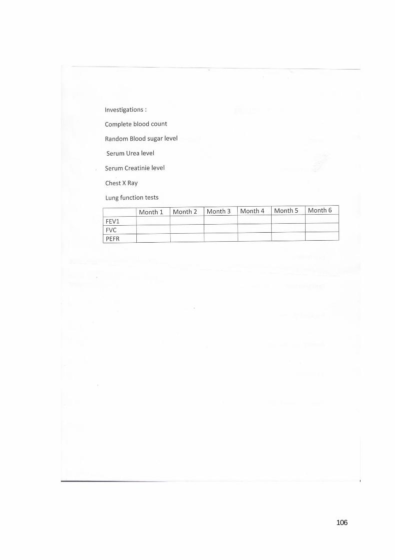
106

107
Patient information sheet in tamil
108
Patient Information sheet in English
This tablet is prescribed for Chronic Obstructive Pulmonary Disease which comprises
of increase in cough, breathlessness, and sputum production for a long period of time.
This tablet should be taken at the most for 6 months to obtain benefit from the disease.
The tablet 0f 500 micro gram should be taken orally, once daily. It can be taken before
food or after food.

109
Patient consent form in Tamil

110
Patient consent form in English
Name of the Participant:
Name of the Principal Investigator:
Name of the Institution:
Documentation of the informed consent:
I _____________________________ have read the information in this form (or it hasbeen read to me). I was free to ask any questions and they have been answered. I amover 18 years of age and, exercising my free power of choice, hereby give my consentto be included as a participant in :
1. I have read and understood this consent form and the information provided to me.
2. I have had the consent document explained to me.
3. I have been explained about the nature of the study.
4. I have been explained about my rights and responsibilities by the investigator.
5. I have been informed the investigator of all the treatments I am taking or havetaken in the past ________ months including any native (alternative) treatment.
6. I have been advised about the risks associated with my participation in this study.
7. I agree to cooperate with the investigator and I will inform him/her immediately ifI suffer unusual symptoms.
8. I have not participated in any research study within the past________month(s).
9. I am aware of the fact that I can opt out of the study at any time without having togive any reason and this will not affect my future treatment in this hospital.

111
10. I am also aware that the investigator may terminate my participation in the studyat any time, for any reason, without my consent.
12. I hereby give permission to the investigators to release the information obtainedfrom me as result of participation in this study to the sponsors, regulatoryauthorities, Govt. agencies, and IEC. I understand that they are publicly presented.
13. I have understood that my identity will be kept confidential if my data arepublicly presented.
14. I have had my questions answered to my satisfaction.
15. I have decided to be in the research study.
I am aware that if I have any question during this study, I should contact theinvestigator. By signing this consent form I attest that the information given in thisdocument has been clearly explained to me and understood by me, I will be given acopy of this consent document.
Name and signature / thumb impression of the participant (or legal representativeif participant incompetent)
Name _________________________ Signature_________________
Date_____________
Name and Signature of the investigator or his representative obtaining consent:
Name _________________________ Signature_________________
Date________________

112
Plagiarism certificate

113