a review of 120 becker permanent tissue expanders in ... · asymmetry or hypoplasia was...
TRANSCRIPT

British Journal of Plastic Surgery (1996), 49, 346-351 0 1996 The British Association of Plastic Surgeons
A review of 120 Becker permanent tissue expanders in reconstruction of the breast
I. G. Camilleri, C. M. Malata, S. Stavrianos and N. R. McLean
Department of Plastic and Reconstructive Surgery, Newcastle General Hospital, Newcastle upon Tyne, UK
S UMMAR Y. In reconstructive breast surgery, the permanent tissue expander has become popular because it avoids expander-implant exchange and gives the patient some control over the final breast size. It may, however, be associated with a number of complications. We therefore analysed the clinical notes of 111 consecutive recipients of Becker breast expanders with respect to complications and their possible predispos- ing factors.
120 prostheses were inserted in 111 consecutive patients with a mean age of 42.6 years. Median follow-up was 12 months (range 8 to 22). The commonest indication was postmastectomy breast reconstruction (81%) followed by congenital hypoplasia (14%) and acquired breast asymmetry following repeated biopsies (3%). Overexpansion before size adjustment was achieved after an average of 8 expander inflations. Complications included capsular contracture (9%), local tumour recurrence (8%), wound dehiscence (8%), filling port failure (6%), infected prostheses (4.5%) and ruptured implants (1.6%). The significant predisposing factors to wound dehiscence/infection were heavy smoking and radiotherapy (P < 0.05, x2 test). Expansion rate was not a factor. 89% of patients expressed satisfaction with the final aesthetic result.
Despite the excellent results obtained with this technique, caution must be exercised in heavy smokers and the previously irradiated.
In the 1980s Hilton Becker described the use of an inflatable breast prosthesis that could be left as a permanent implant once the required inflation volume had been achieved.‘q2 The technique was an improve- ment on that previously described by Radovan in 1978 in which a temporary tissue expander needed to be replaced with a permanent breast implant.3,4 Since then, improvements in the design and reliability of the device have led to the Becker tissue expander/ implant as we know it today. This consists of an outer silicone-filled chamber enveloping an inner, saline fillable compartment which is connected via a length of silicone tubing to a remote filling port through which the implanted device is inflated. Upon completion of expansion, the filling port and tube are removed via a small incision often under local anaesthesia.‘*‘,‘-’
The present silicone gel breast implant controversy makes the use of the Becker expander more attractive as it contains a relatively smaller amount of silicone gel compared to standard breast prostheses (25% in the Mentor Siltex, 50% in the Mentor Siltex 50). Additionally, the use of the permanent expander has a number of advantages over methods utilising autol- ogous tissue. 5-8 It takes less time to perform, and in practice adds only half an hour to a mastectomy if reconstruction is undertaken as a primary procedure. Latissimus dorsi and TRAM flap reconstruction are accompanied by more extensive scarring both locally and at the donor site, which is unacceptable to some patients. Being a smaller surgical procedure, Becker expander reconstruction leads to a shorter hospital
stay, hence contributing to the cost effectiveness of the procedure. In addition, it allows the patient some degree of control over the final size and shape of her breast.4-7
This retrospective case note study was undertaken to review our unit’s experience with the use of the Becker tissue expander in reconstructive surgery of the breast and to determine whether any recommen- dations could be made in order to improve future results. To this end a detailed statistical analysis of the complications and their possible predisposing factors was carried out.
Patients and methods
Patients undergoing breast reconstruction with Becker expanders were identified from the operating theatre records. Their case notes and expansion charts were carefully reviewed and a note of the operation details, the expansion protocol and postoperative complications was made. An attempt was then made to correlate the complications with potential predis- posing factors such as age, adjuvant (preoperative) radiotherapy or chemotherapy, smoking, interval between mastectomy and reconstruction, antibiotic use, expander size and type, intraoperative inflation and expansion technique or course. Nonparametric analysis (Chi squared test) was used to determine the significance of these predisposing factors against the development of complications. Major complications
346
Becker nermanent tissue exnanders 341
were defined as those requiring a specific treatment, readmission to hospital and/or reoperation.’
Expander insertion
The majority of the Becker expanders used in this study were of the Mentor Siltex type (Mentor Corporation, Goleta CA.). These are available in three types, a Standard (smooth surfaced) and a Siltex (textured surface), both of which contain sili- cone gel volume of 25% nominal implant size, and the Siltex Becker 50 type which is also textured and contains silicone gel volume at 50% nominal implant size. In this series, only 5% of the prostheses were smooth surfaced. This reflects our preferential use of textured implantsr0~r2 after they became commer- cially available. The base size of the other breast was used as an indicator of the expander volume, using the manufacturer’s reference tables for guidance. The technique of insertion was as follows:
A submuscular or subglandular pocket (in congeni- tal hypoplasia)r3, was created through a small infra- mammary incision. A latissimus dorsi flap was used if the patient was judged to have poor quality skin, usually following radiotherapy or, in the case of immediate reconstruction, where a large area of skin had been excised during the mastectomy. Medially the inferior attachments of pectoralis major to the ribs were detached, haemostasis was achieved and the cavity was irrigated with half-strength povidone iodine solution. No attempt was made to close the medial fibres of the pectoralis major, although in most cases the inferior border of the muscle was sutured over the expander. A vacuum drain was inserted and the Becker implant was positioned after being immersed in half-strength povidone iodine solu- tion. An average of 150 cc of saline was injected into the expander intraoperatively to obliterate the dead space and the skin was closed using layered subcut- aneous and subcuticular vicryl. All patients received prophylactic flucloxacillin for 48 hours as per unit policy.
Postoperatively, the patient was fitted with a sup- port garment and allowed home when the drains were removed, usually at around the second or third postoperative day.
Expansion protocol
All expander inflations were carried out at weekly intervals under aseptic conditions on the dressings unit. Patients had overexpansion of up to 200% as originally described by Becker1 before the start of deflation and the devices were left overexpanded for up to 6 months. Expander deflation took an average of two outpatient visits and was stopped when the patient was satisfied with her breast size and sym- metry. The majority (68%) of patients had their filling port subsequently removed under local anaesthesia, the remainder as a day case under general anaesthesia at their request.
Patient satisfaction was routinely assessed at reg- ular follow-ups and any dissatisfaction on the
Table 1 Indications for Becker expanders in 111 patients
Indication No. of patients (‘54)
Mastectomy 90 (81) Congenital asymmetry 16 (14) Acquired asymmetry 3 (3) Implant rupture 2 (2)
patient’s behalf was retrieved from the follow-up notes. This, however, did not involve any grading.
Results
A total of 120 prostheses were inserted in 111 patients over the 4-year period ( 1989-1993) for the indications given in Table 1. The average patient age at operation was 42.6 years (range 19-64). 37 patients (33%) were heavy smokers (more than 20 cigarettes per day) while 28 patients (3 1%) had previously received adju- vant radiotherapy. Of the 90 postmastectomy breast reconstructions, 23% were immediate. 9 patients underwent bilateral breast reconstruction.
Table 2 shows the expander location. Most of the expanders used for postmastectomy breast recon- struction were placed in the subpectoral position while in contrast the favoured position for congenital asymmetry or hypoplasia was the retroglandular pos- ition (8%). The latissimus dorsi pedicled flap was used to cover the prosthesis in 36 of the postmastec- tomy breast reconstructions.
Tissue expansion was started after an average of 18.4 days postoperatively (range 2 to 21 days). The average duration of expansion was 53.4 days (range 21 to 63 days). Approximately 8 weekly visits were needed to achieve the required overexpansion volume prior to size adjustment at 6 months.
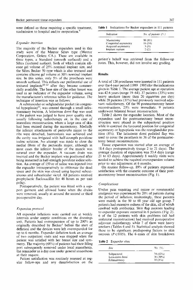
On routine follow-up, 89% of patients expressed satisfaction with the cosmetic outcome of their post- mastectomy breast reconstruction (Fig. 1).
Complications
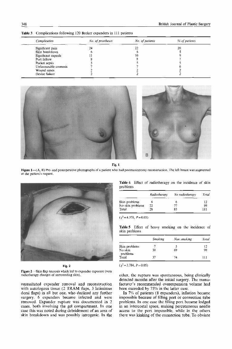
Undue pain requiring oral opiate or nonsteroidal analgesics was experienced by 20% of patients during the period of inflation; interestingly, these patients were mainly in the 50 to 60 year old age group. 7 patients had excessive redness of the skin, all of which resolved with antibiotics. Skin flap necrosis leading to expander exposure occurred in 6 patients (Fig. 2). 6 of the 12 patients with skin problems (all had unilateral reconstruction) had received postoperative adjuvant radiotherapy while 7 of them were heavy smokers (Tables 4 and 5). Statistical analysis showed these to be significant predisposing factors to skin necrosis (P60.05). The 6 cases of skin breakdown
Table 2 Expander sites
Site
Subpectoral Latissimus dorsi Submammary
No. (YY)
14 (62%) 36 (30%) 10 ( 8%)
348 British Journal of Plastic Surgery
Table 3 Complications following 120 Becker expanders in 111 patients
Complication No. of prostheses No. of patients % of patients
Significant pain 24 22 20 Skin breakdown 6 6 5 Significant capsule 11 10 9 Port failure 8 8 7 Pocket sepsis 5 5 5 Unfavourable cosmesis 1 7 6 Wound sepsis 2 2 2 Device failure 2 2 2
Fig. 1
Figure l-(A, B) Pre- and postoperative photographs of a patient who had postmastec at the patient’s request.
:tomy reconstruction. The left breast was augmented
Fig. 2 (x2=3.784, P=O.O5)
Figure 2-Skin flap necrosis which led to expander exposure (note radiotherapy changes of surrounding skin).
necessitated expander removal and reconstruction with autologous tissue (2 TRAM flaps, 3 latissimus dorsi flaps) in all but one, who declined any further surgery. 6 expanders became infected and were removed. Expander rupture was documented in 2 cases, both involving the gel compartment. In one case this was noted during debridement of an area of skin breakdown and was possibly iatrogenic. In the
other, the rupture was spontaneous, being clinically detected months after the initial surgery. The manu- facturer’s recommended overexpansion volume had been exceeded by 75% in the latter case.
In 7% of patients (8 expanders), inflation became impossible because of filling port or connection tube problems. In one case the filling port became lodged in an intercostal space, making percutaneous needle access to the port impossible, while in the others there was kinking of the connection tube. To obviate
Table 4 Effect of radiotherapy on the incidence of skin problems
Radiotherapy No radiotherapy Total
Skin problems 6 6 12 No skin problems 22 71 99 Total 28 83 111
(x2=4.378, P=O.O3)
Table 5 Effect of heavy smoking on the incidence of skin problems
Skin problems No skin
problems Total
Smoking
I 30
31
Non smoking Total
5 12 69 99
14 111
Becker permanent tissue expanders 349
this problem we now routinely close off the filling dome in a separate subcutaneous pocket and use a smaller length of tubing. All these cases required replacement of the filling port or the prosthesis to enable continued expansion.
In 7 patients tumour recurrence developed in the early months following reconstruction. Three had local recurrence in the mastectomy scar and four patients developed bone or lung metastases. Local recurrence was treated by surgical excision and those with distant metastases were referred for radiotherapy or chemotherapy. In no case was detection or treat- ment of tumour recurrence delayed by the recon- structive procedure.
Significant capsular contracture (Baker grade III & IV14) was detected in 10 patients (11 expanders) on follow-up. Six patients required open capsulotomy and replacement of the Becker expander with a textured permanent implant while two declined further surgery. During the course of follow-up, 7 expanders were judged as being cosmetically unsatis- factory. In two instances the implant was too high while the other three failed to develop an adequate degree of ptosis. The latter were treated with masto- pexy of the contralateral breast.
11 expanders in 11 patients were replaced following port malfunction, sepsis, or skin flap necrosis; 7 were substituted with similar devices and 4 with permanent implants. 8 (73%) of these patients needed to be admitted within 24 hours of being seen on the dress- ings unit.
Discussion
One hundred and twenty Becker expanders have been used in 111 patients, the main indication being for breast reconstruction following mastectomy as orig- inally described by Becker1,2 in 1984 and subsequently by others.‘. 15-18 The permanent tissue expander was also found to be effective in the correction of breast asymmetry similar to the reports by others with a standard Radovan-type expander.‘3g’9,20 In the present study, Becker expander inflation began at 18 days postoperatively which is longer than recommended by Becker’ who described starting infation at 2 to 4 days postoperatively. It was possible to remove the injection domes under local anaesthesia in all but those patients who requested a general anaesthetic. Other authors have performed port removal under general anaesthesia only if further surgery, e.g. pocket revision, nipple reconstruction, was required. The overexpansion routinely employed in this series was found to be effective in achieving breast ptosis and the consequent creation of a sub- mammary fold without surgery. This technique also gave patients some control over the final breast size. The overexpansion may have been instrumental in achieving the low rate (9”/) of capsular contracture by inhibiting myofibroblast function. Primary breast reconstruction by tissue expansion at the time of mastectomy is now well established21m24 although careful patient selection is clearly important.25 Although the numbers are not comparable and the
results not statistically significant, the trend is one of a small increase in the incidence of unfavourable results in cases of primary reconstruction.26 The reason for this is unclear at present but may be because many of these cases required flap cover as well as the expander.
Pain during expansion is related to excessive inflation pressure9*27 and in general little pain occurs during the implant expansion. However, post- expansion pain (early or late) may be as high as 50%.28 Gibney’ reported that permanent tissue expanders (similar in type to that described by Becker) reduced intra-expansion pain in a series of 100 post- mastectomy breast reconstructions. In the present study the 20% incidence of significant post-expansion pain is intermediate between the preceding two studies.
Expander exposure is the most devastating compli- cation of tissue expansion.29 In this study it was managed by immediate expander removal and replacement with a new device together with autolog- ous flap reconstruction. If sepsis was also a feature, a new implant was not inserted and the patient was treated with systemic antibiotics until the infection had subsided.
98% of patients in our series received prophylactic flucloxacillin at the time of expander insertion and in addition the expander pocket was irrigated and the expander immersed in half-strength povidone iodine solution prior to insertion. Gibney’ reported a 3% infection rate whereas our rate was marginally higher at 4.5%.
The Becker expander is said to cause less trans- and post-expansion capsular contracture.‘5’31’32 Only 2 out of 49 patients (4%) in Becker’s early series had significant capsular contracture (i.e. Baker III and IV), which was identical to that reported by Gibney in 1989.5 In a number of series a capsular contracture rate of just under 30% has been reported.33*34 In a series of 52 breasts with a 29”/0 capsular contracture rate, capsular contracture was uninfluenced by the speed of expansion or the degree of overexpansion33 and this was confirmed in this study. The 9% capsular rate in the present study is higher than that reported by Becker’ and Gibney5 but less than that of Leone et al. ( 13%).16 In most of our cases, capsulotomy and substitution of the Becker prosthesis with a conven- tional textured implant was performed with good result.
Expander failure is very rare as the reliability of Becker expanders is much better than in early designs. In one case we found that the implant had ruptured at the junction of the two chambers while in another case the implant was accidentally perforated during an attempt at closing a partial wound dehiscence. The filling ports have proved very reliable, with problems caused by kinking of the connection tube and in one case the port lodging itself in an intercostal space, making expansion impossible. We did not need to resort to ultrasound or magnetic detectors to locate the filling ports3’ as we tended to utilise the larger, more easily palpated version.
Skin flap necrosis is a serious but fortunately uncommon complication of any type of tissue expan-

350 British Journal of Plastic Suraerv
Fig. 3
Figure 3-Pre- and postoperative photographs of a congenital hypoplastic breast showing good projection and symmetry.
sion g,2g Tissue expansion temporarily reduces blood flow’in the overlying skin and ischaemia ensues if the expansion is too rapid, too prolonged, or large vol- umes of fluid are used. In the presence of factors that further compromise blood supply to the tissues such as radiation36m3g, or heavy smoking4’, necrosis may supervene.
In this study, the significant predisposing factors to skin problems were heavy smoking and previous radiotherapy. Cohen et a1.40, in a retrospective review of 73 consecutive breast reconstructions, found that cigarette smoking correlated with a higher incidence of unfavourable results. These findings were also observed by Sharpe and Coleman.41
Poor candidates for breast reconstruction using this method include the previously irradiated’5,41,42, obese patients, those with atrophic and tight skin with poor muscle” and patients having subcutaneous mastectomy. 41 Although tissue expansion can be suc- cessful in patients who have had chest wall radio- therapy, it is best avoided in those who have visible radiation dermatitis as they are more prone to compli- cations41 In their series of 19 previously irradiated patients undergoing subcutaneous mastectomies and reconstruction by tissue expansion, Sharpe and Coleman reported the very high overall complication rate of 50%.41 Bilateral total mastectomy with rela- tively conservative skin excision to allow bilateral expander reconstructions is a better option in many of these patients.41
Our results suggest that Becker prostheses do not conceal local tumour recurrence and it has also been the experience of others I8 that the natural course of breast carcinoma is not affected by these devices. Only 3 patients developed local recurrence and this correlated with the pathological features of the orig- inal mastectomy specimen.
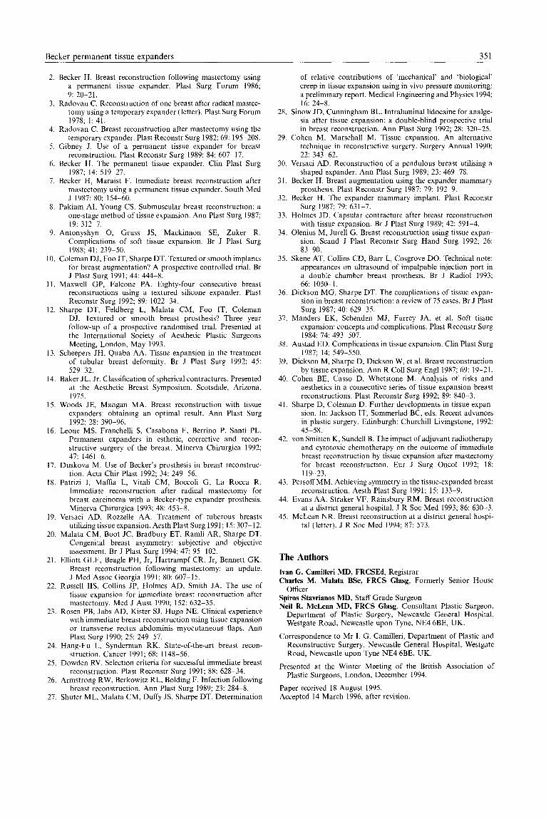
Achieving symmetry and a satisfactory degree of breast ptosis (Fig. 3) using tissue expansion remains
a problem in some patients43, particularly those with large breasts. We achieved an 89% patient satisfaction as regards general breast contour with the patient wearing a brassiere, which in most instances had the effect of lifting the contralateral breast. Only 5 cases had to be revised on cosmetic grounds alone. However it was interesting to note that the majority of our patients (85%) turned down nipple reconstruc- tion, saying that they were happy with just a “breast mound”. Of these, 42% used a prosthetic nipple. Nipple reconstruction was performed on 15 patients (17 breasts). This was accompanied by mastopexy of the normal side in 12 patients.
Conclusions
In conclusion, a series of 120 Becker expanders used in 111 consecutive patients is presented giving the indications and advantages of this method, as well as unfavourable results. Radiotherapy and heavy smok- ing were found to be statistically significant risk factors predisposing to skin problems. Interestingly, the expansion rate did not affect the incidence of skin problems. We conclude that, in our experience, breast reconstruction using the Becker expander is a reliable alternative to other reconstructive methods but good patient selection is essential for satisfactory results and caution must be exercised in treating heavy smokers or patients who have had radiotherapy. An adequate repertoire of plastic surgical reconstructive techniques is an essential back-up in case alternative methods of reconstruction are needed should this method fai1.44,45
References
1. Becker H. Breast reconstruction using an inflatable breast implant with detachable reservoir. Plast Reconstr Surg 1984; 73: 678-83.
Becker Dermanent tissue exDanders 351
2. Becker H. Breast reconstruction following mastectomy using a permanent tissue expander. Plast Surg Forum 1986; 9: 20-21.
3. Radovan C. Reconstruction of one breast after radical mastec- tomy using a temporary expander (letter). Plast Surg Forum 1978; 1: 41.
4. Radovan C. Breast reconstruction after mastectomy using the temporary expander. Plast Reconstr Surg 1982; 69: 1955208.
5. Gibney J. Use of a permanent tissue expander for breast reconstruction. Plast Reconstr Surg 1989; 84: 607717.
6. Becker H. The permanent tissue expander. Clin Plast Surg 1987; 14: 519-27.
7. Becker H, Maraist F. Immediate breast reconstruction after mastectomy using a permanent tissue expander. South Med J 1987; 80: 154-60.
8. Pakiam AI, Young CS. Submuscular breast reconstruction: a one-stage method of tissue expansion. Ann Plast Surg 1987; 19: 312-7.
9. Antonyshyn 0, Gruss JS, Mackinnon SE. Zuker R. Complications of soft tissue expansion. Br J Plast Surg 1988; 41: 239950.
IO. Coleman DJ, Foo IT, Sharpe DT. Textured or smooth implants for breast augmentation? A prospective controlled trial. Br J Plast Surg 1991; 44: 444-8.
1 I, Maxwell GP, Falcone PA. Eighty-four consecutive breast reconstructions using a textured silicone expander. Plast Reconstr Surg 1992; 89: 1022234.
12. Sharpe DT, Feldberg L, Malata CM, Foo IT, Coleman DJ. Textured or smooth breast prosthesis? Three year follow-up of a prospective randomised trial. Presented at the International Society of Aesthetic Plastic Surgeons Meeting, London, May 1993.
13. Scheepers JH, Quaba AA. Tissue expansion in the treatment of tubular breast deformity. Br J Plast Surg 1992; 45: 529932.
14. Baker JL, Jr. Classification of spherical contractures. Presented at the Aesthetic Breast Symposium, Scotsdale, Arizona. 1975.
15. Woods JE, Mangan MA. Breast reconstruction with tissue expanders: obtaining an optimal result. Ann Plast Surg 1992; 28: 390-96.
16. Leone MS, Franchelli S, Casabona F, Berrino P, Santi PL. Permanent expanders in esthetic, corrective and recon- structive surgery of the breast. Minerva Chirurgica 1992; 47: 1461-6.
17. Duskova M. Use of Becker’s prosthesis in breast reconstruc- tion. Acta Chir Plast 1992; 34: 249956.
18. Patrizi I, Maffia L, Vitali CM, Boccoli G, La Rocca R. Immediate reconstruction after radical mastectomy for breast carcinoma with a Becker-type expander prosthesis. Minerva Chirurgica 1993; 48: 453-8.
19. Versaci AD, Rozzelle AA. Treatment of tuberous breasts utilizing tissue expansion. Aesth Plast Surg 1991; 15: 307-12.
20. Malata CM, Boot JC, Bradbury ET, Ramli AR, Sharpe DT. Congenital breast asymmetry: subjective and objective assessment. Br J Plast Surg 1994; 47: 95-102.
21. Elliott GLF, Beagle PH, Jr, Hartrampf CR, Jr, Bennett GK. Breast reconstruction following mastectomy: an update. J Med Assoc Georgia 1991; 80: 607715.
22. Russell IIS, Collins JP, Holmes AD, Smith JA. The use of tissue expansion for immediate breast reconstruction after mastectomy. Med J Aust 1990; 152: 632-35.
23. Rosen PB, Jabs AD, Kister SJ, Hugo NE. Clinical experience with immediate breast reconstruction using tissue expansion or transverse rectus abdominis myocutaneous flaps. Ann Plast Surg 1990; 25: 249957.
24. Hang-Fu L, Synderman RK. State-of-the-art breast recon- struction. Cancer 1991: 68: 1148-56.
25. Dowden RV. Selection criteria for successful immediate breast reconstruction. Plast Reconstr Surg 1991; 88: 628834.
26. Armstrong RW, Berkowitz RL, Bolding F. Infection following breast reconstruction. Ann Plast Surg 1989; 23: 284-8.
27. Shuter ML, Malata CM, Duffy JS, Sharpe DT. Determination
of relative contributions of ‘mechanical’ and ‘biological’ creep in tissue expansion using in vivo pressure monitoring: a preliminary report. Medical Engineering and Physics 1994; 16: 24-8.
28. Sinow JD, Cunningham BL. Intraluminal lidocaine for analge- sia after tissue expansion: a double-blind prospective trial in breast reconstruction. Ann Plast Surg 1992; 28: 320-25.
29. Cohen M, Marschall M. Tissue expansion. An alternative technique in reconstructive surgery. Surgery Annual 1990; 22: 343-62.
30. Versaci AD. Reconstruction of a pendulous breast utilising a shaped expander. Ann Plast Surg 1989; 23: 469978.
3 I. Becker H. Breast augmentation using the expander mammary prosthesis. Plast Reconstr Surg 1987; 79: 192-9.
32. Becker H. The expander mammary implant. Plast Reconstr Surg 1987; 79: 631-7.
33. Holmes JD. Capsular contracture after breast reconstruction with tissue expansion. Br J Plast Surg 1989; 42: 591-4.
34. Olenius M, Jurell G. Breast reconstruction using tissue expan- sion. Stand J Plast Reconstr Sum Hand Sure 1992: 26: 83-90.
35. Skene AT, Collins CD, Barr L, Cosgrove DO. Technical note: appearances on ultrasound of impalpable injection port in a double chamber breast nrosthesis. Br J Radio1 1993; 66: 1050-l.
36. Dickson MG, Sharpe DT. The complications of tissue expan- sion in breast reconstruction: a review of 75 cases. Br J Plast Surg 1987: 40: 629935.
37. Manders EK, Schenden MJ, Furrey JA, et al. Soft tissue expansion: concepts and complications. Plast Reconstr Surg 1984; 74: 493-507.
38. Austad ED. Complications in tissue expansion. Clin Plast Surg 1987; 14: 5499550.
39. Dickson M, Sharpe D, Dickson W, et al. Breast reconstruction by tissue expansion. Ann R Co11 Surg Engl 1987; 69: 19-21.
40. Cohen BE, Casso D, Whetstone M. Analysis of risks and aesthetics in a consecutive series of tissue expansion breast reconstructions. Plast Reconstr Surg 1992; 89: 840-3.
41. Sharpe D, Coleman D. Further developments in tissue expan- sion In: Jackson IT, Sommerlad BC, eds. Recent advances in plastic surgery. Edinburgh: Churchill Livingstone, 1992: 45558.
42. von Smitten K, Sundell B. The impact of adjuvant radiotherapy and cytotoxic chemotherapy on the outcome of immediate breast reconstruction by tissue expansion after mastectomy for breast reconstruction. Eur J Surg Oncol 1992; 18: 119.-23.
43. Persoff MM. Achieving symmetry in the tissue-expanded breast reconstruction. Aesth Plast Surg 1991; 15: 133-9.
44. Evans AA, Straker VF, Rainsbury RM. Breast reconstruction at a district general hospital. J R Sot Med 1993; 86: 630-3.
45. McLean NR. Breast reconstruction at a district general hospi- tal (letter). J R Sot Med 1994; 87: 573.
The Authors
Ivan G. CamiIIeri MD, FRCSEd, Registrar Charles M. Malata BSc, FRCS Glasg, Formerly Senior House
Officer Spiros Stavrianos MD, Staff Grade Surgeon Neil R. McLean MD, FRCS Glasg, Consultant Plastic Surgeon,
Department of Plastic Surgery, Newcastle General Hospital, Westgate Road, Newcastle upon Tyne, NE4 6BE, UK.
Correspondence to Mr I. G. Camilleri, Department of Plastic and Reconstructive Surgery, Newcastle General Hospital, Westgate Road, Newcastle upon Tyne NE4 6BE, UK.
Presented at the Winter Meeting of the British Association of Plastic Surgeons, London, December 1994.
Paper received 18 August 1995. Accepted 14 March 1996, after revision.