a unique case of latissimus dorsi tendon avulsion injury
TRANSCRIPT
Central Annals of Sports Medicine and Research
Cite this article: Waters G, D’Alessandro P, McGrath B, Leith J (2018) A Unique Case of Latissimus Dorsi Tendon Avulsion Injury in an Athlete Cycling Ana-bolic Steroids and Successful Delayed Repair Utilizing an Intracortical Button Technique. Ann Sports Med Res 5(1): 1127.
*Corresponding authorGeorgina Waters, Department of Orthopaedic Surgery, Fiona Stanley Fremantle Hospitals Group; 11 Robin Warren Drive, Murdoch, Western Australia 6150, Tel: +61439985663; Email:
Submitted: 14 January 2018
Accepted: 02 February 2018
Published: 03 February 2018
ISSN: 2379-0571
Copyright© 2018 Waters et al.
OPEN ACCESS
Keywords•Latissimus dorsi•Anabolic steroids•Avulsion•Athlete•Surgical repair
Abstract
The Latissimus Dorsi is a large muscle of the pectoral girdle that is a powerful adductor of the arm, and also acts to elevate the trunk when the arms are fixed. Reported avulsion injuries of its tendinous insertion at the humerus treated with surgical repair have been rarely reported, the literature showing fewer than 15 cases. This is a unique case report of a late presenting Latissimus Dorsi avulsion injury in a power-lifting athlete cycling a wide range of anabolic steroids. We show that utilizing a dual incision approach can allow for safe and effective mobilization of a retracted tendon. Furthermore, robust fixation using an intracortical button technique can produce a successful repair with restoration of full function, even in the most challenging patients.
Case Report
A Unique Case of Latissimus Dorsi Tendon Avulsion Injury in an Athlete Cycling Anabolic Steroids and Successful Delayed Repair Utilizing an Intracortical Button TechniqueGeorgina Waters1*, Peter D’Alessandro1,2,3, Benjamin McGrath4, and Jordan Leith5
1Fiona Stanley Fremantle Hospitals Group; Perth, Western Australia2School of Surgery, University of Western Australia, Western Australia3Orthopaedic Research Foundation of Western Australia, Western Australia4Hunter Sports and Joint Replacement; New South Wales, Australia5Department of Orthopaedics, University of British Columbia, Canada
ABBREVIATIONSAAS: Androgenic Anabolic Steroid; MRI: Magnetic Resonance
Imaging
INTRODUCTIONThe Latissimus Dorsi muscle is one of the largest and
most powerful muscles in the body. It originates from a broad attachment to the thoracolumbar spine and iliac crest, culminating in a relatively small insertion in the medial part of the bicipital groove of the humerus. The musculotendinous region of the tendon forms the bulk of the posterior axillary fold. It is a powerful adductor of the arm, while also pulling the trunk upwards and forwards when the arms are fixed [1,2]. Tendon avulsion injuries have been most commonly reported in the Biceps, Triceps and Pectoralis Major tendons in the arm; and Hamstring, Quadriceps, Patella and Achilles Tendons in the leg. Risk factors include age, overuse and muscle-tendon overload associated with androgenic anabolic steroid (AAS) use. The influence of high dose anabolic steroid use on the risk of tendon avulsion injuries is widely accepted [3]. This is most
likely due to an overload phenomenon of the musculotendinous complex created by a hypertrophic muscle combined with impaired collagen metabolism and ultrastructure that produces an abnormal tendon [3].
Latissimus Dorsi injuries are extremely uncommon and rarely encountered by the Orthopaedic Surgeon. There are only a small number of reported cases in the literature, in military personnel [4] or high level athletes [1,5-10]. To our knowledge this is the first reported case of a Latissumus Dorsi tendon injury associated with anabolic steroid use. Reported mechanisms of injury include either violent pull-up with hyperabduction and eccentric loading in water skiing, wrestling and rock climbing; or high velocity adduction and concentric loading during follow through in a tennis serve and baseball pitch [8,10].
There is no uniform approach to treatment, with non-operative management considered most commonly for recreational or lower level athletes, as surrounding muscle compensation for a weaker latissimus dorsi enables acceptable function for most activities [11]. Chronic pain with activity and an obvious axillary cosmetic deformity have been noted previously
Central
Waters et al. (2018)Email:
Ann Sports Med Res 5(1): 1127 (2018) 2/6
with non-operative management. For competitive athletes in particular, surgical management has been recommended to minimize the risk of subtle yet important strength and functional deficits that may prevent successful return to higher level sport [10, 11].
CASE PRESENTATION
History
Our patient, a 28-year-old right hand dominant male competitive power lifter who admitted to regularly cycling anabolic androgenic steroids and testosterone over the previous 12 months to enhance his performance (Table 1). During this period he had also noted increasing mood lability, aggression and increased body acne, all noted side effects of AAS use [12]. During a boxing sparring session, while reaching for a right jab punch that involved high velocity flexion and slight abduction of his arm, he felt sudden pain and tearing sensation in his posterior axilla. He presented to our clinic at approximately 15 weeks after his original injury, with the delay due to original misdiagnosis as a muscle strain. His major complaint was of weakness in arm flexion, adduction and trunk elevation, along with significant posterior axillary pain and cosmetic asymmetry.
Examination
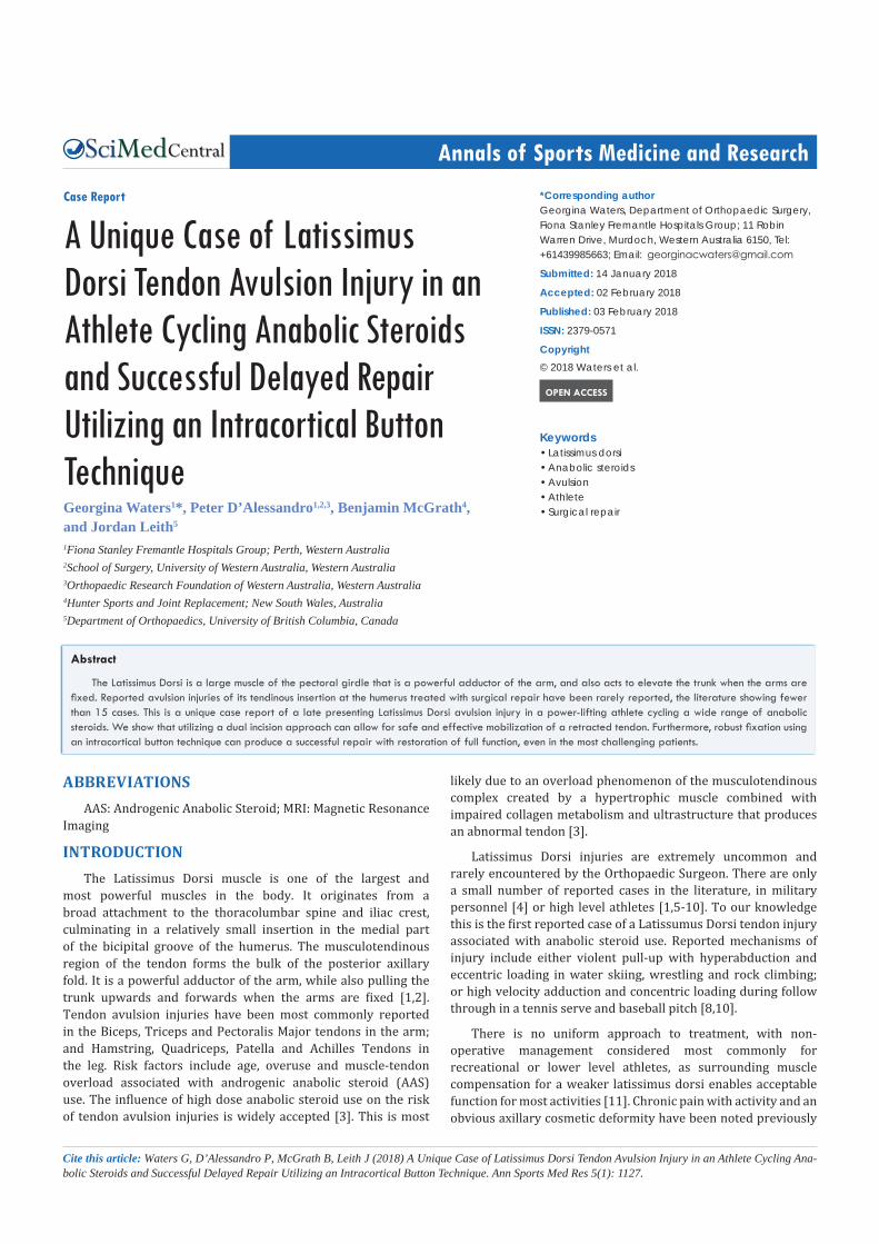
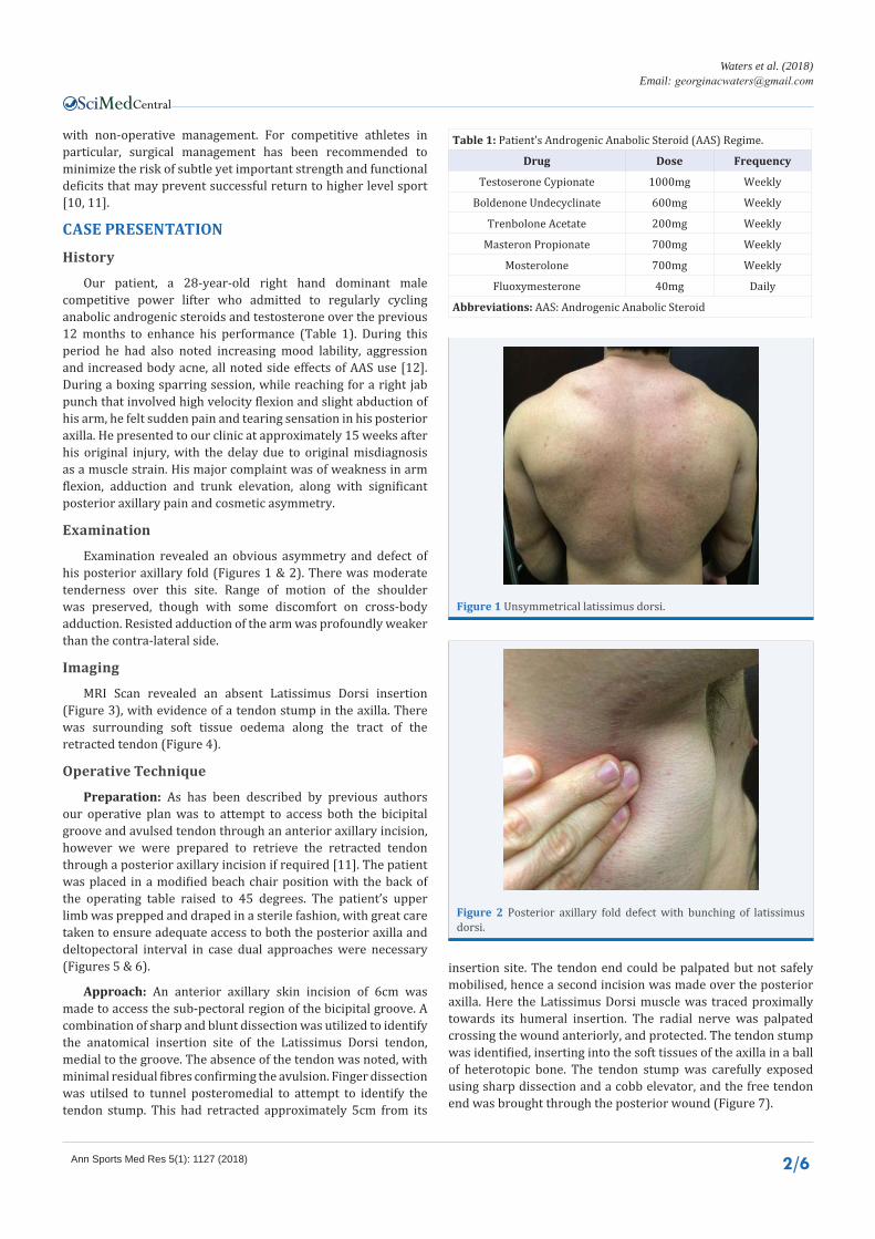
Examination revealed an obvious asymmetry and defect of his posterior axillary fold (Figures 1 & 2). There was moderate tenderness over this site. Range of motion of the shoulder was preserved, though with some discomfort on cross-body adduction. Resisted adduction of the arm was profoundly weaker than the contra-lateral side.
Imaging
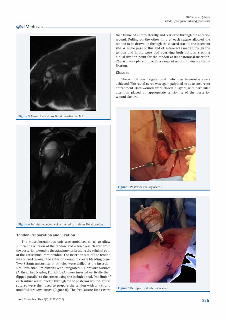
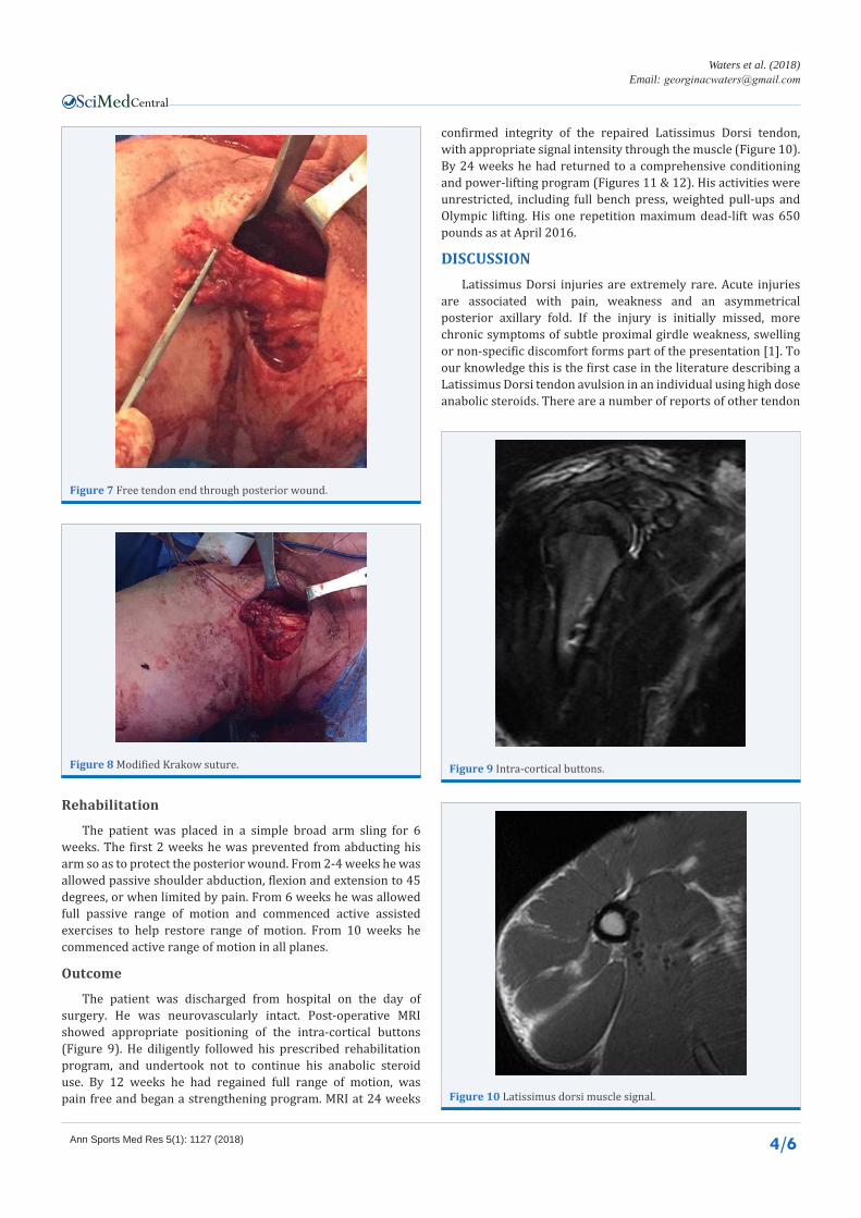
MRI Scan revealed an absent Latissimus Dorsi insertion (Figure 3), with evidence of a tendon stump in the axilla. There was surrounding soft tissue oedema along the tract of the retracted tendon (Figure 4).
Operative Technique
Preparation: As has been described by previous authors our operative plan was to attempt to access both the bicipital groove and avulsed tendon through an anterior axillary incision, however we were prepared to retrieve the retracted tendon through a posterior axillary incision if required [11]. The patient was placed in a modified beach chair position with the back of the operating table raised to 45 degrees. The patient’s upper limb was prepped and draped in a sterile fashion, with great care taken to ensure adequate access to both the posterior axilla and deltopectoral interval in case dual approaches were necessary (Figures 5 & 6).
Approach: An anterior axillary skin incision of 6cm was made to access the sub-pectoral region of the bicipital groove. A combination of sharp and blunt dissection was utilized to identify the anatomical insertion site of the Latissimus Dorsi tendon, medial to the groove. The absence of the tendon was noted, with minimal residual fibres confirming the avulsion. Finger dissection was utilsed to tunnel posteromedial to attempt to identify the tendon stump. This had retracted approximately 5cm from its
Table 1: Patient’s Androgenic Anabolic Steroid (AAS) Regime.
Drug Dose Frequency
Testoserone Cypionate 1000mg Weekly
Boldenone Undecyclinate 600mg Weekly
Trenbolone Acetate 200mg Weekly
Masteron Propionate 700mg Weekly
Mosterolone 700mg Weekly
Fluoxymesterone 40mg Daily
Abbreviations: AAS: Androgenic Anabolic Steroid
Figure 1 Unsymmetrical latissimus dorsi.
Figure 2 Posterior axillary fold defect with bunching of latissimus dorsi.
insertion site. The tendon end could be palpated but not safely mobilised, hence a second incision was made over the posterior axilla. Here the Latissimus Dorsi muscle was traced proximally towards its humeral insertion. The radial nerve was palpated crossing the wound anteriorly, and protected. The tendon stump was identified, inserting into the soft tissues of the axilla in a ball of heterotopic bone. The tendon stump was carefully exposed using sharp dissection and a cobb elevator, and the free tendon end was brought through the posterior wound (Figure 7).
Central
Waters et al. (2018)Email:
Ann Sports Med Res 5(1): 1127 (2018) 3/6
Figure 3 Absent Latissimus Dorsi insertion on MRI.
Figure 4 Soft tissue oedema of retracted Latissimus Dorsi tendon.
Figure 5 Posterior axillary access.
Figure 6 Deltopectoral interval access.
Tendon Preparation and Fixation
The musculotendinous unit was mobilised so as to allow sufficient excursion of the tendon, and a tract was cleared from the posterior wound to the attachment site along the original path of the Latissimus Dorsi tendon. The insertion site of the tendon was burred through the anterior wound to create bleeding bone. Two 3.5mm unicortical pilot holes were drilled at the insertion site. Two titanium buttons with integrated 5 Fibrewire Sutures (Arthrex Inc; Naples, Florida USA) were inserted vertically then flipped parallel to the cortex using the included tool. One limb of each suture was tunneled through to the posterior wound. These sutures were then used to prepare the tendon with a 4 strand modified Krakow suture (Figure 8). The free suture limbs were
then tunneled anterolaterally and retrieved through the anterior wound. Pulling on the other limb of each suture allowed the tendon to be drawn up through the cleared tract to the insertion site. A single pass of this end of suture was made through the tendon and knots were tied overlying both buttons, creating a dual fixation point for the tendon at its anatomical insertion. The arm was placed through a range of motion to ensure stable fixation.
Closure
The wound was irrigated and meticulous haemostasis was achieved. The radial nerve was again palpated so as to ensure no entrapment. Both wounds were closed in layers, with particular attention placed on appropriate tensioning of the posterior wound closure.
Central
Waters et al. (2018)Email:
Ann Sports Med Res 5(1): 1127 (2018) 4/6
Figure 7 Free tendon end through posterior wound.
Figure 8 Modified Krakow suture.
Rehabilitation
The patient was placed in a simple broad arm sling for 6 weeks. The first 2 weeks he was prevented from abducting his arm so as to protect the posterior wound. From 2-4 weeks he was allowed passive shoulder abduction, flexion and extension to 45 degrees, or when limited by pain. From 6 weeks he was allowed full passive range of motion and commenced active assisted exercises to help restore range of motion. From 10 weeks he commenced active range of motion in all planes.
Outcome
The patient was discharged from hospital on the day of surgery. He was neurovascularly intact. Post-operative MRI showed appropriate positioning of the intra-cortical buttons (Figure 9). He diligently followed his prescribed rehabilitation program, and undertook not to continue his anabolic steroid use. By 12 weeks he had regained full range of motion, was pain free and began a strengthening program. MRI at 24 weeks
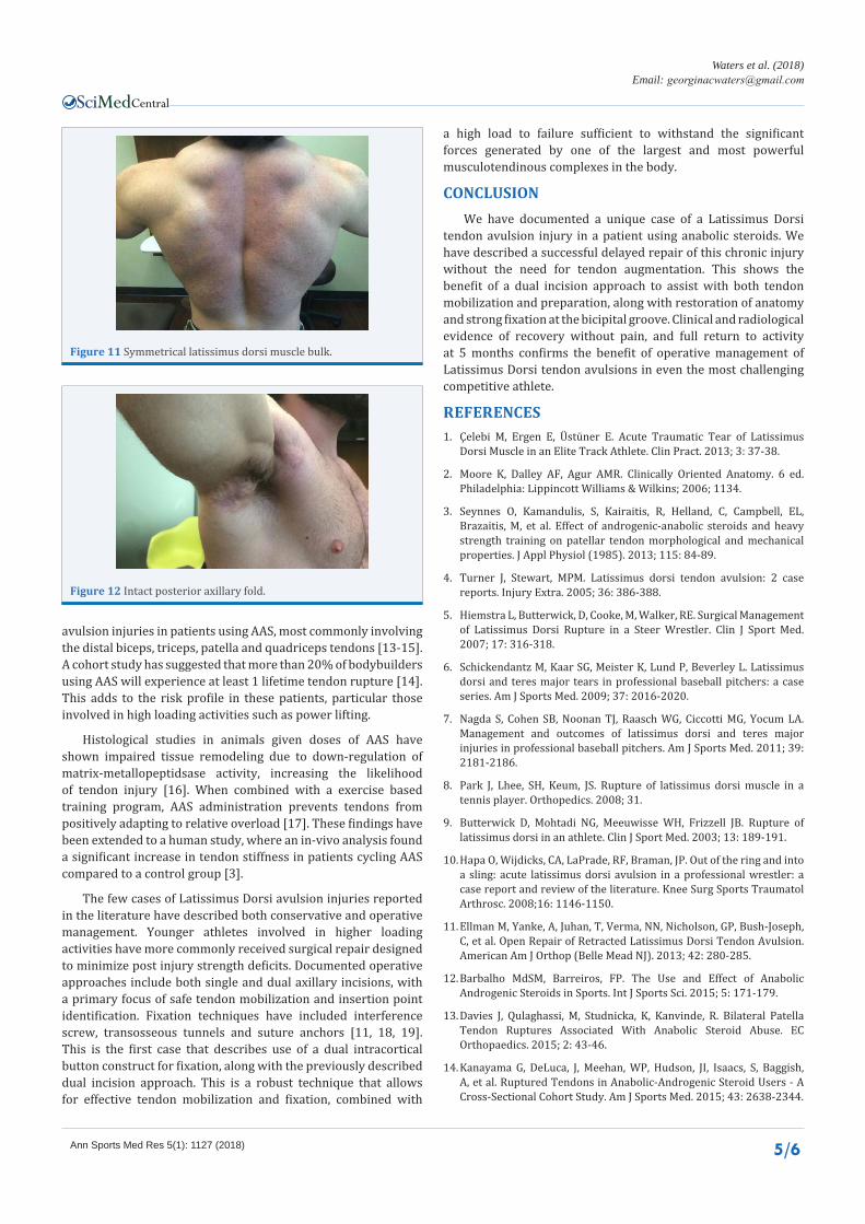
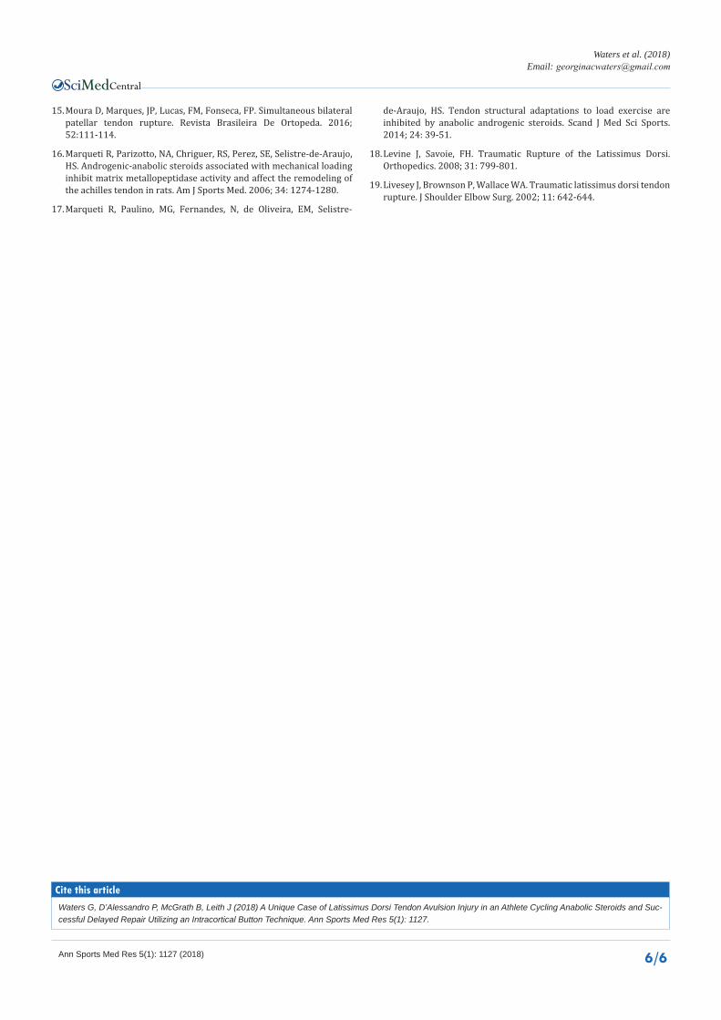
confirmed integrity of the repaired Latissimus Dorsi tendon, with appropriate signal intensity through the muscle (Figure 10). By 24 weeks he had returned to a comprehensive conditioning and power-lifting program (Figures 11 & 12). His activities were unrestricted, including full bench press, weighted pull-ups and Olympic lifting. His one repetition maximum dead-lift was 650 pounds as at April 2016.
DISCUSSIONLatissimus Dorsi injuries are extremely rare. Acute injuries
are associated with pain, weakness and an asymmetrical posterior axillary fold. If the injury is initially missed, more chronic symptoms of subtle proximal girdle weakness, swelling or non-specific discomfort forms part of the presentation [1]. To our knowledge this is the first case in the literature describing a Latissimus Dorsi tendon avulsion in an individual using high dose anabolic steroids. There are a number of reports of other tendon
Figure 9 Intra-cortical buttons.
Figure 10 Latissimus dorsi muscle signal.
Central
Waters et al. (2018)Email:
Ann Sports Med Res 5(1): 1127 (2018) 5/6
Figure 11 Symmetrical latissimus dorsi muscle bulk.
Figure 12 Intact posterior axillary fold.
avulsion injuries in patients using AAS, most commonly involving the distal biceps, triceps, patella and quadriceps tendons [13-15]. A cohort study has suggested that more than 20% of bodybuilders using AAS will experience at least 1 lifetime tendon rupture [14]. This adds to the risk profile in these patients, particular those involved in high loading activities such as power lifting.
Histological studies in animals given doses of AAS have shown impaired tissue remodeling due to down-regulation of matrix-metallopeptidsase activity, increasing the likelihood of tendon injury [16]. When combined with a exercise based training program, AAS administration prevents tendons from positively adapting to relative overload [17]. These findings have been extended to a human study, where an in-vivo analysis found a significant increase in tendon stiffness in patients cycling AAS compared to a control group [3].
The few cases of Latissimus Dorsi avulsion injuries reported in the literature have described both conservative and operative management. Younger athletes involved in higher loading activities have more commonly received surgical repair designed to minimize post injury strength deficits. Documented operative approaches include both single and dual axillary incisions, with a primary focus of safe tendon mobilization and insertion point identification. Fixation techniques have included interference screw, transosseous tunnels and suture anchors [11, 18, 19]. This is the first case that describes use of a dual intracortical button construct for fixation, along with the previously described dual incision approach. This is a robust technique that allows for effective tendon mobilization and fixation, combined with
a high load to failure sufficient to withstand the significant forces generated by one of the largest and most powerful musculotendinous complexes in the body.
CONCLUSIONWe have documented a unique case of a Latissimus Dorsi
tendon avulsion injury in a patient using anabolic steroids. We have described a successful delayed repair of this chronic injury without the need for tendon augmentation. This shows the benefit of a dual incision approach to assist with both tendon mobilization and preparation, along with restoration of anatomy and strong fixation at the bicipital groove. Clinical and radiological evidence of recovery without pain, and full return to activity at 5 months confirms the benefit of operative management of Latissimus Dorsi tendon avulsions in even the most challenging competitive athlete.
REFERENCES1. Çelebi M, Ergen E, Üstüner E. Acute Traumatic Tear of Latissimus
Dorsi Muscle in an Elite Track Athlete. Clin Pract. 2013; 3: 37-38.
2. Moore K, Dalley AF, Agur AMR. Clinically Oriented Anatomy. 6 ed. Philadelphia: Lippincott Williams & Wilkins; 2006; 1134.
3. Seynnes O, Kamandulis, S, Kairaitis, R, Helland, C, Campbell, EL, Brazaitis, M, et al. Effect of androgenic-anabolic steroids and heavy strength training on patellar tendon morphological and mechanical properties. J Appl Physiol (1985). 2013; 115: 84-89.
4. Turner J, Stewart, MPM. Latissimus dorsi tendon avulsion: 2 case reports. Injury Extra. 2005; 36: 386-388.
5. Hiemstra L, Butterwick, D, Cooke, M, Walker, RE. Surgical Management of Latissimus Dorsi Rupture in a Steer Wrestler. Clin J Sport Med. 2007; 17: 316-318.
6. Schickendantz M, Kaar SG, Meister K, Lund P, Beverley L. Latissimus dorsi and teres major tears in professional baseball pitchers: a case series. Am J Sports Med. 2009; 37: 2016-2020.
7. Nagda S, Cohen SB, Noonan TJ, Raasch WG, Ciccotti MG, Yocum LA. Management and outcomes of latissimus dorsi and teres major injuries in professional baseball pitchers. Am J Sports Med. 2011; 39: 2181-2186.
8. Park J, Lhee, SH, Keum, JS. Rupture of latissimus dorsi muscle in a tennis player. Orthopedics. 2008; 31.
9. Butterwick D, Mohtadi NG, Meeuwisse WH, Frizzell JB. Rupture of latissimus dorsi in an athlete. Clin J Sport Med. 2003; 13: 189-191.
10. Hapa O, Wijdicks, CA, LaPrade, RF, Braman, JP. Out of the ring and into a sling: acute latissimus dorsi avulsion in a professional wrestler: a case report and review of the literature. Knee Surg Sports Traumatol Arthrosc. 2008;16: 1146-1150.
11. Ellman M, Yanke, A, Juhan, T, Verma, NN, Nicholson, GP, Bush-Joseph, C, et al. Open Repair of Retracted Latissimus Dorsi Tendon Avulsion. American Am J Orthop (Belle Mead NJ). 2013; 42: 280-285.
12. Barbalho MdSM, Barreiros, FP. The Use and Effect of Anabolic Androgenic Steroids in Sports. Int J Sports Sci. 2015; 5: 171-179.
13. Davies J, Qulaghassi, M, Studnicka, K, Kanvinde, R. Bilateral Patella Tendon Ruptures Associated With Anabolic Steroid Abuse. EC Orthopaedics. 2015; 2: 43-46.
14. Kanayama G, DeLuca, J, Meehan, WP, Hudson, JI, Isaacs, S, Baggish, A, et al. Ruptured Tendons in Anabolic-Androgenic Steroid Users - A Cross-Sectional Cohort Study. Am J Sports Med. 2015; 43: 2638-2344.
Central
Waters et al. (2018)Email:
Ann Sports Med Res 5(1): 1127 (2018) 6/6
15. Moura D, Marques, JP, Lucas, FM, Fonseca, FP. Simultaneous bilateral patellar tendon rupture. Revista Brasileira De Ortopeda. 2016; 52:111-114.
16. Marqueti R, Parizotto, NA, Chriguer, RS, Perez, SE, Selistre-de-Araujo, HS. Androgenic-anabolic steroids associated with mechanical loading inhibit matrix metallopeptidase activity and affect the remodeling of the achilles tendon in rats. Am J Sports Med. 2006; 34: 1274-1280.
17. Marqueti R, Paulino, MG, Fernandes, N, de Oliveira, EM, Selistre-
de-Araujo, HS. Tendon structural adaptations to load exercise are inhibited by anabolic androgenic steroids. Scand J Med Sci Sports. 2014; 24: 39-51.
18. Levine J, Savoie, FH. Traumatic Rupture of the Latissimus Dorsi. Orthopedics. 2008; 31: 799-801.
19. Livesey J, Brownson P, Wallace WA. Traumatic latissimus dorsi tendon rupture. J Shoulder Elbow Surg. 2002; 11: 642-644.
Waters G, D’Alessandro P, McGrath B, Leith J (2018) A Unique Case of Latissimus Dorsi Tendon Avulsion Injury in an Athlete Cycling Anabolic Steroids and Suc-cessful Delayed Repair Utilizing an Intracortical Button Technique. Ann Sports Med Res 5(1): 1127.
Cite this article