a sign to heaven: avr lead elevation and myocardial...
TRANSCRIPT
Case Study TheScientificWorldJOURNAL (2011) 11, 662–665 ISSN 1537-744X; DOI 10.1100/tsw.2011.63
*Corresponding author. ©2011 with author. Published by TheScientificWorld; www.thescientificworld.com
662
A Sign to Heaven: aVR Lead Elevation and Myocardial Infarction
Amir M. Nia, Natig Gassanov, Hannes Reuter, and Fikret Er
Department of Internal Medicine III, University of Cologne, Cologne, Germany
E-mail: [email protected]
Received December 13, 2010; Revised February 11, 2011; Accepted February 11, 2011; Published March 22, 2011
Isolated ST-segment elevation only in the aVR lead, reflecting an acute myocardial infarction due to a left main coronary artery occlusion, was ignored as part of physicians’ training in emergency medicine for a long time. The recognition of aVR lead elevation is becoming more accepted as a mandatory diagnostic tool, in particular for physicians working at emergency departments. We report a typical myocardial infarction with total occlusion of the proximal part of the left anterior coronary artery, presenting with ST-segment elevation in the aVR lead, which was misinterpreted as diffuse ischemia. The lacking mandatory awareness of this entity endangered prompt and correct treatment.
KEYWORDS: aVR lead elevation, ST-segment elevation in aVR, left main coronary artery obstruction, LMCA obstruction, proximal LAD stenosis
CASE
A 63-year-old man was referred from a tertiary hospital’s emergency triage to our cardiology department
with the diagnosis of acute coronary syndrome with global ST-segment depressions. On admission, his
blood pressure was 100/60 mmHg with a heart rate of 90 beats per minute. He complained of moderate
chest pain of several days duration, but 1 h before admission, he noticed an increasingly severe thoracic
discomfort, which caused him to seek medical attention. In his history, Parkinsonism, arterial
hypertension, and hyperlipidemia were known. Prior to the last days, he had never felt angina pectoris.
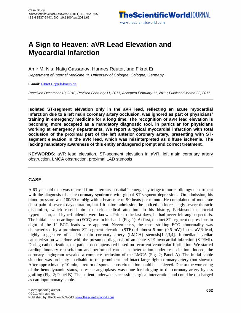
The initial electrocardiogram (ECG) was in his hands (Fig. 1). At first, distinct ST-segment depressions in
eight of the 12 ECG leads were apparent. Nevertheless, the most striking ECG abnormality was
characterized by a prominent ST-segment elevation (STE) of almost 5 mm (0.5 mV) in the aVR lead,
highly suggestive of a left main coronary artery (LMCA) stenosis[1,2,3,4]. Immediate cardiac
catheterization was done with the presumed diagnosis of an acute STE myocardial infarction (STEMI).
During catheterization, the patient decompensated based on recurrent ventricular fibrillation. We started
cardiopulmonary resuscitation and performed cardiac catheterization under resuscitation. Indeed, the
coronary angiogram revealed a complete occlusion of the LMCA (Fig. 2; Panel A). The initial stable
situation was probably ascribable to the prominent and intact large right coronary artery (not shown).
After approximately 10 min, a return of spontaneous circulation could be achieved. Due to the worsening
of the hemodynamic status, a rescue angioplasty was done for bridging to the coronary artery bypass
grafting (Fig. 2; Panel B). The patient underwent successful surgical intervention and could be discharged
as cardiopulmonary stable.

Nia et al.: aVR Lead Elevation and Myocardial Infarction TheScientificWorldJOURNAL (2011) 11, 662–665
663
FIGURE 1. 12-Lead ECG reveals a prominent ST-segment elevation in the aVR lead.
A B
FIGURE 2. Coronary angiograms. (A) Anterior-posterior view displays a complete occlusion of the LMCA. (B)
Right anterior oblique 30°/caudal 20° view displays a marginal reperfusion after rescue angioplasty of the
LMCA.

Nia et al.: aVR Lead Elevation and Myocardial Infarction TheScientificWorldJOURNAL (2011) 11, 662–665
664
The 12-lead ECG is a widely available bedside test, especially in the emergency department, for
urgent triage. Although each lead provides specific information, the aVR lead has been frequently
overlooked in the past[2,5,6]. A review of the literature uncovers that aVR lead changes are widely
ignored[5,7,8,9]. A very interesting study was performed to investigate ECG interpreters’ disregard of the
aVR lead[5]. An experienced medical staff was asked to interpret complex ECGs, but the aVR lead had
been replaced by the –aVR lead (reversed aVR lead with putative positive vector) on all of these
recordings. The vast majority of interpreters (80–94%) did not detect when the aVR lead had been
reversed[5]. Probably, the usually negative QRS vector of the aVR lead may lead to its underestimation.
However, the tracing in this lead can be used to obtain a unique view directly into the right ventricular
outflow tract and the basal portion of the interventricular septum[6]. Thus, it is important to use the aVR
lead as an essential part of the ECG interpretation[2,3,10]. There are further aVR lead findings worthy of
discussion, such as PR-segment elevation indicating acute pericarditis or prominent R’ waves indicating
tricyclic antidepressant poisoning[10,11]. Moreover, several levels of evidence substantiate that STE in
the aVR lead is highly associated with the left main, the left anterior descending coronary artery (LAD),
and 3-vessel coronary artery disease[1,2,3,4,12]. Yamaji et al. reported that STE in the aVR lead greater
than that in lead V1 may be useful for predicting acute LMCA obstruction, which is a rare angiographic
finding and requires immediate intensive treatment[3]. During the last years, this finding could be
confirmed by other colleagues[2,4,12]. The most likely explanation of less STE in lead V1 in patients with
LMCA disease compared to those with LAD disease may be the result of additive posterior wall ischemia
in LMCA-diseased patients[3]. Contrary to patients with LAD disease, LMCA obstruction induces
posterior wall ischemia through disturbance of left circumflex artery blood flow[3]. Due to this, the
posterior wall–induced electrical force counterbalances the ischemia-induced electrical force in the
anterior wall, leading to more prominent STE in the aVR lead than in the chest leads[2,3].
As exemplified in our case, STE in the aVR lead is often misinterpreted and typically neglected.
Apparently, this case report displays that the general criteria for STEMI might be incomplete and that an
isolated STE in the aVR lead reflects a STEMI due to occlusion of the LMCA[2,3,4]. We think that it is
mandatory that this “Sign to Heaven” be known and recognized by all physicians interpreting ECGs in
their daily clinical work.
REFERENCES
1. Kosuge, M., Kimura, K., Ishikawa, T., Ebina, T., Shimizu, T., Hibi, K., Toda, N., Tahara, Y., Tsukahara, K., Kanna,
M., Okuda, J., Nozawa, N., Ozaki, H., Yano, H., and Umemura, S. (2005) Predictors of left main or three-vessel
disease in patients who have acute coronary syndromes with non-ST-segment elevation. Am. J. Cardiol. 95, 1366–
1369.
2. Wong, C.K., Gao, W., Stewart, R.A., Benatar, J., French, J.K., Aylward, P.E., and White, H.D. (2010) aVR ST
elevation: an important but neglected sign in ST elevation acute myocardial infarction. Eur. Heart J. 31, 1845–1853.
3. Yamaji, H., Iwasaki, K., Kusachi, S., Murakami, T., Hirami, R., Hamamoto, H., Hina, K., Kita, T., Sakakibara, N.,
and Tsuji, T. (2001) Prediction of acute left main coronary artery obstruction by 12-lead electrocardiography. ST
segment elevation in lead aVR with less ST segment elevation in lead V(1). J. Am. Coll. Cardiol. 38, 1348–1354.
4. Kuhl, J.T. and Berg, R.M. (2009) Utility of lead aVR for identifying the culprit lesion in acute myocardial infarction.
Ann. Noninvasive Electrocardiol. 14, 219–225.
5. Pahlm, U.S., Pahlm, O., and Wagner, G.S. (1996) The standard 11-lead ECG. Neglect of lead aVR in the classical
limb lead display. J. Electrocardiol. 29(Suppl), 270–274.
6. Gorgels, A.P., Engelen, D.J., and Wellens, H.J. (2001) Lead aVR, a mostly ignored but very valuable lead in clinical
electrocardiography. J. Am. Coll. Cardiol. 38, 1355–1356.
7. Sugishita, K., Shimizu, T., Kinugawa, K., Harada, K., Ikenouchi, H., Matsui, H., Kohmoto, O., Takahashi, T., and
Omata, M. (1997) Chronic total occlusion of the left main coronary artery. Intern. Med. 36, 471–478.
8. Radwaner, B.A., Geringer, R., Goldmann, A.M., Schwartz, M.J., and Kemp, H.G., Jr. (1987) Left main coronary
artery stenosis following mediastinal irradiation. Am. J. Med. 82, 1017–1020.
9. Er, F. and Erdmann, E. (2010) 68-Year-old patient with exercise-induced syncope. Dtsch. Med. Wochenschr. 135,
262; author reply 262.
10. Williamson, K., Mattu, A., Plautz, C.U., Binder, A., and Brady, W.J. (2006) Electrocardiographic applications of lead
aVR. Am. J. Emerg. Med. 24, 864–874.

Nia et al.: aVR Lead Elevation and Myocardial Infarction TheScientificWorldJOURNAL (2011) 11, 662–665
665
11. Babai Bigi, M.A., Aslani, A., and Shahrzad, S. (2007) aVR sign as a risk factor for life-threatening arrhythmic events
in patients with Brugada syndrome. Heart Rhythm 4, 1009–1012.
12. de Winter, R.J., Verouden, N.J., Wellens, H.J., and Wilde, A.A. (2008) A new ECG sign of proximal LAD occlusion.
N. Engl. J. Med. 359, 2071–2073.
This article should be cited as follows:
Nia, A.M., Gassanov, N., Reuter, H., and Er, F. (2011) A sign to heaven: aVR lead elevation and myocardial infarction.
TheScientificWorldJOURNAL 11, 662–665. DOI 10.1100/tsw.2011.63.

Submit your manuscripts athttp://www.hindawi.com
Stem CellsInternational
Hindawi Publishing Corporationhttp://www.hindawi.com Volume 2014
Hindawi Publishing Corporationhttp://www.hindawi.com Volume 2014
MEDIATORSINFLAMMATION
of
Hindawi Publishing Corporationhttp://www.hindawi.com Volume 2014
Behavioural Neurology
EndocrinologyInternational Journal of
Hindawi Publishing Corporationhttp://www.hindawi.com Volume 2014
Hindawi Publishing Corporationhttp://www.hindawi.com Volume 2014
Disease Markers
Hindawi Publishing Corporationhttp://www.hindawi.com Volume 2014
BioMed Research International
OncologyJournal of
Hindawi Publishing Corporationhttp://www.hindawi.com Volume 2014
Hindawi Publishing Corporationhttp://www.hindawi.com Volume 2014
Oxidative Medicine and Cellular Longevity
Hindawi Publishing Corporationhttp://www.hindawi.com Volume 2014
PPAR Research
The Scientific World JournalHindawi Publishing Corporation http://www.hindawi.com Volume 2014
Immunology ResearchHindawi Publishing Corporationhttp://www.hindawi.com Volume 2014
Journal of
ObesityJournal of
Hindawi Publishing Corporationhttp://www.hindawi.com Volume 2014
Hindawi Publishing Corporationhttp://www.hindawi.com Volume 2014
Computational and Mathematical Methods in Medicine
OphthalmologyJournal of
Hindawi Publishing Corporationhttp://www.hindawi.com Volume 2014
Diabetes ResearchJournal of
Hindawi Publishing Corporationhttp://www.hindawi.com Volume 2014
Hindawi Publishing Corporationhttp://www.hindawi.com Volume 2014
Research and TreatmentAIDS
Hindawi Publishing Corporationhttp://www.hindawi.com Volume 2014
Gastroenterology Research and Practice
Hindawi Publishing Corporationhttp://www.hindawi.com Volume 2014
Parkinson’s Disease
Evidence-Based Complementary and Alternative Medicine
Volume 2014Hindawi Publishing Corporationhttp://www.hindawi.com