a study on the parasite diversity and community …
TRANSCRIPT

A STUDY ON THE PARASITE DIVERSITY AND COMMUNITY ECOLOGY OF
THREE SPECIES OF TEXAS FRESHWATER TURTLES
A Thesis
by
WESLEY JAMES NEELY
Submitted to the Office of Graduate and Professional Studies of
Texas A&M University
in partial fulfillment of the requirements for the degree of
MASTER OF SCIENCE
Chair of Committee, Norman O. Dronen
Committee Members, Toby J. Hibbitts
Thomas M. Craig
Head of Department, David J. Caldwell
August 2018
Major Subject: Wildlife and Fisheries Sciences
Copyright 2018 Wesley James Neely

ii
ABSTRACT
In this study, the metazoan parasites of three species of freshwater turtles (the
spiny softshell, Apalone spinifera, the common snapping turtle, Chelydra serpentina,
and the red-eared slider, Trachemys scripta elegans) were surveyed at 16 sites across the
state of Texas. A total of 42 species of metazoan parasites were recovered from 15 A.
spinifera, nine C. serpentina, and 55 T. s. elegans, representing 16 new host-parasite
associations and 17 new locality records. The synonymy of Acanthostomum
nuevoleonensis by Brooks (1980) is refuted and the species is redescribed. Two new
species of monogenean worms in the genus Neopolystoma are reported, one from C.
serpentina and A. spinifera and another from T. s. elegans. Through non-metric
multidimensional scaling and analysis of similarities, A. spinifera was found to contain a
significantly distinct parasite community from C. serpentina and T. s. elegans. A range
of water parameters (ammonia, carbon dioxide, chloride, dissolved oxygen, hardness,
nitrite, nitrate, pH, salinity, temperature, and turbidity) were recorded on each sampling
trip and compared to parasite abundance and diversity. Ammonia levels were positively
correlated with abundance of acanthocephalans. Carbon dioxide levels were negatively
correlated with parasite diversity and monogenean abundance. Chloride levels were
negatively correlated with parasite diversity. Dissolved oxygen levels were positively
correlated with parasite diversity and monogenean abundance. Turbidity was positively
correlated with parasite abundance, acanthocephalan abundance, and digenean
abundance, and negatively correlated with parasite diversity. Parasite abundance was

iii
significantly lower in west Texas and western river basins, and lower in rivers than
ponds. Acanthocephalan abundance was significantly lower in rivers than ponds. Leech
abundance was highest in the Trinity river basin. Turbidity had the strongest correlations
in this study. As water clarity increased, diversity increased and abundance of certain
taxa decreased, indicating clearer water may have greater food web diversity and
healthier hosts. This study adds valuable data on host-parasite associations, parasite
distributions, and parasite ecology of turtles in the state of Texas. Many of these findings
are likely transferable to other host taxa and should be studied in greater depth. Parasite
diversity is not well known, even in common species, highlighting the need for more
diversity surveys.

iv
DEDICATION
This thesis is dedicated to my educators and mentors, past and present. Thank you for
believing in me and pushing me to greater things.

v
ACKNOWLEDGEMENTS
This project would not have been possible without the contributions of a number
of people. First and foremost I would like to thank my advisor, Dr. Norman Dronen, for
teaching me so many valuable skills, always being available, offering direction and
inspiration, and being my biggest advocate. I would also like to thank my committee
members, Dr. Toby Hibbitts and Dr. Thomas Craig for their invaluable assistance with
parasite and turtle identification, equipment acquisition, trapping methodology, and
writing edits during the course of this research.
My lab team, Charlayna Cammarata, Kelsey Garner, and Travis Doggett have
been excellent, both in the field and the lab. Rain or shine, heat or snow, you all
persevered and got work done. Additional thanks to Angie Achorn and Alexis
Pitsnukanh for assistance with parasite processing and measurement.
The Texas Ecolabs program funded this entire project and provided access to the
majority of sampling locations, a distribution that was vital in completing this project. In
addition, thanks to Russell Hanna, Frank Orsak, Bill Hudgins and Katherine Metter,
Shirley Hesse, Nirmal Bual, Frank and Diane Arthur, Tyler Summers, Steve Lumpee,
Susan Finkelstein, Bill McMorris, Kay Ann McKinney, Suzanne Harter, and John Avery
for access to their properties, lodging, and answering all my many questions. Thanks to
Andy Newman, Dr. Terry Blankenship, Steve Chernosky, Dr. Delbert Gatlin, and Brian
Ray for providing access to additional sampling locations and lodging.

vi
Special thanks to Dr. Mike Kinsella for invaluable assistance with nematode
identification, Bill Moser and Dr. Jessica Light for pointers on leech identification, and
Dr. Stephen Curran for identifying all pentastomids.
I am thankful for my friends here and back home, Ryan Metts, Jordan Rogan,
Aleyda Galan, Helen Davis, John Pistone, Amanda McKenna, Jerry Huntley, Danielle
Walkup, Johanna Harvey, Bridgett Benedict, Kinley Prince, Cale and Sam Neff, Nick
Klock, Dillon Hollifield, David Saenz, Zach Grey, Liz Marchio, Connor Adams, Nikki
Roach, John Godwin, Taylor Hyatt, Shishir Basant, Marley McDonough, Rika Muhl,
Alejandra Maldonado, Beth Godwin, and Chris Downs, all contributing to my
completion of this project in their own ways. Thanks to Adrian Castellanos and Luke
Bower for assistance with data analysis in R and answering random questions. Thanks to
Charlayna Cammarata, Whitney Preisser, and Jason Martina for assistance with idea
development, data analysis, and writing edits.
None of this could have been possible without my awesome, supportive family,
Les, Helen, and Rebekah Neely, Todd and Catherine Johnston, Jim and Catherine
Moore, Jimmy, Stacie, and Jamie Burnett, Taylor Vaughn, and Mackenzie Holombo, for
too many reasons to name.

vii
CONTRIBUTORS AND FUNDING SOURCES
This work was supervised by a thesis committee consisting of Dr. Norman O.
Dronen (advisor) and Dr. Toby J. Hibbitts of the Department of Wildlife and Fisheries
Sciences and Dr. Thomas M. Craig of the Department of Veterinary Medicine and
Biomedical Sciences.
All work for the thesis was completed by the student, in collaboration with
Charlayna Cammarata. Undergraduate assistants Kelsey Garner and Travis Doggett
assisted with a large portion of field and lab work for this project.
Graduate study was supported by a teaching assistantship from Texas A&M
University. This project was funded in full by Texas Ecolabs.

viii
TABLE OF CONTENTS
Page
ABSTRACT .......................................................................................................................ii
DEDICATION .................................................................................................................. iv
ACKNOWLEDGEMENTS ............................................................................................... v
CONTRIBUTORS AND FUNDING SOURCES ............................................................vii
TABLE OF CONTENTS ............................................................................................... viii
LIST OF FIGURES ............................................................................................................ x
LIST OF TABLES ............................................................................................................ xi
CHAPTER I INTRODUCTION ....................................................................................... 1
CHAPTER II PARASITE DIVERISTY AND COMMUNITY STRUCTURE IN
TEXAS FRESHWATER TURTLES ................................................................................. 8
II.1 Introduction ............................................................................................................. 8 II.2 Materials and methods ........................................................................................... 12
II.2.1 Field materials and methods ........................................................................... 12
II.2.2 Lab materials and methods ............................................................................. 13 II.2.3 Parasite processing .......................................................................................... 14
II.2.4 Statistical analyses .......................................................................................... 15 II.3 Results ................................................................................................................... 16
II.3.1 Host-community analysis ............................................................................... 18 II.3.2 Host-parasite associations ............................................................................... 24 II.3.3 Parasite taxonomy ........................................................................................... 37 II.3.4 Regional associations ...................................................................................... 55
II.4 Discussion .............................................................................................................. 63
CHAPTER III ENVIRONMENTAL AND REGIONAL INFLUENCES ON
PARASITE ASSEMBLAGES IN TEXAS FRESHWATER TURTLES ........................ 68
III.1 Introduction .......................................................................................................... 68 III.2 Methods ................................................................................................................ 72
III.2.1 Field materials and methods .......................................................................... 72

ix
III.2.2 Lab materials and methods ............................................................................ 75 III.2.3 Parasite processing ........................................................................................ 76 III.2.4 Statistical analysis ......................................................................................... 77
III.3 Results .................................................................................................................. 79
III.3.1 Sample sites ................................................................................................... 79 III.3.2 Capture data ................................................................................................... 81 III.3.3 Regional and water quality analyses ............................................................. 82 III.3.4 Host measurement analyses ........................................................................... 94
III.4 Discussion ............................................................................................................ 97
CHAPTER IV SUMMARY AND FUTURE DIRECTIONS ........................................ 104
REFERENCES ............................................................................................................... 107
APPENDIX .................................................................................................................... 121

x
LIST OF FIGURES
Page
Figure 1: Map of the collection locations of turtles across Texas. ................................... 17
Figure 2: Plotted nMDS ordination showing parasite species grouping by host
species. .............................................................................................................. 19
Figure 3: Plerocercoid protoscolex showing sucker arrangement. .................................. 32
Figure 4: Illustration of Acanthostomum nuevoleonensis. ............................................... 39
Figure 5: Illustration of Neopolystoma n. sp. 1. ............................................................... 44
Figure 6: Illustration of Neopolystoma n. sp. 2. ............................................................... 47
Figure 7: Illustration of Polystomoides coronatum. ......................................................... 50
Figure 8: Map of the study sites across Texas A) within ecoregions and B) within
river basins. ....................................................................................................... 75
Figure 9: Linear regressions for turbidity (water clarity), trend lines plotted with 95%
confidence intervals. ......................................................................................... 85
Figure 10: Linear regressions for carbon dioxide, trend lines plotted with 95%
confidence intervals. ......................................................................................... 86
Figure 11: Linear regressions for dissolved oxygen, trend lines plotted with 95%
confidence intervals. ......................................................................................... 87
Figure 12: Linear regression for chloride levels and taxonomic diversity, trend line
plotted with 95% confidence interval. .............................................................. 88
Figure 13: Linear regression for ammonia levels and acanthocephalan abundance,
trend line plotted with 95% confidence interval. .............................................. 89
Figure 14: Linear regressions for body size of A. spinifera in relation to parasite
abundance, trend line plotted with 95% confidence interval. ........................... 95

xi
LIST OF TABLES
Page
Table 1: Metazoan parasites recovered from 15 Apalone spinifera, nine Chelydra
serpentina, and 55 Trachemys scripta elegans. ................................................ 20
Table 2: A comparison of the body measurements of Acanthostomum species,
including specimens collected in this study. ..................................................... 42
Table 3: A comparison of the body measurements of Polystomoides coronatum
reported by Leidy (1888), Stunkard (1917), Price (1939), and the current
study. ................................................................................................................. 54
Table 4: Significance coefficients (p-values and R2 values) from simple linear
regressions on parasite abundance and diversity. ............................................. 92
Table 5: Significance coefficients (p-values and R2 values) from simple linear
regressions on parasite taxa. ............................................................................. 92
Table 6: Significance coefficients (p-values) from one-way ANOVAs. Significant
values are bolded. ............................................................................................. 93

1
CHAPTER I
INTRODUCTION
Parasite diversity is a key component in understanding ecosystem complexity.
With conservative estimates of around 40% of known species being parasitic, parasitism
is the most common life strategy (Dobson et al., 2008). The high number of unsampled
host species and amount of cryptic speciation potentially uncovered through genetic
analysis indicates that this number is likely higher (Jousson et al., 2000; Steinauer et al.,
2007). Helminths, or parasites in the phyla Acanthocephala, Nematoda, Platyhelminthes,
and the subclass Pentastomida, often have complex life cycles, sometimes traveling
through many hosts throughout development (e.g. Parker et al., 2003; Poulin, 2011).
Endohelminths, or internal helminths, are associated with predator-prey relationships, as
they are typically transmitted through consumption. Current estimates indicate that
parasites are involved in nearly 75% of all trophic linkages in food webs due to their
complex life cycles and dependence on hosts (Lafferty, 2008; Lafferty et al., 2006).
Consequently, healthy ecosystems with greater numbers of trophic linkages are believed
to be higher in parasite diversity. Parasite diversity is therefore a good indicator of
ecosystem diversity and total ecosystem health (Hudson et al., 2006; Marcogliese, 2005).
Despite these findings, parasite assemblages remain highly understudied, with many new
species being described every year and with many life cycles completely unknown
(Blasco-Costa and Poulin, 2017; Dobson et al, 2008; Poulin, 2014).

2
Previous research suggests that parasitic species may be trophic regulators in the
same capacity as top predators (e.g. Dougherty et al., 2016; Lafferty et al. 2006);
however, most conservation plans do not implement any efforts to preserve parasite
diversity, and often work to eradicate parasites to alleviate stressors on threatened
species. In order to conserve the total diversity in an ecosystem, parasite diversity must
be taken into account when making conservation plans. For this to be possible
knowledge of the parasites species present in a given ecosystem is vital. Studies on
parasite diversity are difficult as they generally require collection and euthanasia of a
large number of hosts. In spite of the challenges, studies on parasite communities are
needed to understand the full breadth of diversity in ecosystems (Hudson et al., 2006;
Dobson et al., 2008).
Environmental factors can alter parasite assemblages in significant ways. Some
environmental factors associated with changes in parasite abundance and diversity are
temperature, dissolved oxygen, turbidity, salinity, nutrient pollution, metal pollution,
pesticide/herbicide pollution, and habitat alteration (e.g. Bourque and Esch, 1974;
Lafferty and Kuris, 1999; Banu and Khan, 2004; Nachev and Sures, 2009; Shea et al.,
2012; Chapman et al., 2015; Ahmad et al., 2016). Due to the complex and diverse nature
of parasite life cycles, the effects of environmental factors are variable, and often
contradictory. Bourque and Esch (1974) found that nematode abundance responded
differently to thermal pollution between two wetlands. Goednkegt et al. (2015) reported
trematode infectivity to increase with increasing temperatures; however, predation on
cercaria also increased with increasing temperatures, which then decreased trematode

3
infectivity. In their review, Lafferty and Kuris (1999) found a variety of possible
outcomes on parasite-host interactions impacted by environmental stressors. Pollutants
can increase parasite infectivity by increasing host susceptibility or decrease parasite
infectivity by decreasing host survival and therefore parasite transmission. Zargar et al.
(2012) found varying intensities of monogeneans on fish across polluted lakes, with
decreasing intensities in one polluted and eutrophied lake and increasing intensities at a
different polluted and eutrophied lake. Current trends in environmental degradation and
climate change point to a change in currently observed parasite diversity (e.g. Brooks
and Hoberg, 2007; Strona, 2015; Cizauskas et al. 2017). With the convoluted nature of
environmental effects on parasites, it is vital to continue research in smaller systems that
can be used to clarify the bigger picture.
The state of Texas can be broken up into 12 major ecological regions and 15
major river basins. Ecological regions, or ecoregions, are large stretches of land that are
grouped based on the native vegetation, hydrology, and geochemistry (Griffith et al.,
2007). Aquatic communities can be characterized by the ecoregion in which they reside,
as aquatic community assemblages tend to vary greatly among different ecoregions
(Warry and Hanau, 1993; Stoddard, 2005). The ecoregions found within the state of
Texas are the Arizona/New Mexico Mountains, Chihuahuan Desert, High Plains,
Southwestern Tablelands, Central Great Plains, Cross Timbers, Edwards Plateau,
Southern Texas Plains, Texas Blackland Prairies, East Central Texas Plains, Western
Gulf Coastal Plains, and South Central Plains. These ecoregions have many
characteristic features such as the vegetative communities (hardwood forest, prairie,

4
scrublands, etc.) and soil characteristics (sand or clay, acidic or basic, shallow or deep,
etc.) (Griffith et al., 2007).
The river basins delimit the area drained by each major river and its tributaries
and may cross multiple ecoregions (Bureau of Economic Geology, 1996). The separation
between these basins can often be a determining factor in the range of aquatic species, as
seen in freshwater mussel diversity (Burlakova et al., 2011). The river basins found in
the state of Texas are the Brazos, Canadian, Colorado, Cypress, Guadalupe, Lavaca,
Neches, Nueces, Red, Rio Grande, Sabine, San Antonio, San Jacinto, Sulphur, and
Trinity Basins. The water chemistry and biotic communities may change across the river
basin, since the common river basin is the only connecting factor (Ford et al. 2016).
Texas can also be viewed in respects of latitudinal and longitudinal gradients. North and
East Texas are typically wetter while South and West Texas are typically drier (Texas
Commission on Environmental Quality, 2018), which could lead to shifts in parasite
diversity and abundance due to changes in intermediate host abundance and larval
dispersal (Janzen and Schoener, 1968; Froeschke et al. 2010).
In the state of Texas, only four metazoan parasite surveys have been conducted
on freshwater turtles (Harwood, 1932; Everhart, 1957; Dinuzzo, 1981; McAllister et al.,
2008). Harwood (1932) conducted a survey of the endohelminths of 50 species of
amphibians and reptiles over the course of two and a half years in the vicinity of
Houston, Texas. Over the course of this study, eight species of turtles were collected: 16
red-eared sliders, Trachemys scripta elegans (Wied, 1839), 16 Mississippi mud turtles,
Kinosternon subrubrum hippocrepis Gray, 1856, 14 three-toed box turtles, Terrapene
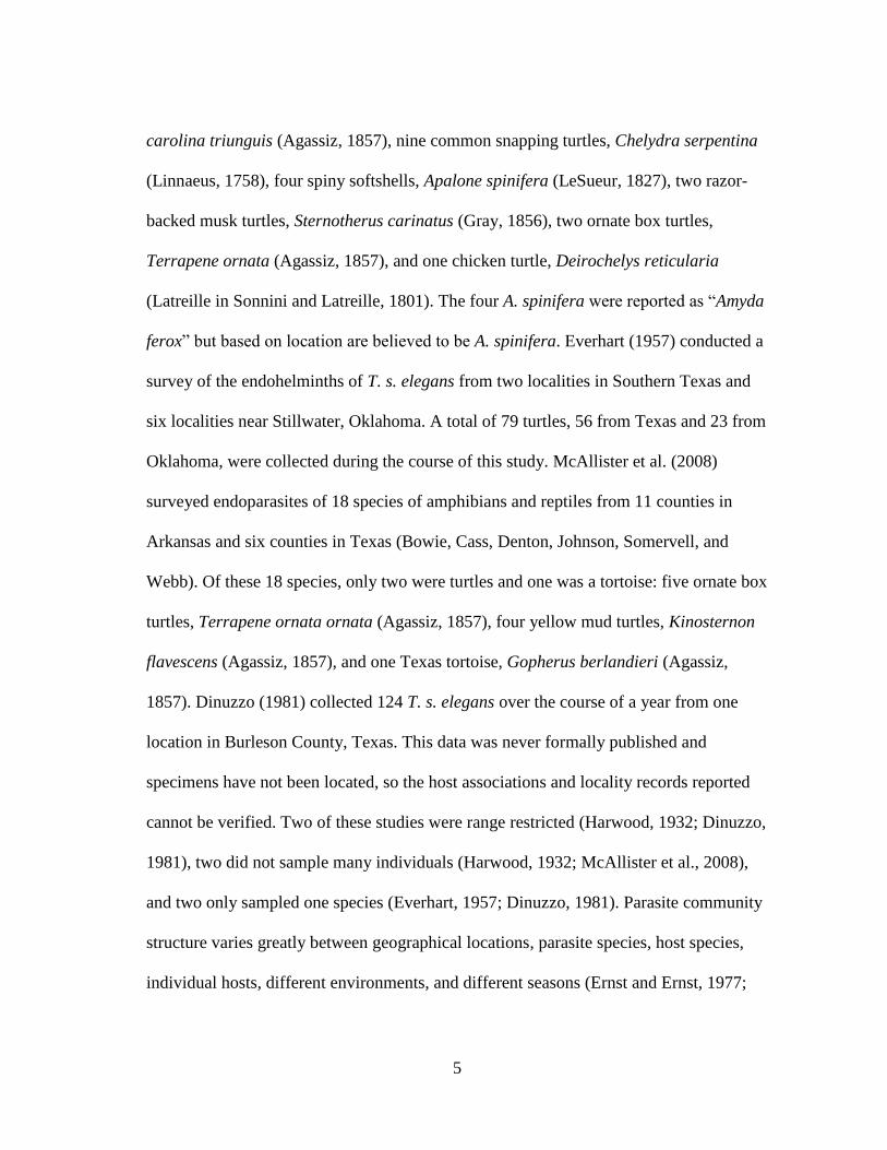
5
carolina triunguis (Agassiz, 1857), nine common snapping turtles, Chelydra serpentina
(Linnaeus, 1758), four spiny softshells, Apalone spinifera (LeSueur, 1827), two razor-
backed musk turtles, Sternotherus carinatus (Gray, 1856), two ornate box turtles,
Terrapene ornata (Agassiz, 1857), and one chicken turtle, Deirochelys reticularia
(Latreille in Sonnini and Latreille, 1801). The four A. spinifera were reported as “Amyda
ferox” but based on location are believed to be A. spinifera. Everhart (1957) conducted a
survey of the endohelminths of T. s. elegans from two localities in Southern Texas and
six localities near Stillwater, Oklahoma. A total of 79 turtles, 56 from Texas and 23 from
Oklahoma, were collected during the course of this study. McAllister et al. (2008)
surveyed endoparasites of 18 species of amphibians and reptiles from 11 counties in
Arkansas and six counties in Texas (Bowie, Cass, Denton, Johnson, Somervell, and
Webb). Of these 18 species, only two were turtles and one was a tortoise: five ornate box
turtles, Terrapene ornata ornata (Agassiz, 1857), four yellow mud turtles, Kinosternon
flavescens (Agassiz, 1857), and one Texas tortoise, Gopherus berlandieri (Agassiz,
1857). Dinuzzo (1981) collected 124 T. s. elegans over the course of a year from one
location in Burleson County, Texas. This data was never formally published and
specimens have not been located, so the host associations and locality records reported
cannot be verified. Two of these studies were range restricted (Harwood, 1932; Dinuzzo,
1981), two did not sample many individuals (Harwood, 1932; McAllister et al., 2008),
and two only sampled one species (Everhart, 1957; Dinuzzo, 1981). Parasite community
structure varies greatly between geographical locations, parasite species, host species,
individual hosts, different environments, and different seasons (Ernst and Ernst, 1977;

6
Esch and Gibbons, 1967; Poulin, 2006; Readel et al., 2008). For this reason, it is useful
for studies to cover multiple host species across broader geographic and temporal ranges
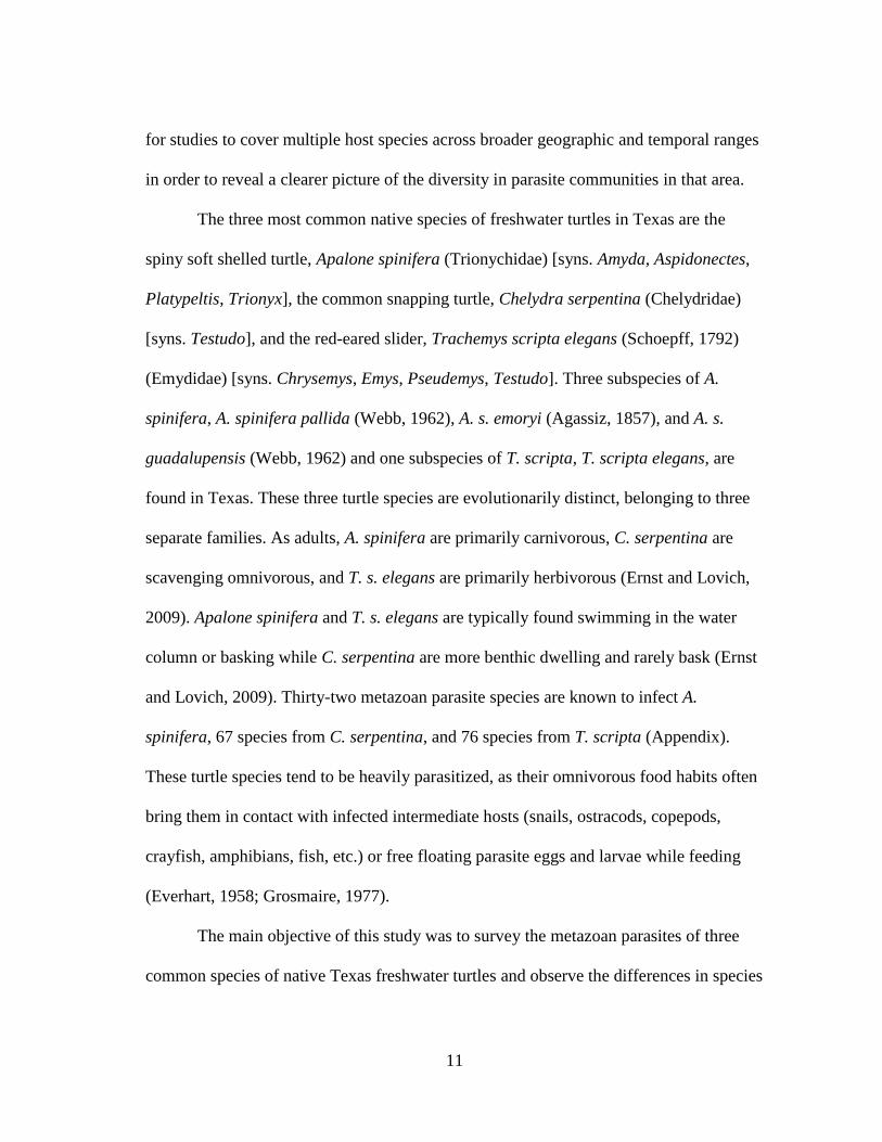
in order to reveal a clearer picture of the diversity in parasite communities in that area.
The three most common native species of freshwater turtles in Texas are the
spiny soft shelled turtle, Apalone spinifera (Trionychidae) [syns. Amyda, Aspidonectes,
Platypeltis, Trionyx], the common snapping turtle, Chelydra serpentina (Chelydridae)
[syns. Testudo], and the red-eared slider, Trachemys scripta elegans (Schoepff, 1792)
(Emydidae) [syns. Chrysemys, Emys, Pseudemys, Testudo]. Three subspecies of A.
spinifera, A. s. pallida (Webb, 1962), A. s. emoryi (Agassiz, 1857), and A. s.
guadalupensis (Webb, 1962) and one subspecies of T. scripta, T. s. elegans, are found in
Texas. These three turtle species are evolutionarily distinct, belonging to three separate
families. As adults, A. spinifera are primarily carnivorous, C. serpentina are scavenging
omnivorous, and T. s. elegans are primarily herbivorous (Ernst and Lovich, 2009).
Apalone spinifera and T. s. elegans are typically found swimming in the water column or
basking while C. serpentina are more benthic dwelling and rarely bask (Ernst and
Lovich, 2009). Thirty-two parasite species are known to infect A. spinifera, 67 species
from C. serpentina, and 76 species from T. scripta (Appendix). These turtle species tend
to be heavily parasitized, as their omnivorous food habits often bring them in contact
with infected intermediate hosts (snails, ostracods, copepods, crayfish, amphibians, fish,
etc.) or free floating parasite eggs and larvae while feeding (Everhart, 1958; Grosmaire,
1977).

7
The main objective of this study was to survey the metazoan parasites of A.
spinifera, C. serpentina, and T. s. elegans from Texas, reporting the differences in
species assemblages across the state and analyzing the host-parasite-environment
relationships in these community assemblages. In addition, samples were collected from
the same site as a previous study on parasites of T. s. elegans (Dinuzzo, 1981), and the
parasite assemblages observed between these temporally distant surveys are compared.
Through this project, the knowledge of the distributions and host associations of
metazoan parasites in Texas freshwater turtles has been clarified, and the effects of
environmental factors on parasite assemblages in aquatic ecosystems have been
elucidated.

8
CHAPTER II
PARASITE DIVERISTY AND COMMUNITY STRUCTURE IN TEXAS
FRESHWATER TURTLES
II.1 Introduction
Parasite diversity is a key component in understanding ecosystem complexity.
With around 40% of known species being parasitic, parasitism is the most common life
strategy (Dobson et al., 2008). The high number of unsampled host species and amount
of cryptic speciation potentially uncovered through genetic analysis indicate that this
number is likely higher (Jousson et al., 2000; Steinauer et al., 2007). Helminths, or
parasites in the phyla Acanthocephala, Nematoda, Platyhelminthes, and the subclass
Pentastomida, often have complex life cycles, sometimes traveling through many hosts
throughout development (e.g. Parker et al., 2003; Poulin, 2011). Endohelminths, or
internal helminths, are associated with predator-prey relationships, as they are typically
transmitted through consumption. It is believed that parasites are involved in nearly 75%
of all trophic linkages in food webs due to their complex life cycles and dependence on
their hosts (Lafferty, 2008; Lafferty et al., 2006). Consequently, healthy ecosystems with
greater numbers of trophic linkages are believed to be higher in parasite diversity.
Parasite diversity is therefore a good indicator of ecosystem diversity and total
ecosystem health (Hudson et al., 2006; Marcogliese, 2005). Despite these findings,
parasite assemblages remain highly understudied, with many new species being

9
described every year and with many life cycles completely unknown (Blasco-Costa and
Poulin, 2017; Dobson et al, 2008; Poulin, 2014).
Previous research suggests that parasitic species may be trophic regulators in the
same capacity as top predators (e.g. Dougherty et al., 2016; Lafferty et al. 2006);
however, most conservation plans do not implement any efforts to preserve parasite
diversity, and often work to eradicate parasites to alleviate stressors on threatened
species. In order to conserve the total diversity in an ecosystem, parasite diversity must
be taken into account when making conservation plans. For this to be possible
knowledge of the parasites species present in a given ecosystem is vital. Studies on
parasite diversity are difficult as they generally require collection and euthanasia of a
large number of hosts. In spite of the challenges, studies on parasite communities are
needed to understand the full breadth of diversity in ecosystems (Hudson et al., 2006;
Dobson et al., 2008).
Parasite diversity is particularly understudied in reptile, amphibian, and fish hosts
(Dobson et al., 2008). In the state of Texas, only four metazoan parasite surveys have
been conducted on freshwater turtles (Harwood, 1932; Everhart, 1957; Dinuzzo, 1981;
McAllister et al., 2008). Harwood (1932) conducted a survey of the endohelminths of 50
species of amphibians and reptiles over the course of two and a half years in the vicinity
of Houston, Texas. Over the course of this study, eight species of turtles were collected:
16 red-eared sliders, Trachemys scripta elegans (Wied, 1839), 16 Mississippi mud
turtles, Kinosternon subrubrum hippocrepis Gray, 1856, 14 three-toed box turtles,
Terrapene carolina triunguis (Agassiz, 1857), nine common snapping turtles, Chelydra

10
serpentina (Linnaeus, 1758), four spiny softshells, Apalone spinifera (LeSueur, 1827),
two razor-backed musk turtles, Sternotherus carinatus (Gray, 1856), two ornate box
turtles, Terrapene ornata (Agassiz, 1857), and one chicken turtle, Deirochelys
reticularia (Latreille in Sonnini and Latreille, 1801). The four A. spinifera were reported
as “Amyda ferox” but based on location are believed to be A. spinifera. Everhart (1957)
conducted a survey of the endohelminths of T. s. elegans from two localities in Southern
Texas and six localities near Stillwater, Oklahoma. A total of 79 turtles, 56 from Texas
and 23 from Oklahoma, were collected during the course of this study. McAllister et al.
(2008) surveyed endoparasites of 18 species of amphibians and reptiles from 11 counties
in Arkansas and six counties in Texas (Bowie, Cass, Denton, Johnson, Somervell, and
Webb). Of these 18 species, only two were turtles and one was a tortoise: five ornate box
turtles, Terrapene ornata ornata (Agassiz, 1857), four yellow mud turtles, Kinosternon
flavescens (Agassiz, 1857), and one Texas tortoise, Gopherus berlandieri (Agassiz,
1857). Dinuzzo (1981) collected 124 T. s. elegans over the course of a year from one
location in Burleson County, Texas. This data was never formally published and
specimens have not been located, so the host association and locality records reported
cannot be verified. Two of these studies were range restricted (Harwood, 1932; Dinuzzo,
1981), two did not sample many individuals (Harwood, 1932; McAllister et al., 2008),
and two only sampled one species (Everhart, 1957; Dinuzzo, 1981). Parasite community
structure varies greatly between geographical locations, parasite species, host species,
individual hosts, different environments, and different seasons (Ernst and Ernst, 1977;
Esch and Gibbons, 1967; Poulin, 2006; Readel et al., 2008). For this reason, it is useful
11
for studies to cover multiple host species across broader geographic and temporal ranges
in order to reveal a clearer picture of the diversity in parasite communities in that area.
The three most common native species of freshwater turtles in Texas are the
spiny soft shelled turtle, Apalone spinifera (Trionychidae) [syns. Amyda, Aspidonectes,
Platypeltis, Trionyx], the common snapping turtle, Chelydra serpentina (Chelydridae)
[syns. Testudo], and the red-eared slider, Trachemys scripta elegans (Schoepff, 1792)
(Emydidae) [syns. Chrysemys, Emys, Pseudemys, Testudo]. Three subspecies of A.
spinifera, A. spinifera pallida (Webb, 1962), A. s. emoryi (Agassiz, 1857), and A. s.
guadalupensis (Webb, 1962) and one subspecies of T. scripta, T. scripta elegans, are
found in Texas. These three turtle species are evolutionarily distinct, belonging to three
separate families. As adults, A. spinifera are primarily carnivorous, C. serpentina are
scavenging omnivorous, and T. s. elegans are primarily herbivorous (Ernst and Lovich,
2009). Apalone spinifera and T. s. elegans are typically found swimming in the water
column or basking while C. serpentina are more benthic dwelling and rarely bask (Ernst
and Lovich, 2009). Thirty-two metazoan parasite species are known to infect A.
spinifera, 67 species from C. serpentina, and 76 species from T. scripta (Appendix).
These turtle species tend to be heavily parasitized, as their omnivorous food habits often
bring them in contact with infected intermediate hosts (snails, ostracods, copepods,
crayfish, amphibians, fish, etc.) or free floating parasite eggs and larvae while feeding
(Everhart, 1958; Grosmaire, 1977).
The main objective of this study was to survey the metazoan parasites of three
common species of native Texas freshwater turtles and observe the differences in species

12
assemblages across the state. In addition, samples were collected from the same site as a
previous study on parasites of T. s. elegans (Dinuzzo, 1981), and the parasite
assemblages observed between these temporally distant surveys are compared. This
project is crucial to understanding the diversity present in aquatic ecosystems so that
changes in these assemblages can be monitored in the future.
II.2 Materials and methods
II.2.1 Field materials and methods
Turtles of the species A. spinifera (A. s. pallida and A. s. emoryi), C. serpentina,
and T. s. elegans were captured using baited hoop nets ranging in size from 1.5 m long
by 0.75 m in diameter to 1.8 m long by 0.9 m in diameter. Nets were set in shallow areas
along the banks of the bodies of water and anchored using 1.2 m metal rebar poles and
baited with deer, chicken, or fish. Nets were left for around 24 hours to allow time for
turtles to catch the scent of the bait and enter the trap. Bycatch, such as fish, alligators, or
non-target turtle species, were immediately released when encountered. Target turtles
were transported in plastic tubs with 15 cm diameter holes cut out for aeration and a
damp sponge in the bottom to prevent desiccation to the Laboratory of Parasitology,
Department of Wildlife and Fisheries Science at Texas A&M University in College
Station, Texas for euthanasia and necropsy. Two sites in west Texas were over six hours
from College Station, so on these trips turtles were processed in the field. Specific GPS
locations were recorded for each collection location using the Garmin eTrex 30 GPS
unit. Capture and euthanasia of turtles was approved by the Institutional Animal Care

13
and Use Committee of Texas A&M University, reference number 040564 and
collections were carried out under Texas Parks and Wildlife Department, scientific
research permit number SPR-0716-172.
A separate aspect of this project was to analyze environmental influences on parasite
diversity. Each time turtles were collected, a range of environmental variables and water
parameters were recorded and correlated with parasite abundance and diversity. These
data are reported in chapter two.
II.2.2 Lab materials and methods
In the lab, turtles were weighed, measured (carapace length, carapace width, shell
depth, circumference, and weight), and euthanized using an intracoelomic injection of
50% MS222 solution at a dosage of 1 mL/kg followed by an overdose of KCl injected
into the brain, following the methods by Conroy et al. (2009). After the initial injection
of MS222, turtles were monitored until the legs and neck were limp (usually around 30
minutes after injection) before KCl was administered. The spinal cord was severed
before necropsy commenced. The combination of a bone saw and aviation wire cutters
were used to cut between the carapace and plastron, and then a scalpel was used to
separate the plastron from the skin and musculature. All external surfaces were checked
for leeches and other metazoan ectoparasites, which were collected when found. All
internal organs including the esophagus, stomach, small intestine, large intestine, heart,
lungs, liver, gall bladder, gonads, kidneys, bladders, and spleen were removed and
searched individually for metazoan parasites under a dissecting microscope. Spirorchiid

14
blood flukes were collected following a modification of the methods outlined by Snyder
and Clopton (2005). After processing turtles, carcasses were donated to the Biodiversity
Research and Teaching Collection at Texas A&M University where they are
permanently housed.
II.2.3 Parasite processing
All metazoan parasites were relaxed in a Stentor dish with 7% saline solution.
Soft-bodied helminths were heat-fixed under light coverslip pressure, placed in a petri
dish with AFA (alcohol-formaldehyde-acetic acid) and left overnight, and stored in 70%
ethanol until further processing. Hard-bodied parasites such as nematodes, pentastomids,
and mites were moved from saline directly to 70% ethanol. Acanthocephalans were
placed in tap water in the refrigerator overnight to relax the specimens and then placed
directly into 70% ethanol. Moving female acanthocephalans into tap water frequently
induced oviposition which facilitated egg measurements and offered a more
unobstructed view of the internal structures. Eggs laid by gravid females were examined
directly and measured to facilitate identification of species in multiple species infections.
Leeches were removed and placed in tap water to which increasing concentrations of
ethanol were added until the leeches were flat. They were then placed in 70% ethanol for
permanent storage and identification.
Heat-fixed specimens were stained in Semichon’s carmine, destained in acid
alcohol, dehydrated through a graded ethanol series (70%, 80%, 95%, 100%, 100%),
cleared in xylene, and mounted on a slide in Canada balsam. Nematodes and

15
acanthocephalans were moved from 70% ethanol to a mixture of equal amounts of 70%
and glycerine for clearing, temporarily mounted on a slide in glycerine for identification,
and subsequently stored in a vial in glycerine for future observations. Where sample size
permitted, a small subset of specimens was placed directly in 95% ethanol for future
molecular analysis.
Spirorchiid blood flukes were flat-fixed in 95% ethanol and will be analyzed
morphologically and molecularly for a separate project. They were not included in any
of the reported diversity in this paper.
Parasites were keyed out to genus using the available dichotomous keys (Khalil
et al., 1994; Gibson et al., 2002; Jones et al., 2005; Bray et al., 2008; Anderson et al.,
2009). For species level identification, body measurements were made and compared to
original parasite descriptions. Leeches were keyed to species using the keys by Klemm
(1985) and Moser et al. (2016).
II.2.4 Statistical analyses
Ecological terms follow Bush et al. (1997). Prevalence, mean intensity, median
intensity, and 95% confidence intervals were calculated in the online application
Quantitative Parasitology (Reiczigel et al., 2013). This software accounts for the non-
normal distributions characteristic of parasite communities. Confidence intervals are
only given for median intensity when the sample size was larger than five. Taxonomic
diversity indices were calculated for each sample location using R version 3.1.4
(taxondive function in vegan package; R Core Team. 2017). Parasite communities of the

16
three host species were analyzed using an analysis of similarities [ANOSIM] (anosim
function in vegan package) and non-metric multidimensional scaling [nMDS]
(metaMDS function in vegan package). This analysis was also performed among the two
subspecies of A. spinifera captured in this study, A. s. pallida and A. s. emoryi. The
purpose of nMDS is to collapse the community data into two dimensions for
visualization and interpretation (Kruskal, 1964). This method differs from other
ordination methods, such as principal components analysis (PCA), in using rank orders
instead of Euclidean distances. ANOSIM is a multivariate method of data analysis that
can be used to compare variation in species abundance among a grouping variable, such
as host species (Clarke, 1993). These two analyses give quantitative and visual
representation of the differences in community data.
II.3 Results
A total of 15 A. spinifera (11 A. s. pallida and four A. s. emoryi), nine C.
serpentina, and 55 T. s. elegans were collected and necropsied for this study. Turtles
were collected from 16 properties in 13 different towns across Texas, USA: Barksdale,
Bryan, College Station, Comstock, Del Valle, Franklin, Gladewater, Glen Rose,
Humble, Iola, Leander, Sinton, and Streetman (Fig. 1). All turtles examined in this study
were infected with at least two species of parasite except one A. spinifera which was
only infected with S. contorta. As many as 10 species were recovered from a single
individual host. Every organ system except the reproductive tract was found to be

17
infected with at least one species, with the small intestine being the most commonly
infected site.
Figure 1: Map of the collection locations of turtles across Texas. A) Apalone spinifera, B) Chelydra
serpentina, and C) Trachemys scripta elegans. Darker points indicate more captures in that location.
Cumulatively, five species of acanthocephalans, nine species of nematodes, 14
species of trematodes, two species of cestodes, five species of monogeneans, five species
of leeches, one species of pentastomid, and one species of mite were recovered.

18
Acanthocephalans and nematodes were the most abundant parasites while trematodes
were the most diverse. Nematodes of the genus Spiroxys were the most common parasite
of A. spinifera, recovered from 100% of turtles. Nematodes of the genus Falcaustra
were the most common parasite of C. serpentina, recovered from 100% of turtles.
Acanthocephalans of the genus Neoechinorhynchus were the most common parasite of
T. s. elegans, recovered from 91% of turtles. Sixteen new host records and 17 new
locality records are recorded herein. Table 1 lists the parasites recovered in this study
with the prevalence, intensity, site of infection, and locality. Spirorchiid blood flukes are
being analyzed as part of a separate project and so are not included.
Occasionally, chironomid larvae were recovered from the intestines of turtles.
These larvae were typically dead, and could usually be found in the debris on the
carapace of the turtle as well, and were likely ingested during feeding. On one occasion,
a large number of live chironomid larvae were found covering the carapace and
throughout the digestive tract of two T. s. elegans collected in Humble, Texas. Tokeshi
(1993) stated that these organisms have evolved commensal relationships with many
slow-moving benthic organisms, which could explain this finding. These specimens
could not be identified but were saved in 70% ethanol and will be deposited in a
museum collection.
II.3.1 Host-community analysis
Analysis of the parasite communities between the three host species was
performed to determine if parasite species assemblages were distinct among host
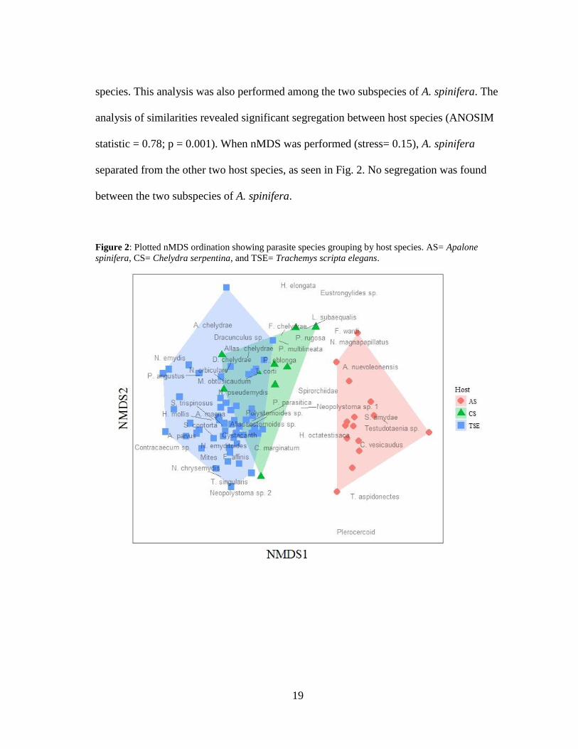
19
species. This analysis was also performed among the two subspecies of A. spinifera. The
analysis of similarities revealed significant segregation between host species (ANOSIM
statistic = 0.78; p = 0.001). When nMDS was performed (stress= 0.15), A. spinifera
separated from the other two host species, as seen in Fig. 2. No segregation was found
between the two subspecies of A. spinifera.
Figure 2: Plotted nMDS ordination showing parasite species grouping by host species. AS= Apalone
spinifera, CS= Chelydra serpentina, and TSE= Trachemys scripta elegans.

20
Table 1: Metazoan parasites recovered from 15 Apalone spinifera, nine Chelydra serpentina, and 55 Trachemys scripta elegans. All localities are towns
in Texas, USA. For median intensity, confidence intervals could only be calculated for sample sizes greater than five. Bolded species name indicates a
new host association and bolded locality indicates the first report in Texas.
Host Species Site of infection Localities Prevalence %
[95% CI]
Mean intensity
(range) [95% CI]
Median intensity
[95% CI]
Apalone
spinifera
(n=15)
Acanthocephala Cystacanth Liver Glen Rose 6.67 1 1
Neoechinorhynchus
magnapapillatus
Small intestine, Large intestine
College Station 6.67 11 11
Neoechinorhynchus
sp.
Small intestine Glen Rose 6.67 1 1
Nematoda Eustrongylides sp. Mesentery College Station 6.67 2 2
Falcaustra wardi Small intestine, Large intestine
College Station 6.67 22 22
Spiroxys amydae Esophagus, Stomach,
Small intestine, Stomach cysts
College Station, Comstock,
Del Valle, Glen Rose,
73.33 [44.9-92.2] 34.91 (3-120) [17-
65.1]
14 [3-66]
Spiroxys sp. Stomach, Stomach cysts Comstock, Del Valle 26.67 [7.8-55.1] 20.75 (1-71) [1-
53.5]
5.5
Trematoda Acanthostomum nuevoleonensis
Small intestine Del Valle 6.67 54 54
Allassostomoides sp. Small intestine Glen Rose 6.67 2 2
Cephalogonimus
vesicaudus
Stomach, Small intestine College Station, Del Valle,
Glen Rose
46.67 [21.3-73.4] 33.57 (1-82)
[14.2-59.6]
26 [1-82]
Teloporia
aspidonectes
Small intestine College Station 6.67 1 1
Telorchis corti Small intestine College Station, Glen Rose 13.33 [1.7-40.5] 7 (1-13) [1-7] 7
Cestoda Plerocercoid Liver Comstock 6.67 6 6
Testudotaenia
testudo
Small intestine, Large
intestine Comstock, Del Valle, Glen
Rose
53.33 [26.6-78.7] 7.13 (1-23) [3.38-
14.3]
3 [1-13]
Monogenea Neopolystoma sp. 1 Conjunctival sac of eye Comstock 6.67 1 1
Polystomoides
coronatum
Mouth, Trachea College Station, Comstock,
Del Valle, Glen Rose,
53.33 [26.6-78.7] 1.25 (1-3) [1-1.75] 1 [1-1]
Hirudinea
Helobdella
octatestisaca Carapace, Skin Comstock 13.33 [1.7-40.5] 1.5 (1-2) [1-2] 1.5
Placobdella
parasitica Carapace, Plastron, Skin
College Station, Del Valle,
Glen Rose 26.67 [7.8-55.1] 3.25 (1-9) [1-7] 1.5
Placobdella rugosa Carapace, Plastron, Skin College Station 13.33 [1.7-40.5] 5 (4-6) [4-5] 5
Arthropoda Levisunguis
subaequalis
Lungs, Trachea College Station, Del Valle 20 [4.3-48.1] 9 (5-16) [5-12.7] 6

21
Table 1: Continued
Host Species Site of infection Localities Prevalence %
[95% CI]
Mean intensity
(range) [95% CI]
Median intensity
[95% CI]
Chelydra
serpentina
(n=9)
Acanthocephala Neoechinorhynchus
sp.
Small intestine, Large
intestine
College Station, Iola,
Streetman
33.33 [7.5-70.1] 4.67 (1-12) [1-
8.33]
1
Nematoda Eustrongylides sp. Body cavity, Liver College Station 11.11 2 2
Dracunculus
globocephalus
Kidney, Bladder College Station, Streetman 22.22 [2.8-60] 3 (2-4) [2-3] 3
Dracunculus sp. Kidney, Rectum epithelium
College Station, Iola 22.22 [2.8-60] 1 1
Falcaustra chelydrae Small intestine, Large
intestine
College Station, Iola 66.67 [29.9-92.5] 235.5 (2-516)
[91.7-385]
218 [2-516]
Falcaustra affinis Large intestine Leander 11.11 14 14
Falcaustra sp. Small intestine, Large intestine
College Station, Streetman 22.22 [2.8-60] 234.5 (16-453) [16-453]
234.5
Serpinema
trispinosus
Esophagus, Small intestine College Station, Iola, Leander,
Streetman
77.78 [40-97.2] 10.14 (1-42)
[3.86-27.1]
5 [1-42]
Spiroxys contorta Stomach College Station 11.11 1 1
Trematoda Allassostomoides
chelydrae
Large intestine College Station, Iola 22.22 [2.8-60] 2 (1-3) [1-2] 2
Auridistomum
chelydrae
Small intestine College Station 11.11 1 1
Telorchis corti Small intestine College Station 11.11 4 4
Monogenea Neopolystoma sp. 1 Conjunctival sac of eye Iola, Streetman 22.22 [2.8-60] 1.5 (1-2) [1-2] 1.5
Polystomoidella
oblonga
Bladder College Station, Iola 44.44 [13.7-78.8] 2.25 (1-6) [1-3.5] 1
Hirudinea Helobdella elongata Skin College Station 11.11 3 3
Placobdella
parasitica
Carapace, Plastron, Skin,
Eye, Rectum
College Station, Iola,
Streetman
44.44 [13.7-78.8] 17 (10-24) [11.5-
20.5]
17
Placobdella
multilineata
Carapace Gladewater 11.11 1 1
Placobdella rugosa Carapace, Plastron, Skin College Station, Iola 44.44 [13.7-78.8] 108.75 (3-333) [3.5-274]
49.5
Arthropoda Levisunguis
subaequalis
Lungs, Trachea, Bladder College Station 33.33 [7.5-70.1] 19 (10-34) [10-27] 13
Trachemys scripta
elegans
(n=55)
Acanthocephala Cystacanth Mouth cyst, Small intestine cysts
College Station, Leander 5.45 [1.1-15.1] 3.33 (1-8) [1-5.67] 1
Neoechinorhynchus
chrysemydis
Small intestine, Large
intestine Gladewater, Glen Rose,
Leander
14.55 [6.5-26.7] 65.4 (4-227)
[20.5-148]
25.5 [4-181]
Neoechinorhynchus emydis
Small intestine, Large
intestine
College Station, Leander,
Sinton
9.09 [3-20] 204.8 (12-740)
[37.6-594]
48
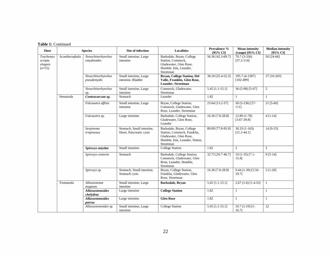
22
Table 1: Continued
Host Species Site of infection Localities Prevalence %
[95% CI]
Mean intensity
(range) [95% CI]
Median intensity
[95% CI]
Trachemys
scripta
elegans (n=55)
Acanthocephala Neoechinorhynchus
emyditoides
Small intestine, Large
intestine
Barksdale, Bryan, College
Station, Comstock,
Gladewater, Glen Rose, Humble, Iola, Leander,
Streetman
56.36 [42.3-69.7] 79.7 (3-336)
[57.2-114]
50 [24-66]
Neoechinorhynchus
pseudemydis
Small intestine, Large
intestine, Bladder Bryan, College Station, Del
Valle, Franklin, Glen Rose,
Leander, Streetman
38.18 [25.4-52.3] 195.7 (4-1587)
[102-499]
37 [10-203]
Neoechinorhynchus
sp.
Small intestine, Large
intestine
Comstock, Gladewater,
Streetman
5.45 [1.1-15.1] 36 (5-98) [5-67] 5
Nematoda Contracaecum sp. Stomach Leander 1.82 1 1
Falcaustra affinis Small intestine, Large intestine
Bryan, College Station, Comstock, Gladewater, Glen
Rose, Leander, Streetman
23.64 [13.2-37] 50 (5-236) [27-111]
21 [5-60]
Falcaustra sp. Large intestine Barksdale, College Station,
Gladewater, Glen Rose, Leander
16.36 [7.8-28.8] 12.89 (1-78)
[3.67-39.8]
4 [1-14]
Serpinema
trispinosus
Stomach, Small intestine,
Heart, Pancreatic cysts
Barksdale, Bryan, College
Station, Comstock, Franklin, Gladewater, Glen Rose,
Humble, Iola, Leander, Sinton,
Streetman
89.09 [77.8-95.9] 30.33 (1-163)
[21.2-44.1]
14 [9-23]
Spiroxys amydae Small intestine College Station 1.82 2 2
Spiroxys contorta Stomach Barksdale, College Station, Comstock, Gladewater, Glen
Rose, Leander, Humble,
Streetman
32.73 [20.7-46.7] 10 (1-35) [7.1-15.4]
9 [5-14]
Spiroxys sp. Stomach, Small intestine, Stomach cysts
Bryan, College Station, Franklin, Gladewater, Glen
Rose, Streetman
16.36 [7.8-28.8] 9.44 (1-39) [3.56-19.7]
3 [1-20]
Trematoda Allassostoma magnum
Small intestine, Large intestine
Barksdale, Bryan 5.45 [1.1-15.1] 2.67 (1-6) [1-4.33] 1
Allassostomoides
chelydrae
Large intestine College Station 1.82 1 1
Allassostomoides
parvus
Large intestine Glen Rose 1.82 1 1
Allassostomoides sp. Small intestine, Large
intestine
College Station 5.45 [1.1-15.1] 10.7 (1-19) [1-
16.7]
12

23
Table 1: Continued
Host Species Site of infection Localities Prevalence %
[95% CI]
Mean intensity
(range) [95% CI]
Median intensity
[95% CI]
Trachemys
scripta
elegans (n=55)
Trematoda Clinostomum
marginatum
Small intestine Leander 1.82 1 1
Dictyangium
chelydrae
Large intestine College Station, Franklin,
Sinton
10.91 [4.1-22.2] 4.7 (1-11) [2.67-
7.67]
4 [1-11]
Heronimus mollis Lungs Bryan 1.82 2 2
Macravestibulum obtusicaudum
Small intestine College Station, Sinton 9.09 [3-20] 9.4 (1-41) [1.2-25.4]
2
Protenes angustus Small intestine Bryan, Glen Rose, Leander,
Sinton
7.27 [2-17.6] 2.25 (1-5) [1-4.25] 1.5
Telorchis corti Small intestine College Station, Gladewater, Leander, Sinton, Streetman
14.55 [6.5-26.7] 47.43 (1-234) [9.38-132]
11.5 [1-48]
Telorchis singularis Small intestine Bryan, Gladewater, Leander 7.27 [2-17.6] 8 (1-17) [2.68-
14.2]
7
Telorchis sp. Small intestine Comstock 1.82 17 17
Monogenea Neopolystoma orbiculare
Bladder, Rectum Barksdale, Bryan, College
Station, Comstock, Del Valle,
Gladewater, Leander, Sinton
29.09 [17.6-42.9] 2.81 (1-8) [1.88-4] 2 [1-3]
Neopolystoma sp. 2 Conjunctival sac of eye Comstock 1.82 3 3
Polystomoides
coronatum
Mouth Barksdale, Bryan, College
Station, Comstock, Franklin, Gladewater, Glen Rose,
Leander, Sinton, Streetman
56.36 [42.3-69.7] 3.87 (1-25) [2.9-
6.75]
3 [2-4]
Hirudinea Helobdella
octatestisaca
Carapace, Skin Barksdale, Comstock, Del
Valle
5.45 [1.1-15.1] 2 (1-3) [1-2.67] 2
Placobdella
parasitica
Carapace, Plastron, Skin Barksdale, Bryan, College
Station, Comstock, Del Valle,
Gladewater, Glen Rose,
Humble, Leander, Streetman
40 [27-54.1] 15.78 (1-136)
[5.95-39.3]
4 [1-8]
Placobdella rugosa Carapace, Plastron, Skin,
Rectum
College Station 7.27 [2-17.6] 4.25 (1-7) [1.75-6] 4.5
Placobdella sp. Plastron, Skin College Station, Gladewater, Glen Rose
5.45 [1.1-15.1] 1 1
Arthropoda Mite Skin Glen Rose 3.64 [0.4-12.5] 9 (3-15) [3-9] 9
Levisunguis
subaequalis
Lungs, Trachea, Dorsal
muscle, Stomach College Station, Leander,
Sinton
9.09 [3-20] 5.6 (1-11) [2-9.2] 4

24
II.3.2 Host-parasite associations
The following is a list of the parasite species recovered in this study with
prevalence and site of infection for each host species.
Acanthocephala
Encysted acanthocephalans (cystacanths) were recovered from the liver of 1 of
15 A. spinifera and the lining of the mouth and small intestine of 3 of 55 T. s. elegans.
These could not be identified to species.
Eocanthocephala: Neoechinorhynchida: Neoechinorhynchidae
Neoechinorhynchus chrysemydis Cable and Hopp, 1954 were recovered from the
small and large intestine of 8 of 55 T. s. elegans. This is the first report this parasite from
Texas.
Neoechinorhynchus emydis (Leidy, 1850) were recovered from the small and
large intestine of 5 of 55 T. s. elegans.
Neoechinorhynchus emyditoides Fisher, 1960 were recovered from the small and
large intestine of 31 of 55 T. s. elegans. This was the most common acanthocephalan
species recovered in this study.
Neoechinorhynchus magnapapillatus Johnson, 1969 were recovered from the
small and large intestine of 1 of 15 A. spinifera. This is the first report of N.
magnapapillatus from A. spinifera and the first report of this parasite from Texas.
Neoechinorhynchus pseudemydis Cable and Hopp, 1954 were recovered from the
small and large intestine of 21 of 55 T. s. elegans. In one turtle, N. pseudemydis was also

25
recovered from the bladder. This turtle had the highest abundance of acanthocephalans
(1,587) and it is likely that these worms were overflowing into the bladder from the large
intestine. This is the first report this parasite from Texas.
Neoechinorhynchus sp. were recovered from the small and large intestine of 1 of
15 A. spinifera, 3 of 9 C. serpentina, and 3 of 55 T. s. elegans. These worms were either
all larval or only males, precluding specific identification.
Nematoda: Enoplea: Dioctophymatoidea: Dioctophymidae
Eustrongylides sp. were recovered from the mesentery of 1 of 15 A. spinifera and
the body cavity and liver of 1 of 9 C. serpentina. These nematodes were larval and were
surrounded in thickened, cyst-like tissue along the length of their bodies. This is the first
report of Eustrongylides sp. from A. spinifera. Molecular analysis is being conducted to
determine the specific identity of these specimens.
Secernentea: Ascaridida: Kathlaniidae
Falcaustra affinis (Leidy, 1856) were recovered from the large intestine of 1 of 9
C. serpentina and the small and large intestine of 13 of 55 T. s. elegans.
Falcaustra chelydrae Harwood, 1932 were recovered from the small and large
intestine of 6 of 9 C. serpentina.
Falcaustra wardi (Mackin, 1936) were recovered from the small and large
intestine of 1 of 15 A. spinifera. This is the first report of F. wardi from A. spinifera and
the first report of this parasite in Texas.

26
Falcaustra sp. were recovered from the small and large intestine of 2 of 9 C.
serpentina and the large intestine of 9 of 55 T. s. elegans. These worms were either all
larval or only females, precluding specific identification.
Anisakidae
Contracaecum sp. was recovered from the stomach of 1 of 55 T. s. elegans. This
parasite, typically found in piscivorous birds such as herons, was larval, the turtle being
a dead end host. This is the first report of Contracaecum sp. from T. s. elegans.
Spirurida: Camallanidae
Serpinema trispinosus (Leidy, 1851) were recovered from the esophagus and
small intestine of 7 of 9 C. serpentina and the stomach and small intestine of 49 of 55 T.
s. elegans. Additionally, larval worms were also recovered from the heart and pancreatic
cysts of 2 of 55 T. s. elegans. These were likely intermediate stages in a migration
through the host. These parasites were typically highly aggregated at the duodenum.
This was the most common helminth species of C. serpentina and T. s. elegans in this
study. Serpinema trispinosus and S. microcephalus have both been reported from all
three turtle species in past studies, however, Baker (1979) clarified the distinction in
morphology and locality between these two species, with S. trispinosus having different
ridge patterns in the buccal cavity and being found in North America. It is likely that all
S. microcephalus reported from North American turtles are actually specimens of S.
trispinosus. A review of the catalogued specimens would be necessary to confirm this
hypothesis.

27
Dracunculidae
Dracunculus globocephalus (Mackin, 1927) were recovered from the kidney and
bladder of 2 of 9 C. serpentina. This is the first report of this parasite from Texas.
Dracunculus sp. were recovered from the kidney and rectal epithelium of 2 of 9 C.
serpentina. These worms were all female and therefore unidentifiable, but are likely D.
globocephalus based on host species and location.
Gnathostomatidae
Spiroxys amydae Cobb, 1929 were recovered from the esophagus, stomach,
stomach cysts, and small intestine of 11 of 15 A. spinifera and the small intestine of 1 of
55 T. s. elegans. Adults in the stomach were typically found in a single mass of
individuals and always associated with ulcers in the stomach lining. The worms
recovered from cysts were always larval and likely intermediate in a migration through
the host. This was the most common helminth species of A. spinifera. This is the first
report of this parasite from T. s. elegans.
Spiroxys contorta (Rudolphi, 1819) were recovered from the stomach of 1 of 9 C.
serpentina and the stomach of 18 of 55 T. s. elegans. Adults in the stomach were
typically found in a single mass of individuals and always associated with ulcers in the
stomach lining.
Spiroxys sp. were recovered from the stomach and stomach cysts of 4 of 15 A.
spinifera and the stomach, stomach cysts, and small intestine of 9 of 55 T. s. elegans.
These worms were all larval and therefore unidentifiable, but are likely S. contorta in T.
28
s. elegans and S. amydae in A. spinifera based on typical host associations. The worms
recovered from cysts were likely intermediate in a migration through the host.
Platyhelminthes: Trematoda: Diplostomida: Clinostomatidae
Clinostomum marginatum (Rudolphi, 1819) was recovered from the small
intestine of 1 of 55 T. s. elegans. This parasite is typically found in herons and egrets, so
was likely a dead-end infection in the turtle. This is the first report of this parasite in T. s.
elegans.
Echinostomida: Heronimidae
Heronimus mollis (Leidy, 1856) were recovered from the lungs of 1 of 55 T. s.
elegans. The genus Heronimus has undergone significant taxonomic debate, but is
currently considered to be monotypic.
Microscaphidiidae
Dictyangium chelydrae Stunkard, 1943 were recovered from the large intestine
of 5 of 55 T. s. elegans. This is the first report of this parasite from Texas.
Paramphistomatidae
Allassostoma magna Stunkard, 1916 were recovered from the small and large
intestine of 3 of 55 T. s. elegans. Large red blisters were found on the intestinal lining
where these trematodes were attached. This is the first report of this parasite from Texas.
Allassostomoides chelydrae (MacCallum, 1919) were recovered from the large
intestine of 2 of 9 C. serpentina and 1 of 55 T. s. elegans. This is the first published
report of this parasite from T. s. elegans and the first record in Texas. Dinuzzo (1981)

29
reported this trematode from T. s. elegans in his thesis, but as this data was never
published and specimens cannot be located, his record is insufficient.
Allassostomoides parvus (Stunkard, 1916) was recovered from the large intestine
of 1 of 55 T. s. elegans. This is the first report of this parasite from T. s. elegans and the
first record in Texas.
Allassostomoides sp. were recovered from the large intestine of 1 of 15 A.
spinifera and the small and large intestine of 3 of 55 T. s. elegans. These worms were all
immatures, precluding specific identification. This is the first report of Allassostomoides
sp. from A. spinifera.
Pronocephalidae
Macravestibulum obtusicaudum Mackin, 1930 were recovered from the small
intestine of 5 of 55 T. s. elegans. This is the first report of this parasite in Texas.
Teloporia aspidonectes (MacCallum, 1917) was recovered from the small
intestine of 1 of 15 A. spinifera. This is the first report of this parasite in Texas.
Plagiorchiida: Auridistomidae
Auridistomum chelydrae (Stafford, 1900) was recovered from the small intestine
of 1 of 9 C. serpentina. Auridistomum georgiense Bogitsh, 1959 was described from C.
serpentina in Georgia. This species differed from A. chelydrae based on a larger overall
size, lobed testes, and the lack of a prominent Laurer’s canal. Body size and testis shape
are notoriously variable characteristics in many trematodes, and the Laurer’s canal is
often not visible, even in well-fixed specimens. Further morphological and molecular
work is needed to determine if A. georgiense is truly a distinct species.

30
Cephalogonimidae
Cephalogonimus vesicaudus Nickerson, 1912 were recovered from the stomach
and small intestine of 7 of 15 A. spinifera. These parasites were typically associated with
the duodenum, but were found throughout the small intestine when infection intensity
was high.
Cryptogonimidae
Acanthostomum nuevoleonensis Caballero and Caballero, 1964 were recovered
from the small intestine of 1 of 15 A. spinifera. This is the first report of A.
nuevoleonensis in the USA. This parasite was synonymized with A. megacetabulum
Thatcher, 1963 by Brooks (1980), who stated that the size difference in oral spines and
sucker sizes were likely due to host induced effects. Tkach and Snyder (2003) pointed
out the tenuous nature of this synonymy, as chitonous elements are unlikely to vary
greatly within a single species. The major differences between the two species are the
size of oral sucker, pharynx, ventral sucker, and oral spines, with the oral spine length
being much greater in A. megacetabulum (68 versus 16-32). For this reason, the
synonymy (Brooks 1980) is rejected and A. nuevoleonensis is redescribed in the next
section.
Telorchiidae
Protenes angustus (Stafford, 1900) were recovered from the small intestine of 4
of 55 T. s. elegans. The genera Telorchis and Protenes have been the subject of much
taxonomic debate. MacDonald and Brooks (1989) placed P. angustus in the genus
Telorchis based on a morphological character tree which placed this species in a clade

31
with Telorchis corti. The unusual location of the genital pore, different than any member
of the genus Telorchis, would indicate that this is a generic level trait, and so P. angustus
is left in the genus Protenes for this study.
Telorchis corti Stunkard, 1915 were recovered from the small intestine of 2 of 15
A. spinifera, 7 of 9 C. serpentina, and 8 of 55 T. s. elegans. This species of trematode is
a generalist known to infect many species of reptiles and amphibians. A large number of
species were synonymized with this species by Wharton (1940) and MacDonald and
Brooks (1989); however, some of these synonymys were rejected after molecular
analysis of specimens (Pulis et al., 2011). A full molecular revision of this genus is
needed to clarify the taxonomy and reveal defining morphological traits.
Telorchis singularis (Bennett, 1935) were recovered from the small intestine of 4
of 55 T. s. elegans. This was the largest trematode species recovered in this study, with
individuals as large as 17 millimeters long.
Telorchis sp. were recovered from the small intestine of 1 of 55 T. s. elegans.
These worms were all immatures with underdeveloped reproductive systems and no
eggs, precluding specific identification.
Cestoda: Onchoproteocephalidea: Proteocephalidae
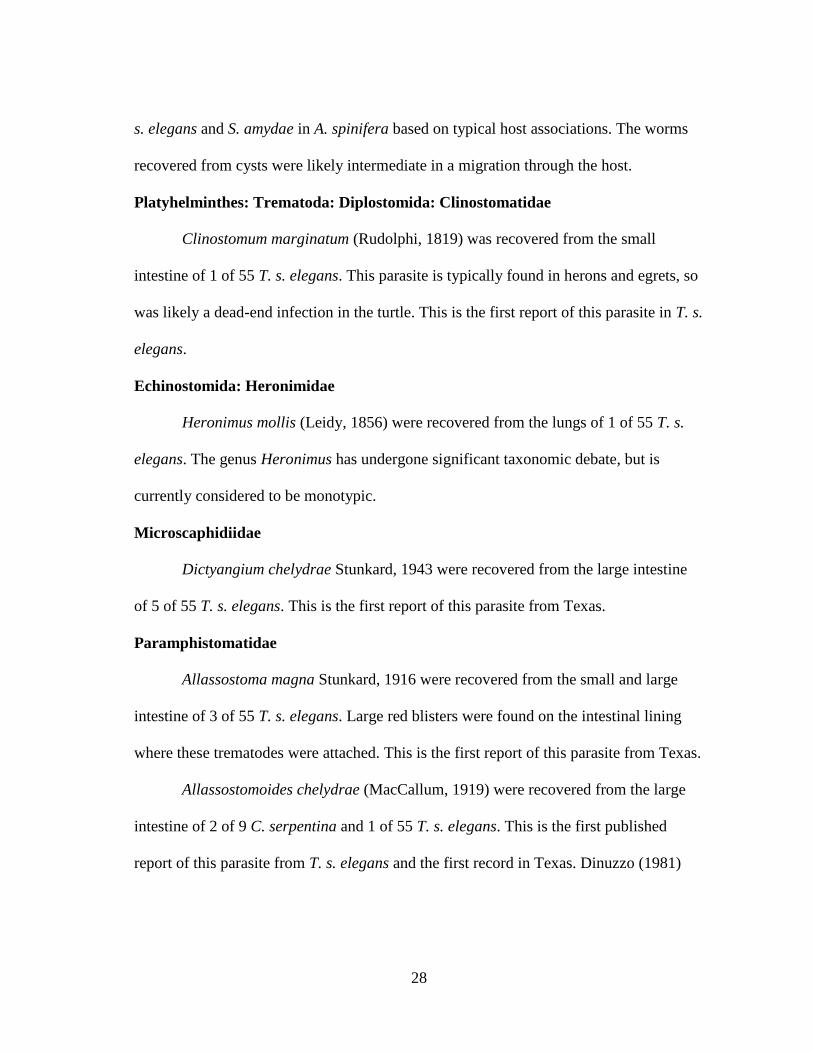
Encysted plerocercoids (Fig. 3) were recovered from the liver of 1 of 15 A.
spinifera. The protoscoleces of these cestodes contained four lateral suckers and one
apical sucker, placing them in the Family Proteocephalidae. Collaboration is ongoing to
determine the specific identity of this parasite through molecular analysis, which will be
detailed in a future report.
32
Figure 3: Plerocercoid protoscolex showing sucker arrangement. Recovered from the liver of Apalone
spinifera.
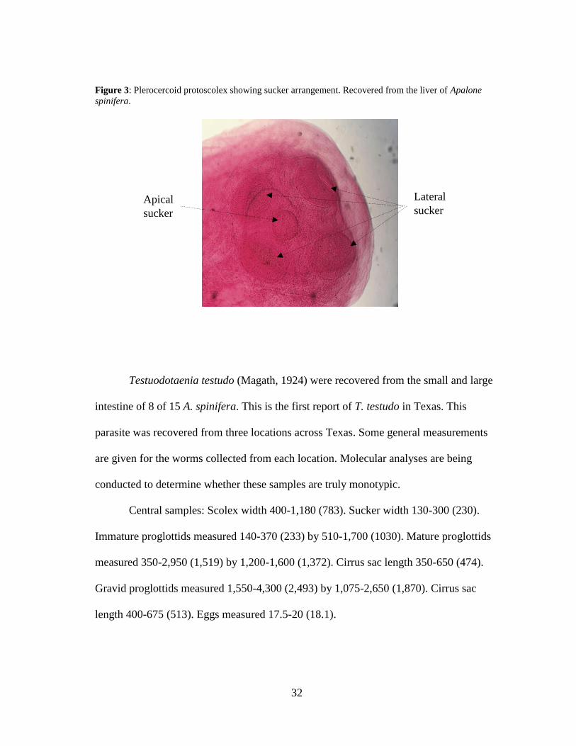
Testuodotaenia testudo (Magath, 1924) were recovered from the small and large
intestine of 8 of 15 A. spinifera. This is the first report of T. testudo in Texas. This
parasite was recovered from three locations across Texas. Some general measurements
are given for the worms collected from each location. Molecular analyses are being
conducted to determine whether these samples are truly monotypic.
Central samples: Scolex width 400-1,180 (783). Sucker width 130-300 (230).
Immature proglottids measured 140-370 (233) by 510-1,700 (1030). Mature proglottids
measured 350-2,950 (1,519) by 1,200-1,600 (1,372). Cirrus sac length 350-650 (474).
Gravid proglottids measured 1,550-4,300 (2,493) by 1,075-2,650 (1,870). Cirrus sac
length 400-675 (513). Eggs measured 17.5-20 (18.1).
Lateral
sucker
s
Apical
sucker

33
Northern samples: Scolex width 480-870 (683). Sucker width 200-270 (239).
Immature proglottids measured 100-460 (221) by 410-1,150 (633). Mature proglottids
measured 900-1,100 (993) by 600-1,600 (1,025). Cirrus sac length 200-490 (318).
Gravid proglottids measured 1,350-4,175 (2,355) by 1,080-1,500 (1,327). Cirrus sac
length 460-810 (593). Eggs measured 15-25 (20.8).
Western samples: Scolex width 510-700 (590). Sucker width 200-240 (218).
Immature proglottids measured 100-400 (234) by 530-960 (665). Mature proglottids
measured 680-1580 (1040) by 720-1,280 (952). Cirrus sac length 230-440 (375). Gravid
proglottids measured 1,225-3,375 (2,405) by 900-1,600 (1,179). Cirrus sac length 380-
590 (470). Eggs measured 17.5-25 (20.8).
Monogenea: Polyopisthocotylea: Polystomatidae
Neopolystoma orbiculare (Stunkard, 1916) were recovered from the bladder and
rectum of 16 of 55 T. s. elegans. At the two sites where this parasite was found in the
rectum, they were only recovered from this location within the host. This could be an
insight into the life history of this parasite, possibly traveling to the rectum for release of
eggs.
Neopolystoma n. sp. 1 were recovered from the conjunctival sac of the eye of 1
of 15 A. spinifera and 2 of 9 C. serpentina. This is the first report of Neopolystoma from
the eye of A. spinifera, and the first report in C. serpentina in Texas. A description of
this new species follows in the next section.
Neopolystoma n. sp. 2 were recovered from the conjunctival sac of the eye of 1
of 55 T. s. elegans. This is the first report of Neopolystoma from the eye of T. s. elegans.

34
Based on measurements these specimens represent a new species. A description of this
new species follows in the next section.
Polystomoides coronatum (Leidy, 1888) were recovered from the mouth of 8 of
15 A. spinifera and the mouth of 31 of 55 T. s. elegans. In a single A. spinifera, one
individual was found in the trachea near the connection to the lungs. This worm had
likely moved to this location from the mouth, as it is not a typical location for
monogenean infection in turtles. The original description of this species (Leidy, 1888)
and later redescriptions (Stunkard, 1917; Price, 1939) are lacking in key morphological
characters. While many synonyms have been accepted for this species, the ranges for
some of the key morphological characters are too large to represent a monotypic group,
particularly the number and length of the genital spines (Bychowsky, 1957; Timmers
and Lewis, 1978; Lenis and Garcia-Prieto, 2009). A full revision comparing morphology
and genetics is needed to clarify the taxonomy of this species. Collaboration is ongoing
to determine whether the specimens in the current study are truly a single species
through molecular analysis, which will be detailed in a future report. A redescription of
this species based on morphology is located in the next section.
Polystomoidella oblonga (Wright, 1879) were recovered from the bladder of 4 of
9 C. serpentina. Although this parasite was recovered from T. s. elegans in a past study
(Acholonu, 1969), it appears to be host specific to C. serpentina at the sites sampled in
this study.

35
Annelida: Clitellata: Rhynchobdellida: Glossiphoniidae
Helobdella elongata (Castle, 1900) were recovered from the skin of 1 of 9 C.
serpentina. This is the first report of this species on C. serpentina. This species was only
recovered one time during this study and it is possible that it was using that turtle as a
substrate and not a host. See notes on H. octatestisaca below.
Helobdella octatestisaca Lai and Chang, 2009 were recovered from the carapace
and skin of 2 of 15 A. spinifera and 3 of 55 T. s. elegans. This is the first record of H.
octatestisaca from A. spinifera. These leeches have typically been considered predators
of small invertebrates. Those found on turtles were thought to be depredating leeches of
the genus Placobdella (Richardson et al. 2017). However, Stark et al. (2017) found that
Helobdella stagnalis were facultative parasites of four amphibian species in Europe. The
fact that the specimens of H. octatestisaca in this study were recovered from individuals
that had no other leech parasites and appeared to have blood-filled ceca indicates that
this species may have a facultative relationship with turtles.
Placobdella multilineata Moore, 1953 was recovered from the carapace of 1 of 9
C. serpentina. This is the first record of this parasite from Texas. Only one individual of
this species was recovered in this study.
Placobdella parasitica (Say, 1824) were recovered from the carapace, plastron,
and skin of 4 of 15 A. spinifera, the carapace, plastron, skin, eye, and rectum of 4 of 9 C.
serpentina, and the carapace, plastron, and skin of 22 of 55 T. s. elegans. This was the
most common species of leech recovered in this study.

36
Placobdella rugosa (Verrill, 1874) were recovered from the carapace, plastron,
and skin of 2 of 15 A. spinifera, the carapace, plastron, and skin of 4 of 9 C. serpentina,
and the carapace, plastron, skin, and rectum of 4 of 55 T. s. elegans. This is the first
report of this species on A. spinifera.
Placobdella sp. were recovered from the plastron and skin of 3 of 55 T. s.
elegans. These were likely P. parasitica but due to poor fixation were unable to be
identified.
Arthropoda: Arachnida (Acari)
Two T. s. elegans, collected on the same day in the same location, were found to
be hosting a number of parasitic mites. These mites were located on the skin around the
cloaca and axillae of the two turtles. These mites appear to represent a new species and a
description of these specimens is currently in progress.
Maxillopoda (Pentastomida): Porocephalida: Sebekidae
Levisunguis subaequalis Curran et al., 2014 were recovered from the lungs and
trachea of 3 of 15 A. spinifera, 3 of 9 C. serpentina, and 2 of 55 T. s. elegans.
Additionally, larval specimens were recovered encysted in the dorsal muscle, lung, and
stomach wall of three T. s. elegans from separate locations. In one C. serpentina, a
single pentastomid was found in the bladder, likely the result of an aberrant migration to
the lungs. The only location where this parasite was found as an adult in C. serpentina
and T. s. elegans had an unusually high abundance of the mosquitofish, Gambusia affinis
(Baird and Girard, 1853), the known intermediate host of L. subaequalis (Curran et al.,

37
2014). It is likely that opportunistic feeding on mosquitofish by C. serpentina and T. s.
elegans resulted in infection at this site. These parasites were typically associated with
excess mucus in the lungs of the host turtle. This is the first report of L. subaequalis
from T. s. elegans and C. serpentina, the first record since its description, and the first
record in Texas.
II.3.3 Parasite taxonomy
Redescription of Acanthostomum nuevoleonensis (Fig. 4)
Redescription [based on 20 gravid adults, measurements in micrometers with ranges
followed by means]:
Body elongate, widest between ventral sucker and testes, 1,450-3,200 (2645) by
290-610 (461). Forebody 460-770 (604), comprising 19-32% (23%) of total body length.
Tegument spinous, with regularly spaces spines diminishing in size and number toward
posterior end. Oral sucker bell-shaped, 130-185 (159) by 120-180 (151). Spines
triangular in shape with widest end embedded in cutaneous tissue, 18-22 (20) in number,
20-36.3 (26.2) by 7.5-12.5 (10.2). Spines regularly spaced around circumference of oral
sucker, up to half often missing, likely lost during removal from host. Ventral sucker
sub-spherical, 90-170 (135) by 115-190 (153). Oral sucker approximately the same size
as ventral sucker, 0.87-1.13 (0.99) width ratio. Cutaneous invaginations present anterior
and posterior to ventral sucker. Prepharynx short if present, 0-60 (14) long. Pharynx
round, close to oral sucker, 80-110 (101) by 50-100 (81). A number of dark-staining
cells located at posterior end of pharynx, possibly digestive glands. Oral sucker

38
approximately twice the size of pharynx, 1.5-2.4 (1.89) width ratio. Esophagus thick and
sinuous, 80-210 (125) long. Bifurcation located just anterior to the ventral sucker.

39
Figure 4: Illustration of Acanthostomum nuevoleonensis. OS= Oral sucker, OSp= Oral spines, P=
Pharynx, Ex= Exretory bladder, Es= Esophagus, GP= Genital pore, VS= Ventral sucker, C= Cecae, SV=
Seminal vesicle, U= Uterus, Vt= Vitellaria, O= Ovary, SR= Seminal receptacle, T= Testis, and A= anus.
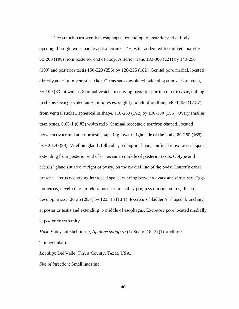
40
Ceca much narrower than esophagus, extending to posterior end of body,
opening through two separate anal apertures. Testes in tandem with complete margins,
60-200 (108) from posterior end of body. Anterior testis 130-300 (221) by 140-250
(199) and posterior testis 150-320 (256) by 120-225 (182). Genital pore medial, located
directly anterior to ventral sucker. Cirrus sac convoluted, widening at posterior extent,
55-100 (83) at widest. Seminal vesicle occupying posterior portion of cirrus sac, oblong
in shape. Ovary located anterior to testes, slightly to left of midline, 540-1,450 (1,137)
from ventral sucker, spherical in shape, 110-250 (192) by 100-180 (156). Ovary smaller
than testes, 0.63-1 (0.82) width ratio. Seminal receptacle teardrop-shaped, located
between ovary and anterior testis, tapering toward right side of the body, 80-250 (166)
by 60-170 (89). Vitelline glands follicular, oblong in shape, confined to extracecal space,
extending from posterior end of cirrus sac to middle of posterior testis. Ootype and
Mehlis’ gland situated to right of ovary, on the medial line of the body. Laurer’s canal
present. Uterus occupying intercecal space, winding between ovary and cirrus sac. Eggs
numerous, developing protein-tanned color as they progress through uterus, do not
develop in size, 20-35 (26.3) by 12.5-15 (13.1). Excretory bladder Y-shaped, branching
at posterior testis and extending to middle of esophagus. Excretory pore located medially
at posterior extremity.
Host: Spiny softshell turtle, Apalone spinifera (LeSueur, 1827) (Testudines:
Trionychidae).
Locality: Del Valle, Travis County, Texas, USA.
Site of infection: Small intestine.

41
Representative DNA sequences: Specimens are currently being processed for genetics
and voucher sequences will be uploaded to GenBank as soon as they are available.
Remarks:
Acanthostomum nuevoleonensis can be differentiated from A. megacetabulum
mainly by the size of oral spines, the size of the oral sucker, and the length of the eggs
(Table 2). This study is the first report of this species outside of Mexico. The recovery of
this parasite from the type host in a geographically distant location indicates that the
originally described specimens were not an accidental host switch, as inferred by Brooks
(1980).

42
Table 2: A comparison of the body measurements of Acanthostomum species, including specimens
collected in this study. All measurements are in micrometers.
Measurements
A. megacetabulum
(Thatcher, 1963)
Yamaguti 1971
A. nuevoleonensis
Caballero and
Caballero, 1964
A. nuevoleonenesis
(this study)
Body Length 1800-3900 1427-1647 1450-3200
Body Width 390-600 311-348 290-610
Forebody Length 460-770
Oral Spine Number 19-21 18-20 18-22
Oral Spine Length 69 16-25 20-36.3
Oral Spine Width 17 8 7.5-12.5
Oral Sucker Length 270-320 107-119 130-185
Oral Sucker Width 250-320 102-107 120-180
Prepharynx Length 0-130 37-39 0-60
Pharynx Length 140-160 74-82 80-110
Pharynx Width 130-140 74-78 50-100
Esophagus Length 41-82 80-210
Ventral Sucker Length 94-102 90-170
Ventral Sucker Width 190-200 94-115 115-190
Ovary Length 190-200 131-148 110-250
Ovary Width 140-180 94-98 100-180
Seminal Receptacle
Length 220-280 49-82 80-250
Seminal Receptacle
Width 140-180 29-86 60-170
Anterior Testis Length 190-230 86-111 130-300
Anterior Testis Width 140-220 111-123 140-250
Posterior Testis Length 140-250 107-123 150-320
Posterior Testis Width 190-230 102-115 120-225
Egg Length 31-33 25-29 24.2-29.2
Egg Width 14-15 14-16 12.5-14.2

43
Description of Neopolystoma n. sp. 1 (Fig. 5)
Type host: Common snapping turtle, Chelydra serpentina (Linnaeus, 1758) (Testudines:
Chelydridae).
Type locality: Iola, Brazos County, Texas, USA.
Other host: Spiny softshell turtle, Apalone spinifera (LeSueur, 1827) (Testudines:
Trionychidae).
Other localities: Streetman, Navarro County, Texas, USA; Comstock, Val Verde
County, Texas, USA.
Site of infection: Conjunctival sac of the eye.
Type material: The holotype and two paratypes will be deposited in the Harold W.
Manter Laboratory of Parasitology, University of Nebraska, Lincoln, Nebraska, USA.
Prevalence and intensity: Across all sites, prevalence in C. serpentina was 22.22% (2 of
9) and A. spinifera was 6.67% (1 of 15). For sites where the parasites were recovered,
prevalence in C. serpentina was 66.67% (2 of 3) and A. spinifera was 25.00% (1 of 4).
Mean intensity for C. serpentina was 2 (1–2) and for A. spinifera was 1.
Description [based on 3 sexually mature worms, all measurements in micrometers unless
otherwise stated]:
Delicate worms, able to extend considerably when alive, firmly attached to
conjunctiva of host’s eye. Body elongate, 2,525–2,825 (2,713.3) by 760–880 (813.3) at
greatest width. Width at vagina 740–850 (796.7). Haptor compact, rounded, 650–710
(686.7) by 800–850 (830.0). Haptor length to body length ratio 0.23–0.28 (0.25).

44
Figure 5: Illustration of Neopolystoma n. sp. 1. FO= False oral sucker, P= Pharynx, Ex= Exretory
bladder, GP= Genital pore, SV= Seminal vesicle, E= Egg, O= Ovary, GIC= Genito-intestinal canal, V=
Vagina, T= Testis, Vt= Vitellaria, C= Cecae, and HS= Haptoral sucker.

45
Six haptoral suckers, muscular with a ring of plate-like skeletal structures, 240–
250 (246.7) in diameter. Marginal hooklets not visible. Mouth subterminal, ventral.
False oral sucker 230–330 (277) by 300–370 (346.7). Pharynx muscular, round, often
overlapping with false oral sucker, 180–230 (213.3) by 220–270 (246.7). A mass of
darkly stained cells are congregated at posterior edge of pharynx. Cecae bifurcating at
posterior edge of pharynx and extending posteriorly, terminating short of anterior edge
of the haptor, remaining unjoined, comprising 49.1–55.6% (53.3%) of total body length.
Testis round to oblong, 160–240 (186.7) by 160–195 (171.7), located medially, posterior
to ovary. Seminal vesicle located posterior to genital bulb, bulbous when filled with
sperm, attached to testis by a long narrow canal. Genital bulb small, located posterior to
bifurcation of intestinal caeca, 40–60 (51.7) wide. Genital spines 8 in number, 7.5–11.25
(9.5) in length, curved with crescent-shaped roots. Vaginae present, located
approximately one third of body length from anterior end, lateral to testis, connecting
anterior to testis by a narrow canal. Ovary longer than wide, comma shaped, 180–220
(196.7) by 80–110 (100.0). Uterus absent, ootype confined between testis and genital
pore, containing a single spindle-shaped egg measuring 260–270 (265) by 120. Vitellaria
in a continuous field stretching from posterior edge of pharynx to anterior edge of
haptor, comprising 57.3–60.5% (59.1%) of total body length. Vitellaria absent in area
taken up by ovary, testis, and uterus. Genito-intestinal canal wide, joining with cecum at
level of the posterior edge of ovary. Ootype, genito-intestinal canal, and vaginae joining
medially above testis. Excretory bladders circular, prominent, lateral to bifurcation of the
cecae.

46
Remarks:
Neopolystoma n. sp. 1 differs from all described species in the genus
Neopolystoma based on a number of characteristics (Table 1). Neopolystoma n. sp. 1
differs from N. orbiculare, N. domitilae, N. rugosa, N. terrapenis, N. cayensis, N.
cyclovitellum, N. exhamatum, N. kreffti, N. macleayi, N. novaeguineae, N. chelodinae, N.
grossi, N. elizabethae, N. scorpioides, N. cribbi, N. liewi, N. palpebrae, N.
queenslandensis, N. spratti, and N. tinsleyi in at least six of the following measurements:
body size, haptor size, false oral sucker width, pharynx size, ovary size, testis size,
vitelline follicle extent, genital bulb width, number of genital spines, genital spine
length, number of eggs, egg size, haptoral sucker width, and ratio of haptor to body
length. Neopolystoma n. sp. 1 is most similar to N. fentoni and N. guianensis.
Neopolystoma n. sp. 1 is larger in size than N. fentoni, has fewer eggs, and a smaller
haptor length to body length ratio. Neopolystoma n. sp. 1 has a smaller testis size,
smaller egg size, and vitelline follicles that do not reach the false oral sucker than N.
guianensis.
In 2011, Platt et al. redescribed N. liewi, an eye monogenean from Asian turtles,
and reported a tendency for wide variation in morphological characters due to coverslip
fixation. The authors recommended that new species only be described based on
specimens fixed in hot formalin without coverslip pressure. According to Platt et al.
(2011), only false oral sucker width, genital bulb width, number of genital spines, genital
spine length, number of eggs, egg size, haptoral sucker width, and ratio of haptor to body
length are still acceptable measurements for comparison when specimens are fixed under

47
coverslip pressure. At least two of these measurements differ between Neopolystoma sp.
1 and all other described species.
Two specimens of Polystomoides sp. collected from a spiny softshell turtle were
heat fixed without coverslip pressure to check if this method was viable. The two
specimens curled and twisted, making them essentially useless for morphological
measurement. For this reason, all of the specimens of Neopolystoma sp. 1 were fixed
with light coverslip pressure.
Description of Neopolystoma n. sp. 2 (Fig. 6)
Type host: Red-eared slider turtle, Trachemys scripta elegans (Wied, 1839) (Testudines:
Emydidae).
Type locality: Comstock, Val Verde County, Texas, USA.
Site of infection: Conjunctival sac of the eye.
Type material: The holotype and two paratypes will be deposited in the Harold W.
Manter Laboratory of Parasitology, University of Nebraska, Lincoln, Nebraska, USA.
Prevalence and intensity: Across all sites, prevalence was 1.82% (1 of 56). For sites
where the parasites were recovered, prevalence was 33.33% (1 of 3). Intensity was 3.
Description [based on 3 sexually mature worms, all measurements in micrometers unless
otherwise stated]:
Delicate worms, more so than Neopolystoma n. sp. 1, able to extend considerably
when alive, firmly attached to conjunctiva. Body elongate with tapered ends, 2,660–
3,070 (2,800) long by 570–740 (657) at greatest width. Width at vagina 570–740 (647).

48
Figure 6: Illustration of Neopolystoma n. sp. 2. FO= False oral sucker, P= Pharynx, Ex= Exretory
bladder, GP= Genital pore, SV= Seminal vesicle, E= Egg, O= Ovary, GIC= Genito-intestinal canal, V=
Vagina, T= Testis, Vt= Vitellaria, C= Cecae, and HS= Haptoral sucker.

49
Haptor compact, wider than long, 540–650 (603) by 710–730 (720). Haptor
length to body length ratio 0.19–0.26 (0.23). Six haptoral suckers, muscular with a ring
of plate-like skeletal structures, 185–245 (225) in diameter. Marginal hooklets small,
curved, 15 long. Mouth subterminal, ventral. False oral sucker 260–270 (263) by 320–
400 (360). Pharynx muscular, round, often overlapping with false oral sucker, 170–200
(187) by 220–240 (233). A layer of darkly stained cells congregated at posterior edge of
pharynx. Cecae bifurcating at posterior edge of pharynx and extending posteriorly,
terminating short of anterior edge of haptor, remaining unjoined, comprising 52.9–
58.9% (55.9%) of total body length. Testis round to lobate, 180–230 (197) by 110–160
(130), located medially, posterior to ovary. Seminal vesicle located posterior to genital
bulb, bulbous when filled with sperm, attached to testis by a long narrow canal. Genital
bulb small, located posterior to bifurcation of intestinal caeca, 20–25 (23.3) wide.
Genital spines 8 in number, 11.25 in length, curved with crescent-shaped roots. Vaginae
present, located approximately one third of body length from anterior end, lateral to
testis, connecting anterior to testis by a narrow canal. Ovary longer than wide, 160–260
(197) by 70–100 (80). Uterus absent, ootype confined between testis and genital pore,
containing a single spindle-shaped egg measuring 160–300 (225) by 70–125 (95).
Vitellaria in a continuous field stretching from anterior edge of pharynx to before
anterior edge of haptor, comprising 62.5–67.1% (61.5%) of total body length. Vitellaria
absent in area taken up by ovary, testis, and uterus. Genito-intestinal canal wide, joining
with cecum at level of posterior edge of the ovary. Ootype, genito-intestinal canal, and

50
vaginae joining medially above testis forming a bell-shaped atrium. Excretory bladders
reduced, barely visible, lateral to bifurcation of cecae.
Remarks:
This description is based on three individuals taken from the conjunctival sac of a
single T. s. elegans. These monogeneans were poorly fixed and no molecular specimen
was saved, but key morphological characters allow for description of this species. The
major difference between this species and Neopolystoma n. sp. 1 are the body width,
haptor size, vitelline follicle extent, and genital bulb width.
Redescription of Polystomoides coronatum (Fig. 7)
Hosts: Red-eared slider, Trachemys scripta elegans (Wied, 1839) (Testudines:
Emydidae); Spiny softshell turtle, Apalone spinifera (LeSueur, 1827) (Testudines:
Trionychidae).
Localities: Barksdale, Edwards County; Bryan and College Station, Brazos County;
Comstock, Val Verde County; Del Valle, Travis County; Gladewater, Upshur County;
Glen Rose, Somervell County; Humble, Harris County; Leander, Williamson County;
and Streetman, Navarro County, Texas, USA.
Site of infection: Mouth.
Redescription [based on 72 sexually mature worms, all measurements in micrometers
unless otherwise stated]:
Hardy worms, light yellow when alive, egg large and orange if present, firmly
attached to mucosa at back of host’s mouth.
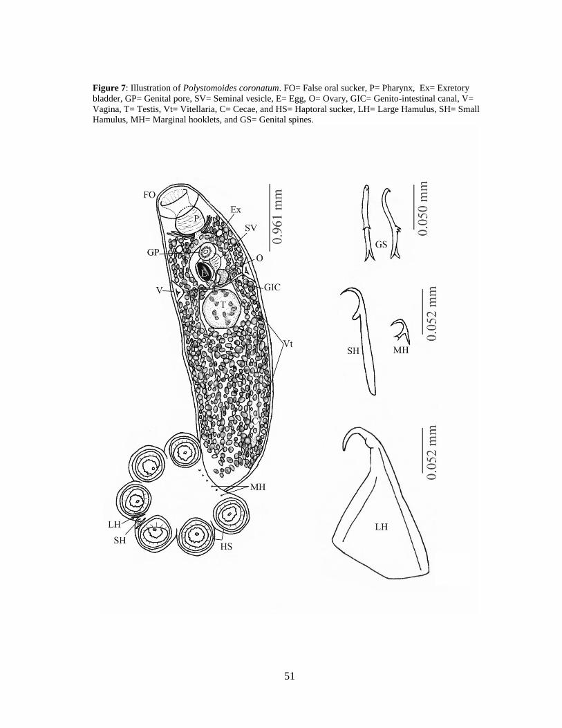
51
Figure 7: Illustration of Polystomoides coronatum. FO= False oral sucker, P= Pharynx, Ex= Exretory
bladder, GP= Genital pore, SV= Seminal vesicle, E= Egg, O= Ovary, GIC= Genito-intestinal canal, V=
Vagina, T= Testis, Vt= Vitellaria, C= Cecae, and HS= Haptoral sucker, LH= Large Hamulus, SH= Small
Hamulus, MH= Marginal hooklets, and GS= Genital spines.

52
Body linguiform with tapered ends, 1,830–7,425 (3,894) by 530–1,550 (938),
with greatest width at vagina. Haptor variable in shape, wider than long with suckers
extending past the outer margins, 590–1,550 (1,056) by 790–2,050 (1,252). Haptor
length to body length ratio 0.17–0.40 (0.28). Six haptoral suckers, muscular with a ring
of plate-like skeletal structures, 200–550 (351) in diameter. Large Hamulus 95–233
(161) in length. Small Hamulus 31.3–161.5 (80.6) in length. Marginal hooklets small,
curved, 12.5–25 (19.2). Mouth subterminal, ventral. False oral sucker 180–600 (311) by
260–760 (443). Pharynx muscular, round, often overlapping with false oral sucker, 140–
610 (302) by 180–760 (365). A layer of darkly stained cells are congregated at the
posterior edge of the pharynx. Cecae only visible in a few specimens, bifurcating at
posterior edge of pharynx and extending posteriorly, terminating short of anterior edge
of haptor, remaining unjoined. Testis round to oblong, 150–840 (448) by 120–760 (401),
located medially, posterior to the ovary. Seminal vesicle located posterior to genital
bulb, bulbous when filled with sperm, attached to testis by a long narrow canal. Genital
bulb large, located posterior to bifurcation of cecae, 70–350 (173). Genital spines 28–40
(34) in number, 27–57 (41) in length, curved with branching roots. Vaginae well
defined, located approximately one third of body length from anterior end, lateral to
testis. Ovary longer than wide, 70–430 (187) by 70–240 (118). Uterus absent, ootype
confined between the testis and genital pore, containing a single pear-shaped egg
measuring 50–270 (179) by 35–200 (134). Vitellaria in a continuous field stretching
from posterior edge of the pharynx to anterior edge of haptor. Vitellaria absent in area
taken up by the ovary, testis, and uterus. Genito-intestinal canal joining with cecum at

53
posterior edge of ovary. Ootype, genito-intestinal canal, and vaginae joining medially
above testis, forming a small round atrium. Excretory bladders round, prominent, lateral
to the bifurcation of cecae.
Remarks:
Polystomoides coronatum is redescribed based on morphological data from the
current study. Juveniles were determined to be individuals with no egg and under 2,000
µm in length. A large number of adults had no egg present (51%). It was noted that eggs
were often released into the saline solution before specimens were heat fixed, which
could result in this low number of gravid adults. The uterus when adults had recently
oviposited was well developed but appeared collapsed, which was also used to
differentiate juveniles from eggless adults.
The specimens recovered in this study match in measurements with the
measurements of P. coronatum in the original description (Leidy, 1888) and both
redescriptions (Stunkard, 1917; Price, 1939), with an expansion of the ranges (Table 3).
The number of genital spines was found to be variable (28–40), but not as variable as
reported by Price (1939) (14–40). Future molecular analyses will clarify the validity of
the identifications in this chapter.

54
Table 3: A comparison of the body measurements of Polystomoides coronatum reported by Leidy (1888),
Stunkard (1917), Price (1939), and the current study.
Characteristics P. coronatum
(Leidy, 1888)
P. coronatum
(Stunkard,
1917)
P. coronatum
(Price, 1939)
P. coronatum this
study
Body length 4,000–6,000 3,150 3,000–6,400 1,830–7,425 (3,894)
Width 830 765–1,600 530–1,550 (938)
Haptor length 590–1,550 (1,056)
Haptor width 1,240 970–1,800 790–2,050 (1,252)
False oral sucker length 160 133–306 180–600 (311)
False oral sucker width 400 323–765 260–760 (443)
Pharynx length 274–460 140–610 (302)
Pharynx width 300 304–595 180–760 (365)
Ovary length 133–435 70–430 (187)
Ovary width 94 65–114 70–240 (118)
Testis length 285–680 150–840 (448)
Testis width 300 190–525 120–760 (401)
Genital bulb width 190 70–350 (173)
Number of genital spines 32 14–40 28–40 (34)
Genital spine length 20–26 26.5–57.3 (41.2)
Number of eggs 1 1
Egg length 228–250 50–270 (179)
Egg width 35–200 (134)
Haptoral sucker width 370 340–510 200–550 (351)
Large Hamulus length 132 95–197 95–233 (161)
Small Hamulus length 51 45–95 31.3–161.5 (80.6)
Marginal hooklet length 20 20–25 12.5–25 (19.2)
Haptor length:body length
ratio 0.17–0.40 (0.28)

55
II.3.4 Regional associations
The following is a list of the locations sampled in this study with the number of
each turtle species collected, diversity index for that location, and parasite species
recovered. For each location, the GPS coordinates, ecoregion, and river basin are given.
Barksdale (29.73762, -100.0294)
This site was located in West Texas in the Edwards Plateau ecoregion and
Nueces river basin. Water bodies on this property were lotic.
Three T. s. elegans were collected from this location. There were nine species of
parasites recovered, with a taxonomic diversity index of 86.7. The parasite species
recovered from this site were N. emyditoides, F. affinis, S. trispinosus, S. contorta, A.
magna, N. orbiculare, P. coronatum, H. octatestisaca and P. parasitica.
Bryan (30.74547, -96.33327)
This site was located in East Texas in the East Central Texas Plains ecoregion
and Brazos river basin. Water bodies on this property were lentic.
Three T. s. elegans were collected from this location. There were 12 species of
parasites recovered, with a taxonomic diversity index of 83.2. The parasite species
recovered from this site were N. emyditoides, N. pseudemydis, F. affinis, S. trispinosus,
S. contorta, A. magna, H. mollis, P. angustus, T. corti, N. orbiculare, P. coronatum, and
P. parasitica.
56
TAMU Campus, College Station (30.61368, -96.331683)
This site was located in East Texas in the East Central Texas Plains ecoregion
and Brazos river basin. Water bodies on this property were lentic.
Three A. spinifera (A. s. pallida) were collected from this location. There were
nine species of parasites recovered, with a taxonomic diversity index of 86.8. The
parasite species recovered from this site were N. magnapapillatus, Eustrongylides sp., F.
wardi, S. amydae, T. corti, P. coronatum, P. parasitica, P. rugosa, and L. subaequalis.
Three C. serpentina were collected from this location. There were 11 species of
parasites recovered, with a taxonomic diversity index of 82.7. The parasite species
recovered from this site were Neoechinorhynchus sp., Eustrongylides sp., D.
globocephalus, F. chelydrae, S. trispinosus, S. contorta, P. oblonga, H. elongata, P.
parasitica, P. rugosa, and L. subaequalis.
Four T. s. elegans were collected from this location. There were 11 species of
parasites recovered, with a taxonomic diversity index of 81.3. The parasite species
recovered from this site were N. emyditoides, N. pseudemydis, F. affinis, S. trispinosus,
S. amydae, T. corti, N. orbiculare, P. coronatum, P. parasitica, P. rugosa, and L.
subaequalis.
Aquaculture Facility, College Station (30.54398, -96.43777)
This site was located in East Texas in the East Central Texas Plains ecoregion
and Brazos river basin. Water bodies on this property were lentic.

57
One A. spinifera (A. s. pallida) was collected from this location. There were four
species of parasites recovered, with a taxonomic diversity index of 66.5. The parasite
species recovered from this site were S. amydae, C. vesicaudus, T. aspidonectes, and P.
coronatum.
Two C. serpentina were collected from this location. There were seven species of
parasites recovered, with a taxonomic diversity index of 75.7. The parasite species
recovered from this site were D. globocephalus, F. chelydrae, S. trispinosus, Al.
chelydrae, Au. chelydrae, T. corti, and P. oblonga.
Six T. s. elegans were collected from this location. There were 11 species of
parasites recovered, with a taxonomic diversity index of 79.9. The parasite species
recovered from this site were N. emydis, N. emyditoides, N. pseudemydis, F. affinis, S.
trispinosus, S. contorta, Al. chelydrae, D. chelydrae, M. obtusicaudum, T. corti, and N.
orbiculare.
Private Property 1, College Station (30.56647, -96.1665)
This site was located in East Texas in the East Central Texas Plains ecoregion
and Brazos river basin. Water bodies on this property were lentic.
One A. spinifera (A. s. pallida) was collected from this location. There were three
species of parasites recovered, with a taxonomic diversity index of 81.1. The parasite
species recovered from this site were S. amydae C. vesicaudus, and P. parasitica.
Two T. s. elegans were collected from this location. There were eight species of
parasites recovered, with a taxonomic diversity index of 70.5. The parasite species
58
recovered from this site were N. emyditoides, N. pseudemydis, S. trispinosus, S. contorta,
D. chelydrae, M. obtusicaudum, P. coronatum, and P. parasitica.
Private Property 2, College Station (30.55704, -96.20074)
This site was located in East Texas in the East Central Texas Plains ecoregion
and Brazos river basin. Water bodies on this property were lentic.
Three T. s. elegans were collected from this location. There were 11 species of
parasites recovered, with a taxonomic diversity index of 83.1. The parasite species
recovered from this site were N. emyditoides, N. pseudemydis, F. affinis, S. trispinosus,
S. contorta, Allassostomoides sp., D. chelydrae, M. obtusicaudum, N. orbiculare, P.
coronatum, and P. parasitica.
Comstock (29.65582, -100.92505)
This site was located in West Texas in the Chihuahuan Desert ecoregion and Rio
Grande river basin. Water bodies on this property were both lentic and lotic.
Four A. spinifera (A. s. emoryi) were collected from this location. There were six
species of parasites recovered, with a taxonomic diversity index of 79.5. The parasite
species recovered from this site were S. amydae, T. testudo, Proteocephalan
pleroceroids, Neopolystoma sp. 1, P. coronatum, and H. octatestisaca.
Three T. s. elegans were collected from this location. There were 10 species of
parasites recovered, with a taxonomic diversity index of 84.1. The parasite species
recovered from this site were N. emyditoides, F. affinis, S. trispinosus, S. contorta,

59
Telorchis sp., N. orbiculare, Neopolystoma sp. 2, P. coronatum, H. octatestisaca, and P.
parasitica.
Del Valle (30.2213, -97.59953)
This site was located in Central Texas in the Texas Blackland Prairie ecoregion
and Colorado river basin. Water bodies on this property were lentic.
Two A. spinifera (A. s. pallida) were collected from this location. There were six
species of parasites recovered, with a taxonomic diversity index of 77.0. The parasite
species recovered from this site were S. amydae, A. nuevoleonensis, C. vesicaudus, T.
testudo, P. coronatum, and L. subaequalis.
One T. s. elegans was collected from this location. There were three species of
parasites recovered, with a taxonomic diversity index of 57.8. The parasite species
recovered from this site were N. pseudemydis, N. orbiculare, H. octatestisaca, and P.
parasitica.
Franklin (31.05375, -96.32432)
This site was located in East Texas in the East Central Texas Plains ecoregion
and Brazos river basin. Water bodies on this property were lentic.
One T. s. elegans was collected from this location. There were seven species of
parasites recovered, with a taxonomic diversity index of 73.7. The parasite species
recovered from this site were N. pseudemydis, S. trispinosus, S. contorta, Al. parvus, P.
coronatum H. octatestisaca, and P. parasitica.

60
Gladewater (32.57858, -94.9633)
This site was located in East Texas in the South Central Plains ecoregion and
Sabine river basin. Water bodies on this property were lentic.
Seven T. s. elegans were collected from this location. There were 11 species of
parasites recovered, with a taxonomic diversity index of 82.8. The parasite species
recovered from this site were N. chrysemydis, N. emyditoides, F. affinis, S. trispinosus,
S. amydae, S. contorta, T. corti, T. singularis, N. orbiculare, P. coronatum, and P.
parasitica.
Glen Rose (32.24048, -97.83173)
This site was located in Central Texas in the Cross Timbers ecoregion and
Brazos river basin. Water bodies on this property were lotic.
Four A. spinifera (A. s. pallida) were collected from this location. There were
eight species of parasites recovered, with a taxonomic diversity index of 79.2. The
parasite species recovered from this site were Neoechinorhychus sp., S. amydae,
Allassostomoides sp., C. vesicaudus, T. corti, T. testudo, P. coronatum, and P.
parasitica.
Five T. s. elegans were collected from this location. There were 11 species of
parasites recovered, with a taxonomic diversity index of 79.2. The parasite species
recovered from this site were N. chrysemydis, N. emyditoides, N. pseudemydis, F. affinis,

61
S. trispinosus, S. contorta, Al. parvus, P. angustus, P. coronatum, P. parasitica, and
mites.
Humble (29.92578, -95.23422)
This site was located in East Texas in the South Central Plains ecoregion and San
Jacinto river basin. Water bodies on this property were lentic.
Two T. s. elegans were collected from this location. There were four species of
parasites recovered, with a taxonomic diversity index of 74.5. The parasite species
recovered from this site were N. emyditoides, S. trispinosus, S. contorta, and P.
parasitica.
Iola (30.69922, -96.05138)
This site was located in East Texas in the East Central Texas Plains ecoregion
and Brazos river basin. Water bodies on this property were lentic.
Two C. serpentina were collected from this location. There were nine species of
parasites recovered, with a taxonomic diversity index of 80.9. The parasite species
recovered from this site were Neoechinorhynchus sp., D. globocephalus, F. chelydrae, S.
trispinosus, Al. chelydrae, Neopolystoma sp. 1, P. oblonga, P. parasitica, and P. rugosa.
One T. s. elegans was collected from this location. There were three species of
parasites recovered, with a taxonomic diversity index of 70.5. The parasite species
recovered from this site were N. emyditoides, S. trispinosus, and P. parasitica.
62
Leander (30.63028, -97.89038)
This site was located in Central Texas in the Edwards Plateau ecoregion and
Brazos river basin. Water bodies on this property were both lentic and lotic.
One C. serpentina was collected from this location. There were two species of
parasites recovered, with a taxonomic diversity index of 58.7. The parasite species
recovered from this site were F. affinis and S. trispinosus.
Six T. s. elegans were collected from this location. There were 16 species of
parasites recovered, with a taxonomic diversity index of 74.6. The parasite species
recovered from this site were N. chrysemydis, N. emydis, N. emyditoides, N.
pseudemydis, Contracaecum sp., F. affinis, S. trispinosus, S. contorta, C. marginatum, P.
angustus, T. corti, T. singularis, N. orbiculare, P. coronatum, P. parasitica, and L.
subaequalis.
Sinton (28.12983, -97.39733)
This site was located in Central Texas in the Western Gulf Coastal Plains
ecoregion and San Antonio Nueces river basin. Water bodies on this property were both
lentic and lotic.
Three T. s. elegans were collected from this location. There were nine species of
parasites recovered, with a taxonomic diversity index of 81.5. The parasite species
recovered from this site were N. emydis, S. trispinosus, D. chelydrae, M. obtusicaudum,
P. angustus, T. corti, N. orbiculare, P. coronatum, and L. subaequalis.

63
Streetman (31.94903, -96.236817)
This site was located in East Texas in the East Central Texas Plains ecoregion
and Trinity river basin. Water bodies on this property were lentic.
One C. serpentina was collected from this location. There were seven species of
parasites recovered, with a taxonomic diversity index of 72.8. The parasite species
recovered from this site were Neoechinorhynchus sp., D. globocephalus, F. chelydrae, S.
trispinosus, Neopolystoma sp. 1, P. oblonga, P. parasitica, and P. multilineata.
Four T. s. elegans were collected from this location. There were eight species of
parasites recovered, with a taxonomic diversity index of 80.4. The parasite species
recovered from this site were N. emyditoides, N. pseudemydis, F. affinis, S. trispinosus,
S. contorta, T. corti, P. coronatum, and P. parasitica.
II.4 Discussion
Through this survey, the distributions and host associations of a number of
metazoan parasite species have been elucidated at sites that had never been sampled. The
fact that 17 new locality records and 16 new host associations are reported underscores
our lack of understanding of parasite diversity and the clear need for more surveys.
While some taxa may appear to be well sampled for parasites, parasite assemblages
seem to be changing (Brooks and Hoberg, 2007; Parmesan, 2006) and need to be
continually monitored. In the same manner that this paper can be compared to the study
by Dinuzzo (1981), future studies will be able to compare to these results to track
community changes. In this study many parasites were found in unusual sites within

64
hosts, emphasizing the need for thorough necropsy of hosts. Even sites that are typically
uninfected (trachea, eyes, etc.) should be searched, as they may reveal dead-end
infections, parasites mid-migration, or new sites of infection.
Parasite diversity was variable across sample sites and largely dependent on
sample size. Taxonomic diversity was a more consistent measure of diversity across
sample sites than species richness, as it is not influenced as heavily by species rarity.
This measure of diversity is a useful comparative measure for studies where samples
sizes are limited (Reiczigel et al, 2013). Data on environmental variables, climatic
regions, and host body measurements were also collected during this study, and will be
compared to parasite abundance and diversity in the next chapter.
Through nMDS and ANOSIM, A. spinifera was found to contain a significantly
different parasite assemblage than C. serpentina and T. s. elegans. Interestingly, A.
spinifera and C. serpentina have more similar feeding habits (as determined by gut
content analysis in this study), and A. spinifera and T. s. elegans have more similar
habitat use, being aerial baskers (Ernst and Lovich, 2009). This suggests that the parasite
diversity observed in this study may be primarily driven by the evolutionary history
between host and parasite, as C. serpentina and T. s. elegans are more closely related
(Shaffer et al., 1997). The two subspecies of A. spinfera, A. s. pallida and A. s. emoryi,
sampled in this study were analyzed but no differences were seen in the parasite
communities. This could possibly be due to sample size since only four A. s. emoryi
were collected. Research has shown distinct molecular differentiation between the

65
allopatric Apalone subspecies, and molecular analyses might reveal cryptic parasite
diversity between these hosts (e.g. Weisrock and Janzen, 2000; McGaugh et al., 2008).
The last survey of freshwater turtle parasites in Texas was conducted by
McAllister et al. (2008). This study only sampled four individual K. flavescens, a species
not sampled in the current study. Dinuzzo (1981) is the most recent survey of T. s.
elegans, and was conducted at a sample site used in the current study. Over the three
decades since that study, there have been many global environmental changes in the
form of urbanization, pollution, and increased annual temperatures due to global
warming (Burrows et al., 2011; Grimmond, 2007). These changes will likely have
impacts on parasite diversity and distributions, which will go unnoticed without regular
surveys (Bellard et al., 2012; Bourque and Esch, 1974; Carlson et al., 2017; Cizauskas et
al., 2016; Hautier et al., 2015; King et al., 2010).
In his thesis, Dinuzzo (1981) recovered 11 helminth species (excluding the
Spirorchiidae) from 124 T. s. elegans at the TAMU aquaculture facility in College
Station, Texas. Six T. s. elegans were collected from the same location in this study, and
11 helminth species (excluding the Spirorchiidae) were recovered. Dinuzzo reported one
species of acanthocephalan (N. emyditoides), three species of nematodes (S.
microcephalus, S. contortus, and F. affinis), five species of trematodes (Al. chelydrae, H.
mollis, T. corti, T. robustus, and P. angustus), and two species of monogeneans (P.
coronatum and N. orbiculare). In this study, three species of acanthocephalans (N.
emydis, N. emyditoides, and N. pseudemydis), three species of nematodes (S. trispinosus,
S. contortus, and F. affinis), four species of trematodes (Al. chelydrae, D. chelydrae, M.
66
obtusicaudum, and T. corti), and one species of monogenean (N. orbiculare) are
reported. The record of S. microcephalus was likely a misidentification of S. trispinosus,
since Baker (1979) clarified that S. microcephalus is found in the old world and S.
trispinosus is found in the new world. The intensities of parasite species are similar
between these two studies, with lower intensities of trematodes and monogeneans and
higher intensities of nematodes and acanthocephalans. It would appear that a community
shift has occurred in this system, with two added species of acanthocephalans and two
added species of trematodes. Species losses cannot be confirmed due to the low sample
size in the current survey. This community shift could be the result of turtle immigration
from nearby water bodies, or a change in prevalence of species already present. Dinuzzo
(1981) sampled 124 individuals from a relatively small area, so it is unlikely that parasite
species were missed, and plausible that turtles have immigrated to the system since
1981, carrying new parasite species with them. Six species of spirorchiid blood flukes
were recovered by Dinuzzo (1981), with prevalences up to 67% and intensities ranging
from 122 individuals. This is contrary to the results of the current study, with a
prevalence of 17% (only recovered from one turtle) and an intensity of three individuals.
Spirorchiid blood flukes were found to be extremely delicate in the current study and
began to die immediately after being removed from the host. The free living stages may
have similar environmental sensitivities, and changes in the environment at the
aquaculture facility could have reduced the abundance and diversity of this group.
Turtles are still being collected from this site and blood flukes will be described with
species identifications in a later report.

67
As the current study had low numbers of turtles collected per location, number of
species sampled, and locations sampled, more surveys are still necessary to uncover the
distributions of parasites of more host species across a broader area in Texas. It would be
beneficial for all collecting ventures, for museum specimens and other projects, to save
internal organs for parasites recovery. In addition to this, more data should be collected
and reported when sampling for parasites. Host-parasite associations are still largely
understudied and some factors not included in the study may be elucidated in future
studies.
When sample size permitted, a small subset of specimens was saved in 95%
ethanol for molecular analysis. These analyses are currently being conducted on the
acanthocephalans, trematodes, cestodes, and monogeneans. This may lead to revision of
this data as these identifications are based solely on morphological characters, which can
fail to detect cryptic speciation. Nematode and pentastomid specimens are available for
any interested parties who would like to add valuable genetic information on
understudied species.

68
CHAPTER III
ENVIRONMENTAL AND REGIONAL INFLUENCES ON PARASITE
ASSEMBLAGES IN TEXAS FRESHWATER TURTLES
III.1 Introduction
Parasitism is the most common life strategy, with an estimated 40% of known
species being parasitic, and many taxa consisting mostly or entirely of parasitic species
(e.g. Acanthocephala, Annelida, Arthropoda, Nematoda, Pentastomida, Platyhelminthes,
Protozoa) (Dobson et al., 2008). With the high amount of cryptic speciation being
uncovered through genetic analysis in parasites, the actual number of parasitic species is
likely much higher than the estimates (Jousson et al., 2000; Steinauer et al., 2007).
Helminths, the most common metazoan parasites recovered in the current study, have
complex life cycles, sometimes traveling through many hosts throughout development
from larvae to adults (Parker et al., 2003; Poulin, 2011). Helminths rely on predator prey
relationships, as they are typically transmitted through consumption of infected hosts
(Dronen, 1994). It is believed that overall parasites are involved in 75% of all trophic
linkages in food webs due to their complex life cycles and dependence on their hosts
(Lafferty et al., 2006; Lafferty, 2008). Consequently, healthy ecosystems with greater
numbers of trophic linkages are higher in parasite diversity than those where the
numbers of hosts in food chains have been reduced through pollution, disease, or natural
causes (Johnson and Thieltges, 2010). Parasite diversity can therefore be a good
indicator of ecosystem diversity and total ecosystem health (Marcogliese, 2005; Hudson

69
et al., 2006; Madanire-Moyo et al., 2012). Despite these findings, parasite diversity
remains highly understudied, with new species being described every year, many life
cycles completely unknown, and many host taxa, such as Neotropical groupers,
completely unstudied (Dobson et al, 2008). Research suggests that parasitic species may
be trophic regulators in the same capacity as top carnivores (e.g. Lafferty et al. 2006;
Dougherty et al., 2016); however, most conservation plans do not implement any efforts
to preserve parasite diversity, and often work to eradicate parasites to alleviate stressors
on threatened species (Gomez and Nichols, 2013).
Environmental factors can alter parasite assemblages in significant ways. Some
environmental factors associated with changes in parasite abundance and diversity are
season, temperature, dissolved oxygen, turbidity, salinity, nutrient pollution, metal
pollution, pesticide/herbicide pollution, and habitat alteration (e.g. Bourque and Esch,
1974; Dronen et al., 1982; Lafferty and Kuris, 1999; Banu and Khan, 2004; Nachev and
Sures, 2009; Shea et al., 2012; Chapman et al., 2015; Ahmad et al., 2016). Due to the
complex and diverse nature of parasite life cycles, the effects of environmental factors
are variable, and often contradictory. Bourque and Esch (1974) found that nematode
abundance responded differently to thermal pollution between two wetlands. Goednkegt
et al. (2015) reported trematode infectivity to increase with increasing temperatures;
however, predation on cercaria also increased with increasing temperatures, which then
decreased trematode infectivity. In their review, Lafferty and Kuris (1999) found a
variety of possible outcomes on parasite-host interactions impacted by environmental
stressors. Pollutants can increase parasite infectivity by increasing host susceptibility or

70
decrease parasite infectivity by decreasing host survival and therefore parasite
transmission. Zargar et al. (2012) found varying intensities of monogeneans on fish
across polluted lakes, with decreasing intensities in one polluted and eutrophied lake and
increasing intensities at a different polluted and eutrophied lake. Current trends in
environmental degradation and climate change point to a change in currently observed
parasite diversity (e.g. Brooks and Hoberg, 2007; Strona, 2015; Cizauskas et al. 2017).
With the convoluted nature of environmental effects on parasites, it is vital to continue
research in smaller systems that can be used to clarify the bigger picture.
The state of Texas can be broken up into 12 major ecological regions and 15
major river basins. Ecological regions, or ecoregions, are large stretches of land that are
grouped based on the native vegetation, hydrology, and geochemistry (Griffith et al.,
2007). Aquatic communities can be characterized by the ecoregion in which they reside,
as aquatic community assemblages tend to vary greatly among different ecoregions
(Warry and Hanau, 1993; Stoddard, 2005). The ecoregions found within the state of
Texas are the Arizona/New Mexico Mountains, Chihuahuan Desert, High Plains,
Southwestern Tablelands, Central Great Plains, Cross Timbers, Edwards Plateau,
Southern Texas Plains, Texas Blackland Prairies, East Central Texas Plains, Western
Gulf Coastal Plains, and South Central Plains. These ecoregions have many
characteristic features such as the vegetative communities (hardwood forest, prairie,
scrublands, etc.) and soil characteristics (sand or clay, acidic or basic, shallow or deep,
etc) (Griffith et al., 2007).

71
The river basins delimit the area drained by each major river and its tributaries
and may cross multiple ecoregions (Bureau of Economic Geology, 1996). The separation
between these basins can often be a determining factor in the range of aquatic species, as
seen in freshwater mussel diversity (Burlakova et al., 2011). The river basins found in
the state of Texas are the Brazos, Canadian, Colorado, Cypress, Guadalupe, Lavaca,
Neches, Nueces, Red, Rio Grande, Sabine, San Antonio, San Jacinto, Sulphur, and
Trinity Basins. The water chemistry and biotic communities may change across the river
basin, since the common river basin is the only connecting factor (Ford et al. 2016).
Texas can also be viewed in respects of latitudinal and longitudinal gradients. North and
east Texas are typically wetter while south and west Texas are typically drier (Texas
Commission on Environmental Quality, 2018), which could lead to shifts in parasite
diversity and abundance due to changes in intermediate host abundance and larval
dispersal (Janzen and Schoener, 1968; Froeschke et al. 2010).
The objective of this study was to analyze the host-parasite-environment
relationships in the parasites of three common species of native Texas freshwater turtles:
the spiny soft shelled turtle, Apalone spinifera (Trionychidae) [syns. Amyda,
Aspidonectes, Platypeltis, Trionyx], the common snapping turtle, Chelydra serpentina
(Chelydridae) [syns. Testudo], and the red-eared slider, Trachemys scripta elegans
(Schoepff, 1792) (Emydidae) [syns. Chrysemys, Emys, Pseudemys, Testudo]. Three
subspecies of A. spinifera, A. spinifera pallida (Webb, 1962), A. s. emoryi (Agassiz,
1857), and A. s. guadalupensis (Webb, 1962) and one subspecies of T. scripta, T. scripta
elegans, are found in Texas. These three turtle species are evolutionarily distinct,

72
belonging to three separate families. These turtle species tend to be heavily parasitized,
as their omnivorous food habits often bring them in contact with infected intermediate
hosts (snails, ostracods, copepods, crayfish, amphibians, fish, etc.) or free floating
parasite eggs and larvae while feeding (Everhart, 1958; Grosmaire, 1977). Through this
project, the effects of environmental factors on parasite assemblages in aquatic
ecosystems have been elucidated for the parasite taxa of these three turtle species.
III.2 Methods
III.2.1 Field materials and methods
Turtles of the species A. spinifera (A. s. pallida and A. s. emoryi), C. serpentina,
and T. s. elegans were captured using baited hoop nets ranging in size from 1.5 m long
by 0.75 m in diameter to 1.8 m long by 0.9 m in diameter. Nets were set in shallow areas
along the banks of the bodies of water and anchored using 1.2 m metal rebar poles and
baited with deer, chicken, or fish. Nets were left for around 24 hours to allow time for
turtles to catch the scent of the bait and enter the trap. Bycatch, such as fish, alligators, or
non-target turtle species, were immediately released when encountered. Target turtles
were transported in plastic tubs with 15 cm diameter holes cut out for aeration and a
damp sponge in the bottom to prevent desiccation to the Laboratory of Parasitology,
Department of Wildlife and Fisheries Science at Texas A&M University in College
Station, Texas for euthanasia and necropsy. Two sites in west Texas were over six hours
from College Station, so on these trips turtles were processed in the field. Specific GPS
locations were recorded for each collection location using the Garmin eTrex 30 GPS

73
unit. Capture and euthanasia of turtles was approved by the Institutional Animal Care
and Use Committee of Texas A&M University, reference number 040564 and
collections were carried out under Texas Parks and Wildlife Department, scientific
research permit number SPR-0716-172.
Whenever turtles were captured, a set of water parameters (i.e. temperature,
turbidity, ammonia, carbon dioxide, chloride, dissolved oxygen, hardness, nitrite, nitrate,
pH, and salinity) were taken from the location of capture. Ammonia, carbon dioxide,
chloride, dissolved oxygen, hardness, nitrite, nitrate, and pH were analyzed using a
colorimetric test kit (HACH: Ten-Parameter Test Kit, Model FF-2). Salinity was
analyzed using a salinity refractometer (HACH: Refractometer, Salinity, FG100sa). A
Secchi disk was used to measure turbidity as water clarity. In addition, aquatic
vegetation, sediment composition, and water body type (lentic or lotic) were noted.
Capture sites were located across seven ecoregions and eight river basins in
Texas (Fig. 8). One site was located in the Chihuahuan Desert and was characterized by
arid shrubland with sand and gravel soils and both lentic and lotic water bodies. One site
was located in the Cross Timbers and was characterized by open prairie with clay and
gravel soils and lotic a lotic water body. Two geographically distant sites were located
in the Edwards Plateau and were characterized by juniper and oak wooded grassland
with clay and gravel soils and both lentic and lotic water bodies. One site was located in
the Texas Blackland Prairies and was characterized by wooded grassland with clay and
sand soils. Eight sites were located in the East Central Texas Plains and were
characterized by oak woodlands with sand and clay soils and lentic water bodies. One

74
site was located in the Western Gulf Coastal Plains and was characterized by shrub and
grasslands with sand and clay soils and both lentic and lotic water bodies. Two sites
were located in the South Central Plains and were characterized by mixed pine forests
with sand and clay soils and lentic water bodies.
Nine sites were located in the Brazos Basin, one site in the Colorado Basin, one
site in the Nueces Basin, one site in the Rio Grande Basin, one site in the Sabine Basin,
one site in the Nueces Basin, one site in the confluence of the San Antonio and Nueces
Basins, one site in the San Jacinto Basin, and one site in the Trinity Basin. Lotic sites
located in the upper Nueces and Brazos basins were markedly clearer and colder than
other lotic and lentic sites.

75
Figure 8: Map of the study sites across Texas A) within ecoregions and B) within river basins.
III.2.2 Lab materials and methods
In the lab, turtles were weighed, measured (carapace length, carapace width, shell
depth, circumference, and weight), and euthanized using an intracoelomic injection of
50% MS222 solution at a dosage of 1 mL/kg followed by an overdose of KCl injected
into the brain. After the initial injection of MS222, the turtle was monitored until the legs
B
A

76
and neck were limp (usually around 30 minutes) before KCl was administered. The
combination of a bone saw and aviation wire cutters were used to cut between the
carapace and plastron, and then a scalpel was used to separate the plastron from the skin
and musculature. All external surfaces were checked for leeches and other metazoan
ectoparasites, which were removed when found. All internal organs including the
esophagus, stomach, small intestine, large intestine, heart, lungs, liver, gall bladder,
gonads, kidneys, bladders, and spleen were removed and searched individually for
metazoan parasites under a dissecting microscope.
III.2.3 Parasite processing
Any metazoan parasites found were relaxed in a Stentor dish with 7% saline
solution.
Soft-bodied helminths were heat-fixed in 7% under light coverslip pressure,
placed in a petri dish with AFA (alcohol-formaldehyde-acetic acid) and left overnight,
and stored in 70% ethanol until further processing. Hard bodied parasites such as
nematodes, pentastomids, and mites were moved from saline directly to 70% ethanol.
Acanthocephalans were placed in tap water in the refrigerator overnight to relax the
specimens and then placed directly into 70% ethanol. Moving female acanthocephalans
into tap water frequently induced oviposition which facilitated egg measurements and
offered a more unobstructed view of the internal structures. Eggs laid by gravid females
were examined directly and measured to facilitate identification of species in multiple
species infections. Leeches were removed and placed in tap water to which increasing

77
concentrations of ethanol were added until the leeches were flat. They were then placed
in 70% ethanol for permanent storage and identification.
Heat-fixed specimens were stained in Semichon’s carmine, destained in acid
alcohol, dehydrated through a graded ethanol series (70%, 80%, 95%, 100%, 100%),
cleared in xylene, and mounted on a slide in Canada balsam. Nematodes and
acanthocephalans were moved from 70% ethanol to a mixture of equal amounts of 70%
and glycerine for clearing, temporarily mounted on a slide in glycerine for identification,
and subsequently stored in a vial in glycerine for future observations. Where sample size
permitted, a small subset of specimens was placed directly in 95% ethanol for future
molecular analysis.
Parasites were keyed out to genus using the available dichotomous keys (Khalil
et al. 1994, Gibson et al. 2002, Jones et al. 2005, Bray et al. 2008, Anderson et al. 2009).
For species level identification, body measurements were made and compared to original
parasite descriptions. Leeches were keyed to species using the keys by Klemm (1985)
and Moser et al. (2016).
III.2.4 Statistical analysis
All statistical analyses were performed in R version 3.4.1 (R Core Team, 2017).
For water parameters, simple linear regressions (lm function) were performed on
parasite abundance, species richness, and taxonomic diversity. Significant relationships
were then plotted for visualization of relationships (ggplot function in the package
ggplot2). Significance was analyzed at the 95% level. Abundances were log-transformed
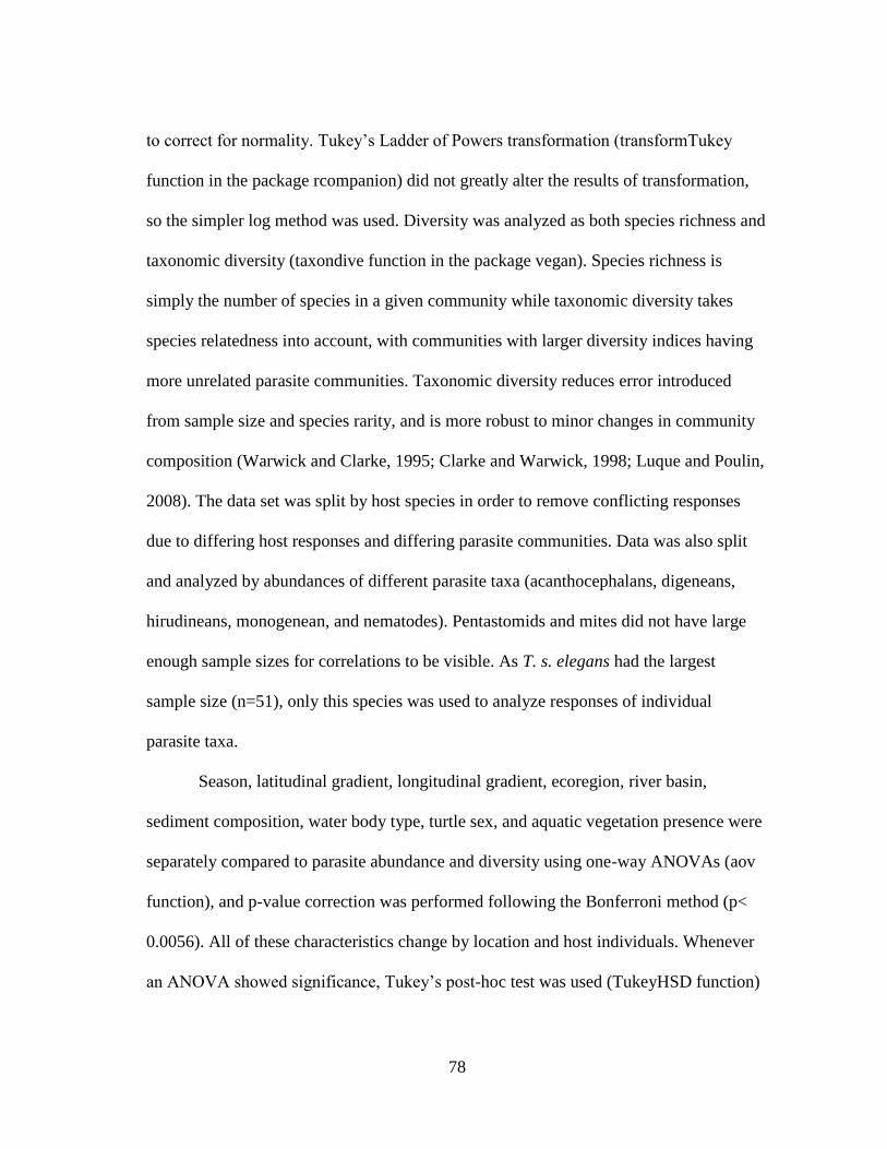
78
to correct for normality. Tukey’s Ladder of Powers transformation (transformTukey
function in the package rcompanion) did not greatly alter the results of transformation,
so the simpler log method was used. Diversity was analyzed as both species richness and
taxonomic diversity (taxondive function in the package vegan). Species richness is
simply the number of species in a given community while taxonomic diversity takes
species relatedness into account, with communities with larger diversity indices having
more unrelated parasite communities. Taxonomic diversity reduces error introduced
from sample size and species rarity, and is more robust to minor changes in community
composition (Warwick and Clarke, 1995; Clarke and Warwick, 1998; Luque and Poulin,
2008). The data set was split by host species in order to remove conflicting responses
due to differing host responses and differing parasite communities. Data was also split
and analyzed by abundances of different parasite taxa (acanthocephalans, digeneans,
hirudineans, monogenean, and nematodes). Pentastomids and mites did not have large
enough sample sizes for correlations to be visible. As T. s. elegans had the largest
sample size (n=51), only this species was used to analyze responses of individual
parasite taxa.
Season, latitudinal gradient, longitudinal gradient, ecoregion, river basin,
sediment composition, water body type, turtle sex, and aquatic vegetation presence were
separately compared to parasite abundance and diversity using one-way ANOVAs (aov
function), and p-value correction was performed following the Bonferroni method (p<
0.0056). All of these characteristics change by location and host individuals. Whenever
an ANOVA showed significance, Tukey’s post-hoc test was used (TukeyHSD function)

79
to determine the relationship between variables. Tukey’s test compares the differences
between group’s means to determine which groups from an ANOVA differ significantly.
No difference was found in the parasite diversity among the two subspecies of A.
spinifera captured in this study, A. s. pallida and A. s. emoryi, and so they were analyzed
together.
III.3 Results
III.3.1 Sample sites
Turtles were collected from 16 properties across seven ecoregions and eight river
basins. The properties were located in 13 towns across Texas: Barksdale, Bryan, College
Station (Aquaculture Facility, Private Property 1, Private Property 2, and TAMU
Campus), Comstock, Del Valle, Franklin, Gladewater, Glen Rose, Humble, Iola,
Leander, Sinton, and Streetman. Sites varied in the type of water bodies present.
The Barksdale site, located in the Edwards Plateau ecoregion and Nueces river basin,
had a single vegetated river with gravel sediment.
The Bryan site, located in the East Central Texas Plains ecoregion and Brazos
river basin, had a single vegetated pond with primarily clay sediment.
The Aquaculture Facility site, located in the East Central Texas Plains ecoregion
and Brazos river basin, had a vegetated oxbow pond with primarily silt sediment and fish
ponds with rubber liner substrate and no vegetation.
The Private Property 1 site, located in the East Central Texas Plains ecoregion
and Brazos river basin, had a single vegetated pond with primarily silt sediment.

80
The Private Property 2 site, located in the East Central Texas Plains ecoregion
and Brazos river basin, had a single vegetated pond with primarily clay sediment.
The TAMU Campus site, located in the East Central Texas Plains ecoregion and
Brazos river basin, had a single vegetated pond with primarily silt sediment.
The Comstock site, located in the Chihuahuan Desert ecoregion and Rio Grande
river basin, had a vegetated pond with primarily clay sediment and an unvegetated river
with primarily loam sediment.
The Del Valle site, located in the Texas Blackland Prairies ecoregion and
Colorado river basin, had a vegetated pond with primarily silt sediment and a vegetated
lake with primarily sand sediment.
The Franklin site, located in the East Central Texas Plains ecoregion and Brazos
river basin, had a vegetated lake with primarily sand sediment.
The Gladewater site, located in the South Central Plains ecoregion and Sabine
river basin, had a vegetated pond with primarily silt sediment and a vegetated pond with
primarily sand sediment.
The Glen Rose site, located in the Cross Timbers ecoregion Brazos river basin,
had a single vegetated river with primarily rock and gravel sediment.
The Humble site, located in the South Central Plains ecoregion and San Jacinto
river basin, had a single unvegetated pond with primarily clay sediment.
The Iola site, located in the East Central Texas Plains ecoregion and Brazos river
basin, had a single vegetated pond with primarily silt sediment.

81
The Leander site, located in the Edwards Plateau ecoregion and Brazos river
basin, had two vegetated ponds, both with primarily gravel sediments.
The Sinton site, located in the Western Gulf Coastal Plains ecoregion and San
Antonio-Nueces river basin, had a vegetated pond and an unvegetated river, both with
primarily clay sediment.
The Streetman site, located in the East Central Texas Plains ecoregion and
Trinity river basin, had a single vegetated pond with primarily sand sediment.
III.3.2 Capture data
A total of 15 A spinifera (11 A. s. pallida and four A. s. emoryi), nine C.
serpentina, and 55 T. s. elegans were collected and necropsied for this study. Four T. s.
elegans were collected without water parameter data, so only 51 individuals of this
species are used for water parameter analyses. A total of 18,853 individual parasites
were recovered in this study: 8,271 acanthocephalans, 8,408 nematodes, 947 trematodes,
191 monogeneans, 906 leeches, 112 pentastomids, and 18 mites. A total of 42 different
parasitic species were recovered in this study: five species of acanthocephalans, nine
species of nematodes, 14 species of trematodes, two species of cestodes, five species of
monogeneans, five species of leeches, one species of mite, and one species of
pentastomid. The detailed localities, taxonomy, and host associations are listed out in the
previous chapter (Table 1).

82
III.3.3 Regional and water quality analyses
Environmental parameters were highly variable across sites. Ammonia levels
were highest (> 2 mg/L) at the Streetman and TAMU campus sites. Chloride levels were
highest (> 200 mg/L Cl-) at the Aquaculture Facility and Sinton sites. Carbon dioxide
levels were highest (> 200 mg/L) at the Aquaculture Facility. Dissolved Oxygen levels
were lowest (< 4 mg/L) at the TAMU campus, Aquaculture Facility, and Leander sites.
Hardness levels were highest (> 500 mg/L CaCO3) at the TAMU Campus, Glen Rose,
and Comstock, with Glen Rose and Comstock having levels over 1850 mg/L CaCO3 on
one occasion. Nitrites were only detected on one occasion at three properties, Bryan, Del
Valle, and TAMU campus, with levels over 2.0 mg/L NO2- at the Bryan site. Nitrate
levels were highest (>4 mg/L NO3-) at the Del Valle site. Salinity was only detected (25
ppt) in the river at the Sinton site. Turbidity measured as water clarity (cm) was highest
(< 20 cm visibility) at the Aquaculture Facility, Humble, Private Property 1, Private
Property 2, and TAMU Campus sites. The Barksdale and Glen Rose sites and one pond
at the Comstock site had the highest visibility, with water being clear to the bottom. The
pH levels varied across sites from 6.5 to 9.0. The Barksdale, Bryan, Aquaculture
Facility, Private Property 1, Private Property 2, TAMU campus, Comstock, Del Valle,
Glen Rose, Humble, Iola, Leander, and Streetman sites were all basic (>7). The
Gladewater site was measured as acidic (6.5) in the small pond and basic (7.5) in the
lake. The Sinton site was measured as acidic (6.75) in the small pond and basic (7.25) in
the river. The Franklin site was only sampled once and was neutral (7). Temperature

83
(C) varied by season, averaging 21C in the spring, 28C in the summer, 24C in the
fall, and 17C in the winter.
For water parameters, simple linear regressions were performed on parasite
abundance, species richness, and taxonomic diversity. The data set was split by host
species in order to remove conflicting responses due to different host responses and
parasite communities. Parasite abundance and diversity were not significantly correlated
to any water parameters for A. spinifera and C. serpentina, which could be a result of the
small sample sizes in both groups (N=15 and N=9). Turbidity was significantly
positively correlated with abundance (Fig. 9A, p=7.47e-6, R2=0.34) and significantly
negatively correlated with taxonomic diversity (Fig. 9B, p= 0.022, R2= 0.1) for parasites
of T. s. elegans (n=51). Parasite species richness and taxonomic diversity were
significantly negatively correlated with carbon dioxide (Fig. 10A, p=0.018, R2= 0.11;
Fig. 10B, p=0.0024, R2= 0.17). Taxonomic diversity was significantly negatively
correlated with chloride (Fig. 12, p= 0.0075, R2= 0.14) and significantly positively
correlated with dissolved oxygen (Fig. 11A, p= 0.029, R2= 0.094). When data was
analyzed by parasitic taxa abundances, only acanthocephalans, digeneans, and
monogeneans showed significant relationships. Acanthocephalan abundance was
significantly positively correlated with ammonia (Fig. 13, p= 0.042, R2= 0.082) and
turbidity (Fig. 9C, p= 0.0024, R2= 0.17). Digenean abundance was significantly
positively correlated with turbidity (Fig. 9D, p=0.049, R2= 0.077). Monogenean
abundance was significantly positively correlated with dissolved oxygen (Fig. 11B, p=

84
0.039, R2= 0.084) and significantly negatively correlated with carbon dioxide (Fig. 10C,
p= 0.024, R2= 0.10).

85
Figure 9: Linear regressions for turbidity (water clarity), trend lines plotted with 95% confidence intervals. Significance coefficients are included on
each plot. A) Correlation between turbidity and parasite abundance, B) Correlation between turbidity and taxonomic diversity, C) Correlation between
turbidity and acanthocephalan abundance, and D) Correlation between turbidity and digenean abundance.
A B
C D

86
Figure 10: Linear regressions for carbon dioxide, trend lines plotted with 95% confidence intervals. Significance coefficients are included on each plot.
A) Correlation between carbon dioxide levels and species richness, B) Correlation between carbon dioxide levels and taxonomic diversity, and C)
Correlation between carbon dioxide levels and monogenean abundance.
A B
C
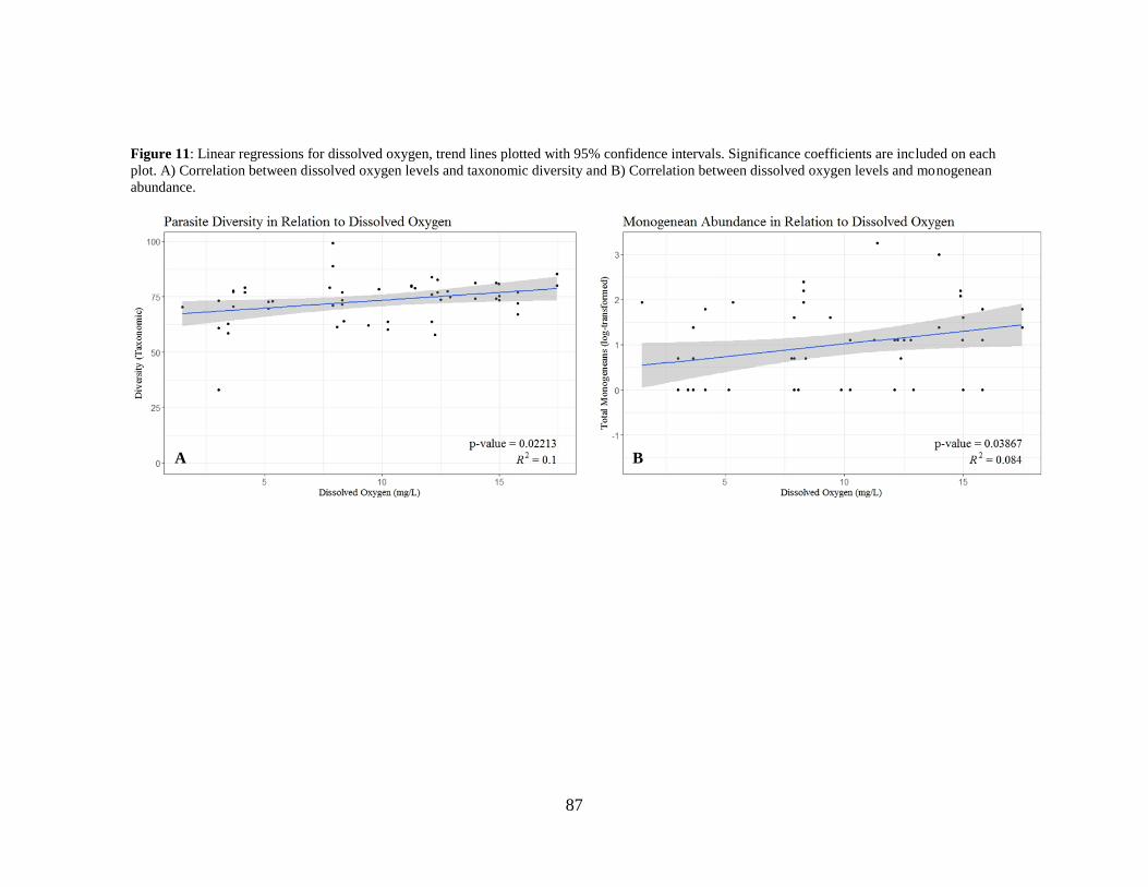
87
Figure 11: Linear regressions for dissolved oxygen, trend lines plotted with 95% confidence intervals. Significance coefficients are included on each
plot. A) Correlation between dissolved oxygen levels and taxonomic diversity and B) Correlation between dissolved oxygen levels and monogenean
abundance.
A B

88
Figure 12: Linear regression for chloride levels and taxonomic diversity, trend line plotted with 95% confidence interval. Significance coefficients are
included on the plot.
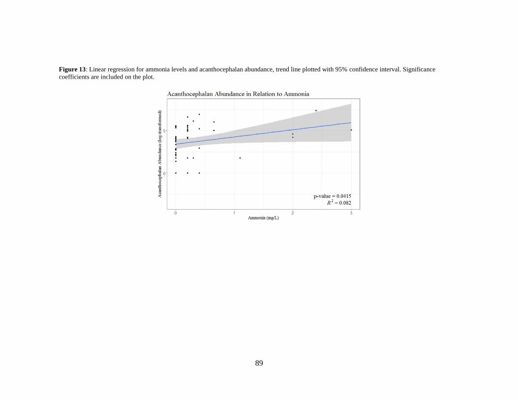
89
Figure 13: Linear regression for ammonia levels and acanthocephalan abundance, trend line plotted with 95% confidence interval. Significance
coefficients are included on the plot.

90
Season, latitudinal gradient, longitudinal gradient, ecoregion, river basin, water
body type, sediment type, and aquatic vegetation presence were all compared to parasite
abundance and diversity using one-way ANOVAs, with Bonferroni p-value correction
(p< 0.0056).
Total parasite abundance among all three host species, scaled logarithmically for
normality, was significantly different among longitudinal gradient (p=0.00028) and river
basins (p=0.00055). For longitudinal gradient, the West had significantly lower
abundance than Central (p=0.0056) and the East (p=0.00016). For river basins, post-hoc
tests showed no significant differences in abundance between basins. Total parasite
abundance was significantly lower in lotic versus lentic systems (p=0.00046).
Total species richness and taxonomic diversity were not significantly correlated
with any environmental variables.
Parasite abundance in T. s. elegans, scaled logarithmically for normality, was
significantly different among river basins (p=0.0013) and water body type (p=0.00039).
For river basins, post-hoc tests showed no significant differences in abundance between
basins. Parasite abundance was significantly lower in lotic versus lentic systems
(p=0.00039).
Species richness and taxonomic diversity in T. s. elegans were not significantly
different among any environmental variables.
When data was analyzed by parasitic taxa abundances, only acanthocephalans,
leeches, and nematodes showed significant relationships. Acanthocephalan abundance
91
was significantly different among water body type (p=0.00065). For water body type,
lentic systems had significantly higher acanthocephalan abundance than lotic systems.
Leech abundance was significantly different among river basins (p=0.0045). For
river basins, leech abundance was significantly higher in the Trinity basin than the
Brazos basin (p=0.0012) and the San Antonio Nueces basin (p=0.0052).
All significance coefficients from the linear regressions and ANOVAs are
reported in tables 4 through 6.

92
Table 4: Significance coefficients (p-values and R2 values) from simple linear regressions on parasite abundance and diversity. Significant values are
bolded. Parasite Abundance Parasite Diversity (Richness) Parasite Diversity (Taxonomic)
Total TS only Total TS only Total TS only
p-
value R2 p-value R2
p-
value R2
p-
value R2
p-
value R2
p-
value R2
Ammonia 0.3 0.015 0.038 0.085 0.719 0.0018 0.45 0.012 0.86 0.00041 0.67 0.0038
CO2 0.43 0.0085 0.93 0.00017 0.16 0.026 0.018 0.11 0.19 0.023 0.0025 0.17
Chloride 0.71 0.0019 0.99 2.28E-06 0.55 0.0049 0.74 0.0024 0.08 0.041 0.0075 0.14
DO 0.6 0.004 0.28 0.024 0.14 0.029 0.14 0.044 0.66 0.0027 0.029 0.094
Hardness 0.071 0.044 0.13 0.046 0.095 0.038 0.11 0.051 0.77 0.0012 0.87 0.00053
pH 0.5 0.0064 0.63 0.0048 0.47 0.007 0.95 7.11E-05 0.77 0.0011 0.68 0.0034
Temperature 0.88 0.00032 0.66 0.0041 0.42 0.009 0.12 0.048 0.75 0.0014 0.57 0.0065
Turbidity 0.0094 0.089 7.47E-06 0.34 0.67 0.0026 0.3 0.022 0.13 0.031 0.022 0.1
Table 5: Significance coefficients (p-values and R2 values) from simple linear regressions on parasite taxa. Significant values are bolded.
Parasite Abundance (TS only)
Acanthocephalans Digeneans Leeches Monogeneans Nematodes
p-value R2 p-value R2 p-value R2 p-value R2 p-value R2
Ammonia 0.042 0.082 0.26 0.026 0.055 0.073 0.28 0.024 0.056 0.072
CO2 0.47 0.011 0.33 0.02 0.072 0.065 0.024 0.1 0.58 0.0064
Chloride 0.083 0.06 0.11 0.052 0.06 0.07 0.74 0.0022 0.82 0.001
DO 0.66 0.004 0.15 0.041 0.28 0.024 0.039 0.084 0.92 0.00022
Hardness 0.64 0.0046 0.31 0.021 0.28 0.024 0.85 0.00079 0.28 0.024
pH 0.38 0.016 0.5 0.0093 0.15 0.041 0.66 0.0041 0.3 0.022
Temperature 0.54 0.0078 0.17 0.039 0.16 0.04 0.92 0.00021 0.41 0.014
Turbidity 0.0024 0.17 0.049 0.077 0.19 0.035 0.74 0.0023 0.41 0.014

93
Table 6: Significance coefficients (p-values) from one-way ANOVAs. Significant values are bolded. See Appendix for full table of significance
coefficients from Tukey’s post-hoc tests. Parasite Abundance
Parasite Diversity
(Richness)
Parasite Diversity
(Taxonomic) Parasite Abundance (TS only)
Total TS only Total TS only Total TS only Acanthocephalans Digeneans Leeches Monogeneans Nematodes
Lentic/Lotic 0.00046 0.00039 0.32 0.72 0.88 0.092 0.00065 0.41 0.27 0.19 0.084
Sex 0.11 0.072 0.11 0.29 0.13 0.61 0.26 0.19 0.91 0.58 0.092
Aquatic Veg 0.13 0.41 0.05 0.16 0.016 0.057 0.1 0.6 0.028 0.91 0.6
Season 0.53 0.61 0.062 0.057 0.403 0.061 0.83 0.065 0.089 0.18 0.03
Latitude 0.83 0.14 0.95 0.41 0.54 0.53 0.12 0.12 0.41 0.29 0.25
Longitude 0.00028 0.0087 0.037 0.32 0.035 0.029 0.28 0.49 0.14 0.76 0.39
Ecoregion 0.009 0.022 0.27 0.62 0.88 0.63 0.03 0.19 0.4 0.097 0.08
River Basin 0.00055 0.0013 0.46 0.84 0.48 0.077 0.0059 0.57 0.0045 0.32 0.021
Sediment 0.039 0.13 0.38 0.78 0.33 0.2 0.55 0.12 0.034 0.04 0.67

94
III.3.4 Host measurement analyses
Simple linear regressions were also performed between parasite abundance,
species richness, and taxonomic diversity and turtle weight, straight carapace length,
straight carapace width, shell depth, curved carapace length, curved carapace width, and
circumference. For A. spinifera, parasite abundance was significantly positively
correlated with turtle weight (Fig. 14E, p=0.035, R2= 0.30), straight carapace length
(Fig. 14A, p=0.017, R2= 0.36), straight carapace width (Fig. 14C, p=0.016, R2= 0.37),
curved carapace length (Fig. 14B, p=0.016, R2= 0.37), curved carapace width (Fig. 14D,
p=0.016, R2= 0.37). No correlation was seen in C. serpentina or T. s. elegans. ANOVAs
were performed between host sex and melanism, and parasite abundance, species
richness, and taxonomic diversity, but no significant relationships were found.

95
Figure 14: Linear regressions for body size of A. spinifera in relation to parasite abundance, trend line plotted with 95% confidence interval.
Significance coefficients are included on the plot. A) Correlation between straight carapace length and parasite abundance, B) Correlation between
curved carapace length and parasite abundance, C) Correlation between straight carapace width and parasite abundance, D) Correlation between curved
carapace width and parasite abundance, and E) Correlation between weight and parasite abundance.
A B
C D

96
Figure 14: Continued.
E

97
III.4 Discussion
Many studies have shown through both surveys and meta-analysis that
environmental changes are impacting parasite diversity and distributions (e.g. Bourque
and Esch, 1974; Grimmond, 2007; King et al., 2010; Burrows et al., 2011; Bellard et al.,
2012; Hautier et al., 2015; Cizauskas et al., 2016; Carlson et al., 2017). Of the three
major diversity surveys of parasites of Texas freshwater turtles (Harwood, 1932;
Everhart, 1957; Dinuzzo, 1981), only one unpublished thesis reported any ecological
data (Dinuzzo, 1981). Season and water temperature as well as host sex and age class
were compared to the abundances of different parasitic taxa in T. s. elegans. Dinuzzo
found that female turtles had higher parasite abundances than male turtles, which was
attributed to their larger size. Adult turtles had more parasites than juveniles, which was
also attributed to body size. In the current study, no differences were found between host
sex or body size (age class) on parasite abundance or diversity in T. s. elegans. A
significant increase in parasite abundance was found in A. spinifera with increasing body
size. Dinuzzo also found that parasites had lower intensities in the winter compared to
the summer for all taxa but the trematodes, which had higher abundances in the winter.
In the current study, trematodes and nematodes were found to have higher abundances in
the spring, with no seasonality in acanthocephalans, monogeneans, and leeches. In
addition, taxonomic diversity was higher in the summer than the fall. The difference
observed in seasonality between the study by Dinuzzo (1981) and the current study
could be attributed to a number of factors including confounding factors from multiple

98
highly variable study sites, different species assemblages responses, or climatic
differences between the two temporally distant studies.
Trachemys scripta elegans was the most common host species captured during
this study. This turtle was in high abundance at most sample sites, often seen basking or
swimming when setting and collecting traps. Apalone spinifera was the second most
common species, and was seen swimming in the river sites more often than T. s. elegans.
Chelydra serpentina were only seen when captured in this study, and more than two
were never captured in a single trap night. Apart from the three target species of turtles,
only one Texas cooter, Pseudemys texana, and one razor-backed musk turtle,
Sternotherus carinatus, were captured during the course of this study. As a result of the
larger sample size, T. s. elegans revealed the clearest picture of the effects of
environmental parameters and regionality on parasite diversity and abundance. Parasite
abundance and species richness were variable across samples sites, and were largely
dependent on sample size. Taxonomic diversity was a more consistent measure of
diversity across sample sites than species richness, as it is not influenced as heavily by
species rarity. This measure of diversity is a useful comparative measure for studies
where samples size is limited (Reiczigel et al., 2013).
Many environmental and host physiological factors contribute to the abundance
and diversity of parasites in a given ecosystem. For this reason it is doubtful that any
single factor has any major significance in the determination of species assemblage
dynamics. Many of the statistically significant correlations presented in this study are
likely due to covariation of parameters at sites, and many of the important variables were

99
likely not measured in this study. Most correlation coefficients (R2) presented with the
linear models were low, indicating very little of the observed variation is explained by
the model. The possible contributions of individual factors are discussed here, although
the contribution may be insignificant in reality, and these hypotheses should be tested in
future lab and fieldwork. Multivariate methods are currently being used to analyze which
factors are contributing the most to the overall variance between sites, and will be
reported in a future paper.
Turbidity, measured as water clarity, had the strongest correlations of any
environmental parameters in this study, and is likely the strongest predictor of parasite
abundance and diversity. Turbidity was negatively correlated with taxonomic diversity,
while it was positively correlated with total parasite abundance, abundance of
acanthocephalans, and abundance of digeneans. This increase in diversity in clearer
water could be a result of reduced diversity of intermediate host species, increased
predation on free living larval stages, reduced motility of free living larval stages, direct
mortality of free living larval stages due to toxins often associate with turbid water, or
cryptic interactions between host health and water quality. The increase in abundance in
more turbid waters could be a result of increased abundance of specific intermediate host
species or a greater availability of niche space within the host due to decreases in
diversity. Abundance of acanthocephalans and digeneans was higher in more turbid
waters, following the trend seen in total parasite abundance. Izyumova (1979) found that
turbidity can negatively affect crustaceans through direct mechanical damage by
suspended particles to the feeding appendages of larval stages. Conversely, Yilman and

100
Kulkoyluoglu (2006) found that certain species of ostracod crustaceans, the known
intermediate host for acanthocephalans, increased in abundance with increasing
turbidity. An increase in the intermediate host abundance of acanthocephalans would
increase the abundance in turtle hosts, explaining the positive correlation between
turbidity and abundance seen in the current study. Turbidity decreases direct mortality of
cercaria from UV radiation and predation, and increases the abundance of snail
intermediate hosts by reducing predation and increasing food availability (Zbikowska et
al., 2006; Shah et al., 2013). An increase in cercaria survival and snail intermediate host
abundance could explain an increase in overall trematode abundance in more turbid
environments.
Carbon dioxide levels were negatively correlated with species richness,
taxonomic diversity, and monogenean abundance. Parasitic nematode larvae respond to
carbon dioxide as a host seeking stimulus (Sciacca et al., 2002). Schistosoma larvae,
both miracidia and cercaria, exhibit host seeking responses to chemical stimuli including
amino acids, fatty acids, ammonia, and several different glycoproteins (Sukhdeo and
Sukhdeo, 2004; Haeberlein and Haas, 2008). If aquatic parasites which have larvae that
must seek hosts, like monogeneans, rely on the same stimulus, high ambient carbon
dioxide levels could interfere with this life cycle and reduce the abundance of specific
parasites, therefore reducing the overall diversity.
Dissolved oxygen levels were positively correlated with taxonomic diversity and
monogenean abundance. Monni and Cognetti-Varriale (2002) found that eels have a
stronger immune response against monogeneans in oxygen rich water, leading to lower

101
levels of infection. Chapman et al. (2000) found higher prevalence of monogeneans in
fish inhabiting oxygen deficient environments, which could be due to a high tolerance on
the part of the parasite or a reduced immune response in the fish. The data in the current
study contrast these findings. Some larval helminths have inhibited development in
oxygen deficient environments (Thorson, 1969). It is likely that turtle monogeneans
respond differently to oxygen levels than fish monogeneans. It may be that turtle
monogenean adults and larvae require higher oxygen levels to persist. Laboratory studies
observing survival and infectivity of monogeneans in water with different levels of
carbon dioxide and oxygen could clarify the mechanisms behind this relationship.
Chloride levels were negatively correlated with taxonomic diversity. High levels
of chlorides are known to interfere with osmoregulation in aquatic organisms (Karraker
and Gibbs, 2011). This reduction in taxonomic diversity indicates a decrease in the
number of parasite taxa, and could be due to toxic effects of chloride on certain free
swimming larval stages, certain intermediate hosts, or adults within the host as water is
ingested.
Ammonia levels were positively correlated with acanthocephalan abundance. At
high levels, ammonia can be toxic to aquatic organisms by halting their ability to
properly excrete waste (U.S. Environmental Protection Agency, 2013). The increase in
acanthocephalan levels could be due to a decrease in host immunity, allowing higher
numbers of acanthocephalans to invade. This correlation is confounding, as higher
ammonia levels would reduce the survival of intermediate host species and free
swimming larvae. Na et al. (2009) showed that certain ostracods have a high tolerance to

102
increased aquatic ammonia levels. If ammonia is reducing the abundances of other
aquatic zooplankton, ostracods may be in high abundance in environments with raised
ammonia levels, increasing the abundance of acanthocephalan parasites. More in depth
field studies would be necessary to discover if this correlation observed is simply
coincidental and that other factors are not causing the increased abundance. Laboratory
experiments testing varying ammonia levels on the acanthocephalan life cycle could
clarify the mechanisms behind this correlation.
Longitudinal gradient, river basin, and water body type were all significantly
correlated with parasite abundance, while no variables were significantly correlated with
diversity. Differences between parasite abundance across a longitudinal gradient and
river basins were in an east to west distribution, with higher parasite abundance in the
East. It is likely that the difference is due to the change in climate from east to west
Texas, with lower precipitation in the west reducing the transmission of parasites. Lotic
systems (rivers) had significantly lower parasite abundance than lentic systems (ponds
and lakes). Higher flow in lotic systems could possibly inhibit transmission of free-
swimming larval stages and therefore reduce the abundance of parasites within the hosts
in those systems.
Host measurement analyses revealed that parasite abundance was higher in larger
A. spinifera. Interestingly, this trend was not observed in the other two host species.
There could be acquired immune responses to parasitism with age in C. serpentina and
T. s. elegans that are not present in A. spinifera.

103
As this study was restricted in the number of turtles collected, number of species
sampled, and locations sampled, more examination is still necessary to uncover the
distributions of parasites of more host species across a broader area in Texas. The
titration water parameter tests used in this study relied on rough visual estimations of
color change for quantification of water parameters, only offering an estimate of the
actual parameter value. Similar studies using more accurate data collection methods
would be valuable, as they may reveal a more accurate representation of species
responses to water parameters. It would be beneficial for all collecting ventures, for
museum specimens and other projects, to save internal organs for examination for
parasites. In addition to this, more data should be collected and reported when sampling
for parasites. Associations between parasites and the environment are largely
understudied and some factors not visible in the current study may be elucidated in a
future meta-analysis.
104
CHAPTER IV
SUMMARY AND FUTURE DIRECTIONS
In this study, a total of 78 turtles of three different species were collected across
the state of Texas and examined for parasites. All turtles were infected with at least one
species of parasite, with an average of four species per turtle. A total of 42 species of
metazoan parasites were recovered, with acanthocephalans and nematodes typically
more abundant and trematodes typically more diverse. Sixteen new host records and 17
new locality records are reported. Two new species of Neopolystoma are reported and
Polystomoides coronatum and Acanthostomum nuevoleonensis are redescribed. When
parasite communities were analyzed between host species, A. spinifera was found to
contain a significantly different parasite assemblage than C. serpentina and T. s. elegans.
Parasite abundance was higher in more turbid waters and diversity was higher in less
turbid waters with lower carbon dioxide and chloride levels and higher dissolved oxygen
levels. Acanthocephalans had higher abundance in more turbid waters and water with
higher ammonia levels, digeneans had higher abundance in more turbid waters, and
monogeneans had higher abundances in water with higher dissolved oxygen and lower
carbon dioxide levels. Parasite abundance was higher in eastern parts of Texas and lentic
water bodies. Acanthocephalans were primarily driving the abundance differences
observed. Parasite abundance was significantly higher in large A. spinifera, but no
correlation was seen with body size in C. serpentina or T. s. elegans. Many of the
correlations discovered in this study relate to changes in the quality of freshwater

105
ecosystems. In water of higher quality (lower turbidity, ammonia, carbon dioxide,
chloride, higher dissolved oxygen), parasite diversity will be higher, since the food web
will be more dynamic, maintaining a greater diversity of parasite life cycles. In lower
water quality, more resilient species become hyper-abundant due to less competition for
resources within the host and possible reduced host immunity to infection.
This project began as a simple community diversity survey. Due to the paucity of
molecular data on many parasites, when sample size permitted specimens were saved in
95% ethanol for analysis. Future directions of this project include analyzing molecular
specimens of specific taxa (acanthocephalans, trematodes, cestodes, and monogeneans)
to uncover any cryptic species and reveal gene flow between populations. Collaboration
is currently ongoing with Dr. Vasyl Tkach to study the genetics of many of the
platyhelminth species reported in this survey, which will likely lead to revision of some
identifications. Acanthocephalans will be genetically analyzed in collaboration with Dr.
Florian Reyda and Dr. Anna Phillips to reveal details in morphological differences
across sexes and between populations. A thorough molecular and morphological analysis
of the mites recovered in this study is being conducted by Dr. Ray Fisher in order to
describe this new species. All spirorchiid blood flukes collected in this study will be
analyzed both morphologically and molecularly by Charlayna Cammarata for her
dissertation. Nematode, leech, and pentastomid specimens are available for any
interested parties who would like to add valuable genetic information on some
understudied species.
106
This study highlights the need for further parasite surveys, particularly in
undersampled locations and understudied host taxa. Even in locations and hosts that
have been well studied, regular surveys are vital to reveal changes in parasite community
assemblages.
107
REFERENCES
Acholonu, A. D. 1969. Acanthocephala of Louisiana turtles with a redescription of
Neoechinorhynchus stunkardi Cable and Fischer, 1961. Proceedings of the
Helminthological Society of Washington 36: 177–183.
Ahmad, I. Afshan, K., Ramzan, M., Hayat, S., Shakeel, et al. 2016. Effect of Water
Quality Parameters on Isopod Parasite Alitropus typus (Aegidae) of Ectotherms
in Chashma Lake, Pakistan. Pakistan Journal of Zoology 48: 769–779.
Anderson, R. C., Anderson, R. C., Chabaud, A. G., and Willmott, S. 2009. Keys to the
Nematode Parasites of Vertebrates. CAB International, Wallingford, U.K., 463 p.
Baker, M. R. 1979. Serpinema spp. (Nematoda: Camallanidae) from turtles of North
America and Europe. Canadian Journal of Zoology 57: 934–939.
Banu, A. N. H., and Khan, M. H. 2004. Water Quality, Stocking Density and Parasites of
Freshwater Fish in Four Selected Areas of Bangladesh. Pakistan Journal of
Biological Sciences 3: 436–440.
Bellard, C., Bertelsmeier, C., Leadley, P., Thuiller, W., and Courchamp, F. 2012.
Impacts of climate change on the future of biodiversity. Ecology letters 15: 365–
377.
Blasco-Costa, I., and Poulin, R. 2017. Parasite life-cycle studies: A plea to resurrect an
old parasitological tradition. Journal of Helminthology 91: 647–656.

108
Bourque, J. E., and Esch, G. W. 1974. Population ecology of parasites in turtles from
thermally altered and natural aquatic communities. In Thermal Ecology
Symposium. Augusta, Georgia, USA. p 551–561.
Bray, R. A., Gibson, D. I., and Jones, A. 2008. Keys to the Trematoda, Volume 3. CAB
International, Wallingford, U.K., 805 p.
Brooks, D. R. 1980. Revision of the Acanthostominae Poche, 1926 (Digenea:
Cryptogonimidae). Zoological Journal of the Linnean Society 70: 313–382.
Brooks, D. R., and Hoberg, E. P. 2007. How will global climate change affect parasite–
host assemblages? Trends in Parasitology 23: 571–574.
Bureau of Economic Geology. 1996. River Basins of Texas.
Burlakova, L. E., Karatayev, A. Y., Karatayev, V. A., May, M. E., Bennett, D. L., et al.
2011. Biogeography and conservation of freshwater mussels (Bivalvia:
Unionidae) in Texas: Patterns of diversity and threats. Diversity and
Distributions 17: 393–407.
Burrows, M. T., Schoeman, D. S., Buckley, L. B., Moore, P., Poloczanska, E. S., et al.
2011. Interpretation of recent Southern Hemisphere climate change. Science 334:
652–655.
Bush, A. O., Lafferty, K. D., Lotz, J. M., and Shostak, A. W. 1997. Parasitology meets
ecology on its own terms: Margolis et al. Revisited. The Journal of Parasitology
83: 575.
Bychowsky, B. E. 1957. Monogenetic trematodes, their systematics and phylogeny. (In
Russian). Proceedings of the USSR Academy of Sciences: 1–509.

109
Carlson, C. J., Burgio, K. R., Dougherty, E. R., Phillips, A. J., Bueno, V. M., et al. 2017.
Parasite biodiversity faces extinction and redistribution in a changing climate.
Science Advances 3: 1–12.
Chapman, J. M., Marcogliese, D. J., Suski, C. D., and Cooke, S. J. 2015. Variation in
parasite communities and health indices of juvenile Lepomis gibbosus across a
gradient of watershed land-use and habitat quality. Ecological Indicators 57:
564–572.
Chapman, L. J. Lanciani, C. A., and Chapman, C. A. 2000. Ecology of a diplozoon
parasite on the gills of the African cyprinid Barbus neumayeri. African Journal of
Ecology 38: 312–320.
Cizauskas, C. A., Carlson, C. J., Burgio, K. R., Clements, C. F., and Dougherty, E. R.
2017. Parasite vulnerability to climate change: an evidence-based functional trait
approach. Royal Society Open Science 4: 1–12.
Clarke, K. R. 1993. Non-parametric multivariate analyses of changes in community
structure. Australian Journal of Ecology 18: 117–143.
Clarke, K. R., and Warwick, R. M. 1998. A taxonomic distinctness index and its
statistical properties. Journal of Applied Ecology 35: 523–531.
Conroy, C. J., Papenfuss, T., Parker, J., and Hahn, N. E. 2009. Use of Tricaine
Methanesulfonate (MS222) for Euthanasia of Reptiles. Journal of the American
Association for Laboratory Animal Science 48: 28–32.
Curran, S. S., Overstreet, R. M., Collins, D. E., and Benz, G. W. 2014. Levisunguis
subaequalis n. g., n. sp., a tongue worm (Pentastomida: Porocephalida:

110
Sebekidae) infecting softshell turtles, Apalone spp. (Testudines: Trionychidae) in
the southeastern United States. Systematic Parasitology 87: 33–45.
Dinuzzo, A. R. 1981. Studies on the population biology and host-specificity of helminth
parasites of aquatic turtles from Burleson County, Texas and vicinity. Texas
A&M University, 125 p.
Dobson, A., Lafferty, K. D., Kuris, A. M., Hechinger, R. F., and Jetz, W. 2008. Homage
to Linnaeus: How many parasites? How many hosts? Proceedings of the National
Academy of Sciences 105: 11482–11489.
Dougherty, E. R., Carlson, C. J., Bueno, V. M., Burgio, K. R., Cizauskas, C. A. 2016.
Paradigms for parasite conservation. Conservation Biology 30: 724–733.
Dronen, N. O. 1994. Utilization of the trophic structure of small pond ecosystems by
helminths of frogs, Rana catesbeiana and Rana utricularia. The Texas Journal of
Science 46: 173–186.
Dronen, N. O., Underwood, H. T., and Rubec, L. A. 1982. Seasonal Fluctuations in the
Population Density of Alloglossidium corti (Digenea: Macroderoididae) in
Channel Catfish from the Little Brazos River, Texas. The Southwestern
Naturalist 27: 73–78.
Ernst, C. H., and Lovich, J. E. 2009. Turtles of the United States and Canada Second
Edi. The Johns Hopkins University Press, Baltimore, 827 p.
Ernst, E. M., and Ernst, C. H. 1977. Synopsis of helminths endoparasitic in native turtles
of the United States. Bulletin of the Maryland Herpetological Society 13: 1–75.

111
Esch, G. W., and Gibbons, J. W. 1967. Seasonal Incidence of Parasitism in the Painted
Turtle, Chrysemys picta marginata Agassiz. The Journal of Parasitology 53:
818–821.
Everhart, B. A. 1957. Notes on the helminths of Pseudemys scripta elegans (Wied,
1838) in areas of Texas and Oklahoma. Proceedings of the Oklahoma Academy
of Science 38: 38–43.
Ford, D. F., Walters, A. D., Williams, L. R., Williams, M. G., and Ford, N. B. 2016.
Mussel assemblages in streams of different sizes in the Neches River Basin of
Texas. Southeastern Naturalist 15: 26–40.
Froeschke, G., Harf, R., Sommer, S., and Matthee, S. 2010. Effects of precipitation on
parasite burden along a natural climatic gradient in southern Africa —
implications for possible shifts in infestation patterns due to global changes.
Oikos 119: 1029–1039.
Gibson, D. I., Jones, A., and Bray, R. A. 2002. Keys to the Trematoda, Vol. 1. CAB
International, Wallingford, U.K., 521 p.
Goedknegt, M. A., Welsh, J. E., Drent, J., and Thieltges, D. W. 2015. Climate change
and parasite transmission: How temperature affects parasite infectivity via
predation on infective stages. Ecosphere 6: 1–9.
Gomez, A., and Nichols, E. 2013. Neglected wild life: Parasitic biodiversity as a
conservation target. International Journal for Parasitology: Parasites and Wildlife
2: 222–227.
112
Griffith, G. E., Bryce, S. B., Omernik, J. M., and Rogers, A. 2007. Ecoregions of Texas.
Austin, TX, 125 p.
Grimmond, S. 2007. Urbanization and global environmental change: local effects of
urban warming. Geographical Journal 173: 83–88.
Grosmaire, E. K. 1977. Aspects of the natural history of freshwater turtles within the
lower Rio Grande Valley of Texas. Texas A&M University, 102 p.
Haeberlein, S., and Haas, W. 2008. Chemical attractants of human skin for swimming
Schistosoma mansoni cercariae. Parasitology Research 102: 657–662.
Harwood, P. D. 1932. The helminths parasitic in the amphibia and reptilia of Houston,
Texas, and vicinity. Proceedings of the National Museum 81: 1–71.
Hautier, Y., Tilman, D., Isbell, F., Seabloom, E. W., Borer, E. T., et al. 2015.
Anthropogenic environmental changes affect ecosystem stability via biodiversity.
Science 348: 336–340.
Hudson, P. J., Dobson, A. P., and Lafferty, K. D. 2006. Is a healthy ecosystem one that
is rich in parasites? Trends in Ecology and Evolution 21: 381–385.
Izyumova, N. A. 1979. The fish parasite fauna of the Volga. Monographiae Biologicae
33: 340–345.
Janzen, D. H., and Schoener, T. W. 1968. Differences in insect abundance and diversity
between wetter and drier sites during a tropical dry season. Ecology 49: 96–110.
Johnson, P. T. J., and Thieltges, D. W. 2010. Diversity, decoys and the dilution effect:
how ecological communities affect disease risk. The Journal of Experimental
Biology 213: 961–970.

113
Jones, A., Bray, R. A., and Gibson, D. I. 2005. Keys to the Trematoda, Volume 2. CAB
International, Wallingford, U.K. 768 p.
Jousson, O., Bartoli, P., and Pawlowski, J. 2000. Cryptic speciation among intestinal
parasites (Trematoda: Digenea) infecting sympatric host fishes (Sparidae).
Journal of Evolutionary Biology 13: 778–785.
Karraker, N. E., and Gibbs, J. P. 2011. Road deicing salt irreversibly disrupts
osmoregulation of salamander egg clutches. Environmental Pollution 159: 833–
835.
Khalil, L. F., Jones, A., and Bray, R. A. 1994. Keys to the Cestode Parasites of
Vertebrates. CAB International, Wallingford, U.K., 751 p.
King, K. C., Mclaughlin, J. D., Boily, M., and Marcogliese, D. J. 2010. Effects of
agricultural landscape and pesticides on parasitism in native bullfrogs. Biological
Conservation 143: 302–310.
Klemm, D. J. 1985. Freshwater leeches (Annelida: Hirudinea). In A Guide to the
Freshwater Annelida (Polychaeta, Naidid and Tubificid Oligochaeta, and
Hirudinea) of North America. D. J. Klemm (ed.). Kendall/Hurt Publishing Co. p
198.
Kruskal, J. B. 1964. Multidimensional scaling by optimizing goodness of fit to a
nonmetric hypothesis. Psychometrika 29: 1–27.
Lafferty, K. D., Dobson, A. P., and Kuris, A. M. 2006. Parasites dominate food web
links. Proceedings of the National Academy of Sciences 103: 11211–11216.
114
Lafferty, K. D., Allesina, S., Arim, M., Briggs, C. J., De Leo, G., et al. 2008. Parasites in
food webs: The ultimate missing links. Ecology Letters 11: 533–546.
Lafferty, K. D., and Kuris, A. M. 1999. How environmental stress affects the impacts of
parasites. Limnology and Oceanography 44: 925–931.
Leidy, J. 1888. Entozoa of the Terrapin. Proceedings of the Academy of Natural
Sciences of Philadelphia 40: 127–128.
Lenis, C., and Garcia-Prieto, L. 2009. Polystomoides magdalenensis n. sp.
(Monogenoidea: Polystomatidae), a Parasite of Buccal Cavity of Trachemys
callirostris callirostris (Testudinata: Emydidae) from Colombia. The Journal of
Parasitology 95: 850–854.
Luque, J. L., and Poulin, R. 2008. Linking ecology with parasite diversity in Neotropical
fishes. Journal of Fish Biology 72: 189–204.
MacDonald, C., and Brooks, D. R. 1989. Revision and phylogenetic analysis of the
North American species of Telorchis Luehe, 1899 (Cercomeria: Trematoda:
Digenea: Telorchiidae). Canadian Journal of Zoology 67: 2301–2320.
Madanire-Moyo, G. N., Luus-Powell, W. J., and Olivier, P. A. 2012. Diversity of
metazoan parasites of the Mozambique tilapia, Oreochromis mossambicus
(Peters, 1852), as indicators of pollution in the Limpopo and Olifants River
systems. Onderstepoort Journal of Veterinary Research 79: 1–9.
Mangiafico, S. 2018. rcompanion: Functions to Support Extension Education Program
Evaluation. R package version 1.13.0. https://CRAN.R
project.org/package=rcompanion

115
Marcogliese, D. J. 2005. Parasites of the superorganism: Are they indicators of
ecosystem health? International Journal for Parasitology 35: 705–716.
McAllister, C. T., Bursey, C. R., and Trauth, S. E. 2008. New Host and Geographic
Distribution Records for Some Endoparasites (Myxosporea, Trematoda,
Cestoidea, Nematoda) of Amphibians and Reptiles from Arkansas and Texas,
U.S.A. Comparative Parasitology 75: 241–254.
McGaugh, S. E., Eckerman, C. M., and Janzen, F. J. 2008. Molecular phylogeography of
Apalone spinifera (Reptilia, Trionychidae). Zoologica Scripta 37: 289–304.
Monni, G., and Cognetti-Varriale, A. M. 2002. Antigenicity of Pseudodactylogyrus
anguillae and P. bini (Monogenea) in the European eel (Anguilla anguilla, L.)
under different oxygenation conditions. Fish and Shellfish Immunology 13: 125–
131.
Moser, W. E. Govedich, F. R., Oceguera-Figueroa, A., Richardson, D. J., and Phillips,
A. J. 2016. Phylum Annelida; Subclass Hirudinida, Fourth Edition. In Keys to
Nearctic Fauna. J. H. Thorp and D. C. Rogers (eds.). Elsevier. p 244–259.
Na, Y., Shimei, C., Erchao, L., Jiayan, C., and Liqiao, C. 2009. Tolerance of
Physocypria kraepelini (Crustacean, Ostracoda) to water-borne ammonia,
phosphate and pH value. Journal of Environmental Sciences 21: 1575–1580.
Nachev, M., and Sures, B. 2009. The endohelminth fauna of barbel (Barbus barbus)
correlates with water quality of the Danube River in Bulgaria. Parasitology 136:
545–552.
116
Oksanen, J., Blanchet, F. G., Friendly, M., Kindt, R., Legendre, P., et al. 2017. Vegan:
Community Ecology Package. R package version 2.4.4. https://CRAN.R-
project.org/package=vegan
Parker, G. A., Chubb, J. C., Ball, M. A., and Roberts, G. N. 2003. Evolution of complex
life cycles in helminth parasites. Nature 425: 480–484.
Parmesan, C. 2006. Ecological and evolutionary responses to recent climate change.
Annual Review of Ecology, Evolution, and Systematics 37: 637–669.
Platt, T. R., Firth, A., and Sharma, R. S. K. 2011. Redescription of Neopolystoma liewi
Du Preez and Lim, 2000 (Monogenea: Polystomatidae), from Cuora amboinensis
(Testudines: Geomydidae) with notes on specimen preparation. Comparative
Parasitology 78: 286–290.
Poulin, R. 2014. Parasite biodiversity revisited: Frontiers and constraints. International
Journal for Parasitology 44: 581–589.
Poulin, R. 2011. Evolutionary ecology of parasites Second Edi. Princeton University
Press, Princeton, 332 p.
Poulin, R. 2006. Variation in infection parameters among populations within parasite
species: Intrinsic properties versus local factors. International Journal for
Parasitology 36: 877–885.
Price, E. W. 1939. North American monogenetic trematodes. IV. The family
Polystomatidae (Polystomatoidea). Proceedings of the Helminthological Society
of Washington 6: 80–92.

117
Pulis, E. E., Tkach, V. V., and Newman, R. A. 2011. Helminth Parasites of the Wood
Frog, Lithobates sylvaticus, in Prairie Pothole Wetlands of the Northern Great
Plains. Wetlands 31: 675–685.
R Core Team. 2017. R: A language and environment for statistical computing. R
Foundation for Statistical Computing, Vienna, Austria.
Readel, A. M., Phillips, C. A., Wetzel, M. J. 2008. Leech parasitism in a turtle
assemblage: Effects of host and environmental characteristics. Copeia 2008:
227–233.
Reiczigel, J., Rozsa, L., Reiczigel, A., and Fabian, I. 2013. Quantitative Parasitology
(QPweb).
Richardson, D. J., Moser, W. E., Hammond, C. I., and Barger, M. A. 2017. Host
associations of Helobdella octatestisaca (Hirudinida: Glossiphoniidae) and the
first report of this leech in the United States. Comparative Parasitology 84: 18–
20.
Sciacca, J., Forbes, W. M., Ashton, F. T., Lombardini, E., Gamble, H. R., et al. 2002.
Response to carbon dioxide by the infective larvae of three species of parasitic
nematodes. Parasitology International 51: 53–62.
Shaffer, H. B. Meylan, P., and McKnight, M. L. 1997. Tests of turtle phylogeny:
Molecular, morphological, and paleontological approaches. Systematic Biology
46: 235–268.
Shah, H. B., Yousuf, A. R., Chishti, M. Z., and Ahmad, F. 2013. Metacercariae of
Clinostomum schizothoraxi Kaw, 1950 (Digenea: Clinostomatidae) in Carassius
118
carassius (Linnaeus) under different environmental conditions. Folia
Parasitologica 60: 163–168.
Shea, J., Kersten, G. J., Puccia, C. M., Stanton, A. T., Stiso, S. N., et al. 2012. The use of
parasites as indicators of ecosystem health as compared to insects in freshwater
lakes of the Inland Northwest. Ecological Indicators 13: 184–188.
Snyder, S. D., and Clopton, R. E. 2005. New methods for the collection and preservation
of spirorchiid trematodes and polystomatid monogeneans from turtles.
Comparative Parasitology 72: 102–107.
Stark, T., Brouwer, D., Ploeg, R., and Lenders, T. 2017. First record of phoresy or
possible parasitism by the fresh water leech Helobdella stagnalis
(Glossiphoniidae) on Lissotriton helveticus (Caudata : Salamandridae) in the
Netherlands. Herpetology Notes 10: 717–719.
Steinauer, M. L., Nickol, B. B., and Ortí, G. 2007. Cryptic speciation and patterns of
phenotypic variation of a highly variable acanthocephalan parasite. Molecular
Ecology 16: 4097–4109.
Stoddard, J. L. 2005. Use of ecological regions in aquatic assessments of ecological
condition. Environmental Management 34: 61–70.
Strona, G. 2015. Past, present and future of host-parasite co-extinctions. International
Journal for Parasitology: Parasites and Wildlife 4: 431–441.
Stunkard, H. W. 1917. Studies on North American Polystomidae, Aspidogastridae, and
Paramphistomidae. Illinois Biological Monographs 84: 115.

119
Sukhdeo, M. V., and Sukhdeo, S. C. 2004. Trematode behaviours and the perceptual
worlds of parasites. Canadian Journal of Zoology 82: 292–315.
Texas Commission on Environmental Quality. 2018. Texas Climatology 1974-2003.
Thorson, R. E. 1969. Environmental stimuli and the responses of parasitic helminths.
BioScience 19: 126–130.
Timmers, S. F., and Lewis Jr., P. D. 1979. Helminths of Chrysemys picta belli in
Manitoba including Polystomoides pauli sp. n. (Monogenea: Polystomatidae).
Canadian Journal of Zoology 57: 1046–1051.
Tkach, V. V., and Snyder, S. D. 2017. Acanthostomum macroclemidis n. sp. (Digenea:
Cryptogonimidae: Acanthostominae) from the Alligator Snapping Turtle,
Macroclemys temmincki. The Journal of Parasitology 89: 159–167.
Tokeshi, M. 1993. On the evolution of commensalism in the Chironomidae. Freshwater
Biology 29: 481–489.
U.S. Environmental Protection Agency. 2013. Aquatic Life Ambient Water Quality
Criteria for Ammonia - Freshwater 2013. Washington, DC, 242 p.
Warry, N. D., and Hanau, M. 1993. The use of terrestrial ecoregions as a regional-scale
screen for selecting representative reference sites for water quality monitoring.
Environmental Management 17: 267–276.
Warwick, R. M., and Clarke, K. R. 1995. New “biodiversity’’ measures reveal a
decrease in taxonomic distinctness with increasing stress.” Marine Ecology
Progress Series 129: 301–305.

120
Weisrock, D. W., and Janzen, F. J. 2000. Comparative Molecular Phylogeography of
North American Softshell Turtles (Apalone): Implications for regional and wide-
scale Historical Evolutionary Forces. Molecular Phylogenetics and Evolution 14:
152–164.
Wharton, G. W. 1940. The Genera Telorchis, Protenes, and Auridistomum (Trematoda:
Reniferidae). The Journal of Parasitology 26: 497–518.
Wickham, H. 2009. ggplot2: Elegant Graphics for Data Analysis. Springer-Verlag, New
York. http://ggplot2.org
Yılmaz, F., and Külköylüoğlu, O. 2006. Tolerance, optimum ranges, and ecological
requirements of freshwater Ostracoda (Crustacea) in Lake Aladağ (Bolu,
Turkey). Ecological Research 21: 165–173.
Zargar, U. R., Chishti, M. Z., Yousuf, A. R., and Fayaz, A. 2012. Infection level of
monogenean gill parasite, Diplozoon kashmirensis (Monogenea,
Polyopisthocotylea) in the Crucian Carp, Carassius carassius from lake
ecosystems of an altered water quality: What factors do have an impact on the
Diplozoon infection? Veterinary Parasitology 189: 218–226.
Zbikowska, E., Kobak, J., Zbikowski, J., and Kaklewski, J. 2006. Infestation of Lymnaea
stagnalis by digenean flukes in the Jeziorak Lake. Parasitology Research 99:
434–439.

121
APPENDIX
Previously reported metazoan parasites of Apalone spinifera (32 species), Chelydra serpentina (67 species), and Trachemys scripta (76 species). Site of
infection and locality is given for each species when given in the literature.
Host Species Site of Infection Localities
Apalone
spinifera
Acanthocephala Neoechinorhynchus chrysemydis Small intestine Louisiana
Neoechinorhynchus emyditoides Small intestine Louisiana
Neoechinorhynhus sp. Not given Louisiana
Cestoda Cylindrotaenia americana Intestine Oklahoma
Testudotaenia testudo Intestine Louisiana, Indiana, Illinois, Minnesota, Nebraska
Proteocephalus trionyechinum Mouth Oklahoma
Monogenea Polystomoides coronatum Intestine Louisiana, Texas, Massachusetts
Nematoda Cosmocercoides dukae Intestine Oklahoma
Cucullanus emydis Intestine, Rectum Oklahoma
Falcaustra chelydrae Stomach, Intestine Texas
Oswaldocruzia leidyi Stomach, Small intestine Oklahoma
Serpinema microcephalus Stomach, Small intestine Louisiana, Oklahoma, Texas
Serpinema trispinosus Stomach Oklahoma, Tennessee
Spiroxys amydae Stomach Mississippi, Texas
Spiroxys constricta Stomach, Stomach cyst Louisiana
Spiroxys contorta Small intestine Michigan, Ohio, Oklahoma, Tennessee, Texas
Trematoda Acanthostomum nuevoleonenis Bile ducts Mexico
Amphimerus ovalis Intestine Iowa, Minnesota
Cephalogonimus vesicaudus Intestine Nebraska, Oklahoma, Mississippi, Texas
Cotylaspis cokeri Mesentery Oklahoma
Haplorhynchus evaginatus Oviduct, Intestine Tennessee
Teloporia aspidonectes Stomach Illinois, Michigan, Massachusetts, NY

122
Continued.
Host Species Site of Infection Localities Host
Apalone
spinifera
Trematoda Telorchis attenuatus Intestine Oklahoma, Mexico
Telorchis corti Intestine Nebraska, Oklahoma
Telorchis erectus Vasculature Oklahoma
Vasotrema amydae Vasculature Indiana, Michigan, Nebraska
Vasotrema attenuatum Vasculature Nebraska
Vasotrema brevitestis Arteries Nebraska
Vasotrema longitestis Arteries Oklahoma, Tennessee
Vasotrema robustum Small intestine Indiana, Michigan, Nebraska, Tennessee
Hirudinea Placobdella ornata Not given Unknown
Placobdella parasitica Not given Alabama
Placobdella sp. Not given Illinois
Arthropoda Levisunguis subaequalis Lungs Florida, Louisiana
Chelydra
serpentina
Acanthocephala Acanthocephalus sp. Not reported Illinois
Neoechinorhynchus chrysemydis Small intestine Indiana
Neoechinorhynchus emydis Small intestine Oklahoma
Neoechinorhynchus pseudemydis Small intestine Illinois, Tennessee
Neoechinorhynchus sp. Not given Louisiana
Monogenea Neopolystoma domitilae Bladder, Cloaca Mexico
Neopolystoma orbiculare Bladder Louisiana, Oklahoma
Polystomoidella oblonga Bladder Louisiana, Maryland, Nebraska, Oklahoma, Canada, Florida,
Iowa
Polystomoidella whartoni Bladder Texas
Polystomoides coronatum Mouth Illinois, Louisiana, Oklahoma
Nematoda Aplectana sp. Lower intestine Louisiana, Ohio
Atractis carolinae Rectum Texas
Capillaria serpentina Intestine, Rectum Oklahoma, Texas

123
Continued.
Host Species Site of Infection Localities Host
Chelydra
serpentina
Nematoda Cruzia testudinis Rectum Texas
Dracunculus globocephalus Body cavity, Mesentery, Pelvic
fasciae
Costa Rica, Illinois, Ohio, Oklahoma, Tennessee
Eustrongylides sp. Rectum epithelium Ohio
Falcaustra affinis Intestine, Rectum Texas, Wisconsin
Falcaustra chelydrae Intestine, Rectum Costa Rica, Illinois, NY, Oklahoma, Tennessee, Texas
Falcaustra sp. Not given Illinois
Falcaustra wardi Intestine Oklahoma
Foleyella sp. Peritoneum Ohio
Icosiella quadrituberculata Stomach cysts Georgia
Klossinemella caballeroi Large intestine Costa Rica
Serpinema microcephalus Stomach, Small intestine Illinois, Iowa, Louisiana, NC, NY, Ohio, Oklahoma,
Tennessee, Texas, Wisconsin
Serpinema trispinosus Stomach, Small intestine Texas, Oklahoma, Illinois, Tennessee
Spiroxys constricta Stomach Wisconsin
Spiroxys contorta Stomach Illinois, Ohio, Oklahoma
Spiroxys sp. Stomach Iowa
Trematoda Allassostoma magnum Intestine, Cloaca USA
Allassostomoides chelydrae Rectum Louisiana, Nebraska
Allassostomoides parvus Intestine, Cloaca Florida, Illinois, Maryland, Nebraska, Ohio, Oklahoma,
Wisconsin, Canada
Amphimerus ovalis Bile ducts North America
Amphimerus sp. Intestine, Bladder Kentucky
Auridistomum chelydrae Intestine Florida, Nebraska, Ohio, Oklahoma, Wisconsin, Canada
Auridistomum georgiense Esophagus Georgia
Cercaria ramonae Intestine Experimental

124
Continued.
Host Species Site of Infection Localities Host
Chelydra
serpentina
Trematoda Cotylaspis stunkardi Digestive tract NC
Crepidostomum cooperi Large intestine, Cloaca Oklahoma
Dictyangium chelydrae Small intestine Louisiana, Oklahoma
Diplostomulum scheuringi Small intestine Virginia
Eustomus chelydrae Circulatory system Michigan, NY, Wisconsin
Haplorhynchus foliorchis Arteries Nebraska
Haplorhynchus gracilis Vasculature of lungs Indiana, Wisconsin
Haplorhynchus stunkardi Lungs Nebraska
Heronimus mollis Intestine Illinois, Indiana, Iowa, Louisiana, Minnesota, Nebraska, NC,
Ohio, Oklahoma, Texas, Canada
Herpetodiplostomum delillei Not given Mexico
Learedius sp. Intestine Tennessee
Macravestibulum eversum Intestine Experimental (did not reach maturity)
Microphallus opacus Intestine Ohio
Microphallus ovatus Heart Ohio
Neascus sp. Heart Oklahoma
Spirorchis haematobium Mesentery Mississippi, Nebraska, Indiana, Iowa, Louisiana, Maryland,
NC, NJ, NY, Ohio, Oklahoma, Tennessee, Texas, Wisconsin,
Mississippi
Spirorchis magnitestis Stomach, Intestine Illinois, Tennessee
Spirorchis minutum Stomach Tennessee
Telorchis aculeatus Not given Oklahoma
Telorchis attenuatus Stomach Ohio, Oklahoma, Illinois, Mexico
Telorchisbonnerensis Intestine Experimental
Telorchis caudatus Intestine NC
Telorchis clava Small intestine Oklahoma

125
Continued.
Host Species Site of Infection Localities Host
Chelydra
serpentina
Trematoda Telorchis corti Small intestine Illinois, Iowa, Louisiana, Nebraska, Ohio, Oklahoma,
Wisconsin, Mexico
Telorchis singularis Small intestine Louisiana
Telorchis sp. Not given Iowa, Wisconsin
Hirudinea Actinobdella annectens Not given Ontario
Desserobdella picta Not given Wisconsin
Placobdella ali Not given Connecticut
Placobdella hollensis Not given Minnesota
Placobdella parasitica Not given Alabama, Illinois, North Carolina, Ohio, Ontario
Placobdella multilineata Not given Illinois, Arkansas, Oklahoma
Placobdella ornata Not given Illinois, Ontario
Placobdella papillifera Not given Illinois
Placobdella rugosa Not given Unknown
Arthropoda Cloacarus faini Cloaca Kansas
Trachemys
scripta
Acanthocephala Leptorhynchoides sp. Not given Iowa
Neoechinorhynchus chelonos Small intestine SC
Neoechinorhynchus chrysemydis Small intestine Alabama, Arkansas, Louisiana, NC, SC, Tennessee,
Louisiana, Texas
Neoechinorhynchus emydis Intestine Illinois, Oklahoma, Texas
Neoechinorhynchus emyditoides Small intestine Alabama, Arkansas, Illinois, Louisiana, Mississippi, NC, SC,
Texas, Virginia, Mexico
Neoechinorhynchus
magnapapillatus
Intestine Alabama, NC
Neoechinorhynchus moleri Small intestine Florida
Neoechinorhynchus pseudemydis Small intestine Alabama, Arkansas, Illinois, Indiana, Louisiana, Mississippi,
Missouri, NC, SC, Tennessee, Texas, Virginia
Neoechinorhynchus schmidti Small intestine Mexico

126
Continued.
Host Species Site of Infection Localities Host
Trachemys
scripta
Acanthocephala Neoechinorhynchus stunkardi Intestine Arkansas, Illinois
Neoechinorhynchus sp. Not given Alabama, Illinois, Louisiana, SC
Cestoda Cyclophyllidean cysticerci Not given NC
Proteocephalan plerocercoid Not given Louisiana
Testudotaenia testudo Intestine Oklahoma
Monogenea Neopolystoma domitilae Bladder, Cloaca Mexico
Neopolystoma orbiculare Bladder Arkansas, Illinois, Louisiana, NC, Oklahoma, Tennessee,
Texas, Mexico
Polystomoidella oblonga Bladder Louisiana
Polystomoidella sp. Bladder Louisiana
Polystomoides coronatum Mouth Illinois, Louisiana, NC, Oklahoma, Texas, Mexico
Polystomoides scriptanus Mouth Florida, North Carolina
Polystomoides soredensis Mouth Indiana, Maine, North Carolina
Polystomoides sp. Not given Iowa
Nematoda Aplectana sp. Intestine Louisiana
Cissophyllus penitus Intestine North America
Cucullanus cirratus Intestine Oklahoma
Dracunculus globocephalus Mesentery USA
Dracunculus sp. Mesentery Louisiana
Falcaustra affinis Intestine, Rectum Illinois, Mexico, Arkansas, Texas
Falcaustra chelydrae Intestine, Rectum SC, Tennessee
Falcaustra concinnae Intestine, Rectum Texas
Falcaustra gracile Stomach North America
Falcaustra procera Intestine, Rectum Oklahoma, Texas
Falcaustra sp. Not given Illinois
Falcaustra tricirratus Not given Arkansas, Oklahoma, Texas

127
Continued.
Host Species Site of Infection Localities Host
Trachemys
scripta
Nematoda Gnathostoma procyonis Muscle cyst Louisiana
Icosiella quadrituberculatus Stomach cyst Georgia
Oxyuroidea sp. Not given Alabama
Serpinema microcephalus Stomach, Small intestine Florida, Illinois, Louisiana, Oklahoma, SC, Tennessee, Texas
Serpinema sp. Not given Alabama, Iowa, SC
Serpinema trispinosus Stomach, Small intestine Arkansas, Mexico, Oklahoma, Tennessee, Texas, Wisconsin
Spiroxys constricta Stomach Louisiana
Spiroxys contorta Stomach Arkansas, Illinois, Louisiana, Oklahoma, Tennessee, Texas,
Mexico
Spiroxys sp. Not given Alabama, Iowa, SC
Trematoda Allassostoma magnum Intestine, Cloaca Illinois, Louisiana, Oklahoma
Caballerodiscus resupinatus Large intestine Mexico
Caballerodiscus tabascensis Large intestine, Cloaca Mexico, Panama
Cephalogonimus vesicaudus Intestine Oklahoma
Cotylaspis sp. Small intestine Louisiana
Dictyangium chelydrae Large intestine, Cloaca Mexico, Arkansas
Henotosoma haematobium Heart Tennessee
Heronimus mollis Lungs Arkansas, Texas, Canada, Tennessee, Illinois, Indiana,
Louisiana
Macravestibulum eversum Intestine Experimental
Macravestibulum kepneri Small intestine Texas
Macravestibulum obstusicaudatum Intestine Oklahoma, SC, Tennessee
Macravestibulum sp. Intestine SC
Pneumatophilus variabilis Lungs Louisiana
Protenes angustus Intestine Louisiana, Texas
Spirorchis artericola Heart Illinois, Louisiana, NC, Oklahoma, Tennessee, Texas
Spirorchis blandingioides Mesentery Tennessee

128
Continued.
Host Species Site of Infection Localities Host
Trachemys
scripta
Trematoda Spirorchis elegans Submucosa of esophagus Illinois, NC, Oklahoma, Mississippi
Spirorchis parvum Heart Arkansas
Spirorchis pseudemydae Mesentery Tennessee
Spirorchis scripta Heart, Arteries NC, Tennessee
Spirorchis sp. Submucosa of esophagus Illinois
Telorchis attenuatus Stomach Arkansas, Oklahoma, Louisiana, Texas, Mexico
Telorchis bonnerensis Intestine Experimental
Telorchis chelopi Not given New York Aquarium
Telorchis corti Small intestine Arkansas, Illinois, Iowa, Louisiana, Mexico, Oklahoma,
Panama, SC, Texas
Telorchis dissimilis Stomach, Intestine Arkansas, Mexico
Telorchis membranaceus Intestine Mexico
Telorchis nematoides Intestine Wisconsin
Telorchis robustus Small intestine Louisiana, Texas, Tennessee
Telorchis scabrae Not given New York Aquarium
Telorchis singularis Small intestine Arkansas, Louisiana, Oklahoma, Texas
Telorchis sp. Not given Iowa, SC
Unicaecum dissimile Blood vessels Tennessee
Unicaecum ruszkowskii Mesentery, Small intestine Mississippi, NC, Tennessee
Hirudinea Helobdella octatestisaca Not given Texas
Placobdella parasitica Not given Texas, Alabama
Placobdella ali Not given Connecticut
Placobdella ornata Not given Unknown
Placobdella rugosa Not given Texas
Placobdella multilineata Not given Illinois

129
Continued.
Host Species Site of Infection Localities Host
Trachemys
scripta
Arthropoda Amblyomma dissimile Not given Panama
Amblyomma sabanerae Not given Panama
Caminacarus chrysemys Cloaca Louisiana
Cistudinomyia cistudinis -- Chrysemys picta in Knipling 1937 cited as Chrysemys
scripta in Mitchell, 2007

130

131
Significance coefficients for the results of one-way ANOVAS and Tukey’s post-hoc test. Significance was considered at the 5% level, with Bonferroni’s
correction (p< 0.0056). Significant correlations are bolded.
Parasite
Abundance
Parasite Diversity
(Richness)
Parasite Diversity
(Taxonomic) Parasite Abundance (TSE only)
Total TSE
only Total TSE only Total
TSE
only Acanthocephalans Digeneans Leeches Monogeneans Nematodes
Lentic/Lotic 0.00046 0.00039 0.32 0.72 0.88 0.092 0.00065 0.41 0.27 0.19 0.084
Sex 0.11 0.072 0.11 0.29 0.13 0.61 0.26 0.19 0.91 0.58 0.092
Aquatic Veg 0.13 0.41 0.05 0.16 0.016 0.057 0.1 0.6 0.028 0.91 0.6
Season 0.53 0.61 0.062 0.057 0.403 0.061 0.83 0.065 0.089 0.18 0.03
Spring-Fall 0.51 0.57 0.05 0.11 0.38 0.33 1 0.04 0.98 0.47 0.098
Summer-Fall 0.99 0.99 0.14 0.079 0.51 0.047 1 0.47 0.18 0.24 1
Winter-Fall 0.98 1 0.42 0.54 0.73 0.79 0.83 0.79 0.98 0.25 0.73
Summer-Spring 0.59 0.65 0.81 0.92 0.96 1 1 0.27 0.7 1 0.058
Winter-Spring 0.8 0.68 0.8 0.71 0.97 0.84 0.87 0.28 0.89 1 0.02
Winter-Summer 1 1 1 0.9 1 0.56 0.87 0.99 0.14 0.98 0.74
Latitude 0.83 0.14 0.95 0.41 0.54 0.53 0.12 0.12 0.41 0.29 0.25
North-Central 0.87 0.14 0.98 0.42 0.51 0.56 0.64 0.36 0.41 0.39 0.87
South-Central 0.92 0.58 0.95 0.75 0.99 0.97 0.34 0.61 1 0.4 0.4
South-North 0.99 0.97 0.98 1 0.78 0.68 0.11 0.15 0.73 0.88 0.22
Longitude 0.00028 0.0087 0.037 0.32 0.035 0.029 0.28 0.49 0.14 0.76 0.39
East-Central 0.44 0.55 0.14 0.34 0.61 0.76 0.48 0.65 0.16 0.75 0.9
West-Central 0.0056 0.045 0.7 1 0.91 0.023 0.38 0.82 0.95 1 0.36
West-East 0.00016 0.006 0.069 0.62 0.97 0.057 0.84 0.51 0.35 0.91 0.56
Ecoregion 0.009 0.022 0.27 0.62 0.88 0.63 0.03 0.19 0.4 0.097 0.08
CT-CD 0.77 1 0.94 1 0.92 0.98 1 1 1 0.68 1
ECTP-CD 0.016 0.51 0.3 0.99 0.88 0.91 0.92 0.72 0.99 0.84 0.99
EP-CD 0.76 1 0.91 1 0.96 0.99 1 1 1 0.21 0.98
SCP-CD 0.93 1 1 1 0.82 1 1 1 0.99 0.35 1

132
Continued.
Parasite
Abundance
Parasite Diversity
(Richness)
Parasite Diversity
(Taxonomic) Parasite Abundance (TSE only)
Total TSE
only Total TSE only Total
TSE
only Acanthocephalans Digeneans Leeches Monogeneans Nematodes
TBP-CD 0.86 0.89 1 1 1 0.55 0.91 1 1 0.98 0.47
WGCP-CD 1 1 0.83 1 1 0.96 0.75 0.56 0.98 1 0.92
ECTP-CT 0.52 0.14 0.94 0.84 1 1 0.32 0.61 0.92 0.99 1
EP-CT 1 0.97 1 0.99 1 1 0.99 1 1 0.98 1
SCP-CT 1 1 1 1 1 1 1 1 0.92 1 1
TBP-CT 1 0.79 0.99 1 1 0.83 0.71 1 0.99 1 0.21
WGCP-CT 0.99 1 1 0.99 1 1 0.92 0.51 0.99 0.74 0.53
EP-ECTP 0.45 0.39 0.96 0.99 1 1 0.61 0.65 0.67 0.39 1
SCP-ECTP 0.31 0.11 0.69 0.65 1 0.97 0.47 0.79 1 0.72 1
TBP-ECTP 0.97 1 0.81 0.95 1 0.82 1 0.92 1 1 0.14
WGCP-ECTP 0.49 0.26 1 1 1 1 0.048 0.98 0.61 0.9 0.31
SCP-EP 1 0.99 1 0.98 1 1 1 1 0.75 1 1
TBP-EP 1 0.95 0.99 0.99 1 0.72 0.88 1 0.98 0.99 0.14
WGCP-EP 0.99 0.97 1 1 1 1 0.52 0.59 1 0.26 0.33
TBP-SCP 1 0.85 1 1 0.99 0.64 0.83 1 1 1 0.2
WGCP-SCP 1 1 0.97 0.97 1 0.99 0.66 0.67 0.64 0.42 0.49
WGCP-TBP 0.99 0.78 0.95 0.98 1 0.92 0.32 0.79 0.91 0.99 0.91
River Basin 0.00055 0.0013 0.46 0.84 0.48 0.077 0.0059 0.57 0.0045 0.32 0.021
CB-BB 1 1 0.94 0.98 1 0.87 0.98 0.99 0.99 1 0.09
NB-BB 0.021 0.0053 1 1 0.53 0.12 0.13 0.93 1 1 0.32
RGB-BB 0.034 0.7 0.52 1 0.96 0.83 0.99 0.93 1 0.74 0.96
SANB-BB 0.68 0.39 1 1 1 1 0.095 0.95 0.95 0.81 0.17
SB-BB 0.2 0.058 0.99 0.97 0.98 0.77 0.36 0.99 0.9 1 1

133
Continued.
Parasite
Abundance
Parasite Diversity
(Richness)
Parasite Diversity
(Taxonomic) Parasite Abundance (TSE only)
Total TSE
only Total TSE only Total
TSE
only Acanthocephalans Digeneans Leeches Monogeneans Nematodes
SJB-BB 1 1 0.93 0.91 1 1 0.99 1 0.95 0.77 1
TB-BB 0.96 1 1 1 1 0.94 0.98 1 0.0012 1 1
NB-CB 0.47 0.21 1 1 0.67 0.16 0.34 1 1 1 0.86
RGB-CB 0.87 0.89 1 1 1 0.52 0.93 1 1 1 0.48
SANB-CB 0.99 0.77 0.97 0.99 1 0.92 0.31 0.87 0.89 1 0.93
SB-CB 0.97 0.65 1 1 0.98 0.54 0.64 1 1 1 0.24
SJB-CB 0.99 1 1 1 1 0.93 1 1 1 0.96 0.25
TB-CB 0.93 1 0.95 1 1 0.63 1 1 0.86 1 0.26
RGB-NB 0.96 0.67 1 1 0.31 0.97 0.81 1 1 0.8 0.98
SANB-NB 0.91 0.87 1 1 0.78 0.45 1 0.68 0.98 0.84 1
SB-NB 0.88 0.83 1 1 0.96 0.84 0.98 1 1 1 0.81
SJB-NB 0.15 0.1 1 1 0.88 0.62 0.21 1 0.99 0.98 0.79
TB-NB 0.017 0.032 1 1 0.93 0.83 0.1 1 0.073 1 0.83
SANB-RGB 1 1 0.89 1 1 0.96 0.76 0.68 0.98 1 0.94
SB-RGB 1 1 0.99 1 0.8 1 0.99 1 1 0.74 1
SJB-RGB 0.41 0.88 1 1 1 0.99 0.92 1 0.99 0.3 1
TB-RGB 0.052 0.8 0.78 1 0.92 1 0.9 1 0.072 0.81 1
SB-SANB 1 1 1 1 1 0.98 0.96 0.84 0.65 0.8 0.63
SJB-SANB 0.79 0.71 0.96 0.97 1 1 0.18 0.9 0.73 0.35 0.65
TB-SANB 0.44 0.56 1 1 1 0.99 0.083 0.92 0.0051 0.86 0.67
SJB-SB 0.61 0.52 1 1 1 0.99 0.5 1 1 0.92 1
TB-SB 0.16 0.28 0.99 1 1 1 0.3 1 0.11 1 1
TB-SJB 1 1 0.94 0.99 1 1 1 1 0.6 0.94 1

134
Continued.
Parasite
Abundance
Parasite Diversity
(Richness)
Parasite Diversity
(Taxonomic) Parasite Abundance (TSE only)
Total TSE
only Total TSE only Total
TSE
only Acanthocephalans Digeneans Leeches Monogeneans Nematodes
Sediment 0.039 0.13 0.38 0.78 0.33 0.2 0.55 0.12 0.034 0.04 0.67
Gravel-Clay 0.99 0.91 0.95 0.92 0.98 0.99 0.98 0.27 0.61 0.1 0.81
Loam-Clay 0.45 1 0.41 1 0.36 1 1 0.5 1 0.79 1
Sand-Clay 1 0.98 0.98 0.88 1 1 1 0.53 0.61 0.5 1
Silt-Clay 0.71 0.62 1 1 0.81 0.36 0.86 1 0.69 0.54 0.82
Loam-Gravel 0.58 1 0.68 1 0.54 0.98 0.96 0.98 0.94 0.098 0.93
Sand-Gravel 0.99 1 1 1 0.98 0.97 0.97 1 0.034 0.93 0.89
Silt-Gravel 0.25 0.1 0.92 0.89 0.98 0.53 0.44 0.31 1 0.79 1
Sand-Loam 0.41 1 0.69 1 0.34 1 1 0.96 0.91 0.26 1
Silt-Loam 0.038 0.84 0.31 1 0.75 0.67 1 0.57 0.96 0.28 0.94
Silt-Sand 0.71 0.25 0.97 0.83 0.79 0.25 0.89 0.62 0.046 1 0.9

135
Full table of significance coefficients for host measurement analyses in relation to parasite abundance. Significant correlations are bolded. Parasite Abundance
AS only CS only TSE only
p-value R2 p-value R2 p-value R2
Weight 0.0351 0.3 0.81 0.0087 0.45 0.012
SCL 0.017 0.36 0.76 0.014 0.5 0.0093
SCW 0.016 0.37 0.82 0.0082 0.2 0.034
Depth 0.39 0.084 0.38 0.16 0.1 0.057
CCL 0.016 0.37 0.64 0.034 0.18 0.037
CCW 0.016 0.37 0.77 0.013 0.091 0.057
Circumference 0.089 0.29 0.87 0.0049 0.17 0.039
Full table of significance coefficients for host measurement analyses in relation to parasite species richness. Significant correlations are bolded.
Parasite Diversity (Richness)
AS only CS only TSE only
p-value R2 p-value R2 p-value R2
Weight 0.33 0.074 0.6 0.042 0.92 0.00021
SCL 0.22 0.11 0.82 0.0076 0.66 0.0041
SCW 0.15 0.15 0.94 0.00086 0.54 0.0076
Depth 0.95 0.00043 0.78 0.018 0.47 0.011
CCL 0.21 0.12 0.75 0.015 0.51 0.0091
CCW 0.17 0.14 0.2 0.22 0.5 0.0091
Circumference 0.45 0.066 0.39 0.13 0.5 0.0096

136
Full table of significance coefficients for host measurement analyses in relation to parasite taxonomic diversity. Significant correlations are bolded.
Parasite Diversity (Taxonomic)
AS only CS only TSE only
p-value R2 p-value R2 p-value R2
Weight 0.55 0.028 0.83 0.007 0.97 3.40E-05
SCL 0.74 0.009 0.85 0.0056 0.81 0.0012
SCW 0.62 0.019 0.62 0.037 0.78 0.0016
Depth 0.93 0.00099 0.46 11 0.99 6.97E-06
CCL 0.73 0.0092 0.99 5.19E-05 0.89 3.70E-04
CCW 0.72 0.01 0.44 0.088 0.78 1.60E-03
Circumference 0.8 0.0077 0.49 0.081 0.81 1.20E-03