abdominal comparment syndrome
TRANSCRIPT

Dr. D. W. DaughertyDr. D. W. DaughertyDepartment of SurgeryDepartment of Surgery
ABDOMINAL COMPARTMENT ABDOMINAL COMPARTMENT SYNDROMESYNDROME

• Abdominal Compartment syndrome occurs as a result of the accumulation of fluid in the abdominal space from trauma or surgical procedures, or the increasing of abdominal contents due to tissue edema from an inflammatory process or massive fluid resuscitation or from tumor growth.
• As this pressure increases within the abdomen capillary perfusion is compromised and tissue ischemia and/or death occurs.
• If undetected or untreated multi-organ failure and patient death may ensue.
ABDOMINAL COMPARTMENT SYNDROMEABDOMINAL COMPARTMENT SYNDROME

Defined as sustained or repeatedly elevated abdominal pressure.
Defined as >12 mmHg and is graded.
INTRA-ABDOMINAL HYPERTENSION (IAH)INTRA-ABDOMINAL HYPERTENSION (IAH)

Grade I 12 – 15 mmHg Grade II 16 – 20 mmHg Grade III 21 – 25 mmHg Grade IV >25 (ACS)
IAH GRADESIAH GRADES

Sustained pressure, >12 mmHg that has significant effects on abdominal organs and cardiac output with subsequent dysfunction of both abdominal and extra-abdominal organs
INTRA-ABDOMINAL HYPERTENSION (IAH)INTRA-ABDOMINAL HYPERTENSION (IAH)

ABDOMINAL COMPARTMENT SYNDROMEABDOMINAL COMPARTMENT SYNDROME
IAH exists when intra-abdominal pressure (IAP) exceeds a measured numeric parameter. The generally accepted pressure to diagnose IAH is > 12 mmHg.
ACS exists when IAH is accompanied by manifestations of organ dysfunction, with reversal of these pathophysiologic changes upon abdominal decompression. Usually occurs at > 25 mmHg.

APP – Abdominal perfusion pressure MAP – Mean arterial pressure IAP – Intra-abdominal pressure APP = MAP – IAP A critical IAP that leads to organ failure is variable
by patient & a single threshold cannot be applied globally to all patients
APP is superior to IAP, arterial pH, base deficit & lactate in predicting organ failure & patient outcomes
DEFINITIONSDEFINITIONS

A sustained IAP > 20 mmHg (with or without an APP of < 60 mmHg) that is associated with new organ dysfunction/failure
Adverse physiological effects caused by massive interstitial and retroperitoneal swelling which leads to organ or multi-organ failure
Historically IAPs as high as 40 mmHg had been acceptable; therefore, most clinicians are concerned when IAP reaches 20 – 25 mmHg
ABDOMINAL COMPARTMENT SYNDROMEABDOMINAL COMPARTMENT SYNDROME

Primary ACS: associated with injury or disease in abdomen/pelvis requiring early surgical or interventional radiological screening
Secondary ACS: from conditions not originating in the abdomen/pelvis
Recurrent ACS: the re-development of ACS following previous surgical or medical treatment of primary or secondary ACS
ABDOMINAL COMPARTMENT SYNDROMEABDOMINAL COMPARTMENT SYNDROME

Primary causes Abdominal trauma with bleeding Pancreatitis Ruptured abdominal aortic aneurysm Retroperitoneal hematoma Obstructions/ileus Pneumoperitoneum Abcesses Visceral edema
PRIMARY CAUSESPRIMARY CAUSES
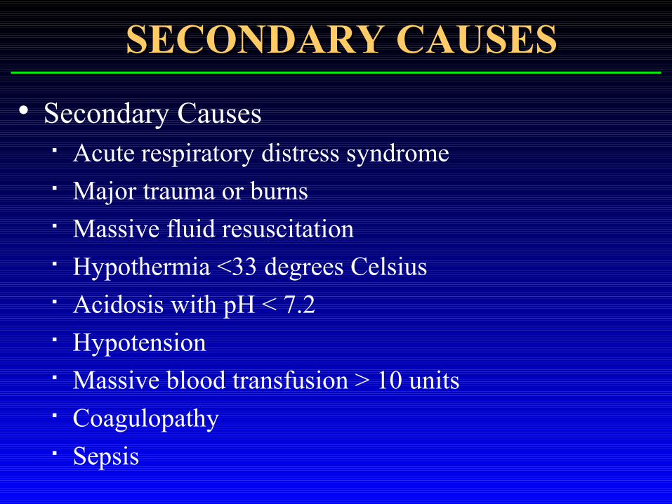
Secondary Causes Acute respiratory distress syndrome Major trauma or burns Massive fluid resuscitation Hypothermia <33 degrees Celsius Acidosis with pH < 7.2 Hypotension Massive blood transfusion > 10 units Coagulopathy Sepsis
SECONDARY CAUSESSECONDARY CAUSES
Chronic Causes Obesity Liver failure with ascities Malignancies
CHRONIC CAUSESCHRONIC CAUSES

Ischemia Inflammatory response
Capillary leak
Tissue Edema (Including bowel wall and mesentery)
Intra-abdominal hypertension
Fluid resuscitation
INJURY / INSULTINJURY / INSULT

PATHOPHYSIOLOGYPATHOPHYSIOLOGY
The adverse physiologic effects of IAH impact multiple organ systems. These include: Pulmonary Cardiovascular Renal Splanchnic Musculoskeletal (abdominal wall) Central Nervous System

Pulmonary compliance suffers with resultant progressive reduction in total lung capacity, functional residual capacity and residual volume.
These changes have been demonstrated with IAP above 15 mmHg.
PULMONARY DYSFUNCTIONPULMONARY DYSFUNCTION
Respiratory failure secondary to hypoventilation results from progressive elevation in IAP.
Ultimately, pulmonary organ dysfunction is manifest by hypoxia, hypercapnia and increasing ventilatory pressure.
PULMONARY DYSFUNCTIONPULMONARY DYSFUNCTION
Elevated IAP is consistently correlated with reduction in cardiac output. This has been demonstrated with IAP above 20 mmHg
Reduction in cardiac output is a result of decreased cardiac venous return from direct compression of the inferior vena cava and portal vein.
CARDIAC DYSFUNCTIONCARDIAC DYSFUNCTION

Increased intrapleural pressures resulting from transmitted intra-abdominal forces produce elevations in measured hemodynamic parameters. This includes central venous pressure and pulmonary artery wedge pressure (PAWP).
Significant hemodynamic changes have been demonstrated with IAP above 20 mmHg.
CARDIAC DYSFUNCTIONCARDIAC DYSFUNCTION
Graded elevations in IAP are associated with incremental reductions in measured renal plasma flow and glomerular filtration rate.
This results in a decline in urine output, beginning with oliguria at IAP of 15-20 mmHg and progressing to anuria at IAP above 30 mmHg. The mechanism by which renal function is compromised by elevated IAP is multifactorial.
RENAL DYSFUNCTIONRENAL DYSFUNCTION

The adverse renal physiology associated with IAH is pre-renal and renal. Prerenal derangements result from altered cardiovascular function and reduction in cardiac output with decreased renal perfusion.
Renal parenchymal compression produces alterations in renal blood flow secondary to elevated renal vascular resistance. This occurs by compression of renal arterioles and veins.
RENAL DYSFUNCTIONRENAL DYSFUNCTION

Impaired liver and gut perfusion have also been demonstrated with elevation in IAP.
Severe progressive reduction in mesenteric blood flow has been shown with graded elevation in IAP from approximately 70% of baseline at 20 mmHg, to 30% at 40 mmHg.
PORTO-SYSTEMIC VISCERAL PORTO-SYSTEMIC VISCERAL DYSFUNCTIONDYSFUNCTION

Intestinal mucosal perfusion as measured by laser flow probe has been shown to be impaired at IAP above 10 mmHg.
Metabolic changes that result from impaired intestinal mucosal perfusion have been shown by tonometry measurements that demonstrate worsening acidosis in mucosal cells with increasing IAH.
PORTO-SYSTEMIC VISCERAL PORTO-SYSTEMIC VISCERAL DYSFUNCTIONDYSFUNCTION

Similarly, measured abnormalities in intestinal oxygenation have been shown with elevations of IAP above 15 mmHg.
Impairment in bowel tissue oxygenation occurs without corresponding reductions in subcutaneous tissue oxygenation, indicating the selective effect of IAP on organ perfusion.
PORTO-SYSTEMIC VISCERAL PORTO-SYSTEMIC VISCERAL DYSFUNCTIONDYSFUNCTION

Impaired bowel perfusion has been linked to abnormalities in normal physiologic gut mucosal barrier function, resulting in a permissive effect on bacterial translocation. This may contribute to later septic complications associated with organ dysfunction and failure.
PORTO-SYSTEMIC VISCERAL PORTO-SYSTEMIC VISCERAL DYSFUNCTIONDYSFUNCTION

Adverse effects of IAP on hepatic arterial, portal, and microcirculatory blood flow have also been shown with pressures above 20 mmHg.
A progressive decline in perfusion through these vessels occurs as IAP increases, despite cardiac output and systemic blood pressure being maintained at normal levels.
PORTO-SYSTEMIC VISCERAL PORTO-SYSTEMIC VISCERAL DYSFUNCTIONDYSFUNCTION

Splanchnic vascular resistance is a major determinant in the regulation of hepatic arterial and portal venous blood flow.
Elevated IAP can become the main factor in establishing mesenteric vascular resistance and ultimately abdominal organ perfusion
PORTO-SYSTEMIC VISCERAL PORTO-SYSTEMIC VISCERAL DYSFUNCTIONDYSFUNCTION
Although technically not a component of the abdominal cavity itself, the abdominal wall is also adversely impacted by elevations in IAP. Significant abnormalities in rectus muscle blood flow have been documented with progressive elevations in IAP.
Clinically, this derangement is manifest by complications in abdominal wound healing, including fascial dehiscence, and surgical site infection.
PORTO-SYSTEMIC VISCERAL PORTO-SYSTEMIC VISCERAL DYSFUNCTIONDYSFUNCTION
Elevations in intracranial pressure (ICP) have been shown in both animal and human models with elevated IAP.
These pressure derangements have been shown to be independent of cardiopulmonary function and appear to be primarily related to elevations in central venous and pleural pressures.
CENTRAL NERVOUS SYSTEM CENTRAL NERVOUS SYSTEM DYSFUNCTIONDYSFUNCTION

Direct measurement of IAP by means of an intra-peritoneal catheter
Bedside measurement of IAP has been accomplished by transduction of pressures from indwelling femoral vein, rectal, gastric, and urinary bladder catheters
MEASUREMENT OF IAPMEASUREMENT OF IAP

In 1984 Kron et al reported a method by which to measure IAP at the bedside: He used an indwelling Foley catheter. Sterile saline (50-100cm3) is injected into the empty
bladder through the indwelling Foley catheter. The sterile tubing of the urinary drainage bag is cross-
clamped just distal to the culture aspiration port.
MEASUREMENT OF IAPMEASUREMENT OF IAP
The end of the drainage bag tubing is connected to the Foley catheter.
The clamp is released just enough to allow the tubing proximal to the clamp to flow fluid from the bladder, then reapplied.
A 16-gauge needle is then used to Y-connect a manometer or pressure transducer through the culture aspiration port of the tubing of the drainage bag.
Finally, the top of the symphysis pubic bone is used as the zero point with the patient supine
MEASUREMENT OF IAPMEASUREMENT OF IAP


INCIDENCEINCIDENCE The exact incidence of ACS is yet to be
established, but it is clearly increased in certain population groups.
In one prospective series of 145 patients who were identified as being at risk for development of the ACS the incidence was reported as 14%.
The incidence following primary closure after repair of ruptured abdominal aortic aneurysm is reported in one series as 4%.

RISK FACTORS FOR ACSRISK FACTORS FOR ACS
Severe penetrating and blunt abdominal trauma Ruptured abdominal aortic aneurysm Retroperitoneal hemorrhage Pneumoperitoneum Neoplasm Pancreatitis Massive ascites Liver transplantation Abdominal wall burn eschar

DIAGNOSISDIAGNOSIS
Clinical manifestations of organ dysfunction include respiratory failure that is characterized by impaired pulmonary compliance, resulting in elevated airway pressures with progressive hypoxia and hypercapnia.
Some authors report pulmonary dysfunction as the earliest manifestation of ACS.

Hemodynamic indicators include elevated heart rate, hypotension, normal or elevated PAWP and central venous pressure, reduced cardiac output and elevated systemic and pulmonary vascular resistance.
Impairment in renal function is manifest by oliguria progressing to anuria with resultant azotemia.
Renal insufficiency as a result of IAH is only partly reversible by fluid resuscitation.
DIAGNOSISDIAGNOSIS

Elevated IAP is an additional clinical manifestation of ACS. Clinical confirmation of IAH requires bedside measurements indicative of IAP.
Experimental and clinical data indicate that IAH is present above an IAP of 20 mmHg.
DIAGNOSISDIAGNOSIS

PREVENTIONPREVENTION
The earliest and potentially most effective means of addressing this disorder is by recognition of patients who are at risk and pre-emptive interventions designed to minimize the chances for development of IAH.
Various types of mesh closures of the abdominal wall and other alternative means of abdominal content coverage have been described.
There is evidence that ACS may be preventable by use of absorbable mesh in high-risk injured patients undergoing laparotomy.
PREVENTIONPREVENTION

Achieving optimal resuscitation rather than over-resuscitation is a potentially preventable complication in intensive care management.
Multiple indicators of effective resuscitation have been evaluated. Lactate, base deficit, and gastric mucosal pH appear to be reliable indicators to guide resuscitative interventions.
PREVENTIONPREVENTION

SICU MANAGEMENTSICU MANAGEMENT
Identifying patients in the intensive care unit (ICU) at risk for developing ACS with constant surveillance can help lead to prevention.
A further strategy is based on recognition of IAH and resultant organ dysfunction.
A four-stage grading scheme base on IAP has been developed, tested, and proposed as a useful ACS management tool.

Grade Bladder pressure Recommendation
(mmHg)
I 12-15 Maintain Normovolemia
II 16-20 Hypervolemic Resuscitation
III 21-25 Decompression
IV (ACS) >25 Decompression and Re-exploration
SICU MANAGEMENTSICU MANAGEMENT

Alternative means for surgical decision making are based on clinical indicators of adverse physiology, rather than on a single measured parameter.
In the setting of IAH, abdominal decompression has been recommended with any coexisting deterioration in pulmonary, cardiovascular, or renal function.
SICU MANAGEMENTSICU MANAGEMENT
A decision to perform the decompression in the ICU is a function of the ventilatory requirements of the patient and the risk associated with transport to the operating room. Although optimal respiratory support may be available in the ICU, this location is generally suboptimal for controlling surgical bleeding.
DECOMPRESSIONDECOMPRESSION

Abdominal decompression may itself precipitate adverse physiologic and metabolic events that should be anticipated.
These include a large increase in pulmonary compliance with resultant elevation in minute ventilation and respiratory alkalosis unless appropriate ventilatory changes are instituted.
'Washout' of accumulated intra-abdominal products of anaerobic metabolism may result in acidosis and hyperkalemia.
DECOMPRESSIONDECOMPRESSION

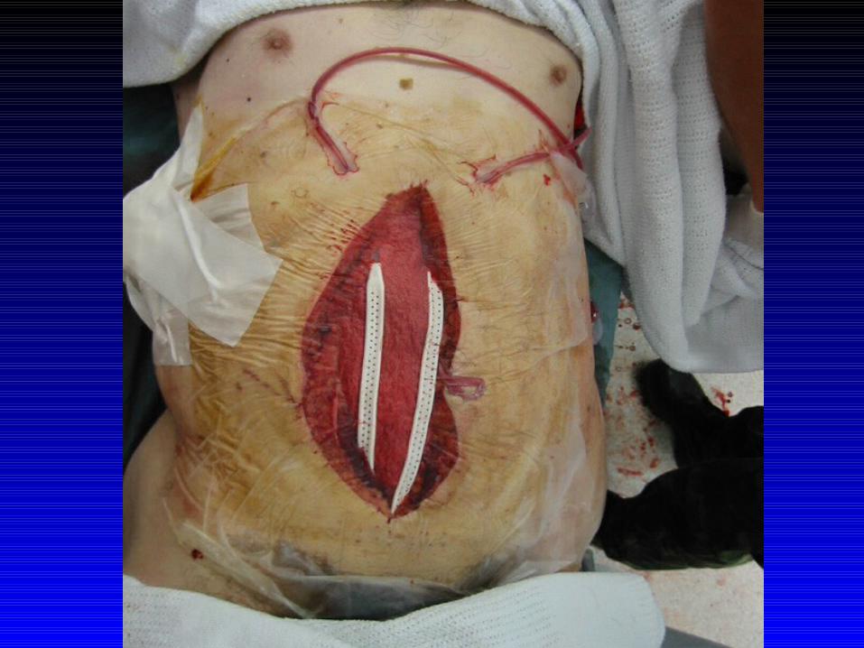
Under most circumstances following abdominal decompression, immediate primary fascial closure is obviated.
Alternative means for coverage of the abdominal contents include skin closure with towel clips or suture, abdominal wall advancement flaps, plastic or silicone coverage, and mesh interposition grafts.
DECOMPRESSIONDECOMPRESSION
Patients undergoing decompressive laparotomy are by definition at risk for future re-development of ACS, and strong consideration should be given to providing for re-exploration and a staged closure.
DECOMPRESSIONDECOMPRESSION

This may include fascial closure after a period of 7–10 days versus placement of split thickness skin grafts on a granulating surface followed by delayed repair of the resulting abdominal wall hernia after several months.
Finally, early management of the open abdomen must include recognition for significant fluid losses and fluid replacement.
DECOMPRESSIONDECOMPRESSION


OUTCOMEOUTCOME
ACS is a condition with a potentially high lethality that must be recognized early and effectively managed in order to optimize outcome.
Most deaths associated with ACS are due to sepsis or multiple organ failure.

Mortality associated with this condition has been reported in 10.6 to 68% of patients.
In one series, non-survivors tended toward a more fulminant course, with the majority of deaths occurring within the first 24h of injury.
OUTCOMEOUTCOME

CONCLUSIONCONCLUSION Abdominal compartment syndrome is defined as
intra-abdominal hypertension associated with organ dysfunction.
Adverse physiology has been demonstrated in pulmonary, cardiovascular, renal, musculoskeletal/integumentary, and central nervous system function.

Identification of Patients at Risk
Early Recognition
Appropriately Assessed and Staged
Well Timed Intervention
CONCLUSIONCONCLUSION