about omics group - d2cax41o7ahm5l.cloudfront.net · control 2,5% pocdpe. ... suggested mechanism...
TRANSCRIPT

About OMICS Group
OMICS Group is an amalgamation of Open AccessPublications and worldwide international scienceconferences and events. Established in the year 2007 withthe sole aim of making the information on Sciences andtechnology ‘Open Access’, OMICS Group publishes 500online open access scholarly journals in all aspects ofScience, Engineering, Management and TechnologyScience, Engineering, Management and Technologyjournals. OMICS Group has been instrumental in taking theknowledge on Science & technology to the doorsteps ofordinary men and women. Research Scholars, Students,Libraries, Educational Institutions, Research centers and theindustry are main stakeholders that benefitted greatly fromthis knowledge dissemination. OMICS Group also organizes500 International conferences annually across the globe,where knowledge transfer takes place through debates,round table discussions, poster presentations, workshops,symposia and exhibitions.

OMICS International
Conferences
OMICS International is a pioneer and leading science eventorganizer, which publishes around 500 open access journalsand conducts over 500 Medical, Clinical, Engineering, LifeSciences, Pharma scientific conferences all over the globeannually with the support of more than 1000 scientificassociations and 30,000 editorial board members and 3.5associations and 30,000 editorial board members and 3.5million followers to its credit.
OMICS Group has organized 500 conferences, workshopsand national symposiums across the major cities includingSan Francisco, Las Vegas, San Antonio, Omaha, Orlando,Raleigh, Santa Clara, Chicago, Philadelphia, Baltimore,United Kingdom, Valencia, Dubai, Beijing, Hyderabad,Bengaluru and Mumbai.

World Congress on Pharmacology
Brisbane , 20-22 July
Novel Biologically Active Polyethers from Novel Biologically Active Polyethers from
Different Species of Boraginaceae Family and Different Species of Boraginaceae Family and
Their Synthetic Derivatives: Prospective Their Synthetic Derivatives: Prospective
Therapeutic Agents.Therapeutic Agents.
Dr. Karen Mulkijanyan
Head of the Department of Pharmacological Research of Tbilisi State Medical University
I.Kutateladze Institute of Pharmacochemistry, Tbilisi, Georgia
Therapeutic Agents.Therapeutic Agents.

Boraginaceae species
Anchusa italica
(Italian bugloss)
Symphytum asperum
(prickly or rough comfrey)
Symphytum caucasicum
(Caucasian comfrey)

Introduction
Extracts from the plants belonging to Boraginaceae family – Symphytum
asperum, S.caucasicum and Anchusa italica have been used in folk
medicine for treatment of different kinds of disorders and wounds due to
analgesic, antimicrobial and anti-inflammatory effects. Aforenamed extracts
contain allantoin, claimed to be a cell proliferation-stimulating agent
responsible for the wound-healing properties of Symphytum, and, on the
other hand, hepatotoxic pyrrolizidine alkaloids which strongly restrict
internal use of comfrey extracts.
The first representative of a new class of natural polyethers - regularThe first representative of a new class of natural polyethers - regular
dihydroxycinnamate-derived polymer
POLY[OXY-1-CARBOXY-2-(3,4-DIHYDROXYPHENYL)ETHYLENE (POCDPE)
has been detected in high-molecular watersoluble fractions of roots, stems
and leaves of Comfrey - Symphytum asperum (SA) S. caucasicum (SC), S.
officinale (SO) and Bugloss (Anchusa) – Anchusa italica (AI).
In addition a monomer of POCDPE – 3-(3,4-dihydroxyphenyl)glyceric acid
(SM) has been synthesized
Some of the results concerning the biological activity of POCDPE and SM
are presented below.

Extraction and fractionation of SA, SC, SO and AI
polysaccharides from raw material
The fractionation procedure by ultrafiltration allows to remove most ballast
polysaccharides and to obtain water-soluble high-molecular (>1000 kDa)
preparations (HMP).

Poly[oxyPoly[oxy--11--carboxycarboxy--22--(3,4(3,4--dihydroxyphenyl)ethylenedihydroxyphenyl)ethylene
V.Barbakadze et al. Molecules, 2005, V. 10, N 9, P. 1135-1144; V.Barbakadze et al. Chem. Nat. Compds. 2009, V. 45, N 1, P. 6-10.
Symphytum asperum, S.caucasicum R=H;
Anchusa italica R=H, CH3.
Symnthetic monomer

TLC detection of pyrrolizidine alkaloids in raw
material (A) and highmolecular fractions (B)
a – roots, b – stems, c - leaves
Solvent system : chloroform-methanol-25% ammonia (85:14:1, v/v/v)
Detection: UV light (λ254 nm);
Spray: Ehrlich’s reagent

WOUND HEALING
• Mouse excisional wound model. Two 1 cm diameter skin rags are
cut out on depilated dorsal skin area. Operation is carried out
under ether anesthesia. Treatment of animals began through 24 h
after the injury. Wounds are treated with 0.1 ml of ointment per
wound once a day.
• Mouse skin burn model. Area and depth standardized skin burns
are caused on depilated skin area under ether anesthesia using
special device with the temperature controller and contact
electrical heater (1 sm2 square copper plate). The temperature of a
contact plate - 1500С, exposition time – 10 sec. At these conditions
burn corresponds to IIIA-degree in accordance with clinical
classification of burns. Treatment of animals began through 24 h
after burn induction.
• Wound healing effect was estimated by the reduction of injured
area in relation to initial and calculated under the formula:
D = (Sexp / Sin) х 100 %, where
S in - initial wound area on day 1.
S exp - wound area on day of measurement.
The obtained data were processed statistically using Student’s t-test

150
200
250
300
Burn
are
a (
mm
2)
Healing effect of 2,5% POCDPE ointment
(skin burn model in rats)
0
50
100
150
1 2 3 4 5 6 7 8 9 10 11 12 13 14 15 16 17 18
Burn
are
a (
mm
Days
Control 2,5% POCDPE

Estimation of wound area
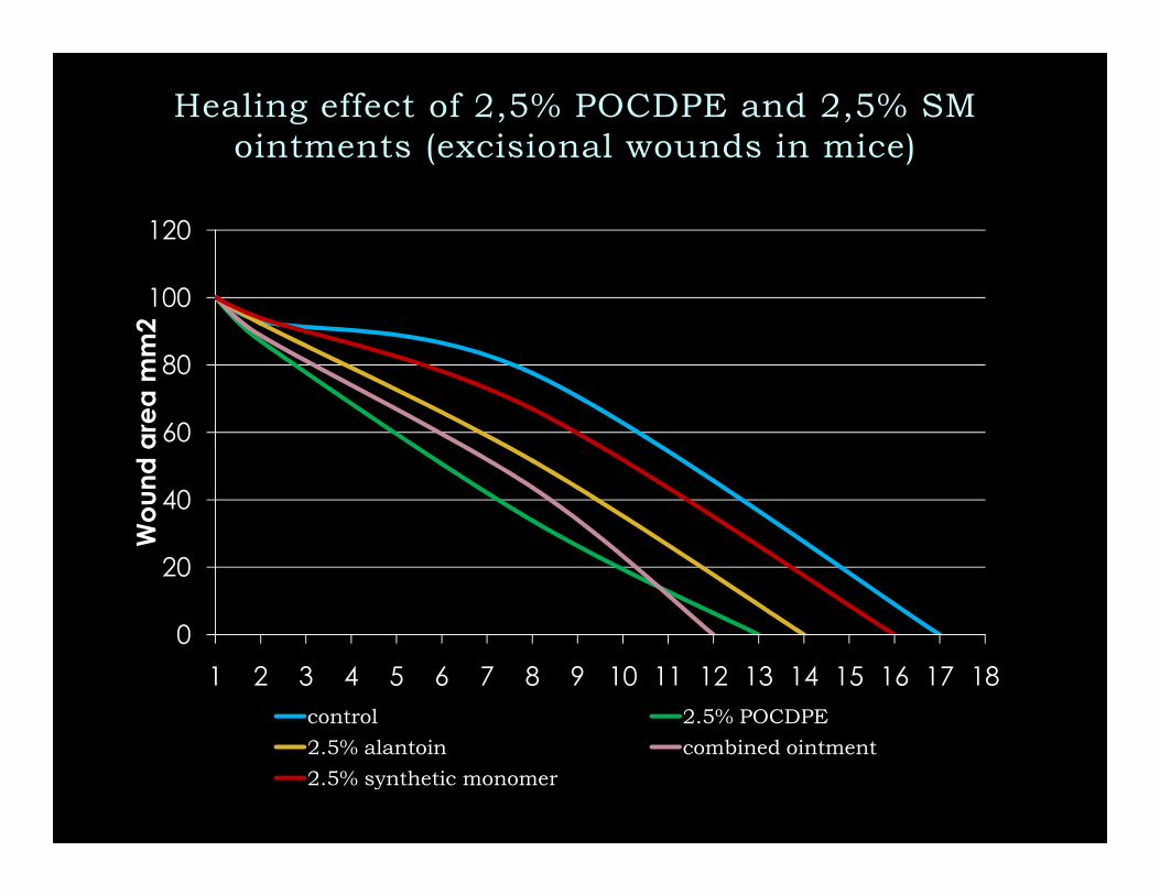
Healing effect of 2,5% POCDPE and 2,5% SM
ointments (excisional wounds in mice)
60
80
100
120
Wo
und
are
a m
m2
0
20
40
1 2 3 4 5 6 7 8 9 10 11 12 13 14 15 16 17 18
Wo
und
are
a m
m2
control 2.5% POCDPE
2.5% alantoin combined ointment
2.5% synthetic monomer

50
60
70
80
90
100
Wound area mm2
Healing effect of 10% POCDPE dry ointment
(burn wounds in mice)
0
10
20
30
40
50
1 2 3 4 5 6 7 8 9 10 11 12 13 14 15 16 17 18
Wound area mm
control POCDPE dry ointment ointment base
60
80
100
Burn area(mm2)
60
80
100
120
Wound area (mm2)
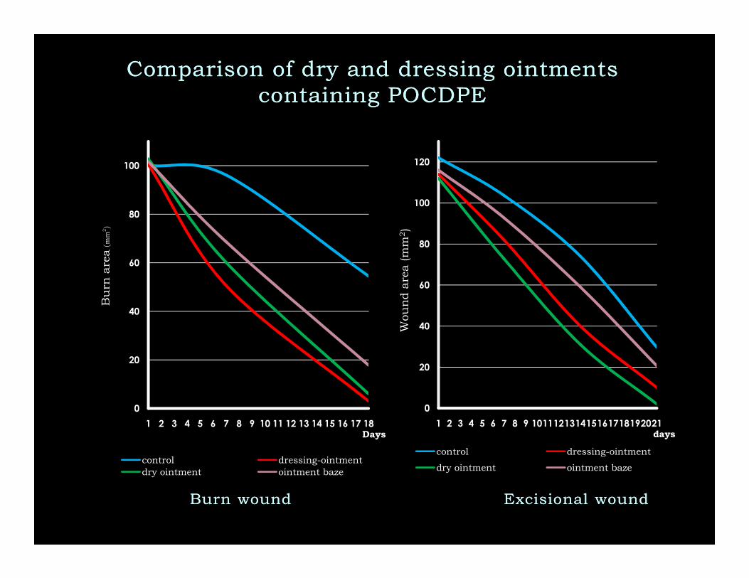
Comparison of dry and dressing ointments Comparison of dry and dressing ointments
containing POCDPEcontaining POCDPE
0
20
40
1 2 3 4 5 6 7 8 9 10 11 12 13 14 15 16 17 18
Burn area
Days
control dressing-ointment
dry ointment ointment baze
0
20
40
60
1 2 3 4 5 6 7 8 9 101112131415161718192021
Wound area (mm
days
control dressing-ointment
dry ointment ointment baze
Burn woundBurn wound ExcisionalExcisional woundwound
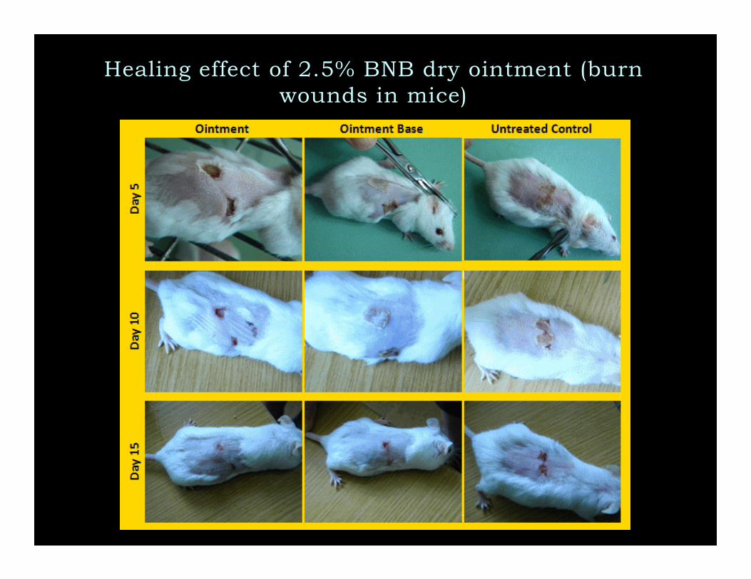
Healing effect of 2.5% BNB dry ointment (burn
wounds in mice)

Suggested mechanism of wounnd healing and anti-
inflammatory action of BNB
Besides generation of superoxide anions by stimulated PMNs, these radicals may also arise inchronic wounds where ischemic conditions may convert the enzyme xanthine dehydrogenase intoxanthine oxidase (XO) which catalyses the conversion of oxygen into superoxide anions causingtissue damage. During this process XO converts hypoxanthine (HX) to xanthine and subsequently touric acid. So, scavenging of superoxide anions either produced by PMNs or through XO is regardedbeneficial for wound healing and in inflammatory process.

30
40
50
60
70
80
90
% of Inhibition
Effects of SA and SC (100 mcg/ml) on PCacells growth after 48 hours
LNCap
22Rv1
PC36
8
10
12
14
16
18
20
Increase of cell death (fold)
Effects of SA and SC (100 mcg/ml) on PCacells death after 48 hours
LNCap
22Rv1
PC3
In vitro anti-cancer efficacy of BNB from
Symphytum asperum (SA) and S.caucasicum
(SC)
S. Shrotriya et al. American Association for Cancer Research 100th Annual Meeting, Denver, Colorado, USA. Abstracts. 2009, N 921.
0
10
20
30
SA SC
% of Inhibition
PC3
0
2
4
6
SA SC
Increase of cell death (fold)
In androgen-dependent (LNCaP) and -independent (22Rv1 and PC3) human prostate
cancer (PCa) cells SA treatment (100 mcg/ml for 48h) decreases the live cell number by 65, 64
and 35% (a) and increases the cell death by 16, 8 and 12 folds (b) in LNCaP, 22Rv1 and PC3
cells, respectively. Similarly, SC treatment (100 mcg/ml for 48h) decreased the live cell number
by 87, 25 and 33% and increased the cell death by 19, 10 and 9 folds in LNCaP, 22Rv1 and
PC3 cells, respectively.
ba

In vivo anti-cancer efficacy of BNB from
Symphytum asperum (SA) and S.caucasicum
(SC)
80
100
Inhibition (%)
Inhibition of 22RV1 xenograft growth in athymic nude mice
0
20
40
60
SA SC
Inhibition
2.5 mg/kg
5 mg/kg
Oral gavage feeding of SA (2.5 and 5.0 mg/Kg body weight) and SC (2.5 and 5.0 mg/Kg body
weight) 5 days/week for 5 weeks caused a marked time-dependent inhibition in 22RV1 tumor
xenograft growth which accounts for 46% and 59% decrease in SA treated animals and 75%
and 88% decrease in SC treated animals, respectively.
S. Shrotriya et al. Carcinogenesis. v.33 no.8 pp.1572–1580, 2012

TNF-a secretion and B16M cell adhesion in B16M-CM-treated HSE
cells in vitro.
60
0
5
10
15
20
25
30
35
40
1 2 3 4% B
16M
Cell
Adhesio
n to H
SE
Cells
*
*
**
The BNB significantly
induced TNF-α production
0
10
20
30
40
50
60
1 2 3 4TN
Fa
lph
a C
oncentr
ation
(pg/1
06
ce
lls)
Polymer1µg/ml
B16M-CM-TreatedHSE Cells
Polymer1µg/ml
UntreatedHSE Cells
Differences in the percent of adhering cells and TNF-a production versus untreated HSE cells (*) and versus VEGF-
treated HSE (**) P<.001 by ANOVA
*
*
*induced TNF-α production
from normal and tumor-
activated HSE
cells, supporting its
potential as immune
defense modulator.

TNF-α α α α secretion and B16M cell adhesion in VEGF-treated HSE cells in vitro.
0
5
10
15
20
25
1 2 3 4
% B
16M
Cell
Adhesio
n to H
SE
Cells *
**
BNB completely abrogated the
adhesion of murine B16
melanoma cells to tumor-
activated HSE, without any
detectable effect on basal
condition-cultured HSE.
Consistent with these anti-
adhesive effects, the BNB also
0
10
20
30
40
50
60
1 2 3 4
1 2 3 4
Polymer1µg/ml
VEGF-TreatedHSE Cells
Polymer1µg/ml
UntreatedHSE Cells
TN
Fa
lph
a C
oncentr
ation
(pg/1
06
ce
lls)
Differences in the percent of
adhering cells and TNF-a
production versus untreated
HSE cells (*) and versus
VEGF-treated HSE (**) P<.001
by ANOVA
*
**
adhesive effects, the BNB also
prevented melanoma cell
adherence to recombinant
VEGF-treated HSE

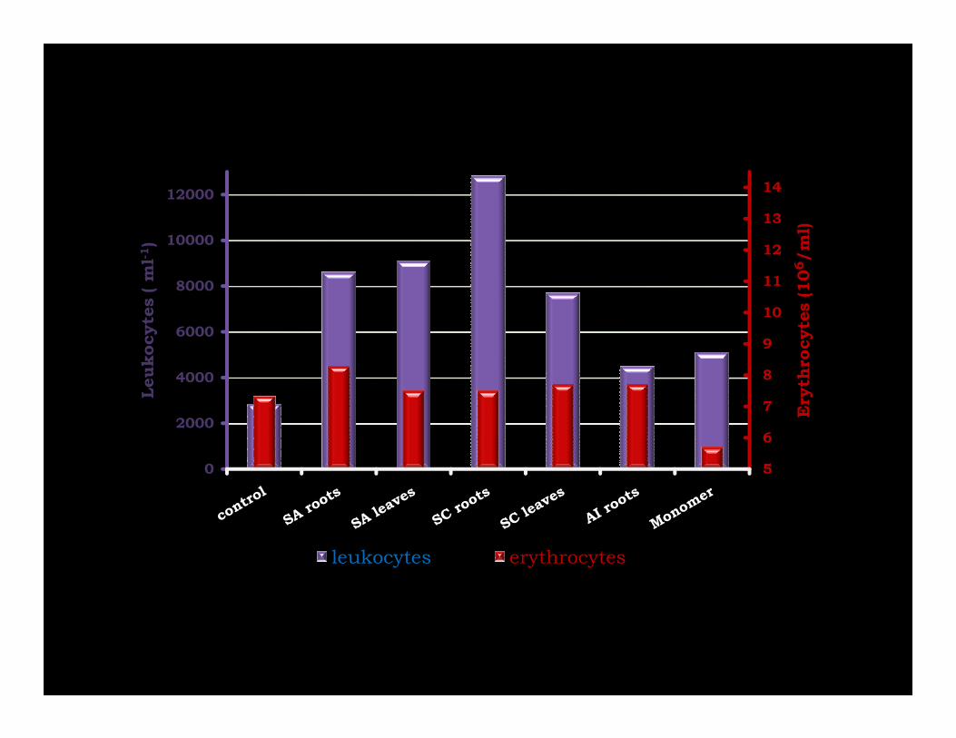
Stimulation of leucopoiesis in experimental
cyclophosphamide induced leucopenia
8000
10000
12000
14000
Leukocytes (ml-1)
0
2000
4000
6000
8000
Leukocytes (ml
BNB

Established effects of BNB

Established major effects of POCDPE
� Burn and wound healing effect due to the shortening of the
second phase of wound healing - the inflammatory response.
K.Mulkijanyan et al. Bull. Georg. Natl. Acad. Sci. 2009, V. 3, N 3, P. 114-117.
� The strong efficacy against prostate cancer cells suggesting their
high potential in prostate cancer patients.
S. Shrotriya et al. Carcinogenesis. v.33 no.8 pp.1572–1580, 2012
�Abrogation of the adhesion of melanoma cells to tumor-conditioned �Abrogation of the adhesion of melanoma cells to tumor-conditioned
medium- and VEGF-activated endothelial cells.
V.Barbakadze et al. Bull. Georg. Natl. Acad. Sci. 2008, V. 2, N 3, P. 108-112.
� Haematopoietic efficacy of polymer: in mice drug-induced leukopenia
the polymer caused significant stimulation of leucopoiesis.
M. Moistsrafishvili, et al. Investigation of Georgian biologically active compounds
of plant and mineral origin. Tbilisi, 2010, Issue 2(17) p.91-93.
�Antioxidant activity and anticomplementary activity due to the
inhibition of xantine oxidase and complement convertase, respectively .
V.Barbakadze et al. Pharmaceutical Chemistry J. 2007, V.41, N 1, P. 14-16.

Pre-clinical investigation of BNB revealed wide
spectrum of biological activity including, but not
limited to antiinflammatory, burn and wound
healing, and anticancer.
Strong efficacy of BNB in different experimental
Conclusion
Strong efficacy of BNB in different experimental
models suggests its high therapeutic potential.

Acknowledgements
I would like to express my gratitude to :
� Dr. V.Barbakadze, Dr. M.Merlani, Dr. L.Amiranashvili, Dr.
L.Gogilashvili, Dr. Zh.Novikova, Dr. M.Sulakvelidze, and
M.Moisttrafishvili, (Tbilisi State Medical University Institute of
Pharmacochemistry, Tbilisi, Georgia);
� Prof. A.Bakuridze, Prof.
D.Berashvili, Dr.G.Mikaia, Dr.N.Kurdiani, and S.Gokadze (Tbilisi
State Medical University Faculty of Pharmacy, Tbilisi, Georgia);
� Prof. F.Vidal-Vanaclocha (Basque Country
University, Bizkaia, Spain);
� Prof. R.Agarwal, Dr. G.Deep, S.Shrotriya, and N.Walia (Colorado
University, Denver, USA);
and
� Civil Research & Development Foundation (USA), Georgian
Research & Development Foundation, Sh. Rustaveli National
Science Foundation for financial support in frame of grant projects
CRDF-GRDF-GEB-3344-TB-06; GNSF-ST08-6-469 and SRNSF-
AR/109/8-403/11

THANK YOU FOR
YOUR ATTENTIONYOUR ATTENTION
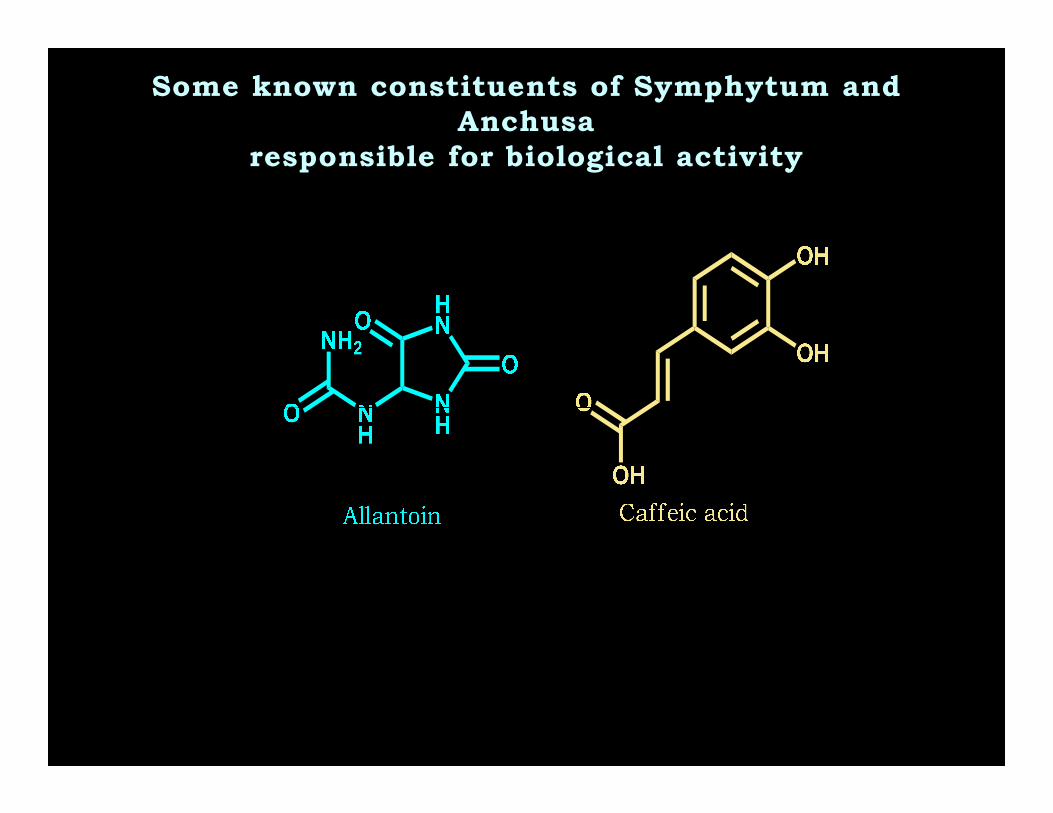
Some known constituents of Symphytum and
Anchusa
responsible for biological activity

Overview of toxic pyrrolizidine alkaloids found
in Symphytum and Anchusa
N
N
CH2OHOH
A
C C
OHC
CH
CH3
OHHO
CH3H3C
C C
OHC
CH
CH3
OHHO
CH3H3C
C C
OHC
CH
CH3
OHHO
CH3H3C
C C
O
C
CH3CH3
H
H
C CH3
O
(-)-viridofloric
(-)-viridofloric
tiglic
symphytine
lycopsamine
7-acetyllycopsamine
R1 R2
H2CR1O OR2
N
B
OHHO
C C
O
CH
CH
CH3
OH
HO
CH3H3C
C C
O
CH
CH
CH3
OH
HO
CH3H3C
C C
OHC
CH
CH3
OHHO
CH3H3C
H
C CH3
O
C CH
O
C
CH3
CH3
(-)-viridofloric
(+)-trachelanthic
(-)-viridofloric
(+)-trachelanthic
acetic
cenecioic
acetic
symviridine
intermedine
7-acetylintermedine
necic acid(s)
N
A – pyrrolizidine, the necine moiety of PAs
B – retronecine;
PAs of Symphytum are mono- or di-esters
of retronecine
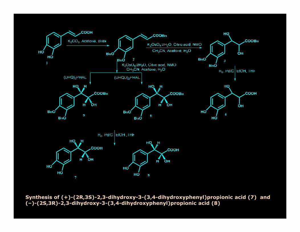
Synthesis of (+)-(2R,3S)-2,3-dihydroxy-3-(3,4-dihydroxyphenyl)propionic acid (7) and (–)-(2S,3R)-2,3-dihydroxy-3-(3,4-dihydroxyphenyl)propionic acid (8)


HPLC analysis of HMP-SA on
column Polysep 6000; injection – 9
ul; detection – RI.
Separation by HPLC on Polysep 6000 confirmed
our previous supposition that polysaccharides
contents are not covalently bounded with phenolic
polymer. However it is very difficult to completely
separate by GFC the polysaccharides from
polymer. This phenomenon can be explained due to
the presence of hydrogen bonds between phenolic
polymer and residual polysaccharides which will
hold the polysaccharides together with the phenolic
polymer during fractionation by ultrafiltration and
GFC. The polymer is chemically simple, but its
molecules can form with each other and with the
molecules of residual polysaccharides complex
macromolecular associates up to their
HPLC analysis of HMP-SA (top) and
HMP-AI (bottom) on column Biosep
4000; injection - 20 ul; detection – RI.
HPLC analysis of mixture of standard
proteins on column Biosep 4000;
injection - 5 ul; detection – UV.
macromolecular associates up to their
supramolecular organization due to hydrogen
bonds

8
9
10
11
12
13
14
4000
6000
8000
10000
12000
Erythrocytes (106/ml)
Leukocytes ( m
l-1)
5
6
7
8
0
2000
4000
Erythrocytes (
Leukocytes ( m
l
leukocytes erythrocytes

Mechanism of action of SA & SC phenolic polymers
Molecular studies suggested that the anti-cancer effects of SA and SC phenolic polymers
are mainly through decreasing androgen receptor, enhancing CDK inhibitors expression,
and inducing apoptosis in PCA cells.
SA and SC caffeic acid-derived polymer suppressed the growth and induced death in PCA
cells, with only marginal cytotoxicity towards non-neoplastic human prostate epithelial
cells. New phenollic polymer of SC caused G1 arrest in PCA cells through modulating the
expression of cell cycle regulators, especially an increase in CDK inhibitors (p21 and p27).
In addition, SA and SC high molecular phenolic polymers induced apoptotic death by
activating caspases, and also strongly decreased AR and PSA expression. In vivo, SC
phenolic polymer feeding, strongly inhibited 22Rv1 tumors growth by 76 and 88% at 2.5
and 5 mg/kg body weight doses, respectively, without any toxicity, together with a strong
decrease in PSA level in plasma; and a decrease in PCNA, AR, and PSA expression but
increase in p21/p27 expression and apoptosis in tumor tissues from SC phenolic polymer-
fed mice.

SA and SC modulate cell cycle progression in PCA cells
and induce apoptosis
PC3 LNCaP
G1 S G2 M G1 S G2 M
SA +++ ++ ++
+ ++
SC +++ ++ ++
++++
Fluorescence Activated Cell Sorting (FACS) analysis showed that the growth inhibition by these Fluorescence Activated Cell Sorting (FACS) analysis showed that the growth inhibition by these
compounds was associated with a strong induction of cell cycle arrest in PCA cells. FACS analysis showed
that these compounds differentially modulate the cell cycle progression in PCA cells, which was based
upon the dose, treatment duration and cell type. SA treatment (1-100 µg/ml) resulted in a significant G1-phase arrest after 24 and 48h of treatment accompanied with a decrease in S-phase population in both
PC3 and LNCaP cells. SC treatment (1-100 µg/ml) for 72h in PC-3 cells resulted in a strong S-phase arrest; while in LNCaP cells SC treatment for 24 and 48h caused a moderate G1-phase arrest at lower
doses (1-50 µg/ml), and a strong G2/M-phase arrest at higher dose (100 µg/ml). In 22Rv1 SC (100 µg/ml)
caused G2/M arrest at 24 h, and S and G2/M phase arrest at 48 h. SA and SC induce cell cycle arrest in
human PCA cells LNCaP and 22Rv1 via modulating the expression of key cell cycle regulatory molecules,
which regulate G1/S/G2M phase of cell cycle. Treatment of LNCaP and 22Rv1 cells with SA and SC
resulted in decrease the expression of cyclins D1, D3, A, and E along with cdk4 at 48h (50-100 µg/ml), however all the compounds increased the level of p21 and p27. Besides, these compounds have inhibitory
effect on p53 level in prostate cancer cells, and we are examining the mechanistic details underlying this
biological effect. SA and SC induced 42, 70% apoptosis in 22RV1 cells and 65, 70% apoptosis in LNCap
cells when analyzed by AnnexinV/PI. SA and SC induced apoptosis involves caspase-3, caspase-9 and
PARP cleavage in LNCaP and 22Rv1 cells.S. Shrotriya et al. American Association for Cancer Research 100th Annual Meeting, Denver, Colorado, USA. Abstracts. 2009, N 921.


Effect of BNB on the inflammatory response of tumor-activated hepatic sinusoidal endothelium
�The polymer abrogates the adhesion of melanoma cells to
tumor-conditioned medium- and VEGF-activated endothelial
cells.cells.
�This antiadhesive effect occurs in the presence of high
concentrations of TNF-α suggesting that the compound block
down-stream the effect of other proadhesive mediators of
tumor factor- or VEGF-induced effects.

Methods:B16 Melanoma (B16M) cell adhesion assay to primary cultured hepatic sinusoidal endothelial(HSE) cells. B16M cells were labeled with 10 µg/ml 2',7'-bis-(2-carboxyethyl)-5,6-carboxyfluorescein-acetoxymethylester (BCECF-AM) solution. This non-fluorescent sterase substrate BCECF-AM is accumulated by tumor cells and hydrolyzed to the fluorescent product BCECF which becomes trapped inside the live cells. After gently washing,2x105 cells/well were added to 24-well plate primarycultured HSE cells and 8 minutes later, the wells were washed three times with fresh medium. Cell adherence was calculated from absorbance at 485 nm using a fluorometric microplate reader(Multiskan Ascent, Thermo Labsystems). The number of adhered cells (registered in fluorescence arbitrary units) was expressed as percentage of the initial number of cells, and calculated for eachwell as follows: Fluorescence after well washing/(Fluorescence before washing – non-specificwell as follows: Fluorescence after well washing/(Fluorescence before washing – non-specificfluorescence before tumor cell addition)
Quantification of TNF-α. Release of TNF-α from primary cultured HSE cells was measured using theELISA kit from R&D Systems based on anti-mouse TNF-α monoclonal antibody, as suggested by the manufacturer (R&D Systems, Minneapolis, MN). Cultured HSE cells were incubated in the presence or absence of B16M-CM or 10 ng/ml murine VEGF for 8 hours. In some experiments, both untreated and treated HSE cells received 1 µg/ml PDGA, 30 minutes before B16MCM or VEGF (A). Cell adhesion assays were performed as described in Methods. (B) HSE supernatants were removed before cell adhesion and TNF-α concentration was measured by ELISA. Data represent the mean ±SD of 2 separate experiments performed using 2 different preparations of HSE cells, each in 3 replicates (n=6).Differences in the percent of adhering cells and TNF-α production versus untreated HSE cells (*) and versus B16M-CM or VEGF-treated HSE (**) were statistically significant (P<.001) by ANOVA and Bonferroni´s posthoc test.

Let us meet again..
We welcome you all to our future conferences of
OMICS International
3rd World Congress on PharmacologyOn
at August 08-10, 2016 at Birmingham, UK
http://pharmacology.pharmaceuticalconferences.com/