academic year 2012 tooth
TRANSCRIPT

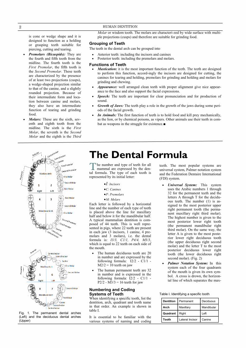
Dental Arches and Quadrants The twenty deciduous teeth in the young mouth and the thirty two permanent teeth in the adult mouth are arranged in two arches called the “Dental Arches”, one upper and one lower. Each arch contains half of the number of the teeth (10 de-ciduous then later 16 permanent teeth). (Fig. 1)
The upper jaw is called the “Maxilla” and the teeth in this arch is called the “Upper of Maxillary Teeth”. On the other hand, the lower jaw is called the “Mandible” and the teeth in this arch is called the “Lower or Mandibular Teeth”.
The imaginary vertical line which equally divides the body into right and left halves is called the midline or the “Midsagittal Plan”. This line also divides each dental arch into right and left seg-ments, referred to as “Quadrants”. The permanent teeth and the deciduous teeth are equally arranged into four quadrants, as follows:
Upper right quadrant or Maxillary right quadrant.
T eeth are more than projection in the mouth that allow you to grind the food and
prepare it for the initial phase of digestive process, nor do they merely serve for a
limited time before ending between the claws of an extracting forceps.
, man, like most mammals is supplied through his life by two sets of teeth, a deciduous
or primary set, followed by a permanent set. They are permanent teeth and it is your
future responsibility to see that they remain permanent.
Teeth are one of the most important elements of the masticatory system. The student is
expected to thoroughly know and understand the basic external morphology of every
tooth, as well as, their proper arrangement and relation to each other and their relation to
fixed points in the skull and the mandible.■
Tooth Morphology & Physiology
Human Dentit ion
Editor
Prof. Maged Lotfy
OMS, School of Dentistry,
MOI University, Eldoret, Kenya
(Formerly) Chairman, OMS Dept,
AinShams Univ., Cairo, Egypt
E.mail: [email protected]
FREE NOT FOR SALE
Issued as part of the scientific cooperation between Egypt and Kenya
Digested from “Fundamentals of Tooth Morphology and Physiology”. Ed. A. Ads, M. El-Zainy, S. Shenaishen and A. Lotfy, Oral Biology Dept., Faculty of Dentistry,
Ainshams University, Cairo, Egypt.
Dental Formula & Coding
Systems
Upper left quadrant or Maxillary left quadrant.
Lower left quadrant or Mandibular left quadrant.
Lower right quadrant or Mandibu-lar right quadrant.
Types and Forms of Teeth Teeth vary in form, this variation reflects
differences in function. Based on forms
and functions the teeth are classified into
incisors, canines, premolars and molars.
(Fig. 1)
Incisors: They are the four front
teeth in each arch. The Central
incisor is the first tooth next to the
midline. The Lateral Incisor is the
second. The form of the incisors is
more or less similar to a chisel.,
which makes them suitable for
cutting of incising food. The side of
the tooth toward the tongue, the
lingual surface, is shaped like a
shovel, to aid in guiding the food
into the mouth.
Canine (Cuspid): It is the third
tooth from the midline. The canine
TOOTH
MORPHOLOGY
Academic Year 2012
Ainshams University
A in Shams University, an institute of higher education located in Cairo, Egypt.
Founded in 1950, the university provides
education at the undergraduate, graduate and
post-graduate levels.
Ain Shams University, was founded in July
1950, making it the third-oldest non-sectarian native public Egyptian university (ancient
Islamic universities such as Al-Azhar and
private institutions such as the American
University in Cairo are older),
In 1950, there were only eight faculties: facul-
tIes of Arts, Law, Commerce, Science, Engi-neering, Medicine, Agriculture, in addition to
Women's college. In 1969, the faculty of
Education, known since 1880 as Teachers' college, became the ninth faculty in the uni-
versity. In 1973 the faculty of Al-Alsun was
made the tenth member in the university. In 1994, a decree was issued for the establish-
ment of two more faculties; the faculty of
Pharmacy and the faculty of Dentistry, the actual study was started the following year in
both faculties.
2 HUMAN DENTITION
is cone or wedge shape and it is
designed to function as a holding
or grasping teeth suitable for
piercing, cutting and tearing.
Premolars (Bicuspids): They are
the fourth and fifth tooth from the
midline. The fourth tooth is the
First Premolar, the fifth tooth is
the Second Premolar. These teeth
are characterized by the presence
of at least two projections (cusps),
a wedge-shaped projection similar
to that of the canine, and a slightly
rounded projection. Because of
their intermediate form and loca-
tion between canine and molars,
they also have an intermediate
function of tearing and grinding
food.
Molars: These are the sixth, sev-
enth and eighth tooth from the
midline. The sixth is the First
Molar, the seventh is the Second
Molar and the eighth is the Third
T he number and type of teeth for all mammal are expressed by the den-
tal formula. The type of each tooth is represented by its initial letter:
I: Incisors
C: Canines
P: Premolars
M: Molars Each letter is followed by a horizontal line and the number of each type of teeth is placed above the line for maxillary half and below it for the mandibular half. A typical mammalian dentition is com-posed of 44 teeth. This is well repre-sented in pigs, where 22 teeth are present in each jaw (3 incisors, 1 canine, 4 pre-molars and 3 molars), i.e. the dental formula is: I3/3, C1/1, P4/4, M3/3, which is equal to 22 teeth on each side of the mouth.
The human deciduous teeth are 20 in number and are expressed by the following formula: I2/2 - C1/1 - M2/2 = 10 teeth on jaw
The human permanent teeth are 32 in number and is expressed in the following formula: I2/2 - C1/1 - P2/2 - M3/3 = 16 teeth for jaw
Numbering and Coding Systems of Teeth When identifying a specific tooth, list the dentition, arch, quadrant and tooth name in that order. An example is shown in table I.
It is essential to be familiar with the
various systems of naming and coding
The Dental Formula teeth. The most popular systems are
universal system, Palmer notation system
and the Federation Dentaire International
(FDI) system.
Universal System: This system uses the Arabic numbers 1 through 32 for the permanent teeth and the letters A through T for the decidu-ous teeth. The number (1) is as-signed to the most posterior upper right permanent tooth (the perma-nent maxillary right third molar). The highest number is given to the most posterior lower right tooth (the permanent mandibular right third molar). On the same way, the letter A is given to the most poste-rior lower right deciduous tooth (the upper deciduous right second molar) and the letter T to the most posterior deciduous lower right tooth (the lower deciduous right second molar). (Fig. 2)
Palmer Notation System: In this
system each of the four quadrants
of the mouth is given its own sym-
bol. A cross is drown, the horizon-
tal line of which separates the max-
Table I. Identifying a specific tooth
Dentition Permanent Deciduous
Arch Maxillary Mandibular
Quadrant Right Left
Tooth Lateral Incisor Canine
Fig. 1. The permanent dental arches (Left) and the deciduous dental arches (Upper)
Molar or wisdom tooth. The molars are characteri-zed by wide surface with multi-
ple projections (cusps) and therefore are suitable for grinding food.
Grouping of Teeth
The teeth in the dental arch can be grouped into
Anterior teeth: including the incisors and canines Posterior teeth: including the premolars and molars.
Functions of Teeth Mastication: it is the most important function of the teeth. The teeth are designed
to perform this function, accord-ingly the incisors are designed for cutting, the
canines for tearing and holding, premolars for grinding and holding and molars for
grinding and chewing.
Appearance: well arranged clean teeth with proper alignment give nice appear-
ance to the face and also support the facial expressions.
Speech: The teeth are important for clear pronunciation and for production of
sound.
Growth of Jaws: The teeth play a role in the growth of the jaws during some peri-
ods of the facial growth.
In Animals: The first function of teeth is to hold food and kill prey mechanically,
as the lion, or by chemical poisons, as vipers. Other animals use their teeth in com-
bat as weapons in the struggle for existence.■

3 TOOTH MORPHOLOGY & PHYSIOLOGY
Fig.2. Universal system of tooth identification. Permanent dentition (left) and deciduous dentition (right)
illary teeth above from the mandibular teeth below. The vertical line represents the
midline of the mouth and separates the right from the left side. Accordingly the
deciduous (upper) and permanent (lower) dentitions is represented as shown in
figure 3.
The Federation Dentaire International (FDI): It is a simple bi-digital system in
which each tooth is referred to by two digits the first digit represent the quadrant
of the mouth and the second digit represent the tooth. The maxillary right quadrant
is given number “1”, maxillary left quadrant “2”, mandibular left quadrant “3”,
and mandibular right quadrant “4”. For deciduous dentition the maxillary right
quadrant is given number “5”, maxillary left quadrant “6”, mandibular left quad-
rant “7” and mandibular right quadrant “8”. The type of each tooth is represented
Fig. 3. Plamer notation system. Decidu-ous teeth (upper) and permanent teeth
(lower)
Fig. 4. Federation Dentaire International system. Permanent teeth (upper) and
deciduous teeth (lower)
Table II. Various tooth Identification systems
Macro Anatomy of Teeth Each tooth has “Crown” and “Root” portion. The crown is covered with enamel and the root is covered with cementum. The root is embedded in the jaw bones. The crown and root joined at the “Cementoenamel Junction”. The line demarcate this junction is called the “Cervical Line”. (Fig 5)
Crown and Root of the Tooth Anatomical Crown: is that portion of the tooth which is covered by enamel.
Clinical Crown: Is that portion of the tooth which is visible in the mouth regard-less whether or not it corresponds to the anatomical crown in length, as the clinical crown may change it length through life.
Anatomical Root: It is that portion of the tooth which is covered by cementum.
Clinical Root: It is that portion of the tooth which is not visible in the mouth. As with the clinical crown the clinical root may change its length throughout life.
Forms of the Root The teeth may have single root, double roots or treble roots.
Single Root: All the anterior teeth and the premolar teeth, except the upper first
premolar, are single rooted teeth.
Double Roots: There is a bifurca-
tion, dividing the root portion into
two extensions or roots as found in
lower molars and upper first pre-
molar.
Treble Roots: There is a trifurca-
tion dividing the root portion into
three roots as the case in upper
molars.
The undivided portion of the root is
called the “trunk”. The root portion of
the tooth is held in its position relative to
other teeth in the dental arch by being
firmly anchored in the bony process of
the jaw serves to support the teeth and is
termed “Alveolar Process”.
The bony space in the alveolar process in
which the roots of an erupted tooth is
found is called the dental “Socket”. On
the other hand, the bony space in which
the developing unerupted tooth is found
is called “Crypt”.
The tooth is attached to the bony alveo-
lus by a strong ligament known as the
Morphological & Structural
Features of Teeth
also by numbers from 1-to-5,
where 1 is the central incisor and 5
is the second molar. Accordingly
the deciduous (upper) and perma-
nent (lower) dentitions is repre-
sented as figure 4.■

4 HUMAN DENTITION
“Periodontal Ligament”. The soft fi-
brous tissue which covers the alveolar
process and surrounds the necks of the
teeth is termed the “Gingiva” of the
“Gum”.
Structural Elements of the Tooth The four tissues that constitute the tooth
are the “Enamel”, “Dentin”,
“Cementum” and “Pulp”. The first three
tissues are hard mineralized tissues com-
posed of organic matrix embedded by
crystalline forms of calcium phosphate
salt. The pulp is soft connective tissue.
(Fig. 6)
Enamel
The enamel covers the outer surface of
the crown. It is thickest over the tip of
the crown and becomes thinner until it
ends at the cervical line. Enamel is the
most mineralized and hardest tissue in
the human body. This dense mineraliza-
tion gives the enamel the ability to resist
the wear that the corn of the tooth is
subjected to. The enamel is very smooth,
a characteristic that gives the crown a
self-cleaning ability.
Cementum It is a bony like substance that covers the root of the tooth. Its main function is to provide a medium for the attachment of the tooth to the alveolar bone. Cemen-tum is less dense and less hard than enamel and dentin, but denser and harder than bone. The cementum is quite thin at the cervical line but increase slightly in thickness at the apex of the roots.
Dentin Dentin forms the main bulk of the body of the tooth. It is wrapped in an envelop of enamel, which covers the crown, an an envelop of cementum, which covers the root. Dentin is a hard dense calcified tissue that is softer than enamel but harder than cementum and bone. The junction of enamel and dentin is called “Dentino-Enamel Junction” and the junction between cementum and dentin is called “Dentino-Cemental Junction”.
Pulp The pulp is the nourishing, sensory and dentin reparative system of the tooth. It is made up of connective tissue, blood vessels, lymphatic and nerves. The pulp tissues is housed in the pulp cavity in the center of the tooth, that is surrounded by dentin.
Anatomically the pulp cavity con-sists of two parts, the “Pulp Cham-ber” housed within the crown por-
tion and the “Pulp Ca-nal” which is located within the root portion of the tooth.
The constricted opening
of the pulp canal is called
the “Apical Foramen”
and it sis possible for a
pulp canal to have two or
more branches which
make their exist at or near
the apex of the root, these
are called “Multiple Fo-
ramina” or “Supp-
lementary Canals”. How-
ever many roots may
have more than one canal
that end n a common fora-
men.
The shape of the pulp ca-
nal or canals follows that
of the respective root. It
tapers from the apex to the
final constriction at the
apical foramen
The “Pulp Chamber” is
always a single cavity and
is centered in the crown,
with an outline that
roughly conforms with that
of the crown. There are
prolongations in the roof
of the pulp chamber that
correspond to various
cusps of the crown of posterior
teeth and the developmental lobes
of the anterior teeth of young per-
sons, these are called “Pulp
Horns” and it disappear by age.■
Surface Characteristics
of Teeth
T he crown of the tooth is divided into surfaces which are named according to the direction in which they face. The anterior teeth , incisors and canines, have four
surfaces and a ridge, while the posterior teeth, premolars and molars, have five surfaces. The surfaces are named as follow:
Facial or Labial / Buccal Surface: this is the outer surface of the anterior teeth, which is facing the face or the lip. The term “labial surface” is more commonly used. The outer surface of the posterior teeth, premolars and molars, is termed the “buccal surface”.
Palatal / Lingual Surface: it is the inner surface of all teeth, anterior and poste-rior. That of the maxillary tooth is termed “palatal surface” because it face the palate, while the inner surface of the mandibular teeth is termed “lingual surface” because it faces the tongue.
Mesial and Distal Surfaces: The surface of the tooth that faces the midline is termed “mesial surface” while that faces away from the midline toward the back of the mouth is termed “distal surface”. The term “Proximal Surface” denotes any surface between two adjacent teeth, it could be mesial or distal. The area of the mesial and/or the distal surface which touches its neighbor in the arch is termed the “Contact Area”.
Fig. 6. Structure elements of teeth and their supporting structures 1. Enamel, 2. Dentino-enamel junction, 3. Dentin, 4. Pulp horn, 5. Pulp chamber, 6. Gingiva, 7. Cementum, 8. Pulp canal, 9. Periodontal ligament, 10. Alveolar bone
1
2
3
4
6
7
8
9
10
5
Fig. 5. Macro anatomy of the tooth

5 TOOTH MORPHOLOGY & PHYSIOLOGY
Incisal Edge or Ridge and Occlu-sal Surface: The cutting edge of the anterior teeth that is used for mastication is termed the “incisal edge or ridge”, while in the poste-rior teeth it is termed the “occlusal surface”.
Division of Tooth Surfaces For purpose of facilitating localization of various areas within a specific surface of the tooth, the surface is divided into thirds in horizontal direction, as well as, in mesiodistal and bucco-lingual /palatal directions. (Fig. 7)
Line Angles and Point Angles “Line angles” are imaginary lines formed by the junction of two surfaces. The line where surface A met surface B is called AB line angle. If A is the labial surface and B is the mesial surface of an anterior tooth, accordingly the line that is formed by the meeting of them is termed “Mesio-Labial Line Angle”.
“Point Angle”, on the other hand, is the point where three surfaces met. The point where the labial and mesial surface join with the incisal ridge of an anterior tooth is termed “Mesio-Labio-Incisal Point Angle”. (Fig. 8)
Crown Elevations Cusps: This are conical or pyrami-
dal projections on the crown por-tion of the tooth that makes up a major division of its occlusal sur-face. Cusps are found in premolars, molars and canines and each cusp represent a calcified developmental lobe. (Figs 9 and 10)
Tubercle: It is a small elevation on some portion of the crown which is produced by extra formation of enamel. It is mostly present in the second deciduous molar and the first permanent molars.
Cingulum: It is a convex bulk of the cervical third of the lingual surface of the anterior teeth that
Fig. 7. Divisions of toot surfaces. Vertical divisions into thirds (upper) and buccal and proximal horizontal divisions (lower)
represent the lingual developmental lobe.
Ridges: This are linear elevations on the surfaces of the crown and are named according to their loca-tion and/or shape. Several types can be identified as follows:
Marginal Ridge: Is the linear elevation which is found on the mesial and distal boundaries of the lingual surface of the ante-rior teeth and the mesial and
distal boundaries of the occlu-sal surface of the posterior teeth.
Triangular Ridge: Is a linear ridge which descends from the tip of the cusp toward the cen-tral area of the occlusal surface of the posterior teeth.
Transverse Ridge: Is the union of two triangular ridges which transversly cross the occlusal surface of posterior teeth usu-ally the lower first premolar.
Oblique Ridge: It is the union between two triangular ridges of two cusps, not facing each others and crossing the occlu-sal surface of the maxillary molars. It extends obliquely from mesiolingual cusp to distobuccal cusp.
Fig. 8. Line angles (upper) and point an-gles (lower) . Line Angles Labioincisal, 2. Mesiolingual, 3. Mesiolabial, 4. Lingoinicsal, 5. Distolingual, 6. Distolabia, 7. Mesio-occlusal, 8. Mesilingual, 9. Mesiobucca, 10. Bucco-occlusal, 11. Linguo-occlusal, 12. Distolingual, 13. Distobuccal, 14. Disto-occlusal Point Angles 1. Mesiolabioincisal, 2. mesiolinguoin-cisal, 3. distolabioincisal, 4. distolinguoin-cisal, 5. mesiolinguocclusal, 6. Mesiobuc-coocclusal, 7. Destolinguooclusal, 8. dis-tobuccoocclusal
Fig. 9. Examples of major cusps of lower second premolar (upper), Lower first mo-lar (middle) and upper first molar (lower)
1
2
6
5
4 3
7 14
13
12
11 10
9
8
3 2
4 1 7
6
5
8

6 HUMAN DENTITION
Other ridges are named according to their site and present as elevations are “incisal, labial, lingual, buccal and cer-vical ridges”.
Crown Depressions Fossa: This is an irregular depres-
sion or concavity and is named according to its shape and location.
Central Fossa: present on the occlusal surface of molars and formed by the converging of ridges and cusps that terminate at the center as a depression.
Lingual Fossa: Is a depres-sion formed on the lingual surface of the anterior teeth.
Mesial or Distal triangular Fossa: Found on the occlu-sal surfaces of premolars and molars, mesial or distal to the triangular ridges.
Sulcus: It is a long depression or valley between ridges and cusps, the inclines of which meets at an angle. A Sulcus has a developmen-tal groove at the junction of its inclines.
Developmental Grooves: It is a groove or line in the bottom of the Sulcus which denotes union of the primary parts or lobes of the crown of the tooth. (Figs 11 and 12)
Supplemental Grooves: These are groves which branch from the de-velopmental grooves, they do not indicate union between primary lobes.
Pits: These are small pinpoint de-pression located at the junction of developmental grooves and/or at their terminals.
Other Terms Inclined Planes: These are slopes
from cusp tips to the developmental grooves.
Height of Contour: This is the point of maximum convexity of tooth surface.
Developmental Lobe: Is the first site of calcium deposition. Each tooth begins it development from four or more growth centers which are known as “Developmental Lobes”:
Fig. 10. Ridges of upper first permanent molar.
Fig. 11. Developmental grooves of lower 5 (upper), lower first molar (middle) and upper first molar (lower).
Fig. 12. Fossae and pits of lower second premolar (upper), lower first molar (middle) and upper first molar (lower).
Fig. 13. General outline of some of the lobes. a: Labial aspect of maxillary central incisor, mesial lobe (1), labial lobe (2) and distal lobe (3), b and e: Mesial and occlusal aspect of maxillary first premolar, mesial lobe (1), buccal lobe (2), distal lobe (3) and lingual lobe (4), c: Occlusal aspect of mandibular first molar, mesiobuccal lobe (1), distobuccal lobe (2), distal lobe (3), mesiolingual lobe (4) and distolingual lobe (5), d: Occlusal aspect of maxillary first molar, mesiobuccal lobe (1), distobuccal lobe (2), mesiolingual lobe (3), distolingual lobe (4) and fifth lobe (fifth cusp) (5)
The anterior teeth, the maxillary premolars and the mandibular first premolar de-velop from four developmental lobes, three labial and one lingual.
The mandibular second premolar may be two-cuspid, and show the same number and arrangement of the developmental lobes as the mandibular first premolar, or three-cuspid and, therefore, have five lobes three labial and two lingual.
All molar teeth have two buccal and two lingual lobes, except the mandibular first molar which may have a fifth buccal lobe.
The lobes grow until they fuse, and the line of fusion is marked by a line that is termed the “Developmental Groove”, that can be seen on the tooth after its erup-tion but soon disappear due to wear and attrition.
The developmental lobes are represented by cusp, cingulum and mamelon.■

7 TOOTH MORPHOLOGY & PHYSIOLOGY
Development of Teeth During the six week of fetal life tiny teeth germs begin to grow within the alveolar proc-
ess of the fetus. “Tooth Germs” are small clumps of cells that have the ability to form
dental (tooth) tissues i.e. enamel, dentin, cementum and pulp. (Fig. 14)
From the deepest layer of oral epithelium a band called the “Dental Lamina” ex-
tends deep inside the jaw all around.
From the dental lamina epithelium bud out and named the “Dental Organ”, which
is the first sign of tooth development.
The mesodermal tissue around each dental organ become influenced by its grow-
ing cells forming localized area of mesoderm called the “Dental Papillae”, which
is seen in the concavity of the dental organ.
A mesoderm tissue also encircle each dental organ and dental papilla forming the
“Dental Sac”.
The dental organ, dental papilla and dental sac are called the “Tooth Germ”.
Cells forming the “Enamel” are differentiated from the “Dental Organ”.
The cells forming “Dentin and Pulp” are differentiated from the “Dental Papilla”.
Cells that form “Cementum, Periodontal Ligament and Alveolar Bone” are de-
rived from the “Tooth Sac”.
The dental lamina of each jaw gives off ten dental organs of deciduous teeth. Lingual to
the deciduous dental organs, “Successional Laminae” are extended to form the perma-
nent successors. These teeth are the permanent incisors, canines and premolars. The
development of the dental organ begins at the fifth month intrauterine for the permanent
central incisors and ends at about the age of ten months for the second premolar. The
tooth germs for the developing permanent incisors and canines are in a position lingual
to the deciduous roots, while that for the premolar are within the bifurcation of the de-
ciduous molar roots.
Calcification
Each cell is specialized to form one of the hard dental tissues, enamel, dentin and
cementum, first lays down a soft organic
matrix. This is followed by deposition of
mineral salts, mostly calcium, circulating
in the blood, into this matrix in the form
of globules called “Calcospherites”. The
calcospherites enlarge and fuse together
forming the calcified dental tissues. This
calcification process continue till about
the fourth year of life for the deciduous
teeth and for the permanent dentition
until the twenty-fifth year of life.
Eruption
The development of the crown and root
takes place within a bony “Crypt” in the
jaw bone. After the formation of the
crown and about 1/3 of the root, the
tooth starts to erupt and penetrate the
oral mucous membrane. The tooth con-
tinue to erupt and as it reaches the occlu-
sal plane 2/3 of it root becomes formed.
When the tooth is newly erupted, the
dental pulp is large and then becomes
progressively smaller.
Formation of the tooth is said to be com-
pleted when the apex of the root is
formed. This occur between 1-11/2 years
after the emergence of the tooth in the
mouth for the deciduous teeth and be-
tween 2-3 years for the permanent teeth.
After the tooth reach the occlusal plane it
continue to erupt and more of the crown
become exposed as the tooth moves
occlusally. Formation of the root dentin
and cementum continue after the tooth is
completely formed.
As for the chronology of eruption of
teeth the following rules is to be consid-
ered:
Eruption of mandibular teeth usu-
ally precede that of maxillary teeth.
Teeth in both jaws erupt in pairs,
one on the right and one on the left.
Teeth erupts slightly earlier in girls
than in boys.
Deciduous Dentition
At the age of 6 months, the deciduous
mandibular central incisors show up in
the mouth. The usual sequence of erup-
tion of deciduous dentition are: Central
Incisors, 6 months lower and 7 months
upper, lateral incisors, 7 months lower
Development, Calcification
& Eruption of Teeth
Fig. 15. Eruption of teeth.
Fig. 14. Early stage of tooth development. 1. permanent tooth bud, 2. Dental lamina, 3. Enamel organ, 4. Dental papilla, 5. Dental sac
5
4
3
2
1

8 HUMAN DENTITION
Fig. 16. diagrammatic representation for the dentition stages.
Table II. Permanent dentition chronology
Tooth E.O
App. S. Cal.
Cr. Comp.
Erup-tion
Root Comp.
1 U 5 MIU
3-4 M 4-5 Y 7-8 Y 10 Y
L 5 MIU 6-7 Y 9 Y
2 U 5 MIU 10-12 M
4-5 Y 8-9 Y 11 Y
L 5 MIU 3-4 M 7-8 Y 10 Y
3 U 6 MIU
4-5 M 6-7 Y 11-12 Y 14-15 Y
L 6 MIU 9-10 Y 12-14 Y
4 U 7 MIU 18-21 M
5-6 Y 10-11Y
12-13 Y L 7 MIU 21-24 M 10-12 Y
5 U 8 MIU 24-27 M
6-7 Y 10-12 Y
13-15 L 8 MIU 27-30 M 11-12 Y
6 U 4 MIU
At Birth 3-4 Y
6-7 Y 9-10 Y L 4 MIU 2.5-3 Y
7 U 1Y 2.5-3 Y
7-8 Y 12-13 Y 14-16 L 1Y 2.5-3 Y
8 U 4Y 7-9 Y 12-16
Y 17-21 Y 18-25
L 4Y 8-10 Y
E.O.App, Enamel organ apperarance - S.Cal., Start calcification -
Cr.Comp., Crown completed - MIU, Month interuterine - Y, Year
DESCRIPTION OF PERMANENT TEETH T o describe a tooth it advisable to start with the chronology of the tooth followed by
its type and function, number of lobes, relation of the tooth, number of surfaces
and roots, geometric outline and anatomy of each surface, root curvatures and anatomy
The Incisors General Features
There are eight permanent incisors, four maxillary (upper) and four mandibular
(lower). The maxillary consist of two centrals and two laterals, as do the mandibu-
lar.
When viewed from the labial or the lingual aspect the crown of all incisors is
trapezoidal in shape. The longer parallel side of the trapezoid is at the icisal edge
and the shorter side is close to the cementoenamel junction. Whrn viewed from the
proximal side the crown is triangular in shape with the base represented by the
cervical portion.
The labial and lingual crest of curvatures are at the cervical third of the crown.
Another common feature of all newly erupted incisors is the presence of rounded
portions on the incisal ridge called “Mamelons”. Each mamelon forms the incisal
ridge of one of the labial primary lobes (each incisor has four primary lobes, three
labial and one lingual). After normal use the mamelons wear down into a flat
ridge, therefore the term “Incisal Edge” is more appropriate than ridge.
The main function of incisors is to incise and cut food material during the process
of mastication.
Maxillary Incisors Maxillary Central
Incisor This tooth has the functions of incising food material as well as esthetic. It has four lobes, three mamelons and a cin-gulum. The two central incisors make contact mesially with each other and distally with the mesial surface of the lateral incisor. It has four surfaces, labial, palatal, mesial and distal, and incisal aspect. Chronology of the upper central incisor is listed in table I.
Labial Aspect The maxillary central incisor is the most widest anterior tooth. The geometric outline of the crown is trapezoid. The mesial outline of the crown is straight or slightly convex with the crest of the cur-vature at the contact area approaching the mesioincisal angle. The mesioincisal angle is relatively sharp.
The distal outline of the crown is more convex than the mesial outline wit the crest of curvature being higher toward the cervical line as the distal contact area approaching the middle third. The disto-incisal angle is round.
and 8 months upper, first molar, 12
months lower and 14 months upper,
canines, 16 months lower and 18 months
upper, second molar, 20 months lower
and 24 months upper.
Although deciduous teeth are temporary
yet they are important for the following
reasons:
Normal function on both sides of
the dental arches is important for
normal jaw development.
To guide the first permanent molars
into their normal position.
Deciduous teeth contribute to the
health and wellbeing of the individ-
ual during an important period of
growth between 6-12 years.
Permanent Dentition
The permanent tooth in its follicle at-
tempt to force its way into the position
held by its predecessor. The pressure
brought to bear against the deciduous
root result in its resorption. Root resorp-
tion of deciduous teeth will continue
until the crown looses its anchorage,
becomes loose and finally exfoliated.
The first tooth of the permanent dentition
to erupt and emerge in oral cavity is the
first mandibular molar. The first perma-
nent molars are called the “Six Years
Molars” because they erupt at the age of
6 years, just distal to the second decidu-
ous molar. The chronology of the perma-
nent dentition is shown in table II.■

9 TOOTH MORPHOLOGY & PHYSIOLOGY
Table III. Chronology of Upper Central incisor
Dental Organ Appearance 5 MIU
Start Calcification 3-4 M
Enamel Completed 4-5 Y
Eruption 7-8 Y
Root Completed 10 Y
Fig. 17. Surface characteristics of upper right central incisor. Cing, cingulum - DMR, distal marginal ridge - DMR, distal mar-ginal ridge, IM, incisal margin - LF lingual fossa - CA, contact
Fig. 18 . Pulp cavity characteristics of upper right central incisor.
The incisal margin is generally straight
and nearly perpendicular to the long axis
of the tooth. In newly erupted tooth this
margin is characterized by the presence
of three mamelons. The cervical line is
semicircular with the convexity of the
root.
The labial surface is convex in all direc-
tions with the highest point of curvature
(crest of curvature) located in the cervi-
cal third. The convexity tends to de-
crease to almost a flat surface from the
middle one third to the incisal ridge.
There are two faint but distinguished
shallow grooves extending cervically
from the incisal edge and fading out in
the middle third. They are termed the
mesio-labial and disto-labial develop-
mental grooves and they separate the
three mamelons.
The root of the upper central incisor is
cone-shaped with blunt apex and regular
outline mesially and distally. The root is
usually 2-3 mm longer than the crown
which is 10-11 mm long.
Lingual Surface The crown dimension from this aspect is
narrower mesio-distally than from the
labial aspect since the mesial and distal
surfaces converge toward the lingual
surface i.e. lingual convergence.
The lingual outline is the reverse of that
of the labial aspect. While the labial
surface is smooth the lingual surface is
irregular. The cervical line is similar to
that on the labial surface. Immediately below it is a smooth large convexity called the
“cingulum”, it represents the lingual lobe. The cingulum is located slightly toward the
distal surface.
The mesial and distal margins take the form of linear ridges that extend from the incisal
line angle to the cingulum. They are refered to as mesial and distal marginal ridges.
The lingual fossa is a shallow smooth concavity below the cingulum that involves the
largest part of the middle and incisal part of the lingual surface. It is bordered proxi-
mally by the mesial and distal marginal ridges, incisally by the incisal edge and cervi-
cally by the cingulum. The lingual fossa may show few irregular lines. In some teeth
poorly defined ridges extends from the cingulum to the lingual fossa. The lingual aspect
of the root is convex, conical in shape and narrower than the labial aspect.
Mesial Aspect The mesial surface of the crown is triangular in shape, its base at the cervix and the apex
at the incisal ridge. The incisal ridge of the crown is on a line that bisects the center of
the root. This alignment is characteristic for maxillary central and lateral incisors.
The labial outline of the crown is convex cervically (the cervical ridge) then it becomes
somewhat flattened or slightly curved toward the incisal ridge. The lingual outline, on
the other hand, is convex at the cingulum, then becomes concave at the lingual fossa
and it become slightly convex again at the linguo-incisal ridge. The mesial surface is
convex with the maximum convexity at the junction of the incisal and middle thirds (the
contact area). The cervical curvature is greater on the mesial surface than any other
tooth in the mouth. For the average crown length the curvature is 3-4 mm.
The root from the mesial aspect is cone-shaped with blunt apex.
Distal Aspect There is a little difference between the distal and mesial surfaces. The curvature of the
cervical line is less on the distal surface (about 1 mm less). This is a characteristic for
most teeth. The distal surface shows maximum convexity located at the center of the
middle third (the contact area).
Incisal Aspect The crown shows a triangular shape with its apex at the lingual surface and the base
placed labially. The incisal ridge can be seen clearly slopping lingually. The labial sur-
face of the crown from this aspect is broad and flat in comparison with the lingual sur-
face, especially toward the incisal third. Labially the cervical portion of the crown is
convex (the cervical ridge).
The lingual outline tapers lingually toward the cingulum. The cingulum is shifted dis-
tally; accordingly a line drawn from the mesio-incisal angle to center of the cingulum is
longer than a line drawn from the disto-incisal angle to the center of the cingulum. A
view of the crown from this aspect superimposes it over the root entirely, so that the
later is not visible.
10 HUMAN DENTITION
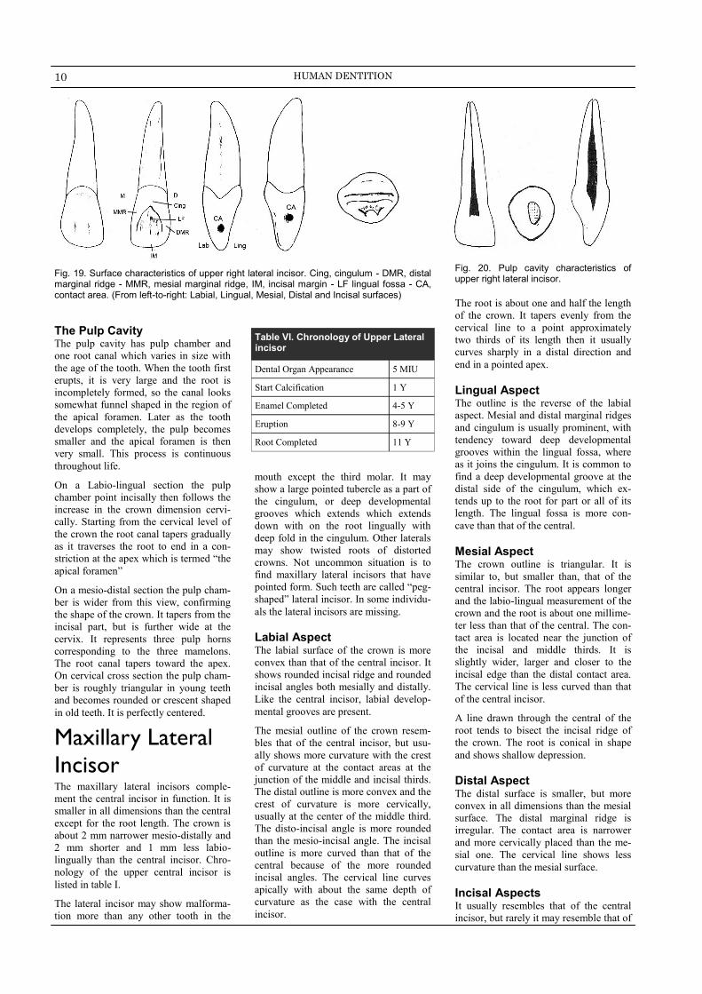
Fig. 19. Surface characteristics of upper right lateral incisor. Cing, cingulum - DMR, distal marginal ridge - MMR, mesial marginal ridge, IM, incisal margin - LF lingual fossa - CA, contact area. (From left-to-right: Labial, Lingual, Mesial, Distal and Incisal surfaces)
Fig. 20. Pulp cavity characteristics of upper right lateral incisor.
The Pulp Cavity The pulp cavity has pulp chamber and
one root canal which varies in size with
the age of the tooth. When the tooth first
erupts, it is very large and the root is
incompletely formed, so the canal looks
somewhat funnel shaped in the region of
the apical foramen. Later as the tooth
develops completely, the pulp becomes
smaller and the apical foramen is then
very small. This process is continuous
throughout life.
On a Labio-lingual section the pulp
chamber point incisally then follows the
increase in the crown dimension cervi-
cally. Starting from the cervical level of
the crown the root canal tapers gradually
as it traverses the root to end in a con-
striction at the apex which is termed “the
apical foramen”
On a mesio-distal section the pulp cham-
ber is wider from this view, confirming
the shape of the crown. It tapers from the
incisal part, but is further wide at the
cervix. It represents three pulp horns
corresponding to the three mamelons.
The root canal tapers toward the apex.
On cervical cross section the pulp cham-
ber is roughly triangular in young teeth
and becomes rounded or crescent shaped
in old teeth. It is perfectly centered.
Maxillary Lateral
Incisor The maxillary lateral incisors comple-
ment the central incisor in function. It is
smaller in all dimensions than the central
except for the root length. The crown is
about 2 mm narrower mesio-distally and
2 mm shorter and 1 mm less labio-
lingually than the central incisor. Chro-
nology of the upper central incisor is
listed in table I.
The lateral incisor may show malforma-
tion more than any other tooth in the
mouth except the third molar. It may
show a large pointed tubercle as a part of
the cingulum, or deep developmental
grooves which extends which extends
down with on the root lingually with
deep fold in the cingulum. Other laterals
may show twisted roots of distorted
crowns. Not uncommon situation is to
find maxillary lateral incisors that have
pointed form. Such teeth are called “peg-
shaped” lateral incisor. In some individu-
als the lateral incisors are missing.
Labial Aspect The labial surface of the crown is more
convex than that of the central incisor. It
shows rounded incisal ridge and rounded
incisal angles both mesially and distally.
Like the central incisor, labial develop-
mental grooves are present.
The mesial outline of the crown resem-
bles that of the central incisor, but usu-
ally shows more curvature with the crest
of curvature at the contact areas at the
junction of the middle and incisal thirds.
The distal outline is more convex and the
crest of curvature is more cervically,
usually at the center of the middle third.
The disto-incisal angle is more rounded
than the mesio-incisal angle. The incisal
outline is more curved than that of the
central because of the more rounded
incisal angles. The cervical line curves
apically with about the same depth of
curvature as the case with the central
incisor.
The root is about one and half the length
of the crown. It tapers evenly from the
cervical line to a point approximately
two thirds of its length then it usually
curves sharply in a distal direction and
end in a pointed apex.
Lingual Aspect The outline is the reverse of the labial
aspect. Mesial and distal marginal ridges
and cingulum is usually prominent, with
tendency toward deep developmental
grooves within the lingual fossa, where
as it joins the cingulum. It is common to
find a deep developmental groove at the
distal side of the cingulum, which ex-
tends up to the root for part or all of its
length. The lingual fossa is more con-
cave than that of the central.
Mesial Aspect The crown outline is triangular. It is
similar to, but smaller than, that of the
central incisor. The root appears longer
and the labio-lingual measurement of the
crown and the root is about one millime-
ter less than that of the central. The con-
tact area is located near the junction of
the incisal and middle thirds. It is
slightly wider, larger and closer to the
incisal edge than the distal contact area.
The cervical line is less curved than that
of the central incisor.
A line drawn through the central of the
root tends to bisect the incisal ridge of
the crown. The root is conical in shape
and shows shallow depression.
Distal Aspect The distal surface is smaller, but more
convex in all dimensions than the mesial
surface. The distal marginal ridge is
irregular. The contact area is narrower
and more cervically placed than the me-
sial one. The cervical line shows less
curvature than the mesial surface.
Incisal Aspects It usually resembles that of the central
incisor, but rarely it may resemble that of
Table VI. Chronology of Upper Lateral incisor
Dental Organ Appearance 5 MIU
Start Calcification 1 Y
Enamel Completed 4-5 Y
Eruption 8-9 Y
Root Completed 11 Y

11 TOOTH MORPHOLOGY & PHYSIOLOGY
Table V. Chronology of mandibular central incisor
Dental Organ Appearance 5 MIU
Start Calcification 3-4 M
Enamel Completed 4-5 Y
Eruption 6-7 Y
Root Completed 9 Y
Fig. 21. Surface characteristics of lower central incisor. (left upper) A, labial as-pect, B, lingual aspect, C, Mesial aspect, D, Distal aspect and E, incisal aspect. (right lower) characteristics of lingual and incisal surfaces. IR, incisal ridge, MMR, mesial marginal ridge, DMR, distal mar-ginal ridge, C, cingulum, LF, lingual fossa, CL, cervical line. (upper right) pulp cham-ber characteristics
a small canine. The cingulum and incisal
edge or ridge may be large. The labio-
lingual dimension may be greater than
usual in comparison with that of the
central incisor, resembling that of a ca-
nine. All maxillary lateral incisors show
more convexity labially and lingually
from the incisal aspect than the maxillary
central incisors and the cingulum is cen-
tered.
Pulp Cavity It consists of pulp chamber and a root
canal. The chamber is quite similar to
that of the central incisor, but without the
three sharp pulp horns. More often the
pulp chamber ends incisally as one round
horn or two less sharp plup horns, a me-
sial and distal.
Mandibular
Incisors The mandibular incisors are four in num-
ber and have smaller mesio-distal dimen-
sions than any other tooth. The contact
areas are near the incisal edge both dis-
tally and mesially. The labial surfaces
are inclined lingually so that the incisal
ridges are lingual to a line bisecting the
root.
Mandibular
Central Incisors The mandibular central incisor is the
smallest in the dental arch. The crown
has little more than half of the mesio-
distal dimension of the maxillary central
incisor; however, the labio-lingual di-
ameter is only about one millimeter less
than that of the maxillary central incisor.
The crown is shorter than that of the
maxillary central by about 1.5 mm.
Labial Aspect It is trapezoidal in outline with smallest side at the cervix. The mesial and distal outline of the crown make a straight drop downward from the incisal angles to the contact areas which are close to the in-cisal edge. The mesial and distal sides then taper evenly from the contact area to the narrow cervix. The mesio-incisal and disto-incisal angles are sharp. The incisal margin is straight and at right angle to the long axis of the tooth.
The labial surface is convex both mesio-distally and inciso-cervically with defi-nite convexity in the cervical one third where he height of contour is located (cervical ridge) and a flattened surface at the incisal third. Mesio-labially and disto-labially developmental grooves are very
faint if present. The cervical line is sym-metrically curved towardthe root with distal diviation.
The mesial and distal root outlines are straight and are continuous with the me-sial and distal outlines of the crown. They slop down to the apical portion and terminate in small pointed taper in most cases curving distally.
Lingual Aspect The outline of the crown is the reverse of the labial surface, and is narrower. It presents a cingulum much smaller than that of the maxillary anteriors. The lin-gual fossa is shallow and the mesial and distal marginal ridges are less prominent. The cingulum is placed more cervically and is centered. It is smooth withno ac-cessory ridges, grooves or pits. The root is slightly narrower than labially.
Mesial Aspect Labial outline of the crown is straight above the cervical curvature, sloping rapidly from the crest of curvature to the incisal edge. The lingual outline shows smooth convexity at the cingulum then it becomes straight line inclined labially for a short distance to join a concave line at the middle third of the crown. This extends upward to join the rounded out-
line of a narrow incisal edge. The curva-tures above the cervical line labially and lingually are less than the maxillary inci-sor.
The incisal margin is straight of slightly
rounded, and its center is located just
lingual to the center of the root. The
contact area is very close to the incisal
edge. The cervical line shows a marked
curvature incisally about one third the
length of the crown.
The root outlines labially and lingually
are straight with the crown outline from
the cervical line. The root start to taper in
the middle third to either a bluntly
rounded or pointed root end. The mesial
surface of the root is flat just below the
cervical line. Most of these roots have a
broad developmental depression for the
most of the root length which is deeper at
the junction of the middle and apical
thirds. In rare cases the root apex is bifid.
Distal Aspect It is the reverse of the mesial aspect. The
cervical line curves incisally about 1 mm
less than on the mesial. The developmen-
tal depression is more marked with well
defined developmental groove at its cen-
ter.
Incisal Aspect Form this view, the tooth is four sided or
diamond shaped. The incisal edge is
straight and the mesial and distal halves
are identical. The cingulum is slightly
shifted towards the distal portion The
crown appeared centered over the root.
The incisal ridge is perpendicular to a
line bisecting the crown labiolingually.
Pulp Cavity In labio-lingual section the outline of the

12 HUMAN DENTITION
Table VI. Chronology of Mandibular lateral incisor
Dental Organ Appearance 5 MIU
Start Calcification 3-4 M
Enamel Completed 4-5 Y
Eruption 7-8 Y
Root Completed 10 Y
The Canines T he canines – two maxillary and two mandibular – bear a close resemblance to each
other. They are the longest teeth in the mouth and placed at the corners of the
mouth and therefore referred to as the corners stones of the mouth.
The canines are well anchored in the bone by their extremely long roots. The crown is
bulkier than that of the incisors, and the middle labial lobe is highly developed incisally
forming a strong well formed cusp and labial ridge.
The crowns and roots of the canines are markedly convex on most surfaces. When
viewed from the proximal aspects they show a triangular shape, however, from labial
and lingual aspects they take pentagonal shape.
The position and form of these teeth and their anchorage in the bone, along with a bone
ridge over the labial portion of the root, called “canine eminence”, have a osmotic value
and ensure facial expression. In function the canines support the incisors and premolars
in holding and tearing the food material.
Maxillary Canine The crown and root of the maxillary canine are narrow mesiodistally than the central
incisor. The cervico-incisal length of the crown is much longer than any other anterior
tooth, with the exception of the maxillary central incisor. Table V list the chronological
data of maxillary canine.
The mesial half of the crown of the maxillary canine resemble a portion of an incisor
and contact with lateral incisor. The distal half, on the other hand, resembles a portion
of a premolar and contact the first premolar. The incisal portion of the crown is thicker
labiolingually then that of the incisors, and the cingulum shows greater development.
Labial aspect It is pentagonal in outline. The mesial outline is convex from the cervix to the center of
the contact area at the junction between the middle and incisal thirds of the crown.
The distal outline between the cervical line and the distal contact area which is situated
at the center of the middle third. The mesial contact area is at a lower level than the
distal.
The cusp has a mesial and distal slopes. The mesial one is shorter and shows tendency
toward concavity, while the distal slope shows tendency toward convexity. The cervical
line is convex root wise. Faint mediolabial and distolabial developmental grooves can
be seen.
The labial surface is smooth except for a shallow depression mesially and distally divid-ing the crown into its three labial lobes. The middle lobe is more developed than the other two which result in formation of a ridge on the labial surface , the labial ridge, which runs from the cervical line to the tip of the cusp in a curved manner inclined me-sially at its center. The areas mesial to the crest of this ridge exhibits convexity while area distal to it tends toward concavity.
Fig. 22. Surface character-istics of lower right lateral incisor. A, labial aspect, B, lingual aspect, C, mesial aspect, D, Distal aspect and E, ncisal aspect.
pulp cavity conform to the crown and
root outline. The mesiodistal section is
narrow and has two pulp horns directed
to the mesial and distal angles of the
incisal edge. Crow section of the root at
the cervical line shows an oval canal
usually constricted nesiodistally and
wide labiolingually.
Mandibular
Lateral Incisor The mandibular lateral incisor has almost
the same form as the mandibular central
incisor, however, some variations exist.
Table IV list the chronological data of
mandibular lateral incisor.
Labial aspect The tooth resembles the central incisor
except that it is slightly larger by o.5 mm
in all directions and is fan shaped. The
mesial side is often longer than the distal
side, causing distal sloping of the incisal
edge. The distoincisal angle is more
rounded than its counterpart in the man-
dibular central.
The distal contact area is more toward
the cervical line than the mesial contact
area to contact properly with the canine.
The crown is larger than that of the cen-
tral and the root is longer by about 1.5
mm.
Lingual Aspect Similar to that of the central incisor but
the mesial outline and the mesial mar-
ginal ridge are longer than the distal.
The cingulum is deviated distal to the
center of the lingual surface.
Proximal Aspects (Mesial and Distal) Differ from the center counterpart in the
following:
The distal surface of the lateral
incisor is shorter than the mesial
surface.
Both cervical line curvatures are
slightly less than that of the central
incisor.
The distal contact area is more
cervically located than the mesial
one.
Root depressions are seen on both
the mesial and distal surfaces.
Incisal Aspect The mandibular lateral incisor appears to
be rotated over their root axes because
the distal developmental lobe is larger
and more mesially located than the distal
lobe. This is because the tooth has to
curve distally to fit into mandibular arch
because it has to fit inside the maxillary
arch. The incisal edge follows the curva-
ture of the mandibular dental arch. The
cingulum is shifted distally and the in-
cisal ridge follows the curvature of the
dental arch.■

13 TOOTH MORPHOLOGY & PHYSIOLOGY
Table VII. Chronology of maxillary canine
Dental Organ Appearance 6 MIU
Start Calcification 4-5 M
Enamel Completed 6-7 Y
Eruption 11-12 Y
Root Completed 14-15 Y
The root appears slender form the labial aspect and is conical with blunt apex. The root may show either mesial or dis-tal curvature, mostly distal, near the apex. The labial surface of the root is smooth and convex.
Lingual aspect The crown and root are narrower lin-gually than labially. The cervical line from is less convex than on the labial surface. Below the cervical line is a well developed cingulum. The mesial and distal marginal ridges are strongly devel-oped.
The area incisal to the cingulum is con-cave forming the lingual fossa. Very often the lingual fossa show a well de-veloped lingual ridge extending from the cusp tip to a point near the cingulum dividing the lingual fossa into mesial and distal.
The root is narrow when viewed from the lingual aspect than the labial and is smooth and convex.
Mesial aspect The outline of the crown is wedge shaped with the greatest measurement at the cervical third. The wedge point at the tip of the cusp. The labial outline is more convex from the cervical line to the cusp tip than any other maxil-lary anterior tooth. The lingual outline is convex below the cervical line, representing the cingulum then continue as a straight line curves toward the cusp.
The relation of the tip of the cusp to the long axis of the root is different from that of maxillary incisors. A line bisecting the cusp is on long axis of the tooth or labial to it.
The mesial surface of the crown is convex at all points except for small circumscribed area above the contact area where the surface is concave of flat to the cervical line.
The outline of the root is conical with tapered blunt or pointed apex. The root may curve labially at apical third. The mesial surface of the root appears broad with a shallow de-velopmental depression for part of the root length which helps to anchor the tooth to the alveolus and prevent rotation.
Distal aspect This aspect is similar to the mesial aspect except that the cervical line shows less curva-ture toward the cusp tip. The distal marginal ridge is heavier and more irregular than the mesial one and the contact area is more cervically located in the middle third. In addi-tion, the surface is more concave above the contact area and the developmental groove is more pronounced.
Incisal aspect The labiolingual dimension is greater than the mesiodistal dimension. The cusp tip is
labial and mesial to the center of the crown. The ridge of the meddle labial lobe is very
noticeable from the incisal aspect. It attains its greatest convexity at the cervical third of
the crown, becoming broader and flatter at the middle and incisal thirds.
Pulp cavity It consists of the pulp chamber and a single root canal. Labiolingual section shows a narrow pulp chamber that points incisally. The root canal is wide in the cervical half of the root than any other tooth. The canal then narrows to average width on its way to the apical foramen.
On mesio-distal section the pulp cavity is much narrower and similar to those of the incisors. It has much longer and tapered root canal.
On cervical cross section the pulp cavity appears even narrower and the root canal is eleptical rather than round and centered over the root. The canal is wider labiolingually than mesiodistally.
Mandibular canine The mandibular canine resembles the maxillary one in that they have the same wedge shaped outline, long crown and a well developed cingulum. They differ fro the maxil-lary canine, however, in some aspects. Table VI lists the chronological data of mandibu-lar canine.
Labial aspect The crown is narrower mesiodistally by about o.5 mm than the maxillary one. Labiolin-gually the crown and root is slightly less than those of the maxillary canine.
On the other hand, the length of the mandibular canine is similar toe the maxillary ca-nine, but the crown is longer by 1 mm and the root is shorter by 1 mm. the effect of
Fig. 23. Surface characteristics of maxillary left canine (left up-per) A, labial aspect, B, lingual aspect, C, Mesial aspect, D, Distal aspect and E, incisal aspect. (right upper) character-istics of lingual and incisal sur-faces. CL, cervical line, C, cin-gulum, MMR, mesial marginal ridge, MLF, mesial lingual fossa, MCR, Mesial cusp ridge, DCR, distal cusp ridge, LR, lingual ridge, DLF, distal lingual fossa, DMR, distal marginal ridge, MCR, Mesial cusp ridge.
Fig. 24. pulp cavity characteristics of max-illary left canine.

14 HUMAN DENTITION
greater crown length is emphasized by the narrowness of the crown mesiodis-tally and the height of the contact area above the cervix.
The mesial outline of the crown is nearly straight with the mesial outline of the root. The mesial contact area is near the mesioincisal angle.
The distal outline is convex cerivcoin-cisaaly with more rounded and obtuse distoincisal angle. It is shorter than the mesial outline and the contact area is located more cervically than the distal one but still more incisalo than the max-illary canine.
The cusp tip and ridges of the mandibu-lar canine are not as well developed as the maxillary canine. The cusp tip is on the center of the root and the distal cusp slope is longer than the mesial one and inclined more cervically.
The cervical line is more symmetrically contoured than the maxillary one .
The crown surface is flat mesial and distal to the labial ridge at the incisal third. Crown outline is concave distally and convex mesially
The root is shorter by 1-2 mm than the maxillary canine and has a more sharply pointed apex that may be directed me-sially of distally.
Lingual aspect The lingual surface of the crown is flat-ter, smoother and regular simulating mandibular incisors. The cingulum and the ridges as well as the lingual fossa are less pronounced than in maxillary ca-nine.
The lingual portion of the root is nar-rower than that of the maxillary canine, it is about one half or a little more in width than the labial portion.
Mesial aspect The labial outline of the crown is less curved with the crest of the curvature at the cervical third. The lingual outline is also less curved. The cusp tip is centered over the root or slightly lingually in-clined (that of the maxillary canine is labilly inclined).
The mesial developmental depression on the root is more pronounced and some-
times the root is bifurcated at the apical third.
Distal aspect There is little difference between distal and mesial aspect which are:
The distal marginal ridge is more pronounced than the mesial
The cervical line has less curvature.
The distal contact area is more cervical.
More pronounced developmental depression on the root.
Incisal aspect The outline of the incisal surface is less curved than in the maxillary canine. The cusp tip, mesial cusp ridge and the con-tact area are inclined lingually, while those of the maxillary canine extends nearly a straight line mesiodistally. The cingulum is shifted distally.
Pulp cavity Pulp cavityiy is similar to that of the upper canine but smaller. The root may show one or two root canals - labial and lingual canal - that join at the apex.or have separate foramina when the root is bifurcated. There are always two canals. ■
Fig. 25. Surface characteristics of mandibular left canine MCR, mesial cusp ridge, DCR, distal cusp ridge, LR, lingual ridge, MMR, mesial marginal ridge, MLF, mesial lingual fossa, C, cingulum, CL, cervical line, DLF, distal lingual fossa, SMR, distal marginal ridge (From left-to-right: labial, liongual, mesial, distal and incisal surfaces)
Fig. 26. pulp cavity characteristics of man-dibular left canine.
The
Premolars T he premolars are intermediate teeth
between molars and canines. They
succeed the deciduous molars. The term
“bicuspid” is often used in place of pre-
molars, but this is inaccurate since the
mandibular second premolar may show
three cusps. There are eight premolars,
two in each quadrant.
The premolars are developed from four
lobes except the mandibular second pre-
molar, the three cusp form, which devel-
oped from five lobes, three buccal and
two lingual.
The buccal cusp of the first premolar is
long and sharp assisting the canine in its
function of tearing. The second premo-
lars have cusps less sharp which make
them efficient in gridding teeth much
like molars.
The marginal ridges of the crown are in a
more horizontal plane and are considered
part of the occlusal surface of the crown
rather than the lingual surface as in case
of anterior teeth. The crown and root of
the premolars are shorter than those of
the canines.
Maxillary
Premolars First Maxillary
Premolar This tooth has two cusps, buccal and
lingual, which are sharply defined. The
buccal cusp is about 1 mm longer than
the lingual cusp and the crown is shorter
than that of the canine by 1.5-2 mm.
table VII lists the chronological data of
the maxillary first premolar.
Table VIII. Chronology of Mandibular canine.
Dental Organ Appearance 6 MIU
Start Calcification 4-5 M
Enamel Completed 6-7 Y
Eruption 9-10 Y
Root Completed 12-13 Y

15 TOOTH MORPHOLOGY & PHYSIOLOGY
Table VIX. Chronology of maxillary first premolar
Dental Organ Appearance 7MIU
Start Calcification 1.5-1.7 Y
Enamel Completed 5-6 Y
Eruption 10-11 Y
Root Completed 12-13 Y
Table X. Common characteristics of posterior teeth.
Have occlusal surface instead of in-
cisal ridge.
Larger bucco-lingual than mesio-distally
Broader contact area.
Less curvature of cervical line mesially
and distal.
Shorter crown compared with incisors and canines
Marginal ridges oriented horizontally
and are part of the occlusal surface.
This tooth is the only premolar which normally has two roots (about 80%), a buccal and a lingual, with two pulp canals, although occasionally there is only one root (20%). It resembles the canine from the buccal aspect but the root is shorter, the mesial slope of the buccal cusp is longer than the distal slope (opposite to the maxillary canine) and the mesiodistal diameter is less than that of the canine. The first premolar presents some characteristics common to all posterior teeth as differentiated from the anterior teeth. (Table VIII)
Buccal aspect The crown is roughly trapezoid with the smallest uneven side directed ervically. The width of the crown mesiodistally is less at the cervix than at the level of contact areas by 2mm.
The mesial outline of the crown is slightly concave from the cervical line to the mesial contact area and also to the cusp tip. The mesial cusp slpe is longer than the distal and the contact area is just cervical to the junction between the occlusal middle one third.
The distal outline is more straight below the cervical line and the contact area is broader and more occlusally placed than the mesial. The cervical line is curved with the crest of the curvature near the center of the root and toward it.
The buccal cusp is long with pointed tip located distal to the midline and divide the occlusal border into a long, straight or concave, mesial cusp ridge and a short convex distal cusp ridge.
The buccal surface of the first maxillary premolar is convex, showing strong develop-ment of the middle lobe with a continuous ridge from the cusp tip to the cervical mar-gin. This ridge is called the buccal ridge. Two developmental grooves, mesiobuccal and distobuccal, are located on both sides of the buccal ridge and mark the union of the developmental lobes.
The buccal root outline of the maxillary first premolar is similar to that of the canine but is shorter by 3-4 mm.
Lingual aspect The crown converges towards the lingual cusp which is shorter and narrower mesiodis-tally than the buccal one. The tip of the lingual cusp is pointed and located slightly to-ward the mesial side. The mesial slope is shorter than the distal.
The mesial and distal outlines are convex and continuous with the mesial and distal slopes of the lingual cusp and become more straight as they join the mesial and distal sides of the lingual root.
The crown as seen from the lingual aspect is smooth spheroid and convex at all points with no definite lingual ridge and no developmental grooves. The lingual height of con-tour is located at the middle third. Since the lingual cusp is shorter than the buccal one, the tips of both cusps can be seen with their mesial and distal slopes from the lingual aspect.
The cervical line is similar to that of the buccal aspect. The lingual aspect of the root, or the lingual aspect of the lingual root if two roots are present, is smooth and convex with blunt apex.
Mesial aspect It is trapezoidal in shape, however, the longest of the uneven sides is toward the cervical portion and the shortest toward the occlusal portion. Another characteristic of all the maxillary posterior teeth is that the measurement between the tip of the buccal cusp to that of the lingual cusp is less than the buccolingual measurement of the root at its cervi-cal portion, i.e. the tips of the cusps are well within the confines of the root trunk.
The cervical line is curved and regular with average curvature of 1 mm. which is similar to all posterior teeth. The buccal outline of the crown is convex below. The crest of curvature is at the junction of the cervical and middle thirds of the crown. It continue as a line of less convexity to the tip of the buccal cusp. The tip of the buccal cusp is located directly below the center of the buccal root.
The lingual outline is curved smoothly from the tip of the lingual cusp to the cervical line. The crest of the curvature is near the center of the middle third. The tip of the lin-gual cusp is on a line with the lingual border of the lingual root.
The lingual cusp is always shorter than the buccal cusp by about 1 mm. the mesial mar-ginal ridge is convex and is located at about the level of the junction of the middle and occlusal thirds.
The mesial contact area is circular in shape and located at the junction of the middle and occlusal thirds and slightly near the buccal.
The distinguishing features of this tooth from the mesial aspect are:
The presence of “Mesial Develop-mental Depression” which extends cervically from the contact area and continue to include the cervical line then joins a deep depression be-tween the root bifurcation, known as “Canine Fossa”. The maxillary second premolar do not have this feature.
The presence of a well defined developmental grooves in the enamel of the mesial marginal ridge. This marginal groove is con-tinuous with the central groove of the occlusal surface of the crown, crossing the marginal ridge just lingual to the contact area and ter-minating a short distance cervical to the mesial marginal ridge. This groove is called the “Mesial Mar-ginal Developmental Groove”.
The buccal outline of the buccal root is straight above the cervical line with tendency toward lingual inclination. The lingual outline is also straight and may show buccal or lingual inclination. The root trunk is long making up about half the length of the root. The bifurca-tion of the roots begins at a more occlusal point mesially than dis-tally.
The mesial surface of the root trunk is smoothly convex buccally and lingually with deep developmental groove and depression at or below the bifurcation. In case of one rooted tooth this depression is no-ticed for most of the root length.

16 HUMAN DENTITION
Fig. 27. Maxillary right first premolar, mesial and occlusal aspect. BR, buccal root; MDD, mesial developmental depression; BCR, buccal cervcal ridge; MCA, mesial contact area; BC, buccal cusp; LC, lingual cusp; MMDG, mesial marginal developmental groove; CL, cervical line; LR, lingual root; DBCR, distobucca; cusp ridge; DMR, distal marginal ridge; DTF, distal triangular fossa; DLCR, distolingual cusp ridge; MLCR, mesiolingual cusp ridge; CDG, central developmental groove; MTF, mesial triangular fossa; MMR, mesial marginal ridge; MBCR, mesiobucaal cusp ridge.
Distal aspect The crown outline is similar to the me-sial aspect. The crown surface is convex at all points. The distal contact area is wider buccolingually than ocluusocervi-cally and slightly near the buccal. It is larger and more cervically located than the mesial contact area.
The height of contour is in the cervical third buccally and middle third lingually. The curvature of the cervical line is less than in the mesial surface. Also there is no evidence of deep developmental grove and the root trunk is flattened above the cervical line. The bifurcation is more toward the apical third with no developmental groove leading to it.
Occlusal aspect The occlusal aspect has a hexagonal outline. Two equal buccal sides, mesial side shorter than the distal and mesiolin-gual shorter than distolingual. It is wider buccally than lingually and buccolin-gually than mesiodistally. The buccal margin is convex with a prominent buc-cal ridge at the crest of the curvature.
The mesiolingual and distolingual cusp ridges are continuous with the mesial and distal marginal ridges. The crest of the buccal ridge is some what distal to the lingual ridge, while the crest of the distal contact area is somewhat buccal to that of the mesial one.
The occlusal surface shows two well developed cusps, the lingual one is more pointed while the buccal one is larger. Each cusp has four cusp ridges named according to their location, buccal, lin-gual, distal and mesial ridges. The buccal cusp ridge descends from the cusp tip cervically onto the buccal surface. The lingual cusp ridge of the buccal cusp and buccal cusp ridge of the buccal cusp descends from the cusp tip to the central area of the occlusal surface. The two triangular ridges of the buccal and lin-gual cusps are separated by the central developmental groove.
The primary grooves on the occlusal
aspect are sharp and deep. A well-
defined central developmental groove
divides the tooth mesiodistally. It ex-
tends from the distal to the mesial mar-
ginal ridge where it joins the mesial mar-
ginal developmental groove which
crosses the mesial marginal ridge and
ends on the mesial surface of the crown.
Two developmental grooves join the
central groove just inside the mesial and
distal triangular fossae. These are called
the mesiobuccal and distobuccal devel-
Fig.28. Oc-clusal aspect of upper first premolar is hexagonal in outline.
Fig. 28. Maxillary first premolar, occlusal aspect. DBCR, distobuccal cusp ridge; BTR, buccal triangular ridge; DBDG, dis-tobuccal developmental groove; DTF, distal triangular fossa; DMR, distal mar-ginal ridge; DLCR, distolingual cusp ridge; CG, central groove; TLC, tip of lingual cusp; LTR, lingual triangular ridge; MLCR, mesiolingual cusp ridge; MMR, mesial marginal ridge; MMDG, mesial marginal developmental groove; MTF, mesial trian-gular fossa; MBDG, mesiobuccal develop-mental groove; MBCR, mesiobuccal cusp ridge; TBC, tip of buccal cusp.
Fig. 29. Pulp cavity characteristics of the upper first premolar. Mesiodistal section (upper left) cross section (upper middle); buccolingual section (upper right): varia-tions of root canals (lower)
opmental grooves. The junction of the
grooves are pointed and are named the
mesial and distal developmental pit. Just
inside the mesial and distal marginal
ridges are the mesial and distal triangular
fossae.
Pulp cavity In the bucco-lingual section the pulp
chamber is broad buccolingually with
well developed pulp horns. It presents a
funnel-like opening leading to the root
canals. The floor of the pulp chamber is
below the level of the cement-enamel
junction. The lingual root canal is larger
than the buccal and both taper evenly
toward the apical foramen.
The mesiodistal section is similar to the
pulp cavity of the maxillary canine. It is
relatively narrow and taper evenly to the
apical foramen.

17 TOOTH MORPHOLOGY & PHYSIOLOGY
Table XI. Chronology of maxillary sec-ond premolar
Dental Organ Appearance 8MIU
Start Calcification 2-2.3 Y
Enamel Completed 6-7 Y
Eruption 10-12Y
Root Completed 13-15 Y
Fig. 30. Surface characteristics of maxillary left second premolar from left-to-right buccal, mesial, distal and lingual aspect, and occlusal aspect (lower left).
Fig. 31. Pulp cavity characteristics of the upper second premolar. Mesiodistal section (left) cross section (middle); buccolingual section (right).
A transverse section at the cementoe-
namel junction shows the the characteris-
tic kidney shaped root trunk and the root
is wider buccolingually than mesidis-
tally.
Maxillary Second
Premolar This tooth supplements the first maxil-lary premolar in function and very simi-lar to it. The second premolar is single rooted in 85% of cases and has a less angular and rounded crown from all aspect than that of the first premolar. The crown is shorter cervico-occlusally but the root is little longer than that of the first premolar. Table IX list the chrono-logical data of the maxillary second pre-molar.
Buccal aspect The buccal cusp is shorter and less pointed than in the first premolar. The mesial slope of the buccal cusp is shorter than the distal slope, which is the oppo-site for the first premolar.
Lingual aspect Very similar to the first molar with very little variations.
Mesial aspect Cusps are shorter than in the first premo-lar and are almost of the same length. The distance between the cusp tips is wider which widens the occlusal surface buccolingually. Crown is convex with no developmental depression and a shallow developmental groove is seen on the root. No developmental groove crosses the mesial marginal ridge.
Distal aspect The distal aspect shows very little varia-tions from that of the first premolar.
Occlusal aspect The occlusal surface outline is more rounded or oval than angular, as the oc-clusal outline of the first premolar. The distance between the cusp tips is wider, the central developmental groove is shorter and more irregular with multiple supplemental grooves radiating from the central groove. The mesial and distal triangular fossae are nearer to each other.
Pulp cavity On buccolingual section the pulp chamber is wide with well defined pulp horns. There
are two pulp canals, buccal and lingual. The canals are very wide at their junction with
the pulp chamber and then narrow until reaches the mid-root where it constricts rapidly
to the apical foramen.
The tooth may show single broad pulp canal divided at mid-root into two canals by
dentin island then the canals join again near the apical foramen. Mesiodistally the pulp
chamber is similar to that of the first premolar. Cervical cross section display an oval
root trunk instead of kidney shaped rott trunk of the first premolar.
Mandibular Premolars They are four in number, two in each side of the mandible. As a rule the mandibular
first premolars are always smaller than second premolars. The opposite is true for max-
illary premolars.
Mandibular First Premolar The mandibular first premolar has many of the characteristics of a small canine. It has
one sharp buccal cusp which is the only part occlude with maxillary teeth. The lingual
cusp is small and nonfunctioning resembling a well developed cingulum. (Table X) On
the other hand the mandibular second premolar has more of the characteristics of a
small molar.
Buccal aspect The outline is roughly trapezoidal with the
smallest of the uneven sides cervically.
The mesial outline of the crown is straight
or slightly convex till it joins the curvature
of the mesial contact area. The outline of
the mesial slope of the buccal cusp usually
shows some convexity.
The distal outline of the crown is slightly
concave above the cervical line to a point
where it is continuous with the curvature
of the distal contact area. This curvature
is broader than that of the mesial contact
area. The distal slope of the buccal cusp
shows some concavity.
Table XIII. Characteristics of first man-dibular premolar resembling canine
The buccal cusp is long and sharp and is
the only part occluding
The mesiodistal and buccolingual meas-
ures are similar to the canine
The occlusal surface slopes sharply lin-
gually in a cervical direction
The mesiobuccal cusp ridge is shorter then
the distobuccal one.
The outline form of the occlusal aspect
resembles the incisal aspect of the canine
Table XII. Chronology of mandibular first premolar
Dental Organ Appearance 7MIU
Start Calcification 1.7-2 Y
Enamel Completed 5-6 Y
Eruption 10-11Y
Root Completed 12-13 Y
Table XIV. Characteristics of first man-dibular premolar resembling second premolar
Crown and root outline from the buccal
aspect are similar but the buccal cusp is longer
Contacts areas are at the same level
Curvatures of cervical line are similar
Roots are about the same length

18 HUMAN DENTITION
The tip of the buccal cusp is pointed and
is located a little mesial to the central of
the crown buccally. The cervical line is
curved toward the root.
The buccal surface of the crown is more
convex than the maxillary premolars.
The middle buccal lobe of the crown is
well developed, resulting in a large
pointed buccal cusp with a buccal ridge
continuous from the cervical margin to
the cusp tip. Mesial and distal to the
ridge a developmental depression may be
seen separating the three lobes. The me-
sial cusp ridge is shorter than the distal
one.
The contact areas are broad and are al-
most at the same level being just cervical
to the junction between the occlusal and
middle thirds.
The root outline resembles that of the
canine but is shorter by about 3-4 mm an
shows a distal curvature at the apex.
Lingual aspec Lingual aspect is much narrower mesio-
distally than buccal since the crown and
root taper markedly toward the lingual
side. The lingual surface is evenly con-
vex in all directions.
The mesial and distal outlines are con-
cave between the cervical line and the
contact areas. The contact areas and
marginal ridges are pronounced and
extend out above the narrow cervical
portion of the crown. The mesial mar-
ginal ridge is more cervically located
than the distal one.
The cervical line is slightly convexto-
ward the root. The occlusal surface
slopes greatly toward the lingual surface
down to the short lingual cusp. Accord-
ingly most of the occlusal surface is seen
from this aspect.
A mesiolingual developmental groove
separates the mesial marginal ridge from
the lingual cusp. The groove fads out at
the junction of the middle and cervical
thirds.
The root tapes evenly from the cervix to
a pointed apex. A narrow smooth convex
ridge is seen through the full length of
the root. Often developmental depression
in the root with developmental grooves
mesially and distally are seen.
Mesial aspect Crown outline is rhomboidal which is a
characteristic of all mandibular posterior
teeth from mesial and distal aspect.
The surface of the crown presents an
overhang above the root in a lingual
direction. The tip of the lingual cusp is
on a line with the lingual border of the
root (both the buccal and lingual roots of
the maxillary premolars are within the
confines of the root trunk).
The tip of the buccal cusp is nearly cen-
tered or a little buccal to the center of the
root.
The buccal outline of the crown is con-
vex with the crest of curvature near the
cervical one third. This feature is a char-
acteristic for buccal surface of all poste-
rior teeth. The lingual outline is less
convex and the crest of curvature ap-
proaches the middle third.
The mesial marginal ridge has a sharp
inclination lingually in a cervical direc-
tion (45o), the lingual border of which
merges with the developmental depres-
sion mesiolingually. This depression
harbors the mesiolingual developmental
groove.
The contact area is ovoid in shape and is
located to the junction of the occlusal
and middle thirds and slightly buccally.
Some of the mesial portion of the occlu-
sal surface is seen from this aspect, this
include mesial portion of the buccal cusp
triangular ridge and mesial marginal
ridge.
The cervical line curves occlusally and
the crest of curvature is about 1mm in
extgent and is centered buccolingually.
The mesia surface is convex at the me-
sial contact area then becomes sharply
concave till the cervical line.
The root outline tapers to a pointed apex
in line with the tip of the buccal cusp.
Deep developmental groove is shown in
the center of the root which may end in
bifurcation at the apex.
Distal aspect In differes from the mesia aspect in the
following:
Distal marginal ridge is more oc-
clusally located and nearly at line
angle to the axis of the crown and
root.
The distal contact area is broader
than the mesial and is centered
buccolingually in the middle one
third.
Curvature of cervical line is nearly
the same as in the mesial aspect.
The root surface is more convex
distally.
A shallow developmental depres-
sion is centered on the root but with
no developmental groove
Occlusal aspect This aspect shows considerable varia-
tions on the outline. It may be diamond-
shaped, similar to the incisal aspect of
canine, or circular similar to some sec-
ond premolars. The characteristics com-
mon to all mandibular first premolars
from the occlusal aspect regardless the
type are listed in table XIII.
Fig. 32. mandibular right first premolar, mesial and occlusal aspect. BC, buccal cusp, BTR, buccal triangular ridge, LC, lingual cusp; MLDG, mesiolingual devel-opmental groove; CL, cervical line; BCR, buccal cervical ridge; MCA, mesial con-tact area; MMR, mesial marginal ridge; MBCR, mesiobuccal cusp ridge; CDG, central developmental groove; DMR, distal marginal ridge.
Fig. 33. Surface characteristics of mandibular right first premolar from left-to-right buccal, mesial, distal, lingual and occlusal aspect.

19 TOOTH MORPHOLOGY & PHYSIOLOGY
Table XV. Characteristics of occlusal aspect of first mandibular premolar
The middle buccal lobe makes the major
bulk of the crown.
The lingual marginal ridge is convex and is
interrupted mesially by the mesiolabial
developmental groove.
The mesiobuccal and distobuccal line
angles are prominent even though rounded.
The curvature representing the contact
areas are broad and the distal is broader.
The crown converges sharply to the center
of the lingual surface, which gives the
crown a triangular appearance with the
base at the buccal cusp and the apex at the lingual cusp.
The marginal ridges are well developed.
The lingual cusp is small.
The buccal triangular ridge is heavy and
the lingual one form a transverse ridge.
Mesial and distal triangular fossae are two
irregular depressions containing pits.
The mesiolingual developmental groove
constricts the mesial surface of the crown and create a smaller contact area.
The mesial triangular fossa of the occlusal
surface is more linear in form.
The mesiobuccal groove extends bucally
from mesiotriangular fossa and the mesio-
lingual groove extends from it lingually.
The distal triangular fossa is more circular.
The central developmental groove extends
from the mesial to the distal pits of the fossae.
The pulp cavity It resemble that of the canine but smaller
in dimensions.
In buccolingual section the pulp chamber
is wide with clear pulp horn under the
buccal cusp. The pulp horn directed to
the lingual cusp is missing. The root
canal is broad tapering evenly and is
more constricted at halfway point of root
length.
Mesiodistally the pulp chamber and root
canal are narrow and the canal taper
evenly until the constricted apical fora-
men. This is typical for all premolars.
Cervical cross section shows wider pulp
canal bucculingually ehan mesiodistally.
The outline form may be oval, rectangu-
lar or triangular. Occalsionally two cnals
may be seen.
Fig. 34. pulp cavity characteristics of lower right first premolar. Mesiodistal section (upper left), buccolingual section (upper right) and cervical cross section (upper middle) and possible variation (lower).
Mandibular second premolar The crown of the mandibular second premolar is wider buccolingually by 0.5 mm than
the mandibular first premolar. Its lingual cusps are more developed, and both marginal
ridges are higher. This produces a more efficient occlusion with the maxillary antago-
nist. Therefore, a mandibular second premolar functions more like molar than a canine.
There are two common forms which this tooth assumes;
The three cusp type, which is the most common and appears more angular from
the occlusal aspect.
The two-cusp type appears more rounded from the occlusal aspect.
The single root of the second premolar is longer (by 0.5 mm) than that of the first pre-
molar with developmental groove buccally in many cases.
Buccal aspect From the buccal aspect the crown resembles a first premolar in its general shape. It pre-
sents shorter buccal cusp than the first premolar with the mesiobuccal and distobuccal
cusp riges more rounded and less pointed cusp tip which is little mesial to the tooth
center. The mesial and distal contact areas are nearly at the same level but are broader
and placed higher occlusally. The buccal ridge isles prominent that than that of the man-
dibular first premolar.
The root is longer and broader mesiodistally than the first premolar and ends in more
blunt apex. The single root may show a developmental groove.
Lingual aspect The lingual lobes are developed to a greater degree making the cusp or cusps longer. In
the three cusp type, there are a mesiolingual and a distolingual cusps. The former is the
wider and longer of the two cusps. The cusps are divided by a lingual groove.
In the two cusp type, the single lingual cusp is higher than on a mandibular first premo-
lar. There is no groove, but a developmental depression is seen distolingually where the
lingual cusp ridge joins the distal marginal ridge.
Table XVI. Chronology of mandibular second premolar
Dental Organ Appearance 8MIU
Start Calcification 21/3-21/2 Y
Enamel Completed 6-7 Y
Eruption 11-12Y
Root Completed 13-15 Y
The lingual surface of the crown of all
mandibular second premolars is smooth
and spheroidal, having a bulbous form
above the constricted cervical portion.
The root is much wider lingually than
that of the first premolar. This results in
less convergence towards lingual. It is
smooth and convex.
Mesial aspect The second premolar differs from the
first from the mesial aspect in the follow-
ing ways;
The crown and root are thicker
buccalingually than the first premo-
lar.
The buccal cusp is shorter and its
tip located more to the buccal side.
The lingual lobe or lobes develop-
ment is greater. The tip of the lin-
These ancient Indians forceps were used for extraction of teeth and were named for the resemblance to the head of an animal. The right forceps is called “Cat” and the left “Jackal”. The forceps were used to extract teeth as well as foreign bodies such as arrow heads.

20 HUMAN DENTITION
gual cusp is on line with the lingual border of the root.
The marginal ridge is at right angle to the long axis of the tooth.
Less of the occlusal surface may be seen.
There is no mesio-lingual developmental groove on the crown portion.
The root is longer and in most cases slightly convex on the mesial surface with a
more blunt apex.
Distal aspect More of the occlusal surface can be seen because the distal marginal ridge is at a lower
level than the mesial marginal ridge.
As a general rule, the crown of all posterior teeth (maxillary and mandibular) are tipped
distally to the long acis of the root. So, much of the occlusal surface may be seen from
the distal aspect.
In the three-cusp type both lingual cusps are seen since the distolingual cusp is shorter
than the mesiolingual cusp.
The root is conical in shape and may show deeper developmental depression.
Occlusal aspect In both the two and three-cusp forms, the buccal cusp is similar, however, the outline of
each type shows some variations from the occlusal aspect.
The occlusal characteristics of the three cusp type:
It appears square lingual to the buccal cusp ridge. It has three distinct cusps, the
buccal cusp is the largest then the mesiolingual cusp. The distolongual cusp is the
smallest.
Each cusp has a well defined triangular ridges separated by deep developmental
grooves. These grooves converge in a central pit and from a ‘’Y’’ shape on the
occlusal surface. Three pits may be present, a central, a mesial and distal.
The central pit is located in the center buccolingually and slightly distal to the
midway point between the mesial and distal marginal ridges.
From the central pit, a mesial and distal developmental grooves travel in a me-
siobucal and distobuccal directions respectively ending in the mesialand distal
triangular fossae.
The lingual developmental groove extends lingually between the two lingual cusps
and ends on the lilnguall surface of the crown just below the lingual cusps. The
mesiolongualcusp is wider mesiodistally than the distolingual cusp.
Supplemental grooves and depressions are often seen radiating from the develop-
mental groove.
The mesial and distal marginal ridges are confluent with the cusp ridges.
The occlusal characteristics of the two-cusp type as compared with the three-cusp type
are:
The outline of the crown is rounded.
The lingual surface of the crown is more convex and tapers toward the lingual side
(lingual convergence)
Fig. 35. Surface characteristics of mandibular left second premolar from left-to-right buccal, mesial, distal, lingual and occlusal aspect.
Fig. 36. Pulp cavity characteristics of lower left sec-ond premolar. Mesiodistal section (left), buccolingual section (middle), cervical cross section (next to left) and mesiodistal section of three cusp type (right).
The mesiolingual and distolingual
line angles are rounded.
There is only one well-developed
lingual cusp located directly oppo-
site to th buccal cusp in a lingual
direction.
There is no lingual developmental
groove.
A central developmental groove is seen
on the occlusal surface traveling in ame-
siodistal direction. This groove may be
straight and often crescent-shaped. The
groove pattern can be either a „‟U‟‟ or
„‟H‟‟ groove pattern, depending on
whether the central developmental
groove is straight mesiodistally or curves
buccally at its ends. The central groove
of the two-cusp from terminates in a
mesial and distal fossae. There is usually
no central pit, a mesial or distal pit is
much more likely. Usually the two cusp
type shows a transverse ridge.
THE TOOTH IS A
NOBLE ORGAN THAT
CAN NOT BE REPLACED
The pulp cavity In buccolingual section it resembles that of the mandibular first premolar, but is larger in size, and the roof of the pulp chamber is pointed to accommodate more than one pulp horn. The lingual pulp horn is smaller than the buccal pulp horn.
In mesiodistal section it resembles that of the mandibular first premolar except for the added size and length.
The pulp chamber has three pulp horns in the three cusp type. The largest is the buccal pulp horn then the mesio lingual then the distolingual.
In cervical cross section the outline of the pulp cavity follows that of the root and most commonly is rectangular.■

21 TOOTH MORPHOLOGY & PHYSIOLOGY
The Molars General features
There are twelve permanent molars three in each quadrant. They are the largest
and strongest teeth in the mouth by their crown bulk size and their root anchorage
in the bone.
The molars have No deciduous predeccesors. Generally the lower first molars are
formed from five lobes, but second and some third molars may have only four.
Each cusp of a molar is formed from its own lobe.
The most developed of the molar is the first molar. The third molar exhibits the
most variable morphology in the mouth.
Molars are used in grinding food, but most important they support and maintain
the vertical dimension of the face.
Maxillary Molars They are six in number, three in each side, the first, second, and third molars.
Maxillary First Molar The normal location of the first permanent molar is at center of the fully developed
adult dental arch antero-posteriorly. Table XV shows the chronology of the first maxil-
lary molar.
It is the largest tooth in maxillary arch. It has four well developed cusps (two buccal and
two lingual) and a fifth supplemental nonfunctioning elevation which is called Tubercle
of Carabelli. This fifth cusp serves to identify the maxillary first molar. The crown of
this tooth is wider buccolingually than mesiodistally.
There are three wel developed and widely separated roots, two buccal and one palatal
(lingua). They give this tooth the maximum anchorage against forces.
Buccal aspect The crown is roughly trapezoid. The cervical line is the shortest of the uneven sides.
The tips of all cusps are usually visible from this aspect. Part of the distal side is seen
due to the obtuse distobuccal line angle.
The mesial outline of the crown follows a nearly straight path downward and mesially,
curving occlusally as it reaches the crest of contour of the mesial surface which is the
contact area. The contact area is just Cervical to the junction of the occlusal and middle
thirds. Then, the mesial outline curves occluaslly and distally and becomes correspond-
ing with the outline of the mesial slope of the mesiobuccal cusp.
The distol outline is convex, from the cervical line to the contact area is in the center of
the middle third then it curves occlusally and mesially to outline and distal slope of the
distobuccal cusp.
The cervical line is slightly curved with the curvature root wise. This line is not so
smooth and regular as found in some other teeth.
Although the mesiobuccal cusp is broader than the distobuccal cusp, the distobuccal
cusp is usually sharper and longer. The mesial slope of the mesiobuccal cusp meets its
distal slope at an obtuse angle. The mesial slope os the distobuccal cusp meets its disal
slope at approximately right angle.
Separating the two buccal cusps is the buccal developmental groove. This shallow
groove runs in occluso-apical direction parallel to the long axis of the distobuccal root.
It terminates at a point nearly half the distance from its origin as a horizontal groove.
The buccal surface is characterized by buccal ridges on each buccal cusp. They are con-
vex areas which extend cervically about half its length. It is also characterized by the
buccal cervical ridge which extends horizontally from mesial to distal in the entire cer-
vical third.
The molar roots originate as a single root on the base of the crown, and then they divide
into three roots. The common root base is called root trunk. Usually the palatal toot is
the longest and the two buccal roots are approximately equal in length. The root are
about twice as long as the crown and their greatest extremities mesiodistally is less than
the calibration of the crown mesiodistally.
Table XVII. Chronology of maxillary first molar
Dental Organ Appearance 4MIU
Start Calcification At birth
Enamel Completed 3-4 Y
Eruption 6-7Y
Root Completed 9-10 Y
Fig. 37. Maxillary right first molar, buccal aspect. DBR, distobuccal root; LR, lingual root; MBR, mesiobuccal root; CL, cervical line, DBR distobuccal cusp; MLC, mesio-lingual cusp; BDG, buccal developmental groove; MBC, mesiobuccal cusp
There is a deep developmental groove
buccally on the root trunk which starts at
the bifurcation and progresses downward
to end in a shallow depression at the
cervical line.
All three roots may be seen from the
buccal aspect. The point of bifurcation of
the two buccal roots is located approxi-
mately 4.mm above the cervical line.
The roots are not straight; the buccal
roots are curved halfway between the
point of bifurcation and the apices.
The mesiobuccal root curves distally
starting at the middle third to the apex.
Its long axis at an acute angle distally
with the cervical line. It has a tendancy
toward curvature mesially at the middle
third.
Lingual Aspect It is Trapezoid in shape. The crown sur-
face shows more convexity occlusocervi-
cally than the buccal surface. The hight
of contour is located in the middle third
of the surface. The lingual cusps are the
only ones to be seen from this aspect.
The mesiolingual cusp is much larger. Its
mesiodistal width is about 3/5th of the
mesiodistal diameter of the crown, the
distolingual cusp making up the remain-
ing 2/5th.
The mesial outline is straight from the
cervical line to the contact area and

22 HUMAN DENTITION
meets the mesial slope of the mesiolingual cusp at 90o. The two cusp slopes meet at
obtuse angle. The distal outline of the crown is smoothly curved. The cervical line is
slightly convex rootwise in an irregular manner.
The fifth cusp (Tubercle of Carabelli) appears attached to the mesiolinguall surface of
the mesiolingual cusp. The fifth cusp is separated from the mesiolingual cusp by an
irregular developmental groove. This is called the fifty cusp developmental groove.
The distolingual cusp is smooth and spherioidal without angulation on the mesial and
distal slopes. The lingual developmental groove separates the two large lingual cusps
and runs cervically to end at the center of the lingual surface of the crown. From this
point a shallow depression extends cervically and continues in an apical direction on the
lingual root till the middle third.
All three roots are visible from the lingual aspect. The lingual portion of the crown lin-
gually. The lingual root is conical with blunt rounded apex. The apex of the lingual root
is on line with the lingual groove of the crown.
Mesial aspect It is trapezoid in shape with the smallest uneven side located occlusally. The buccal
outline of the crown starting from the cervical line is curved with the crest of curvature
within the cervical third. As it progresses downward, it becomes less convex to circum-
scribe the mesiobuccalcusp. Only the mesiobuccal, mesiolingual and the fifth cusps are
seen. Also only two roots are seen, the mesiobuccal and the palatal roots.
The lingual outline is curved with the crest of curvature located near the middle third. If
the tubercle is well developed, the lingual outline dips inward to illustrate it. If it is un-
developed the lingual outline continues from the crest of curvature as a smooth curved
arc to the tip of the mesiolingual cusp.
The mesial marginal ridge, which is confluent with the mesiobuccal and mesiolingual
cusp ridges, is irregular and curved cervically about one-fifth the crown length. The
cervical line is irregular, curving occlusally not more than 1mm.
The mesial contact area is above the marginal ridge approximately cervical to the junc-
tion of the middle and occlusal thirds of the crown and some what toward the buccal
side. Just above it, a shollow concavity is found which may continue to the mesial sur-
face of the root trunk at its cervical third.
The mesiobuccal root is broad and flattened on its mesial surface. The width of this root at the point of bifurcation is about 2/3rds of the crown width at the cervical line buccalin-gually. The buccal outline of this root is curved, but its lingual outline is straight to end at a blunt apex.
The lingual or (palatal) root is longer than the mesiobuccal root by 1.0mm but it is nar-rower from this aspect. Its buccal outline is concave and its lingual outline is convex. At its middle and apical thirds, it is outside of the confines of the greatest crown projection. It is banana shaped.
The level of the bifurcation is a little closer to the cervical lie than is found between the roots buccally. There is smooth depression extends from the bifurcation to the cervical line.
Distal aspect The gross outline of this aspect is similar to that of the mesial aspect, but the following variation is noted.
The buccolingual measurement of the crown on the distal aspect is less than that of the mesial asplect.
The distal marginal ridge dips sharply in a cervical direction exposing part of the four cusps.
The cervical line is almost straight.
The distalsurface is generally convex except for a small concave area near the distobccal root at the cervical third. This concavity continues over the distobuccal root and end at the bifurcation point.
The distobuccal root is narrow at its base than either of the others. Its outline fol-lows a concave path from the cervical line for a short distance than makes a con-vex arc to the blunt apex. The lingual outline of this root is slightly concave from the apex to the bifurcation.
The bifurcation is more apical. The area from the cervical line to bifurcation is 5mm or more in extent.
Fig. 38. maxillary right first molar, mesial aspect. LR, lingual root; FC, fifth cusp; MLC, mesiolingual cusp; MMR, mesial marginal ridge; MBC, mesiobuccal cusp; MCA, mesial contact area;CRm cervical ridge; MBR, mesiobuccal root.
Fig. 39. maxillary right first molar, distal aspect. DBR, distobuccal root; MBR, mesiobuccal root; BDG, buccal develop-mental groove;DBC, distobuccal cusp; DCA, distal contact area; CL, cervical line; LR, lingual root; DLC, distolingual cusp; DMR, distal marginal ridge.
Occlusal aspect It is Rhomboidal in outline, the crown is
wider mesialy than distally, and wider
lingually than buccally (this the only
molar that is wider lingually than buc-
cally).
The mesiobuccal and distolinguall angles
are acute in comparison to the mesiolin-
gaul and distobuccal angles which are
comparatively obtuse.
The four major cusps are well developed.
The tubercle of Carabelli on the lingual
surface of the mesiolingual cusp is small
and nonfunctional. It may be indistinct,
and instead, a developmental line in the
enamel is present.
The mesiolingual cusp is the largest
cusp, followed by the rounded mesiobuc-
call, the sharp distobuccal, the small
distolingual , and the nonfunctional fifth
cusp (tubercle of Carabelli).

23 TOOTH MORPHOLOGY & PHYSIOLOGY
Fig. 40. Surface characteristics of maxillary right first molar. From left-to-right buccal, me-sial lingual, distal and occlusal aspects.
Fig. 41. Maxillary right first molar, occlusal aspect (upper) and developmental grooves (lower). (Upper) MBCR, mesiobuccal cusp ridge; CF, central fossa (shaded area); MTF. Mesial triangular fossa (shaded area); MMR, mesial marginal ridge; MLCR, me-siolingual cusp ridge;DF, distal fossa; DTF, distal triangular fossa (shaded area); DMR, distal marginal ridge; DBCR, distobuccal cusp ridge; DLCR, distolin-gual cusp ridge; OR, oblique ridge. (Lower) BG, buccal groove; BGCF, buccal groove of central fossa; CGCF, central groove of central fossa; FCG, fifth cusp groove; LG, lingual groove; DOG, distal oblique groove; TGOR, transverse groove of oblique ridge;CP, central pit.
The occlusal surface is within the con-fines of the cusp ridges and the marginal ridges.
There are two major fossae, a central fossa which is triangular concave area mesial to the oblinque ridge, and a distal fossa which is to the distal marginal ridge.
The central development pit lies in rteh central fossa. The buccal developmental groove radiates from this pit buccally between the two bucclcusp. The central development groove proceeds in a mesial direction originating in the central pit and termination at the mesial triangular fossa. The mesial pit is found in the me-sial triangular groove and crosses the mesial marginal ridge.
From the mesial pit the following devel-opmental grooves radiate.
The central developmental groove to the central fossa.
The mesiobuccal groove.
The mesiolingual groove.
Another developmental groove may be seen radiating from the central pit dis-tally and crosses the oblique ridge to end at the distal fossa. This is called the Transverse groove of the oblique ridge.
From the distal pit the following devel-opmental grooves radiate:
The distal groove that joins the distal triangular and distal fossa.
The distal marginal groove that may extends over te marginal ridge into the distal surface.
The distolingual groove.
The distobuccal groove.
The oblique ridge traverses the occlusal surface from the top of the mesiol;ingual cusp to the top of the distobuccal cusp. It is reduced in height in the center of the occlusal surface and is some times crossed by the transverse groove of the oblique ridge.
The mesial and distal marginal ridges are irregular elevations of enamel which are confluent with the adjacent cusp ridges.
Pulp cavity It contains a pulp chamber and three or four root canals.
In buccolingual section yhe chamber is broad and rectangular in shape and the two pulp horns are prominent and extend to the mesiobuccal and mesiolingual cusps. The floor is smooth with canal openings widening out as they enter the
pulp chamber. The lingual canal is large and accessible, while the mesiobuccal canal is small and shorter and it may have supplementary branches and multi-ple foraminae, but mostly the mesiobuc-cal root has two root canals which either oopen in one or two aical foramina. The disto buccal root has only one root canal.
In mesiodistal section The pulp chamber is not wide, and the pulp horns extend to the mesio-buccal and distobuccal cusps. The mesiobuccal and distobuccal canals are narrow and tapering to the apex. The pulp chamber the two root canals are centered within the tooth outlines.
In cervical cross section The outline is rhomboidal in shape with rounded cor-ners. The canals of this tooth form a triangular patern. A line drawn between the mesiobuccal and the palatal canals makes the base of the triangle.
In midroot cross section The palatal root canal is rounded in outline. The disto-buccal canal is oval and smaller. The mesiobuccal canal or canals are elon-gated oval or kidney shaped. ■
Fig. 42. Characteristics of pulp cavity of upper first molar. Buccal, distal, and lingual aspect (upper) and cross section at the cervical line (left)

24 HUMAN DENTITION
Maxillary Second
Molar This tooth assists the maxillary first mo-lar in function, and it has the same from with some varioations.
Buccal aspect The crown is a little shorter cervico-occlusally (by about 0.5mm) narrower mesiodistally (by 1mm), but of the same measurement bucco-lingually.
The buccal groove is located further distally, with a resultant larger and longer mesiobuccal cusp, and a shorter, smaller and sharper distobuccal cusp.
The buccal roots are about the same length. They are more nearly parallel ad are inclined distally more than those of the maxillary first molar so that the end of the distobuccal root is slightly distal to the distal extremity of the crown.
The apex of the mesiobuccal root is on a line with the buccal groove of the crown instead of the tip of the mesiobuccal cusp as found on the first molar.
Lingual aspect The mesilingaul cusp is smaller and
not well developed as in the first maxillary molar. No fifth cusp is present.
The distolingual cusp is smaller.
The buccal cusps may be seen be-tween the lingual cusps.
The apex of the lingual root is in line with the distolingual cusp in-stead of the lingual groove as was found on the first molar.
There is no developmental groove on the palatal root.
Mesial aspect No tubercle of Carabelli. It shows less divergent roots (being within the con-fines of buccolingual crown outline). In addition the cervical concavity is not present.
Distal aspect It is similar to the mesial except that the distobuccal cusp is smaller than in the first molar. The apex of the lingual root is in line with the distolingualcusp.
Occlusal aspect The crown is more constricted mesiodis-tally and has no tubercle of Carabelli. There are two major types of crown from:
Rhomboidal form which is most frequent type resembling the maxillary first molar except that:
The rhomboid form is more accen-tuated.
The acute angle is less acute, and the obtuse angles are greater.
The distobuccal and distolingual cusps are smaller.
Heart shaped form resemble the maxil-lary third molar. The distolingaul cusp being poorly developed or sometime missing.
It is common to find supplemental grooves as well as grooves and pits on
Fig. 43. Surface characteristics of maxillary left second molar. From left-to-right buccal, mesial lingual, distal and occlusal aspects.
the occlusal surface of the maxillary second molar which make the surface more wrinkled.
Pulp cavity It is similar to the first maxillary molar
both in the mesiodistal and buccolingual
cross sections. It is not common to find
two root canals in the mesiobuccal root.
Maxillary Third Molar The maxillary third molar is the most variable tooth in the upper arch. It v aries in size, shape, contour and surface de-tails. The most common crown shape is the hear-shape, which is generally smaller and more rounded in all dimen-sions than the second molar and also the crown is shorter and narrower.
The distolingual cusp is very small and poorly developed or may be absent pre-senting an occlusal table with three cusps. The groove pattern is variable and may show many supplementary grooves and the distolingual groove is absent.
The root form and number are extremely variable, but are smaller in all dimen-sions. The most common is the three root type where they are often fused
Pulp cavity Due to the variation in shape of this
tooth the pulp cavity also subjected to
great variation in shape and size. How-
ever, the pulp cavity roughly follows the
shape of the tooth.
Table XVIII. Chronology of maxillary second molar
Dental Organ Appearance One Year
Start Calcification 2.5-3 Y
Enamel Completed 7-8 Y
Eruption 12-13 Y
Root Completed 14-16 Y
Table XIX. Chronology of maxillary third molar
Dental Organ Appearance 4Y
Start Calcification 7-9 Y
Enamel Completed 12-16 Y
Eruption 17-21 Y
Root Completed 18-25 Y
Fig. 45. Surface characteristics of maxillary left second molar. From left-to-right buccal, mesial lingual, distal and occlusal aspects.

25 TOOTH MORPHOLOGY & PHYSIOLOGY
Mandibular Molars They are six in number, three on each side of the mandible. (The are larger than any
mandibular teeth. The crowns are quadrilateral being some what longer mediodistally
than buccolingually). The crowns of the mandibular molars are shorter cervico-
occlusally than the other mandibular teeth. The posses two roots, one mesial and one
distal.
Mandibular First Molar It is the largest tooth in the mandibular arch. It has five well-developed cusps, two buc-
cal two lingual and distal cusp, so it is formed of five lobes. Occasionally four cuspid
first molar is formed with the distobuccal and distal cusp0s fused with little or no disto-
buccal developmental groove.
It has two well developed roots, one mesial and one distal which are very buccolin-
gually. The roots are widely separated at the aspices. The most abnormal finding is to
see a thin slender long root originated lingual and adjacent to the mesial root.
Buccal aspect The outline is trapezoid with the long side occlusally. It is wider mesiodistally than
occlusocervically. The five cusps ar in view from this aspect (the two buccal cusps, the
buccal portion of the distal cusp and the tips of the two lingual cusps). The develop-
mental grooves appear on the crown.
The mesiobuccal developmental groove which demarcates the mesiobuccal cusp from
the distobuccal cusp is situated a little mesial to the root bifurcation. It originates from
the occlusal and extends onto the buccal surface of the crown ending with a pit.
This distobuccal development groove is longer than the other and separates the the dis-
tobuccal cusp from the distal cusp from the distal cusp. It has its terminus near the cer-
vical third of the crown.
The cervical ridge is a heavy mesiodistal ridge along thte cercival thir of the surface. It
is more prominent in its mesila portion. The cervical line is regular in outline and
curved root wise.
The mesial outline of the crown is convex in the occlusal third to the contact area and
becomes concave to the cervical line. The mesial contact area is located at the junction
of the occlusal and middle thirds.
The distal ouline is more convex in the occlusal portion than the mesial outline, and
straight from the cervical line to the distal contact area which is near the crown centre
cervico-occlusallu.
The buccal cusps are flat with less curved ridge (which is characteristic for madnibular
moral) . The mesiobuccal cusp is the widest mesiodistally and the distobuccal cusp, is
less wide.
The mesial root curves mesially from a point shortly below the cervical line to the mid-
dle third, then curves distally to tapered apex which is located below the mesiobuccal
cusp. The distal outline of the mesial root is concave from the bifurcation of the trunk
to the apex.
The distal root is less curved than mesial. The apex is more pointed and is located be-
low or distal to the distal contact area of the crown.
Both roots are wider misiodistally at the buccal surgace than they are lingually. Develp-
mental deprssions are present on ht emesial and distal sides. The point of bifurcation of
the two roots is located approximately 3mm below the cervical line. There is a deep
developmental depression on the root trunk from the bifurcation ending immediately
above the cervical line.
Lingual aspect It is trapezoid and is smaller than the buccal aspect due to the convergence of the me-
sial and distal surfaces. The mesial outline of the crown is convex from the cervical line
to the marginal ridge with the crest of curvature at the contact area.
The distal outline is straight or slightly concave above the cervical line to the distal
contact area and becomes convex to the occlusal margin. The contact area is lower than
on the mesial side located in the middle third.
It shows two pointed lingual cusps with
high cusp ridges as well as the lingual
part of the distal cusp. The mesiolingual
cusp is the widest mesiodistally and its
cusp tip is higher than the distolingual
cusp. The mesiolingual and distolingual
cusp ridges form obtuse angles at the
cups tips. The tow lingual cusps are
separated by the lingual developmental
groove which extends downward on the
lingual surface of the crown for a short
distance to the junction of the occlusal
and middle thirds. The distal cusp is at a
lower level than the mesiolingual and
distolingual cusps.
The cervical line is irregular and is
sharply pointed toward the bifurcation of
the root.
The roots are longer by 1mm lingually
than buccally. The root trunk is also
1mm longer, the root bifurcation starts at
about 4 mm. It shows deep developmen-
tal depression form the point of bifurca-
tion progresses cercially and fades out
entirely immediately below the cervical
line. The bifurcation groove of the
crown.
Mesial aspect It is rhomboidal, wider at the cervical
margin than at the occlusal margin.
From this aspect the mesiobycccal and
mesiolinguals cusps together with the
mesial root are seen. The entire crown
has a lingual tilte in relation to the root
axis (the maxillary posterior teeth have
the centre of the occlusal surgaces be-
tween the cusps in line with the root
axis).
The buccal outline is convex immedi-
ately above the cervical line which out-
lines the buccal cervical ridge. Above
the cervical rideg, the buccal contour
becomes flat till the contour of the me-
siobuccal cusp. The mesiobuccal cusp is
located directly above the buccal third of
the mesial root.
The lingual outline is straight from the
cervical line to join the curvature at the
middle third then becomes curved till tip
of the mesiolingual cusp. The crest of
curvature is locted at the centre of the
middle third of the crown. The mesiolin-
gual cusp tip is located directly above the
Table XX. Chronology of mandibular first molar
Dental Organ Appearance 4 MIU
Start Calcification At birth
Enamel Completed 2.5-3 Y
Eruption 6-7 Y
Root Completed 9-10 Y
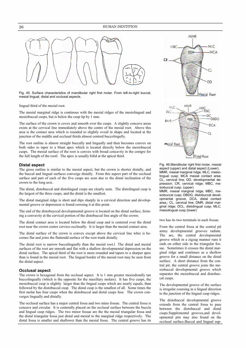
26 HUMAN DENTITION
Fig. 46.Mandibular right first molar, mesial aspect (upper) and distal aspect (Lower). MMR, mesial marginal ridge; MLC, mesio-lingual cusp; MCA mesial contact area; CL, cervical line; DD, developmental de-pression; CR, cervical ridge; MBC, me-siobuccal cusp. (upper) MMR, mesial marginal ridge; MBC, me-siobuccal cusp; DBDG, distobuccal devel-opmental groove, DCA, distal contact area,; CL, cervical line; DMR, distal mar-ginal ridge; DCL, distolingual cusp; MLC, mesiolingual cusp (lower)
lingual third of the mesial root.
The mesial marginal ridge is continous with the mesial ridges of the mesiolingual and
mesiobuccal cusps, but is below the cusp tip by 1 mm.
The surface of the crown is covex and smooth over the cusps. A slightly concave areas
exists at the cervical line immediately above the centre of the mesial root. Above this
area is the contact area which is rounded to slightly ovoid in shape and located at the
junction of the middle and occlusal thirds almost centred buccolingully.
The root outline is almost straight buccally and lingually and then becomes convex on
both sides to taper to a blunt apex which is located directly below the mesiobuccal
cusps. The mesial surface of the root is convex with broad concavity in the centgre for
the full length of the rootl. The apex is usually bifid at the apical third.
Distal aspect The gross outline is similar to the mesial aspect, but the crown is shorter distally, and
the buccal and lingual surfaces converge distally. From this aspect part of the occlusal
surface and part of each of the five cusps are seen due to the distal inclination of the
crown to the long axis.
The distal, distobuccal and distolingual cusps are clearly seen. The distolingual cusp is
the largest of the three cusps, and the distal is the smallest.
The distal marginal ridge is short and dips sharply in a cervical direction and develop-
mental groove or depression is found corssing it at this point.
The end of the distobuyccal developmental groove is located on the distal surface, form-
ing a convavity at the cervical portion of the distobuccal line angle of the crown.
The distal contact area is located below the distal cusp and is centered over the distal
root near the crown center cervico-occlusally. It is larger than the mesial contact area.
The distal surface of the crown is convex except above the cervical line whre is be-
comes flat and joins the flattendd surface of the root trunk distally.
The distal root is narrow buccalingually than the mesial root.l The distal and mesial
surfaces of the root are smooth and flat with a shallow developmental depression on the
distal surface. The apical third of the root is more rounded and tapers to a sharper apex
than is found in the mesial root. The lingual border of the mesial root may be seen from
the distal aspect.
Occlusal aspect The crown is hexagonal from the occlusal aspect. It is 1 mm greater mesiodistally tan
buccolingually (which is the opposite for the maxillary molars). It has five cusps, the
mesiobuccal cusp is slightly larger than the lingual cusps which are nearly equals, than
followed by the distobuccal cusp. The distal cusp is the smallest of all. Some times the
first molar has four cusps when the distobuccal and distal cusps fuse. The crown con-
verges lingually and distally.
The occlusal surface has a major central fossa and two mino fossae. The central fossa is
concave and circular. It is centerally placed on the occlusal surface between the buccla
and lingual cusp ridges. The two minor fossae are the the mesial triangular fossa and
the distal triangular fossa just distal and mesial to the marginal ridge respectively. The
distal fossa is smaller and shallower than the mesial fossa. The central groove has its
Fig. 45. Surface characteristics of mandibular right first molar. From left-to-right buccal, mesial lingual, distal and occlusal aspects.
two has its two terminals in each fossae.
From the central fossa at the central pit
amny developmental grooves radiate.
The are, the central developmental
groove which in a zigzag manner runs it
ends on either side in the triangular fos-
sae. Sometimes it crosses the distal mar-
ginal ridge and continues as a shollow
groove for a small distance on the distal
surface. A short distance from the cen-
tral pit, the central groove joins the me-
siobuccal developmental groove which
separates the mesiobuccal and distobuc-
cal cusps.
The developmental groove of the surface
is irregular coursing in a lingual direction
to the junction of the lingual cusp ridges.
The distobuccal developmental groove
extends from the central fossa to pass
between the distobuccal and distal
cusps.Supplemental grooves,and devel-
opmental pits may also found on the
occlusal surface.Buccal and lingual sup-

27 TOOTH MORPHOLOGY & PHYSIOLOGY
Fig. 47. Mandibular right first molar, occlu-sal aspect. DBCR, distobuccal cusp ridge; DBDG, distobuccal developmental groove; DCR, distal cusp ridge; DTF, distal triangular fossa (shaded area), DLCR, distolingual cusp ridge; LDG, lingual developmental groove; MLCR, mesiolingual cusp ridge; MTF, mesial triangular ridge (shaded area); SG, supplemental groove; MBCR, mesiobuccal cusp ridge, MBDG, me-siobuccal developmental groove (upper) Central fossa (shaded area); CP, central pit; DMR, distal marginal ridge; DP, distal pit; CDG, central developmental groove; MP, mesial pit; MMR, mesial marginal ridge, (lower)
plimantal grooves join the terminal of the
central groove in the mesial and distal
triangular fossae.
Pulp cavity In buccolingual section the pulp chamber
is wide with prominent pulp horns.Some
times the pulp chamber is quite deep
with the floor extending well down.The
mesil root may show broad root canal
(but very thin mesiodistally) which be-
comes narrow at the apical end of the
root to a pointed apical foramen.Mor
likely this root present two separate ca-
nals which join in a common apical
opening,or the two canels open in two
separated apical foramina.
In mesiodiscatl section the pulp cham-
ber is wide and accommodate pulp
horns.The mesial root show considerable
curvature which contains the most con-
stricted canal mesiodistally. The distal
root presents shorter, rounder and more
open root canal.
In cross section at the cervical
part of the pulp chamber is rec-
tangular in shape.
Mandibular Second
Molar The Second molar supplements the first
molar in function and its anatomy differs
in the following details.
The second molar is slightly smaller than the first molar in all dimensions, although a second molar with larger crown or longer root may be found.
The crown has four well developed cusps, two buccal and two lingual of nearly equal development. The distobuccal cusp is larger than the same one of the first molar.
The tooth has two well developed roots,one mesial and distal. They are less wide buccalingually than the roots of the first molar and they are not so widely separated.
The differences between the second
and first molars are as follow:-
■ Buccal aspect
The crown is some what shorter cervico-occlussally and narrower mesiodistally than the first molar.
The buccal cervical ridge is less prominent.
There is one development groove, the buccal developmental groove between the mesiobuccal and dis-tobuccal cusps.
The roots may be shorter and closer together and their axes are parallel. They are inclined distally in relation to the occlusal plane of the crown than is found on the first molar. The roots may be used for all or part of their length.
■ Lingual aspect.
The crown and root of the second molar converge far less lingually than the first molar.
The two lingual cusps, mesiolin-gual and distolingual are nearly the same size.
The lingual developmental groove crosses from the occlisal surface onto the ligual durface and fades out near the junction of the middle and occlusal thirds,between the lingual cups.
The curvatures of the contact area are more noticeable from the lin-gual aspect. They are at slight lower level specially the distal
Fig. 48. characteristics of pulp cavity of lower first molar.
Fig. 49. Surface characteristics of mandibular left second molar. From left-to-right buccal, mesial lingual, distal and occlusal aspects.
Table XXI. Chronology of maxillary second molar
Dental Organ Appearance 1Y
Start Calcification 2.5-3 Y
Enamel Completed 7-8 Y
Eruption 11-13 Y
Root Completed 14-15 Y

28 HUMAN DENTITION
contact area than those of the first molar.
The cervical line is irregular me-siodistally.
■ Mesial aspect
It is Rhomboidal in shape with lin-gual tilt.
The buccal cervical ridge is less pronounced.
The occlusal surface is more con-stricted buccolingally.
The mesial root is less broad and some what pointed apically.
■ Distal aspect
There is no distal cusp and disto-buccal groove.
The distal root is less broad.
The contact area is therefore cen-tered on the distal surface buc-coligually as well as cervico-occlussaly.
■ Occlusal aspect
The occlusal aspect of the second molae differs considerably from the first molar. The outline form of the occlusal surface is rectangular wider mesiodistally than buccolingually.
The second molar exhibit more curvature of the outline of the crown distally than mesially show-ing a semicircular outline of the
Fig. 50. Characteristics of pulp cavity of lower second molar. From left-to-right buccal , mesial, mesial variation and distal sections
B L
D M
L
B
disto-occlusal surface in compari-son with a square outline mesially.
The occlusal surface shows four cusps. The distobuccal cusp is less pronounced than the mesiobuccal cusp. There is no distobuccal de-velopmental groove buccally or occlusally.The distal cusp is absent. The lingual cusps are equal.
The buccal and lingual development grooves meet the central groove at right angles at the central pit,so forming a cross dividing the crown into four nearly equal parts. This groove is known as crossi form
fissure.
There are major central fossa and two minor fossae ( the mesial and distal triangular fossae). Three pits are present, a mesial a distal, and a central.
There are more secondary grooves than on the first molar so the occlu-sal surface is wrinkled.
Pulp cavity.
As for the mandibular firs molar, except that the root canals ap-peared shorter, straighter with less spread.
The cervical cross section is not as square as the first molar as the crown tapers distally in the second molar.
Mandibular Third Molar The mandibular third molars are ex-tremely variable in size and shape of both crown and root portions.
There are two basic forms:
Type I resembles the permanent second molar 50%. It has four cusps and the same general occlu-sal pattern and contour. Ordinarily the size is the same, but range from very small larger than any other molar.
Type II resembles the permanent first molar (40%) with five cusps and a similar occlusal pattern and contours. In 10% of cases it has more than five or less than four cusps.
The roots are extremely variable in size,number and curvature.Single fused roots are common.Most often,root length is less than other mandibular molars regardless of the crown size. The most common root torm reveals two short curved roots.
Pulp cavity. If it is well developed and is comparable to the second molar in shape the pulp cavity will resemble the second mo-lar.However,if it differs,it follows the general outline of the tooth.
Fig. 51. Surface characteristics of mandibular right third molar. From left-to-right buccal, mesial lingual, distal and occlusal aspects.
POLYPHYODENT A polyphyodEnt is an animal whose
teeth are continuously replaced. Most
toothed fishes are polyphyodonts.
Sharks replace teeth throughout their
life. They have teeth arranged in rows.
When a tooth is lost it is replaced by
the one next to it. Sharks may have
35000 to 50000 tooth in lifetime.
Reptiles and most other vertebrates are
also polyphyodonts, but very fewmam-
mals are polyphydent.

29 TOOTH MORPHOLOGY & PHYSIOLOGY
DESCRIPTION OF THE
DECIDUOUS DENTITION
T he deciduous teeth or primary,milk,baby and temporary teeth are twenty in num-ber,10 in each jaw ( 4 incisors, 2 canines and 4 molars).
They start to erupt at the age of six monthe by the apperarence of the first deciduous mandibular incisors in the oral cavity.At about 2 years old,the last deciduous tooth which is the second molar,starts to apperare.At three year of age all the deciduos teeth are erupt and are in function.
As the term “deciduous” implies, teeth are shed in order to make way for their perma-nent successors.The process of exfoliation takes place between the sixth and twelfth years.One to two years after complete root formation resorption begins at the root apex of the deciduous teeth and continues cervical until resorption of the entire root has taken place and the crown is lost from lack of support.
The anterior deciduous teeth are replaced by the anterior permanent teeth, while the deciduous molars are replaced by the permanent premolars.
The life span of all the deciduous teeth in the oral cavity are from 6 months to 12 years of age, therefore, from 6 months to 6 years the oral cavity contains only deciduous teeth.
From 6-12 years, the oral cavity contains mixed dentition (deciduous and permanent). From 12 years on, the oral cavity contains only permanent teeth.
Deciduous Anterior Teeth Maxillary central incisor The deciduous central incisor closely resembles its permanent successor except for the following:-
It is smaller than the permanent.
Its crown shows greater mesiodistal width than the cervico incisal length which is the opposite for the permanent. The labiolingual dimension at the cervical third is only 1mm. less than cervicoincisally.
The lingual surface shows well developed marginal ridges and cingulum.
The cervical ridges of enamel are pronounced on the cervical third of the crown both labially and lingually.
From the proximal aspect, the crown appears thick in relation to its total length.
From the incisal aspect, the crown appears much wider mesiodistally than labiolin-gually.
The root length is greater in comparison with the crown length than that of the perma-nent. A lingual ridge is evident on the full length of the root. On the mesal and distal surfaces of the root, there are central development depression for about the whole length of the root.
Maxillary lateral incisor It is similar to the central incisor with some variation.
The crown is smaller in all dimention.
The cervico –incisal length of the crown is greater than its mesiodistal width.
The root is similar in shape to the central incisor but it is much longer in proportion to its crown.
Mandibular central incisor They are similar to their successors except that:
They are much smaller.
The crown is wide in proportion to its length in comparison with the permanent.
The mesial and distal sides of the crown taper evenly from the contact areas.
From the mesial aspect, the incisal ridge is centered over the root. The labial and lin-gaual cervical contours are quite convex, much more than those of the permanent mandibular incisors.
The root is almost twice the crown length and evenly tapered down to a pointed apex. It is very narrow and conical in shape.
Fig. 52. Surface characteristics of primary maxillary and mandibular anterior teeth. Lingual aspect (upper), lingual aspect (middle) and mesial aspect lower.

30 HUMAN DENTITION
From the incisal aspect the incisal ridge bisects the crown labiolingually unlke perma-nent mandibular central incisors.
Mandibular lateral incisor The fundamental outlines of the deciduous mandibular lateral incisors and similar to thoseoyu of the deciduous mandibular central except for:-
It is somewhat larger in all measurements except labiolingually where the two teeth are identical in measurement.
The cingulum,marginal ridges and lingual fossa are more developed.
The incisal ridge slopes downwards distally, and its distal margin is more rounded.
Maxillary canine It is similar to the permanent maxillary canine except for:-
It is small in size.
The crown is more constricted at the cervix in relation to its mesiodistal width and more conves on its mesial distal surfaces.
The contact areas placed at about the same level nearly at the center of the crown
cervico – incisally.
The cusp of the deciduous canine is much longer and shaper than the permanent. The mesial slope of the cusp is longer than the distal slope which is the opposite for the permanent.
The tip of the cusp is more distal in relation to a line bisecting the crown mesiodis-tally.
The root is about twice as long as the crown and more slender than that of its perma-nent successor.
Mandibular canine It is similar to the permanent mandibular canine except for;
It is smaller.
The mesial slope of the cusp is shorter than the distal slope. This is true for both the permanent canines and premolars except the maxillary first premolar.
It differs from the deciduous maxillary canine in the following:
It is thicker at the neck of the tooth.
The cervical ridge labially and the cingulum lingually are not quite pro-nounced.
The mesial cusp slope is shorter than the distal cusp slope.
Deciduous Posterior Teeth Maxillary first molar This tooth presents four cusps mostly and three roots.The mesiolingual cusp is the larg-est and the distolingual cusp is the smallest which may be even absent resulting in a three cusped molar.The first molar is much smaller thatn the deciduous maxillary sec-ond molar.
Buccal aspect It present narrow cervical portion of crown and root than that of the same portion
of the permanent molars.
At the contact areas mesially and distally, the crown shows its widest measure-ments. The measurement at the cervix is less than that at the contact areas mesio-distally by about 2mm.
The occlusal margin is slight scalloped, but with no definite cusp form.
The buccal surface is smooth with a slight evidence of developmental grooves.
The furcation of the roots begins almost immediately at the site of cervical line. This characteristic is not possessed by the permanent molars.
The roots are slender and long and spread widely. All three roots are seen from this aspect. The mesial root is longer than the distal one.Each of the three roots has a single root canal.
The buccal cervical ridge is very developed particularly mesially.
Lingual aspect The crown converges considerably in a lingual direction.
The outline lingually is similar to the buccal aspect.
The mesiolingual cusp is the longest and sharpest, while the distolingual cusp is poorly defind or even absent. The distobuccal cusp may be seen from this aspect.
All three roots are in view and the lingual one is the longest.
Fig. 53. Surface characteristics of primary anterior teeth, incisal aspect.
Mesial aspect The buccolongual dimestion of the
crown at the cervical third is greather than the same dimention at the occlusal third.This characteristic is true of all molar teeth,but it is more evident on the deciduous teeth (the crown is con-stricted at the occlusal third).
The pronounced convexity of the buc-cal cervical ridge is a outstanding char-acteristic of this tooth.
The mesiolingual cusp is shaper and longer in size than the mesiobuccal cusp.
The cervical line is curved slightly toward the occlusal surface.
The mesiobuccal and lingual roots are the only visible from this aspect.The root is long,slender and extends lin-gually to a marked degree then curves sharply in a buccal direction above the meddle third.
Distal aspect The crown appears narrower distally
than mesially.
The distobuccal cusp is long and sharp, while the diatolingual cusp is poorly developed.
The buccal cervical ridge seen from the mesial aspect does not continue dis-tally.
The cervical line may curve occlusally or may run straight.
The distobuccal and lingual roots are seen this aspect as well as the apical part of the mesiobuccal root.
Occlusal aspect It is nearly rectangukar in shape with
the shortest sides representing the proximal sides.(mesxial and distal)
The crown outline converges lingual and distally.
The occlusal surface shows a sulcus with its central groove connecting a central fossa and a mesial triangular fossa.
A distal development grove connects the central fossa and the distal triangu-lar fossa.
The mesial and distal triangular fossae are just inside the meaisl and distal marginal ridges with a mesial and distal pit in them.

31 TOOTH MORPHOLOGY & PHYSIOLOGY
Supplimantal grooves radiate from the mesial and distal pits, one to abuccal, and one to a lingual directions.A third supplemental groove extends from the pit toward the marginal ridge and may extend over it.
This tooth may show an oblique ridge connecting the mesiolingual and disto-buccal cusps. This ridge may be ill defined and the central developmental groove extends from the mesila pit to the distal developmental groove.
The distal margine ridge is thin and poorly developed in comparison with the mesial marginal ridge.
There is a well defined buccal develop-mental groove dividing the mesio buc-cal and distobuccal cusp occlusally.
Maxillary second molar It resembles the masillary first perma-nent moloar that it is smaller in size.
Buccal aspect This is toothe shows well defined buc-
cal cusp separated by a buccal develop-mental groove.
The crown is narrower at the cervic in comparison with its mesiodistal meas-urement at the contact areas.
The roots appear slender and much longer and heavier relatively than those of the permanent maxillary first molar.
The bifurcation of the roots is close to the cervical line.
Lingual aspect There is lingual convergence of the
crown and root.
It shows two well developed cusps,the mesiolingual and the distolingual cusps are separated by a short lingual devel-opment groove.
A supplemental poorly developed to the large mesiolingual cusps and is outlined by developmental groove lin-gually.
The cervical line is nearly straight.
All three roots are visible from this aspect; the ligual root is larger and thicker than the two buccal roots.
Mesial aspect The outline resembles that of the per-
manent molars.
The crown appears short because of its thickness buccolingually in comparison with its length.
The mesiolingual is cusp larger than the mesiobuccal cusp.
The lingual root extends lingually be-yond the crown outline.The point of bifurcation between the mesiobuccal and lingual roots is 2-3mm. Apical to the cervical line.
The curvature at the cervical portion is pronounced lingually,but it resembles that of the permanent maxillary first molar buccally.In this,it differs entirely from the prominent curvature found on the deciduous maxillary first molar at the cervical third buccally.
Distal aspect It resembles that of the permanent maxil-lary first molar.
Occlusal aspect It is somewhat rhomboidal and
resembles the permanent maxillary first molar except that the buccal surface is rather flat.
The developmental grooves be-tween the cusps are less marked than that found on the permanent molar.
Mandibular first molar It varies much from all the teeth and appears strange and primitive. It pos-sesses four cusps and two roots.
Buccal aspect It shows two cusps separated by devel-
opmental depression instead of groove.The mesiobuccal cusp is larger than the distobuccal cusp.
The mesial outline of the crown is straight from the convex contact area to the cervix constricting the crown very little at the cevix.
The distal outline converges toward the contact are extend distally to a marked degree.
The distal portion of the crown is shorter than the mesial portion because of dipping of the cervical line apically over the mesial root.
There is a cervical enamel buccally (buccal cervical ridge) which is over development mesially. This makes the cervical line dips downward in order to outline this ridge.
The two roots are long, slender and they spread greatly at the apical third beyond the outline of the crown.The distal root is shorter than the mesial one and their bifurcation is very close to the cervical line.
Lingual aspect The crown and root converge lingually
to a marked degree on the mesial sur-face, while the opposite is true distally.
The mesiolingual cusp is long and sharp,while the distolingual cusp is rounded with alingual developmental groove separating them.
The mesial marginal ridge is well de-veloped to the extent that it might al-most considered as another small cusp lingually.
Fig. 55. Maxillary primary molars, charac-teristics of occlusal surface, first molar (upper) and second molar (lower|). MPC, mesiobuccal cusp; MTF, mesial triangular fossa; MP, mesial pit; CP, central pit; MLC, mesiolingual cusp; DLC, distolin-gual cusp; DTF, distal triangular fossa; DDG, distal developmental groove; BDG, buccal developmental groove; DBC, disto-buccal cusp; CDG, central developmental groove; FC, fifth cusp; LDG, lingual devel-opmental groove; OR, oblique ridge
Fig. 54. Surface characteristics of primary molars, buccal aspect (left), lingual aspect

32 HUMAN DENTITION
The crown length mesially and distally is nearly equal.
The cervical line is nearly straight.
Mesial Aspect It is rhomboidal in outline.
The buccal outline shows prominent conxity representing the cervical ridge, then it flattens to the cusp tip.
The buccal cusp is placed over the root base while the lingual outline of the crown extends out lingually beyond the confines of the root base.This design places the cusps in a favourable posi-tion for proper occlusion with the upper molars.
The mesiobuccal cusp and the mesio-lingual cusp as well as the well devel-oped mesial marginal ridge are seen.
The cervical line extends upward in a buccolingual direction.
The buccal and lingual outlines of the root drop straight down from the crown approximately parallel each other for over half the root length them taper at the apical third to a square end.
A developmental depression in usually extends almost through the full length of the root.
Distal aspect The crown has an equal length buccally
and lingually and the cervical line is straight.
The distal cusps are shorter and less sharper than the mesial cusps.
The distal marginal ridge is less devel-oped than the mesial marginal ridge.
There is less curvature at the cervical third (no prominent buccal cervical ridge).
The distal root is rounder, shorter and taper more apically.
Occlusal aspect. It is rhomboidal in outline.
All four cusps are seen, the mesiolin-gual cusp is the largest.
The prominence mesiobuccally (C.R) is noticeable from this aspect.
From a central pit, the following grooves are seen,
A buccal developmental groove extends buccally separating the two buccal cusps. It does not ex-tend on the buccal surface.
A central developmental groove extends mesially ending in mesial pit in the mesial triangular fossa and extends distally ending in a distal pit in the distal triangular fossa.
A lingual developmental groove extends lingually separating the two lingual cusps. It does not extend to the lingual surface.
There are supplemental grooves radiating from the mesial and dis-tal pits buccally and lingually.
Mandibular secons
molar It resembles the permanent mandibular
first molar except for the following.
It is smaller insize.
The deciduous molar present narrow mesiodistal clibration at the cervix than does the permanent molar.
The mesiobuccal and distobuccal de-velopmental grooves divide the buccal surface of the crown occlusally into three cuspal protions almost equal in size (mesiobuccal and distobuccal and distal cusps)
The mesial portion of the crown seems to be little higher than the distal portion of the crown lingually which gives the impression of being tipped distally.
There is a prominent buccal cervical ridge,then the buccal surface flattened occlusal to the ridge.
Proximally, the crown is rhomboidal in outline.
The mesial marginal ridge is high,while the distal marginal ridge dips down more sharply and is shorter buccolin-gually than the mesial marginal ridge.
The grown is norrower buccolingually in comparison with its mesiodistal measurement than is the permanent tooth.
The root flares out greatly mesiodis-tally at the middle and apical thirds.They are twice as long as the crown.Ther bifurcation starts at a point immediately below the cervical line.
The roots are more slender than those of the permanent molars.
The occlusal surface shows a rectangu-lar outline that converges lingually and distally.
The major differences between the de-
ciduous and permanent teeth
The crowns of the deciduous teeth are lighter in color than are the permanent teeth.
The deciduous teeth are smaller in size than the permanent.
The crowns of the deciduous teeth are more bulbous.
The crowns of the deciduous anterior teeth are wider mesiodistally in com-parison with their crown length than are the permanent.
The crown widths of the deciduous molars are larger in all directions in comparison with the root trunks and cervices. The crown and roots are lender at thr “neck” mesiodiswtally than are those of permanent molars.
The buccal and lingual surfaces of
deciduous molars taper occlusally
above the cervical curvature much
more than do the permanent molar
surfaces. This results in a much nar-
rower occlusal table of the occlusal
surface buccolingually.
The buccal cervical ridge of enamel is
quite prominent on the facial aspect of
the deciduous anteriors and molar teeth
(especially on the first molars).
The roots of the deciduous anterior
teeth are narrower and longer in com-
parison with crown length, as well as
tooth length and width, than are the
permanent teeth roots.
The roots of the deciduous molars are
relatively longer and more slender than
the roots of the permanent teeth. They
are more divergent and flare more ex-
tending out beyond projected outlines
of the crown. The roots thin out rapidly
as the apices are approached.
In cross section, the deciduous teeth
shows the following differences:
The enamel is relatively thin and has a
consistent depth.
The dentin thickness between the pulp
chamber and the enamel is much less
than in the permanent teeth.
The pulp chambers are relatively large,
and the pulp horns are high occlusally
placing them much closer to enamel
than the permanent teeth.
Fig. 56. Mandibular primary molars, char-acteristics of occlusal surface, first molar (upper) and second molar (lower). MBC, mesiobuccal cusp; CDG, central develop-mental groove; DBC, distobuccal cusp, BDG, buccal developmental groove, CP, central pit;; DLC, distolingual cusp; LDg, lingual developmental groove; MLC, mesi-olingual cusp; MP, mesial pit; MBC, me-siobuccal cusp; DBDG, distobuccal devel-opmental groove; DP, distal pit; MBDG, mesiobuccal developmental groove; DC distal cusp.