accepted date : 18-mar-2015 article - dott. riccardo bono article this article has been accepted for...
TRANSCRIPT

Acc
epte
d A
rtic
le
This article has been accepted for publication and undergone full peer review but has not been through the copyediting, typesetting, pagination and proofreading process, which may lead to differences between this version and the Version of Record. Please cite this article as doi: 10.1111/bjd.13861 This article is protected by copyright. All rights reserved.
Accepted Date : 18-Mar-2015 Article type : Clinical and laboratory investigations
Pigmented nodular melanoma: The predictive value of dermoscopic
features using a multivariate analysis
Running head: Pigmented nodular melanoma
M.A. Pizzichetta,1 H. Kittler,2 I. Stanganelli,3 R. Bono,4 S. Cavicchini, 5 V. De Giorgi, 6
G. Ghigliotti,7 P. Quaglino, 8 P. Rubegni, 9 G. Argenziano10 and R. Talamini 11 for the Italian
Melanoma Intergroup (IMI)*
Author Affiliations:
1 Division of Medical Oncology – Preventive Oncology, Centro di Riferimento Oncologico, National Cancer Institute, Aviano, Italy
2 Department of Dermatology, University of Vienna, Vienna, Austria 3 Skin Cancer Unit, Istituto Tumori Romagna (IRST), Meldola, Italy 4 Istituto Dermopatico Immacolata, IRCCS, Roma
5 Dept. of Dermatology, Fondazione Ospedale Maggiore Policlinico IRCCS, Milan, Italy 6
Dept. of Dermatology , University of Florence, Italy 7 Clinic of Dermatology, IRCCS San Martino- Ist, Genova, Italy 8
Dermatologic Clinic, Dept Medical Sciences, University of Torino, Italy 9
Dept. of Dermatology, University of Siena, Italy 10 Skin Cancer Unit, Arcispedale Santa Maria Nuova IRCCS, Reggio Emilia, Italy)
11 Unit of Epidemiology and Biostatistics, Centro di Riferimento Oncologico, National Cancer Institute, Aviano, Italy
*Members of the Italian Melanoma Intergroup (IMI): D. Guardoli (Skin Cancer Unit, Arcispedale Santa Maria Nuova IRCCS, Reggio Emilia, Italy) M. Alaiback (Dept. of Dermatology , University of Padova, Italy) S. Astorino (Div. of Dermatology, Celio Hospital, Rome, Italy) F. Ayala (National Cancer Institute, IRCCS, Naples, Italy) M T. Corradin (Div. of Dermatology, Pordenone Hospital, Italy) M. Gonzales, P. Zampieri (Div. of Dermatology, Merano Hospital, Italy) S. Magi, L. Mazzoni (Skin Cancer Unit, Istituto Tumori Romagna - IRST, Meldola, Italy) S. Seidenari, G. Pellacani (Dept. of Dermatology , University of Modena and Reggio Emilia, Modena, Italy) F. Specchio (Dept. of Dermatology ,University Tor Vergata, Roma, Italy) D. Serraino (Unit of Epidemiology,Centro di Riferimento Oncologico, National Cancer Institute, Aviano, Italy H.P. Soyer (Dermatology Research Centre, The University of Queensland, School of Medicine, Princess Alexandra Hospital, Brisbane, Australia Corresponding author: Maria Antonietta Pizzichetta, MD, Division of Medical Oncology –
Acc
epte
d A
rtic
le
This article is protected by copyright. All rights reserved.
Preventive Oncology, Centro di Riferimento Oncologico – IRCCS, Via Franco Gallini, 2 33081 Aviano (Italy); Tel. +39-0434-659611; Fax: +39 / 0434 / 659840; E-mail: [email protected] Funding Sources Statement
On behalf of all the authors, I, (Dr. Maria A. Pizzichetta) hereby declare that I had no funding sources to support this work. Conflict of interest Statement None declared.
Bulleted Statements:
What’s already known about this topic?
• Nodular melanoma (NM) often exhibits features associated with deep tumor
extension and less commonly displays the classic dermoscopic features of
superficial spreading melanoma (SSM).
What does this study add?
• The study identifies dermoscopic features which are significantly associated with
pigmented NM compared to pigmented SSM and non-melanoma nodular lesions.
• The authors validates with a multivariate analysis the dermoscopic features leading to
a significant increased likelihood of a diagnosis of pigmented NM.
Summary
Background Nodular Melanoma (NM), representing 10%-30% of all melanomas, plays a
major role in global mortality related to melanoma. Nonetheless, the literature on dermoscopy
of NM is scanty.
Objectives Assessment of odds ratios (ORs) to quantify dermoscopic features of pigmented
NM versus pigmented superficial spreading melanoma (SSM), and pigmented nodular non
melanocytic and benign melanocytic lesions.
Acc
epte
d A
rtic
le
This article is protected by copyright. All rights reserved.
Methods Digitized images of 457 pigmented skin lesions from patients with
histopathological diagnosis of NM (75), SSM (93), and nodular non melanocytic and benign
melanocytic lesions (289), (i.e., 39 basal cell carcinoma, 85 seborrheic keratosis, 81 blue
nevi, and 84 compound/dermal nevi), were retrospectively collected and blindly evaluated by
three observers to assess the presence or absence of global patterns and dermoscopic criteria.
Results The multivariate analysis showed that ulceration (OR, 4.07), homogeneous
disorganized pattern (OR, 10.76), and homogeneous blue pigmented structureless areas (OR,
2.37) were significantly independent prognostic factors for NM vs SSM. The multivariate
analysis of dermoscopic features of NM vs non melanocytic and benign melanocytic lesions
showed that the positive correlating features leading to a significant increased NM risk were:
asymmetric pigmentation (OR, 6.70), blue-black pigmented areas (OR, 7.15), homogeneous
disorganized pattern (OR, 9.62), a combination of polymorphous vessels and milky red
globules/areas (OR, 23,65) and polymorphous vessels combined with red homogeneous areas
(OR, 33.88).
Conclusions Dermoscopy may be helpful in improving the recognition of pigmented NM by
revealing asymmetric pigmentation, blue-black pigmented areas, homogeneous disorganized
pattern, and abnormal vascular structures including polymorphous vessels, milky red
globules/areas and red homogeneous areas.
Introduction
Nodular melanoma (NM) represents 10 - 30% of all melanomas and nearly 50% of all
melanomas thicker than 2 mm, and it plays a major role in the global mortality related to this
cancer.1 Unlike other melanoma subtypes, NM appears to lack the initial radial growth phase,
beginning with vertical growth. 2
Acc
epte
d A
rtic
le
This article is protected by copyright. All rights reserved.
The “ABCD” warning signs for melanoma are better at detecting superficial spreading
melanoma (SSM) than NM, as the latter is often small in diameter, symmetric, with regular
borders and less colour variegation, and it is frequently amelanotic.1,3 For this reason, the
EFG rule (Elevation, Firmness on palpation, continuous Growth over 1 month), summarizing
the most frequent features of NM, has been introduced for the identification of this subtype of
melanoma.4 Dermoscopically, in a study of 10 NM lesions, an asymmetric color and pattern
distribution was observed in all lesions; in addition, all lesions exhibited at least 3 colours,
though the number of colors and structures was significantly lower in the NM group than in
the SSM group.5 In a study of 11 thin NM, most lesions had a homogeneous disorganized
asymmetric pattern or a featureless pattern; although many dermoscopic features seen in SSM
were frequently absent, some features such as a blue-white veil, structureless areas, atypical
vessels and pink veil were often identified.4 In a large series of NM, Menzies et al6 found that
pigmented NM compared with non-nodular invasive melanoma, more frequently displayed a
symmetrical pigmentation pattern, large-diameter vessels, areas of homogeneous blue
pigmentation, symmetrical shape, predominant peripheral vessels, blue-white veil, pink
colour, black colour, and milky red/pink areas.
In this study, 457 pigmented skin lesions, including 75 NM, were evaluated dermoscopically
to examine the predictive value of dermoscopic features of NM using a multivariate analysis.
Patients and Methods
Between January 2007 and December 2011, all consecutive cases of histopathologically
confirmed pigmented NM, pigmented SSM, pigmented nodular benign melanocytic lesions
(e.g. compound /dermal nevus, blue nevus), and pigmented nodular non-melanocytic lesions
(e.g. seborrheic keratosis, basal cell carcinoma) seen at the 15 participating Italian centers,
were collected for this study, with the aim to identify dermoscopic features which were

Acc
epte
d A
rtic
le
This article is protected by copyright. All rights reserved.
significantly associated with pigmented NM compared to pigmented SSM and non-melanoma
nodular lesions. In this study only “pigmented” lesions were considered, that were defined as
those having black, dark brown, gray, or blue structures that occupied more than 25% of the
total surface area of the lesion.7
NM was defined as an invasive melanoma that lacks significant intraepidermal tumor cells
beyond the margins of the dermal invasive component. 6
There were no SSM with a nodular component in our series of NM as well as no in situ
melanoma in the non-nodular melanoma set.
Two separate files were provided for each case, one containing the dermoscopic images and a
second one containing all patient-related information such as gender, age at diagnosis, skin
lesion site, type of dermoscopy (polarized or non-polarized), date of excision, clinical
diagnosis, dermoscopic diagnosis and histopathological diagnosis.
By the end of December 2011, all the dermoscopic images (N=560) from the 15 centers were
merged into a database at the Epidemiology and Biostatistics Unit (R.T.) of the Centro di
Riferimento Oncologico, Aviano (Italy), with a new identification link to the patient
information on clinical features and diagnosis. Of the 560 submitted images, 457 were found
to be of sufficiently good quality for the evaluation of the dermoscopic criteria. Of these, 310
(67.8%) were taken with a camera using non-polarized dermoscopy and 147 (32.2%) with a
camera using polarized dermoscopy. All the dermoscopic images were examined to assess
the presence or the absence of global patterns and specific dermoscopic criteria in NM, non-
nodular melanoma, non-melanocytic and benign melanocytic lesions. We assessed the lesions
using the features reportedly associated with melanoma, basal cell carcinoma, seborrheic
keratosis, common nevi, and blue nevi;8-10 all features assessed were also considered in the
analysis and in the tables. All cases were evaluated by a panel of three blinded observers; the
dermoscopic features were scored based on the agreement of the two observers (M.A.P. and
Acc
epte
d A
rtic
le
This article is protected by copyright. All rights reserved.
R.B.) and in the case of disagreement between them, a third observer (I.S.) was consulted.
The evaluation of the dermoscopic criteria as well as the final dermoscopic diagnosis was
made when 3/3 or 2/3 observers agreed.
Statistical analysis
The statistical analysis was performed by means of the SAS 9.1 (SAS Institute Inc., Cary,
NCI) statistical software.11 Unconditional logistic regression models were used to assess odds
ratios (OR) and their corresponding 95% confidence intervals (CI) to quantify dermoscopic
features of pigmented NM versus pigmented non nodular melanoma and pigmented nodular
non melanocytic and benign melanocytic lesions. Variables that resulted statistically
significant at the univariate analysis were included in the multivariate model. In addition, the
Chi-square test or Fisher exact test was used, when appropriate, to evaluate differences in
clinical characteristics and melanoma thickness. Results were considered to be statistically
significant when p-values were less than or equal to 0.05 (two-sided).
Results
Patients demographics and classification of lesions
The diagnostic categories of the study lesions are shown in Table 1. Of 457 lesions, 75 were
NM, 93 were SSM and 289 were non-melanocytic (39 basal cell carcinoma and 85 seborrheic
keratosis) plus benign melanocytic lesions (81 blue nevi and 84 compound/dermal nevi). The
study included 457 patients (236 males, 221 females) with a median age of 51 years
(range:11-95) (Table 2). The sites of the skin lesions included: head and neck (107 cases);
anterior trunk (90 cases); back (129 cases); lower limbs (80 cases); and upper limbs (51
cases). The median ages were 61 years, (range 21-92), 57 years, (range 16-92), and 46 years
(range 11-95), for NM, SSM, and non-melanocytic and benign melanocytic lesions
respectively (Table 2).
Acc
epte
d A
rtic
le
This article is protected by copyright. All rights reserved.
No differences emerged between NM and SSM in the distribution by sex, age and sites of the
melanomas. By contrast, the median Breslow thickness of NM (3.40 mm, range: 0.05 -11.00)
was significantly greater than that of the SSM (0.80 mm, range: 0.01-5.00; p<0.0001) (Table
2).
Dermoscopic features of pigmented NM vs pigmented SSM
Table 3A and 3B show the univariate and multivariate analyses of the dermoscopic features
(positive 3A and negative 3B) of pigmented NM vs pigmented SSM. The univariate analysis
was used to identify the relevant factors (i.e. listed according to p value) in determining NM.
The multivariate analysis was used to estimate the independent effect of each factor which
had a significant result in the univariate analysis. The multivariate analysis showed that
ulceration, homogeneous disorganized pattern and homogeneous blue pigmented
structureless areas were significant independent prognostic factors for NM. The presence of
ulceration was 37.3 % in the NM and 10.8 % in the SSM group and led to a significantly
increased risk of diagnosing NM (OR, 4.07; 95% CI, 1.71-9.69). Moreover, significant risks
of diagnosing NM were also found when the lesion was characterized by the presence of
homogeneous disorganized pattern (OR, 10.76; 95% CI, 2.69 - 42.99) and homogeneous
blue pigmented structureless areas (OR, 2.37; 95% CI, 1.08 - 5.22) (Table 3A). Conversely,
when evaluating the negative features, the presence of peripheral light brown structureless
areas was 12% in NM and 38.7 % in the SSM group, leading to a significantly reduced risk of
NM (OR, 0.26; 95% CI, 0.11- 0.65) (Table 3B).
Acc
epte
d A
rtic
le
This article is protected by copyright. All rights reserved.
Dermoscopic features of pigmented NM vs pigmented nodular non melanocytic and
benign melanocytic lesions.
Tables 4A and 4B show the univariate and multivariate analyses of the dermoscopic features
(i.e. listed according to p value), of pigmented NM vs pigmented nodular non melanocytic
and benign melanocytic lesions. The multivariate analysis showed that the significant positive
correlating features, leading to a significant increased risk of NM, were asymmetric
pigmentation (OR, 6.70; 95% CI, 1.49 - 30.11), blue-black pigmented areas, (OR, 7.15;
95% CI, 1.54- 33.30), homogeneous disorganized pattern (OR, 9.62; 95% CI, 1.62- 57.13),
the combination of polymorphous vessels and milky-red globules/areas (OR, 23.65; 95% CI,
1.65 – 339.93), and the combination of polymorphous vessels and red homogeneous areas
(OR, 38.88; 95% CI, 1.72- 877.07) (Table 4A). By contrast, the homogeneous pattern was a
negative correlating feature, leading to a significant reduced risk of NM (OR, 0.05; 95% CI,
0.01- 0.22) (Table 4B) .
Discussion
Based on our results, a large number of features such as ulceration, homogeneous
disorganized pattern, homogeneous blue pigmented structureless areas, multiple (≥ 3)
colours, the combination of polymorphous vessels and milky red globules/areas and
symmetric shape were significantly more frequent in pigmented NM compared to pigmented
SSM only in the univariate analysis. When we compared our results with those of Menzies et
al6, they appeared in agreement in relation to the areas of homogeneous blue pigmentation
and symmetrical shape, which were positively correlated with NM in both studies, while
Menzies et al6 also found symmetrical pigmentation pattern, pink colour, blue-white veil and
black color positively correlated with NM. Regarding negative features, we found a large
number of features such as atypical network, multicomponent pattern, peripheral light brown
Acc
epte
d A
rtic
le
This article is protected by copyright. All rights reserved.
structureless areas, asymmetric shape, irregular streaks and the combination of linear
irregular, dotted and milky red globules/area that were negatively correlated with NM in
comparison with SSM only in the univariate analysis. When we compared our results with
those of Menzies et al6, the atypical network was the only feature in agreement. By contrast
in this study,6 other negative correlating features of pigmented NM were found such as
pigment network/pseudonetwork, multiple blue-gray dots (granularity), scarlike
depigmentation, irregular brown dots/globules, tan colour, irregular shape depigmentation,
and irregular dots/globules of any colour. The inconsistencies between the two studies could
depend on the different sample sizes of both NMs and SSMs and on different terminology
used to describe some features regarding the vascular pattern, the regression structures,
(including both multiple blue-gray dots (granularity) and scar-like depigmentation,) instead
of multiple blue-gray dots (granularity) or scar-like depigmentation, as well as the irregular
dots/globules, instead of irregular dots/globules of any color or irregular brown
dots/globules.
The most striking results in our study were that the only significant features distinguishing
pigmented NM from pigmented SSM in the multivariate analysis were ulceration,
homogeneous disorganized pattern, and homogeneous blue pigmented structureless areas.
The overall homogeneous pattern is typically exhibited by common and blue nevi, consisting
of a diffuse homogeneous tan, brown, or blue structureless pigmentation.12-13 By contrast, in
NM, in agreement with that reported by other authors, 4,14 the colours are distributed in a
disorganized and asymmetric fashion, characterizing the overall disorganized homogeneous
pattern ( Figure 1).

Acc
epte
d A
rtic
le
This article is protected by copyright. All rights reserved.
Regarding the homogeneous blue pigmented structureless areas, some authors use a unifying
definition of blue-white structures over raised areas associated with dense melanin within
melanocytes in the dermis;14 the uniform distribution of blue hue may be focal in the case of
homogeneous blue pigmented structureless areas or diffuse in the case of blue-whitish veil.15
( Figure 2).
Concerning negative features, the only significant negatively correlated feature of pigmented
NM in the multivariate analysis were peripheral light brown structureless areas, which are
already reported to be associated with thin melanoma.16
When comparing the univariate analyses of the positive features of pigmented NM vs
pigmented nodular non-melanocytic and benign melanocytic lesions in both our study as well
as that of Menzies and colleagues, 6 most of the features were in agreement, i.e. peripheral
black dots/globules, irregular black dots/globules, blue-white veil, pseudopods, homogeneous
blue pigmentation, multiple colors, black colour, irregular blotches, irregular dots/globules,
blue-black structures, and asymmetric shape. The differences between the two studies
concerned some positive correlating features found only in our study, such as linear irregular
vessels + milky red globules/areas, more than one shade of pink, red homogeneous areas,
asymmetric pigmentation pattern, multicomponent pattern, regression structures, ulceration,
homogeneous disorganized pattern, atypical network, shiny white structures, predominant
blue clods, featureless pattern, peripheral light brown structureless areas and irregular
depigmentation. Conversely, some positive correlating features found by Menzies and
colleagues 6 did not correspond to those evaluated in our study, such as pink color, abrupt
edge, blurred “out of focus” colours, red-blue colour, multiple brown dots and central black
dots/globules. Regarding negative features, the agreement emerged only for milia-like cysts
and comedo-like openings, while arborizing vessels, multiple blue globules, leaf like areas,
Acc
epte
d A
rtic
le
This article is protected by copyright. All rights reserved.
and large blue-gray ovoid nests, were negatively associated with NMs only in Menzies’study
6. The different terminology of some features as well as the different diagnostic categories of
lesions included in the two studies may explain the differences in the results between these
two different series; our series did not include, in relation to benign melanocytic lesions,
Spitz nevi and deep penetrating nevi, and in relation to non-melanocytic lesions,
hemangioma, dermatofibroma and other nodular lesions.
Asymmetric pigmentation, blue-black pigmented areas, homogeneous disorganized pattern,
the combination of polymorphous vessels and milky red globules/areas and polymorphic
vessels associated with red homogeneous areas were significantly more frequent in
pigmented NM compared to pigmented nodular non-melanocytic and benign melanocytic
lesions in the multivariate analysis. In agreement with our results, Argenziano et al 17 found
that blue-black pigmented areas, defined as a combination of structureless blue areas, black
dots/ globules, and blotches, involving at least 10% of the lesion surface, were significantly
associated with NM, (Figures 1,2,3). These areas correspond to different histopathologic
correlates, with blue area corresponding to pigmented melanocytes in the deep dermis and
black areas, arising from superficial intraepidermal melanin or a dense dermal proliferation of
pigmented melanocytes under a thinned epidermis that may predict ulceration.18 This could
also explain why we found ulceration to be significantly more frequent in NM compared to
SSM. Abnormal vascular structures, including polymorphic vessels, milky red globules/areas
and red homogeneous areas are also significant relevant features of NM which have been
reported to be associated with NM by other authors. 19 Polymorphous vessels are defined as
having more than one morphological type of vessel; the most frequent combination of vessel
types seen in melanoma is linear irregular and dotted vessels, as the thickness increases, the
vascular polymorphism increases with hairpin, linear coiled (glomerular), linear helical (cork-
Acc
epte
d A
rtic
le
This article is protected by copyright. All rights reserved.
screw like), and arborizing vessels (seemingly emerging from the dermal plexus of the
adjacent skin, possibly because vertical growth cannot be maintained by further elongation of
the capillary loops), associated with milky red globules/areas and/or red homogeneous areas (
Figure 4). 19 Milky red globules have been defined as unfocused large ovoid or polygonal
structures of pink-white colour, often showing a central linear irregular or corkscrew vessel,
separated from each other by blurred whitish lines, that may be seen within or near areas with
a milky red color, the so-called milky red areas, also knows as pink veil or more than one
shade of pink that probably correspond to areas with increased vascular volume (Figure 2 ).
19-20 The red homogeneous areas were seen in NM and in pyogenic granuloma as structureless
areas of red homogeneous colour covering the structures lying below ( Figure 3 ). 19,21The
milky red globules/areas as well as the red homogeneous areas probably represent an
increased vascular volume reflecting neoangiogenesis. 19-21
Dermoscopy may be helpful in improving the recognition of pigmented NM by revealing
asymmetric pigmentation, blue-black pigmented areas, homogeneous disorganized pattern,
and abnormal vascular structures including polymorphic vessels, milky red globules/areas
and red homogeneous areas. In conclusion, the abnormal vascular structures (Figure 4) as
well as the blue-black pigmented areas (Figure 1) may be the only clue for the correct
diagnosis of pigmented NM in examining asymmetrically pigmented lesions with a
homogeneous disorganized pattern.
Acknowledgements
The authors wish to thank Luigina Mei and Anna Maria Colussi for their editorial assistance
and Mauro Mazzocut for technical support.
Acc
epte
d A
rtic
le
This article is protected by copyright. All rights reserved.
References
1 Shaikh WR, Xiong M, Weinstock MA. The contribution of nodular subtype to melanoma mortality in the united states, 1978 to 2007. Arch Dermatol 2012; 148:30-36.
2 Clark WH Jr, From L, Bernardino EA, Mihm MC. The histogenesis and biologic behaviour of primary human malignant melanomas of the skin. Cancer Res. 1969; 29:705-727.
3 Chamberlain AJ, Fritschi L, Giles GG et al. Nodular type and older age as the most significant associations of thick melanoma in Victoria, Australia. Arch Dermatol 2002; 138: 609-614.
4 Kalkhoran S, Milne O, Zalaudek I et al. Historical, clinical, and dermoscopic characteristic of thin nodular melanoma. Arch Dermatol 2010; 146:311-318.
5 Segura S, Pellacani G, Puig S et al. In vivo microscopic features of nodular melanomas: dermoscopy, confocal microscopy, and histopathologic correlates. Arch Dermatol 2008; 144:1311-1320.
6 Menzies SW, Moloney FJ, Byth K, et al. Dermoscopic evaluation of nodular melanoma. Jama Dermatol 2013; 149:699-709.
7 Menzies SW, Kreusch J, Byth K, et al. Dermoscopic evaluation of amelanotic and hypomelanotic melanoma. Arch Dermatol 2008; 144:1120-1127.
8 Rao BK, Ahn CS. Dermatoscopy for melanoma and pigmented lesions. Dermatol Clin 2012; 30:413-434.
9 Altamura D, Menzies SW, Argenziano G et al. Dermatoscopy of basal cell carcinoma: morphologic variability of global and local features and accuracy of diagnosis. J Am Acad Dermatol 2010; 62:67-75.
10 Longo C, Scope A, Lallas A et al. Blue Lesions. Dermatol Clin 2013; 31:637-647.
11 Armitage P, Berry G, Matthews JNS. Statistical methods in medical research. 4th ed. Blackwell Science Publ, 2002.
12 Marghoob AA. Pattern analysis In: Atlas of Dermoscopy (Marghoob AA,
Malvehy J, Braun RP, eds). London: Informa Healthcare, 2012; 98-11.
13 Henning S, Dusza SW, Wang SQ et al. The CASH (color, architecture, symmetry, and homogeneity) algorithm for dermoscopy. J Am Acad Dermatol 2007; 56:45-52.
14 Rose SE, Argenziano G, Marghoob AA. Melanoma difficult to diagnose via Dermoscopy. G Ital Dermatol Venereol 2010; 145:111-26. 15 Massi D, De Giorgi V, Carli P, Santucci M. Diagnostic significante of the blue hue in dermoscopy of melanocytic lesions. Am J Dermatopathol.2001; 23:463-469.

Acc
epte
d A
rtic
le
This article is protected by copyright. All rights reserved.
16 Annessi G, Bono R, Sampogna et al. Sensitivity, specificity, and diagnostic accurancy of three dermoscopic algorithmic methods in the diagnosis of doubtful melanocytic lesions: the importance of light brown structureless areas in differentiating atypical melanocytic nevi from thin melanomas. J Am Acad Dermatol 2007; 56:759-67
17 Argenziano G, Longo C, Cameron A et al. Blue-black rule: a simple dermoscopic
clue to recognize pigmented nodular melanoma. British Journal of Dermatology 2004; 165:1251-1255
18 Longo C, Farnetani F, Moscarella E et al. Can non invasive imaging tools potentially predict the risk of ulceration in invasive melanomas showing blue and black colors? Melanoma Research 2013; 23:125-131 19 Zalaudek I, Kreusch J, Giacomel J et al. How to diagnose non pigmented skin tumors: A review of vascular structures seen with dermoscopy. Part 1. Melanocytic skin tumors. J Am Acad Dermatol 2010; 63:361-74 20 Marghoob AA, Braun R. Proposal for a recise 2-step algorithm for the classification of lesions of the skin using dermoscopy. Arch Dermatol 2010; 146:426-428. 21 Zaballos P, Carulla M, Ozdemir F et al. Dermoscopy of pyogenic granuloma: a morphological study. British Journal of Dermatology 2010; 163:1229-1237 Figure Legends
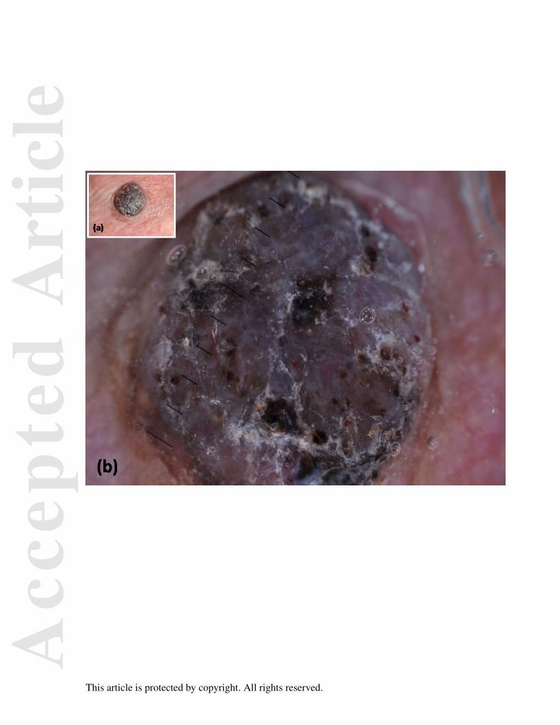
Fig 1. Clinical and dermoscopic image of a nodular melanoma, 2.4 mm thick on the cheek of
an 80-year-old woman. (a) In the clinical image (inset), a blue-grayish scaly symmetrical
nodule can be observed. (b) An overall homogeneous disorganized pattern consisting of a
diffuse homogeneous blue pigmentation plus foci of irregular black dots/globules and
blotches can be seen. The presence of additional features and colors distributed in disarray
and in an asymmetric fashion help to distinguish NM from blue nevus. (Original
magnification x 10.)
Fig 2. Clinical and dermoscopic images of an ulcerated nodular melanoma, 5.8 mm thick on
the leg of a 69-year-old woman. (a) In the clinical image (inset), a black-grayish symmetrical
nodule can be observed. (b) In the polarized dermoscopic image of the same melanoma, blue
Acc
epte
d A
rtic
le
This article is protected by copyright. All rights reserved.
pigmented structureless areas with different shades of blue-white pigmentation are intermixed
with black dots/globules (blue-black pigmented areas) and black hemorrhagic crusts from
ulceration in the center of the lesion; framed is the area with polymorphous vascular pattern
and milky-red globules/areas. (c) Magnified detail of polymorphous vascular pattern, having
linear irregular vessels (white arrow), irregular hairpin vessels (black arrows), combined with
milky-red globules (circle), appearing as unfocused polygonal structures of milky red color
with a central linear irregular vessel and separated from each other by blurred whitish lines
that may be seen within or near unfocused areas of milky red color, the so-called milky red
areas, also knows as pink veil or more than one shade of pink structures (arrowheads).
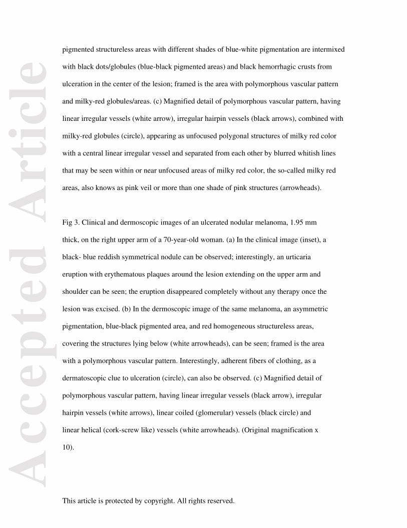
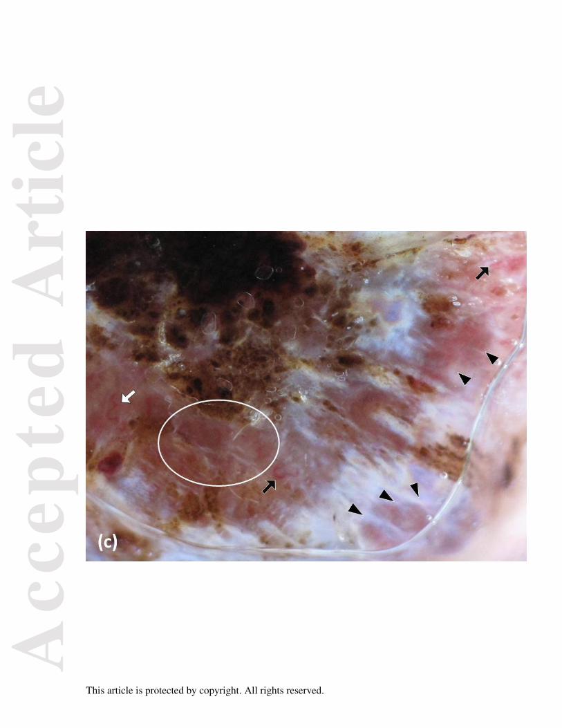
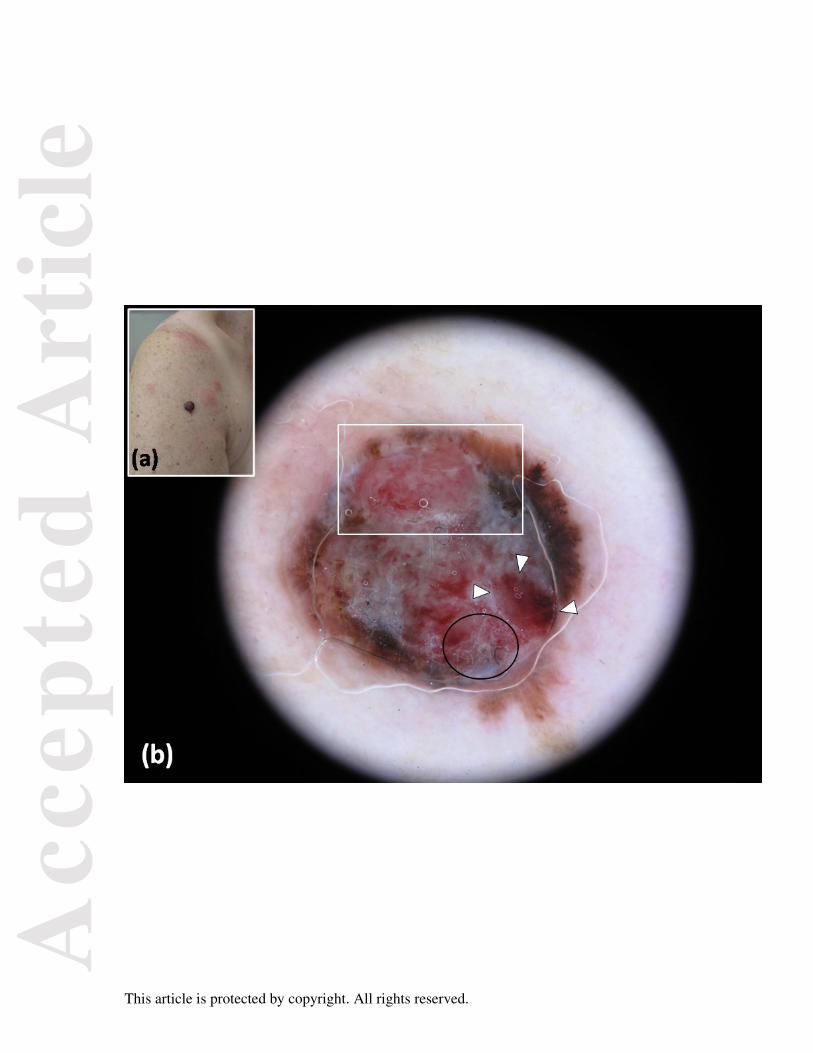
Fig 3. Clinical and dermoscopic images of an ulcerated nodular melanoma, 1.95 mm
thick, on the right upper arm of a 70-year-old woman. (a) In the clinical image (inset), a
black- blue reddish symmetrical nodule can be observed; interestingly, an urticaria
eruption with erythematous plaques around the lesion extending on the upper arm and
shoulder can be seen; the eruption disappeared completely without any therapy once the
lesion was excised. (b) In the dermoscopic image of the same melanoma, an asymmetric
pigmentation, blue-black pigmented area, and red homogeneous structureless areas,
covering the structures lying below (white arrowheads), can be seen; framed is the area
with a polymorphous vascular pattern. Interestingly, adherent fibers of clothing, as a
dermatoscopic clue to ulceration (circle), can also be observed. (c) Magnified detail of
polymorphous vascular pattern, having linear irregular vessels (black arrow), irregular
hairpin vessels (white arrows), linear coiled (glomerular) vessels (black circle) and
linear helical (cork-screw like) vessels (white arrowheads). (Original magnification x
10).
Acc
epte
d A
rtic
le
This article is protected by copyright. All rights reserved.
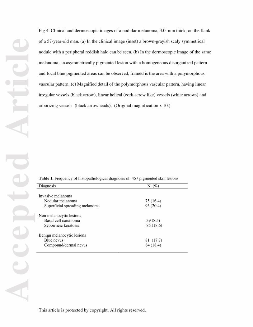
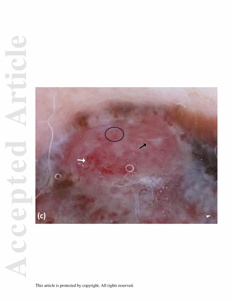
Fig 4. Clinical and dermoscopic images of a nodular melanoma, 3.0 mm thick, on the flank
of a 57-year-old man. (a) In the clinical image (inset) a brown-grayish scaly symmetrical
nodule with a peripheral reddish halo can be seen. (b) In the dermoscopic image of the same
melanoma, an asymmetrically pigmented lesion with a homogeneous disorganized pattern
and focal blue pigmented areas can be observed, framed is the area with a polymorphous
vascular pattern. (c) Magnified detail of the polymorphous vascular pattern, having linear
irregular vessels (black arrow), linear helical (cork-screw like) vessels (white arrows) and
arborizing vessels (black arrowheads), (Original magnification x 10.)
Table 1. Frequency of histopathological diagnosis of 457 pigmented skin lesions
Diagnosis N. (%) Invasive melanoma
Nodular melanoma 75 (16.4) Superficial spreading melanoma 93 (20.4)
Non melanocytic lesions Basal cell carcinoma 39 (8.5) Seborrheic keratosis 85 (18.6)
Benign melanocytic lesions Blue nevus 81 (17.7) Compound/dermal nevus 84 (18.4)
Acc
epte
d A
rtic
le
This article is protected by copyright. All rights reserved.
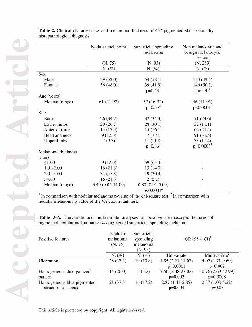
Table 2. Clinical characteristics and melanoma thickness of 457 pigmented skin lesions by histopathological diagnosis Nodular melanoma
Superficial spreading
melanoma
Non melanocytic and benign melanocytic
lesions (N. 75) (N. 93) (N. 289) N. (%) N. (%) N. (%) Sex
Male 39 (52.0) 54 (58.1) 143 (49.5) Female 36 (48.0) 39 (41.9) 146 (50.5)
p=0.431 p=0.701
Age (years) Median (range) 61 (21-92) 57 (16-92) 46 (11-95)
p=0.552 p<0.00012 Sites
Back 26 (34.7) 32 (34.4) 71 (24.6)Lower limbs 20 (26.7) 28 (30.1) 32 (11.1) Anterior trunk 13 (17.3) 15 (16.1) 62 (21.4) Head and neck 9 (12.0) 7 (7.5) 91 (31.5) Upper limbs 7 (9.3) 11 (11.8) 33 (11.4)
p=0.861 p=0.00031 Melanoma thickness (mm)
≤1.00 9 (12.0) 59 (63.4) - 1.01-2.00 16 (21.3) 13 (14.0) - 2.01-4.00 34 (45.3) 19 (20.4) ->4.00 16 (21.3) 2 (2.2) - Median (range) 3.40 (0.05-11.00) 0.80 (0.01-5.00) -
p<0.00012 1 In comparison with nodular melanoma p-value of the chi-square test. 2 In comparison with nodular melanoma p-value of the Wilcoxon rank test. Table 3-A. Univariate and multivariate analyses of positive dermoscopic features of pigmented nodular melanoma versus pigmented superficial spreading melanoma Positive features
Nodular melanoma
(N. 75)
Superficial spreading melanoma
(N. 93)
OR (95% CI)1
N. (%) N. (%) Univariate Multivariate2
Ulceration 28 (37.3) 10 (10.8) 4.95 (2.21-11.07) p=0.0001
4.07 (1.71-9.69) p=0.002
Homogeneous disorganized pattern
15 (20.0) 3 (3.2) 7.50 (2.08-27.02) p=0.002
10.76 (2.69-42.99)p=0.0008
Homogeneous blue pigmented structureless areas
28 (37.3) 16 (17.2) 2.87 (1.41-5.85) p=0.004
2.37 (1.08-5.22) p=0.03
Acc
epte
d A
rtic
le
This article is protected by copyright. All rights reserved.
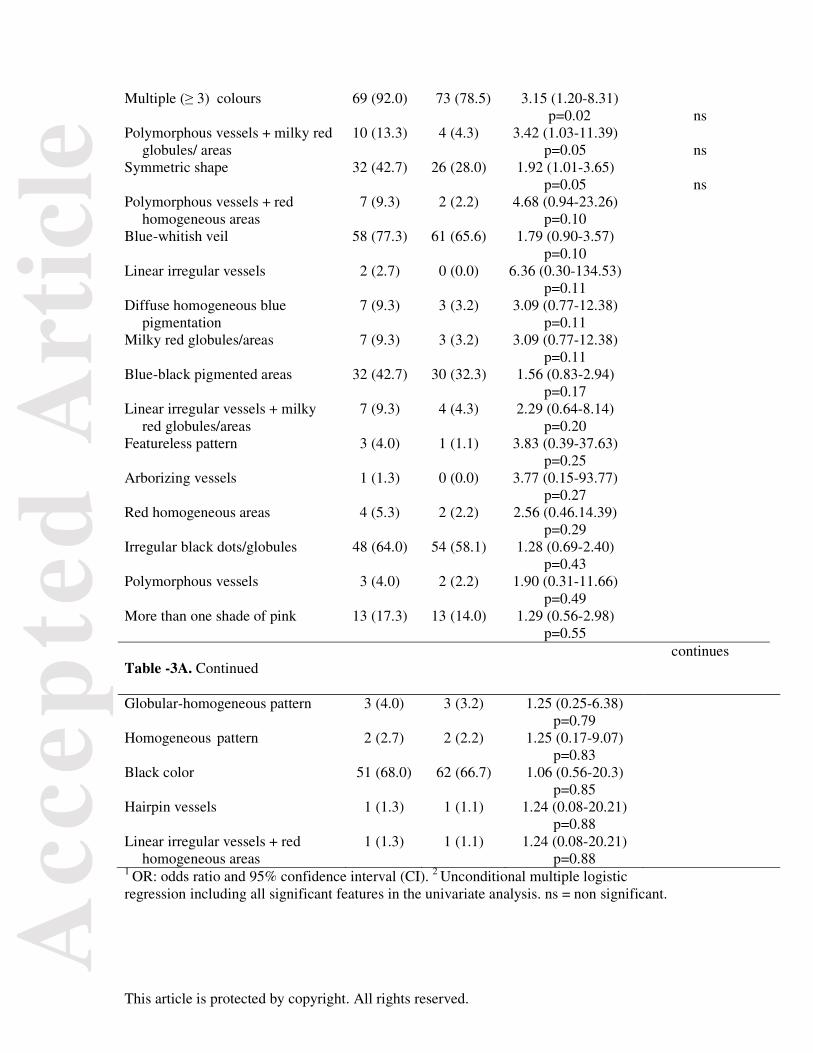
Multiple (≥ 3) colours
69 (92.0) 73 (78.5) 3.15 (1.20-8.31) p=0.02
ns
Polymorphous vessels + milky red globules/ areas
10 (13.3) 4 (4.3) 3.42 (1.03-11.39) p=0.05
ns
Symmetric shape 32 (42.7) 26 (28.0) 1.92 (1.01-3.65) p=0.05
ns
Polymorphous vessels + red homogeneous areas
7 (9.3) 2 (2.2) 4.68 (0.94-23.26) p=0.10
Blue-whitish veil 58 (77.3) 61 (65.6) 1.79 (0.90-3.57) p=0.10
Linear irregular vessels 2 (2.7) 0 (0.0) 6.36 (0.30-134.53) p=0.11
Diffuse homogeneous blue pigmentation
7 (9.3) 3 (3.2) 3.09 (0.77-12.38) p=0.11
Milky red globules/areas 7 (9.3) 3 (3.2) 3.09 (0.77-12.38) p=0.11
Blue-black pigmented areas 32 (42.7) 30 (32.3) 1.56 (0.83-2.94) p=0.17
Linear irregular vessels + milky red globules/areas
7 (9.3) 4 (4.3) 2.29 (0.64-8.14) p=0.20
Featureless pattern 3 (4.0) 1 (1.1) 3.83 (0.39-37.63) p=0.25
Arborizing vessels 1 (1.3) 0 (0.0) 3.77 (0.15-93.77) p=0.27
Red homogeneous areas 4 (5.3) 2 (2.2) 2.56 (0.46.14.39) p=0.29
Irregular black dots/globules 48 (64.0) 54 (58.1) 1.28 (0.69-2.40) p=0.43
Polymorphous vessels 3 (4.0) 2 (2.2) 1.90 (0.31-11.66) p=0.49
More than one shade of pink 13 (17.3) 13 (14.0) 1.29 (0.56-2.98) p=0.55
continues Table -3A. Continued Globular-homogeneous pattern 3 (4.0) 3 (3.2) 1.25 (0.25-6.38)
p=0.79
Homogeneous pattern 2 (2.7) 2 (2.2) 1.25 (0.17-9.07) p=0.83
Black color 51 (68.0) 62 (66.7) 1.06 (0.56-20.3) p=0.85
Hairpin vessels 1 (1.3) 1 (1.1) 1.24 (0.08-20.21) p=0.88
Linear irregular vessels + red homogeneous areas
1 (1.3) 1 (1.1) 1.24 (0.08-20.21) p=0.88
1 OR: odds ratio and 95% confidence interval (CI). 2 Unconditional multiple logistic regression including all significant features in the univariate analysis. ns = non significant.

Acc
epte
d A
rtic
le
This article is protected by copyright. All rights reserved.
Table 3-B. Univariate and multivariate analyses of negative dermoscopic features of pigmented nodular melanoma versus pigmented superficial spreading melanoma Negative features
Nodular melanoma
(N. 75)
Superficial spreading melanoma
(N. 93)
OR (95% CI)1
N. (%) N. (%) Univariate Multivariate2 Atypical network 17 (22.7) 50 (53.8) 0.25 (0.13-0.50)
p<0.0001
ns Multicomponent pattern 34 (45.3) 70 (75.3) 0.27 (0.14-0.52)
p<0.0001
ns Peripheral light brown
structureless areas 9 (12.0) 36 (38.7) 0.22 (0.10-0.49)
p=0.0002 0.26 (0.11-0.65)
p=0.004 Asymmetric shape 39 (52.0) 69 (74.2) 0.38 (0.20-0.72)
p=0.003
ns Irregular streaks 22 (29.3) 47 (50.5) 0.41 (0.21-0.77)
p=0.006 ns Linear irregular + dotted vessels
+ milky red globules/areas 2 (2.7) 11 (11.8) 0.20 (0.04-0.95)
p=0.04
ns Shiny white structures 13 (17.3) 28 (30.1) 0.49 (0.23-1.03)
p=0.06 Irregular dots/globules 58 (77.3) 82 (88.2) 0.46 (0.20-1.05)
p=0.06 Milia-like cysts 0 (0.0) 5 (5.4) 0.11 (0.06-1.96)
p=0.07
Linear irregular + dotted vessels 1 (1.3) 7 (7.5) 0.17 (0.02-1.38) p=0.10
Depigmentation /Structureless areas
40 (53.3) 59 (63.4) 0.66 (0.35-1.22) p=0.19
Comedo-like openings 0 (0.0) 1 (1.1) 0.41 (0.02-10.17) p=0.37
Symmetric pigmentation pattern 3 (4.0) 6 (6.5) 0.60 (0.15-2.50) p=0.49
Peripheral black dots /globules 34 (45.3) 45 (48.4) 0.89 (0.48-1.63) p=0.69
Asymmetric pigmentation pattern 69 (92.0) 87 (93.6) 0.79 (0.25-2.57) p=0.70
Regression structures 54 (72.0) 68 (73.1) 0.95 (0.48-1.87) p=0.87
Irregular blotches 49 (65.3) 61 (65.6) 0.99 (0.52-1.87) p=0.97
Predominant blue clods 7 (9.3) 9 (9.7) 0.98 (0.35-2.78) p=0.97
1 OR: odds ratio and 95% confidence interval (CI). 2 Unconditional multiple logistic regression including all significant features in the univariate analysis. ns= non significant.

Acc
epte
d A
rtic
le
This article is protected by copyright. All rights reserved.
Table 4-A. Univariate and multivariate analyses of positive dermoscopic features of pigmented nodular melanoma versus pigmented non melanocytic and benign melanocytic lesions Positive features
Nodular melanoma
(N. 75)
Non melanocytic and benign melanocytic
lesions (N. 289)
OR (95% CI)1
N. (%) N. (%) Univariate Multivariate2
Linear irregular vessels + milky red globules/areas
7 (9.3) 0 (0.0) 63.39 (3.58-1123.46) p<0.0001
ns
More than one shade of pink 13 (17.3) 1 (0.4) 60.39 (7.76-470.18) p<0.0001 ns
Red homogeneous areas 4 (5.3) 0 (0.0) 36.44 (1.94-648.62) p<0.0001 ns
Asymmetric pigmentation 69 (92.0) 94 (32.5) 23.86 (10.00-56.93) p<0.0001
6.70 (1.49-30.11) p=0.01
Irregular blotches 49 (65.3) 26 (9.0) 19.06 (10.22-35.56) p<0.0001
ns
Irregular black dots/globules 48 (64.0) 26 (9.0) 17.98 (9.67-33.44) p<0.0001
ns
Multicomponent pattern 34 (45.3) 13 (4.5) 17.61 (8.58-36.11) p<0.0001 ns
Regression structures 54 (72.0) 37 (12.8) 17.51 (9.51-32.26) p<0.0001 ns
Blue-black pigmented areas 32 (42.7) 12 (4.2) 17.18 (8.22-35.90) p<0.0001
7.15 (1.54-33.30) p=0.01
Peripheral black dots/ globules
34 (45.3) 15 (5.2) 15.15 (7.59-30.22) p<0.0001
ns
Ulceration 28 (37.3) 11 (3.8) 15.06 (7.02-32.29) p<0.0001
ns
Blue-whitish veil 58 (77.3) 57 (19.7) 13.89 (7.52-25.64) p<0.0001 ns
Multiple (≥ 3) colours 69 (92.0) 132 (45.7) 13.67 (5.75-32.49) p<0.0001 ns
Black color 51 (68.0) 42 (14.5) 12.50 (6.96-22.44) p<0.0001
ns
Irregular streaks 22 (29.3) 12 (4.2) 9.58 (4.47-20.54) p<0.0001
ns
Irregular dots/globules 58 (77.3) 110 (38.1) 5.55 (3.08-10.02) p<0.0001
ns
Homogeneous disorganized pattern 15 (20.0) 13 (4.5) 5.31 (2.40-11.74) p<0.0001
9.62 (1.62-57.13)p=0.01
Asymmetric shape 39 (52.0) 52 (18.0) 4.94 (2.87-8.50) p<0.0001 ns
continue

Acc
epte
d A
rtic
le
This article is protected by copyright. All rights reserved.
Table 4-A. continued Atypical network 17 (22.7) 18 (6.2) 4.41 (2.15-9.08)
p<0.0001
ns Homogeneous blue pigmented
structureless areas 28 (37.3) 35 (12.1) 4.32 (2.41-7.77)
p<0.0001
ns Polymorphous vessels + milky red
globules/areas 10 (13.3) 1 (0.4) 44.30 (5.57-352.16)
p=0.0003 23.65 (1.65-339.93)
p=0.02 Shiny white structures 13 (17.3) 15 (5.2) 3.83 (1.73-8.46)
p=0.0009 ns Polymorphous vessels + red
homogeneous areas 7 (9.3) 1 (0.4) 29.63 (3.59-244.79)
p=0.002 38.88 (1.72-877.07)
p=0.02 Milky red globules/areas 7 (9.3) 1 (0.4) 29.63 (3.59-244.79)
p=0.002
ns Predominant blue clods 7 (9.3) 8 (2.8) 3.57 (1.25-10.19)
p=0.02
ns Featureless pattern 3 (4.0) 1 (0.4) 12.00 (1.23-117.05)
p=0.03
ns Peripheral light brown structureless
areas 9 (12.0) 14 (4.8) 2.68 (1.11-6.45)
p=0.03 ns Depigmentation/structureless areas 40 (53.3) 114 (39.5) 1.75 (1.05-2.93)
p=0.03 ns Linear irregular + dotted vessels +
milky red globules/areas 2 (2.7) 1 (0.4) 7.89 (0.71-88.22)
p=0.10
Linear irregular + dotted vessels 1 (1.3) 0 (0.0) 11.66 (0.47-289.08) p=0.10
Polymorphous vessels 3 (4.0) 3 (1.0) 3.97 (0.79-20.09) p=0.10
Linear irregular vessels + red homogeneous areas
1 (1.3) 2 (0.7) 1.94 (0.17-21.68) p=0.59
1 OR: odds ratio and 95% confidence interval (CI). 2 Unconditional multiple logistic regression including all significant features in the univariate analysis. ns=non significant.
Acc
epte
d A
rtic
le
This article is protected by copyright. All rights reserved.
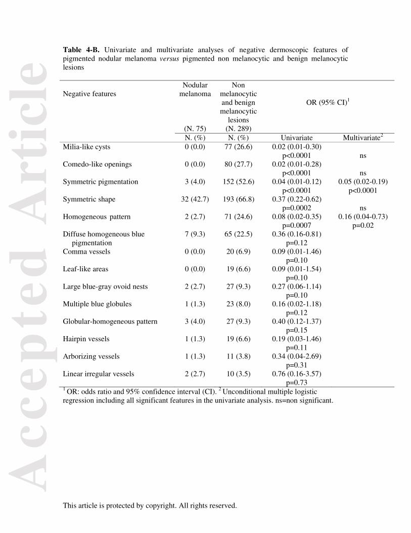
Table 4-B. Univariate and multivariate analyses of negative dermoscopic features of pigmented nodular melanoma versus pigmented non melanocytic and benign melanocytic lesions Negative features
Nodular melanoma
(N. 75)
Non melanocytic and benign melanocytic
lesions (N. 289)
OR (95% CI)1
N. (%) N. (%) Univariate Multivariate2
Milia-like cysts 0 (0.0) 77 (26.6) 0.02 (0.01-0.30) p<0.0001
ns
Comedo-like openings 0 (0.0) 80 (27.7) 0.02 (0.01-0.28) p<0.0001 ns
Symmetric pigmentation 3 (4.0) 152 (52.6) 0.04 (0.01-0.12) p<0.0001
0.05 (0.02-0.19)p<0.0001
Symmetric shape 32 (42.7) 193 (66.8) 0.37 (0.22-0.62) p=0.0002
ns
Homogeneous pattern 2 (2.7) 71 (24.6) 0.08 (0.02-0.35) p=0.0007
0.16 (0.04-0.73) p=0.02
Diffuse homogeneous blue pigmentation
7 (9.3) 65 (22.5) 0.36 (0.16-0.81) p=0.12
Comma vessels 0 (0.0) 20 (6.9) 0.09 (0.01-1.46) p=0.10
Leaf-like areas 0 (0.0) 19 (6.6) 0.09 (0.01-1.54) p=0.10
Large blue-gray ovoid nests 2 (2.7) 27 (9.3) 0.27 (0.06-1.14) p=0.10
Multiple blue globules 1 (1.3) 23 (8.0) 0.16 (0.02-1.18) p=0.12
Globular-homogeneous pattern 3 (4.0) 27 (9.3) 0.40 (0.12-1.37) p=0.15
Hairpin vessels 1 (1.3) 19 (6.6) 0.19 (0.03-1.46) p=0.11
Arborizing vessels 1 (1.3) 11 (3.8) 0.34 (0.04-2.69) p=0.31
Linear irregular vessels 2 (2.7) 10 (3.5) 0.76 (0.16-3.57) p=0.73
1 OR: odds ratio and 95% confidence interval (CI). 2 Unconditional multiple logistic regression including all significant features in the univariate analysis. ns=non significant.
Acc
epte
d A
rtic
le
This article is protected by copyright. All rights reserved.

Acc
epte
d A
rtic
le
This article is protected by copyright. All rights reserved.
Acc
epte
d A
rtic
le
This article is protected by copyright. All rights reserved.
Acc
epte
d A
rtic
le
This article is protected by copyright. All rights reserved.
Acc
epte
d A
rtic
le
This article is protected by copyright. All rights reserved.

Acc
epte
d A
rtic
le
This article is protected by copyright. All rights reserved.

Acc
epte
d A
rtic
le
This article is protected by copyright. All rights reserved.