acetabular fraacture management with surgical approaches
TRANSCRIPT

Acetabular Fracture :Classification & Surgical Approaches

The Pioneers
Robert Judet Emile Letournel Marvin Tile
Anatomical reduction and stable
internal fixation
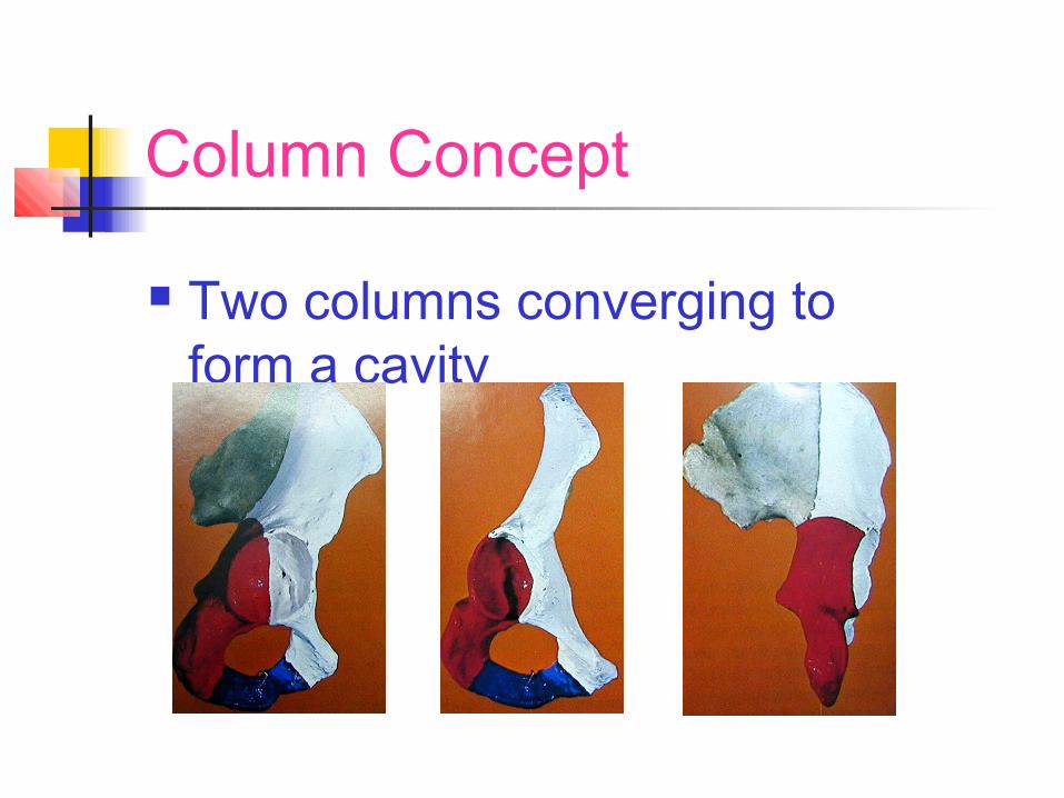
Column Concept
Two columns converging to form a cavity

The Acetabular Cavity Two walls – anterior and posterior Roof Floor

Plain Radiograph

Judet’s Iliac Oblique View

Judet’s Iliac Oblique View

Judet’s Obturator Oblique View

Judet’s Obturator Oblique View

CT Scan
Size of fragment Degree of displacement Amount of articular surface Site of origin of fragment Marginal impaction

CT Scan
Position of femoral head- dislocation, subluxation, perfect reduction
Incarcerated fragment

3 D CT SCAN

Letournel Classification
Most acetabular fractures fit into one of ten types
Five simple fracture patterns Five associated fracture patterns

Simple Fractures
Posterior wall fracture Posterior column fracture Anterior wall fracture Anterior column fracture Transverse fracture

Posterior wall Fracture Includes posterior
articular surface and retro acetabular surface

Posterior Column Fracture Extends from
PSIS to ischio pubic ramus
Involves posterior articular surface and ilio ischial line

Anterior Wall Fracture
Uncommon Separation of anterior part of
articular surface along with a large part of middle third of anterior column
Anterior hip dislocation can be associated

Anterior Column Fracture Extends from
symphysis pubis to iliac crest
Most commonly fracture line exits below AIIS
Often comminution into the quadrilateral plate

Transverse Fracture Across anterior and posterior columns Superior segment – ilium, acetabular roof Inferior segment – ischiopubic segment May be associated with central dislocation

Combined Fracture Types
Posterior column and posterior wall fracture
Transverse and posterior wall fracture T-shaped fracture Anterior column or wall and posterior
hemi transverse fracture Complete both-column fracture

Associated Posterior Wall and Posterior Column Fracture
Posterior column fracture is usually undisplaced or minimally displaced
Primary fracture – posterior wall

Associated Posterior Wall And Posterior Column Fracture
Anterior Column intact
Posterior dislocation Displaced posterior
wall Ilio ischial line
disrupted

Associated Transverse and Posterior Wall Fracture
Commonly posterior dislocation
Sometimes central dislocation
Highest incidence of pre op sciatic palsy and AVN of femoral head

T Shaped Fractures
Transverse and vertical components
Acetabular cavity is split into at least 3 fragments

T Shaped Fractures

Both Column Fractures All segments of
fractured acetabulum are detached from the ilium

AO Classification Type A – Fractues of a single wall or
column Type B –Fractures involve both columns Type C –Both column fractures with
articular fragments seperated from ilium

AO Classification

AO Classification

AO Classification

Injury Pattern Affecting Prognosis
High or low energy trauma Involvement of acetabular dome Comminution and displacement Joint dislocation Damage to femoral head Both-column fractures and transverse with
posterior wall fractures have worst results , primarily because of imperfect reduction

Indications For Conservative Management Non displaced or displaced
<3mm Displaced fracture in
unimportant part of acetabulum – low anterior column, low transverse
Secondary congruence in both column fractures

Indications for operative treatment Fracture Displaced >3mm Irreducible fracture dislocation Intra articular fragment
interfering with joint movement Instability of the joint To prepare the joint for hip
replacement

Contraindications to surgery Severe osteoporosis Very old patients Severe associated injuries Poor local skin condition Limited experience of the surgeon

Timing Of Surgery Urgent closed reduction of dislocation Stabilise the patient before ORIF Ideally within 7 days Poor results after 3 weeks

Kocher Langenbeck Approach Posterior wall fracture Injuries with associated
posterior wall fracture Posterior column fracture

Kocher Langenbeck Approach Transverse fracture
where major displacement is posterior
T shaped fracture with major displacement posterior

Kocher Langenbeck Approach

Ilioinguinal Approach Anterior wall
fracture Anterior column
fracture Fractures
associated with anterior wall and column fractures

Ilioinguinal Approach

Ilio inguinal Approach Incise along anterior two thirds of iliac
crest, extending to the pubic symphysis Elevate abdominal muscles from iliac
crest Open inguinal canal

Ilioinguinal Approach Detach deep abdominal
muscles from inguinal ligament and pubis
Open iliopsoas sheath Incise iliopectineal fascia Lateral retraction of
iliopsoas and medial retraction of iliac vessels

Dangers Of Ilio Inguinal Approach Lat cut nerve of thigh,
femoral nerve Corona mortis Internal iliac vein, femoral
vein Lymphatics Abdominal wall
weakness

EXTENSILE APPROACHES
Used in complex fracture patterns Triradiate Extensile Approach Carnesile Extensile approach
Extensile Ileofemoral approach

Triradiate Approach Described by Mears & Rubash Reduction & repair of Complex Acetabular fracturesAvoids ischemic necrosis Of Hip Abductors

Triradiate Approach

Carnesile approach

Carnesile approach

Complications Of Acetabular Surgery
Imperfect reduction, inadequate fixation Avascular necrosis Infection Nerve injury Heterotopic ossification Thromboembolism
Learning Curve Of The Surgeon Parallels The Suffering Curve Of The Patient

Thank You Thank You For Your For Your Kind Kind AttentionAttention