acid denaturation of horse carbonylhemoglobin in the … · · 2003-02-07acid denaturation of...
TRANSCRIPT

THEJOURNAI. OF BIOLOGICA~~CHEYISTRY Vol. 241, No. 17, Issue of September 10, pp. 3988-3996, 1966
Printed in U.S.A.
Acid Denaturation of Horse Carbonylhemoglobin in the Absence of Oxygen*
(Received for publication, January 24, 19G6)
JACINTO STEINHARDT,HELGA POLET,AND FATEMAH MOEZIE
From the Department of Chemistry, Georgetown University, Washington, D. C. dOOO?’
SUMMARY
Unlike ferrihemoglobin (Hb+), denatured carbonylhemo- globin (COHb) has almost the same absorption spectrum in the visible and near ultraviolet region as the native form; the very slight difference’appears only in the Soret band. Ki- netic and equilibrium experiments under anaerobic condi- tions show that COHb is considerably more resistant to acid denaturation at 25’ than is Hb+ although the rates of de- naturation of both proteins depend on pH in much the same way. These conclusions are based on assays of denatured protein by precipitation at the isoionic point, by the measure- ments of unmasking of imidazole groups inaccessible in the native protein, and by changes in optical rotatory dispersion. Air, when present, oxidizes the denatured protein practically instantaneously, but it has no discernible effect on the de- naturation rate. It does, however, predictably shift the equilibria by removing the initial product, unoxidized acid- denatured protein. COHb, unlike Hbf, is not stabilized by formate, but another stabilizer of Hb+, azide, produces com- plex effects. Bromphenol blue greatly accelerates the rate of denaturation and shifts the denaturation equilibrium to higher pH. The possible significance of the absence of a marked spectral change is discussed in terms of a possible nonbonded clathrate structure for the prosthetic group in COHb. Changes in the Cotton effects in the Soret band differ in acid-denatured COHb and Hb+, and there are differences in the regenerability of the denatured proteins. These facts add support to the evidence that conformation differences exist in the native forms; thus the difference in stability between COHb and Hb+ need not be solely the direct consequence of the positive charge on the prosthetic group of the latter as previously postulated.
Although the earliest reports of the all-or-none unmasking of basic prototropic groups in the acid denaturation of proteins were based on investigations of horse carbonylhemoglobin (l-3),
* This work was supported by National Institutes of Health Grant GM 10607. Part of the material in this paper was pre- sented at the Chicago meeting of the American Society of Bi- ological Chemists in April, 1964. Some of the preliminary meas- urements were made by Mrs. Irene Pigman to whom thanks are due.
most of the subsequent quantitative studies of this phenomenon in the heme proteins were based on work with ferrihemoglobin (see References 4 and 5 for reviews). The latter protein was used because the rapid oxidation of the iron in heme proteins which occurs at acid pH can be a disturbing source of error which is completely avoided by working with the already oxidized ferri form.
We have returned to studies of the denaturation of COHbl by dilute acid because (a) interpretation of the stabilizing effects of certain iron-ligands on ferrihemoglobin (6) led to an expectation that the ferro forms should be more stable than the free ferri forms;2 and (b) because the earlier indication, based on spectro- photometry, that COHb was characterized by an apparent in- crease in stability to acid denaturation as the concentration of dissolved oxygen was progressively reduced, led to the possibility that it might be very much more stable than ferrihemoglobin if oxygen was completely excluded.
We therefore present the results of a more complete thermo- dynamic and kinetic study of COHb in as close an approach to anaerobic conditions as could be attained in our laboratory. It has led to a confirmation that COHb is indeed considerably more resistant to acid denaturation than ferrihemoglobin. It has also led, more unexpectedly, to the realization that COHb may be completely denatured by any of a number of critical criteria and still undergo only minor changes in its visible andnear ultraviolet absorption spectrum.
EXPERIMENTAL PROCEDURE
Crystalline Carbonylhemoglobin
Horse COHb from single animals was crystallized in a CO atmosphere at room temperature by the method of Ferry and Green (8). After three crystallizations and washing with dis- tilled water, the crystals were dissolved by adding 1 M KOH to a pH of about 7.3, and then stored in stock solutions at concentra- tions between 1 and 7 % on a dry weight basis (drying at 105”). Iron and nitrogen contents, determined as previously described (I), were normal. The molal extinction coefficients(t), measured in a Cary model 14 spectrophotometer, were higher than those
1 The abbreviations used are: COHb, carbonylhemoglobin; Hb+, ferrihemoglobin; OsHb, oxyhemoglobin; HbO, deoxyhemo- globin.
2 The mechanism proposed by one of us for the increased sta- bility conferred by such ligands as azide no longer appears tenable on the basis of x-ray crystallographic studies on myoglobin azide by Stryer, Kendrew, and Watson (7).
by guest on June 24, 2018http://w
ww
.jbc.org/D
ownloaded from
Issue of September 10, 1966 J. Xteinhardt, H. Polet, and F. Moezie 3989
usually reported, as follows: 419 mp, 179,000 to 211,000; 540 rnp, 13,400 to 14,200; 570 rnp, 13,300 to 13,900. One preparation, crystallized only twice, used in the pH-stat experiments, had an t4,g of 158,000.
The slight differences in e between various batches were not associated with any systematic differences in the results reported in this paper. The 630 band of Hb+ appeared to be absent. Errors in e arising from adsorption on pipettes in serial dilutions were avoided by using a Limit variable path length cell to make measurements at the different wave lengths at approximately the same absorbances. Since early measurements showed that anti- foaming agents, such as octanol, affected the stability of Hb+, no antifoam agents were employed even when it was necessary to evacuate or to bubble gasses through the experimental solution.
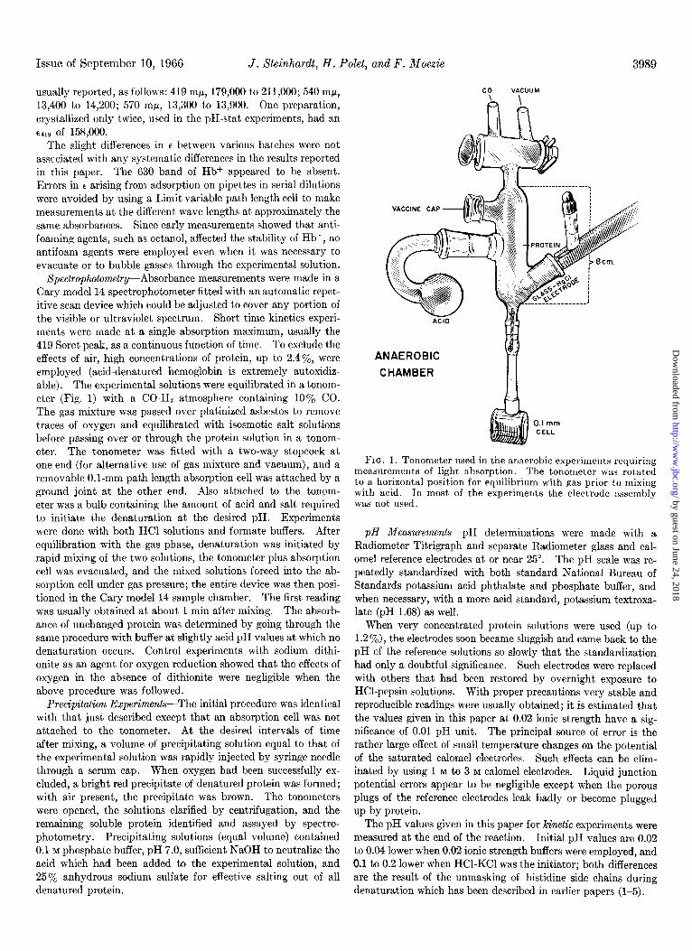
Spectrophotometry-Absorbance measurements were made in a Cary model 14 spectrophotometer fitted with an automatic repet- itive scan device which could be adjusted to cover any portion of the visible or ultraviolet spectrum. Short time kinetics experi- ments were made at a single absorption maximum, usually the 419 Soret peak, as a continuous function of time. To exclude the effects of air, high concentrations of protein, up to 2.4y0, were employed (acid-denatured hemoglobin is extremely autoxidiz- able). The experimental solutions were equilibrated in a tonom- eter (Fig. 1) with a CO-H:! atmosphere containing 10% CO. The gas mixture was passed over platinized asbestos to remove traces of oxygen and equilibrated with isosmotic salt solutions before passing over or through the protein solution in a tonom- eter. The tonometer was fitted with a two-way stopcock at one end (for alternative use of gas mixture and vacuum), and a removable O.l-mm path length absorption cell was attached by a ground joint at the other end. Also attached to the tonom- eter was a bulb containing the amount of acid and salt required to initiate the denaturation at the desired pH. Experiments were done with both HCl solutions and for-mate buffers. After equilibration with the gas phase, denaturation was initiated by rapid mixing of the two solutions, the tonometer plus absorption cell was evacuated, and the mixed solutions forced into the ab- sorption cell under gas pressure; the entire device was then posi- tioned in the Cary model 14 sample chamber. The first reading was usually obtained at about 1 min after mixing. The absorb- ance of unchanged protein was determined by going through the same procedure with buffer at slightly acid pH values at which no denaturation occurs. Control experiments with sodium dithi- onite as an agent for oxygen reduction showed that the effects of oxygen in the absence of dithionite were negligible when the above procedure was followed.
Precipitation Experiments-The initial procedure was identical with that just described except that an absorption cell was not attached to the tonometer. At the desired intervals of time after mixing, a volume of precipitating solution equal to that of the experimental solution was rapidly injected by syringe needle through a serum cap. When oxygen had been successfully ex- cluded, a bright red precipitate of denatured protein was formed; with air present, the precipitate was brown. The tonometers were opened, the solutions clarified by centrifugation, and the remaining soluble protein identified and assayed by spectro- photometry. Precipitating solutions (equal volume) contained 0.1 M phosphate buffer, pH 7.0, sufficient NaOH to neutralize the acid which had been added to the experimental solution, and 250/, anhydrous sodium sulfate for effective salting out of all denatured protein.
co VACUUM
ANAEROBIC 81 >
CHAMBER
n
0.1 mm CELL
FIG. 1. Tonometer used in the anaerobic experiments requiring measurements of light absorption. The tonometer was rotated to a horizontal position for equilibrium with gas prior to mixing with acid. In most of the experiments the electrode assembly was not used.
pH Measurements-pH determinations were made with a Radiometer Titrigraph and separate Radiometer glass and cal- omel reference electrodes at or near 25”. The pH scale was re- peatedly standardized with both standard National Bureau of Standards potassium acid phthalate and phosphate buffer, and when necessary, with a more acid standard, potassium textroxa- late (pH 1.68) as well.
When very concentrated protein solutions were used (up to 1.2 %), the electrodes soon became sluggish and came back to the pH of the reference solutions so slowly that the standardization had only a doubtful significance. Such electrodes were replaced with others that had been restored by overnight exposure to HCl-pepsin solutions. With proper precautions very stable and reproducible readings were usually obtained; it is estimated that the values given in this paper at 0.02 ionic strength have a sig- nificance of 0.01 pH unit. The principal source of error is the rather large effect of small temperature changes on the potential of the saturated calomel electrodes. Such effects can be elim- inated by using 1 M to 3 M calomel electrodes. Liquid junction potential errors appear to be negligible except when the porous plugs of the reference electrodes leak badly or become plugged up by protein.
The pH values given in this paper for kinetic experiments were measured at the end of the reaction. Initial pH values are 0.02 to 0.04 lower when 0.02 ionic strength buffers were employed, and 0.1 to 0.2 lower when HCl-KC1 was the initiator; both differences are the result of the unmasking of histidine side chains during denaturation which has been described in earlier papers (I-5).
by guest on June 24, 2018http://w
ww
.jbc.org/D
ownloaded from

3990 Anaerobic Acid-denaturation of Hemoglobin Vol. 241, No. 17
pa-stat Experiments-The rate of unmasking of basic imidazole groups at 25” f 0.1’ was measured by the automatic addition of acid from a syringe burette controlled by a Radiometer Titri- graph and SBU recorder, operated as a pH-stat. Electrode standardization and provision of fast response were in accordance with the description of pH measurements above. Runs in which electrode standardization drifted over 0.03 unit or in which elec- trode response was sluggish when restandardized were rejected.
Because of the shape of the titration vessel and the numerous insertions fitted to it (stirrer, burette tip, two electrodes, gas flow inlet and outlet), it proved difficult to exclude air to the point that no detectable oxidation of the protein (evidenced by color change) occurred during a run. Recourse was had to the addi- tion of 0.01 M formamidine sulfinic acid (Nutritional Biochemi- cals), a readily autoxidizable dipolar ion, which has the rare property of not affecting the stability of the protein as do known ligands and such indicators as thymol blue and bromphenol blue. Although formamidine sulfinic acid reacts with oxygen slowly and has the disadvantage of buffering in the pH range of interest, its use is far preferable to that of the more common re- ductant, sodium dithionite, which undergoes large time-depend- ent prototropic changes as it decomposes at acid pH. Since the oxidation of formamidine sulfinic acid affects its acid-base proper- ties, some of the time-dependent addition of base is the result of such changes and results in the absence of an end point. An effort has been made to eliminate this latter contribution to the kinetics by subtracting a term proportional to the time (deter- mined at the end of the experiment) from all of the data. In other experiments oxidation was completely prevented by sat- urating the solutions with CO-Hz mixtures in the presence of small amounts of platinized asbestos. These experiments showed unambiguously that unmasking occurred without any detectable change in the color of the solutions. They are less reproducible than the experiments with formamidine sulfinic acid and are, therefore, not shown in this paper.
Xpectropolarimetry-Optical rotatory dispersion measurements were made with a Cary model 60 spectropolarimeter at protein concentrations of 1.2% at a path length of 0.1 mm at 27”. Kinetic measurements were made at the trough at 233 rnE.1 with the anaerobic procedure described under “Spectrophotometry.”
Titration Curves-A rapid flow technique similar to that pre- viously described (1) was employed with measurements at 3 to 4 set after mixing at 25” f 2”. Experiments in which the amounts of dissolved oxygen present were varied showed that the presence or absence of oxygen did not alter the titration curve with acid detectably. In the back-titration, however, it is important to exclude oxygen during the initial acidification in order to assure that the material being back-titrated is not transformed by oxida- tion to a different product. The acidification was carried out in the pH-stat device described above after long equilibration of 0.1% protein with flowing CO-H2 by injecting the initial acid by syringe (pH 2.75 at 0.02 ionic strength) and commencing the automatic back-titration 5 min later. About 20 min were re- quired for each complete curve. Precipitation of denatured pro- tein at pH values approaching 6 showed that appreciable regen- eration had not occurred. The bright red color of the solutions indicated that most of the protein had not been oxidized. More rigorous experiments on the back-titration are continuing.
Stop Flow Kinetics-Efforts were made to get kinetic data by observing the change in pH in electrode-fitted tonometers in the rapid flow titration apparatus and in the pH-stat electrode vessel
after bringing the pH rapidly to values between 3.2 and 3.7. These data are less clear cut than the pH-stat data, simply be- cause the pH, and therefore the rates, change constantly. Therefore, they are not given in the present paper.
RESULTS
The results to be reported here are of two kinds. The first (“Comparison of Spectral with other Criteria of Denaturation”) is concerned with establishing that the denaturation of COHb by acid in the absence of air is not accompanied by the drastic spec- tral changes that accompany denaturation of methemoglobin; we first present, therefore, as a function of pH, the rates of dena- turation determined by several distinct criteria other than light absorption, and the range of pH values in which an equilibrium occurs (i.e. in which denaturation is not complete). These data are of interest in themselves in that they show the large extent to which COHb is more resistant to acid denaturation than Hb+. The second type of data presented (“Observations of Effects of Ligands on Stability of COHb”) does not bear directly on this demonstration, but is germane to the broader subject, already well explored with Hbf, the extent to which combination with various ligands affects the stability.
Comparison of Spectral with Other Criteria of Denaturation
Earlier kinetic measurements of the acid-denaturation of COHb and Hb+ were based on (a) changes in spectrum; (b) in- creases in acid-binding due to the liberation of imidazole groups which are inaccessible to acid in the native protein (these in- creases were estimated from time-dependent changes in the titration curve (9)) ; and (c) time-dependent losses in solubility. We consider each of these below.
The rates of spectrophotometric change when COHb is brought rapidly to pH values below 4.5 are considerably lower than the same rates when Hb+ is acidified to the same pH values (Fig. 14 of Reference 4). The apparent rates with COHb, moreover, are smaller the more stringently oxygen is excluded (2) ; the limiting values reached at high concentrations of dissolved oxygen are nevertheless lower than those for Hb+.
The second property, less affected by the presence of oxygen, is the extent to which the titration curve with acid obtained in the usual way (minutes) departs from the titration curve obtained by a rapid flow method (2 to 3 set) (see Figs. 7 and 8 of Reference 4). By this measure, COHb is much more stable than Hb+; unmasking can be detected with the latter in 2 min at pH 4.1; the former shows no noticeable unmasking in 10 min at pH 3.8. A related measure is the pH at which the rapid flow method fails to catch all of the protein in the masked (native) form. This may be estimated from the pH at which the difference titration curves (slow minus rapid flow) turn downward as the pH is decreased (the pH at which they have lost half of their maximum magnitude may be interpreted as the pH at which the half-period of un- folding is 2 to 3 set). By this criterion, COHb has such a half- period at pH 2.65 at 25” and 0.02 ionic strength. Hb+ reaches this fast reaction rate at pH 3.3. Since the velocity depends on the 2.5 power of concencentration of H+ in the case of Hb+ and on the 3.2 power in the case of COHb, the 0.65 unit difference in pH corresponds to about a 50-fold difference in velocity of de- naturation at pH 3.7 and an even larger difference at higher pH. At higher salt concentrations (0.30) this factor exceeds 500. These measurements have been made much more quantitative
by guest on June 24, 2018http://w
ww
.jbc.org/D
ownloaded from

Issue of September 10, 1966 J. Steinhardt, H. Pokt, and F. Moezie 3991
in the pH-stat experiments which are described later in this paper.
The rate at which precipitable protein is produced as a function of pH differs with the two proteins. This method has proved the most direct and least equivocal of the three described, and is used, among others in this paper, to measure rates, and to es- tablish when COHb is fully denatured.
The first method above (spectrophotometric) has proved to present great difficulties in kinetics experiments with COHb. When oxygen is excluded (or its ratio to protein reduced by evacuation and gaseous exchange in tonometers with the use of high protein concentrations), we may acidify to the same low pH values that rapidly produce denaturation by the above criteria (extensive or complete unmasking, or the rapid conversion of all or most of the native protein to an insoluble form) without pro- ducing more than slight changes in either the visible or the near ultraviolet spectrum, even after periods extending to a day or longer. The same absence of large spectrophotometric effect is found when minimal quantities of sodium dithionite (1 to 2 mg in 10 ml) are used to eliminate the effects of oxygen except for transient changes in absorption in the vicinity of 3500 A which are due to the decomposition of dithionite.
Typical results at 25” and 0.02 ionic strength are shown in Fig. 2. The spectrum at pH 2.81, obtained in the absence of air, shows that only a very slight reduction in the height of the Soret band, and practically no reduction in the heights of the 570 bands occurs after 3 min. The changes continue at an ex- tremely low rate long after the unmasking reaction described below is complete. With air present, the Soret band, the 349 rnp band, and the green bands disappear in a matter of minutes; the Soret band is replaced by a broad diffuse band centered at 370 rnp; the latter band is characteristic of hematin in the presence of protein. It will be shown below that the rate of disappearance of the Soret band, although obviously due to oxidation by oxygen after denaturation, closely parallels the unmasking reaction as measured in a pH-stat or the production of protein insoluble at the isoionic point in the presence of 12.5% sodium sulfate.
Fig. 3 summarizes various kinds of rate data obtained at 25” in the presence of almost 10 times as much dissolved oxygen as would be required to oxidize all of the protein. These data rep- resent the information available at the time the experiments re- ported here were first undertaken. They show that in the pres- ence of oxygen the formation of COHb precipitable at the isoionic point occurred at the same rate as the disappearance of the Soret band of Hb+. The data for the latter in acetate buffers are given, since formate which was used with COHb stabilizes Hbf but not COHb.3 The figure also shows that the criterion of unmasking of imidazole groups yields approximately the same rates as does the fading of the Soret band when oxygen is present, and as does the production of insoluble protein. The three triangles on the figure represent half-periods calculated from the difference between the amounts of acid bound “instantaneously” (3 set) and the amounts bound in 10 min at the same pH values. They are cal- culated from the early COHb titration data referred to in the introduction.
The agreement of the three methods of following denaturation when oxygen is present is compatible with the view that little
3 In the upper left-hand corner of Fig. 3 log rate pH relationships obtained at 0” (spectrophotometric) are given for both COHb and 02Hb merely to establish that the stability of COHb in the pres- ence of oxygen is shared by OzHb.
1 DENATURATION OF COHb
08 ‘-
3000 4000 5000 6000 Wave length in Angstroms
FIG. 2. Absorption spectra of CoHb at various periods of time at 25” after bringing to the pH values shown in the presence and in the absence of air. The product of concentration of protein and path length is 2.2 times as high in the top jigure as in the bottom $gure. The times at. the maximum of the Soret band arc about 2 min higher than the times given (start of scan).
0.0 3.2 3.3 3.4 3.5 3.6 3.7 3.6 3,9 4.0
PH
FIG. 3. Previously published results of kinetics experiments with COHb. Hb+. and OlHb at 25” at 0.02 ionic streneth as af- fected by pk. D&ta represented by triangles are explained in the text.
change in absorption spectrum occurs in the absence of oxygen, but that denatured COHb loses its Soret band extremely rapidly as the result of oxidation by air. This tentative conclusion is confirmed by the experiments in the present paper.
In the sections that follow, the rates of denaturation and equilibria are presented in terms of different criteria of denaturation. These measurements, which establish the con- ditions under which COHb is practically totally denatured, are followed by a demonstration that only slight changes in spectrum accompany denaturation.
Rate of Formation of Insoluble Protein-By exposing the protein
by guest on June 24, 2018http://w
ww
.jbc.org/D
ownloaded from

3992 Anaerobic Acid-denaturation of Hemoglobin Vol. 241, No. 17
5 IO 15 20 Time (minutes)
FIG. 4. Typical anaerobic kinetics experiments with formate buffers by the precipitation technique. D is optical density.
PH
FIG. 5. The relation of denaturation rate to pH at 25” and 0.02 ionic strength. Formate buffers were used in the precipitation and optical rotation experiments. fsa, formamidine sulfinic acid.
to acid in the absence of air and then salting out the altered pro- tein after various periods of time, it should be possible to detect whether unfolding occurs when air is absent; if it does the effect of air would be shown to consist in oxidation of the denatured protein rather than in making the unfolding possible by preceding it. COHb would thus have a measurable denaturation rate, much lower than that of Hb+, but not infinitely slow as the lack of spectral changes would suggest.
Results of typical experiments with 0.02 ionic strength formate buffers at 25”, carried out in sealed 02-free CO-filled tonometers containing concentrated (1.2y0) protein solutions, are shown in Fig. 4. Points relating the rates to pH are shown in Fig. 5 to- gether with a dotted line, taken from Fig. 3, which showed the earlier precipitation and spectrophotometric data (air present).
The agreement is good at the higher pH values, but the new anaerobic experiments show a progressive shift to higher pH val- ues at the faster rates. This apparent shift is probably due in part to a change in pH due to unmasking (removal of hydrogen ions by imidazoles) as the reaction proceeds. With the much lower protein concentrations used earlier, such a shift is almost negligible. At the high protein concentrations used here, the addition of protein to buffer causes a rise in the buffer pH to values which are higher by 0.25 to 0.31 unit (measured after un- masking occurs).
Of the experiments just described only those carried out at the lowest pH values proceeded to completion. The equilibria at- tained at 25” at various pH values are shown in Figs. 6 and 7. The slope in Fig. 7 is 5.2, close to the values shown earlier for Hb+ under comparable conditions (no corrections for irreversible
I I I I I I I I 3.5 4.0
PH
FIG. 6. The equilibrium between native and denat.ured protein (COHb and Hb+) at 25” and 0.02 ionic strength. The bromphenol blue CUTX is described at a later point in the text.
COHb 25”
. r precipitation __
l l
\
0 Formote 0 Formote OHCI OHCI
0 0
\, \,
I l\I I I 3.5 4.0
PH
FIG. 7. Identity of the equilibrium between native and dena- tured COHb at 25” in formate buffer and HCl.
by guest on June 24, 2018http://w
ww
.jbc.org/D
ownloaded from

Issue of September 10, 1966 J. Xteinhardt, H. Polet, and F. Moezie 3993
loss (10)). Half of the protein is in the denatured form at pH 3.59 whereas half of Hb+ is denatured at equilibrium at pH 4.13. Such data could not be obtained in the earlier experiments, since the equilibrium was continually shifted by oxidation, until the reaction had proceeded to completion. The same equilibrium curve is obtained if HCl-KC1 mixtures are used instead of for- mate. The HCl experiments, however, change pH over a wide range as denaturation proceeds, and therefore do not follow first order kinetics. The identity of the equilibria is noteworthy since it shows clearly that formate does not stabilize COHb as it does Hb+. With the latter, the equilibria are shifted 0.35 pH unit lower, relative to HCl or acetic acid, when formate is used.
Whereas the precipitates obtained in the earlier experiments (air present) were dark brown, those obtained in the present ex- periments in the absence of air were always bright red, consistent with the similarity in spectrum of native and denatured COHb.
pH-stat Kinefics-The results of typical unmasking rate meas- urements obtained in the presence of 0.01 M formamidine sulfinic acid are shown in Fig. 8. The relation of rates to pH is shown in Fig. 5 where it may be compared with the early spectrophoto- metric and solubility rate data of Fig. 3 (dotted Zine).4 At the low pH values at which the unmasking reaction should be com- plete, the adjusted time-dependent total increment of acid added corresponds to 28 basic (presumably imidazole) groups per mole of protein. The equilibrium end values, determined for higher pH, are shown in Figs. 6 and 7 together with the points obtained by the precipitation technique; they run closely parallel (there is no evidence of a divergent trend). Since both denaturation rates and equilibria, measured by the distinct criteria, are con- sistent, it must be concluded that regeneration rates, when measured by the same two methods, would also be identical as in the case of Hbf (11). Regeneration from denatured protein that has been allowed to oxidize should not necessarily proceed at the same rate as regeneration from unoxidized denatured pro- tein; in fact, the latter proceeds much more rapidly (10, II), thus oxidized denatured protein differs from denatured oxidized (ferri-) protein.
Unfolding Rates and Equilibria Shown by Optical Rotation-A single experiment was carried out at 233 rnp at pH 3.52 (formate) at 27” as described under “Experimental Procedure.” Fig. 9 shows a Guggenheim treatment (12) of the data expressed as the log of the change in rotation in successive 2-min intervals of a 1.2% protein solution with a O.l-mm path length. It will be noted that the data are first order over more than three half- periods. The half-period of the reaction has been indicated by a + in the assembled kinetic data for 25” (Fig. 5) with which it agrees fairly well. The same data have been used to calculate a single equilibrium point shown by a + in Fig. 6. The latter result, which is consistent with the other equilibria in Fig. 6 de- termined by other means, was arrived at by comparing the end value at pH 3.52 with the difference in optical rotation at the same frequency between the extrapolated zero time value and the end value at pH 3 where unfolding is complete within a minute.
Diflerence Spectrophotometry-The near identity of the spec- trum of native and acid-denatured protein is a crucial and chal- lenging aspect of the phenomena reported in this paper. It has been relatively easy to show that changes in light absorption in
4 The isotherms shown in Fig. 8 were calculated on the basis of end values determined by adjusting for the slow oxidation of formamidine sulfinic acid as explained under “Experimental Procedure.”
2.0 : i i 1 / I I , 1 , , ,
CbHb 25’ 0.02 ionic strength pH -stat
I l pH 3.12 lair)
1 - OpH 3.41 with fsa OpH 3.62 with fsa
I 1 I , I I 1 5 IO 15 Time (minutes)
FIG. 8. Typical kinetics experiments at 25” and 0.02 ionic strength (HCI-KCl) measuring the rate of unmasking. A is the delayed acid uptake. jsa, formamidine sulfinic acid.
levo rotation ot
Q -
F? - -I
I I I 5 IO 15 20 25
Time (minutes)
FIG. 9. Kinetics of denaturation (Guggenheim plot) of COHb at 27O in formate buffer (pH 3.5 and 0.02 ionic strength) followed by changes in the depth of the rotation trough at 233 mp. The ordinate refers to the changes in rotation in successive 2.min in- tervals. The rate and equilibrium points determined in this ex- periment are shown as + in Figs. 5 and 6.
the various bands are small. It is not so easy to show that there are no differences at all if oxidation of the heme is rigorously pre- vented, because the condition of no oxidation was approached closely but in our experiments never reached. Even a small amount of oxidation can produce slight asymmetries and changes in the position of the maximum of the Soret band. It has been possible to examine these differences in a close approach to an- aerobic conditions by comparing the spectrum of COHb at pH 3.37 and 7.4 (Fig. 10) in a rapid flow system containing small amounts of sodium dithionite, which permits scans to be made within 5 to 10 set of mixing. At pH 3.37, the unfolding half- period is about 3 min (Fig. 5) and the fraction denatured at equilibrium is 95% (Fig. 6). A consistent time-dependent shift of about 20 A in the position of the maximum of the Soret band toward longer wave lengths has been found. This is accompa- nied by a time-dependent reduction in the intensity of the maxi- mum on denaturation of about 17% (see the Guggenheim plot in Fig. 11). The change cannot be due to the pH difference rather
by guest on June 24, 2018http://w
ww
.jbc.org/D
ownloaded from

3994 Anaerobic Acid-denaturation of Hemoglobin Vol. 241, No. 17
& /
I 1 4
FIG. 10. Absorption spectrum of COHb in the Soret region (1.2yo, O.l-mm path length) at 25” in the presence of sodium dithi- onite. The pH 7.4 curve is that of the native protein in 0.02 M
KCl. The pH 3.37 curves, at two different times (10 and 660 set) after mixing, represent protein in the process of unfolding in 0.02 ionic strength formate buffer. The pH 3.37 curve for 660 set repre- sents protein that is over 90% unfolded.
-0.8
- I .c
>r t g -1.2
d
E -1.4 ‘a 0
a -I.E F _I
- I.E
P t-
I I 1 i I
Time (minutes)
FIG. 11. Guggenheim plot (12) of the kinetics of denaturation at 419 rnp at pH 3.37 at 25”. Each point represents the log of the difference in optical density between the time plotted and a period 2 min later. Thus, the density difference between 2 min and 4 min is antilog -1.05 which equals 0.089. The ordinate does not give the log of the total density difference between zero time and time (t).
than to unfolding, since it occurs to the same extent at pH 3.1, and also since it is time-dependent with a half-period at pH 3.37 of 2.3 min only slightly shorter than the unfolding half-period determined in other ways. The 370 rnp band, characteristic of acid-denatured Hb+ (4), which is also found when hemin is added to any denatured protein (4), does not appear at all in Fig.10. It is clear, therefore, that the results of these experiments are practically free of the effects of oxidation. Consistent results have been obtained at pH 3.1 but the time-dependence is more difficult to observe.
The significance of the absence of larger effects of denaturation on the spectrum of COHb is treated in the “Discussion.”
Observations of Effects of Ligands on Stability of COHb
Effect of Formate and A&&--It has already been stated in the discussion of Fig. 3 that formate, which stabilizes Hb+, has no effect on the denaturation of COHb. Azide ion, which is a ligand
for all ferriheme compounds, is one of the most effective stabiliz-
ers of Hb+ against acid denaturation (6). On the basis of the mechanism of stabilization proposed, it was predicted that aside should not stabilize ferroheme compounds. pH-stat experiments have therefore been performed with COHb in HCl-KC1 solutions to which 0.004 M sodium azide had been added. The results obtained were unexpected and have not been interpreted. An apparently larger amount of “unmasking” occurs, and the ve- locity is only slightly pH dependent. Further experiments with azide and other anionic and neutral ligands, such as alkyl cgan- ides, are in progress.
Destabilization by Bromphenol Blue-Efforts were made to re- duce the effects of air in the pH experiments by working in evacuated tonometers provided with combination electrodes. It was intended to translate pH drifts into unmasking rates at constantly changing pH. Although a few of these experiments were successful, instability of electrode standardization was ex- perienced, and recourse was had to replacing the glass electrode by an indicator effective in the pH interval of interest (3.1 to 3.8). Bromphenol blue was selected, but the experiments were unsuccessful for two reasons, one of which was purely practical, and the other of which had chemical significance.
The absorption spectra of the protein and of the dye overlap, and even relatively high indicator concentrations (0.04%) result in only small percentage changes in extinction at 570 to 580 rnl.r, the most favorable range for the observation of changes in the indicator. The second reason is more important; the presence of the indicator causes a large increase in the rate of denaturation of the protein, i.e. it destabilizes it instead of stabilizing it as all of the other ligands studied up to now have done. The destabiliz- ing effect of bromphenol blue not only results in increased rates of denaturation, but also gives a corresponding shift in the equilibrium-pH function to higher pH values. The equilibrium results obtained by precipitation experiments with the dye are included in Fig. 6.
Other reports of the effects of a pH indicator on hemoglobins have been recently published. Thus bromthymol blue has been shown to affect the hemoglobin-oxygen equilibrium; the effect has been shown to be due to a greater affinity of the dye to the reduced form than to the oxygenated form (13). The present results may mean that the dye has a higher affinity or a larger binding equivalent to denatured protein than it does to native protein. If so, in view of the fact that “protein error” is a gen- eral characteristic of acid-base indicators, it is likely that brom- phenol blue and some other indicators are subject to a greater “protein error” with denatured protein than with native protein. It seems unlikely therefore that such destabilizing effects have any relation to the prosthetic group. They are likely to be the results of complexing with the globin moiety of apoprotein, and may be as general a characteristic of some classes of indicators as they are of anionic detergents. The heme ligands known to date all stabilize if low spin complexes are formed (6).
DISCUSSION
Interpretation of Denatured COHb Spectrum-Although there have been numerous references to the “identity” of the spectrum
by guest on June 24, 2018http://w
ww
.jbc.org/D
ownloaded from

Issue of September 10, 1966 J. Steinhardt, H. Polet, and F. Moexie 3995
of COHb and denatured COHb or “CO-hemochromogen” (Ref- erences 14 to 16 are among the earliest), no effort seems to have been made to account for the challenging fact that a complete identity of these spectra would be without parallel, and that its quantum chemical explanation would require the assumption that the fields of the chromophore before and after “unfolding” do not differ in symmetry. Heme is known to play an important role in the stabilization of the native form of hemoglobin at room temperature (globin is much less stable than any hemoglobin) ; and it is commonly accepted that (a) iron is linked to an imidaz- ole, and (b) that this bond is broken or disturbed in denaturation. In view of these considerations the profound changes in spectrum which accompany the denaturation of Hb+ are not surprising; the much smaller changes that accompany the denaturation of COHb are harder to explain. Complete identity would imply that CO heme is still bound after denaturation and that the pre- existing iron-nitrogen linkage has not been disturbed. However, it has often been shown that the heme, although partly labile at neutral pH as shown by hybridization and exchange experiments (17, 18), is much more easily extracted by organic solvents at acid pH (19). With complete identity of spectra, one might also be driven to assume that with COHb (and possibly OzHb) no chemical bond is involved in the heme linkage at either pH, but that the prosthetic group exists as a clathrate, a caged but un- bound molecule, within a protein which can be disorganized by acid. Such a model is not inconsistent with the very small spectral effects of unfolding reported here; i.e. acid denatura- tion “wet.s” the heme.
The existence of the Bohr effect (a change in acidity of the protein brought about by combining either CO or 02), however, shows that some kind of combination between prosthetic group and protein must exist. It might be assumed, in the absence of more data on HbO, that it is the deoxy (decarbonyl) prosthetic group which is chemically bound to protein imidazole, and that this bond is broken when the prosthetic group combines with CO or 02. This assumption, however, is more difficult to recon- cile with the fact that oxygenated heme requires protein to stabilize it against oxidation, and that the higher affinities of hemoglobin or myoglobin for CO or 02 depend on the apoprotein and are different from those of free heme (20). The protection from oxidation might be attributed to shielding from water by the protein; when the protein is unfolded by acid, this protection fails and very rapid oxidation can occur. The higher affinity for 02 and CO of the intact protein can only be accounted for, if the clathrate structure is valid, as a characteristic of a favorable partition equilibrium between CO (or 02) dissolved in water, and the same ligands “dissolved” in the apoprotein.
Another possible objection to the clathrate hypothesis is the well known change in the magnetic susceptibility of HbO when it combines with either CO (or 02) as reported by Coryell, Stitt, and Pauling (21). The conversion of ionic iron-nitrogen bonds, involving the iron atom of the prosthetic group, to covalent bonds seems to be well established by this long familiar work. At most only one of the five postulated iron-nitrogen bonds, whether ionic or covalent (high or low spin), involves a nitrogen outside the heme, i.e. a bond to protein, and it has already been suggested that this ionic bond may well exist in the deoxy form. When CO or 02 is combined with the prosthetic groups, all of the ionic bonds disappear but it is not necessary that a low spin bond to apoprotein replace all of them. There is a greater steric flexi- bility in an ionic bond than in a covalent one.
It must be admitted, however, that the concept of a clathrate heme, rather than a protein, in ferrohemoglobins, is highly specu- lative and presents other difficulties in addition to those dis- cussed above. We are left wit,h another alternative, namely that acid-denatured COHb is less completely unfolded than acid-de- natured Hb+, and that the immediate environment of the heme in COHb is much less changed than it is in Hb+, i.e. that in COHb heme remains jirmly attached to protein in acid solutions. The Cotton effects referred to later are consistent with this interpretation. However, either alternative leaves unexplained the profound differences produced by small changes in the pros- thetic group on the stability properties of the conjugated protein.
Significance of Differences in Stability between COHb and Hb+--- COHb and Hb+ are both denatured by exposure to solutions of low pH, and the rates of denaturation at 25” and 0.02 ionic strength have closely similar dependence on concentration of H+. The ferri form, however, is denatured at pH values about 0.55 unit less acid; at low ionic strength at the same pH the rates of denaturation differ by a factor of more than 20 over the ranges studied. This difference can only depend on a difference in the valence state of the iron or on the conversion of the iron-nitrogen bonds in the divalent form to low spin bonds by the combination with CO (or 02). The similar stabilizing effects of anionic ligands, such as CN- and N3, on Hbf are consistent with the formation of low spin bonds also, but the formation of such bonds with negatively charged ions has the concomitant effect of making the electrical field of the iron atom less positive than it would otherwise be in Hb+, i.e. more like that of COHb. It was sug- gested earlier (5) that the changes in stability brought about when such complexes are formed might be explained by changes in the position of the heme resulting from the changes just de- scribed. It is now known that such changes do not occur in myoglobin (7). It is therefore necessary to assume that other conformation differences exist between COHb and Hbf. Since the Cotton effect trough at 233 rnp is alike in the two proteins, such conformation differences probably involve features of the tertiary rather than of the secondary structure. Further in- formation as to their nature may be obtained from studies on Hbo, and on the apoprotein, similar to those described in this paper, as well as by more complete investigations of the Cotton effects in all forms of these proteins. It is already known, for example, from experiments in progress to be reported elsewhere, that the Cotton effects of the two proteins in the region of the Soret band respond quite differently to acid-denaturation (the 406 rnp Cotton effect in Hbf is initially abolished by acid-de- naturation (22), but at pH values above 3.65 a secondary reaction slowly produces a second effect in the same region; on the other hand the 420 rnp Cotton effect of COHb is sharpened and in- tensified by denaturation). Clearly the denaturation products differ greatly, whether or not subsequent oxidation is permitted to occur.
Thus, the difference in stability between COHb and Hbf need not be simply the direct electrostatic consequence of the positive charge on the prosthetic group of the latter as previously postu- lated (6). The different conformations of the native forms of both (23) might differ inherently in stability apart from the charge distribution.
The alternative possibility, that the principal difference in the kinetic behavior of the two proteins lies in the pK of the ioniza- tions which trigger denaturation rather than in the intrinsic stability of the apoprotein, seems rather unlikely. Short, time
by guest on June 24, 2018http://w
ww
.jbc.org/D
ownloaded from

3996 Anaerobic Acid-denaturation of Hemoglobin Vol. 241, No. 17
constant experiments in which the kinetic behavior is followed into the region of half-periods of a few milliseconds would bear on this possibility and furnish other valuable information as well. Such experiments are planned.
It may be well to recall that CO-heme dissolved in glacial acetic acid had a Soret band at about 385 rnp. This maximum moves to longer wave lengths when nitrogenous bases are present (e.g. to 410 rnp in the case of pyridine). A closer study of differ- ence spectra during denaturation may help determine whether appreciable amounts of free heme are produced, possibly tran- siently, in the experiments reported here. There is some evidence of such an effect during acid-denaturation when air is present.
Masked H&-tic&es in COHbThe approximate number of un- masked groups given by the pH-stat data in this paper is 28 rather than the 36 previously estimated from the difference at low pH values between the back-titration curve and the native titration curve (1). The true value is probably even lower; it is probably close to the 22 previously shown for Hb+, by the use of difference titration curves obtained at high salt concentrations (9). Confirmation will require titrations of COHb at high salt concentrations under conditions that assure that the back-titra- tion curves, unlike those published earlier, refer to unoxidized protein. Until this work is complete, it will not be certain that exactly the same number of masked histidines occur in native ferri- and ferrohemoglobins. In the meantime, however, the titration curve of native COHb at 0.02 ionic strength and 25” previously reported (1) has been confirmed. From the kinetic data reported in this paper, it is now clear that this curve is valid for pH values above 3.2 where the half-period of denaturation is slightly greater than 1 min. With such a half-period only a negligible amount of unmasking occurs during the 2 to 3 set required to determine the pH. As the pH is reduced below 3.2, unmasking occurs to a greater and greater extent and is prac- tically complete at pH 2.9 where the curves merge with that of fully denatured protein. The presence of air should have no effect on the valid portion of the titration curve of native protein although in its presence the color of the denatured protein changes. The equilibrium curve, however, is greatly affected by the presence of air. Since some dissolved air was always present in the earlier titrations it must be considered that the equilibrium
unmasking curve obtained in the pH-stat experiments and al- ready described (Fig. 6) is more reliable than the equilibrium ob- tained from the difference titration curve (titration curve at equilibrium minus the titration curve of native protein) as shown in the inset of Fig. 7 of Reference 4 or Fig. 4 of Reference 1.
REFERENCES
1. STEINHARDT, J., AND ZAISER, E. M., J. Biol. Chem., 190, 197 (1951).
2. STEINHARDT, J., ONA, R., AND BEYCHOK, S., Biochemistry, 1, 29 (1962).
3. ZAISER, E. M., AND STEINHARDT, J., J. Am. Chem. Sot., 73, 5568 i1951).
4. STEINHARDT. J.. )IND ZAISER. E. M.. Advan. Protein Chem.. 10, 151 (1955):
5. STEINHARDT, J., AND BEYCHOK, S., in H. NEUR.ITH (Editor), The proteins, Vol. 2, Ed. 2, Academic Press, Inc., New York, 1964, p. 139.
6. STEINHARDT, J., ONA-PX%X~\L, R., BEYCHOK, S., ‘\ND Ho, C., Biochemistry, 2, 256 (19G3).
7. STRYER, L., KENDREW, J. C., AND WATSON, II. C., J. Mol. Biol., 8, 96 (1964).
8. FERRY, R. M., AND GHEETX, A. A., J. Am. Chem. Sot., 69, 509 (1929).
9. BEYCHOK, S., AND STEINH.IRDT, J., J. Am. Chem. Sot., 81, 5679 (1659) :
10. STEINHARDT. J.. AND ZAISEN. E. M.. J. Am. Chem. Sot.. 76, 1599 (1953). ’
11. STEINH.~RDT, J., Z~ISER, E. M., BND BEYCHOK, S., J. Am. Chem. Sot., 80, 4634 (1958).
12. GUGGENHEIM, E. A., Phil. Mag., 7,538 (1926). 13. ANTONINI, E., WYMAN, J., MORETTI, R., AND ROSSI-FBNELLI,
A., Biochim. Biophys. Acta, 71, 124 (1963). 14. MELDRUM, N. U., Biochem. J., 26, 1498 (1931). 15. SCHONBERGER, S., Biochem. Z., 278, 428 (1935). 16. STEINHARDT, J., J. Biol. Chem., 123, 543 (1938). 17. ROSSI-FANELLI, A., AND ANTONINI, E., J. Biol. Chem., 236,
PC4 (1960). 18. BANERJEE, R., Biochem. Biophys. Res. Commun., 8, 114 (1962);
Biochem. Biophys. Acta, 64, 385 (1962). 19. LEWIS, U. J., J. Biol. Chem., 206, 109 (1954). 20. LEMBERG, R., AND LEGGE, J. W., Hematin compounds and bile
pigments, Interscience Publishers, Inc., New York, 1949. 21. CORYELL, C. D., STITT, F., AND PAULING, L., J. Am. Chem.
Sot., 69, 633 (1937). 22. BEYCHOK, S., DE Lozfi, C., AND BLOUT, E. R., J. Mol. Biol..
4, 421 (1962). 23. BEYCHOK, S., Biopolymew, 2, 575 (1964).
by guest on June 24, 2018http://w
ww
.jbc.org/D
ownloaded from

Jacinto Steinhardt, Helga Polet and Fatemah MoezieAcid Denaturation of Horse Carbonylhemoglobin in the Absence of Oxygen
1966, 241:3988-3996.J. Biol. Chem.
http://www.jbc.org/content/241/17/3988Access the most updated version of this article at
Alerts:
When a correction for this article is posted•
When this article is cited•
to choose from all of JBC's e-mail alertsClick here
http://www.jbc.org/content/241/17/3988.full.html#ref-list-1
This article cites 0 references, 0 of which can be accessed free at
by guest on June 24, 2018http://w
ww
.jbc.org/D
ownloaded from