acid ph in tumors and its potential for therapeutic exploitation1 · glycolytic pathway. modifiers...
TRANSCRIPT

[CANCER RESEARCH 49. 4.173-4384. August 15. 1989]
Perspectivesin CancerResearch
Acid pH in Tumors and Its Potential for Therapeutic Exploitation1
Ian F. Tannock2 and Daniela Hot ins
Departments of Medicine and Medical Biophysics, Ontario Cancer Institute and University of Toronto, Toronto, Ontario, Canada M4X IK9
The development of effective therapy for malignant diseasehas been hindered by the lack of consistent differences betweentumor and normal tissue. Thus, unlike antibiotic treatment ofbacteria, it has been difficult to develop therapeutic strategieswhich have major toxic effects against tumors, without causingdamage to normal cells. One major difference between manysolid tumors and surrounding normal tissue is the nutritionaland metabolic environment. The functional vasculature of tumors is often inadequate to supply the nutritional needs of theexpanding population of tumor cells, leading to deficiency ofoxygen and many other nutrients. The production of lactic acidunder anaerobic conditions and the hydrolysis of ATP in anenergy-deficient environment contribute to the acidic microen-vironment which has been found in many types of tumor.
Deficiency of nutrients and acid conditions may contributeto cell death and necrosis within solid tumors. Many cells,however, are known to survive under marginal conditions, andthese cells may be an important cause for failure of conventionaltherapies. Thus cells remote from blood vessels may be resistantto radiation because of hypoxia and they may also be resistantto anticancer drugs because of limited drug access or becausethey have a low proliferative rate. Hypoxia and acidity representtwo factors that might be exploited therapeutically to destroysuch cells.
In the present article we review the evidence for developmentof acid pH within tumors. Regulation of pi I, ' in the face of an
acid load is considered in some detail, and the role of ionexchangers located in the cell membrane in maintaining cellviability and tumor growth ¡sreviewed. Finally we describe theinfluence of pH on cell survival after conventional therapy andintroduce the possibility that new therapies might be targetedagainst the membrane-based mechanisms that regulate pll,under the acidic conditions that prevail in many tumors.
Tumor pH
Measurement of pH in Tissue. Most estimates of pH in tissuehave been obtained by insertion of pH electrodes (1). Probesfor measuring pH in tissue have been constructed with a tipdiameter ranging from about 1 urn to a few mm. Measurementsmade by such electrodes are presumed to reflect predominantlythe pH of the extracellular fluid, with an unknown componentfrom damaged cells and blood released from ruptured capillaries(2). Most electrodes are large compared with the diameter ofindividual cells and cannot easily be used to study variation in
Received 12/22/88; revised 4/18/89; accepted 5/8/89.1Experimental work described in this paper was supported by the Medical
Research Council of Canada.2To whom requests for reprints should be addressed.3MRC Research Fellow; supported previously by a National Cancer Institute
of Canada Studentship. Present address: Department of Cell Biology, Hospitalfor Sick Children, 555 University Avenue. Toronto, Ontario, Canada M5G 1K8.
'The abbreviations used are: pH¡,intracellular pH; pHc, extracellular pH;NMR, nuclear magnetic resonance; DMO, 5,5-dimethyl-2,4-oxazolidinedione;BCECF, 2,7-bis(carboxyethyl)-5(6)-carboxyfluorescein; [K*],, (K*),, intracellularand extracellular concentration of potassium; |Na'|.. |"vr]., intracellular andextracellular concentration of sodium; DIDS, 4,4'-diisothiocyanostilbene-2,2'-disulfonic acid; CCCP, carbonylcyanide-3-chlorophenylhydrazine; CHO, Chinesehamster ovary.
pH over small distances within tumors. Very fine electrodeshave been constructed (3) but are associated with a decrease insignalmoise ratio. Such electrodes have been used to determinepH as a function of depth in mult ¡edInlar spheroids (4).
Recently, "P-NMR spectroscopy has been adapted to measure tissue pH. The method is based on the pH-dependentchemical shift of the resonance frequency due to phosphates.The relative concentrations of H2PO4~ and HPO4:~ are depend
ent on pH; due to rapid chemical exchange between thesespecies (H2PO4~ ^ HPO42~ + FT) the observed NMR spectrum
consists of a single peak the chemical shift of which is pHdependent. Since phosphates are largely intracellular, themethod leads to an estimate of pH¡.Adaptation of NMR tomeasure pi I, in tissue requires that an appropriate surface coilbe placed over or around the tissue of interest (5-9). Themethod is sensitive and provides an average value for pll withinthe magnetic field, but at present it gives little informationabout spatial heterogeneity.
Estimates of pH within Tumors. A comprehensive review ofseveral thousand microelectrode measurements of pH in humanand animal tumors has been provided by Wike-Hooley et al.(10). Their results may be summarized as follows: (a) determination of pH in s.c. tissue and muscle ranged from 7.00 to 8.06,with mean values of 7.52 for human s.c. tissue and 7.32 and7.43 for muscle in dogs and rats, respectively; (b) there is awider range of pH values in malignant tissue, from about pH5.8 to pH 7.6 in both human and rodent tumors. There isconsiderable variation within different regions of the sametumor; (c) in general, tumors are more acidic than normaltissues with median pH values of about 7.0 in tumors and 7.5in normal tissues.
Cumulative distributions of pH obtained in tumors andnormal tissues by Wike-Hooley et al. (10) are reproduced inFig. 1.
In general, more recent estimates of the average pH¡in tumorsobtained by "P-NMR are in the same range as estimates of
pHc obtained by insertion of electrodes. Exceptions includemeasurements of pH¡in some human brain tumors which maybe more alkaline than the surrounding normal brain (9).
Several factors may influence the pH within tumors andnormal tissues. In a further study of 105 human tumors, Wike-Hooley et al. (11) found greater acidity in primary tumors ascompared to their métastasesbut no correlation between tumorpH and histology, degree of differentiation, tumor size, patientage, treatment history, or the presence or absence of ulcération.In contrast, Thistlethwaite et al. (12) found greater acidity inlarger human tumors, but central regions were no more acidicthan the periphery. In several studies of rodent tumors, themean pH has been found to decrease with increasing tumorsize. This effect has been observed with both electrode measurements of pHc (13) and NMR measurements of pH¡(5-7),suggesting that it is not due to acidity in noncellular regions.Large regions of necrosis may in fact be relatively alkaline, dueto breakdown of tissue and release of basic components ofprotein (14, 15).
4373
Research. on February 29, 2020. © 1989 American Association for Cancercancerres.aacrjournals.org Downloaded from

TUMOR pH
Fig. l. Cumulative distribution of measurements of extracellular pH, obtainedby electrode measurements, in tumors and normal tissues. Data reviewed byWike-Hooley et al. (10); reproduced with permission.
The above observations are consistent with the tendency forthe functional vasculature of tumors to decrease during growth(16, 17) leading to increased hypoxia, anaerobic metabolism,and acidosis. A correlation between hypoxia and low pH hasbeen observed in some rodent tumors (3), and both pO2 andpH have been observed to fall at increasing depths in spheroidsand in some rodent tumors (14, 15).
Causes of Acidity within Tumors. Warburg (18) measured theproduction of lactic acid in slices of tumors and normal tissuesand reported a consistently greater production of láclate intumor tissue. On the basis of this observation, he proposed thattumor cells had impairment of respiration, since they dependedon glycolysis for a source of metabolic energy. This hypothesishas not withstood the test of time but stimulated a large numberof studies on tumor metabolism. In general, these studies haveconfirmed an increased rate of glycolysis in tumor cells ascompared to normal cells (19) but have shown that malignantcells were fully able to use respiration as a source of metabolicenergy (20, 21). Pouyssegur et al. (22, 23) showed that mutantcells defective in the glycolytic pathway were completely dependent on oxygen for survival but retained a similar capacityto grow tumors as wild-type cells. Although some cell typesmay utilize glycolysis to produce the majority of their ATPunder aerobic conditions, it is probable that the increased rateof glycolysis and production of lactic acid that is observed inmany tumors results from the existence of hypoxic regions inwhich cells are dependent on anaerobic glycolysis to obtainenergy. Measurements made with oxygen probes, and a largenumber of radiobiological studies, have provided ample evidence for the existence of hypoxic regions in most solid tumors.
Anaerobic glycolysis leads to the formation of 2 mol of lacticacid and 2 mol of ATP for each mol of glucose utilized. Becauselactic acid has a pK., of 3.9, it is dissociated into a lactate aniónand a proton at physiological pH. Careful consideration of thestoichiometry of the glycolytic pathway reveals that the netproduction of protons in this pathway is small and that themain source of protons during anaerobic metabolism at physiological pH originates from the hydrolysis of ATP. At any pHin the range of 6.8-8.0 a total of 2 protons is produced duringone cycle of anaerobic glycolysis plus ATP hydrolysis (24).Production of protons by this mechanism is probably a majorcause of acidity in tumors, although additional pathways mayalso lead to acid production. Steady state aerobic energy metabolism has no net effect on pH, since under physiological conditions respiration consumes the same number of protons asare produced by hydrolysis of ATP (24, 25).
The rate of glycolysis is dependent on environmental conditions. The pathway is inhibited at acidic pH, probably due to
the inhibition of phosphofructokinase, the rate-limiting enzymein glycolysis (26, 27). Transport of lactate out of the cell is alsoinhibited at low extracellular pH (28, 29). Accumulation ofintracellular lactate because of severe acidosis, or because ofinadequate blood flow to remove lactate from tumor tissue, willtend to inhibit glycolysis through end product inhibition of theglycolytic pathway.
Modifiers of Tumor pH. It is possible to decrease the pH inexperimental tumors by measures which are designed to produce systemic acidosis or to modify tumor metabolism or bloodflow. Systemic acidosis may be achieved by increasing the levelof carbon dioxide in inspired air or by providing bicarbonate inthe drinking water. The latter method led to a fall in pH (~0.3unit) in the extracellular fluid of Walker rat tumors (30), butno fall in pH was observed in a different type of rat tumor (31).
A consistent decrease in tumor pH can be achieved by infusion of glucose, with or without insulin to stimulate its cellularuptake (2, 31, 32). The magnitude of the effect depends on theconcentration of glucose, the duration of infusion, and thetumor under investigation, but a decrease of 0.5 pH unit orgreater has been achieved in several experimental tumors. Asimilar effect has been achieved in patients after ingestion of100 g glucose (33). The effect is usually associated with increased production of lactic acid and was not observed afterinfusion of nonmetabolized sugars such as galactose (34). Glucose infusion also leads to decreased blood flow, probablybecause of increased viscosity (2, 32), and i.p. injection ofglucose into rodents leads to decreased blood volume becauseof osmotic shift of fluid into the peritoneal space (35). Theseeffects lead to increased hypoxia and decreased clearance oflactate. Thus stimulation of glycolysis and decreased blood flowfollowing glucose administration probably contribute to increased acidosis within different solid tumors.
Tumor blood flow may be inhibited by several drugs. Arterialvasodilators such as sodium nitroprusside or hydralazine mayselectively increase the perfusion of normal tissues, leading toa corresponding decrease in the perfusion of tumors and a fallin their pH (36, 37). Recent experiments with hydralazine haveshown that the drug can increase tumor hypoxia and enhancethe effectiveness of hypoxia-specific drugs (38), but in anothermodel system hydralazine led to a greater fall in pH of kidneyand liver than of tumors (39). Von Ardenne and Reitnauer (40,41) have reported that several additional compounds (amyg-dalin, /3-glucosidase, and NAD) can enhance the effect of glucose to lower tumor pH, but given the variability among tumorsthese experiments need to be repeated in other systems beforethey are regarded as of general application.
In summary, there is firm evidence that solid tumors tend tobe more acidic than normal tissues and that acidity can beenhanced by glucose administration with or without additionalmeasures. Under such conditions, viability of cells is likely todepend critically on homeostatic mechanisms that maintainpH, within the physiological range. In subsequent sections wewill explore the mechanisms used by cells to regulate pH, in theface of an acid load and the possible implications for tumortherapy.
Regulation of Intracellular pH
Measurement ofpHt
Studies of the regulation of pH¡require accurate methods forits measurement which respond rapidly to changes that occurin response to modification of the cellular environment. The
4374
Research. on February 29, 2020. © 1989 American Association for Cancercancerres.aacrjournals.org Downloaded from

TUMOR pH
most direct method for measuring pH¡is through the use ofmicroelectrodes, which can be inserted into selected large cells(e.g., snail neurons, rat soleus muscle fibers) (42). These electrodes have a tip diameter of ~1 ¿imbut are still too large to be
inserted into most cells.Until recently, the most widely used method for estimating
pi I, in mammalian cells depended on the distribution of anisotopically labeled weak acid or base across the cell membrane.It is assumed that only the uncharged form is membrane per-meant, and the equilibrium distribution on either side of thecell membrane can be calculated using the Henderson-Hassel-
balch equation
pH = pKa + logIA-]
[HA]
where HA is the uncharged form and A is the proton acceptor(43, 44). The weak acid DMO has been used most often. Inpractice [UC]DMO is used to determine its partition between
the inside and the outside of the cell, with a parallel samplecontaining 'H2O and [MC]polyethylene glycol used to determine
cellular water content. The method can be applied widely butsuffers from the following disadvantages (44): (a) cells aredamaged during measurement, so that repeated measurementsare not possible; (b) the slow distribution of DMO and otherindicators renders the method inappropriate for studying rapidchanges in pi I,; and (c) the method determines some averagevalue of pH¡,and not cytoplasmic pH, inasmuch as weak acidsare known to accumulate in organelles such as mitochondria.
The preferred method for measuring pH¡uses a pH-sensitivefluorescent dye such as the derivative of fluorescein BCECF(45). The uncharged acetoxy methyl ester of BCECF diffusesinto the cell where nonspecific esterases cleave the ester groups,leaving the fluorescent, charged (and therefore impermeant)BCECF molecule. BCECF shows minimal leakage, and thereis a linear relationship between its fluorescence intensity andpH¡within the range of 6.5-7.5. The conversion of fluorescenceintensity to pi I, units is carried out with a calibration curveobtained by disrupting the cells with a detergent and recordingthe pH in medium following titration with concentrated acidor base solutions. It is necessary to correct for a slight red shiftthat occurs after the release of the dye by the detergent, andthis can be estimated by use of the ionophore nigericin, whichsets pHi = pH0 when cells are placed in K+-containing solutionsuch that [K+]¡= [K+]c(46, 47).
Rink et al. (45) have compared values of pHEobtained usingBCECF to those obtained by 3 other methods and have foundthem to be accurate and reproducible. The dye is excluded fromorganelles and therefore measures cytoplasmic pH. It allows asimple fluorimetrie reading of pH¡with very rapid responsetime (see Fig. 2) and no apparent damage to cells. It can alsobe adapted for use with flow cytometry, allowing the sorting ofcells with different pH¡(48) and for fluorescence imaging microscopy of intact tissue (49). By dissociating cells from tumorsinto HCO.r-free medium containing amiloride (thereby blocking the major mechanisms for pH¡regulation, see below) followed by loading of cells with BCECF and flow cytometry,Hedley and Jorgensen (50) have attempted recently to measurethe distribution of pi I, in experimental tumors.
Mechanisms Which Regulate pH¡
The main challenge faced by pHrregulatory mechanisms isrelieving the cell of excess protons. This chronic acid loadingoriginates from accumulation of metabolically generated acids
B cI 17.0
6.8
6.6
Fig. 2. Use of the fluorescent probe BCECF to study mechanisms whichregulate pHa. In the fluorimeter trace shown, cells were loaded with BCECF andthen suspended in Na*- and HCOj~-free iV-methyl-n-glucamine medium (58). At
Point A cells were acidified with nigericin (2 i<g/ml) (a process that can be achievedalso by the ammonium prepulse method) and in /; albumin (2.5 mg/ml) wasadded to bind excess nigericin. In C addition of NaCl (100 HIMl allows exchangeof extracellular Na* for intracellular II leading to a rise in pi I,, which in D is
blocked by adding amiloride (0,1 m\i). The slope of the segment CD is a measureof Na*/H* activity. Activity of the HCO.r/CI~ ion exchange mechanisms can be
measured in similar experiments where acid-loaded cells are added to bicarbonate-containing medium in the presence or absence of Na* and with amiloride presentto inhibit Na*/H* exchange.
Fig. 3. Some of the mechanisms which are known to regulate intracellular pHin mammalian cells. When acid is produced within the cell, protons may beremoved by the amiloride-sensitive Na*/H* antiport or by an H*:lactate symport,or they may be buffered by HCOr which enters the cells via the Na*-dependentHCOj"/Cl" exchange agent. When pH¡tends to become alkaline HCO,~ may beremoved via the Na*-independent aniónexchanger. Additional mechanisms prob
ably contribute to regulation of pH¡in some types of cell.
and from passive diffusion of H* (equivalents) into the cell due
to the internally negative membrane potential. To overcomethis chronic acidification, cells have evolved several methods toremove protons.
Short-term homeostasis of pH¡in cells involves the recruitment of rapid H+-consuming mechanisms. These include phys-icochemical buffering, H+-consuming metabolic reactions and
transfer of acids from the cytosol into organelles [see Roos andBoron (44) for review). Cells have also developed several membrane-based ion transport mechanisms for regulating pHj, andthese are illustrated schematically in Fig. 3. Major transportmechanisms which are known to contribute to regulation ofpH¡in many types of cell include the Na+/H+ antiport, the Na+-dependent HCO.r/Cl" exchanger, and the cation-independent
HCOr/Cr exchanger (44, 51-53). The first two of these areinvolved primarily with regulation of pH¡in acid-loaded cells,whereas the latter probably participates in lowering pH¡ofalkaline-loaded cells. Other membrane transporters may contribute to the regulation of pH, by extruding protons from cells;examples include H+ (ATPase) pumps often found in special
ized epithelia and the lactate:proton symport (54).In order to study the regulation of pi I, it is often necessary
to induce intracellular acidification (or alkalinization) by artificial means and then observe the mechanisms which attemptto restore pi I, to the physiological range. One method is to use
4375
Research. on February 29, 2020. © 1989 American Association for Cancercancerres.aacrjournals.org Downloaded from

TUMOR pH
¡onophores such as nigericin which cause intracellular acidification by allowing entry of protons in exchange for K+ which
leaves the cells down its chemical gradient (46). An alternativemethod is the ammonium prepulse technique (55). Exposure ofcells to NH4+ (NH4+ ^ NH, + H+) causes initial cytoplasmic
alkalinization due to entry of NH, and its association withinternal H* (to form intracellular NH4+), followed by a gradualacidification due to slow influx of NH4+. Upon removal ofexternal NH4+, NH., rapidly exits the cells leaving behind H+,
and pi I, falls to a level less than the initial value due to theprevious net entry of NH4+ (44, 55).
Properties of membrane-based exchange agents which areinvolved in regulation of pH¡are described below.
Na*/H* Antiport. The Na+/H+ antiport is a plasma mem
brane-associated transporter found in most animal cells (56). Ithas been implicated in the regulation of pH¡,intracellular Na+
concentration, and cell volume and may also be involved in theearly stages of mitogenesis. The activity of the exchanger maybe estimated from the rate of change of pH¡,measured fluori-metrically after loading cells with BCECF, following additionof Na+ to acid-loaded cells suspended in Na+-free solution (Fig.
2).The Na+/H+ antiport transports Na+ and H+ across the cell
membrane with a 1:1 stoichiometry and is therefore electroneu-tral (57, 58). Although Na+/H+ exchange is reversible, physiological gradients of Na+ and H+ favor Na+ influx and H+ efflux
with consequent cytoplasmic alkalinization. The prevailing inward Na+ gradient is maintained by the Na+/K+ ATPase, whichtherefore indirectly fuels the Na+/H+ exchanger. The Na*/H+
antiport is not dependent on metabolic energy for its operation(59, 60), but its activity is partially inhibited in ATP-depletedcells (61, 62). ATP may influence Na+/H+ exchange activity by
phosphorylation of the antiport or of a regulatory protein.The activity of the Na+/H+ antiport depends on Na+ and H+
concentration. In general, lowering [Na+]c. and pHc leads toprogressive inhibition of the antiport, whereas lowering [Na+]¡
and pHi leads to its stimulation (56, 58). The exchanger becomes inactive at alkaline pH¡.The exchanger appears to havea single binding site for external Na+ but has a second cytoplasmic H+-binding site ("modifier site") that allosterically
controls its activity (63, 64). Such modification is necessarybecause thermodynamic considerations predict that if Na* andH+ were distributed across the plasma membrane according totheir chemical gradient alone, the steep inward Na+ gradient
would drive the resting pH¡to pH¡>8.0.Studies of substrate specificity of the Na+/H+ antiport show
that it can transport Li* and with a lower affinity also NH4+,but not K+, Rb+, Cs+, Mg2"1",Ca2+, or organic cations. The geneencoding the Na+/H+ exchanger has been cloned recently (65).
The structure of the corresponding protein appears to have 10transmembrane spanning segments, with a molecular weight ofabout 100,000 (65). It can also mediate Na+/Na+ exchange
(56).The K+-sparing diuretic amiloride is a weak base which
inhibits Na*/H+ exchange. Its inhibition is asymmetrical and
confined to the extracellular side of the membrane (66). Although amiloride can compete with extracellular Na+, Franchi
et al. (67) have isolated 2 types of mutant fibroblasts whichoverexpress Na+/H+ exchange activity and have used thesemutants to demonstrate that the binding sites for Na" and
amiloride are distinct.At concentrations known to inhibit Na+/H+ activity (—100
tiM), amiloride has been found to inhibit directly a variety ofcellular processes, including several transport systems such as
Na+/K+ ATPase, Na+/Ca2+ exchange, Na+/hexose, and Na+/
amino acid transport, several tyrosine and serine kinases, protein synthesis, and the activity of topoisomerase II (reviewed inRef. 68). A series of amiloride analogues has been synthesized(69). In general, alkyl substitution of the 5-amino group (e.g.,dimethylamiloride, ethylisopropylamiloride) greatly enhancesthe potency of inhibition of Na+/H* exchange (up to 100-fold)(70, 71). The study of inhibition of Na*/H* exchange by one of
these compounds has been used to demonstrate the presence of2 pharmacologically distinct exchange agents in the membraneof porcine kidney cells (72).
Reversible inhibition of Na+/H+ exchange can be achieved
also by guanidinium or its derivatives [e.g., guanochlor (73)]and by several alkaloids [e.g., quinidine and harmaline(74, 75)]. The antiport is inhibited covalently by compoundssuch as the carboxyl group-specific dicyclohexylcarbodiimide(76, 77), the histidine-specific diethylpyrocarbonate, and thesulfhydryl group-specific yV-ethylmaleimide (78). The lattercompounds, however, show reduced specificity towards Na+/H+ exchange relative to amiloride.
Bicarbonate-dependent Transporters. Bicarbonate-dependentpH¡regulation is carried out by Na+-coupled and Na+-inde-pendent HCO3~/C1~ exchange mechanisms. These transporters
are both sensitive to stilbene derivatives such as DIDS and 4-acetamido-4'-isothiocyanostilbene and insensitive to amiloride.
Recent studies showing differential pH dependence (see below)and differential sensitivity to ethacrynic acid and picryIsullbnicacid (79) have demonstrated that these bicarbonate-dependenttransporters are distinct from each other. Their activity can bemeasured by observing changes in pH¡after adding acid-loadedcells containing BCECF to bicarbonate-containing medium, inthe presence or absence of Na+, and in the presence of amilorideor one of its analogues to inhibit Na+/H+ exchange (cf. Fig. 2).
The Na+-dependent HCO.r/Cl" exchange agent participates
in acid extrusion (44, 80-83). Whereas most cells appear toutilize Na+/H+ exchange to regulate pH¡under acid conditions,recent evidence suggests that presence and activity of the Na+-
dependent HCO, /Cr exchanger are quite variable amongmammalian cell lines (84). The transporter is electroneutraland is believed to exchange extracellular Na+ and HCOr forintracellular Cl~ and (possibly) H+, with a stoichiometry of 1Na+:l Cl~:2 acid/base equivalents (44, 85). Entry of HCO.rinto the cell allows buffering of H+ according to the reactionsH* + HCO3~ ^ H2CO3 ^±H2O + CO2, with a net loss ofprotons as CO2 diffuses freely out of the cell. The Na*-depend-ent HCO.r/Cl~ exchanger is quiescent at alkaline pH¡and
becomes activated as pH¡falls below a certain threshold (81-83).
The large inward Na* gradient is sufficient to fuel Na+-
coupled HCO.r/Cr exchange, but there is evidence that exchange activity is inhibited by depletion of ATP in some invertebrate cells (86) and in Chinese hamster ovary cells (62).
The cation-independent HCO.r/Cr exchanger transportsanions across the cell membrane with a 1:1 stoichiometry andis therefore electroneutral (87). In most cells, the inward chloride gradient is greater than that of bicarbonate. Because thestoichiometry of the exchange is 1:1, Cl~ is expected to enter
the cell in exchange for HCO.r, leading to cytoplasmic acidification (88). Accordingly, the HCO.r/Cl" exchanger has been
demonstrated to reduce pH, in a variety of mammalian cells(82, 83), but cells are rarely faced with the problem of alkalineloading under physiological circumstances. HCO.r/Cl~ ex
change was found to be inhibited at acidic pH, in several celltypes in culture (51, 82, 83) and appears to play at most a
4376
Research. on February 29, 2020. © 1989 American Association for Cancercancerres.aacrjournals.org Downloaded from
TUMOR pH
minor role in regulation of pH, under acidic conditions.Additional Mechanisms Which Contribute to Regulation of
pH¡.Additional membrane-based mechanisms probably contribute to regulation of pi I, in mammalian cells, since somecells are able to survive in nominally bicarbonate-free mediumin the absence of Na+/H+ exchange. An ATPase-linked proton
pump plays the major role of regulating pH, in yeast, and arelated mechanism has been found in some specialized epithelialcells (89).
Lactic acid is the end product of the glycolytic pathway andmust be transported out of cells. Since the pKa of lactic acid is3.9, it is dissociated within the range of pH that is consistentwith cell viability. Diffusion across the cell membrane is therefore believed to be a minor process. Láclatemay be transportedby the cation-independent anión exchanger but most of theláclate transport appears lo lake place via a palhway lhatspecifically transports lactate and olher monocarboxylic acidssuch as pyruvale (28, 29, 90, 93). The Iransport is eleclroneu-Iral, probably involving a symporl wilh H+ (92, 93); il Iherefore
contributes to removal of protons, and in ral hepalocytes ilaccounled for about 18% of Na+-independenl prolon efflux
(54). The palhway is inhibiled compelilively by pyruvale andolher substituted monocarboxylic acids and their analogues (94,95), by the compounds a-cyano-3 (or-4)-hydroxycinnamate, byIhiol group reagents such as mersalyl (28, 90), and by bioflavi-noids such as quercelin (96, 97).
Influence of pi I, Regulation on Mitogenesis and Tumor Growth
Role of Na*/H* Exchange in Mitogenesis. The firsl direcl
demonstration lhal cell proliferation could be induced by anincrease in pH¡,mediated by Na*/H+ exchange, came from
sludies on the activation of Ihe sea urchin egg (98). Since Ihen,numerous sludies have presented evidence which suggesls lhalaclivation of the antiporl and/or Ihe consequenl cyloplasmicalkalinizalion can be important precursors of cell proliferation.This evidence has been reviewed recently by Grinstein et al.(68) and is presented briefly below, (a) Many known mitogensand comitogens, including growlh faclors, milogenic leclins,Ihe ras oncogene produci (M, 21,000 prolein), and lumor-promoting phorbol esters, have been shown lo cause rapidaclivalion of Na+/H+ exchange wilhoul prior cyloplasmic aci
dification. This usually occurs, however, in medium thai is freeof bicarbonale. Aclivalion of Ihe Na+/H+ anliport under such
condilions resulls in an elevalion of pH, by 0.15-0.30 uniiabove Ihe resting pH¡of-7.0-7.2 (99-102). The above stimuliappear lo cause an elevation of Ihe Ihreshold pH¡conlrolled byIhe modifier sile, leading lo an alkaline shift in Ihe pi I, sensitivity of the anliport. (b) Amiloride and ils analogues have beenshown lo inhibil egg aclivalion (cell division) in Ihe sea urchin(98) and growlh faclor-induced DNA synlhesis in several mammalian cell lypes (e.g., 70, 96). Inhibition of DNA synlhesiscan also be observed, however, in Ihe absence of exlracellularsodium. In hamster lung fibroblasls, L'Allemain et al. (70) have
demonstrated lhat in bicarbonate-free medium with low[Na*]c, growth faclor-induced DNA synlhesis of G0-arresled
cells was inhibiled by amiloride analogues wilh Ihe same rankorder as lhal for inhibition of the Na*/H+ anliport. (c) In some
cells, il has been claimed lhal DNA synthesis and cell proliferation can be induced by cytoplasmic alkalinizalion in Ihe absence of milogens (97). In addilion, iransfection of fibroblaslswilh the gene coding for the yeast H+-ATPase pump led to
elevalion of pi I, and lo cell transformation, two phenomenathat were not observed upon transfection wilh a defeclive H+-
ATPase gene (103). Thus elevalion of pH¡(bul noi necessarily
aclivalion of Ihe Na+/H+ anliport) may be a primary mecha
nism which initiates proliferation of some types of cell. Incontrasl to the above evidence for Ihe involvemenl of Ihe Na+/H+ anliport (or cellular alkalinizalion) in milogenesis, receñÃ
sludies have demonstrated lhal aclivalion of Ihe anliport andelevalion of pH¡are probably noi sufficienl and may noi evenbe necessary for proliferation of many types of cell. A briefsummary of Ihis evidence is presenled below (see Ref. 68 forreview), (a) A variely of agents and condilions which aclivaleIhe Na+/H+ anliport (e.g., several hormones, osmolic shrinking)
are not milogenic (64, 104). Moreover, cyloplasmic aIkali ni/ation alone does not lead to stimulation of proliferai ion in mostcells (e.g., Ref. 15). (b) In some cells, milogen-induced cellproliferation proceeds normally in Ihe absence of prior cyloplasmic alkalinizalion (106, 107). (c) Amiloride and ils analogues are noi always inhibilory to cell proliferation (107-108)and Ihe inhibilory effecls lhal are observed may be due lononspecific effecls of Ihe drugs, such as direcl inhibilion ofprolein or DNA synlhesis. In lymphocyles (106, 109) and in ahuman breasl cancer cell line (109), exposure lo amilorideanalogues in Ihe absence of bicarbonale, al doses which eliminated Na+/H+ exchange, had no effects on mitogen-slimulaled
expression of Ihe oncogenes c-fos and c-myc, on DNA synlhesis,or on cell growlh. (d) In Ihe presence of bicarbonale, argininevasopression was shown lo slimulale bolh Na+-dependenl and-independenl HCO.,~/Cr exchange, as well as Na+/H+ ex
change, leading lo a nel decrease in [ill, in growlh-slimulaledrenal mesangial cells (110). (e) A reduced growth rate of Na*/H+ exchange-deficienl mutants as compared lo Iheir respeclive
parenlal cells was observed only at physiological or acid pHc inthe absence of bicarbonale (111-113).
Taken logelher, Ihe evidence presenled above suggesls lhalin mosl mammalian cells Ihe Na+/H+ anliport and cyloplasmic
alkalinizalion are noi necessary to stimulate cellular proliteralion. The anliport may be essenlial for cell proliferation onlyunder reslricled condilions such as those in which bicarbonatelevels are low and the Na+-coupled HCO.r/Q" exchanger is
inoperative. Such condilions mighl occur in acidic regions oflumors.
Regulation of pi I, and Tumor Growth. Under the acidic condilions lhat occur in solid lumors, regulation of pH¡may playan important role in mainlaining Ihe viability of lumor cells.Acid extrusion in mosl mammalian cells is carried oui via IheNa*/H+ anliport and Ihe Na*-dependenl HCO.r/Cl~ exchan
ger, bul according lo Ihe Henderson-Hasselbalch equalion,HCO.r concenlralion decreases wilh decreasing pH. Thus, incells surrounded by an acidic environmenl in which bicarbonaleconcentralion is reduced, Ihe Na+/H+ anliport may have an
imporlanl role in controlling pi I,.To sludy the importance of pH¡regulalion for lumor forma-
don and growlh, Ihe aclivily of Ihe pH¡-regulaling ion exchangemechanisms may be eliminated eilher wilh specific inhibilorsor by a mulalion of Ihe gene(s) encoding Ihe transporter protein.A stralegy for Ihe isolation of Na*/H+ exchange-deficienl mu-
lanls was developed by Pouyssegur et al. (l 11) and is illuslraledin Fig. 4. Cells are exposed lo a mulagen, loaded wilh Li+, and
Ihen exposed lo a very low pH„(5.5) in Ihe absence of externalNa+ or Li+. Because Ihe anliport is reversible and can alsoIransport Li+, this leads lo Li+ efflux and H+ influx, resulting
in severe cyloplasmic acidificai ion and dealh of cells possessing,bui noi of cells lacking, the antiporl. Thus far, Na+H+-deficienl
mulanls have been isolaled from Chinese hamsler lung fibroblasls (111) pig kidney epithelial cells (112), mouse L cells
4377
Research. on February 29, 2020. © 1989 American Association for Cancercancerres.aacrjournals.org Downloaded from

TUMOR pH
Physiological conditions[No*]e = l30mM
pHe = 7.4
[Li*.No+]e*0
pH. = 5.5
Cell death— due to
Mutagenizethen load cells
with Li*
Mutant»" survive
at pHe7.7
Fig. 4. Method used to select variant cells which lack Na*/H* exchangeactivity. Cells are exposed to a mutagen and then loaded with Li*. When placedin choline chloride solution at pH, ~5.5, wild type cells exchange Li* for H* anddie from cytoplasmic acidification. This occurs because the Na*/H* antiport isreversible and recognizes Li* as an alternative substrate to Na*. Variant cellswhich lack Na*/H* exchange activity are selected and will grow in alkaline
medium [modified from the report of Pouyssegur et al. ( 111)].
Table I Growth of xenografts from MGH-Ul human bladder cancer cells, andfrom their sodium-proton exchange-deficient (HSPD) sublines
Tumorincidence(%)MGH-Ul
(wild type)HSPD-1HSPD-2HSPD-2 revenantExchange proficient MGH-Ul cells surviving
selection procedure34/35
(97)6/21 (29)0/23 (0)8/8(100)15/16(94)
(114), Chinese hamster ovary cells,5 and human bladder carcinoma MGH-Ul (EJ) cells (113).
Na+/H+-deficient cells were unable to grow in tissue cultureat pHc <7.0-7.2 in media nominally free of bicarbonate,whereas their respective parental (Na+/H+ exchange compe
tent) cells grew well at pHc 0.2-0.4 unit lower (111-113); thisdifferential acid sensitivity was abolished in the presence ofbicarbonate. Thus, in an environment of reduced bicarbonateconcentration in vivo, lack of Na^/H"1"antiport activity might
impair tumor growth.Assessment of tumor growth of Na+/H*-deficient cells has
been carried out in two cell lines: Chinese hamster lung fibro-blasts (115, 116); and the human bladder carcinoma MGH-Ulcells (113). Only the latter is a cell line derived from malignantcells, although the hamster fibroblasts (CCL39) form tumorsin athymic nude mice after a long latency period. Inoculations.c. of Na+/H+-deficient mutants isolated from both CCL39
and MGH-U1 cells into immunodeficient mice led to either acomplete loss or severe retardation of tumor growth relative tothat observed after implantation of their respective parentalcells (Refs. 113 and 116; Table 1). Loss of tumor-formingability of variant MGH-Ul cells could not be attributed topretreatment with a mutagen, because cells which survived thesame isolation procedure but which did not lose Na+/H+ ex
change activity retained their tumorigenic capacity (113). Inaddition, spontaneous revenant cells, isolated from the Na+/H+ exchange-deficient mutants, regained both Na+/H+ ex
change activity and tumorigenicity. Moreover, cells derivedfrom the few slowly growing tumors which developed afterimplantation of Na+/H+ exchange-deficient MGH-Ul variants
have regained antiport activity (113). Taken together, theseresults suggest that the presence of the antiport is important,and may be even required, for growth of MGH-Ul tumors.
In the hamster fibroblasts, although initial tumor incidencewas lower in the antiport-deficient (PS 120) cells relative to theparental CCL39 cells, reimplanting cells excised from PS 120tumors, which retained their Na+/H+-deficient phenotype, led
5 L. Siminovitch. personal communication.
to a substantial recovery of tumorigenic capacity (115, 116).These results suggest that in hamster fibroblasts, the initialacquisition or expression of the tumorigenic phenotype is moreefficient in the presence of the Na+/H+ antiport. In support of
this view, Perona and Serrano (103) have shown recently thattransfection of fibroblasts with the yeast H+-ATPase gene led
to elevation of cytoplasmic pH, to cell transformation, and toincreased tumorigenic capacity.
A plausible (albeit speculative) explanation for the dependency of MGH-U 1 cells, but not of Chinese hamster lung fibroblasts, on Na+/H+ exchange for tumor growth may relate to
differences in the tumor microenvironment that result fromdifferences in growth rate. Parental MGH-Ul cells form rapidlygrowing xenografts which become palpable at 2 weeks aftertransplantation and which grow exponentially with a doublingtime of about 1 week (17). They form regions of necrosis andprobably develop regions of hypoxia and acidity at an earlystage of growth. Survival of constituent tumor cells may thenbe dependent on the presence of Na+/H* exchange, and tumorgrowth would continue from exchange-deficient mutants onlyif revenant cells are selected with Na+/H+ exchange proficiency.
In contrast, Chinese hamster lung fibroblasts form slowly growing tumors in nude mice with a latency period of 4-6 weeks.Although the exchange-proficient cells selected from primarytumors grow more rapidly on reimplantation, the Na+/H+-
deficient cells which are derived from initial tumors continueto produce slowly growing xenografts with a latency of 3-6weeks and doubling times of 1-3 weeks (116). This slower rateof growth may allow the development of an adequate vascula-ture from the host animal, with better nutrition and less tendency to develop acidosis. Thus the tumor-forming Chinesehamster lung fibroblasts may be less dependent for their survivalin vivo on mechanisms which regulate pH¡.
An alternative, albeit less specific, method to assess theimportance of the Na+/H+ antiport for tumor growth is by useof Na+/H+ exchange inhibitors such as amiloride or its ana
logues. Indeed, Sparks et al. (118) have demonstrated significant suppression of growth of the H6 hepatoma and DMA/Jmammary carcinoma in mice treated repeatedly with amiloride,as compared to untreated animals. In vitro, clonogenic capacityof both CHO and MGH-U 1 cells was impaired following acuteexposure of acid-loaded cells to amiloride at low external pH(119). Amiloride and its more potent analogues have beenshown to have several effects on cellular metabolism, and thesecompounds should be tested for their effects on tumor growthin animals.
Participation of the Na*-coupled HCO3~/C1~ exchange
mechanism in pH¡regulation in cells located in acidic regionsof tumors is likely to be dependent on the concentration ofbicarbonate and on the activity of this ion exchange mechanism.There have been no Na+-coupled HCO.r/Cl~ exchange-defi
cient mutants isolated to date, so that studies of tumorigenicitycomparable to those performed with Na+/H+-deficient mutants
have not been carried out.
Tumor pH and Treatment
Many cellular processes depend on pH. These include synthesis of macromolecules and cell proliferation, transport ofmetabolites and drugs, and the activity of enzymes. It is to beexpected, therefore, that the effects of therapeutic agents maydepend on intracellular or extracellular pH (or both). Moreover,the low pH in tumors may contribute to cell death even in theabsence of therapy. The influence of pH on cell viability and
4378
Research. on February 29, 2020. © 1989 American Association for Cancercancerres.aacrjournals.org Downloaded from
TUMOR pH
on the activity of therapeutic agents is reviewed below.Low pH and Cell Death in Untreated Tumors. In tissue
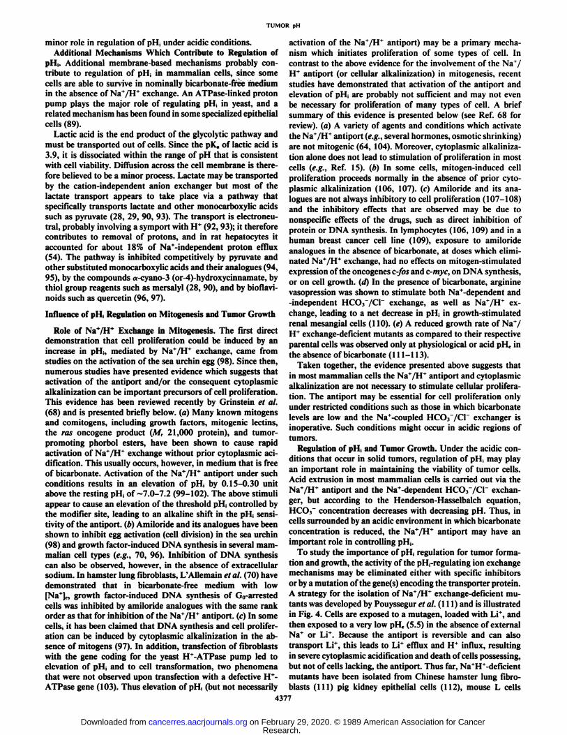
culture, most mammalian cells will not proliferate in mediumat a pH lower than about 6.6 (120, 121). Cell viability asassessed by colony formation also decreases after chronic exposure to low pH, but there is little loss of viability after shortexposure (<6 h) under aerobic conditions to medium at pHc aslow as 6.0 (120, 122). It is expected, however, that hypoxia andacidosis may coexist in some regions of solid tumors, and whenthese conditions were simulated in culture we found a very rapidloss of colony-forming ability in two mammalian cell lines (Ref.122; Fig. 5). Investigation of cell metabolism under these conditions showed that cells had a marked reduction in ATP levels,presumably because of inhibition of glycolysis at low pH andof respiration under hypoxic conditions (122). Whereas cellsincubated at low pHc under aerobic conditions were able tomaintain pi I, about 0.25 unit higher than pi I,, this gradienttended to be lost under hypoxic conditions, probably as a resultof inhibition of the Na+/HT and Na+-dependent HCOr/Cr
exchange mechanisms which regulate pi I, under acidic conditions (62, 123, 124). Inhibition of the exchangers may haveresulted from ATP deprivation as reported for other cells (55,61, 125, 126).
The causes of cell death within solid tumors are poorlyunderstood. The above data are provocative that low pH mayhave a major role in contributing to this process. The decreasedor absent tumor formation from mutant cells lacking Na+/H+
exchange (see above) is consistent with this hypothesis.Ionizing Radiation. Cell survival after ionizing radiation has
been assessed at physiological and at low pH for several mammalian cell lines (127-130). The results of these experimentshave been consistent in demonstrating increased radiation resistance at reduced pHc, although the effect on radiation sensitivity is much less than that due to hypoxia. In two of theabove studies the major effect of low pH, was to increase thewidth of the shoulder region of the cell survival curve (127,128), implying greater capacity for DNA repair under acidicconditions. Such an effect would be magnified when radiationis delivered in multiple small fractions, as in human tumorradiotherapy. In contrast, the studies of Freeman et al. (129,
io t "
io-'246
incubation time (hours)
Fig. 5. Plating efficiency of Chinese hamster ovary cells incubated in air(closed symbols) or hypoxia (N2. open symbols) for up to 6 h at pHe 6.0 (A. A).6.25 (T, V), 6.5 (•.D), or 7.0 (•,O). Mean and range for triplicate plates areplotted, and data are representative of more than 10 experiments. Qualitativelysimilar results have been obtained with other cell lines (modified from Rotin etal. (122)].
131) using CHO cells show little or no effect of low pH inincreasing the width of the shoulder region of the survival curve.
The time at which cells are rendered acidic in relation todelivery of radiation influences their survival. Exposure of CHOcells to acid conditions after irradiation gave increased survivalas compared to acid exposure before or during radiation (132).The probable mechanism is that acid conditions inhibit thefixation of potentially lethal radiation damage (131).
The above results were obtained on exponentially growingcells that were irradiated under aerobic conditions. In solidtumors, many cells may be both acidic and hypoxic, and underthese conditions cell proliferation is inhibited. Freeman andSierra (130) found that low pH did not increase the radioresist-ance of unfed plateau phase cells in culture, and these cells mayprovide a better model for slowly proliferating cells in acidicand nutrient-deprived regions of solid tumors. It seems unlikely,therefore, that acidity adds to radioresistance in hypoxic tumorregions.
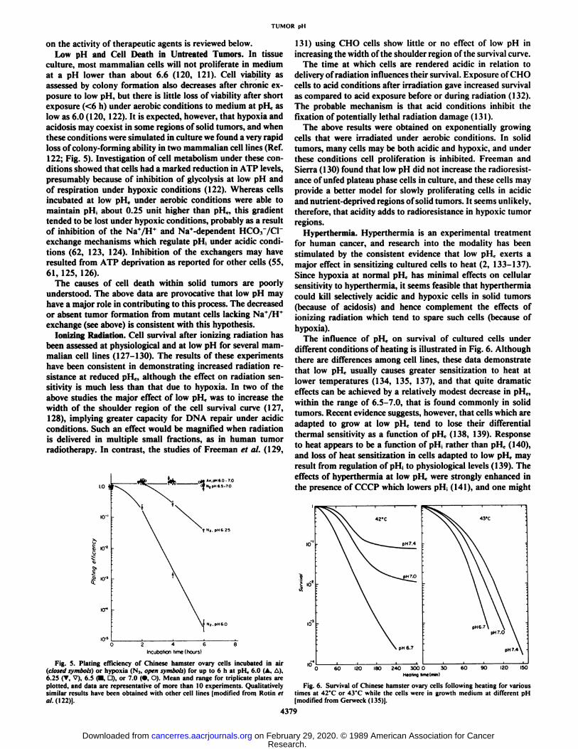
Hyperthermia. Hyperthermia is an experimental treatmentfor human cancer, and research into the modality has beenstimulated by the consistent evidence that low pH,, exerts amajor effect in sensitizing cultured cells to heat (2, 133-137).Since hypoxia at normal pH0 has minimal effects on cellularsensitivity to hyperthermia, it seems feasible that hyperthermiacould kill selectively acidic and hypoxic cells in solid tumors(because of acidosis) and hence complement the effects ofionizing radiation which tend to spare such cells (because ofhypoxia).
The influence of pHc on survival of cultured cells underdifferent conditions of heating is illustrated in Fig. 6. Althoughthere are differences among cell lines, these data demonstratethat low pi I, usually causes greater sensitization to heat atlower temperatures (134, 135, 137), and that quite dramaticeffects can be achieved by a relatively modest decrease in pH„within the range of 6.5-7.0, that is found commonly in solidtumors. Recent evidence suggests, however, that cells which areadapted to grow at low pHt. tend to lose their differentialthermal sensitivity as a function of pHc (138, 139). Responseto heat appears to be a function of pH¡rather than pHc (140),and loss of heat sensitization in cells adapted to low pi I, mayresult from regulation of pH¡to physiological levels (139). Theeffects of hyperthermia at low pHc were strongly enhanced inthe presence of CCCP which lowers pH¡(141), and one might
Fig. 6. Survival of Chinese hamster ovary cells following heating for varioustimes at 42'C or 43'C while the cells were in growth medium at different pH
[modified from Gerweck (135)].
4379
Research. on February 29, 2020. © 1989 American Association for Cancercancerres.aacrjournals.org Downloaded from
TUMOR pH
expect persistence of this effect in cells adapted to low pH,,.Amiloride has also been found to increase thermal sensitivity,perhaps by impairing pH¡regulation, although this effect isobserved both at physiological and at acidic pHc (142).
A major limitation to the application of heat for cancertreatment is the development of thermotolerance, whereby aninitial heat treatment leads to the induction of resistance tosubsequent heat treatments delivered within the next 3-5 days.Maintenance of cells at low pHc after heating was found toinhibit the development of thermotolerance (143, 144), but thisinhibition of thermotolerance at low pHc was not observed incells that were adapted to such conditions (138). Thus bothsingle and fractionated heat treatments would be expected tobe selectively toxic only to cells which have become acutelyacidic within solid tumors, and these selective effects of heatwould be lost as the cells became chronically adapted to lowpHc. Agents which cause acute acidification of cells in thepresence of low pHc might play a therapeutic role in thetreatment of solid tumors by hyperthermia.
The mechanism(s) by which heat treatment kills cells isunknown, although damage to cell membranes and proteindenaturation are believed to play a primary role (145). Recentstudies of pH¡during heating of CHO cells to 42"C or 45°Cat
pHc 6.6 have shown unexpected initial increases in pH¡,although subsequent intracellular acidification was also observed(146, 147). Thus there appears to be no immediate breakdownin mechanisms regulating pH¡,although the effects of heat onspecific membrane exchange mechanisms have not been determined.
Treatment with heat leads to a rapid fall in pH,, in many solidtumors. This effect appears to be due to a marked decrease inblood flow because of vasoconstriction and coagulation necrosis(2, 3, 148, 149). Hyperthermia often stimulates blood flowinitially in normal tissues unless the temperature exceeds ~44°C
(150, 151) and this effect may contribute to selective killing ofcells in tumors, as compared to those in normal tissues, duringsubsequent heat treatment.
Chemotherapy. Anticancer drugs must be transported intocells, by either active transport or passive diffusion, and frequently undergo intracellular metabolism. Since all of theseprocesses, as well as the metabolic pathways that are inhibited,depend on pH, it is expected that the cytotoxic activity ofanticancer drugs may depend on both pHc and pH¡.In particular, where uptake of drugs which contain acidic or basiccharged groups is by passive diffusion, transport will be enhanced at values of pHc that favor the nonionized form of thecompound.
Data on the activity of commonly used anticancer drugs as afunction of varying pHc in cell culture are summarized in Table2. Additional data indicate that low pHe enhances the interaction of several drugs (e.g., bischloroethylinitrosourea, bleomy-cin) when used with hyperthermia, and this effect is muchgreater than the enhancement of either agent used alone (138,154).
The influence of pH,, on the in vivo activity of drugs is likelyto be complex since the more acidic regions of tumors areprobably situated distal from functional blood vessels. Thus thenet effect of anticancer drugs against cells in acidic regions oftumors will depend also on their penetration into tissue fromthe blood vessels through which they are delivered. Penetrationis known to be poor for some drugs such as doxorubicin (162,163).
There have been attempts to use selectively drugs whoseuptake into cells is favored at low pH,. The alkylating agent
triethylenemelamine was used in an attempt to selectively killcells in a solid tumor (presumed hypoxic and acidic) thatsurvived ionizing radiation; although the drug caused delay torecurrence after radiation it did not enhance the probability ofcure (164). Others have shown some increased activity of triethylenemelamine and other alkylating agents for experimentaltumors after treating animals with glucose (165, 166). Thiseffect may be due to a fall in tumor pH but could also beinfluenced by changes in tumor metabolism after treatmentwith glucose.
It is apparent that data relating to the effects of pHc on theactivity of anticancer drugs are incomplete, and most of theavailable data summarized in Table 2 have been obtained (foreach drug) on only one or two cell lines. The use of fluorescentdyes, such as Hoechst 33342, which allow the separation ofcells at different distances from functional blood vessels intumors by flow cytometry, allows study of drug effects indifferent tumor regions (169). Improvements in technologymight allow these methods to be combined with the use offluorescent probes which indicate pH¡in vivo (50) and wouldfacilitate study of the relationship between drug activity andpH¡in tumors.
New Approaches to Therapy. Acidic pH in tumors may enhance the effectiveness of conventional therapeutic modalities.Acidity might also allow the development of new approachestargeted toward the cellular mechanisms that regulate pH¡.Support for the feasibility of this approach derives from theobservation that mutant cells which lack Na+/H+ exchange
have either absent or reduced ability to generate tumors (113,116) and from the demonstration of (acid) pH-dependent cell
killing by agents which impair regulation of pH¡(119, 127).lonophores are compounds which allow transport of ions
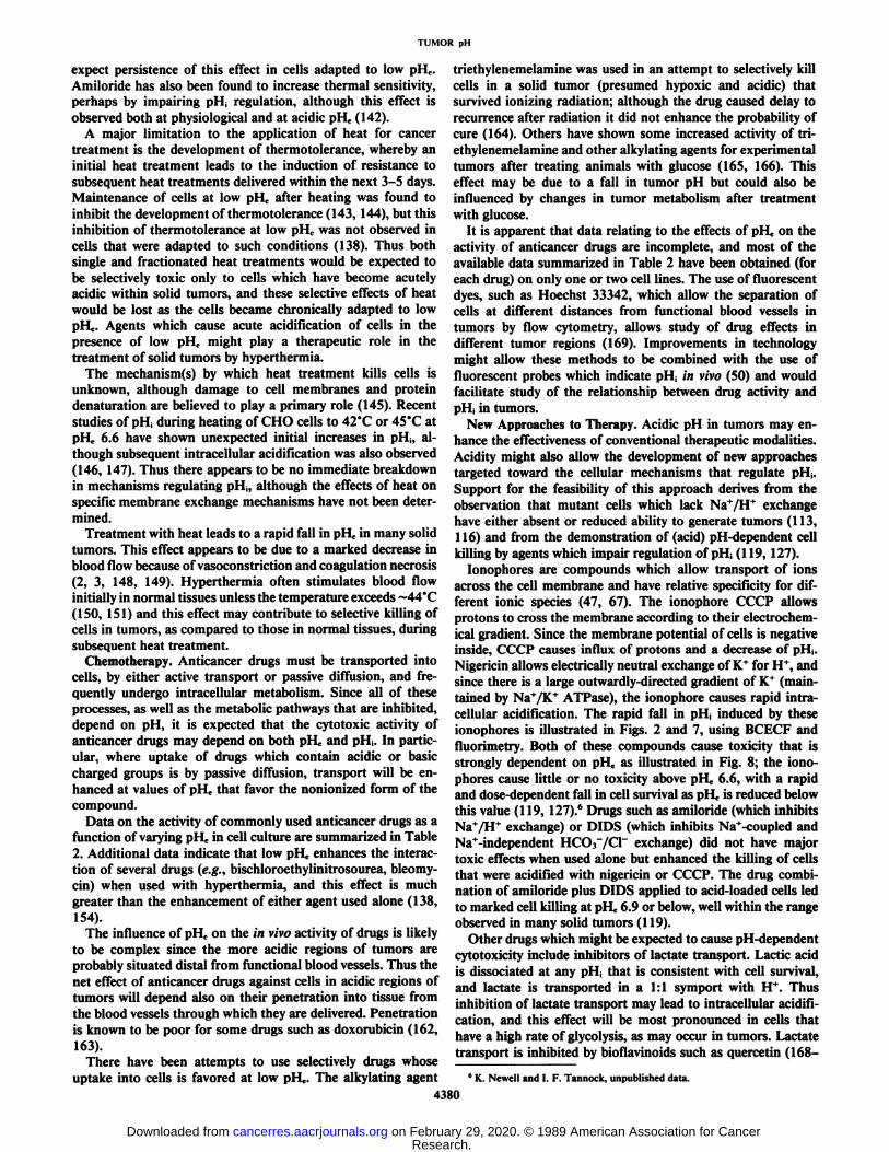
across the cell membrane and have relative specificity for different ionic species (47, 67). The ionophore CCCP allowsprotons to cross the membrane according to their electrochemical gradient. Since the membrane potential of cells is negativeinside, CCCP causes influx of protons and a decrease of pH¡.Nigericin allows electrically neutral exchange of K+ for H+, andsince there is a large outwardly-directed gradient of K+ (maintained by Na+/K+ ATPase), the ionophore causes rapid intra
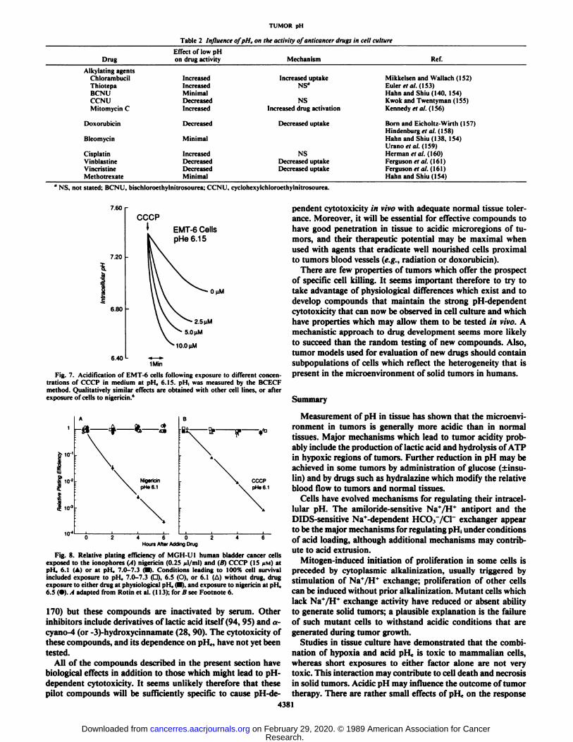
cellular acidification. The rapid fall in pH¡induced by theseionophores is illustrated in Figs. 2 and 7, using BCECF andfluorimetry. Both of these compounds cause toxicity that isstrongly dependent on pHe as illustrated in Fig. 8; the ionophores cause little or no toxicity above pHe 6.6, with a rapidand dose-dependent fall in cell survival as pHc is reduced belowthis value (119, 127).6 Drugs such as amiloride (which inhibitsNa+/H+ exchange) or DIDS (which inhibits Na+-coupled andNa+-independent HCO.r/Cr exchange) did not have major
toxic effects when used alone but enhanced the killing of cellsthat were acidified with nigérianor CCCP. The drug combination of amiloride plus DIDS applied to acid-loaded cells ledto marked cell killing at pHe 6.9 or below, well within the rangeobserved in many solid tumors (119).
Other drugs which might be expected to cause pH-dependentcytotoxicity include inhibitors of lactate transport. Lactic acidis dissociated at any pH, that is consistent with cell survival,and lactate is transported in a 1:1 symport with H". Thus
inhibition of lactate transport may lead to intracellular acidification, and this effect will be most pronounced in cells thathave a high rate of glycolysis, as may occur in tumors. Lactatetransport is inhibited by biofla vino ids such as quercetin (168-
6 K. Newell and I. F. Tannock, unpublished data.
4380
Research. on February 29, 2020. © 1989 American Association for Cancercancerres.aacrjournals.org Downloaded from
TUMOR pH
Table 2 Influence of pH, on the activity of anticancer drugs in cell culture
DrugAlkylating
agentsChlorambucilThiotepaBCNUCCNUMitomycin
CDoxorubicinBleomycinCisplatinVinblastineVincristineMethotrexateEffect
of low pHon drugactivityIncreasedIncreasedMinimalDecreasedIncreasedDecreasedMinimalIncreasedDecreasedDecreasedMinimalMechanismIncreased
uptakeNS"NSIncreased
drugactivationDecreased
uptakeNSDecreased
uptakeDecreaseduptakeRef.Mikkelsen
and Wallach(152)Euleret al.(153)Hahnand Shiu(140.154)Kwokand Twentyman(155)Kennedy
ft al.(156)Born
and Eicholtz-Wirth(157)Hindenburgrtal.(158)Hahn
and Shim MX.154)Uranoet al.(159)Herman
et al. (160)Fergusonet al. (16 1)Fergusonet al. ( 16 1)Hahn
and Shiu(I54)1NS, not stated; BCNU, bischloroethylnitrosourea; CCNU, cyclohexylchloroethylnitrosourea.
7.60
7.20
6.80
6.40
EMT-6 CellspHe6.15
n nM
10.0(iM
1Min
Fig. 7. Acidification of EMT-6 cells following exposure to different concentrations of CCCP in medium at pH, 6.15. pH¡was measured by the BCECFmethod. Qualitatively similar effects are obtained with other cell lines, or afterexposure of cells to nigericin.*
io-3
02460246Hours After Adding Drug
Fig. 8. Relative plating efficiency of MGH-UI human bladder cancer cellsexposed to the ionophores (A) nigericin (0.25 (il/ml) and (B) CCCP (15 /IM) atpHe 6.1 (A) or at pll. 7.0-7.3 <•).Conditions leading to 100% cell survivalincluded exposure to pH, 7.0-7.3 (d). 6-5 (O), or 6.1 (A) without drug, drugexposure to either drug at physiological pll, (•).and exposure to nigericin at pi I,6.5 (•).,1 adapted from Rotin et al. (113); for B see Footnote 6.
170) but these compounds are inactivated by serum. Otherinhibitors include derivatives of lactic acid itself (94,95) and a-cyano-4 (or -3)-hydroxycinnamate (28, 90). The cytotoxicity ofthese compounds, and its dependence on pH.. have not yet beentested.
All of the compounds described in the present section havebiological effects in addition to those which might lead to pH-dependent cytotoxicity. It seems unlikely therefore that thesepilot compounds will be sufficiently specific to cause pH-de-
pendent cytotoxicity in vivo with adequate normal tissue tolerance. Moreover, it will be essential for effective compounds tohave good penetration in tissue to acidic microregions of tumors, and their therapeutic potential may be maximal whenused with agents that eradicate well nourished cells proximalto tumors blood vessels (e.g., radiation or doxorubicin).
There are few properties of tumors which offer the prospectof specific cell killing. It seems important therefore to try totake advantage of physiological differences which exist and todevelop compounds that maintain the strong pH-dependentcytotoxicity that can now be observed in cell culture and whichhave properties which may allow them to be tested in vivo. Amechanistic approach to drug development seems more likelyto succeed than the random testing of new compounds. Also,tumor models used for evaluation of new drugs should containsubpopulations of cells which reflect the heterogeneity that ispresent in the microenvironment of solid tumors in humans.
Summary
Measurement of pH in tissue has shown that the microenvironment in tumors is generally more acidic than in normaltissues. Major mechanisms which lead to tumor acidity probably include the production of lactic acid and hydrolysis of ATPin hypoxic regions of tumors. Further reduction in pH may beachieved in some tumors by administration of glucose (±insu-lin) and by drugs such as hydralazine which modify the relativeblood flow to tumors and normal tissues.
Cells have evolved mechanisms for regulating their intracel-lular pH. The amiloride-sensitive Na+/H* antiport and theDIDS-sensitive Na*-dependent HCO.r/Cl" exchanger appear
to be the major mechanisms for regulating pll, under conditionsof acid loading, although additional mechanisms may contribute to acid extrusion.
Mitogen-induced initiation of proliferation in some cells ispreceded by cytoplasmic alkalinization, usually triggered bystimulation of Na+/H+ exchange; proliferation of other cells
can be induced without prior alkalinization. Mutant cells whichlack NaVH* exchange activity have reduced or absent ability
to generate solid tumors; a plausible explanation is the failureof such mutant cells to withstand acidic conditions that aregenerated during tumor growth.
Studies in tissue culture have demonstrated that the combination of hypoxia and acid pH, is toxic to mammalian cells,whereas short exposures to either factor alone are not verytoxic. This interaction may contribute to cell death and necrosisin solid tumors. Acidic pH may influence the outcome of tumortherapy. There are rather small effects of pHc on the response
4381
Research. on February 29, 2020. © 1989 American Association for Cancercancerres.aacrjournals.org Downloaded from

TUMOR pH
of cells to ionizing radiation but acute exposure to acid pHecauses a marked increase in response to hyperthermia; thiseffect is decreased in cells that are adapted to low pHc. Aciditymay have varying effects on the response of cells to conventionalanticancer drugs. lonophores such as nigericin or CCCP causeacid loading of cells in culture and are toxic only at low pHc;this toxicity is enhanced by agents such as amiloride or DIDSwhich impair mechanisms involved in regulation of pH¡.It issuggested that acid conditions in tumors might allow the development of new and relatively specific types of therapy whichare directed against mechanisms which regulate pi I, under acidconditions.
Acknowledgments
We thank R. P. Hill, K. Newell, and S. Grinstein for their adviceand critical review of the manuscript.
REFERENCES
1. Van den Berg. A. P., Wike-Hooley, J. L., Van Den Berg-Blok, A. E., et al.Tumour pH in human mammary carcinoma. Eur. J. Cancer Clin. Oncol.,18:457-462, 1982.
2. Calderwood, S. K., and Dickson, J. A. pH and tumor response to hyperthermia. Adv. Radiât.Biol., JO: 135-190, 1983.
3. Bicher, H. I., Hetzel, F. W., Sandhu, T. S., et al. Effects of hyperthermiaon normal and tumor microenvironment. Radiology, 137: 523-530, 1980.
4. Acker, H., Carlsson, J., Holtermann, G., et al. Influence of glucose andbuffer capacity in the culture medium on growth and pH in spheroids ofhuman thyroid carcinoma and human glioma origin. Cancer Res., 47:3504-3508, 1987.
5. Ng, T. C, Evanochko, W. T., Hiramoto, R. N., et al. 31PNMR spectroscopyof in vivo tumors. J. Magn. Reson., 49: 271-286, 1982.
6. Evanochko, W. T., Ng. T. C., Lilly, M. B., et al. In vivo 3'P NMR study of
the metabolism of murine mammary 16/C adenocarcinoma and its responseto chemotherapy, X-irradiation, and hyperthermia. Proc. Nati. Acad. Sci.USA, «0:334-338, 1983.
7. Okunieff, P. G., Koutcher, J. A., Gerweck, L., et al. Tumor size dependentchanges in a murine fibrosarcoma: use of in vivo 3IP NMR for noninvasive
evaluation of tumor metabolic status. Int. J. Radiât.Oncol. Biol. Phys., 12:793-799, 1986.
8. Oberhaensli, R. D., Hilton-Jones, D., Bone, B. J., et al. Biochemicalinvestigation of human tumours in vivo with phosphorus-31 magnetic resonance spectroscopy. Lancet, 2: 8-11, 1986.
9. Daly, P. F., and Cohen, J. S. Magnetic resonance spectroscopy of tumorsand potential in vivo clinical applications: a review. Cancer Res., 49: 770-779, 1989.
10. Wike-Hooley, J. L., Haveman, J., and Reinhold, J. S. The relevance oftumour pH to the treatment of malignant disease. Radiother. Oncol., 2:343-366, 1984.
11. Wike-Hooley, J. L., Van Den Berg, A. P., Van Der Zee, J., and Reinhold,H. S. Human tumour pH and its variation. Eur. J. Cancer Clin. Oncol., 21:785-791, 1985.
12. Thisthlethwaite, A. J., Alexander, G. A., Moylan, D. J., Ill, and Leeper, D.B. Modification of human tumor pH by elevation of blood glucose. Int. J.Radiât.Oncol. Biol. Phys., 13:603-610. 1987.
13. Jain, R. K., Shah, S. A., and Finney, P. L. Continuous noninvasive monitoring of pH and temperature in rat Walker 256 carcinoma during normoglycemia and hyperglycemia. J. Nati. Cancer Inst., 73:429-436, 1984.
14. Vaupel, P. W., Frinak, S., and Bicher, H. I. Heterogeneous oxygen partialpressure and pH distribution in C3H mouse mammary adenocarcinoma.Cancer Res., 41: 2008-2013, 1981.
15. Kallinowski, F., and Vaupel, P. pH distributions in spontaneous and iso-transplamed rat tumours. Br. J. Cancer, 58: 314-321, 1988.
16. Catalana, S., Cohen, C., and Sapirstein, L. A. Relation between size andperfusion rate of transplanted tumors. J. Nati. Cancer Inst., 29: 389-394,1962.
17. Tannock, I. F., and Steel, G. G. Quantitative techniques for study of theanatomy and function of small blood vessels in tumors. J. Nati. CancerInst., «.-771-782, 1969.
18. Warburg, O. H. The Metabolism of Tumors. Translated from the Germanedition by F. Dickens. London: Constable, 1930.
19. Burk, D., Woods, M., and Hunter, J. On the significance of glucolysis forcancer growth, with special reference to Morris rat hepatomas. J. Nati.Cancer Inst., 38: 839-863, 1967.
20. Weinhouse, S. On respiratory impairment in cancer cells. Science (Wash.DC), 124:267-26», 1956.
21. McKeehan, W. L. Glycolysis, glutaminolysis and cell proliferation. Cell.Biol. Int. Rep., 6: 635-650, 1982.
22. Pouyssegur, J., Franchi, A., Salomon, J-C., and Silvestre, P. Isolation of aChinese hamster fibroblast mutant defective in hexose transport and aerobic
glycolysis: its use to dissect the malignant phenotype. Proc. Nati. Acad. Sci.USA, 77:2698-2701, 1980.
23. Franchi, A., Silvestre, P., and Pouyssegur, J. A genetic approach to the roleof energy metabolism in the growth of tumor cells: tumorigenicity offibroblast mutants deficient either in glycolysis or in respiration. Int. J.Cancer, 27:819-827, 1981.
24. Hochachka, P. W., and Mommsen, T. P. Protons and anaerobiosis. Science(Wash. DC), 219: 1391-1397, 1983.
25. Busa, W. B., and Nuccitelli, R. Metabolic regulation via intracellular pH.Am. J. Physiol., 246: R409-R438, 1984.
26. Halperin, M. L., Connors, H. P., Relman, A. S., and Karnovsky, M. L.Factors that control the effect of pH on glycolysis in leukocytes. J. Biol.Chem., 244: 384-390, 1969.
27. Wilhelm, G., Schulz, J., and Hofmann, E. pH-dependence of aerobicglycolysis in Ehrlich ascites tumour cells. FEBS Lett., 17:158-162, 1971.
28. Spencer, T. L., and Lehninger, A. L. L-Lactate transport in Ehrlich ascitestumour cells. Biochem. J., 154:405-414, 1976.
29. Dubinsky, W. P., and Racker, E. The mechanism of láclatetransport inhuman erythrocytes. J. Membr. Biol., 44: 25-36, 1978.
30. Guillino, P. M., Grantham, F. H., Smith, S. H., and Haggerty, A. C.Modifications of the acid-base status of the internal milieu of tumors. J.Nati. Cancer Inst., 34: 857-869, 1965.
31. Jahde, E., and Rajewsky, M. F. Tumor-selective modification of cellularmicroenvironment in vivo: effect of glucose infusion on the pH in normaland malignant rat tissues. Cancer Res., 42: 1505-1512, 1982.
32. Sevick, E. M., and Jain, R. K. Blood flow and venous pH of tissue-isolatedWalker 256 carcinoma during hyperglycemia. Cancer Res., 48:1201-1207,1988.
33. Thistlethwaite, A. J., Alexander, G. A., Moylan, D. J., HI, and Leeper, D.B. Modification of human tumor pH by elevation of blood glucose. Int. J.Radiât.Oncol. Biol. Phys., 13:603-610, 1987.
34. Voegtlin, C., Fitch, R. H., Kahler, H., et al. Experimental studies on cancer.I. The influence of the parenteral administration of certain sugars on thepH of malignant tumors. NIH Bull., 164: 1-14, 1935.
35. Vaupel, P. W., and Okunieff, P. G. Role of hypovolemic hemoconcentrationin dose-dependent flow decline observed in murine tumors after intraperi-toneal administration of glucose or mannitol. Cancer Res., 48: 7102-7106,1988.
36. Von Ardenne, M., and Reitnauer, P. G. Verstärkungder mit Glukosinfusionerzeilbaren Tumorubersauerung in vivo durch Natriumnitroprussid. Pharmazie, 34: 447, 1979.
37. Okunieff, P., Kallinowski, F., Vaupel, P., and Neuringer, L. J. Effects ofhydralazine-induced vasodilatation on the energy metabolism of murinetumors studied by in vivo "P-nuclear magnetic resonance spectroscopy. J.Nati. Cancer Inst., 80: 745-750, 1988.
38. Chaplin, D. J., and Acker, B. The effect of hydralazine on the tumorcytotoxicity of the hypoxic cell cytotoxin RSU-1069: evidence for therapeutic gain. Int. J. Radiât.Oncol. Biol. Phys., 13: 579-585, 1987.
39. Tobari, C., Van Kersen, I., and Hahn, G. M. Modification of pH of normaland malignant mouse tissue by hydralazine and glucose, with and withoutbreathing of 5% CO2 and 95% air. Cancer Res., 48: 1543-1547, 1988.
40. Von Ardenne, M., and Reitnauer, P. G. Verstärkung der mit GlukoseInfusion erzeilbaren Tumor Übersäuerungin vivo durch Amygdalin und /3-Glukosidase. Arch. Geschwulstforsch., 45: 135-145, 1975.
41. Von Ardenne, M., and Reitnauer, P. G. Verstärkungder mit Glukoseinfusion erzeilbaren Tumorubersauerung in vivodurch NAD. Arch. Geschwulstforsch., 46:197-203, 1976.
42. Thomas, R. C. Intracellular pH of snail neurones measured with a new pH-sensitive glass micro-electrode. J. Physiol., 238: 159-180, 1974.
43. Wadell, W. J., and Butler, T. C. Calculation of intracellular pH from thedistribution of 5,5-dimethyl-2,4-oxazolidinedione (DMO). Application toskeletal muscle of the dog. J. Clin. Invest., 38: 720-729, 1959.
44. Roos, A., and Boron, W. F. Intracellular pH. Physiol. Rev., 61: 296-434,1981.
45. Rink, T. J., Tsien, R. Y., and Pozzan, T. Cytoplasmic pH and free Mg2* inlymphocytes. J. Cell Biol., 95: 189-196, 1982.
46. Thomas, J. A., Buchsbaum, R. N., Zimniak, A., and Racker, E. IntracellularpH measurements in Ehrlich ascites tumor cells utilizing spectroscopicprobes. Biochemistry 18: 2210-2218, 1979.
47. Pressman, B. C. Biological applications of ionophores. Annu. Rev.Biochem., 45: 501-530, 1976.
48. Musgrove, E., Rugg, C., and Hedley, D. Flow cytometric measurements ofcytoplasmic pH: a critical evaluation of available fluorochromes. Cytometry,7:347-355,1986.
49. Paradiso, A. M., Tsien, R. Y., and Machen, T. E. Digital image processingof intracellular pH in gastric oxyntic and chief cells. Nature (Lond.), 325:447-449, 1987.
50. Hedley, D. W., and Jorgensen, H. B. Flow cytometric measurement ofintracellular pH in B16 tumours; intercell variance and effects of pretreatment with glucose. Exp. Cell Res., 180:106-116, 1989.
51. Moolenaar, W. H., Tertoolen, L. G. J., and De Laat, S. W. The regulationof cytoplasmic pH in human fibroblasts. J. Biol. Chem., 259: 7563-7569,1984.
52. Frelin, C., Vigne, P., Ladoux, A., and Lazdunski, M. The regulation of theintracellular pH in cells from vertebrates. Eur. J. Biochem., 174: 3-14,1988.
53. Madshus, I. H. Regulation of intracellular pH in eukaryotic cells. Biochem.J., 250:1-8, 1988.
4382
Research. on February 29, 2020. © 1989 American Association for Cancercancerres.aacrjournals.org Downloaded from
TUMOR pH
54. Anwer, M. S., and Nolan, K. Characterization of proton efflux pathways in 84.rat hepatocytes. Hepatology (Baltimore), 8: 728-734, 1988.
55. Boron, W. F., and Deweer, P. Intracellular pH transients in squid giantaxons caused by CO2, NH,, and metabolic inhibitors. J. Gen. Physiol., 67: 85.91-112, 1976.
56. Aronson, P. S. Kinetic properties of the plasma membrane Na*-H* exchan
ger. Annu. Rev. Physiol., 47: 545-560, 1985. 86.57. Cala, P. M. Volume regulation by Amphiuma red blood cells. The membrane
potential and its implications regarding the nature of the ion-flux pathways.J. Gen. Physiol., 76: 683-708, 1980. 87.
58. Grinstein. S., Cohen, S., and Rothstein, A. Cytoplasmic pH regulation inthymic lymphocytes by an amiloride-sensitive Na*/H* antiport. J. Gen.Physiol., S3: 341 -368, 1984. 88.
59. Murer, H.. Hopfer, U., and Kinne, R. Sodium/proton antiport in brush-border-membrane vesicles isolated from rat small intestine and kidney. 89.Biochem. J., 154: 597-604, 1976.
60. Kinsella. J. L., and Aronson, P. S. Determination of the coupling ratio for 90.Na*/H* exchange in renal microvillus membrane vesicles. Biochim. Bio-phys. Acta, 689: 161-164, 1982.
61. Cassel, D., Katz, M., and Rotman. M. Depletion of cellular ATP inhibits 91.Na*/H* antiport in cultured human cells. Modulation of the regulatory'
effect of intracellular protons on the antiporter activity. J. Biol. Chem., 261:5460-5466, 1986.
62. Rotin, D. Regulation of intracellular pH as an important determinant of 92.tumor cell viability. Ph.D. Dissertation, University of Toronto, Toronto.Ontario. Canada. 1988. 93.
63. Aronson, P., Nee, J., and Suhm, M. A. Modifier role of internal H* inactivating the Na*/H* exchanger in renal microvillus membrane vesicles.Nature (Lond.), 299: 161-163, 1982.
64. Grinstein, S., and Rothstein, A. Mechanisms of regulation of the Na*/H* 94.exchanger. J. Membr. Biol., 90: 1-12, 1986.
65. Sardet, C., Franchi, A., and Pouyssegur, J. Molecular cloning, primarystructure, and expression of the human growth factor-activatable Na'/H* 95.antiporter. Cell, 56: 271-280, 1989.
66. Grinstein, S., and Smith, J. D. Asymmetry of the Na*/H* antiport of dog
red cell ghosts. Sidedness of inhibition by amiloride. J. Biol. Chem., 262: 96.9088-9092, 1987.
67. Franchi, A., Cragoe, E., Jr., and Pouyssegur, J. Isolation and properties offibroblast mutants overexpressing an altered NaVH* antiporter. J. Biol. 97.Chem., 261: 14614-14620, 1986.
68. Grinstein, S., Rotin, D., and Mason, M. J. Na*/H* exchange and growthfactor-induced cytosolic pH changes. Role in cellular proliferation. Biochim. 98.Biophys. Acta, 988: 73-97, 1989.
69. Cragoe, E. J., Jr., Woltersdorf. O. W., Jr., Bicking, J. B.. et al. Pyrazine 99.diuretics. II. /V-Amidino-3-substituted amino-5-6-halopyrazinecarboxam-ides. J. Med. Chem., 10:66-75, 1967.
70. L'Allemain, G., Franchi, A., Cragoe, E., and Pouyssegur. J. Blockade of 100.the Na*/H* antiport abolishes growth factor-induced DNA synthesis infibroblasts. Structure-activity relationships in the amiloride series. J. Biol.Chem., 259:4313-4319, 1984. 101.
71. Vigne. P., Felin, C., Audinot, M., el al. ['H]Ethylpropylamiloride, a radio-labelled diuretic for the analysis of the \a 11 exchange system. Its usewith kidney cell membranes. EMBO J., 3: 2647-2651, 1984. 102.
72. Haggerty, J. G., Agarwal, N., Reilly, R. F., el al. Pharmacologically differentNa/H antiporters in the apical and basolateral surfaces of cultured porcinekidney cells (LLC-PK,). Proc. Nati. Acad. Sci. USA, 85:6797-6801. 1988.
73. Frelin, C-, Vigne, P., Barbry, P., and Lazdunski, M. Interaction of guani- 103.dinium and guanidinium derivatives with the Vili exchange system. Eur.J. Biochem., 154: 241-245, 1986. 104.
74. Kinsella. J. L., and Aronson, P. S. Properties of the Na*/H* exchanger inrenal microvillus membrane vesicles. Am. J. Physiol., 238: F46I-F469, 105.1980.
75. Parker, J. C. Volume-responsive sodium movements in dog red blood cells.Am. J. Physiol., 244: C324-C330, 1983. 106.
76. Kinsella, J. L., Werle, J., Wilkins, N., and Sacktor, B. Inhibition of Na*/H* exchange by AVV'-dicyclohexylcarbodiimide in isolated rat renal brushborder membrane vesicles. J. Biol. Chem., 262: 7092-7097. 1987. 107.
77. Igarashi, P., and Aronson, P. S. Covalent modification of the renal Na*/H* exchanger by AVV'-dicyclohexylcarbodiimide. J. Biol. Chem., 262: 860-
868, 1987.78. Grinstein, S., Cohen, S., and Rothstein, A. Chemical modification of the 108.
Na+/H* exchange of thymic lymphocytes. Inhibition of jV-ethylmaleimide.Biochim. Biophys. Acta, 812: 213-222, 1985.
79. Madshus, I. H., and Olsnes, S. Selective inhibition of sodium-linked andsodium-independent bicarbonate/chloride antiport in Vero cells. J. Biol. 109.Chem., 262: 7486-7491, 1987.
80. Rothenberg, P., Glaser, L., Schlesinger, P., and Cassel, D. Activation ofNa*/H* exchange by epidermal growth factor elevates intracellular pH inA431 cells. J. Biol. Chem., 25«:12644-12653, 1983. 110.
81. L'Allemain. G., Paris, S., and Pouyssegur, J. Role of a Na*-dependent Cl"/11<'<) i exchange in regulation of intracellular pH in fibroblasts. J. Biol.
Chem., 260: 4877-4883, 1985. 111.82. Tonnessen, T. I., Ludi, J., Sandvig, K., and Olsnes, S. Bicarbonate/chloride
antiport in Vero cells. I. Evidence for both sodium-linked and sodium-independent exchange. J. Cell. Physiol.. /32: 183-191. 1987. 112.
83. Cassel, D., Scharf, O.. Rotman, M., et al. Characterization of Na*-linkedand Na*-independent CI~/HCO.r exchange systems in Chinese hamsterlung fibroblasts. J. Biol. Chem., 263: 6122-6127. 1988. 113.
4383
Reincrtsen, K. V„Tonnessen, T. I.. Jacobsen, J., et al. Role of chloride-bicarbonate antiport in the control of cytosolic pH. Cell-line differences inactivity and regulation of antiport. J. Biol. Chem., 263:11117-11125, 1988.Boron. W. F., and Russell. J. M. Stoichiometry and ion dependencies ofthe intracellular-pH regulating mechanism in squid giant axons. J. Gen.Physiol., 81: 373-399. 1983.Boron. W. F.. Hogan. E.. and Russell. J. M. pH-sensitive activation of theintracellular pH regulation system in squid axons by ATP-7-S. Nature(Lond.), 332: 262-265, 1988.Cabatchnick, Z. I., Knauf. P. A., and Rothstein. A. The anión transportsystem of the red blood cell. The role of membrane protein evaluated by theuse of "probes." Biochim. Biophys. Acta. 5/5: 239-302. 1978.
Boron. W. F. Intracellular pH regulation in epithelial cells. Annu. Rev.Physiol., 48: 377-388, 1986.AI-Awqati. Q. Proton-translocating ATP. Annu. Rev. Cell. Biol., 2: 179-199. 1986.Halestrap. A. P. Transport of pyruvate. NAD. láclateinto human erythro-cytes. Evidence for the involvement of the chloride carrier, and a chloride-independent carrier. Biochem. J.. 756: 193-207. 1976.Deuticke. B., Meyer, E., and Forst. B. Discrimination of three parallelpathways of láclate transport in the human erythrocyte membrane byinhibitors and kinetic properties. Biochim. Biophys. Acta. 684: 96-110.1982.Barbara!. B.. and Podevin, R-A. Stoichiomelry of the renal sodium-i -lactatecotransporter. J. Biol. Chem.. 263: 12190-12193. 1988.Balkovetz, D. F., Leibach. F. M., Mahesh, V. B., and Ganapathy. V. Aproton gradient is the driving force for uphill transport of lactate in humanplacenta! brush-border membrane vesicles. J. Biol. Chem.. 263: 13823-13830. 1988.Johnson, J. H.. Belt, J. A., Dubinsky, W. P., et al. Inhibition of lactatetransport in Ehrlich ascites tumor cells and human erythrocytes by asynthetic anhydride of lactic acid. Biochemistry, 19: 3836-3840, 1980.Johnson, J. H., Zimniak, A., and Racker, E. Inhibition of hexokinase andprotein kinase activities of tumor cells by a chloromethyl ketone derivativeof lactic acid. Biochemistry. 21: 2984-2989. 1982.Moolenaar, W. H., Yarden, Y., De Laat, S. W., and Schlessinger. J.Epidermal growth factor induces electrically silent Na* influx in humanfibroblasts. J. Biol. Chem., 257: 8502-8506, 1982.Zetterbcrg. A., and Engstrom. W. Mitogenic effect of alkaline pH onquiescent, serum-starved cells. Proc. Nati. Acad. Sci. USA, 78:4334-4338,1981.Johnson. J. D.. and Epel, D. Intracellular pH and activation of sea urchineggs after fertilization. Nature (Lond.), 262: 661-664, 1976.Moolenaar. W. H., Tsien, R. Y.. Van der Saag, P. T., and De Laat, S. W.Na*/H* exchange and cytoplasmic pH in the action of growth factors inhuman fibroblasts. Nature (Lond.), 304: 645-648, 1983.Moolenaar, W. H., Tsien, R. Y., Van der Saag, P. T., and De Laat, S. W.Phorbol ester and diacylglycerol mimic growth factors in raising cytoplasmicpH. Nature (Lond.), 312: 371-374, 1984.Paris, S., and Pouyssegur, J. Growth factors activate the Na+/H+ antiporterin quiescent fibroblasts by increasing its affinity for intraccllular H*. J. Biol.Chem.. 259: 10989-10994. 1984.Grinstein. S., Cohen, S., Goetz. J. D.. et al. Characterization of the activation of Na*/H* exchange in lymphocytes by phorbol esters: Change in
cytoplasmic pH dependence of the antiport. Proc. Nati. Acad. Sci. USA,«2:1429-1433, 1985.Perona, R.. and Serrano. R. Increased pH and tumorigenicity of fibroblastsexpressing a yeast proton pump. Nature (Lond.), 334: 438-440, 1988.Moolenaar, W. H., Defize, L. H. K., and De Laat, S. W. Ionic signallingby growth factor receptors. J. Exp. Biol., 124: 359-373, 1986.Grinstein, S., Smith. J. D., Onizuka, R., et al. Activation of Na*/H*exchange and the expression of cellular proto-oncogenes in mitogen andphorbol ester-treated lymphocytes. J. Biol. Chem., 26J: 8658-8665, 1988.Deutsch, C., Taylor, J. S., Price, M., et al. pH homeostasis in humanlymphocytes: modulation by ions and mitogen. J. Cell Biol., 98: 885-894,1984.Haggerty, J. G.. Agarwal. N., Amsler. K., et al. Stimulation by serum of theNa*/H* antiporter in quiescent pig kidney epithelial (LLC-PK,) cells and
role of the antiporter in the reinitiation of DNA synthesis. J. Cell. Physiol.,132: 173-177. 1987.Mills, G. B., Cragoe, E. J., Jr., Gelfand, E. W., and Grinstein, S. Interlcukin2 induces a rapid increase in intracellular pH through activation of a ViH* antiport. Cytoplasmic alkalinization is not required for lymphocyteproliferation. J. Biol. Chem., 260: 12500-12507, 1985.Church, J. G., Mills, G. B., and Buick, R. N. Activation of the Na*/H*antiport is not required for EGF-dependent gene expression, growth inhibition or proliferation in human breast cancer cells. Biochem. J., 257: 151-157, 1989.Ganz, M. B., Boyarsky, G., Sterzel, R. B., and Boron, W. F. Argininevasopressin enhances pH, regulation in the presence of HCOr by stimulating three acid-base transport systems. Nature (Lond.). 337:648-651. 1989.Pouyssegur, J., Sardet. C.. Franchi, A., et al. A specific mutation abolishingNa*/H* antiport activity in hamster fibroblasts precludes growth at neutraland acidic pH. Proc. Nati. Acad. Sci. USA. 81:4833-4837. 1984.Agarwal. N., Haggerty. J. G.. Adelberg, E. A., and Slayman. C. W. Isolationand characterization of a Na*/H* antiporter-deficicnt mutant of LLC-PK,cells. Am. J. Physiol., 257: C825-C830, 1986.Rotin. D., Steele-Norwood, D.. Grinstein, S.. and Tannock. I. Requirement
Research. on February 29, 2020. © 1989 American Association for Cancercancerres.aacrjournals.org Downloaded from

TUMOR pH
of the NaVH* exchanger for tumor growth. Cancer Res., 49: 205-211,
1989.114. Franchi, A., Perucca-Lostanlen, D., and Pouyssegur, J. Functional expres
sion of a human Na*/H* antiporter gene transfected into antiporter-deficientmouse L-cells. Proc. Nati. Acad. Sci. USA, S3: 9388-9892. 1986.
115. Lagarde, A. E., and Pouyssegur, J. M. The Na*/H* antiport in cancer.Cancer Biochem. Biophys., 9: 1-14, 1986.
116. Lagarde. A. E., Franchi. A. J.. Paris, S.. and Pouyssegur, J. M. Effect ofmutations affecting Na*/H* antiport activity on tumorigenic potential ofhamster lung fibroblasts. J. Cell Biochem.. 36: 249-260, 1988.
117. Kovnat, A., Armitage, M., and Tannock, I. F. Xenografts of human bladdercancer in immune-deprived mice. Cancer Res., 42: 3696-3703, 1982.
118. Sparks, R. L., Pool, T. B., Smith. N. K. R., and Cameron. I. L. Effects ofamiloride on tumor growth and intracellular element contact of tumor cellsin vivo. Cancer Res., 43: 73-77, 1983.
119. Rotin, D., Wan, P., Grinstein, S., and Tannock, I. Cytotoxicity of compounds that interfere with the regulation of intracellular pH: a potentialnew class of anticancer drugs. Cancer Res., 47: 1497-1504, 1987.
120. Eagle, H. The effect of environmental pH on the growth of normal andmalignant cells. J. Cell. Physiol., 82: 1-8, 1973.
121. Taylor, I. W., and Hudson, P. J. Cell cycle regulation by environmental pH.J. Cell. Physiol., 121: 517-525, 1984.
122. Rotin, D., Robinson, B., and Tannock, I. F. Influence of hypoxia and anacidic environment on the metabolism and viability of cultured cells: potential implications for cell death in tumors. Cancer Res., 46:2821-2826,1986.
123. Gillies, R. J., Ogino, T., Shulman, R. G.. and Ward, D. C. "P nuclear
magnetic resonance evidence for the regulation of intracellular pH by Ehrlichascites tumor cells. J. Cell Biol., 95: 24-28, 1982.
124. Bowen, J. W., and Levinson, C. H* transport and the regulation of intracellular pH in Ehrlich ascites tumor cells. J. Membr. Biol. 79: 7-18, 1984.
125. Thomas, R. C. Ionic mechanism of the H* pump in a snail neurone. Nature(Lond.). 262: 54-55, 1976.
126. Russell, J. M., and Boron, W. F. Role of chloride transport in regulation ofintracellular pH. Nature (Lond.), 264: 73-74, 1976.
127. Haveman, J. The influence of pH on the survival after \-irradiation ofcultured malignant cells. Effects of carbonylcyanide-3-chlorophenylhydra-zone. Int. J. Radiât.Biol., 37: 201-205, 1980.
128. Rottinger, E. M., Mendonca, M., and Gerweck, L. E. Modification of pHinduced cellular inactivation by irradiation-glial cells. Int. J. Radiât.Oncol.Biol. Phys., 6; 1659-1662, 1980.
129. Freeman, M. L., Holahan, E. V., Highfield, D. P., et al. The effect of pHon hyperthermic and X-ray induced cell killing. Int. J. Radiât.Oncol. Biol.Phys., 7:211-216, 1981.
130. Rottinger, E. M., and Mendonca, M. Radioresistance secondary to low pHin human glial cells and Chinese hamster ovary cells. Int. J. Radiât.Oncol.Biol. Phys., 8: 1309-1314, 1982.
131. Freeman, M. L., and Sierra, E. An acidic extracellular environment reducesthe fixation of radiation damage. Radiât.Res., 97: 154-161, 1984.
132. Holahan, E. V., Stuart, P. K., and Dewey. W. C. Enhancement of survivalof CHO cells by acidic pH after X-irradiation. Radiât.Res., 89: 433-435,1982.
133. Overgaard, J. Influence of extracellular pH on the viability and morphologyof tumor cells exposed to hyperthermia. J. Nati. Cancer Inst., 56: 1243-1250, 1976.
134. Freeman, M. L., Dewey, W. C., and Hopwood, L. E. Effect of pH onhyperthermic cell survival. J. Nati. Cancer Inst., 58: 1837-1839, 1977.
135. Gerweck, L. E. Modification of cell lethality at elevated temperatures. ThepH effect. Radiât.Res., 70: 224-235, 1977.
136. Hahn. G. M. Hyperthermia and Cancer. New York: Plenum PublishingCorp., 1982.
137. Gerweck, L. E., and Richards, B. Influence of pH on the thermal sensitivityof cultured human glioblastoma cells. Cancer Res., 41: 845-849. 1981.
138. Hahn. G. M., and Shiu, E. C. Adaptation to low pH modifies thermal andthermochemical responses of mammaliar cells. Int. J. Hyperthermia, 2:379-387, 1986.
139. Chiù.G. L., and Dewey, W. C. The role of low intracellular or extracellularpH in sensitization to hyperthermia. Radiât.Res., 114: 154-167, 1988.
140. Hofer, K. G., and Mivechi, N. F. Tumor cell sensitivity to hyperthermia asa function of extracellular and intracellular pH. J. Nati. Cancer Inst., 65:621-625. 1980.
141. Haveman, J. The pH of the cytoplasm as an important factor in the survivalof in vivo cultured malignant cells after hyperthermia. Effects of carbonyl-cyanide 3-chlorophenylhydrazone. Eur. J. Cancer, 15: 1281-1288, 1979.
142. Miyakoshi, J., Oda. W., Hirata, M., et al. Effects of amiloride on thermo-sensitivity of Chinese hamster cells under neutral and acidic pH. CancerRes., 46: 1840-1843, 1986.
143. Nielsen, O. S., and Overgaard, J. Effect of extracellular pH on thermo-
toléranceand recovery of hyperthermic damage in vitro. Cancer Res., 39:2772-2778, 1979.
144. Goldin, E. M., and Leeper, D. B. The effect of reduced pH on the inductionof thermotolerance. Radiology, 141: 505-508, 1981.
145. Leeper. D. Molecular and cellular mechanisms of hyperthermia alone orcombined with other modalities. In: J. Overgaard (ed.), Hyperthermic Oncology, 1984. Vol, 2, pp. 9-40. London: Taylor and Francis, 1985.
146. Cook, J. A., and Fox, M. H. Effects of acute pH 6.6 and 42°Cheating onthe intracellular pH of Chinese hamster ovary cells. Cancer Res., 48: 497-502, 1988.
147. Cook, J. A., and Fox, M. H. Intracellular pH of Chinese hamster ovarycells heated at 45.0'C at pH 6.6. Radiât.Res., US: 96-105, 1988.
148. Song, C. W. Physiological factors in hyperthermia. Nati. Cancer Inst.Monogr., 61: 169-176, 1982.
149. Vaupel, P., Muller-Klieser, W., Otte, J., et al. Blood flow, tissue oxygénation, and pH distribution in malignant tumours upon localized hyperthermia. Strahlentherapie, 159: 73-81, 1983.
150. Song, C. W. Effect of local hyperthermia on blood flow and microenviron-ment. A review. Cancer Res., (Suppl.), 44:4721s-4730s, 1984.
151. Reinhold, H. S., and Endrich, B. Tumor microcirculation as a target forhyperthermia. Int. J. Hyperthermia, 2: 111-137, 1986.
152. Mikkelsen, R. B.. and Wallach, D. F. H. Transmembrane ion gradients andthermochemotherapy. In: M. Gautherie and E. Albert (eds.), BiomédicalThermology. pp. 103-107. New York: Allan R. Liss Inc., 1982.
153. Euler, J., Sauerman, G., Priesching, A., and Kloekler, K. Wirkung vonTemperatur, pH und Thio-Tepa auf Angehraten und Thymidineinbau vanAszitestumorzellen. Wien. Klin. Wochenschr., 86: 211-219, 1974.
154. Hahn, G. M., and Shiu, E. C. Effect of pH and elevated temperatures onthe cytotoxicity of some chemotherapeutic agents on Chinese hamster cellsi/i vitro. Cancer Res., 43: 5789-5791, 1983.
155. Kwok, T. T., and Twentyman, P. R. Effects of changes in oxygen tension,pH and glucose concentration on the response to CCNU of EMT6 mousetumor monolayer cells and multicellular spheroids. Int. J. Radiât.Biol.Oncol. Phys., 14: 1221-1229, 1988.
156. Kennedy, K. A., McGurl, J. D., Leondaridis, L., and Alabaster, O. pHdependence of mitomycin C-induced cross-linking activity in EMT6 tumorcells. Cancer Res., 45: 3541-3547, 1985.
157. Born, R., and Eicholtz-Wirth, H. Effect of different physiological conditionson the action of Adriamycin on Chinese hamster cells in vitro. Br. J. Cancer,-«•241-246,1981.
158. Hindenburg, A. A., Stewart, V. J., Baker, M. A., and Taub, R. N. Effectsof pH on cellular accumulation of daunorubicin. Proc. Am. Assoc. CancerRes., 28: 261. 1987.
159. Urano, M., Kahn, J., and Kenton, L. A. Effect of bleomycin on murinetumor cells at elevated temperatures and two different pH values. CancerRes., 48: 615-619, 1988.
160. Herman, T. S., Teicher, B. A., and Collins. L. S. Effect of hypoxia andacidosis on the cytotoxicity of four platinum complexes at normal andhyperthermic temperatures. Cancer Res., 48: 2342-2347, 1988.
161. Ferguson, P. J., Phillips, J. R., Seiner, M., and Cass, C. E. Differentialactivity of vincristine and vinblastine against cultured cells. Cancer Res., 44:3307-3312,1984.
162. Tannock, I. F. Response of aerobic and hypoxic cells in a solid tumor toAdriamycin and cyclophosphamide and interaction of the drugs with radiation. Cancer Res., 42: 4921-4926, 1982.
163. Chaplin, D. J., Durand, R. E., and Olive, P. L. Cell selection from a murinetumour using the fluorescent probe Hoechst 33342. Br. J. Cancer, 51: 569-572, 1985.
164. Tannock, I. F., Marshall, N., and Van Putten, L. M. An attempt at selectivechemotherapy of hypoxic cells: triethylenemelamine and irradiation of aC3H mouse mammary tumour. Eur. J. Cancer, 8: 501-507, 1972.
165. Connors, T. A., Mitchley, B. C. V., Rosenoer, V. M., and Ross, W. J. C.The effect of glucose pretreatment on the carcinostatic and toxic activitiesof some alkylating agents. Biochem. Pharmacol., 15:395-400, 1964.
166. Osinsky, S., Bubnovskaja, L.. and Sergienko, T. Tumor pH under inducedhyperglycemia and efficacy of chemotherapy. Anticancer Res., 7: 199-202,1987.
167. Friedberg, L, Weisman, G. A., and De, B. K. Permeability change intransformed mouse fibroblasts caused by ionophores, and its relationship tomembrane permeabili/:«ion by exogenous ATP. J. Membr. Biol., 83: 251-259, 1985.
168. Suolinna, E.-M., Buchsbaum, R. N., and Racker, E. The effect of flavinoidson aerobic glycolysis and growth of tumor cells. Cancer Res., 35: 1865-1872, 1975.
169. Belt, J. A., Thomas, J. A., Buchsbaum, R. N., and Racker, E. Inhibition ofláclatetransport and glycolysis in Ehrlich ascites tumor cells by bioflavi-noids. Biochemistry, 18: 3506-3511, 1979.
170. Kim, J. H., Kim, S. H., Alfieri, A. A., and Young, C. W. Quercetin, aninhibitor of láclatetransport and a hyperthermic sensitizer of HeLa cells.Cancer Res., 44: 102-106, 1984.
4384
Research. on February 29, 2020. © 1989 American Association for Cancercancerres.aacrjournals.org Downloaded from

1989;49:4373-4384. Cancer Res Ian F. Tannock and Daniela Rotin Acid pH in Tumors and Its Potential for Therapeutic Exploitation
Updated version
http://cancerres.aacrjournals.org/content/49/16/4373
Access the most recent version of this article at:
E-mail alerts related to this article or journal.Sign up to receive free email-alerts
Subscriptions
Reprints and
To order reprints of this article or to subscribe to the journal, contact the AACR Publications
Permissions
Rightslink site. Click on "Request Permissions" which will take you to the Copyright Clearance Center's (CCC)
.http://cancerres.aacrjournals.org/content/49/16/4373To request permission to re-use all or part of this article, use this link
Research. on February 29, 2020. © 1989 American Association for Cancercancerres.aacrjournals.org Downloaded from