acid phosphatase cytochemistry of phagocytizing leukocytes
TRANSCRIPT
INFECTION AND IMMUNITY, Jan. 1971, p. 179-183Copyright ( 1971 American Society for Microbiology
Vol. 3, No. IPrinted in U.S.A.
Acid Phosphatase Cytochemistry of PhagocytizingLeukocytes from Patients with Chronic
Granulomatous DiseaseSTEVEN D. DOUGLAS AND S. S. SPICER
Department of Medicine, Mount Sinai School of Medicinie, of The City Uniiversity of New York, New York,New York 10029, and Department of Pathology, Medical University of South Carolina,
Charleston, South Carolinta 29401
Received for publication 24 September 1970
Studies of neutrophils and eosinophils from normal individuals, patients withchronic granulomatous disease of childhood, and their heterozygous mothersdemonstrate degranulation and vacuolization during phagocytosis of opsonizedEscherichia coli. Acid phosphatase reactivity is demonstrated in some primary gran-ules of neutrophils and around crystalloids of some eosinophil granules and is com-parable for cells from the three groups of individuals studied. Strong acid phos-phatase reactivity is consistently demonstrated in phagocytic vacuoles of both celltypes in cells from normal and affected individuals.
Chronic granulomatous disease (CGD) ofchildhood represents a form of phagocyte dys-function (3, 7, 8, 13) characterized by reducedbactericidal capacity of peripheral blood neutro-phils (18) and monocytes (6). This impairment inhost defense results in severe, recurrent infectionsinvolving all major organ systems; such infectionsmay occur with organisms which are mild patho-gens in the normal host.
Although neutrophils from such patients havebeen shown to have an impairment in the oxida-tive burst, hexose monophosphate shunt activity,and hydrogen peroxide production during phago-cytosis, the fundamental defect in these cells re-mains unknown (4, 7, 14).
Initial electron microscopic studies of neutro-phils from CGD patients reported that these cellsfailed to undergo degranulation during phagocy-tosis (18). Our studies (7, 8), as well as those ofothers (2, 16), have indicated that engulfment ofbacteria into phagocytic vacuoles and fusion ofcytoplasmic granules with phagocytic vacuolesoccur normally in these cells; however, quantita-tive electron microscopic studies have not beendone. The present morphologic and cytochemi-cal study was undertaken to characterize furtherthe role of various granule types and morphologicevents during phagocytosis of bacteria by neutro-phils and eosinophils from normal individuals,CGD patients, and their heterozygous mothers.
MATERIALS AND METHODSPatients. The studies were performed on mixed
leukocytes obtained from the peripheral blood of twohealthy donors, two children with the sex-linked formof CGD, and the heterozygous mothers of these pa-tients (8). The clinical and laboratory features of thesepatients have been described elsewhere (7, 8).
Cell preparation. Mixed buffy coat leukocytes wereincubated with E. coli (ratio 10:1) by using themethods for the isolation of granulocytes and assayof their bactericidal activity as described previously(8). Samples of leukocytes were taken at 5- to 90-min intervals during the incubation.
Electron microscopy. Cells were fixed 30 min in1.5%G glutaraldehyde in 0.1 M sodium cacodylate buf-fer at 4 C. Part of each specimen was postfixed inosmium tetroxide, dehydrated through graded al-cohols, and embedded in Epon for morphologicexamination. Ultrathin sections of these specimenswere stained with uranyl acetate followed by leadcitrate and were examined in AEI 6B or Siemens101 electron microscopes.
Acid phosphatase cytochemistry. A portion of eachglutarldehyde-fixed specimen was sectioned with aSmith-Farquhar tissue sectioner and incubated in amodified Gomori medium (5) for demonstration ofacid phosphatase as described previously (15). Thespecimens were then postfixed with osmium tetroxideand processed routinely as for morphologic examina-tion.
RESULTSAs evidenced by the specimens processed for
morphologic examination, the neutrophils and
179
Dow
nloa
ded
from
http
s://j
ourn
als.
asm
.org
/jour
nal/i
ai o
n 05
Jan
uary
202
2 by
36.
153.
173.
14.
DOUGLAS AND SPICER
.St, *.,JI'
L .n .. .4
qqoX, ii4._...... rg ,
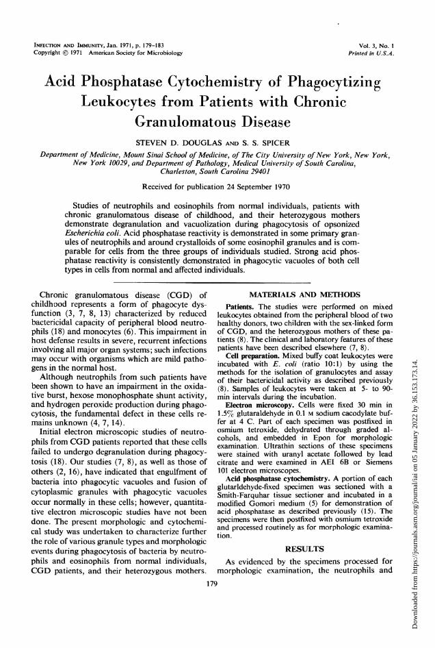
FIG. 1. Buffy coat neutrophil from control subject showing dense reaction product indicative of acid phospha-tase activity in phagocytic vacuoles (v) and cytoplasmic granules. Profiles of the vacuoles vary in morphology andcontent. The largest loosely envelops a bacterium heavily coated with acid phosphatase reaction product. Othersappear nearly empty or contain heterogeneous, moderately dense material with a light scattering of reaction prod-uct. Some relatively large structures with acidphosphatase activity (arrows) probably represent primary (azurophil)granules (which have a tendency toward disruption by the technical processing) but may be small or tangentiallysectioned phagocytic vacuoles. Other smaller profiles of reactive cytoplasmic granules may be tangentially sectionedprimary granules or tertiary granules. Note reactive granules closely bordering and apparently about to fuse withthe upper border of the large, bacterium-laden vacuole. The many relatively small unreactive granules with uniformanid moderately dense content are interpreted as secondary (specific) granules. Since the proportion of acid phos-phatase-positive granules appears smaller than the known proportion ofprimary granules, as identified in humanbuffy coat neutrophils by their peroxidase activity, many of the larger unreactive granules in this cell profile maybe primary granules which the cytochemical procedure failed to demonstrate. Acid phosphatase preparation; un-stained thin section. X25,000.
eosinophils from CGD patients and their mothersresembled those of normal controls in showingincorporation of bacteria into phagocytic vacu-oles. With continued incubation, cells from thesethree groups of individuals also disclosed subse-quent depletion of cytoplasmic granules indicativeof fusion between cytoplasmic granules andphagocytic vacuoles.
Acid phosphatase reaction product was evidentin profiles of structures which were interpretedas probably representing cytoplasmic granules(Fig. 1-3). These acid phosphatase reactive pro-
files were present comparably in neutrophils ob-tained either from normal individuals or frompatients or their mothers. The larger of these re-active structures presumably were primary (azuro-phil) granules (22), but their number-per-cell
profile was fewer than the number of primarygranules usually demonstrated by peroxidase re-activity in mature neutrophils (9), indicatingthat the acid phosphatase procedure failed todemonstrate some of the primary granules. Thelarge population of neutrophil secondary (spe-cific) granules lacked acid phosphatase reactivity(Fig. 1, 3) as anticipated (20, 22). Phagocyticvacuoles were the only other structures whichcontained heavy deposits of reaction product in-dicative of acid phosphatase activity in buffycoat neutrophils. In neutrophils of normal con-trols (Fig. 1) and those of CGD patients (Fig. 2,3) and their mothers, the phagocytic vacuoles dis-closed strong acid phosphatase activity closelysurrounding enclosed bacteria.
Eosinophils from controls (Fig. 4), patients,
0
180 INFEC. IMMUN.
Dow
nloa
ded
from
http
s://j
ourn
als.
asm
.org
/jour
nal/i
ai o
n 05
Jan
uary
202
2 by
36.
153.
173.
14.
CYTOCHEMISTRY OF PHAGOCYTIZING LEUKOCYTES
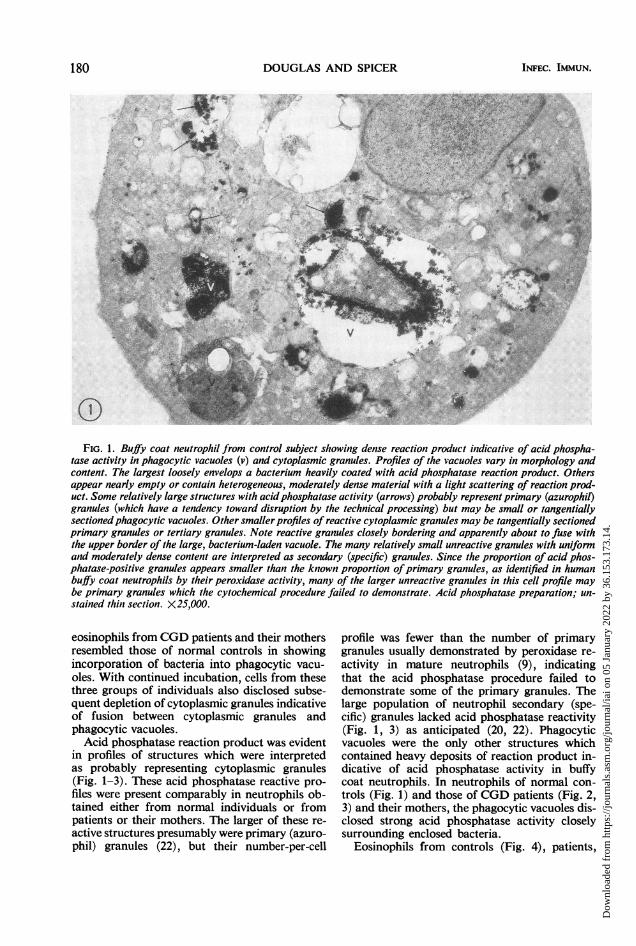
FIG. 2. Like Fig. I except ntettrophil from CGD patienzt. As in the conttrol cell, a few profiles probably represenit-intg initact or partially disrupted primary graniules (short arrows) as well as phagocytic vacuoles (v) exhibit heavydeposits of acid phosphatase reactiont product. A phosphatase-reactive protrusiont (long arrow) ont thze vacuole witha bacterium suiggests a stage of fusioni betweeii a primary graniule anid the vacutole. The htyaloplasm and nucleuisshow extranteous fite deposits of lead phosphate. X 18 750.
FIG. 3. Like Fig. 2. In this phagocytic vacuiole int a nieutrophil from a CGD patienit, acid phosphatase reactiontproduct inivests a phagocytosed bacteriuim. A reactive cytoplasmic graiiiule closely nteighbors the vacuole. X 15,000.
and heterozygotes showed reaction product fillingthe matrix around the crystalloids of a few of thecytoplasmic granules. However, most of thesegranules lacked reaction product indicative ofacid phosphatase activity (Fig. 4, 5). In addition,smaller, rounder, ellipsoid or reniform profiles ofunidentified structures observed infrequently inthe eosinophils appeared partially or completelyfilled with dense reaction product (Fig. 4). Densedeposits of reaction product enveloped bacteriatightly enclosed in phagocytic vacuoles in eosino-phils from controls (Fig. 4), patients (Fig. 5), andheterozygotes.
DISCUSSIONNeutrophils and eosinophils from patients
with CGD and their heterozygous mothers wereshown to undergo formation of phagocytic vacu-oles and fusion of these vacuoles with cytoplasmicgranules, following incubation with opsonizedE. coli. Although the rates of these two activitiesould not be quantitated accurately with thecresent techniques, they did not appear grossly
different in the three groups studied. These ob-servations are consistent with and extend our pre-vious morphologic study (8) and the cytochemicaldemonstration of peroxidase in the phagocyticvacuoles of these cells (2).
Previous cinemicrophotographic studies haveshown the phagocytic capabilities of eosinophilsincubated in the presence of opsonized particles(1). We have provided electron microscopic evi-dence that cytoplasmic granules of eosinophilsfuse with phagocytic vacuoles since a granulecomponent, acid phosphatase, has been demon-strated in the vacuole. The bacteria used in thisstudy, E. coli, lack acid phosphatase (21).
Furthermore, this study is the first electronmicroscopic demonstration of phagocytic vacuoleformation and granule-vacuole fusion (degranu-lation) in eosinophils from CGD patients andtheir heterozygous mothers. Thus, each of theseveral types of phagocytic blood cells from CGDpatients undergoes the normal sequence of mor-phological events during phagocytosis; their im-paired bactericidal action does not result from a
VOL. 3, 1971 181
Dow
nloa
ded
from
http
s://j
ourn
als.
asm
.org
/jour
nal/i
ai o
n 05
Jan
uary
202
2 by
36.
153.
173.
14.
'EQ- s ISK\
V, _ S4 ;V
*ii -
.s'tv= _ 4,4f~e -S_s'J s
..*-..i _ VPr'~ % _' 'I-
FIG.4Buffco ateos omc l se s i hi v o (v.s g c sue m
w_a
crstlli (ln ro) u te rsali-otiiggauls(hr ros akdpst.Dnedpstr
*pp.xw18,7.
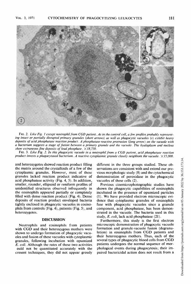
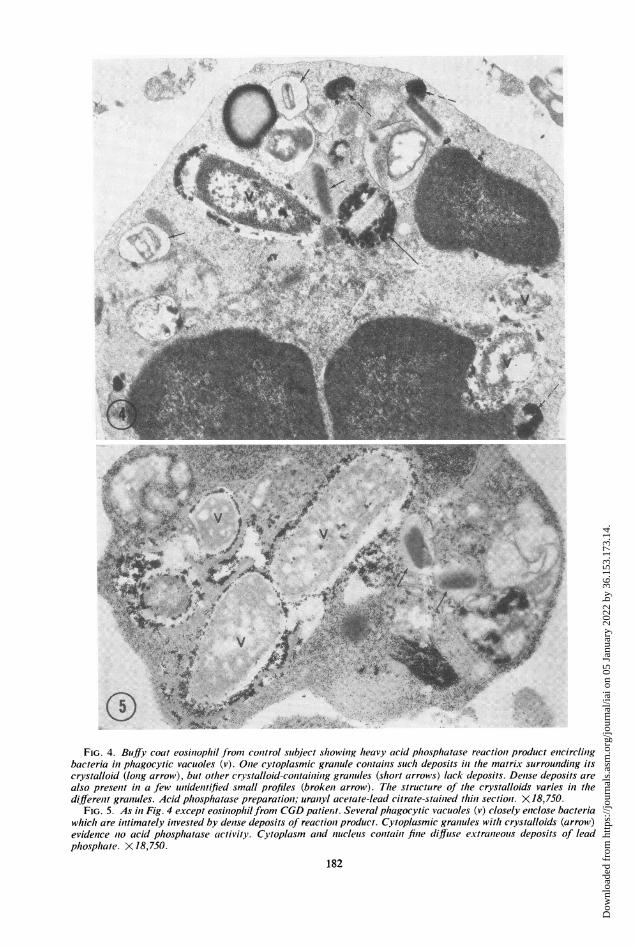
FIcG. 4. Bugfy coat eosintophlil from coiitrol slabject shlowintg hleavy acid phlosphzatase reactiont product enicircliingbacteria inl phagocytic vacuioles (v). Onie cytoplasmic graiitule coiitaiis s uchz deposits inl the mnatrix surroundilig itscrystalloid (lonig arrow), but other ci-ystalloid-conztaininig grantules (s/tort arrows) lack deposits. Denlse deposits arealso preseiit itt a few uniideiitified small profiles (brokeit arrow). T/1e struceture of thle crystalloids varies inl th1ediffereint grantules. Acid phlosphwatase preparatioii; uraniyl acetate-lead citrate-staiiied thiii sectiont. X 18,750.
FIG. 5. AS in Fig. 4 except eosilnop/tilfrom CGD patietit. Several phagocytic vacuoles (v) closely eniclose bacteriawhicht are initimately intvested by dense deposits of reactioit product. Cytoplasmic graiiules witht crystalloids (arrow)evidence nto acid phtosphlatase activity. Cytoplasm anid niucletis conttalii finte difftse extranteolus deposits of leadphtosphzate. X 18,750.
182
Dow
nloa
ded
from
http
s://j
ourn
als.
asm
.org
/jour
nal/i
ai o
n 05
Jan
uary
202
2 by
36.
153.
173.
14.
CYTOCHEMISTRY OF PHAGOCYTIZING LEUKOCYTES
failure in these activities. Bactericidal defectshave been demonstrated in both neutrophils andmonocytes in these patients (3, 7, 8, 13, 14, 18);isolated eosinophils have not yet been studied forbactericidal activity, however. Although the defectin the sex-linked form of CGD is most likely re-lated to the hexose monophosphate shunt andH202 generation, it is now evident that severalforms of phagocyte dysfunction may occur (7, 8).Acid phosphatase does not appear to be deficientin the leukocytes of CGD patients, but it is pro-posed that detailed cytochemical studies may yetdemonstrate the basic defect involved in certainforms of phagocyte dysfunction.The present cytochemical observations on acid
phosphatase agree with previous ultrastructuralobservations in which acid phosphatase activityis occasionally but not usually demonstrable incrystalloid-laden granules of eosinophils (11, 12,17, 19, 20, 22). Although the reason for the vari-able acid phosphatase reactivity in these granulesis not known, the possibility may be consideredthat the enzyme is stored in a latent form in thegranules. The strong acid phosphatase reactivityconsistently demonstrated in the phagocyticvacuoles in eosinophils indicates conversion ofthe enzyme to the expected active form in thesevacuoles. Complexing of the enzyme with theassociated acid mucosubstance in the eosinophilgranule (10) and dissociation of this complex inthe phagocytic vacuole could provide a mecha-nism for such a change in enzyme activity. A simi-lar mechanism may occur in the neutrophilprimary granules which also contain acid phos-phatase (22) and acid mucosubstance (10).
ACKNOWLEDGMENTS
S. D. D. is the recipient of Public Health Service CareerDevelopment Award 5 K04 HE-42575-02.
This study was supported by Public Health Service grantsAI-09338 from the National Institute of Allergyand Infectious Dis-eases and AM-1 1028 from the National Institute of Arthritisand Metabolic Diseases.
LITERATURE CITED
1. Archer, G. T., and J. G. Hirsch. 1963. Motion picture studieson degranulation of horse eosinophils during phagocytosis.J. Exp. Med. 118:287-294.
2. Baehner, R. L., M. J. Karnovsky, and M. L. Karnovsky. 1969.Degranulation of leukocytes in chronic granulomatousdisease. J. Clin. Invest. 48:187-192.
3. Baehner, R. L., and D. G. Nathan. 1968. Quantitativenitroblue tetrazolium test in chronic granulomatous disease.N. Engl. J. Med. 278:971-976.
4. Baehner, R. L., D. G. Nathan, and M. L. Karnovsky. 1970.
Correction of metabolic deficiencies in the leukocytes ofpatients with chronic granulomatous disease. J. Clin.Invest. 49:865-870.
5. Barka, T., and P. J. Anderson. 1962. Histochemical methodsfor acid phosphatase using hexazonium pararosanilin ascoupler. J. Histochem. Cytochem. 10:741-753.
6. Davis, W. C., H. Huber, S. D. Douglas, and H. H. Fudenberg.1968. A defect in circulating mononuclear phagocytes inchronic granulomatous disease of childhood. J. Immunol.101:1093-1095.
7. Douglas. S. D. 1970. Analytical review: disorders of phagocytefunction. Blood 35:851-866.
8. Douglas, S. D., W. C. Davis, and H. H. Fudenberg. 1969.Granulocytopathies: pleomorphism of neutrophil dysfunc-tion. Amer. J. Med. 46:901-909.
9. Dunn, W. B., J. H. Hardin, and S. S. Spicer. 1968. Ultra-structural localization of myeloperoxidase in human neutro-phil and rabbit heterophil and eosinophil leukocytes. Blood32:935-944.
10. Dunn, W. B., and S. S. Spicer. 1969. Histochemical demon-stration of sulfated mucosubstances and cationic proteinsin human granulocytes and platelets. J. Histochem. Cyto-chem. 17:668-674.
11. Enomoto, T., and T. Kitani. 1966. Electron microscopicstudies on peroxidase and acid phosphatase reaction inhuman leukocytes. (In nonnal and leukemic cells anid on thephagocytosis). Acta Haemat. Jap. 29:554-570.
12. Ghidoni, J. J., and A. F. Goldberg. 1966. Light and electronmicroscopic localization of acid phosphatase activity inhuman eosinophils. Amer. J. Clin. Pathol. 45:402-405.
13. Good, R. A., P. G. Quie, D. G. Windhorst, A. R. Page, G. E.Rodey, J. White, J. J. Wolfson, and B. H. Holmes. 1968.Fatal (chronic) granulomatous disease of childhood: ahereditary defect of leukocyte function. Seminars Hemat.5:215-254.
14. Holmes, B., A. R. Page, and R. A. Good. 1967. Studies ofthe metabolic activity of leukocytes from patients with agenetic abnormality of phagocyte function. J. Clin. Invest.46:1422-1432.
15. Horn, R. G., S. S. Spicer, and B. K. Wetzel. 1964. Phago-cytosis of bacteria by heterophil leukocytes: acid andalkaline phosphatase cytochemistry. Amer. J. Pathol.45:327-335.
16. Kauder, E., L. L. Kahle, H. Moreno, and J. C. Partin. 1968.Leukocyte degranulation and vacuole formation in patientswith chronic granulomatous disease of childhood. J. Clin.Invest. 47:1753-1762.
17. Miller, F., and F. Herzog. 1969. Die Lokalisaition von Peroxy-dase und saurer Phosphatase in eosinophilen Leukocytenwahrend der Reifung. Z. Zellforsh. 97:84-110.
18. Quie, P. G., J. G. White, B. Holmes, and R. A. Good. 1967.In vitro bactericidal capacity of human polymorphonuclearleukocytes: diminiished activity in chronic granulomratousdisease of childhood. J. Clin. Invest. 46:668-679.
19. Seemen, P. M., and G. E. Palade. 1967. Acid phosphataselocalization in rabbit eosinophils. J. Cell Biol. 34:745-756.
20. Spicer, S. S,, and J. H. Hardin. 1969. Ultrastructure, cyto-chemistry and function of neutrophil leukocyte granules.A review. Lab. Invest. 20:488-497.
21. Wetzel, B. K., S. S. Spicer, H. F. Dvorak, and L. A. Heppel.1970. Cytochemical localization of certain phosphatases inEscher-ichia coli. J. Bacteriol. 104:529-542.
22. Wetzel, B. K., S. S. Spicer, and R. G. Horn. Fine structurallocalization of acid and alkaline phosphatases in cells ofrabbit blood and bone marrow. J. Histochem. Cytochenm.15:311-334.
VOL. 3,7 1971 183
Dow
nloa
ded
from
http
s://j
ourn
als.
asm
.org
/jour
nal/i
ai o
n 05
Jan
uary
202
2 by
36.
153.
173.
14.