acquired parietal intradiploic encephalocele. case … · n. dobrin et al acquired parietal...
TRANSCRIPT
N. Dobrin et al Acquired parietal intradiploic encephalocele
Acquired parietal intradiploic encephalocele. Case report and review of the literature
N. Dobrin¹, Mihaela Bălinişteanu¹, B. Costăchescu¹, Cornelia Tudorache², A. Chiriac, I. Poeată¹
“Prof. Dr. N. Oblu” Clinical Emergency Hospital, Iaşi, Romania ¹Department of Neurosurgery; ²Department of Radiology
Abstract Very few cases of intradiploic
encephalocele in adulthood have been reported in the literature. In our paper we describe a case of parietal intradiploic encephalocele, which presents with simple partial seizures. Preoperative imaging (CT and MRI) showed brain herniation within the intradiploic space. Diagnosis was confirmed at surgery. Postoperatively the patient recovered from his presenting symptoms.
Keywords: brain herniation, intradiploic encephalocele
Introduction Parietal cephaloceles may develop
spontaneously as congenital maldevelopments or may occur subsequently to acquired processes, such as infection, trauma, neoplasms, surgical procedures. An intradiploic location of these cephaloceles is extremely rare.
In this communication, we report a case of symptomatic parietal intradiploic encephalocele and we debate upon its possible origin and review the pertinent literature.
Case Report History and examination A 75-year-old
man presented to our clinic with a 1-month
history of partial seizures in the right inferior limb. His neurological examination revealed no focal abnormalities. Nothing was found on the examination of his scalp. His medical history revealed type 2 diabetes, high blood pressure, coronary heart disease, hepatitis C, but no head trauma, febrile seizures, stroke, brain tumor, central nervous system infection. His family history was uneventful.
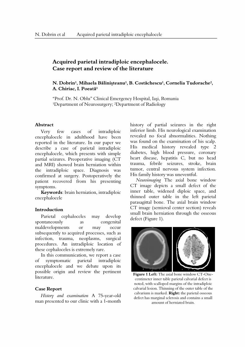
Neuroimaging The axial bone window CT image depicts a small defect of the inner table, widened diploic space, and thinned outer table in the left parietal parasagittal bone. The axial brain window CT image (semioval center section) reveals small brain herniation through the osseous defect (Figure 1).
Figure 1 Left: The axial bone window CT-One-centimeter inner table parietal calvarial defect is noted, with scalloped margins of the intradiploic calvarial lesion. Thinning of the outer table of the calvarium is marked. Right: the parietal osseous defect has marginal sclerosis and contains a small
amount of herniated brain.
Romanian Neurosurgery (2011) XVIII 2
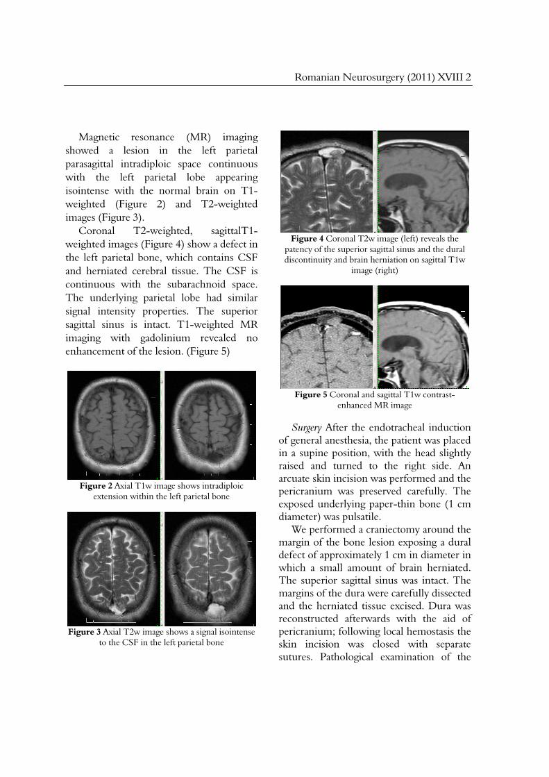
Magnetic resonance (MR) imaging showed a lesion in the left parietal parasagittal intradiploic space continuous with the left parietal lobe appearing isointense with the normal brain on T1-weighted (Figure 2) and T2-weighted images (Figure 3).
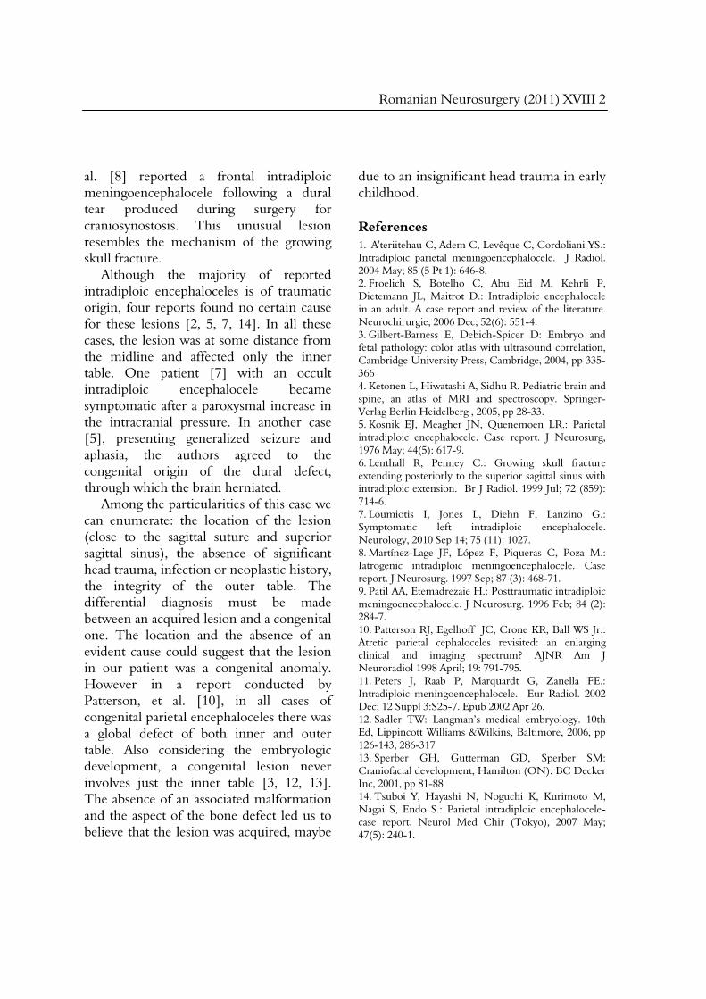
Coronal T2-weighted, sagittalT1-weighted images (Figure 4) show a defect in the left parietal bone, which contains CSF and herniated cerebral tissue. The CSF is continuous with the subarachnoid space. The underlying parietal lobe had similar signal intensity properties. The superior sagittal sinus is intact. T1-weighted MR imaging with gadolinium revealed no enhancement of the lesion. (Figure 5)
Figure 2 Axial T1w image shows intradiploic
extension within the left parietal bone
Figure 3 Axial T2w image shows a signal isointense
to the CSF in the left parietal bone
Figure 4 Coronal T2w image (left) reveals the
patency of the superior sagittal sinus and the dural discontinuity and brain herniation on sagittal T1w
image (right)
Figure 5 Coronal and sagittal T1w contrast-
enhanced MR image
Surgery After the endotracheal induction of general anesthesia, the patient was placed in a supine position, with the head slightly raised and turned to the right side. An arcuate skin incision was performed and the pericranium was preserved carefully. The exposed underlying paper-thin bone (1 cm diameter) was pulsatile.
We performed a craniectomy around the margin of the bone lesion exposing a dural defect of approximately 1 cm in diameter in which a small amount of brain herniated. The superior sagittal sinus was intact. The margins of the dura were carefully dissected and the herniated tissue excised. Dura was reconstructed afterwards with the aid of pericranium; following local hemostasis the skin incision was closed with separate sutures. Pathological examination of the

N. Dobrin et al Acquired parietal intradiploic encephalocele
resected specimen revealed gliotic and normal cerebral cortex.
Postoperative course The patient received 600 mg carbamazepine per day for three months after surgery, and he remained seizure free on his 6-month follow-up.
Discussion and conclusions A cephalocele is defined as a protrusion
of intracranial contents through a defect in the skull or dura. Cephaloceles are classified by their contents (meningocele, meningoencephalocele, hydromeningo-encephalocele) and by the location of the cranial defect through which the herniation occurs. The herniating neural tissue may include meninges, brain parenchyma, ventricles and vascular structures [4]. A cephalocele can result from various causes: infection, trauma, surgery, tumors. Those which develop in absence of an evident cause are congenital or early postnatal maldevelopments termed spontaneous meningoencephaloceles. These spontaneous lesions usually occur at the site of a cranial suture, and most of them represent primary or secondary midline closure defects of the neural tube. The vault of the neurocranium is derived from the paraxial mesoderm. During the 8th week of development, the two parietal bones undergo membranous ossification from two primary centers for each bone, appearing on the parietal tuber. At 4 months, the fusion between these centers is complete. At birth the parietal bones are unilaminar and are separated by the sagittal suture. At the age of 4 there appears the differentiation between inner and outer table and the ossification of the sagittal suture begins at the same time for the two layers, but the outer tables could fuse slowerly and sometimes incompletely. A
complete fusion takes place by the age of 20. Defects in closure of the sagittal suture allow the herniation of the brain, but they involve both tables [3, 12, 13].
Parietal cephaloceles are very rare (1% of all cerebrospinal malformations and 10% of cephaloceles [11]) and if they are congenital, they are usually associated with many anomalies such as corpus calosum agenesis, Chiari II, Dandy-Walker malformation.
The intradiploic encephaloceles are also extremely rare, only few cases are mentioned in the literature. In their case report Patil, et al. [9] presented a 64-year-old man with a posttraumatic intradiploic meningoencephalocele after a head trauma. He bumped his head on a garage door and 8 months post-trauma he came to the hospital with a lump on his head. The authors concluded that their case was a variation of an adult growing skull fracture due to the blunt trauma to the head, an intradiploic arachnoidal cyst containing CSF and brain. Lenthall, et al. [6], also presented a case of a growing skull fracture with the intradiploic extension within the occipital and parietal bone in a 6-month-old baby who sustained a head trauma after falling down the stairs, but, in this case, both inner and outer bone tables were eroded. A'teriitehau, et al. [1] described an intradiploic parietal encephalocele in a 73-year-old woman with no history of significant head trauma, but they attributed the intradiploic defect to a minor trauma, without loss of consciousness, but strong enough to produce the destruction of the inner table and a dural tear. Peters, et al. [11] came to the same conclusion with their patient who showed coordinative problems in his right leg, also without history of head trauma. Martinez-Lage, et
Romanian Neurosurgery (2011) XVIII 2
al. [8] reported a frontal intradiploic meningoencephalocele following a dural tear produced during surgery for craniosynostosis. This unusual lesion resembles the mechanism of the growing skull fracture.
Although the majority of reported intradiploic encephaloceles is of traumatic origin, four reports found no certain cause for these lesions [2, 5, 7, 14]. In all these cases, the lesion was at some distance from the midline and affected only the inner table. One patient [7] with an occult intradiploic encephalocele became symptomatic after a paroxysmal increase in the intracranial pressure. In another case [5], presenting generalized seizure and aphasia, the authors agreed to the congenital origin of the dural defect, through which the brain herniated.
Among the particularities of this case we can enumerate: the location of the lesion (close to the sagittal suture and superior sagittal sinus), the absence of significant head trauma, infection or neoplastic history, the integrity of the outer table. The differential diagnosis must be made between an acquired lesion and a congenital one. The location and the absence of an evident cause could suggest that the lesion in our patient was a congenital anomaly. However in a report conducted by Patterson, et al. [10], in all cases of congenital parietal encephaloceles there was a global defect of both inner and outer table. Also considering the embryologic development, a congenital lesion never involves just the inner table [3, 12, 13]. The absence of an associated malformation and the aspect of the bone defect led us to believe that the lesion was acquired, maybe
due to an insignificant head trauma in early childhood.
References 1. A'teriitehau C, Adem C, Levêque C, Cordoliani YS.: Intradiploic parietal meningoencephalocele. J Radiol. 2004 May; 85 (5 Pt 1): 646-8. 2. Froelich S, Botelho C, Abu Eid M, Kehrli P, Dietemann JL, Maitrot D.: Intradiploic encephalocele in an adult. A case report and review of the literature. Neurochirurgie, 2006 Dec; 52(6): 551-4. 3. Gilbert-Barness E, Debich-Spicer D: Embryo and fetal pathology: color atlas with ultrasound correlation, Cambridge University Press, Cambridge, 2004, pp 335-366 4. Ketonen L, Hiwatashi A, Sidhu R. Pediatric brain and spine, an atlas of MRI and spectroscopy. Springer-Verlag Berlin Heidelberg , 2005, pp 28-33. 5. Kosnik EJ, Meagher JN, Quenemoen LR.: Parietal intradiploic encephalocele. Case report. J Neurosurg, 1976 May; 44(5): 617-9. 6. Lenthall R, Penney C.: Growing skull fracture extending posteriorly to the superior sagittal sinus with intradiploic extension. Br J Radiol. 1999 Jul; 72 (859): 714-6. 7. Loumiotis I, Jones L, Diehn F, Lanzino G.: Symptomatic left intradiploic encephalocele. Neurology, 2010 Sep 14; 75 (11): 1027. 8. Martínez-Lage JF, López F, Piqueras C, Poza M.: Iatrogenic intradiploic meningoencephalocele. Case report. J Neurosurg. 1997 Sep; 87 (3): 468-71. 9. Patil AA, Etemadrezaie H.: Posttraumatic intradiploic meningoencephalocele. J Neurosurg. 1996 Feb; 84 (2): 284-7. 10. Patterson RJ, Egelhoff JC, Crone KR, Ball WS Jr.: Atretic parietal cephaloceles revisited: an enlarging clinical and imaging spectrum? AJNR Am J Neuroradiol 1998 April; 19: 791-795. 11. Peters J, Raab P, Marquardt G, Zanella FE.: Intradiploic meningoencephalocele. Eur Radiol. 2002 Dec; 12 Suppl 3:S25-7. Epub 2002 Apr 26. 12. Sadler TW: Langman’s medical embryology. 10th Ed, Lippincott Williams &Wilkins, Baltimore, 2006, pp 126-143, 286-317 13. Sperber GH, Gutterman GD, Sperber SM: Craniofacial development, Hamilton (ON): BC Decker Inc, 2001, pp 81-88 14. Tsuboi Y, Hayashi N, Noguchi K, Kurimoto M, Nagai S, Endo S.: Parietal intradiploic encephalocele-case report. Neurol Med Chir (Tokyo), 2007 May; 47(5): 240-1.