activation of an arabidopsis resistance protein is speciï¬ed by
TRANSCRIPT

Activation of an Arabidopsis Resistance Protein Is Specified bythe in Planta Association of Its Leucine-Rich Repeat Domainwith the Cognate Oomycete Effector W OA
Ksenia V. Krasileva, Douglas Dahlbeck, and Brian J. Staskawicz*
Department of Plant and Microbial Biology, University of California, Berkeley, California 94720
Activation of plant immunity relies on recognition of pathogen effectors by several classes of plant resistance proteins. To
discover the underlying molecular mechanisms of effector recognition by the Arabidopsis thaliana RECOGNITION OF
PERONOSPORA PARASITICA1 (RPP1) resistance protein, we adopted an Agrobacterium tumefaciens–mediated transient
protein expression system in tobacco (Nicotiana tabacum), which allowed us to perform coimmunoprecipitation experi-
ments and mutational analyses. Herein, we demonstrate that RPP1 associates with its cognate effector ARABIDOPSIS
THALIANA RECOGNIZED1 (ATR1) in a recognition-specific manner and that this association is a prerequisite step in the
induction of the hypersensitive cell death response of host tissue. The leucine-rich repeat (LRR) domain of RPP1 mediates
the interaction with ATR1, while the Toll/Interleukin1 Receptor (TIR) domain facilitates the induction of the hypersensitive
cell death response. Additionally, we demonstrate that mutations in the TIR and nucleotide binding site domains, which
exhibit loss of function for the induction of the hypersensitive response, are still able to associate with the effector in planta.
Thus, our data suggest molecular epistasis between signaling activity of the TIR domain and the recognition function of the
LRR and allow us to propose a model for ATR1 recognition by RPP1.
INTRODUCTION
Plants have evolved amultilevel innate immune system to protect
them against infection by a diverse range of pathogens, including
viruses, bacteria, fungi, oomycetes, and nematodes. Despite the
great evolutionary distance among phytopathogens, the out-
come of the plant–pathogen interactions is controlled by the
same principles: the ability of the pathogen to suppress the plant
immune system to establish infection, and the ability of plants to
recognize the presence of a pathogen and to induce immune
responses that restrict pathogen growth.
The first line of plant defense consists of the integral plasma
membrane receptors known as pattern-recognition receptors,
which recognize the presence of common pathogen-associated
molecular patterns (PAMPs) near the cell surface (Chisholm
et al., 2006; Jones and Dangl, 2006; Trinchieri and Sher, 2007).
Upon associationwith PAMPs, the pattern-recognition receptors
activate a downstream mitogen-activated protein kinase signal-
ing cascade that culminates in transcriptional activation and
generation of the innate immune responses (Chisholm et al.,
2006; Jones andDangl, 2006). This line of defense, called PAMP-
triggered immunity, is commonly suppressed by a successful
pathogen in order to establish infection. To interfere with PAMP-
triggered immunity, pathogens from different kingdoms of life
have evolved effector proteins that are delivered into and func-
tion within the host plant cells (Desveaux et al., 2006; Kamoun,
2006; Dodds et al., 2009). The second layer of plant immunity
depends on the ability of the plant to recognize these pathogen-
derived effectors and trigger a robust resistance response that
normally culminates in a hypersensitive cell death response (HR).
While PAMPs represent conservedmicrobial molecules, effector
molecules constitute very divergent groups of proteins and their
recognition requires constant structural and evolutionary ad-
justment of the corresponding receptors. Effector-triggered
immunity is mediated by a large group of structurally related
intracellular innate immune receptors encoded by resistance (R)
genes. Products of R genes either directly or indirectly recognize
pathogen effectors and induce innate immunity. The major class
of R proteins is characterized by the central nucleotide binding
site (NBS) domain and C-terminal leucine-rich repeats (LRRs).
This group can be further subdivided according to the N-terminal
domain into a TIR-NBS-LRR class, which has an N-terminal
domain with sequence similarity to Drosophila melanogaster Toll
and human Interleukin1 Receptor (TIR), and a CC-NBS-LRR
class, which has a structured coiled-coil domain (CC). Currently,
the data supportingmolecular functions of the TIR/CC, NBS, and
LRR domains in R proteins are sparse, sometimes contradictory,
and scattered across many different proteins and experimental
systems. Therefore, there is a need for in planta data that would
test and clearly define the roles of different domains for a given R
protein.
The NBS domain of R proteins shares sequence similarity with
the mammalian cell death–inducing proteins, including human
apoptotic peptidase-activating factor 1 (Apaf-1), its Caenorhab-
ditis elegans homolog CED-4, and a large group of intracellular
* Address correspondence to [email protected] author responsible for distribution of materials integral to thefindings presented in this article in accordance with the policy describedin the Instructions for Authors (www.plantcell.org) is: Brian J. Staskawicz([email protected]).WOnline version contains Web-only data.OAOpen Access articles can be viewed online without a subscription.www.plantcell.org/cgi/doi/10.1105/tpc.110.075358
The Plant Cell, Vol. 22: 2444–2458, July 2010, www.plantcell.org ã 2010 American Society of Plant Biologists
Dow
nloaded from https://academ
ic.oup.com/plcell/article/22/7/2444/6095973 by guest on 22 Septem
ber 2021

nucleotide oligomerization domain receptors that function in
mammalian innate immunity (Takken et al., 2006). In animal
proteins, nucleotide binding and hydrolysis acts as a molecular
switch that regulates signal transduction by conformational
change, often leading to oligomerization of the protein (Kim
et al., 2005). In plants, the NBS domains of the tomato (Solanum
lycopersicum) R proteins I-2 and Mi-1 were shown to bind and
hydrolyze ATP in vitro, supporting the function of the NBS as a
nucleotide binding switch in R protein activation (Tameling et al.,
2002). In addition, mutations in the conserved P-loop motif
resulted in a loss of nucleotide binding in vitro (Tameling et al.,
2002) and the corresponding loss of HR induction in planta
(Tameling et al., 2002;Wirthmueller et al., 2007). The intactP-loop
motif was also required for effector-induced oligomerization of the
tobacco (Nicotiana tabacum) TIR-NBS-LRR resistance protein N
(Mestre and Baulcombe, 2006). Based on these data, nucleotide
binding is considered to be a molecular switch that regulates the
activity of R proteins (Takken et al., 2006).
The TIR/CC N-terminal domains of R proteins were originally
proposed to act in downstream signaling rather than in initial
perception of the effector molecule. This view was based on the
signaling function of TIRs in the Toll receptors from Drosophila,
human Interleukin1 receptor, and mammalian Toll-Like Recep-
tors (TLRs). In the case of R proteins, this model was supported
by Frost et al. (2004), who showed autoactivation of the TIR
domain of flax (Linum usitatissimum) L10, and by Swiderski et al.
(2009), who demonstrated that the TIR domains of several R
proteins, including Arabidopsis thaliana Resistance to Pseudo-
monas syringae 4 (RPS4) and Recognition of Peronospora
parasitica 1A (RPP1A) as well as tobacco N, show different
degrees of effector-independent HR when transiently expressed
in N. tabacum or Nicotiana benthamiana. However, there also
exists evidence of TIR involvement in determining interactions
with various effectors (Ellis et al., 1999; Burch-Smith et al., 2007).
The importance of the TIR domains for R protein function was
highlighted in several studies that identified mutations in con-
served residues that led to the loss of HR in planta (Dinesh-
Kumar et al., 2000; Swiderski et al., 2009).
The LRR domains are often involved in ligand–receptor inter-
actions in animal systems (Kobe and Deisenhofer, 1993; Alder
et al., 2005). In plant innate immunity, this role of LRR domains is
supported, in the case of some R proteins, by both genetic
analyses and yeast two-hybrid data (Deslandes et al., 2003;
Dodds et al., 2006). Evolutionary analyses suggest that many R
genes and their cognate effectors are evolving under the pres-
sure of diversifying selection (Mondragon-Palomino, 2002; Win
et al., 2007). This often results in numerous duplications and
rearrangements at the R gene loci as well as in high levels of
polymorphism in the LRR domain (Ellis et al., 1999; Allen et al.,
2004). Domain-swapping experiments confirmed the role of the
LRR domain in determining specificity toward different effector
gene variants (Ellis et al., 1999; Dodds et al., 2001; Rairdan and
Moffett, 2006; Shen et al., 2007; Rentel et al., 2008). Direct
protein–protein interaction of the LRR domain with the cognate
effector was demonstrated for rice (Oryza sativa) Pi-ta and the
rice blast fungus (Magnaporthe grisea) effector AvrPita. In this
case, the LRR region was both necessary and sufficient for
interaction in yeast two-hybrid assays and for in vitro binding (Jia
et al., 2000). Despite growing evidence for the direct interactions
between R proteins and their cognate effectors, there has been a
lack of in planta biochemical data supporting the role of the LRR
region in effector recognition.
The RECOGNITION OF PERONOSPORA PARASITICA1
(RPP1) locus, which contains a complex resistance gene cluster,
was originally identified in Arabidopsis thaliana ecotype Wassi-
lewskija (Ws; Botella et al., 1998). Several members of the RPP1
gene family have been shown to specify disease resistance
against Hyaloperonospora arabidopsidis (previously known as
Peronospora parasitica; Botella et al., 1998; Rehmany et al.,
2005; Sohn et al., 2007), including RPP1-WsA, RPP1-WsB,
RPP1-WsC, and RPP1-NdA, while RPP1-like genes from
other ecotypes have been implicated in hybrid incompatibility
(Bomblies et al., 2007). Currently, proteins encoded by only two
RPP1 alleles have been shown to recognize the cognate effector
ARABIDOPSIS THALIANA RECOGNIZED1 (ATR1) from H. arab-
idopsidis (Rehmany et al., 2005). One of those alleles was cloned
from the Arabidopsis ecotype Ws and is denoted RPP1-WsB,
whereas the other, RPP1-NdA, is from Arabidopsis ecotype
Niederzenz (Nd). The R proteins RPP1-WsB and RPP1-NdA
share a common TIR-NBS-LRR domain architecture and are
87% identical at the amino acid level. Although polymorphisms
are present throughout their coding sequences, most of the
differences occur in the LRR region and include both single
amino acid polymorphisms and short insertions and deletions.
ATR1 belongs to a simple locus in H. arabidopsidis with
diverse allelic variants present in different strains of the pathogen
(Rehmany et al., 2005). The ATR1 protein has two regions found
in most oomycete effectors thus far: the N-terminal eukaryotic
signal peptide that targets the effector for secretion, followed by
the putative host-targeting domain defined by the conserved
RXLR motif (Rehmany et al., 2005; Birch et al., 2006). The rest of
the protein lacks sequence similarity to any proteins of known
function. Although this region is thought to contribute to patho-
gen virulence (Sohn et al., 2007), recognition by RPP1-WsB and
RPP1-NdA is its only known function.
In this study, we characterize the molecular mechanisms un-
derlying the recognition of ATR1 by RPP1. We present biochem-
ical evidence for in planta association between RPP1-WsB and
ATR1, which correlates with the ability of RPP1-WsB to recognize
this effector and elicit a resistance response. Our studies reveal
that this association is mediated by the LRR domain of RPP1-
WsB. Moreover, we demonstrate an epistatic relationship be-
tween effector binding ability of the LRR and HR induction by the
TIR and NBS domains. Finally, we exploit natural polymorphisms
in ATR1 to isolate two gain-of-recognition mutants, showing that
the pattern of ATR1 recognition depends on just a fewamino acids
that are subjected to strong diversifying selection.
RESULTS
The Phylogeny of ATR1 Correlates with Its Recognition by
RPP1-WsB and RPP1-NdA
Previous studies established the gene-for-gene recognition
specificities of five ATR1 alleles sequenced from natural
R-Protein/Effector in Planta Association 2445
Dow
nloaded from https://academ
ic.oup.com/plcell/article/22/7/2444/6095973 by guest on 22 Septem
ber 2021

populations of H. arabidopsidis (Figure 1; see Supplemental
Figure 1 online) and two RPP1 alleles isolated from Arabidopsis
ecotypes Ws and Nd (Figure 1A; see Supplemental Figure 2
online; Rehmany et al., 2005). The ATR1 sequences obtained
from different H. arabidopsidis strains contained many polymor-
phisms and were named after the strain they were isolated from;
that is, ATR1-Emoy2 comes from H. arabidopsidis strain Emoy2.
It has been demonstrated that only a subset of ATR1 alleles is
able to elicit a hypersensitive response (HR) in transgenic
Arabidopsis plants carrying either RPP1-WsB or RPP1-NdA.
Thus, transgenic plants carrying RPP1-WsB elicited HR in re-
sponse toATR1-Emoy2,ATR1-Maks9, andATR1-Emco5 but not
in response to ATR1-Cala2 or ATR1-Emwa1, and plants carrying
RPP1-NdA recognized ATR1-Emoy2 but not ATR1-Maks9 (Fig-
ure 1B; Rehmany et al., 2005; Sohn et al., 2007).
We decided tomore closely examine the relationships between
the ATR1 sequence variation and recognition by RPP1. We
constructed a maximum likelihood phylogenetic tree of ATR1
alleles and mapped the pattern of their recognition by RPP1.
Interestingly, the three alleles recognized byRPP1-WsB clustered
together, while the two nonrecognized alleles formed a separate
clade (Figure 1B). Although the number of sequences within both
ATR1 and RPP1 groups of genes is too small to make any
concrete conclusions, such an evolutionary pattern is consistent
with tight coevolution betweenRPP1 and ATR1 and highlights the
need for a deeper sequence sampling of both genes.
Allelic sequences encoding both ATR1 and RPP1 show high
levels of polymorphisms. The most divergent alleles, ATR1-
Emoy2 and ATR1-Emwa1, share only 81.3% sequence identity
at the amino acid level (see Supplemental Figure 1 online).
Rehmany et al. (2005) examined the average pair-wise diver-
gence between ATR1 alleles in a sliding-window analysis and
found that synonymous and nonsynonymous polymorphisms
are distributed unevenly throughout the coding sequence, with
nonsynonymous mutations accumulating in the C-terminal re-
gion of the protein. Similarly, sequence comparison of RPP1
alleles indicated that most of the polymorphisms accumulated in
the LRR portion of the protein (Botella et al., 1998; see Supple-
mental Figure 2 online).
High levels of polymorphisms and evidence of positive selec-
tion in the R gene and its cognate effector suggest a direct
recognition of the effector protein by the corresponding resis-
tance protein. Investigation of the molecular basis of ATR1–
RPP1 interaction in a natural pathosystem presents several
experimental challenges: (1) low levels of protein expression for
both ATR1 and RPP1; (2) small numbers of cells undergoing a
resistance response during the infection; and (3) inability to
genetically manipulate H. arabidopsidis. To circumvent these
problems, we adopted a transient Agrobacterium tumefaciens-
mediated protein expression system in tobacco.
RPP1 Activates ATR1-Dependent HR in Tobacco
Tobacco has been widely used for Agrobacterium-mediated
transient expression of proteins and for assaying the HR. How-
ever, one commonly occurring limitation of the heterologous
Figure 1. Domain Architecture and Allele-Specific Recognition Pattern of ATR1 and RPP1.
(A) Domain organization of the oomycete effector ATR1 and the Arabidopsis resistance protein RPP1. The ATR1 diagram shows the eukaryotic
secretion sequence (SP), host translocation-targeting region (containing RXLR), and C-terminal domain, recognized by RPP1. The RPP1 diagram
highlights the major domains identified by sequence similarity to known protein families: TIR, NBS, and a series of LRRs.
(B) Maximum-likelihood tree illustrating phylogenetic relationships among five ATR1 alleles (left) and the corresponding recognition pattern by RPP1
alleles RPP1-WsB and RPP1-NdA (right). Bootstrap values are shown on the tree branches.
Recognition was demonstrated in the following studies: ain Arabidopsis in cobombardment assay (Rehmany et al., 2005); bin Arabidopsiswith type three
secretion system delivery by Pseudomonas syringae pv tomato DC3000 (Sohn et al., 2007); cin tobacco by Agrobacterium-mediated transient
coexpression (this study). The alignment used is shown in Supplemental Figure 1 online (pdf format) and Supplemental Data Set 1 online (text format).
2446 The Plant Cell
Dow
nloaded from https://academ
ic.oup.com/plcell/article/22/7/2444/6095973 by guest on 22 Septem
ber 2021

plant expression system is autoactivation of the overexpressed
resistance protein even in the absence of the effector. This
autoactivation phenotype was observed in the case of Arabi-
dopsis R proteins Resistance to Pseudomonas syringae 2
(RPS2), RPS4 (Zhang et al., 2004), and Recognition of Perono-
spora parasitica 13 (Rentel et al., 2008) but not RPP1-WsA
(Weaver et al., 2006). We tested the expression of RPP1-WsB in
tobacco, driven either by its native promoter (see Supplemental
Figure 3 online) or by the strong constitutive cauliflower mosaic
virus 35S promoter (35S; Figure 2A). None of the RPP1-WsB
constructs exhibited autoactivation and cell death in the absence
of ATR1. To find a construct amenable to biochemical analyses,
we made a protein fusion between RPP1-WsB and the hemag-
glutinin epitope tag (HA) on both N- and C-terminal ends of the
protein. While protein levels of the RPP1-WsB construct driven
by its native promoter were below the detection limit, we could
detect 35S:RPP1-WsB-HA by protein immunoblot (Figure 2B).
The ATR1 constructs were fused with Citrine tag, a fluorescent
protein variant that is detected by anti-green fluorescent protein
(a-GFP) antibody. Coexpression of the RPP1-WsB-HA together
with full-length ATR1-Emoy2-Citrine, ATR1D15-Emoy2-Citrine
(lacking the signal peptide sequence), or ATR1D51-Emoy2-Citrine
(lacking both the signal peptide sequence and the RXLR motif)
elicited a HR ;20 to 24 h post infiltration (Figure 2A). The full-
length ATR1 protein produced a slower and less pronounced
response. Protein immunoblot analysis showed that while
ATR1D15-Emoy2-Citrine and ATR1D51-Emoy2-Citrine were ex-
pressed at very high levels, the full-length ATR1 protein levels
were below the detection limit (Figure 2C), probably due to partial
secretion of the protein from the plant cells and instability in the
extracellular space. Based on these results, we chose to use
35S:RPP1-WsB-HA and ATR1D51-Citrine for further biochemi-
cal analyses.
Race-Specific Recognition of ATR1 by RPP1 in Tobacco
In order to further characterize whether the tobacco system
reflects the previously observed ATR1/RPP1 recognition spec-
ificities, we coexpressed five ATR1 alleles, ATR1-Emoy2, ATR1-
Maks9, ATR1-Emco5, ATR1-Cala2, and ATR1-Emwa1, with
either RPP1-WsB or RPP1-NdA. We observed the same race-
specific recognition pattern in tobacco as previously described
in Arabidopsis (Rehmany et al., 2005; Sohn et al., 2007) and
expanded the data for RPP1-NdA, which was previously tested
only with ATR1-Emoy2 and ATR1-Maks9. RPP1-WsB induced
anHRwhen coexpressedwith ATR1-Emoy2,Maks9, andEmco5
but not with Cala2 or Emwa1 (Figure 3A); RPP1-NdA induced an
HR only when coexpressed with ATR1-Emoy2 but not with any
other ATR1 allele (Figure 3B). The same recognition specifici-
ties were observed for both 35S:RPP1-WsB and genomic
Figure 2. Coexpression of ATR1 and RPP1 in Tobacco Triggers the HR.
(A) RPP1-WsB-HA induces HR in tobacco upon Agrobacterium-mediated coexpression with full-length ATR1-Emoy2-Citrine, ATR1D15-Emoy2-Citrine,
or ATRD51-Emoy2-Citrine. All constructs were expressed under the control of the constitutive cauliflower mosaic virus 35S promoter. The photograph
was taken 48 h post infiltration.
(B) Protein immunoblot probed with a-HA antibody showing relative levels of protein expression of RPP1-WsB-HA in tobacco driven by either its native
promoter (NP) or cauliflower mosaic virus 35S promoter (35S). Ribulose-1,5-bis-phosphate carboxylase/oxygenase (Rubisco), stained on the blots with
Ponceau S stain, is shown as a loading control.
(C) Protein immunoblot probed with a-GFP antibody showing relative levels of protein expression of the full-length (FL) ATR1-Emoy2-Citrine, ATR1D15-
Emoy2-Citrine, and ATRD51-Emoy2-Citrine in tobacco at 24 h post infiltration. Rubisco is shown as a loading control.
R-Protein/Effector in Planta Association 2447
Dow
nloaded from https://academ
ic.oup.com/plcell/article/22/7/2444/6095973 by guest on 22 Septem
ber 2021

RPP1-WsB construct (see Supplemental Figure 3 online). Protein
gel blot analysis showed that, whereas all ATR1 protein variants
were expressed to equally high levels in the absence of RPP1
(Figure 3C, left panel), protein levels of the recognized alleles
decreased dramatically upon induction of HR (Figure 3C, right
panel). These data indicate that the lack of recognition of a
subset of ATR1 alleles was not due to a lack of protein expres-
sion. All ATR1 variants localized consistently to the cytoplasm
and nucleus, as determined by fluorescence microscopy (see
Supplemental Figure 4 online), indicating that the difference in
recognition was not due to differential localization of ATR1
variants within the plant cell.
RPP1 Coimmunoprecipitates with ATR1
To investigate whether recognition specificity results from phys-
ical interaction between the ATR1 and RPP1 proteins, we tested
ATR1 variants for their ability to associate with RPP1-WsB in
planta. Citrine-tagged ATR1D51 constructs were transiently
coexpressed with 35S:RPP1-WsB-HA in tobacco for 20 to 24 h.
Tissue was collected when the HR began to appear. Protein
extracts were incubated with a-GFP antibodies to immunopre-
cipitate ATR1D51-Citrine. Resulting immunocomplexes were
separated by SDS-PAGE, and RPP1-WsB was detected by
immunoblot using a-HA antibody. Due to the mild conditions of
the nondenaturing protein extraction buffer used for coimmuno-
precipitations, the initial levels of RPP1-WsB protein were very
low in all of the input samples and were usually close to the limit
of detection by a protein gel blot. Therefore, we performed
parallel a-HA immunoprecipitations to ensure that RPP1-WsB
protein was present in all of the samples.
We observed that RPP1-WsB coimmunoprecipitated with the
three variants of ATR1 (Emoy2, Maks9, and Emco5) that also
triggered HR, but it did not associate with the nonrecognized
variants ATR1-Cala2 and ATR1-Emwa1 (Figure 4). These data
show a perfect correlation between the ability of RPP1 to
associate with ATR1 and the activation of the immune response,
suggesting that it is dependent on the ability of RPP1-WsB to
associate with ATR1. Additionally, we observed a slight, but
consistent, decrease in the amount of RPP1-WsB associating
with ATR1-Emco5 as compared with ATR1-Maks9 and ATR1-
Emoy2 (Figure 4).
Unfortunately, we were unable to detect a robust association
between RPP1-NdA and ATR1-Emoy2. In one coimmunoprecip-
itation experiment out of three conducted, we detected a weak
signal corresponding to RPP1-NdA, which was specific for an
Figure 3. Race-Specific Recognition of ATR1 Alleles by RPP1-WsB and RPP1-NdA in Tobacco.
(A) Coinfiltration of RPP1-WsB with ATR1-Emoy2, ATR1-Maks9, and ATR1-Emco5, but not ATR1-Cala2 or ATR1-Emwa1, triggers the HR in tobacco.
(B) Coinfiltration of RPP1-NdA with ATR1-Emoy2, but none of the other four alleles, triggers the HR. Photographs were taken 48 h post infiltration.
(C) Protein immunoblot probed with a-GFP antibody showing relative levels of protein abundance of five ATR1 variants coinfiltrated with empty vector
(EV; left panel) or with RPP1-WsB (right panel). Samples for protein extraction were collected at 20 to 24 h post infiltration, when the HR started to
appear. Rubisco is shown as a loading control.
2448 The Plant Cell
Dow
nloaded from https://academ
ic.oup.com/plcell/article/22/7/2444/6095973 by guest on 22 Septem
ber 2021

association with ATR1-Emoy2, but the level of the signal was just
above the detection limit (data not shown). This could be due to a
much weaker protein–protein interaction between RPP1-NdA
and ATR1-Emoy2.
The LRR Domain of RPP1-WsB Is Necessary and Sufficient
for an Association with ATR1 but Not Sufficient for
Triggering the HR
Next, we decided to determine which domains of RPP1-WsB are
important for an association with ATR1. We made several trun-
cations of RPP1-WsB that carried deletions of the TIR, NBS, and
LRR domains (Figure 5A). Each truncated RPP1 construct was
tagged with HA and coexpressed with Citrine-tagged ATR1D51-
Emoy2 or ATR1D51-Cala2 in tobacco for 24 h. We observed that
only the full-length RPP1-WsB but none of the truncated con-
structs was able to elicit an ATR1-specific HR (Figure 5B). We
performed coimmunoprecipitation experiments to determine
whether any of the RPP1 truncations were able to associate
with ATR1. The results showed that the TIR or TIR-NBS con-
structs without the LRR domain did not coimmunoprecipitate
with ATR1, while the LRR domain alone was sufficient for
interactions and maintained the same binding specificity as
the full-length RPP1-WsB protein (Figure 5C). The NBS-LRR
construct showed decreased protein stability; nonetheless, we
could detect a weak but specific association with ATR1-Emoy
when the blot was exposed for a sufficiently long time (see
Supplemental Figure 5A online). Since the LRR domain alone
coimmunoprecipitated with ATR1-Emoy2 but not with ATR1-
Cala2, we concluded that the LRR domain is sufficient for
interaction with the effector. Similar results were obtained from
coimmunoprecipitation of the LRR of RPP1-WsB and ATR1-
Maks9, ATR1-Emco5, and ATR1-Emwa1 (see Supplemental
Figure 5B online), showing that the LRR domain is sufficient for
determining RPP1-WsB specificity.
Mutants in TIR andNBSThatDoNot InduceHRAreStill Able
to Interact with ATR1
Previous genetic analyses of R proteins identified conserved
amino acid residues in both TIR and NBS domains that are
critical for the activation of HR (Dinesh-Kumar et al., 2000;
Takken et al., 2006). We have made a multiple sequence align-
ment between the TIR domains of several TIR-NBS-LRR resis-
tance genes, including Arabidopsis RPP1-WsB, RPP4, and
RPS4, tobacco N, flax M and L, and human TLR1 (see Supple-
mental Figure 6A online). There are several conserved residues
among those R proteins and TLR1. We used mutational analysis
and identified a conserved Glu (E) at position 158 that was
required for RPP1-WsB to elicit HR (Figure 6B) while not affecting
protein stability. The most conserved motif within the NBS
domain is the P-loop motif (also known as kinase-1a or Walker-
A), which has been shown to affect ATP binding in vitro and was
required for eliciting in planta HR response (Tameling et al.,
2002). The P-loop has the conserved consensus sequence
GxxxxGKS/T (where x is any amino acid), and mutation of the
conserved Lys (K) leads to a loss-of-function phenotype (see
Supplemental Figure 6B online; Takken et al., 2006). We created
a K293L mutation in RPP1-WsB and found that, as with similar
mutations to other R proteins, it abolished the ability of RPP1-
WsB to trigger effector-mediated HR (Figure 6B).
We tested the E158A and K293L mutants of RPP1-WsB for
their ability to coimmunoprecipitate with ATR1. Interestingly, we
discovered that both mutants retained their ability to associate
with the effector (Figure 6C). These data clearly show an epistatic
relationship between the recognition function of the LRR domain
of RPP1-WsB and the subsequent downstream activation of
signaling events mediated by the TIR and NBS domains.
The TIR Domain of RPP1-WsB Elicits
Effector-Independent HR
The TIR domains of several TIR-NBS-LRR proteins, including a
RPP1-WsB paralogue, RPP1-WsA, are able to induce effector-
independent HR in both tobacco and Arabidopsis (Frost et al.,
2004; Swiderski et al., 2009). We were surprised that we did not
observe this phenotype with our TIR-HA construct of RPP1-
WsB, which was equivalent to the constructs described for
RPP1-WsA. However, when we tested the same truncation
variant tagged with GFP, we detected effector-independent HR
that appeared around 48 h post infiltration. Thus, the TIR domain
of RPP1-WsB was sufficient for induction of the HR, yet the
response was slower and weaker than effector-mediated HR
induced by the full-length RPP1 protein (Figure 7A). The TIR-
NBS-GFP protein did not activate effector-independent HR
(Figure 7A), which either could be due to decreased protein
levels of this protein fusion compared with the TIR-GFP (Figure
7D) or may indicate additional negative regulation of the TIR by
Figure 4. RPP1-WsB Associates with ATR1 Alleles in Planta in a
Recognition-Specific Manner.
RPP1-WsB-HA coimmunoprecipitates with the recognized ATR1 variants
ATR1-Emoy2-Citrine, ATR1-Maks9-Citrine, and ATR1-Emco5-Citrine
but not with the virulent variants ATR1-Cala2-Citrine and ATR1-
Emwa1-Citrine. RPP1 and ATR1 alleles were transiently expressed in
tobacco leaves by Agrobacterium infiltration. Tissue samples were
collected at 24 h post infiltration and analyzed by immunoprecipitation
(IP) and protein immunoblot (WB). Top panel, Input probed with a-GFP
for ATR1-Citrine; second panel, a-HA IP showing the presence of RPP1-
WsB protein in all samples; third panel, a- GFP IP samples probed with
a-GFP for immunoprecipitating ATR1; bottom panel, a-GFP IP probed
with a-HA for coimmunoprecipitating RPP1. Rubisco is shown as a
loading control.
R-Protein/Effector in Planta Association 2449
Dow
nloaded from https://academ
ic.oup.com/plcell/article/22/7/2444/6095973 by guest on 22 Septem
ber 2021

the NBS domain. Additionally, in the case of RPP1-WsB, HR
induced by the TIR truncation was dependent upon the presence
of a large fusion tag, the GFP.
The effect of GFP could be attributed to either stabilization of
the fusion protein or its native ability to formweak dimers (Shaner
et al., 2005). The dimerization property of GFP can be disrupted
by a single amino acid substitution, A206K (Shaner et al., 2005),
creating monomeric GFP (mGFP). We tested whether dimeriza-
tion of GFP might contribute to the ability of TIR to elicit
autoactive HR. We observed that the TIR fusion to mGFP was
unable to elicit the response (Figure 7B). This finding suggests a
requirement for TIR domain dimerization prior to triggering the
HR. Protein gel blot analysis showed that the protein expression
of the TIR-mGFP was slightly reduced compared with the wild-
type GFP version (Figure 7E); therefore, we cannot exclude the
possibility that the ability of the TIR to induce the effector-
independent response depends on the relative protein stability.
Additionally, we tested the E158A point mutation and observed
that it abolished the ability of the TIR domain to trigger effector-
independent HR (Figure 7C). Protein gel blot demonstrated that
Figure 5. The LRR Region of RPP1 Is Necessary and Sufficient for Association with ATR1 but Not for Eliciting the HR in Tobacco.
(A) Schematic organization of the RPP1-WsB truncations. Domain abbreviations are described in Figure 1A.
(B) Corresponding phenotypes of the HA-tagged RPP1-WsB deletion constructs coexpressed with ATR1D51-Emoy2 or ATR1D51-Cala2 in tobacco.
The photograph was taken 48 h post infiltration.
(C) Coimmunoprecipitation of RPP1-WsB truncations with ATR1. The LRR domain alone, but not the TIR or TIR-NBS domain, associates with ATR1 in
planta. The NBS-LRR construct is unstable in planta but shows allele-specific binding to ATR1 upon prolonged exposure (see Results). Panels are
labeled as in Figure 4.
2450 The Plant Cell
Dow
nloaded from https://academ
ic.oup.com/plcell/article/22/7/2444/6095973 by guest on 22 Septem
ber 2021

the E158A mutation did not compromise protein stability (Figure
7F); therefore, loss of function in this case is not due to reduced
protein expression. Collectively, these data demonstrate that the
TIR domain of RPP1 is capable of eliciting HR response inde-
pendently of either the nucleotide binding ability of NBS or the
effector binding ability of LRR.
SingleAminoAcidChanges inATR1-Maks9Result inGainof
Recognition by RPP1-NdA
ATR1 alleles Emoy2 and Maks9 differ by five amino acids, yet
only ATR1-Emoy2 and not ATR1-Maks9 is recognized by RPP1-
NdA (Figure 8A). We decided to evaluate the contribution of
those five amino acids to the pathogen’s ability to escape
recognition by RPP1-NdA. We used site-directed mutagenesis
to substitute each amino acid in ATR1-Maks9 to the correspond-
ing residue in ATR1-Emoy2 and tested whether any of those
single amino acid mutations could lead to recognition by RPP1-
NdA. Two single substitutions, E92K and D191G, independently
converted ATR1-Maks9 to a form fully recognized byRPP1-NdA,
while the other three sites did not have any visible effect (Figure
8B). In the reciprocal experiment, when the K92E or G191D
mutation was introduced into ATR1-Emoy2, either substitution
delayed the recognition (the HR started to appear only at 48 h
post infiltration) but did not reduce the intensity of HR once it
started to appear. The double mutant K92E/G191D completely
abolished the recognition of ATR1-Emoy2 by RPP1-NdA (Fig-
ure 8D).
Protein gel blot analysis showed that all of the ATR1 mutant
variants produced protein amounts equal to the wild type
(Figure 8F). Additionally, the mutations did not have any effect
on the recognition by RPP1-WsB (Figures 8C and 8E), sug-
gesting that additional sites can mediate interaction between
ATR1 and RPP1-WsB. This genetic analysis suggests that it is
unlikely that RPP1 monitors the enzymatic activity of ATR1,
since several independent sites in ATR1 can activate recogni-
tion. It is interesting that the identified mutations involved
charged residues, which can alter the charge of the surface
area of the ATR1 molecule, influencing its ability to associate
with a cognate R protein. Additional structural analyses will
help us to understand the significance of those amino acid
residues. This finding illustrates the narrow evolutionary line
between recognition and susceptibility that puts effectors and
R genes under diversifying selection; in this case, a pathogen
can be only one amino acid away from being recognized by the
plant.
DISCUSSION
Pathogen effector recognition by plant resistance proteins is one
of the most important initial events required for a successful
immune response, yet the molecular events underlying recogni-
tion specificity remain enigmatic. Our genetic and molecular
analyses of the oomycete effector protein ATR1 and its cognate
resistance protein RPP1 provide a mechanistic insight into the
perception of an oomycete pathogen inside the plant cell. In this
article, we have demonstrated that the LRR domain of the RPP1-
WsB protein is able to associate with the ATR1 protein in planta.
Interestingly, this association does not require functional TIR or
NBS domains that function in RPP1 activation and downstream
signaling, leading to activation of disease resistance responses.
We also provide in planta data supporting the direct recognition
of a pathogenic effector by a TIR-NBS-LRR–type resistance
protein.
Figure 6. Mutations in the TIR and NBS Domains That Compromise
Induction of HR Do Not Affect in Planta Association between ATR1 and
RPP1.
(A) Schematic diagram of RPP1-WsB, showing positions of the E158A
mutation in the TIR domain and the P-loop K293L mutation in the NBS
domain.
(B) RPP1-WsB-HA E158A and RPP1-WsB-HA K293L are unable to
induce HR in tobacco upon coexpression with ATR1D51-Emoy2-Citrine.
The photograph was taken at 48 h post infiltration.
(C) RPP1-WsB E158A and RPP1-WsB K293L are still able to associate
with ATR1-Emoy2 but not with ATR1-Cala2. Panels are labeled as in
Figure 4.
R-Protein/Effector in Planta Association 2451
Dow
nloaded from https://academ
ic.oup.com/plcell/article/22/7/2444/6095973 by guest on 22 Septem
ber 2021

Evolutionary Histories of ATR1 and RPP1 Suggest
Direct Recognition
Recognition mechanisms of pathogenic effector proteins by
corresponding R proteins have been grouped into at least
three general models. The first model proposes that recogni-
tion occurs though direct binding between the effector and
cognate R protein, manifested in the “ligand and receptor”
model. The direct recognition model is supported by yeast
two-hybrid interaction studies between the flax R proteins L
and M and their corresponding flax rust effectors AvrL and
AvrM (Dodds et al., 2006; Catanzariti et al., 2010). Additional
yeast two-hybrid and in vitro interactions have been demon-
strated for tomato Resistance to Ralstonia solanacearum
1 and Ralstonia solanacearum Pseudomonas outer protein
(Deslandes et al., 2003) and for rice Pi-ta and rice blast AvrPita
(Jia et al., 2000).
The second model suggests that recognition is the result of
indirect binding, in which an effector protein interacts with a host
factor bound by the R protein. This mode of recognition has been
supported by interactions between the Pseudomonas syringae
effectors AvrPto/AvrPtoB, tomato kinase Pto, and the tomato
NBS-LRR protein Pseudomonas resistance fenthion insensitivity
(Prf; Mucyn et al., 2006). The Pto protein interacts directly with
both the bacterial effectors and Prf, providing the bridging factor
in effector recognition.
The third mode of effector recognition is also indirect, as
recognition is achieved via detection of the enzymatic activity
of the effector by the R protein. The most well-described
examples of this type of recognition include the P. syringae
effector AvrRpt2, a bacterial Cys protease that cleaves the
Arabidopsis host factor RPM1 Interacting Protein 4 (RIN4).
This cleavage leads to activation of the Arabidopsis CC-NBS-
LRR protein RPS2 (Axtell et al., 2003; Axtell and Staskawicz,
Figure 7. The TIR Domain Is Sufficient for Triggering the HR in Tobacco.
(A) The TIR truncation (amino acids 1–266) of RPP1-WsB fused to GFP is able to elicit effector-independent HR in tobacco.
(B) An A206K mutation in GFP that disrupts its ability to form dimers compromises the ability of the TIR to autoactivate.
(C) The E158A mutation in the TIR domain compromises its ability to trigger effector-independent HR.
(D) and (E) Protein immunoblots probed with a-GFP antibody showing relative expression levels of the GFP-tagged RPP1 constructs. Samples were
taken at 48 h post infiltration. Rubisco is shown as a loading control. At 48 h post infiltration, HR triggers overall protein degradation (i.e., lower levels of
Rubisco), while levels of TIR-GFP are unchanged.
2452 The Plant Cell
Dow
nloaded from https://academ
ic.oup.com/plcell/article/22/7/2444/6095973 by guest on 22 Septem
ber 2021
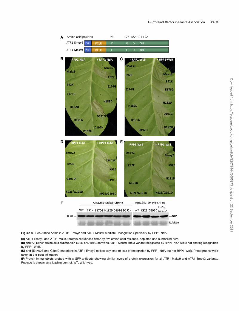
Figure 8. Two Amino Acids in ATR1-Emoy2 and ATR1-Maks9 Mediate Recognition Specificity by RPP1-NdA.
(A) ATR1-Emoy2 and ATR1-Maks9 protein sequences differ by five amino acid residues, depicted and numbered here.
(B) and (C) Either amino acid substitution E92K or D191G converts ATR1-Maks9 into a variant recognized by RPP1-NdA while not altering recognition
by RPP1-WsB.
(D) and (E) K92E and G191D mutations in ATR1-Emoy2 collectively lead to loss of recognition by RPP1-NdA but not RPP1-WsB. Photographs were
taken at 3 d post infiltration.
(F) Protein immunoblots probed with a-GFP antibody showing similar levels of protein expression for all ATR1-Maks9 and ATR1-Emoy2 variants.
Rubisco is shown as a loading control. WT, Wild type.
R-Protein/Effector in Planta Association 2453
Dow
nloaded from https://academ
ic.oup.com/plcell/article/22/7/2444/6095973 by guest on 22 Septem
ber 2021

2003). Similarly, the P. syringae effector AvrPphB, a Cys
protease, cleaves the Arabidopsis protein AvrPphB suscepti-
ble 1 (Shao et al., 2003). Proteolytic activity of AvrPphB is
indirectly detected by the Arabidopsis CC-NBS-LRR protein
RPS5 (Ade et al., 2007), which leads to activation of resistance
signaling. Additional examples of indirect enzymatic activa-
tion include Pseudomonas effectors AvrRpm1 and AvrB,
which induce phosphorylation of the Arabidopsis RIN4 pro-
tein, leading to activation of the CC-NBS-LRR protein Resis-
tance to Pseudomonas maculicola 1 (RPM1; Mackey et al.,
2002).
It has been noted that the evolutionary history of the cognate
effector/R gene pairs correlates with their mode of recognition.
The high levels of diversifying selection in the flax rust effector
alleles and corresponding flax R genes have been commonly
explained by an arms race between the ligand and the receptor
(Dodds et al., 2006; Jones and Dangl, 2006; Barrett et al.,
2009). Indeed, this observation is consistent with the hypoth-
esis that evolutionary selection pressure targets particular
amino acid sites in the effector and the LRR portions of a
resistance protein as a result of a direct interaction between
the two proteins. On the other hand, effectors with enzymatic
activity that induce modifications in the host targets are evolv-
ing under balancing or purifying selection (Van der Hoorn et al.,
2002; Rohmer et al., 2004). Generally, it has been assumed that
the R genes that have evolved to monitor host targets of the
effectors are evolving more slowly and are less prone to
duplications, since their function is to be stably associated
with the invariant host proteins (Rohmer et al., 2004; Jones and
Dangl, 2006).
The evolutionary history of ATR1 and RPP1 matches the
direct recognition model. The ATR1 effector gene is evolving
under very strong levels of diversifying selection (Rehmany
et al., 2005). The RPP1 gene locus shows high levels of
duplications, insertions/deletions, and polymorphisms, which
create alleles with altered specificities, such as RPP1-WsB and
RPP1-NdA. This observation is further supported by the closer
examination of the interactions between the two RPP1 alleles
WsB and NdA and the ATR1 alleles Emoy2 and Maks9. Since
both RPP1-WsB and RPP1-NdA can recognize ATR1-Emoy2,
we can conclude that both RPP1 alleles encode functional
resistance gene products capable of eliciting the defense
response. Similarly, since both ATR1-Emoy2 and ATR1-
Maks9 are recognized by RPP1-WsB, this interaction reveals
that both effector alleles are functional. However, only ATR1-
Emoy2, but not ATR1-Maks9, is recognized by RPP1-NdA.
Moreover, we have demonstrated that this difference in recog-
nition can be overturned by two independent amino acid site
substitutions in ATR1-Maks9. Given this evidence, we con-
clude that it is unlikely that RPP1 recognizes an enzymatic
function of ATR1. This leaves the hypothesis of either recogni-
tion by direct binding or recognition by indirect binding, which
requires additional host proteins. Using similar logic, we can
conclude that if binding is indirect, there should be at least two
different host targets of ATR1, guarded by RPP1-WsB and
RPP1-NdA. We conclude that in light of the evolutionary history
of ATR1 and RPP1, recognition through direct binding remains
the most parsimonious model.
Activation of RPP1 Is Specified by in Planta Association
with ATR1
The lack of published in planta data demonstrating that R
proteins are able to associate with effectors inside the plant
cell has led researchers to question the overall validity of the
direct binding hypothesis. This lack of in planta data for R
protein–effector association may be explained by difficulties
using natural pathosystems in biochemical assays, including
very low levels of protein expression and the low strength or
transient nature of R protein–effector interactions. Here, we
show that the pattern of in planta association between ATR1 and
RPP1-WsB matches that of the effector recognition, suggesting
that the ability of RPP1 to form a protein complex with ATR1 is
required for triggering the hypersensitive cell death resistance
response. At the same time, we are well aware that our in planta
results do not prove direct protein–protein binding. Such a
demonstration remains challenging due to our inability to purify
soluble recombinant RPP1 protein or its LRR domain in the
organisms commonly used for heterologous protein production,
including Saccharomyces cerevisiae, Pichia pastoris, and Esch-
erichia coli. It is possible that RPP1 may require additional plant-
specific factors that provide protein stability, such as HSP90,
SGT1, or RAR1, or yet unidentified factors. Thus, future research
will include isolating the ATR1/RPP1 protein complex from
Arabidopsis and characterizing other interacting protein com-
plex members by mass spectrometry to elucidate the nature of
the interaction between the R protein and its effector.
Roles of Different Domains of RPP1 in Effector Recognition
and Signaling Responses
Although many R proteins share a common CC/TIR-NBS-LRR
domain architecture, the actual biochemical role of each domain
in effector recognition and downstream signaling has been
subject to debate. Based on molecular genetic analyses, the
LRR domain was originally proposed to mediate effector recog-
nition; this is well supported by yeast two-hybrid studies and in
vitro experiments (Deslandes et al., 2003; Dodds et al., 2006).
However, recent in vivo data suggested the TIR domain of
tobacco TIR-NBS-LRR protein N and the CC domain of the
potato (Solanum tuberosum) CC-NBS-LRR protein Rx as the
main regions involved in effector recognition (Burch-Smith et al.,
2007; Rairdan et al., 2008). Despite the fact that these data
contradicted previous studies of N, in which the NBS-LRR
portion was shown to bind the cognate tobacco mosaic virus
effector protein p50 both in vitro and in yeast (Ueda et al., 2006), a
new hypothesis has emerged postulating that theN-terminal CC/
TIR domain is the main determinant of effector–R protein inter-
actions (Burch-Smith and Dinesh-Kumar, 2007; Collier and
Moffett, 2009; Lukasik and Takken, 2009). Having established
the in planta association between the RPP1-WsB and ATR1
proteins, we were able to identify which domain of RPP1 was
responsible for effector recognition. Our deletion analysis
showed that neither TIR nor TIR-NBS could associate with
ATR1 in planta. On the other hand, the LRR domain was both
necessary and sufficient for interaction with ATR1 in an allele-
specific manner. Furthermore, we introduced single amino acid
2454 The Plant Cell
Dow
nloaded from https://academ
ic.oup.com/plcell/article/22/7/2444/6095973 by guest on 22 Septem
ber 2021

substitutions in the TIR domain and the P-loop of the NBS
domain that abolished the ability of RPP1 to trigger effector-
induced HR but did not disrupt the association with ATR1. This
indicates that ATR1 can associate with RPP1 it its inactive state,
which is consistent with the proposed model, in which R protein
activation is “switched on” after perception of the effector
(Takken et al., 2006; Lukasik and Takken, 2009). These data
are also consistent with the step-wise model of R protein
activation, in which the LRR domain associates with the effector
before the activation of other domains and signaling is triggered
(Collier and Moffett, 2009).
Although the LRR portion of RPP1 is sufficient for association
with ATR1, it is not sufficient to induce the hypersensitive cell
death immune response. Mutagenesis of the TIR and NBS
domains of RPP1 revealed that they control the ability of RPP1
to activate the response after association with ATR1. Involve-
ment of the TIR and NBS domains in signaling has been previ-
ously reported for the TIR-NBS-LRR proteins L10, RPS4,
RPP1A, and N (Frost et al., 2004; Zhang et al., 2004; Weaver
et al., 2006; Swiderski et al., 2009). The minimal region that was
sufficient to induce the HR in those proteins included the TIR
domain and the first 20 amino acids of the NBS domain (which
does not include the P-loop motif; Frost et al., 2004; Swiderski
et al., 2009). In this study, we report that the analogous region of
RPP1-WsB (amino acids 1–266) was also able to elicit effector-
independent HR when fused to the large epitope tag, GFP.
Intrigued by the fact that fusion to GFP was required for the
induction of the cell death, we examined whether the dimerizing
property of GFP could contribute to the signaling of the TIR
domain. Indeed, when we constructed a monomeric GFP and
fused it to the TIR domain, the latter lost its ability to activate
effector-independent cell death. These data suggest a hypoth-
esis whereby the activation of the TIR domain is regulated by
oligomerization of RPP1 upon perception of ATR1. Effector-
induced oligomerization of R proteins has been proposed pre-
viously and has been supported by oligomerization of N, which
was dependent on the presence of its cognate effector and the
intact P-loop motif (Mestre and Baulcombe, 2006).
We do not attempt to propose that our data explain how all of
the R proteins would be activated, nor that functions of the
structurally similar domains would be exactly the same in differ-
ent R proteins. Different modes of effector perception predict the
existence of different modes of R protein activation. Indeed, if a
protein is negatively regulated by another host factor, as in the
case of Arabidopsis RPS2 and RIN4 (Axtell and Staskawicz,
2003), the initial mechanism of its activation might be different
from a protein that is autoinhibited due to intramolecular inter-
actions, as demonstrated for the potato R protein Rx (Moffett
et al., 2002; Rairdan and Moffett, 2006). Herein, we provide the
missing biochemical in planta data that support a particular
simple model, in which effector recognition is mediated through
the LRR domain while the TIR domain is involved in signaling.
RPP1 Activation Model
Given these data, we propose the following series of molecular
events that lead to the activation of RPP1-WsB in response to
ATR1. First, the pathogen infects the host and delivers ATR1
protein across the haustorial extracellular matrix into the plant
cell. Recognition of ATR1 ultimately depends on which variant of
ATR1 is delivered and which variant of RPP1 is present in the
host. Our mutational analyses of ATR1 demonstrated that the
pathogen could be just one amino acid away from being recog-
nized. The LRR domain of RPP1 monitors for the presence of
ATR1 inside the plant cell. Upon successful association between
the LRR and the ATR1 variant, RPP1-WsB undergoes a confor-
mational change, possibly oligomerization coupled to ATP bind-
ing by the NBS domain. This activates the TIR domain, which
initiates a signal transduction cascade that leads to the HR and
expression of plant disease resistance.
Several key questions will need to be resolved before we can
claim full understanding of the RPP1–ATR1 interaction: (1) the
composition of the protein complex they form; (2) the structural
basis of effector recognition by the LRR domain; (3) the mode of
TIR domain activation by LRR and NBS domains; and (4) the
signaling cascade leading to the induction of the HR and plant
disease resistance. The elucidation of these molecular mecha-
nisms would be of great benefit to both plant and animal
immunity and will ultimately allow researchers to rationally de-
sign broad-spectrum R genes for applications in agriculture.
METHODS
Multiple Sequence Alignment
Alignment of the amino acid sequences of Arabidopsis thalianawas done
using the MUSCLE algorithm (Edgar, 2004) and visualized with belvu
(Sonnhammer and Hollich, 2005). For phylogenetic analyses, we used an
alignment between coding DNA sequences, in which codons were
aligned corresponding to the amino acid sequence alignment using the
pal2nal algorithm (Suyama et al., 2006).
Phylogenetic Analysis
Phylogenetic analysis was performed using the Phylip 3.66 software
package (Felsenstein, 2005). The unrooted maximum likelihood tree was
constructed from the nucleotide sequence alignment discussed above
using the dnaml algorithm (Felsenstein and Churchill, 1996) with default
parameters (see Supplemental Table 2 online). Bootstrapping was
performed for 1000 replicates using the seqboot algorithm with the
default parameters and the consense algorithm with the user-tree option
(see Supplemental Table 2 online). The tree was visualized using the
TreeView program (Page, 1996).
Strains and Growth Conditions
Escherichia coli DH5a and Agrobacterium tumefaciens GV3101 were
grown in Luria–Bertani medium supplemented with the appropriate
antibiotics at 378C and 288C, respectively. Bacterial DNA transformation
was conducted using chemically competent E. coli (Invitrogen) and
through freeze/thaw transformation of CaCl2-competent Agrobacterium
(Wise et al., 2006). Tobacco (Nicotiana tabacum var Turk) plants were
grown in a controlled growth chamber at 248C with a 16-h-light/8-h-dark
photoperiod before infiltrations and switched to 24 h of light after
infiltrations.
Constructs
The sequences for all of the primers used in this study are shown in
Supplemental Table 1 online in 59 to 39 orientation, restriction sites are
R-Protein/Effector in Planta Association 2455
Dow
nloaded from https://academ
ic.oup.com/plcell/article/22/7/2444/6095973 by guest on 22 Septem
ber 2021

indicated in boldface, the sequence encoding the HA tag is underlined,
and the pENTR/D-TOPO targeting sequence CACC is shown in the
forward primers. All point mutations were introduced by site-directed
mutagenesis using the Quick-Change SDM kit (Stratagene), and primers
are specified in Supplemental Table 1 online.
The open reading frame of RPP1-WsB was amplified via PCR using an
Arabidopsis Ws-0 cDNA template. Cloning of the full-length RPP1-WsB
gene was done in two fragments, taking advantage of the unique NdeI
restriction site within the gene. The reverse primer incorporated an in-
frame HA epitope tag 59 to the stop codon in order to create a C-terminal
fusion protein with RPP1-WsB. The PCR products were directly sub-
cloned in pENTR/D-TOPO vector (Invitrogen). TwoRPP1-WsB fragments
were combined in pENTR/D-/TOPO with a NotI-NdeI digest. Deletion
variants of RPP1 were amplified with the following primers: RPP1-WsB
F/TIR R;RPP1-WsB F/NBSR;RPP1-WsBNBS F/Spe1-HAR;RPP1-WsB
LRR F/Spe1-HA R. A SpeI site was incorporated in the reverse primers,
which allowed creation of a C-terminal fusion with the HA or GFP epitope
tags by restriction digest/ligation. For in planta analyses, all resulting
RPP1-WsB constructs were introduced into pEarleyGate destination
binary vector pEG201 (35S promoter, N-terminal HA tag fusion; Earley
et al., 2006) using LR clonase (Invitrogen). Sequence information for
RPP1-NdA was acquired from Gordon (2002). Unfortunately, we could
not isolate any functional cDNA for RPP1-NdA. The full-length genomic
sequence, including the native promoter and native terminator, was
subcloned in three fragments using the following primers, RPP1-NdA
fragment 1 F/R, fragment 2 F/R, fragment 3 F/R, and joined together by
restriction digest/ligation. An XbaI restriction site was introduced at the 39
of the RPP1-NdA open reading frame by site-directed mutagenesis. The
sequence encoding for a 3xHA epitope tag was PCR-amplified from the
pBJ36-3xVenus-3xHA vector (provided by Jeff Long, Salk Institute),
column-purified, digested with SpeI/XbaI, and ligated into XbaI of RPP1-
NdA. Resulting construct was introduced into pEG301 (no promoter)
using LR clonase (Invitrogen).
All ATR1 variants were amplified from genomic DNA templates ex-
tracted from Hyaloperonospora arabidopsidis spores of the appropriate
pathovar (provided by John McDowell, Thomas Eulgem, and Jonathan
Jones). The ATR1 allelic variants and deletions were amplified by PCR
with the primers indicated in Supplemental Table 1 online and subcloned
in pENTR/D-TOPO vector (Invitrogen). A SpeI site was included in the
reverse primers before the stop codon to facilitate epitope tagging. The
Citrine gene was cloned on the 39 end of all ATR1 alleles using SpeI
digestion. The ATR1 constructs were introduced in the pEG103 (35S
promoter, C-terminal GFP fusion) and pEG202 (35S promoter, N-terminal
Flag tag fusion) binary destination vectors via LR recombination.
Agrobacterium-Mediated Transient Expression
Agrobacteriumwas grown in Luria–Bertani broth cultures (supplemented
with 50 mg/mL gentamycin and 25 mg/mL kanamycin) overnight at 288C
with constant shaking. The cultures were spun down at a tabletop
centrifuge at 10,000 rpm for 2 min. The resulting pellet was resuspended
in 1 mL of induction medium (10 mM MgCl, 10 mM MES, and 150 mM
acetosyringone, adjusted to pH 5.6 with KOH). Bacterial concentrations
were measured and adjusted with induction medium to OD600 = 0.9.
Resulting cultures were preinduced for 2 to 3 h at 288C. For coinfiltrations,
cultures carrying individual constructs were induced separately and
mixed in a 1:1 ratio just before infiltration. Young tobacco leaves were
inoculated with Agrobacterium cultures using a blunt syringe.
Transient Protein Expression in Tobacco
To detect transient protein expression in tobacco, two leaf discs (1.5 cm
diameter) were collected 24 to 48 h post infiltration. The samples were
frozen in liquid nitrogen and ground with a prechilled plastic pestle.
Protein was extracted with 150 mL of the Laemmli buffer (0.24 M Tris-Cl,
pH 6.8, 6% SDS, 30% glycerol, 16% b-mercaptoethanol, 0.006%
bromophenol blue, and 5 M urea). Samples were boiled for 5 min and
centrifuged at maximum speed for 10min in a tabletop centrifuge at room
temperature; supernatants were transferred into fresh tubes before
analysis by SDS-PAGE and immunoblotting as described below.
Coimmunoprecipitations
All constructs were transiently expressed in tobacco using Agrobacte-
rium-mediated transformation with strain GV3101. Leaf tissue was col-
lected 24 h post infiltration, when the first visible signs of HR started to
appear, weighed, and snap-frozen in liquid nitrogen. The weight of the
tissue undergoing HR was estimated based on its surface area. We have
routinely used ;1 g of tissue for each coimmunoprecipitation experi-
ment, which corresponds to about one youngest fully expanded leaf of
3- to 4-week-old tobacco. Each tissue sample was ground with mortar
and pestle to a homogeneous powder in liquid nitrogen, transferred into a
precooled mortar containing 2 mL of the protein extraction buffer (50 mM
Tris-HCl, pH 7.5, 150 mM NaCl, 0.1% Triton X-100, 0.2% Nonidet P-40,
6 mM b-mercaptoethanol, 0.3 mM aprotinin, 10 mM leupeptin, 1 mM pep-
statin A, and a Complete protease inhibitor cocktail [Roche]), and ho-
mogenized with a fresh precooled pestle until the sample was completely
thawed. Resulting samples were transferred to 1.5-mL Eppendorf tubes
and centrifuged for 20 min, 14,000 rpm, and 48C. The supernatant was
split into two fresh Eppendorf tubes and used as input for the a-GFP and
a-HA immunoprecipitations. All the steps in the immunoprecipitations
were performed at 48C.
In order to immunoprecipitate the target protein, either 2 mL of a-GFP
(rabbit, polyclonal; Abcam) or 20 mL of a-HA (mouse, clone 16B12;
Roche) was added to 1mL of protein extract. The antibody–lysatemixture
was incubated with gentle tumbling for 1.5 h at 48C. Next, 50mL of Protein
G beads (Protein G Sepharose for Fast Flow; GE Healthcare), prewashed
three times in the protein extraction buffer, was added to each sample
and incubated with gentle tumbling for 4 h at 48C. Then, the beads were
spun down for 2min at 1000g, washed three timeswith 1mLof the protein
extraction buffer, and supplemented with fresh protease inhibitors and
fresh b-mercaptoethanol. The protein was eluted by boiling for 5 min in
50 mL of the Laemmli buffer. The samples, 5 mL per lane for detecting
immunoprecipitated protein and 25 mL per lane for detecting coimmu-
noprecipitating protein, were separated on 10% SDS-PAGE gels or
commercial NuPAGE SDS gradient 4% to 12% gels (Invitrogen), trans-
ferred to nitrocellulosemembrane (Fisher), andanalyzedby immunoblotting
usingmousea-GFP (cloneB34;Covance) andgoat anti-mouse-horseradish
peroxidase (Bio-Rad) or rat a-HA-horseradish peroxidase (clone 3F10;
Roche). All coimmunoprecipitation experiments were performed at least
three times from different leaf tissue samples and gave robust and repeat-
able results.
Accession Numbers
Sequence data that were used in this paper can be found in the National
Center for Biotechnology Information databases under the following
accession numbers: ATR1-Emoy2 (gi61660946), ATR1-Maks9 (gi61660952),
ATR1-Emco5 (gi61660954), ATR1-Cala2 (gi61660958), ATR1-Emwa1
(gi61660960), RPP1-WsB (gi3860164). The genomic sequence of RPP1-
NdA was cloned based on Gordon (2002) and deposited in the GenBank
database under accession number HM209027.
Supplemental Data
The following materials are available in the online version of this article.
Supplemental Figure 1. Multiple Sequence Alignment of the ATR1
Protein Sequences Used in This Study.
2456 The Plant Cell
Dow
nloaded from https://academ
ic.oup.com/plcell/article/22/7/2444/6095973 by guest on 22 Septem
ber 2021

Supplemental Figure 2. Multiple Sequence Alignment of the Protein
Sequences Coding for RPP1-WsB and RPP1-NdA.
Supplemental Figure 3. Race-Specific Recognition of ATR1 Alleles
by the Genomic RPP1-WsB Construct Expressed under the Control
of Its Native Promoter.
Supplemental Figure 4. Localization of Different ATR1 Alleles in
Tobacco.
Supplemental Figure 5. Additional Coimmunoprecipitation Experi-
ments Showing Interactions between NBS-LRR and LRR Domains of
RPP1-WsB and Different ATR1 Variants.
Supplemental Figure 6. Multiple Sequence Alignment of the TIR
Domain and P-Loop Motif in Different R Proteins and the TIR Domain
of Human TLR1.
Supplemental Table 1. List of Primers Used in This Study for Gene
Amplification and Site-Directed Mutagenesis.
Supplemental Table 2. Set of Parameters Used in dnaml, seqboot,
and consense Algorithms for Constructing the Phylogenetic Tree of
the ATR1 Alleles.
Supplemental Data Set 1. Text File of the Alignment Used for the
Phylogenetic Analysis Shown in Figure 1.
ACKNOWLEDGMENTS
We are grateful to Jim Beynon (University of Warwick) for providing
sequence information and plant material that facilitated making RPP1-
NdA constructs; to John McDowell (Virginia Tech), Thomas Eulgem
(University of California Riverside), and Jonathan Jones (Sainsbury Lab-
oratory) for providing H. arabidopsidis inocula and DNA samples; and to
Kimmen Sjolander (University of California Berkeley) for providing access
to the belvu program used in visualizing multiple sequence alignments.
We also thank Daniil Prigozhin (University of California Berkeley), Sandra
Goritschnig (University of California Berkeley), Peter Dodds (Common-
wealth Scientific and Industrial Research Organisation), Lauriebeth
Leonelli (University of California Berkeley), and Baomin Zhang (University
of California Berkeley) for helpful discussion of the presented material and
critical reading of the manuscript. This work was supported by the
National Science Foundation (grant NSF 2010 0726229).
ReceivedMarch 19, 2010; revised May 8, 2010; accepted June 17, 2010;
published July 2, 2010.
REFERENCES
Ade, J., DeYoung, B.J., Golstein, C., and Innes, R.W. (2007). Indirect
activation of a plant nucleotide binding site-leucine-rich repeat
protein by a bacterial protease. Proc. Natl. Acad. Sci. USA 104:
2531–2536.
Alder, M.N., Rogozin, I.B., Iyer, L.M., Glazko, G.V., Cooper, M.D.,
and Pancer, Z. (2005). Diversity and function of adaptive immune
receptors in a jawless vertebrate. Science 310: 1970–1973.
Allen, R.L., Bittner-Eddy, P.D., Grenville-Briggs, L.J., Meitz, J.C.,
Rehmany, A.P., Rose, L.E., and Beynon, J.L. (2004). Host-parasite
coevolutionary conflict between Arabidopsis and downy mildew.
Science 306: 1957–1960.
Axtell, M.J., Chisholm, S.T., Dahlbeck, D., and Staskawicz, B.J.
(2003). Genetic and molecular evidence that the Pseudomonas
syringae type III effector protein AvrRpt2 is a cysteine protease.
Mol. Microbiol. 49: 1537–1546.
Axtell, M.J., and Staskawicz, B.J. (2003). Initiation of RPS2-specified
disease resistance in Arabidopsis is coupled to the AvrRpt2-directed
elimination of RIN4. Cell 112: 369–377.
Barrett, L., Thrall, P., Dodds, P., Van Der Merwe, M., Linde, C.,
Lawrence, G., and Burdon, J. (2009). Diversity and evolution of
effector loci in natural populations of the plant pathogen Melampsora
lini. Mol. Biol. Evol. 26: 2499–2513.
Birch, P., Rehmany, A., Pritchard, L., Kamoun, S., and Beynon, J.
(2006). Trafficking arms: Oomycete effectors enter host plant cells.
Trends Microbiol. 14: 8–11.
Bomblies, K., Lempe, J., Epple, P., Warthmann, N., Lanz, C., Dangl,
J., and Weigel, D. (2007). Autoimmune response as a mechanism for
a Dobzhansky-Muller-type incompatibility syndrome in plants. PLoS
Biol. 5: e236.
Botella, M.A., Parker, J.E., Frost, L.N., Bittner-Eddy, P.D., Beynon,
J.L., Daniels, M.J., Holub, E.B., and Jones, J.D. (1998). Three genes
of the Arabidopsis RPP1 complex resistance locus recognize distinct
Peronospora parasitica avirulence determinants. Plant Cell 10: 1847–
1860.
Burch-Smith, T., and Dinesh-Kumar, S. (2007). The functions of plant
TIR domains. Sci. STKE 401: pe46.
Burch-Smith, T., Schiff, M., Caplan, J., Tsao, J., Czymmek, K., and
Dinesh-Kumar, S. (2007). A novel role for the TIR domain in asso-
ciation with pathogen-derived elicitors. PLoS Biol. 5: e68.
Catanzariti, A.M., Dodds, P.N., Ve, T., Kobe, B., Ellis, J.G., and
Staskawicz, B.J. (2010). The AvrM effector from flax rust has a
structured C-terminal domain and interacts directly with the M resis-
tance protein. Mol. Plant Microbe Interact. 23: 49–57.
Chisholm, S., Coaker, G., Day, B., and Staskawicz, B. (2006). Host-
microbe interactions: Shaping the evolution of the plant immune
response. Cell 124: 803–814.
Collier, S.M., and Moffett, P. (2009). NB-LRRs work a “bait and switch”
on pathogens. Trends Plant Sci. 14: 521–529.
Deslandes, L., Olivier, J., Peeters, N., Feng, D.X., Khounlotham, M.,
Boucher, C., Somssich, I., Genin, S., and Marco, Y. (2003). Physical
interaction between RRS1-R, a protein conferring resistance to bac-
terial wilt, and PopP2, a type III effector targeted to the plant nucleus.
Proc. Natl. Acad. Sci. USA 100: 8024–8029.
Desveaux, D., Singer, A., and Dangl, J. (2006). Type III effector
proteins: Doppelgangers of bacterial virulence. Curr. Opin. Plant Biol.
9: 376–382.
Dinesh-Kumar, S.P., Tham, W.H., and Baker, B.J. (2000). Structure-
function analysis of the tobacco mosaic virus resistance gene N. Proc.
Natl. Acad. Sci. USA 97: 14789–14794.
Dodds, P., Lawrence, G.J., Catanzariti, A., Teh, T., Wang, C., Ayliffe,
M.A., Kobe, B., and Ellis, J. (2006). Direct protein interaction under-
lies gene-for-gene specificity and coevolution of the flax resistance
genes and flax rust avirulence genes. Proc. Natl. Acad. Sci. USA 103:
8888–8893.
Dodds, P.N., Lawrence, G.J., and Ellis, J.G. (2001). Six amino acid
changes confined to the leucine-rich repeat b-strand/b-turn motif
determine the difference between the P and P2 rust resistance
specificities in flax. Plant Cell 13: 163–178.
Dodds, P.N., Rafiqi, M., Gan, P.H., Hardham, A.R., Jones, D.A., and
Ellis, J.G. (2009). Effectors of biotrophic fungi and oomycetes: Path-
ogenicity factors and triggers of host resistance. New Phytol. 183:
993–1000.
Earley, K., Haag, J., Pontes, O., Opper, K., Juehne, T., Song, K., and
Pikaard, C. (2006). Gateway-compatible vectors for plant functional
genomics and proteomics. Plant J. 45: 616–629.
Edgar, R.C. (2004). MUSCLE: Multiple sequence alignment with high
accuracy and high throughput. Nucleic Acids Res. 32: 1792–1797.
Ellis, J.G., Lawrence, G.J., Luck, J.E., and Dodds, P.N. (1999).
R-Protein/Effector in Planta Association 2457
Dow
nloaded from https://academ
ic.oup.com/plcell/article/22/7/2444/6095973 by guest on 22 Septem
ber 2021

Identification of regions in alleles of the flax rust resistance gene L
that determine differences in gene-for-gene specificity. Plant Cell 11:
495–506.
Felsenstein, J. (2005). PHYLIP (Phylogeny Inference Package) version
3.6. http://evolution.genetics.washington.edu/phylip.html.
Felsenstein, J., and Churchill, G.A. (1996). A hidden Markov model
approach to variation among sites in rate of evolution. Mol. Biol. Evol.
13: 93–104.
Frost, D., Way, H., Howles, P., Luck, J., Manners, J., Hardham, A.,
Finnegan, J., and Ellis, J. (2004). Tobacco transgenic for the flax rust
resistance gene L expresses allele-specific activation of defense
responses. Mol. Plant Microbe Interact. 17: 224–232.
Gordon, A. (2002). Analysis of the RPP1 Resistance Gene Cluster in
Arabidopsis Accession Niederzenz (Nd-1). PhD dissertation (Birming-
ham, UK: University of Birmingham).
Jia, Y., McAdams, S.A., Bryan, G.T., Hershey, H.P., and Valent, B.
(2000). Direct interaction of resistance gene and avirulence gene
products confers rice blast resistance. EMBO J. 19: 4004–4014.
Jones, J., and Dangl, J. (2006). The plant immune system. Nature 444:
323–329.
Kamoun, S. (2006). A catalogue of the effector secretome of plant
pathogenic oomycetes. Annu. Rev. Phytopathol. 44: 41–60.
Kim, H.E., Du, F., Fang, M., and Wang, X. (2005). Formation of
apoptosome is initiated by cytochrome c–induced dATP hydrolysis
and subsequent nucleotide exchange on Apaf-1. Proc. Natl. Acad.
Sci. USA 102: 17545–17550.
Kobe, B., and Deisenhofer, J. (1993). Crystal structure of porcine
ribonuclease inhibitor, a protein with leucine-rich repeats. Nature 366:
751–756.
Lukasik, E., and Takken, F.L. (2009). STANDing strong, resistance
proteins instigators of plant defence. Curr. Opin. Plant Biol. 12:
427–436.
Mackey, D., Holt III, B.F., Wiig, A., and Dangl, J.L. (2002). RIN4
interacts with Pseudomonas syringae type III effector molecules and
is required for RPM1-mediated resistance in Arabidopsis. Cell 108:
743–754.
Mestre, P., and Baulcombe, D.C. (2006). Elicitor-mediated oligomer-
ization of the tobacco N disease resistance protein. Plant Cell 18:
491–501.
Moffett, P., Farnham, G., Peart, J., and Baulcombe, D.C. (2002).
Interaction between domains of a plant NBS2LRR protein in disease
resistance-related cell death. EMBO J. 17: 4511–4519.
Mondragon-Palomino, M. (2002). Patterns of positive selection in the
complete NBS-LRR gene family of Arabidopsis thaliana. Genome
Res. 12: 1305–1315.
Mucyn, T., Clemente, A., Andriotis, V., Balmuth, A., Oldroyd, G.,
Staskawicz, B., and Rathjen, J. (2006). The tomato NBARC-LRR
protein Prf interacts with Pto kinase in vivo to regulate specific plant
immunity. Plant Cell 18: 2792–2806.
Page, R.D. (1996). TreeView: An application to display phylogenetic
trees on personal computers. Comput. Appl. Biosci. 12: 357–358.
Rairdan, G., and Moffett, P. (2006). Distinct domains in the ARC region
of the potato resistance protein Rx mediate LRR binding and inhibition
of activation. Plant Cell 18: 2082–2093.
Rairdan, G.J., Collier, S.M., Sacco, M.A., Baldwin, T.T., Boettrich, T.,
and Moffett, P. (2008). The coiled-coil and nucleotide binding do-
mains of the potato Rx disease resistance protein function in path-
ogen recognition and signaling. Plant Cell 20: 739–751.
Rehmany, A., Gordon, A., Rose, L.E., Allen, R.L., Armstrong, M.R.,
Whisson, S.C., Kamoun, S., Tyler, B.M., Birch, P.R.J., and Beynon,
J.L. (2005). Differential recognition of highly divergent downy mildew
avirulence gene alleles by RPP1 resistance genes from two Arabi-
dopsis lines. Plant Cell 17: 1839–1850.
Rentel, M.C., Leonelli, L., Dahlbeck, D., Zhao, B., and Staskawicz,
B.J. (2008). Recognition of the Hyaloperonospora parasitica effector
ATR13 triggers resistance against oomycete, bacterial, and viral
pathogens. Proc. Natl. Acad. Sci. USA 105: 1091–1096.
Rohmer, L., Guttman, D.S., and Dangl, J.L. (2004). Diverse evolution-
ary mechanisms shape the type III effector virulence factor repertoire
in the plant pathogen Pseudomonas syringae. Genetics 167: 1341–
1360.
Shaner, N.C., Steinbach, P.A., and Tsien, R.Y. (2005). A guide to
choosing fluorescent proteins. Nat. Methods 2: 905–909.
Shao, F., Golstein, C., Ade, J., Stoutemyer, M., Dixon, J.E., and
Innes, R.W. (2003). Cleavage of Arabidopsis PBS1 by a bacterial type
III effector. Science 301: 1230–1233.
Shen, Q., Saijo, Y., Mauch, S., Biskup, C., Bieri, S., Keller, B., Seki,
H., Ulker, B., Somssich, I., and Schulze-Lefert, P. (2007). Nuclear
activity of MLA immune receptors links isolate-specific and basal
disease-resistance responses. Science 315: 1098–1103.
Sohn, K.H., Lei, R., Nemri, A., and Jones, J.D. (2007). The downy
mildew effector proteins ATR1 and ATR13 promote disease suscep-
tibility in Arabidopsis thaliana. Plant Cell 19: 4077–4090.
Sonnhammer, E.L., and Hollich, V. (2005). Scoredist: A simple and
robust protein sequence distance estimator. BMC Bioinformatics
6: 108.
Suyama, M., Torrents, D., and Bork, P. (2006). PAL2NAL: Robust
conversion of protein sequence alignments into the corresponding
codon alignments. Nucleic Acids Res. 34: 609–612.
Swiderski, M., Birker, D., and Jones, J. (2009). The TIR domain of TIR-
NB-LRR resistance proteins is a signaling domain involved in cell
death induction. Mol. Plant Microbe Interact. 22: 157–165.
Takken, F., Albrecht, M., and Tameling, W. (2006). Resistance pro-
teins: Molecular switches of plant defence. Curr. Opin. Plant Biol. 9:
383–390.
Tameling, W.I., Elzinga, S.D., Darmin, P.S., Vossen, J.H., Takken,
F.L., Haring, M.A., and Cornelissen, B.J. (2002). The tomato R gene
products I-2 and MI-1 are functional ATP binding proteins with
ATPase activity. Plant Cell 14: 2929–2939.
Trinchieri, G., and Sher, A. (2007). Cooperation of Toll-like receptor
signals in innate immune defence. Nat. Rev. Immunol. 7: 179–190.
Ueda, H., Yamaguchi, Y., and Sano, H. (2006). Direct interaction
between the tobacco mosaic virus helicase domain and the ATP-
bound resistance protein, N factor during the hypersensitive response
in tobacco plants. Plant Mol. Biol. 61: 31–45.
Van der Hoorn, R.A., De Wit, P.J., and Joosten, M.H. (2002). Balanc-
ing selection favors guarding resistance proteins. Trends Plant Sci. 7:
67–71.
Weaver, M.L., Swiderski, M., Li, Y., and Jones, J. (2006). The
Arabidopsis thaliana TIR-NB-LRR R-protein, RPP1A: Protein locali-
zation and constitutive activation of defence by truncated alleles in
tobacco and Arabidopsis. Plant J. 47: 829–840.
Win, J., Morgan, W., Bos, J., Krasileva, K.V., Cano, L.M., Chaparro-
Garcia, A., Ammar, R., Staskawicz, B.J., and Kamoun, S. (2007).
Adaptive evolution has targeted the C-terminal domain of the RXLR
effectors of plant pathogenic oomycetes. Plant Cell 19: 2349–2369.
Wirthmueller, L., Zhang, Y., Jones, J., and Parker, J. (2007). Nuclear
accumulation of the Arabidopsis immune receptor RPS4 is necessary
for triggering EDS1-dependent defense. Curr. Biol. 17: 2023–2029.
Wise, A.A., Liu, Z., and Binns, A.N. (2006). Three methods for the
introduction of foreign DNA into Agrobacterium. In Agrobacterium
Protocols, 2nd ed, K. Wang, ed (Totowa, NJ: Humana Press Inc.), pp.
43–54.
Zhang, Y., Dorey, S., Swiderski, M., and Jones, J.D. (2004). Expres-
sion of RPS4 in tobacco induces an AvrRps4-independent HR that
requires EDS1, SGT1 and HSP90. Plant J. 40: 213–224.
2458 The Plant Cell
Dow
nloaded from https://academ
ic.oup.com/plcell/article/22/7/2444/6095973 by guest on 22 Septem
ber 2021