active solute transport across frog skin and epithelial cell
TRANSCRIPT

ACTIVE SOLUTE TRANSPORT ACROSS FROG SKIN AND EPITHELIAL CELL SYSTEMS ACCORDING TO THE ASSOCIATION-INDUCTION HYPOTAESIS
GILBERT N. LING
Department of Molecular Biology. Pennsylvania Hospital. 8th and Spruce Streets, Philadelphia, PA 19107
Rcprintcd froin
PHYSIOLOClCA L CHEMISTRY und PH Y.PICS
Volume 13, Number 4.1981
GPO BOX 2044
NEW YORK, N.Y. , IOUOI

356 Physiol. Chem. & Physics 13 (1981)
ACTIVE SOLUTE TRANSPORT ACROSS FROG SKIN AND EPITHELIAL CELL SYSTEMS ACCORDING TO THE ASSOCIATION-INDUCTION HYPOTHESIS
GILBERT N. LING
Department of Molecular Biology, Pennsylvania Hospital, 8th and Spruce Streets, Philadelphia, PA 19107
The phenomenon of transport o f ions, sugars, amino acids, etc. across frog skin and other epithelial systems has been commonly interpreted on the basis o f the membrane- pump theory, according to which asymmetry in solute distribution as well as transport into and out o f all living cells results from the permeability properties and "pump" activities o f the cell membrane. IIZ the present review, certain findings in the field o f transepithelial transport o f soli~tes are given new interpretation on the basis o f molecular mechanisms introduced in the association-indrrction hypothesis, according to which "active transport" of solutes occurs only across bifacial cell systems like frog skin and intestinal epithelium but not in the maintenance o f steady levels o f solutes in unifacial cell svstems such as muscle, nerve, and red blood cells.
INTRODUCTION
Since the cell is the fundamental physio- logic unit of life, some of the features that define life may be revealed by studying the properties of a cell before and after it has ceased living. One method often used to tell a living from a dead cell is based on the dye-exclusion property. Dyes such as nigrosin, trypan blue, and erythrocin B are excluded by living cells but stain dead cells with vivid colors. In short, the dead cell has lost its exclusion property and hence the ability to maintain itself separate from its environment.
Exclusion is not limited to foreign mole- cules such as the dyes. It applies to molecules that may be intrinsic to cell functions but require subtle quantitative balance, as in the case of the asymmetrical distribution of K+ and Na+ between cells and their environ- ment. Chemically almost indistinguishable, this pair of ions is sharply segregated by living cells, which can preferentially accu-
mulate K+ to a level many times higher than . that in the external medium and, by exclu- sion, sustain a level of Na+ only a fraction of that found in the same external medium.
The earliest interpretation of this phenom- enon of asymmetrical K+ and Na+ distribu- tion was based on the assumption that the cell membrane is absolutely impermeable to both K+ and Na+.'s2 In following years, re- visions were made. First K+, and later Na+, were recognized to be permeant. Then, after the 1940s, membrane impermeability as the basis of asymmetrical solute distribution was abandoned, largely in consequence of direct measurements of solute permeability made possible by the development of radioactive labeling and other accurate techniques. There remained two and only two possible interpretations for the asymmetrical solute distribution typified by K+ and Na+: ( I ) the steady-state model, in which the unequal distribution is due to active transport ac- complished by continued operation of en- ergy-consuming membrane pumps; and (2)

the equilibrium model, in which accumula- tion of K+ and exclusion of Na+ reflect the different physicochemical attributes of the environments inside and outside the cell.
A strong argument often cited in favor of the steady-state model arises from the ion-distribution pattern in giant algal cells such as Valonia macrophysa. Living in sea water, which contains nearly 50 times more Na+ than K+,"hese cells may retain 3 times more K+ than Na+.4 The bulk of this K + and Na+ is found in the cell sap contained in the central vacuole. The sap in fact con- tains little more than water and salt ions and is therefore not significantly different in this respect from sea water. Clearly, then, the maintained asymmetry of K + and Na+ distribution in this algal cell can be due only to active transport. Here we have a flawless argument, as far as it goes, but it offers no proof that non-algal cells accumu- late K + and exclude Na+ by the same mechanism. Indeed, giant algal cells with large central vacuoles are not typical of other living cells at all. A large central vacuole is seen solely in old plant cells; young plant cells as well as most procaryotic and eucary- otic cells are "solid b ~ d i e s . " ~
CONCEPTS OF MEMBRANE-PUMP THEORY
A majority of cell physiologists adhered to the steady-state model and conventional membrane-pump theory. Most studies of epi- thelial transport have been built on that theory. Hence epithelial solute transport is widely considered equivalent to selective so- lute accumulation and exclusion in "simpler" cells, all due to membrane pumps. This ap- proach does not take full account of the difference between unifacial "solid" cells (e.g., human red blood cells, frog muscle cells, squid axon) and bifacial hollow cells (e.g., Valonia). Only the Valonia-type cell contains an enclosed body of simple aqueous solution. Only in Valonia-type cell does a
second membrane, the tonoplast, enclose this internal aqueous phase. Fifty years ago, Chambers and HoflerQtudied the osmotic behavior of the isolated central vacuole and showed that the tonoplast has properties quite different from those of the outer plas- ma membrane. Indeed, the perfect osmo- meter-like behavior of plant cells was shown to be due primarily to the tonoplast and not to the plasma membrane.
Of course, giant algal cells are not the only living cell systems that achieve net transport of ions and other small molecules between two similar aqueous phases against concen- tration gradients. Many epithelial tissues do the same. But instead of the continuous pro- toplasmic layer characterizing Valonia, epi- thelial cells joined together in continuous sheets separate the two aqueous phases. Without exception, each of these epithelial cells is bifacial, possessing two different sur- faces each facing one of the two aqueous media.
Proposed Mecl~anism of the Na-Pump '
As pointed out by Ussing and Leaf,7 "It is the properties of the outward-facing mem- brane that are unusual, viz., the selectivity for sodium rather than potassium and the absence of .a sodium pump. . . . The inward facing cell membrane . . . can be assumed to be of the same nature as the sodium-potas- sium exchange pump of red cells, muscle and nerve." This opinion clearly shows that in the conventional view, epithelial Na+ trans- port is the same as in most unifacial non- epithelial cells. But what kind of mechanism, if only in theoretical terms, has been pro- posed for these Na pumps? The anwer is clearly provided by Glynn and Karlish." Covering the literature over a span of 23 years, they begin their review on the sodium pump with these words: "The recent startling growth of the literature on the sodium pump may make a review timely. . . . If the great

mass of work that has been done had led to the general acceptance, even provisionally and even in outline, of a hypothesis account- ing for the working of the pump, we could have described that hypothesis and then con- sidered the evidence for it. Unfortunately no such hypothesis exists. . . ."
I believe that the major reasons behind the difficulty in proposing a mechanism for the Na pump are faulty theoretical assump- tions, including that which conceives the lipid layer as the permeability barrier.
Theories of Epithelial Transport
A number of theories have been offered relating to different aspects of epithelial transport. With the exception of the sketch of a theory communicated by myself in 1965 (see ASSOCIATION-INDUCTION HYPOTHESIS
below) all are based on the membrane-pump theory. They include.
( I ) The "two-membrane theory" of Koefoed-Johnson and Us~ ing .~ This theory was based on the authors' work with isolated frog skin. From electrical potential studies they reached the conclusion that the surface facing the outside has a selective high Na+ permeability and the surface facing the inside has a selective high K+ permeability. Ac- cording to this well-known "two-membrane" theory the inner membrane has essentially the same properties seen in most living cells including ,the possession of the Na-K pump in the form of K,Na-activated ATPase. It is the membrane facing ,the outside solution that is unusual and) functions as the seat of transport regulation through the control of Na+ permeability.
(2) The standing osmotic gradient theory of Diamond and Bossert.Io Certain epithelial membranes like the gall bladder, intestinal mucosa, and renal proximal tubule transport salt ions and water in the form of an isotonic solution. Diamond and Bossert suggested that this secretion of an isotonic fluid is due to the pumping of ions into the bottom of
the spaces formed between the folds of the baso-lateral membrane of the epithelial cells. - The hypertonic solutions thus formed then draw water from the cell, becoming more dilute and eventually isotonic as the fluid moves outward toward the serosal "sink."
(3) Cereijido and Rotunno's theory of pericellcllar pumping.I1 In this theory Cerei- jido and coworkers argue that the transport of Na+ by frog skin involves migration of Na+ along an array of fixed negative sites on the outside surface of the epithelial cells.
(4) The Nu-gradient hypothesis of sugar and amino acid transport. Following the dis- covery that intestinal transport of D-glucose depends on the presence of Na+ in the mucosal fluid,12J3 it was suggested by Crane et al.14 and CraneI5 that glucose, Na+, and a carrier form a ternary complex in the mu- cosal membrane which then dissociates and delivers Na+ and glucose. The Na+ gradient from the mucosal fluid to the cell interior provides the energy for the inward transport . of sugar. That uptake of amino acids by uni- facial duck erythrocytes and Ehrlich ascites cells and bifacial kidney cells require Na+ '
was known even earlier.16J7 Schultz and Curranla further elaborated the Na-gradient hypothesis to include transport of amino acids. Support for the hypothesis came from the study of sugar and amino acid transport into isolated microvilli "vesicles." These iso- lated microvilli transiently take up sugar or amino acids to a greater concentration than found in the surrounding medium, thereby exhibiting an "overshoot," when the sugar or amino acid is added with a high concentra- tion of Na+ but not in the presence of the sugar transport inhibitor, phloridzin. (For references, see below.)
THE ASSOCIATION-INDUCTION
HYPOTHESIS
The bulk of cell K+ and Na+ in most living cells is not found in a separate aque- ous phase as in the central vacuole of Valo-

nia. These ions, rather, are encountered in the cytoplasm, where proteins make up from 15 to 25% of the to.tal weight. According to the association-induction (AI) hypothe- sis,19n20 it is primarily the cell proteins that provide and maintain a different physico- chemical environment in the cell, and it is this different physicochemical environment that gives rise to the accumulation of K + and exclusion of Na+.21 Specifioally, the A1 hypothesis argues that certain proteins pro- vide a network or matrix of extended poly- peptide chains whose alternating positive NH and negative CO groups polarize and orient the bulk of cell water into the state of polarized m ~ l t i l a y e r s . ~ ~ - ~ ~ Water in this state has decreasing solubility as molecular size and complexity of solutes increases. These same and/or other proteins provide p- and y-carboxyl groups for selective ad- sorption of K+ over Na+.24 However, both polarized water (and hence Na+ exclusion)
. and ion adsorption (and hence K+ accumu- lation) depend on the existence of the pro- tein-water-ion system in a cooperative high- energy state, called the living slate. To main- tain this living state, adsorption of ATP (and other key molecules) on certain con- trolling cardinal sites is essential.21
Recently there have been major develop- ments in effor,ts aimed at choosing between 'the two diametrically opposed theories of the living cell: ( a ) the steady-state mem- brane-pump or pump-leak theory and ( b ) the equilibrium-based A1 hypothesis. Leav- ing detailed discussion of this multifaceted, complex problem to a monograph in prepar- ation and several published review^,^^^^"^^ I shall briefly consider here only some criti- cisms of the A1 hypothesis.
( I ) Amount of water of hydration on protein is too small to support the contention that the bulk of cell water differs from nor- mal liquid water.27 It is argued that since most native proteins hydrate to the extent
of 0.2 to 0.3 g water/g dry p r ~ t e i n , ~ ~ . ~ ~ no more than 7 to 8% of the cell water could be significantly affected by the 15 to 25% protein content found in most living cells. According to recent surveys as well as ex- perimental studies, however, this conclusion is valid only when proteins are in the globu- lar ~ o n f o r m a t i o n . ~ 3 ~ ~ ~ . ~ W n the other hand, if for one reason or another the protein exists in an extended conformation, long- range polarization of many more water mole- cules occurs. Water so affected has unusual properties; e.g., reduced solubility for Na+, sugars, and amino acids, in full agreement with the A1 hypothesi~.~"
( 2 ) Strong selective adsorption of K + over Nu+ has not been demonstrated in isolated proteins in ~ i t ro .~O This objection is no longer valid. Ouabain-sensitive, cooper- ative, selective K+ binding over Na+ onto one isolated protein (i.e., K,Na-activated ATPase) has been successfully demonstrated in vitro by Matsui and coworker^.^^^^^ Even more exciting is Edelmann's demomtration of selective accumulation of K + over Na+ in freeze-dried muscle sections in v i t r ~ , ~ ~ a finding dealt with in greater detail below.
( 3 ) If cell K+, which constitutes the bulk of intracellular cation, is in an adsorbed and hence osmotically inactive form, the cell would be unable to maintain normal vol- ume.34s35 The linear polymer, polyethylene oxide (PE0)-which carries no net electric charge-polarizes water in deep layers. A dialysis bag containing PEO-water shrinks in concentrated Na-citrate solutions and swells in dilute Na-citrate solution in a man- ner similar to that of living cells placed in hyper- or hypotonic solutions, even though in the case of the PEO-water system there is no semipermeable membrane covering the polymer, the dialysis membrane being fully permeable to Na- itr rate.^^ This illustrates the point made by the A1 hypothesis to the effect

G . N. LING
TABLE I. Data of K+ Concentration ( c K 1 ) , K + Activity ( u K - ~ ' ) , and Activity Coefficient ( u s ' / c ~ ' ) in Various Epithelial Cells Measured with Intracellular Kf-Sensitive Electrodes
Authors Cells
Khuri et al." Rat distal renal tubule epithelium 46 .52 1.6 136 0.34
Khuri et al."' Rat proximal renal tubules epithelium 54.422.5 136 0.40
Zeuthen and M o n g e S V a b b i t intestinal epithelium 38
WhiteM Conger eel intestinal epithelium 41.621.5 146210 0.28 Kimura and Fujimoto" Bull frog urinary bladder epithelium 39.3 115 0.34
Kimr~ra et aLrn Toad urinary bladder epithelium 4120.5 140 0.29 De Long and Civan" Toad urinary bladder epithelium 4320.6 140 0.3 1
that reduction of cell water activity is due primarily to its interaction with macro-mole- cules, not with K+. Thus maintenance of cell volume is not hampered.
( 4 ) The magnitude of the cellcilar resting potential demands that the bulk o f cell K+ be in a free state.37 This contention has no merit because cellular resting potential has been shown to be independent of intracellular K+ and external C1- c o n ~ e n t r a t i o n , ' " ~ ~ ~ ~ ~ thus contradicting the membrane potential theory of Bern~tein,~O.~l the ionic theory of Hodgkin and K a t ~ , ~ * and the electrogenic potential theory of Mullins and Noda.43s44 On the other hand, the same data are in full quantitative agreement with the A1 model of the cell resting potential as a surface adsorp- tion potential.19,2k38.3%45.""
( 5 ) Mobility of K+ in squid axon and frog muscles is close to that of a solute o f similar ionic strength with some slow-down due to mechanical o b s t r u ~ t i o n . ~ ~ , ~ ~ K+ mo- bility in healthy frog sartorius muscle fiber is only '/s of that in a solution of 0.1 M
KC1.49eterioration of the cells causes in- creases of mobility approaching that in a dilute salt solution.
( 6 ) Using ion-selective intracellular elec- trodes, the K + activity in nerve and muscle
cells is seen to be close to that in a simple KC1 solution of equal ionic ~ t r e n g t h . ~ ~ ~ l This objection to the A1 hypothesis is less readily falsified than those listed above. In the next section, therefore, the question of in- tracellular K+ activity is discussed in detail.
THE PHYSICAL STATE OF K + IN LIVING CELLS
Evidence for K + Adsorption
K h ~ r i ~ ~ wrote in a recent review: "Since intracellular activities of the monovalent ions were determined largely in muscle fibers . . . but no activity values were available for epithelia, renal physiologists generally as- sumed that as in muscle, intracellular K+ is all free. . . . As it turned out, these extrapo- lations with respect to the physical state of intracellular K . . . were invalidated by the intracellular electrochemical techniques."
Table I presents data on intracellular K + activities measured in a variety of epithelial cells. Uniformly, the activity coefficient ob- tained was markedly below that found in a solute solution. Most workers explained the reduced K+ activity as due either to seques- tration of the missing free K+ in subcellular compartments or to adsorption of K+ in the epithelial cells. The first explanation can be
-
shown to be highly unlikely. K+ is by far the

major cation in the cells; most Ca2+ and Mgf are bound; Na+-sensitive microelectrode studies have shown also that there is very little free Na+ activity in the epithelial cells (ca. 20 to'25 m ~ ) (see ref. 52, p. 71). With no significant osmotic activity of other so- lutes to balance the deficit, sequestration of the missing K + into the limited volume of organelles, which make up less than 50% of cell volume, would lead to an impermis- sible osmotic imbalance.* That difficulty leaves the second explanation, K + adsorp- tion, the more reasonable one.
This question must then be raised: is K+ all "free" in muscle cells, as Khuri main- tained? A possible answer, offered some time ago, is that protoplasm is easily damaged or activated by the intrusion of the ion-sensi- tive electrode which excites and causes lib- eration of K + from its adsorbing ~ i t e s .~"~O The fact that the rest of the cytoplasm, more distant from the microelectrode, may remain intact is irrelevant because it is only the K + activity in the immediate vicinity of the mi- croelectrode tip that is measured. The lower K + activity recorded in epithelial cells sug- gests that they are less susceptible to activa- tion by the microelectrode than muscle and nerve cells, which are inherently more ex- citable.
More recently, however, the question of the state of K + in muscle cells has been vastly clarified, mostly through the brilliant work of the young German scientist, Ludwig Edelmann.
*The total intracellular K + concentration is ap- proximately 100 n i ~ . Let us make the generous assumption that the combined volume of mito- chondria, nuclei, and other subcelllilar particles add up to 50% of the cell volume. The measured K + activity is 40 mM. T o explain this low K C activity in terms of sequestration the average K + concen- tration in the subcellular particles would be 100 + (100 - 40) = 160 mM. The sum of measured cytosol K + and N a + would then be 40 + 25 = 65 mM, o r less than one-half of the anticipated subcellular particle osmotic activity and thus un- tenable.
According to the A1 hypothesis, the seats of selective K + adsorption are the P- and 7-carboxyl ,groups of certain intracellular p r o t e i n ~ . ~ ~ # ~ - ' . ~ ~ . ~ ~ In muscle cells, more than 60% of the p- and y-carboxyl groups belong to m y o ~ i n . ~ ' Since in striated muscle, myosin constitutes the A-bands, one could expect K + to be localized primarily in these bands. Furthermore, since it is generally accepted that in E M sections it is also p- and 7-car- boxyl groups that bind and are thus stained by cationic uranyl and lead stains, the A1 hypothesis could further predict that in nor- mal living striated muscle (as well as in other cells) K + will be distributed a t the sites that appear dark in standard fixed EM sections stained with uranium and lead.
Figure 1, assembled from published work of Edelmann,6hhows that these expectations are indeed accurate. Panel A comprises a small segment of frog muscle fixed and stained with uranium in the usual way. Panel B portrays a similar muscle section that had not been fixed or chemically stained. Instead the muscle had been loaded in the living state with electron-dense Cs+. Panels C and D show muscles loaded with thallium (TI+) rather than cesium (Cs+). These TI+- o r Cs+-loaded muscles were then freeze- dried, infiltrated with Spurr's medium, and dry-cut by the method of Edelmann." Panel E shows a cesium-loaded section after it had been washed in distilled water. Panel E shows normal frog muscle with its normal K-content. Figure 1 confirms the expecta- tion that K+ in muscle cells is neither free nor evenly distributed but instead adsorbed on p- and y-carboxyl groups belonging to the A-band and Z-lines.
The validity of these conclusions was fur- ther confirmed by three different laboratories using three additional techniques: ( I ) radio- autography of 134Cs- and 20sT1-loaded, air- dried single muscle fiber~,~"2) low temper- ature (70°K) radioautography of 134Cs-
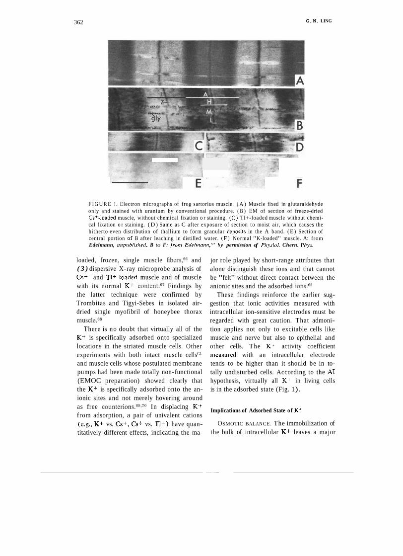
362 G. N. LING
F I G U R E I . Electron micrographs of frog sartorius muscle. ( A ) Muscle fixed in glutaraldehyde only and stained with uranium by conventional procedure. ( B ) EM of section of freeze-dried Cs+-loaded muscle, without chemical fixation o r staining. ( C ) TI+-loaded muscle without chemi- cal fixation o r staining. (D) Same as C after exposure of section to moist air, which causes the hitherto even distribution of thallium to form granular d-posits in the A band. ( E ) Section of central portion of B after leaching in distilled water. ( F ) Normal "K-loaded" muscle. A: from Edelmann, uriprrblisl~erl. B to F: fron~ Edeh~~arrn."' by permission of Pl~ysiol. Chern. Phps.
loaded, frozen, single muscle fibers,66 and ( 3 ) dispersive X-ray microprobe analysis of Cs+- and TI+-loaded muscle and of muscle with its normal K + ~ o n t e n t . ~ ' Findings by the latter technique were confirmed by Trombitas and Tigyi-Sebes in isolated air- dried single myofibril of honeybee thorax muscle.6s
There is no doubt that virtually all of the K+ is specifically adsorbed onto specialized locations in the striated muscle cells. Other experiments with both intact muscle cellsG1
and muscle cells whose postulated membrane pumps had been made totally non-functional (EMOC preparation) showed clearly that the K + is specifically adsorbed onto the an- ionic sites and not merely hovering around as free counter ion^.^^^^^ In displacing K-+ from adsorption, a pair of univalent cations (e.g., K + vs. Cs+, Cs+ vs. 'TI+) have quan- titatively different effects, indicating the ma-
jor role played by short-range attributes that alone distinguish these ions and that cannot be "felt" without direct contact between the anionic sites and the adsorbed ions.65
These findings reinforce the earlier sug- gestion that ionic activities measured with intracellular ion-sensitive electrodes must be regarded with great caution. That admoni- tion applies not only to excitable cells like muscle and nerve but also to epithelial and other cells. The K + activity coefficient measured with an intracellular electrode tends to be higher than it should be in to- tally undisturbed cells. According to the A1 hypothesis, virtually all K + in living cells is in the adsorbed state (Fig. 1 ).
Implications of Adsorbed State o f K +
OSMOTIC BALANCE. The immobilization of the bulk of intracellular K + leaves a major

osmotic deficit in muscle cells. One recalls that it is precisely this anticipated "diffi- culty" that at one time led Hill and many others to conclude that all cell K+ mus.t be free. Since Ca2+ and Mg2+ as well as a sub- stantial portion of Na+ in frog muscle cells are also in an adsorbed and hence osmotic- ally inactive state, there is no recourse but to accept that the osmotic activity (that is, the lowering of activity of water) must arise from cell proteins. This agrees well with the polarized multilayer theory of cell water in which water activity is reduced by long-range polarization and adsorption. In other words, osmotically speaking, "pure" polarized water behaves like normal liquid water in which substantial amounts of ions or other solutes are dissolved.
CELLULAR ELECTRIC POTENTIAL. The conventional view of the cellular electrical potential, as first suggested by 0stwald71 and extensively developed by Bernstein,4O as well
' as later variants introduced by Boyle and C ~ n w a y , ~ ~ Hodgkin and K a t ~ , ~ ~ and Mullins
, and N ~ d a , ~ ~ were all based on the assump- tion that the bulk of intracellular K+ is in the free state. That in fact the bulk of this K+ is in a localized, adsorbed state makes all these theories inapplicable. Again, the ad- sorbed s,tate of cell K+ is in harmony with .the A1 hypothesis' surface adsorption theory of cellular resting potential, which has been experimentally tested and ~ e r i f i e d . ~ ~ , ~ ~
THE SURFACE BARRIER AND
PERMEABILITY
Since Overton introduced his lipoidal membrane the idea that cells and subcellular structures are covered by a mem- brane whose continuous phase is lipid in na- ture has taken deep root. Yet there have long been serious questions about the validity
. of this a s s u m p t i ~ n . ~ ~ , ~ ~ Seven years ago, for example, I presented evidence that not lipid, but a layer of water polarized in multilayers
by surface proteins, constitutes the semiper- meable surface barrier.73
Work by Stillman et al. on squid axon,75 by Maloff et al. on inner mitochondria1 mem- brane,76 and by Ling and Ochsenfeld on frog muscle and egg7? produced additional experimental evidence: no change of K+ permeability was observed in any of these systems in response to K+ ionophores. Since there is no question that under similar condi- tions K+ permeability of phospholipid layers is drastically increased by valinomycin
M ) and monactin M ) , ~ ~ one concludes that the semipermeable barrier in muscle, nerve, and egg cell as well as in the inner membrane of mitochondria is not a phospholipid layer.
In my view of the cell surface barrier, semipermeability depends on the state of multilayer polarization of cell water near the surface. That state in turn depends on certain proteins assuming the extended con- formation. Semipermeability is therefore un- der close control by hormones or drugs that react with receptor sites on these proteins and so control their conformation. The sus- ceptibility of water to allosteric control offers yet another major advantage to the A1 model and suggests new approaches to cell physiol- ogy and pharmacology.
Solute Permeation According to the A1 Hypothesis
Before further comparison with the theo- retical model of active transport of solute and water across bifacial cells can be given, some additional basic concepts of the A1 hypothe- sis need to be introduced. These concepts include ( I ) the molecular mechanism of se- lective solute distribution in living cells, (2) ,the molecular mechanism of selective solute permeability, ( 3 ) cooperativity in adsorp- tion and desorption of solutes and water, and (4) the control of cooperative adsorption- desorption by a cardinal adsorbent, ATP. Fuller accounts of these concepts and sup- portive evidence can be encountered else-

where,10-21.26-i9 but we briefly review them here.
SELECTIVE SOLUTE DISTRIBUTION IN LIV-
ING CELLS. There are two basic modes of existence of an in.tracellular solute: adsorbed on macromolecular sites or dissolved in the cell water. Referring to the particular solute of interest as pi, and its concentration in the cell in moles per kilogram of fresh cells as [~ilin, then
the cell water, and the unfavorable adsorp- tion energy of /3- and y-carboxyl groups in :
comparison to that for K+, which account for the sustained low Na+ concentration in many kinds of cells.
The adsorbed fraction of the ith solute may be, under the simplest condition, limited to just one type of adsorption site. In this case, the concentration of the ith adsorbed solute may be described by a Langmuir ad- sorption isotherm:
where [pi]rree and [pi],, are respectively the concentration of free and adsorbed ith so- lutes in moles per kilogram of fresh cells. The distribution of free ith solute in the cell water follows the Nernst distribution law and thus is described by the linear rela.tion
where [f] is the concentration of intracellular ad~orpt~ion sites in moles per kilogram of fresh cells, and Ki is the adsorption constant in units of (M)-*. If another solute called the jth, and represented as pj, also adsorbs onto the same sites, Eq. 3 becomes
[PtIrree = aqi[PiIex (2) [PiInd = ([fIKi[PiIex)/
(1 + Kifpiles + Ki[~jlen). (4) where a is the percentage water content (v/w) of the cell, [pile, is the equilibration concentration of the ith solute in the external medium, and qi is the average equilibrium distribution coefficient of the ith solute be- tween cell water and the external medium.
Now, if and when the cell water is entirely normal as, say, in a Ringer's solution, qi would be equal to unity for all permeant so- lutes. If, however, the cell is in its normal resting state, then according to the A1 hy-
Equation 4 shows that there is a hyperbolic ,relation between the adsorbed ith solute ' and the external concentration of the ith solute. Further, this fraction shows competi- tion with other solutes like the jth. It also shows saturability; i.e., as [pi],, increases, [pilad approaches but cannot exceed the value of V1.
Combining Eqs. 1,2, and 4, we have
pothesis this water is not normal liquid water but in the state of polarized multi- [ ~ J i n = aqi[~ilex + ([flKi[~~lex)/
layers. As a rule, water in that state has (1 + Kdpilex + Kdpjlex). ( 5 )
solubilities different from those of normal A somewhat simpler version of this equation liquid water, and in polarized water qi varies was first presented by the Russian physiolo-
with the solute involved. In general, small and simple molecules have q-values close to gist, A. S. Tro~chin.*~
or equal to 1. The q-value usually decreases with increasing size and complexity of the
Solute Permeation According to the solute in question. The concentration of hy-
. . ... - - . .~ . .. Membrane-Pumo Theow drated solutes like Na+ in a living cell may be 0.1 or even lower. According to the A1 In the conventional membrane theory, hypothesis, it is the low q-value for Na+ in permeation of a solute molecule into a living

G. N. LING 365
cell may be achieved by three mechanisms: ' (a) free diffusion, ( b ) facilitated diffusion,
and (c) active transport. Free diffusion, as the name indicates, is
envisaged as involving diffusion through the lipid phase of the cell membrane. The rate of solute entry into a cell by free diffusion is usually linearly related to the concentra- tion of the solute in the external medium. As for facilitated diffusion and active trans- port, fundamentally they are alike. For ex- ample, both rest on saturability. The dif- ference between the two mechanisms lies in the relative electrochemical potential of the phase the solute moves into. An active trans- port mechanism of solute movement is con- ceived as being against an electrochemical gradient, while facilitated diffusion is con- ceived as being not against an electrochemi- cal gradient. The notion of an uphill move- ment when applied to unifacial cells is en- tirely dependent on the basic assumption of
- the membrane theory that the cell interior is filled with a dilute aqueous solution. Since this assumption has already been disproven (see above), there is no longer a solid foun- dation for the proposed difference between facilitated diffusion and active transport into single unifacial resting cells such as erythro- cytes, muscle cells, or nerve cells. Thus phe- nomen~logically there are two rather than three types of permeation mechanisms: one depends on saturability; the other does not.
Facilitated diffusion is distinguished from free diffusion by at least three outstanding features. First of these is saturability. As al- ready mentioned, the rate of entry of a solute into living cells by this means does not increase linearly with increasing external concentration of the solute. Instead, the rate levels off to approach a fixed value. The second distinguishing feature is competition. That is, the rate of entry of a solute into living cells is inhibited by other solutes of similar nature. In practice, this trait can be easily and quantitatively assessed with the
aid of, for example, Linewever and Burk double reciprocal plots of rates vs. external concentration, as in enzyme kinetic studies. The third distinguishing feature is specificity. The rate of entry of a solute into living cells often exhibits a high degree of specificity. Thus the steric orientation of one of the five OH groups in a sugar molecule may have a profound influence on the rate of entry of the sugar. I t has been widely con- sidered that facilitated diffusion is mediated through "molecular oarriers" which, like ferry boats, select favored "passengers" and shuttle them back and forth across the lipid membrane barrier. But I have already men- tioned convincing evidence against the con- cept that cells are covered with a continuous sheet of phospholipid. Other such evidence against the lipid barrier concept has been reviewed e l s e ~ h e r e . ~ ~ - ~ ~ With disproof of the universal existence of a continuous lipid layer the concept of "mobile carriers" also falls, for again like ferry boats, such carriers cannot shuttle without a fluid barrier. In- deed, from the viewpoint of conventional membrane theory, one oanncrt imagine a bet- ter "mobile carrier" for K+ than valinomy- cin and monactin. The failure of these "ion- ophores" to cause significant change in K+ permeability in nerve, muscle, and egg cells demands totally different interpretations for the physiological observations of solute per- meation. Such alternative interpretations have been available for some time.10,30.81.82
Permeation Mechanisms According to the
A1 Hypothesis
Since lipid does not constitute a continu- ous surface barrier and a "mobile carrier" cannot exist without a water-immiscible fluid barrier, clearly a different phospholipid model must be sought to explain the satur- able and competitive solute permeation widely observed. Such a model has been sug- gested.'aE3 It will now be briefly reviewed.

POLARIZED WATER IN LIEU OF LIPID
LAYER AS THE SURFACE BARRIER FOR L L ~ ~ ~ -
SATURABLE" SOLUTE ENTRY. The near per- fect semipermeability of Traube's copper ferrocyanide precipitation membrane led Pfeffer to found the membrane theory.' Cop- per ferrocyanide obviously contains no lipid but is a gel composed of a network of crys- talline particles with water-filled interstices on the order of 150 A in width.s4 These "pores" are many times as wide as the diam- eter of sucrose molecules (94 A ) ; yet this membrane is virtually impermeable to su- crose. More recently I showed that the per- meability to water and ten other hydroxylic compounds at three different temperatures through inverted frog skin is in excellent cor- respondence to that through a sheet of cellu- lose acetate.73 Both membranes exhibit high- est permeability for water; both are virtually impermeable to sucrose even though the average pore diameter of the "active7' layer of the cellulose acetate membrane is more than four times as wide as the sucrose mole- cule. These data strongly support the con- cept that water in the state of mulilayers, polarized by a matrix of protein chains car- rying polar sites, provides semipermeable surface barriers in living cells as well as their cogent models.
FIXED POLAR SITES ON SIDE-CHAINS AND
"BACKBONE" OF CELL SURFACE PROTEINS IN
LIEU OF "MOBILE CARRIERS" AND L ' ~ ~ ~ ~ ~ "
AS "GATES" FOR "SATURABLE" SOLUTE EN-
TRY. The A1 hypothesis offers an interpreta- tion of what is conventionally called facili- tated diffusion, as well as of active transport, in terms of an adsorption-desorption mecha- nism on cdl surface adsorption sites. Indeed the mechanism is so simple that it involves few postulations additional to those already described for solute distribution in the bulk phase.
Consider the rate of permeation of labeled K + into a living cell, typically demonstrating
saturability, competition, and a high degree of specificity. Now, the bulk phase distribu- tion of K+ in frog muscle cells is to a first approximation described by Eq. 5. If we consider the cell surface to be primarily a water-protein system like the cdll interior, then in principle the cell surface is a two- dimensional rendition of the three-dimen- sional cell. In other words, like the cell interior, the cell surface will have a continu- ous layer of polarized water mentioned above, and also anionic p- and y-carboxyl groups distributed at regular distances apart. Furthermore, if an instantaneous photograph could be taken, one would find most of the K+ to be associated with the surface anionic sites, as has been demonstrated for the bulk phase K+ (Fig. 1). Few K+ molecules would be found between these sites in the interstices filled with polarized water. If then a motion picture could be taken, one would observe two different modes of entry cor- responding to each of these instantaneous - positions taken by the K + :
(I) Saltatory route. In this mode of entry, K+ enters via the polarized water '
filling the space away from the charged sites. (2) Adsorption-desorption route. As the
name indicates, for this kind of entry the K+ from the outside must first succeed in occupying one of the anionic sites, followed by a librational motion around that site and then eventual desorption and entry into the cell.
Taking both routes into account, the rate of entry vi,,,, of the ith ion in moles per sec per kilogram of fresh cells can be written =19,82
where A is the total surface area of one kilo- gram of cells, ki,,,, is the inward rate con- stant for the entry of the ith solute via the

C. N. LING 367
saltatory route, [ f ] is the molar concentra- tion of surface sites per kilogram of fresh cells, ki.nd is a kinetic rate constant for the desorption of the ith solute from the surface adsorption sites, Kt and K, are respectively the adsorption constants in M-I of the ith and jth solute on the surface sites.
Epstein and HagensVn 1952 first success- fully analyzed alkali-metal ion entry into barley roots using Michaelis-Menton kin- etics, an analysis I soon confirmed for Rb+ entry into frog muscle.s1 Epstein and Hagen like many other investigators who have ex- tended these studies to various types of cells and solutes, adhered to the "mobile carrier" concept. Indeed, with the additional assign- ment of the non-saturable fraction of solute entry as "leaky," the overall rate equation for solute permeation becomes formally ana- logous to Eq. 6. ( For extensive discussion of this subject from the conventional viewpoint, see Chr i s ten~en .~~) However, as mentioned
, #above, the disproof of the lipid layer theory makes untenable the mobile carrier model.
To demonstrate the general validity of Eq. 6 and its basic assumptions, I have shown they can not only describe rate of entry of solutes into living cells but also rate of entry into such inanimate systems as sheets of ion-exchange resin or sheep's ~ o o 1 . ~ ~ ~ ~ ~ ~ ~ ~ These models have in common with the A1 model the attribute of anionic sites fixed in a matrix of polarized water.
While Eq. 6 adequately describes solute entry, especially for neutral solutes, a some- what more complicated equation is required to describe the entry of ions into frog muscle cells by a mechanism called the trip- let adsorption-desorption route. That is to say, in the case where the entrant ion is rather tightly adsorbed by the electrostatic forces, its desorption requires the participa- tion of another free cation. Thus a second K+ from the outside may approach a fixed anion-K+ pair in the right direction, weak- ening the attraction between the fixed anion
and K+, thereby facilitating the latter's en- try into the cell. Indeed, we have shown that the rate of entry of labeled Rb+ into frog muscle cells is facilitated by external K+.u2 Clearly the facilitating effect of the second K+ outweighs the competition offered by K+ against Rb+ adsorption.
SPECIFICITY OF SUGAR PERMEABILITY. AS
noted above, the third characteristic of per- meation phenomen,a is specificity. In the past this has often been attributed to carriers, and is exemplified by sugar entry into cells. Here also, I believe, the present model has distinc- tive advantages over the oarrier model.
There is little question that the high degree of steric specificity for solute permeation rate can be recognized only by a system that pro- vides a complex of closely spaced sites. This strongly suggests that these sites are provided by proteins. In the A1 model, adsorption fol- lowed by libration and desorption would be all th'at is required to achieve a facilitated dif- fusion when such specific sites are available.
SPECIFICITY IN ALKALI-METAL ION ENTRY; VARIABILITY AND THE C-VALUE CONCEPT.
Surfaces of most normal unifacial cells and the base1 lateral surfaces of bifacial cells have greater permeability for K+ than for Na+. However, the frog skin surface facing the outside has a higher permeability for Na+ than for K + . W h a t could be the mo- lecular basis for this specificity difference?
In 1952 I suggested a theoretical model of selective adsorption of K+ over Na+ on fixed anionic sites.24 Stimulated by the later dis- coverys7 that carboxyl ion exchange resin selects Na+ over K+ while sulfonate ion ex- change resin selects K+ over Na+, as well as the work of Eisenman, Rudin, and Casby on glass electrode ion selectivity, I con- structed a theoretical model in which the c-value concept was i n t r o d u ~ e d . ~ ~ ~ ~ ~ In es- sence, the c-value measures the electron density of an anionic oxygen atom; high

c-value is equivalent to a high pK value as in acetic acid, low c-value to a low pK value as in trichloroacetic acid. It was then possible to show that a variation of the c- value produces changes in the preferential order of selectivity among Cs+, Rb+, K+, Na+, Li+, as well as NH4+ and H+ . Thus K+/Na+ preference seen in muscle and nerve as well as basolateral membranes of various epithelial cells corresponds to a fairly low c-value; high Na+ over K+ preference seen in a number of apical cell membranes" bespeaks a high c-value. The essence of this work was presented in 1960" and in full de- tail in 1962.19
Solitary p- and y-carboxyl groups usually have a pH value of 4 to 5; when carboxyl groups are placed in close proximity to each other as in various carboxy rypes of ion-ex- change resin, the pK value may rise to 9 or even higher!@ Thus the high c-value of api- cal membranes may be due to p- and y-car- boxyl groups in close proximity to each other or paired, whereas the low c-value at the basolateral membrane may be due to isola- tion of p- and y-carboxyl groups (for evi- dence, see ref. 89).
In 1962 Eisenman also published a theory of selective ionic adsorption using a much simpler m0de1"~~l to explain the rela- tion between ion specificity of glass elec- trodes and the glass c o m p ~ s i t i o n . ~ ' ~ ~ ~ Ussing and Leaf7 considered Eisenman's theory to explain the different specificity at the two surfaces of epithelia but rejected it on the ground that the theory does not provide enough specificity. They preferred the "close- fit" hypothesis of mull in^^^^^^ in which close- fitting into small holes endows a high degree of specificity in selectivity for Na+ and Li+ over K+ (see ref. 19, p. 548, for tabulated data from literature). Mullins' model re- quired a dehydration of Na+ and Li+ prior to entry into the postulated close-fitting pores-a concept not easily defensible if the cell membrane is a lipid bilayer, and not
defensible at all if the membrane is not a lipid bilayer. It is even more difficult to think of rigid pores in a layer of polarized water that would force the dehydration of Na+ and Li+ before their entry.
On the other hand, the low level of ionic specificity attributed to Eisenman's model is not applicable to the association-induction model, in which a high degree of Na+ as well as K+ selectivity has been theoretically cal- culated.
In brief, I feel that difference in ionic spe- cificity between the two surfaces of the epithelial cells can be adequately explained as a result of the difference in the c-values of the anionic surface sites.
PROTEIN-WATER-ION: THE
COOPERATIVE ASSEMBLY
Cooperativity in Adsorption and Desorption of
So l~~tes and Water
In the preceding sections I have discussed adsorption in relation to selective solute ac- cumulation as well as selective solute perme- ability. Thus far the adsorption sites have been considered non-interacting and there- fore the adsorption is adequately described by the Langmuir adsorption isotherm. In that isotherm, given as a part of Eq. 5, the con- centration of adsorption sites [ f ] and a pair of adsorption constants Ki and Kj (or their ratio, Ki/Kj) determine the adsorption at a fixed ratio of [p,],,/[pj],, in the surrounding medium. In the cooperative adsorption iso- therm, these parameters play comparable roles. However, it is the new parameter, the nearest neighbor interaction energy, that opens the door to coherence in adsorption. Thus if the nearest neighbor interaction en- ergy is positive, it means that if one adsorp- tion site adsorbs K+ it would make the two immediately neighboring sites prefer K+ over Na+. Conversely, if the middle site adsorbs Na+, it would make the two immediately

G . N. LING 369
neighboring sites prefer Na+ more than if the middle site were to adsorb K+. The result is autocooperativity in ion adsorption. When the nearest neighbor interaction energy is large and positive, the whole system of ad- sorption sites will adsorb eithcr all K+ or all Na+. "All-or-none" switching can then occur at a "threshold" value of the external K+/Na+ concentration ratio. Thus auto- cooperativity among the four heme sites in hemoglobin provides the molecular basis for efficient loading and unloading of oxygen be- tween the lung and the respiring tissues. This type of cooperative adsorption isotherm de- scribes the uptake of Kt- or Na+ in various living cells, including frog muscle,"-g7 hu- man lymphocyte^,"^^^ and a variety of smooth muscles (ref. 100, p. 22), all show- ing positive nearest neighbor interaction and a sigrnoid-shaped adsorption curve. Since I have established that initially nearly all intra- cellular K+ is in an adsorbed state (Fig. 1 ), this autocooperativity switching from the K+ to the Na+ state clearly bears a fundamental similarity to the oxygenation and deoxygena- tion of erythrocytes. In both, the seats of interaction are intracellular proteins. As far back as 1908, Moore pointed out the paral- lelism of oxygen accumulation in erythro- cytes and K+ accumulation in cells.lO' In 1965, I sharpened this parallelism by dem- onstrating that both oxygen taken up by erythrocytes and K+ taken up by frog muscle show autocooperative behavior with similar values of nearest neighbor interac- tion energy.g6
Control of Cooperative Adsorption and
Desorption by Cardinal Adsorbents
Autocooperativity provides the basis for the ability of the solute-adsorbing protein to shift its adsorbed solute from one type to another in a stepwise, all-or-none manner. According to the A1 hypothesis, such an autocooperative transition is not the proper-
ty of the protein alone but involves the water molecules, ions, and other substances that in- teract with the protein molecule at different sites. As a result of the propagated electronic redistribution, all or at least a major portion of the properties of the protein-water-ion assembly are changed. Often, however, it is the physical conformation change that is most noticeable. (For a fuller discussion of the various theories of cooperative interac- tion, see ref. 25.) Proteins contain specific sites that exert a controlling influence on the all-or-none transitions. These sites are called cardinal sites, and the specific adsorbents in- teracting with these cardinal sites are called cardinal adsorbents. ATP is a bona fide cardi- nal adsorbent, as are many drugs and hor- mones, -and all share a distinctive ,feature: a small number of cardinal adsorbent molecules can bring about responses involving a much larger number of non-cardinal sites such as the p- and r-~arboxyl groups adsorbing K+ and Na+.
OUABAIN AS A CARDINAL ADSORBENT. Oua- bain, like a number of other cardiac glyco- sides, causes loss of K+ and gain of Na+ in a variety of living cells. In the conventional membrane-pump model, ouabain acts by in- hibiting the Na pump. In fact, it was the par- allel behavior of ouabain's effect on the K+/ Na+ distribution in living cells and its inhi- bition of isolated K,Na-activated ATPase that gave impetus to the extensive work car- ried out under the assumption that this K+, Na+-activated ATPase is in fact the Na pump and that when incorporated into phos- pholipid vesicles this pump can actually translocate both K+ and Na+ against con- centration gradients. Careful analysis of the resultant data revealed major inconsistencies of interpretation, so that an alternative inter- pretation more in accord with the findings was presented in 1980.1°2
That K,Na-activated ATPase can indeed adsorb K+ and Na+ and that this adsorption

FIGURE 2. Effect of ouabain (3.2 x lo-' M ) on equilibrium distribution of K + and Na+. Curves with open (Na+) and filled ( K + ) circles describe equilibrium distribution data for muscles not treated with ouabain. The point of intersection gives K t h ~ of 100. In muscles treated with ouabain (3.2 x lo-' M ) , K E + K has shifted to 21.7.
is indeed sensitive to ouabain have been demonstrated by Matsui et a1.3193%s men- tioned above under INTRODUCTION. The ouabain-sensitive K+ binding on K,Na-acti- vated ATPase also exhibited autocoopera- tivity as had Seen repeatedly demonstrated for K+ binding in intact living cells.
Certainly the data of Matsui et al. indi- cated that this enzyme may be the seat of some K+,Na+-adsorption in some cells. Nevertheless, the demonstration that the A-band containing another ATPase, myosin, is the seat of adsorption of the bulk of intracellular K+ left little doubt that ouabain acts on K+ and Na+ distribution in frog muscle and other cells by changing the rela- tive preference of many proteins for the adsorption of K+ and Na+. Figure 2, from Ling and E30hr,~~ shows that the Yang-Ling cooperative isotherm can quantitatively de- scribe the K+ + Na+ transition in frog muscle in response to 3.26 X lo-" M oua- bain. The figure presents the full range of external K+/Na+ concentration ratios to
demonstrate that the primary effect of oua- bain is to shift the intrinsic equilibration con- stant for the Na+ -, K+ exchange from a value of about 100 to a value of 17; i.e., the - constant changes by a factor of 6.
The normal environment of muscle cells contains 2.5 mM K+ and 100 mM Na+, cor- responding to a [K+],,/[Na+],, ratio of 2.5 X At this ratio of external K+/Na+ concentration, almost all the intracellular anionic sites are occupied by K+. The effect of exposure to 3.26 x M ouabain is to reverse the situation completely; the anionic sites become almost entirely occupied by Na+.
It is clear that ouabain, by its adsorption onto the appropriate cardinal sites, alters selectivity in adsorption of the cooperatively linked anionic sites, causing changes in c-
values in a direction toward reduced prefer- ence for K+ over Na+. Therefore it is to be expected that if one increases external K+ concentration or decreases external Na+ concentration by a factor equal to or greater

than 6, the preferential K+ accumulation seen in normal cells will be restored. That this is true is indicated, of course, by the data of Fig. 2.
ATP AS A CARDINAL ADSORBENT. ATP was long considered to carry special chemical bonds-so-called high energy phosphate bonds. But later work103 established that the enthalpy of hydrolysis of these bonds was not usually high at all. Investigation also es- tablished that the favorable free energy of hydrolysis reflects largely the different affinity it produces for H+, Mg+, and H20.104s106 These findings, though little celebrated, con- tributed substantially to better understanding of ATP, one of the most important biologi- cal compounds. Certainly the old idea that this package of high energy could do work as a sort of universal fuel became no longer tenable.
The A1 hypothesis, on the other hand, by recognizing the role of ATP as a cardinal adsorbent, provides a different mechanism by which ATP energizes biological perfor- mance.
Maintenance of the Living State: Role of ATP
The conventional concept, still, is that hydrolysis of ATP releases energy stored in its "high energy" phosphate bond to support cellular work. Thus a resting muscle, on re- ceiving its package of energy from the hydro- lyzing ATP, enters into the high-energy con- tracted state. When the ATP is used up, the muscle reverts back to its low-energy re- laxed state.
According to the A1 hypothesis, quite the opposite is the case. The resting state is seen as a high-energy state; its maintenance does not depend on a steady stream of decompos- ing ATP but on a steady adsorption of intact ATP onto key cardinal sites. The resting state is a high-energy state much like that of a set mousetrap. It is only when ATP is destroyed, say by hydrolysis, that the mus-
cle seeks its low-energy state, much as a triggered mousetrap seeks its low-energy state.
The simplest and most direct evidence in favor of this concept is the fact that a dead muscle, as a rule, is found in the contracted state. Were the relaxed state the lower en- ergy state, dead muscle all should be fully relaxed and rigor mortis would not occur.
Relaxation and shortening are but two of the changes in muscle tissue that occur when its ATP content changes. A parallel phe- nomenon is the inverse relation observed between muscle shortening and muscle de- sorption of K+ followed by its release into the r n e d i ~ m . ~ ~ . ~ ~ Perhaps the most convinc- ing and elegant demonstration of this inti- mate relation was that performed by Wilde and coworkers using perfused turtle hearts.lo6 They were able to observe that each heart- beat is accompanied by an exact pulse of labeled K+ release.
ATP-induced changes are not limited to contraction-relaxation and K+ desorption- adsorption, biologically important as they are, but include changes in the physical state of intracellular water.
ATP Control of the Physical State of Cell Water
In 1952 I showed that a quantitative relation exists between K+ (and Na+) con- tent and ATP level in frog muscles poi- soned by iodacetate.*-' This observation has been repeatedly confirmed and ex- tended.'"7"107.10s There is also, generally speaking, a reciprocal relation between the K+ and Na+ contents. The latter relation can be explained as due to a mechanism similar to that proposed above for ouabain effects. However, the strict one-for-one ex- change seen in ouabain-induced Na+ for K+ exchange was not observed here. Found
-instead was a parallel between the gain of Na+ with decreasing ATP and the gain of labeled sucrose.79

In terms of the A1 hypothesis, the low level of (hydrated) Na+ as well as sugars and amino acids in normal living cells is due to the polarized multilayered condition of the cell water. Just as ATP acting as a cardinal adsorbent maintains the protein anionic p- and y-carboxyl groups at a c- value at which K+ is preferred over Na+, so the adsorption of ATP on the same or other proteins maintains the c-value ana- logs* of the backbone carbonyl groups (and the cr-value analogs* of the backbone imino groups) at values that favor long-range water polarization. This single unitary cause for solute exclusion (in contrast to the mul- tiple and separate causes required by mem- brane-pump theory) is described as the uni- versality rule.74 The rule states that if for one or another reason the solubility of one normally excluded solute is changed, then the solubility of all other normally excluded solutes should change pari passu. This rule has been demonstrated in D-arabinose dis- tribution and Na+ distribution in frog ova- rian eggsl0"s well as in D-arabinose, su- crose, and Na+ distribution in the IAA- poisoned frog muscles.110 As the egg cells or muscles were dying their ATP levels gradually fell, and a parallel gain of free Na+, free D-arabinose, and free sucrose occurred.
ATP Control of Salt Linkage
Formation and Dissociation
As mentioned above, ouabain causes a stoichiometric displacement of K+ by Na+. The additional Na+ taken up has been shown by NMR spectroscopy to be in an adsorbed state.lll For a while it was asked whether the original assumption used in
identifying bound Na+ might not have been erroneous.l12 However, it turned out that
*
although error had been made it was merely quantitative. The "disappearance" of part of the Na+ signal was indeed due to one-site- one-ion specific adsorption of Na+. Thus Monoi's demonstration that the NMR- invisible Na+ in liver homogenate can be made visible by introduction of competing K+ and Cs+, but not of choline, indicates ion-specific adsorption.ll"f f a + signal dis- appearance were truly due to a diffuse elec- tric field gradient as suggested by Berend- son and Edzes,l12 there would be only va- lency specificity and not the ion specificity that Monoi found. A theoretical argument against the diffuse electric field gradient con- cept was presented by Chang and Woess- ner.l14
The obedience of Na+ and sugar uptake in poisoned frog muscle to the universality rule, however, shows clearly that the loss of adsorbed K+ is accompanied by a gain of free Na+ (and free sucrose) and is there- fore different from ouabain-induced K+ + . Na+ exchange.
But if K+ is lost, what has happened to the p- and r - ~ a r b ~ ~ y l groups (in the mus- cle A-bands and Z-lines) that normally ad- sorb K+? According to the A1 hypothesis, in the absence of ATP these anionic sites [f], became "masked" by forming salt linkages. Specifically, without ATP those sites [f] prefer as counterions fixed cat- ions [f+] in the form of 6-amino groups, guanidyl groups, a-amino groups, and/or histidyl groups, thus forming salt link- ages f+f-.1"G0.115 An expression for the reaction is
*The c'-value is a parameter measuring the posi- tive charge density a t a cationic group. c-value analogs and c'-value analogs refer to the negative and positive charge density at polar groups not bearing net charges."
Still uncertain is the nature of the counter- anion, X-, adsorbed to the fixed cation be- fore the salt-linkage formation. However, in

G. N. LING 373
muscle tissues X- could be creatine phos- phate.
If salt linkages are formed among differ- ent protein molecules, there may be macro- scopic volume shrinkage and loss of water with concomitant change of the state of remaining water. On the other hand, if the salt linkages are formed within a single pro- tein molecule or an aggregate of like pro- tein molecules, the result might be limited to conformation change of the molecule(s).
Synchronized ATP- and ATPase-
Dependent Cyclic Changes
Addition o'f Mg2+ and ATP to a sus- pension of glycerinated Paramecia evoked synchronized beating of the cilia, propelling the dead protozoa in water as if they were alive.l16 An interpretation of this remark- able phenomenon on the basis of the A1 hypothesis was offerede21 In brief, ATP acts as a cardinal adsorbent that ckuses cilia
' orientation in one direction, followed by Mg2+-induced ATP hydrolysis and result-
, ant protein conformation change that causes cilia orientation in another direction. The beating cycle is then re-initiated by adsorp- tion of fresh ATP. This model establishes the need for a continuous supply of fresh ATP to maintain continuous swimming.
Another fascinating illustration of the inherent ability of protoplasm to act in a synchronized and cyclic manner is the ATPase-dependent ion accumulation and swelling cycle demonstrated in rat liver and pigeon heart mitochondria, initiated in the latter by valinomycin or r n ~ n a c t i n . l l ~ ~ ~ ~ ~
The evidence that valinomycin, monactin, etc. do not act as ionophores but rather as cardinal adsorbents has been discussed re- ~ e n t l y . ~ V t only needs to be mentioned here that Sr2+, a simple divalent ion with no ionophore property whatsoever, can initiate cycle changes in much the same way ob- served for valinomycin in other studies.llg
The oscillatory changes at issue are in perfect synchrony in regard to shrinkage and swelling with concurrent loss and gain of K+, and they involve the operation of an ATPase. It is my belief that this basic pro- toplasn~ic trait is essential to a variety of physiological activities including transepi- thelial transport of water and ions.
Cooperative Adsorption-Desorption Model of
Active Transport Across Epithelia and
Other Bifacial Cell Systems
When living cells are incubated in a K+-free or low K+ solution, they gain Na+ and lose K+ until a new equilibrium is reached. In the case of isolated frog mus- cle, a tenfold reduction in the external K+ concentration (from 25 to 0.25 m ~ ) at constant external Naf concentration ( 100 m ~ ) leads to almost total displacement of K+ by Na+ (Fig. 2). If these K+-depleted muscles are returned to a normal Ringer's solution, their Na+ will be stoichiometrically displaced by K+. In this restoration process both Na+ and K+ are "transported" against concentration gradients. This type of phe- nomenon is referred to as "active transport" by adherents of the membrane-pump theory. However, now that the cellular K+ is known to be virtually all adsorbed (Fig. I ) , as is the Na+ that stoichiometrically replaces K+,120 the phenomenon can no longer be regarded as active transport. Actually the phenomenon represents an exchange adsorp- tion much like that seen in the operation of an ion-exchange resin. The displacement of K+ by Na+ adsorbed on Dowex 50 is not an active transport process but merely the consequence of ( I ) a change in the ratio of K+/Na+ concentration in the external me- dium and (2) the fact that electrostatic and other forces, at the ambient concentration ratio, make it more favorable for one ion to congregate in the resin than an alterna- tive ion. Oxygen concentrates in red blood

G. N. LING
cells for exactly the same reasons. If a dialysis bag containing hemoglobin
solution is suspended in an oxygen-contain- ing solution, after a suitable length of time an equilibrium will be reached and the amount of oxygen taken up by the sac is quantitatively defined. If now ATP is added to the internal solution, oxygen will begin to move out of the hemoglobin-containing phase into the external ~ o l u t e . ~ " l ~ l This out- ward migration of oxygen is the consequence of ATP interaction with the oxygen-binding protein, hemoglobin. ATP exercises a long- range effect on the affinity of the heme groups for oxygen, a type of effect termed "allosteric" by Monod et a1.122 (A detailed mechanism for such allosteric effect has been offered by the A1 hypothesis, which referred to the phenomenon as an indirect F-ef fe~t .~O~~l) When ATP is removed from the system, oxygen will once more move into the bag, apparently against a concen- tration gradient. This effect of ATP on the concentration of a solute in the hemoglobin- containing system does not involve the hy- drolysis of ATP, since hemoglobin has no ATPase activity.
Movement of the solute oxygen into and out of the above hemoglobin-containing sys- tem involves participation of the laboratory worker, who introduces or removes ATP from the system and hence brings about the oxygen transport. But one may imagine an- other way to achieve the cyclic back-and- forth transport of oxygen. It would occur if the bag contained first, a specific enzyme, an ATPase, which could destroy and thus remove ATP from the hemoglobin-contain- ing phase; second, another enzyme system that could regenerate ATP; and third, a co- ordinating system that could synchronize the ATP destruction and its subsequent resyn- thesis and readsorption. Examples of bio- logical systems known to provide just this kind of coordination and synchrony are mani- fest in the above-mentioned ATPase-depend-
ent swimming of dead paramecia and in the ATPase-dependent oscillatory changes of ion -
and water uptake and release in isolated mito- chondria suspensions.
In summary, through the use of simple models I have shown how a cyclic change of ATP adsorption and hydrolysis can bring about a cyclic change of selective accumu- lation of a solute from the medium and its subsequent release back into that medium. I shall now try to incorporate this mecha- nism into a theoretical model able to per- form true active transport. The major addi- tional component needed is a one-way valve.
A Detailed Model of Active Transport of
Solutes and Water
As discussed above, the surface perme- ability barrier of the living cell is not a lipid layer but multilayers of water polar- ized by cell surface proteins. It is reason- able, then, that synchronized cyclic ion and +
water release and uptake would involve an alternation between a polarized impermeable state and a depolarized, permeable state of
'
the surface water. If we can imagine that the basolateral or serosal surface has these properties and that the apical or mucosal surface does not undergo cyclic changes but has high c-value anionic sites and hence selective high permeability for Na+, we will then be equipped with the necessary com- ponents for a model of active transport I believe to be in best accordance with avail- able data and published observations.
Figure 3 outlines the proposed model in four stages. The ion to be transported from the external solution, Na+, is represented as solid triangles. The mucosal surface is conscidered to have a higher permeability for Na+ than the resting serosal surface because the serosal surface anionic sites have a high c-value that promotes selective Na+ adsorption. Water in the serosal surface, in '
the cytoplasm, and in the normal cell surface

exists in the polarized multilayered state at the beginning of the cycle when the cell is at rest. In this active transport model the key role is played by the serosal and cytoplas- mic surface proteins, which possess an ATP- binding cardinal site as well as an ATPase activated by the transported ion. It is sug- gested that the ATP-binding cardinal site
is in fact the ATPase site but in a different cooperative state. The model functions in the following sequence:
Stage I. The higher mucosal surface per- meability allows both Na+ and water to enter the cell. Once inside, the Na+ and water will proceed to adsorb onto anionic sites on the cytoplasmic proteins under the
Serosal Surface Mucoral Surface Low Permeability Higher Permeability
a I
Stage 1
Asymmetrical entry from mucoral surface; Cooperative adswption on sites under ATP domination.
Stage 2
a Last adsorption triggers ATP splitting;,
n Cooperation desorption; and water depolarization;
High mucoral permeability. n
n A Stage 3
A Diffusion outward through depolarized l r o ra l surface affecting transport.
n n
Stage 4
Regenerat~on and admrptlon of ATP
FIGURE 3. Cyclic adsorption-desorption model of active transport of Na (&a) across frog skin, intestinal epithelia, and other bifacial systems.

376 G. N. LING
control of the cardinal adsorbent, ATP, and thus existing in an extended state with their backbones favoring multilayer polarization of water and their anionic side chains pre- ferring Na+.
It may be asked how this stipulation can be reconciled with the view that ATP ad- sorption favors K+ (rather than Na+) ad- sorption on cell proteins. The answer is that the ATP control of the c-value ensemble depends on other factors; e.g., secondary cardinal adsorbents. That ATP adsorption favors K+ adsorption is specific only for a specific protein under rigorously defined conditions. It also bears remembering that in active transport across bifacial cells the key cation is not always Na+. Thus id Malpighian tubules of insects, the key ion transported is K+.12Vn any case, the sto- chastic process of Stage 1 continues until the protein enters into the cooperative Na-i. state with a high concentration of adsorbed Na+ locally accumulated.
Stage 2. The autocooperative shift to the Na+ state involves the site adjacent to the cardinal site that also adsorbs Na+. This Na+ adsorption then activates the Na,K- activated ATPase activity of the cardinal site, causing the hydrolysis of ATP, thereby entering Stage 2.
With the hydrolysis of ATP, the protein undergoes an autocooperative desorption of Na+ (possibly of C1- as well) with con- comitant formation of salt linkages, depo- larization, and release of water plus libera- tion of a high concentration of Na+. At the same time or slightly later, the serosal sur- face protein also undergoes change from the extended to a more helical conformation ac- companied by water depolarization.
Stage 3. The depolarization of serosal
Stage 4. This last stage is marked by the regeneration of ATP, its adsorption onto the cardinal site, and autocooperative shift back to the Stage 1 condition favoring Na+ ad- sorption on anionic side chairiS and multi- layer polarization at the serosal membrane. The cycle is now ready to repeat itself.
Discussion of the Proposed Model
CYCLIC CHANGES OF ADSORPTION-DESORP-
TION AS THE BASIS OF ACTIVE TRANSPORT.
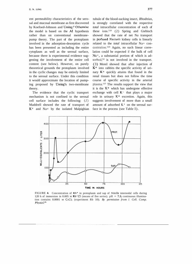
The cyclic changes of adsorption-desorption proposed in this model may not be easily observable in a multicellular epithelium. However, there is evidence of cyclic changes in the case of single giant algal cells (due largely to the careful work of S. C. Brooks.124J25) Thus when Nitella cells were exposed to salt solutions containing radio- active K+, Na+, and Rb+, those ions first accumulated in the protoplasmic layer sur- rounding the central vacuole. Furthermore, the accumulation of these labeled ions was not monotonic but exhibited a distinct pe- riodic increase and decrease, and they reached a concentration in the protoplasm many times higher than that found in the surrounding medium. It was considerably later that the labeled cation reached the cell sap in the central vacuole. Figure 4 (from Brooks12.') shows the time course of labeled Rb+ accumulation in the sap. The rise phase of sap Rb+ coincided with the fall phase of protoplasmic Rb+, whereas the fall phase coincided with the rise phase of the protoplasmic Rb+. Eventually the sap Rb+ reached concentrations "notably exceeding those present in the immersion fluid." At no time was the concentration in the sap higher than that in the protoplasm.
surface water increases serosal surface per- LOCATION OF THE TRANSPORT MECHA-
meability to ions and osmotic flow of water, NISM RELATIVE TO USSING'S TWO-MEMBRANE
permitting rapid exit of both the liberated MODEL. Our model has incorporated the Na+ and released water through the serosal two-membrane theory of Ussing to the ex- surface. tent of recognizing and utilizing the differ-
G. N. LING 377
ent permeability characteristics of the sero- sal and mucosal membrane as first discovered by Koefoed-Johnson and Ussing.Wtherwise the model is based on the A1 hypothesis rather than on conventional membrane- pump theory. The part of the protoplasm involved in the adsorption-desorption cycle has been presented as including the entire cytoplasm as well as the serosal surface, because there is experimental evidence sug- gesting the involvement of the entire cell content (see below). However, on purely theoretical grounds the protoplasm involved in the cyclic changes may be entirely limited to the serosal surface. Under this condition it would approximate the location of pump- ing proposed by Ussing's two-membrane theory.
The evidence that the cyclic transport mechanism is not confined to the serosal cell surface includes the following: ( I ) Maddrell showed the rate of transport of K+ and Na+ by the isolated Malpighian
tubule of the blood-sucking insect, Rhodnius, is strongly correlated with the respective total intracellular concentration of each of these ions.123 (2) Spring and Giebisch showed that the rate of net Na transport in perfused Necturis kidney cells is linearly related to the total intracellular Na+ con- ~ e n t r a t i 0 n . l ~ ~ Again, no such linear corre- lation could be expected if the bulk of cell Na+, a substantial portion of which is ad- sorbed,52 is not involved in the transport. (3) Morel showed that after injection of K+ into rabbits the specific activity of uri- nary K+ quickly attains that found in the renal tissues but does not follow the time course of specific activity in the arterial plasma.12i The results support the view that it is the K+ which has undergone effective exchange with cell K+ that plays a major role in urinary K+ excretion. Again, this suggests involvement of more than a small amount of adsorbed K+ on the serosal sur- face in the process (see Table I).
TIME IN HOURS
FIGURE 4. Concentration of Rbf in protoplasm and sap of Nitella internodal cells during 120 h of immersion in 0.005 M Rb+CI (means of five series). pH = 7.3; continuous illumina- tion contains 0.0001 M CnCI? (experiment Rb 10). By permission from J . Cell. Comp. Physiol.*"

SOURCE OF ENERGY FOR ACTIVE TRANS-
PORT. In the present model, the immediate source of energy is stored in the protein- water-ion assembly in the high-energy rest- ing state, and the ultimate source of energy for transport of solutes and water is that used in the synthesis of ATP from ADP and pi. If one assumes that this synthesis is highly effective, with an efficiency approach- ing 100%, then the energy formed would be proportional to the free energy liberated during ATP hydrolysis. Further, since there is a fixed number of ATP molecules synthe- sized for each molecule of oxygen utilized, the energy for active transport should be quantitatively related also to the extra oxy- gen consumed.
Ussing and Leaf have raised the old ques- tion of '"whether there is a stoichiometric relationship between the number of ions transported and the amount of, say, ATP consumed. . . ." (ref. 7, p. 3) . They cited earlier work of their own and others leading to the conclusion that there may be a cor- relation between Na+ transport and oxygen consumption of, for example, frog skin. On the other hand, the failure to demonstrate such a relation in all tissues may reflect either a predominantly glycolytic source of ATP regeneration or the presence of a high and variable rate of oxygen consumption for cell functions not directly concerned with active transport.
membrane is not necessary and that any confined space lined with a membrane pos- sessing both active salt transport and semi- permeability could function as a local os- motic coupling space. But Hill130 pointed out that the feasibility of such functioning depends critically on the osmotic permea- bility of the membrane involved and that the required permeability range of lo-' to 10' cm2/sec "lies completely outside that of any living . . . membrane studied to date by at least three orders of magnitude."
In the case of NaC1-coupled water trans- port, the present adsorption-desorption mod- el may be represented by two sets of alter- native equations:
- ATP
to serosal side + - + Na+ + nHpO Y- 4+ from mucosal side (8)
- ATP (H20) . fNa+ + ( ~ ~ o ) . f + ~ ~ - y ? f - f - +
+ ATP
to serosal side --)
COUPLING OF ION AND WATER TRANS-
PORT. Gall bladder, proximal kidney tu- bules, and small intestine are examples of "low resistance" epithelia. They transport water in the form of an isotonic fluid. As mentioned above, the standing gradient os- motic flow theory was proposed by Diamond and Bossert.l0 This theory, a refinement of the earlier double membrane theory of local osmosis to explain the apparent coupling of solute and water f l o ~ , ' ~ " ~ ~ ~ argued that contrary to earlier assumptions a second
4+ from mucosal side (9
As shown in Fig. 3, the synchronized de- phosphorylation of ATP at Stage 2 causes simultaneous liberation of Na+ from its ad- sorption on anionic sites f - and of water from its polarized multilayered adsorption on the extended polypeptide chain NHCO sites. A normal free aqueous solution of Na+ and C1- is momentarily and locally released concomitant with the removal of the barrier to ion permeation as well as to

G. N. LING 379
osmotic water flow at the serosal cell ' surface. This is so because cooperative
multilayer polarization of water drastic- ally reduces osmotic permeability but only moderately reduces diffusive permeability whereas depolarization of water increases both. As a result, an essentially isotonic fluid of Na+ is excreted into the lumen of the "low resistance" epithel
i
a. It should be mentioned that solute and
water transport may be rate-limited by either the frequency of the cycles of adsorption and desorption or the mucosal surface perm- eability. Antidiuretic hormones when applied to the outside surface of frog skin and toad bladder greatly increase the rate of Na+ transport as well as osmotic water permea- bility without major effect on diffusional permeability.131 This suggests that such hor- mones may depolarize water at the mucosal surface, thereby increasing both Na+ and water permeability at the external or mu- cosal surface.
While the cyclic adsorption-desorption model given above can explain active coupled salt and water transport with only the serosal surface involved in polariza- tion-depolarization cycling of permeability changes, there is no reason why in different tissues the cycling may not occur at the mucosal rather than the serosal surface, as is known or at least indicated in insect Mal- pighian tubules, midgut, choroid plexus, and gastric mucosa.
CONCILIATION OF "HOMOCELLULAR REGU-
LATION OF CELL Kf AND Na+ COMPOSI-
TION" AND " H ~ M ~ E P ~ T H E L ~ A L Na TRANS-
PORT." S c h u l t ~ l ~ ~ wrote in a recent review on "Transport Across Small Intestine": "The data available at present cannot be readily reconciled with any model that in- vokes a close relation between transepi- thelial Na transport and the homocellular regulation of Na and K composition or with the notion that the epithelial cell can be
adequately represented by a double mem- brane model." (p. 769).
These limitations and conflicts are pri- marily the consequence of the incorrect basic concepts of the membrane-pump theory. The present model does not encounter any inter- nal conflict between a theory of the mecha- nism of selective K+ accumulation and Na+ exclusion in epithelial cells (and algal cells) and the theory of transepithelial Na+ trans- port. Indeed, the transepithelial Na+ trans- port model is built on the foundation of the mechanism for "homocellular" K+ accumu- lation and Na+ exclusion.
COUPLING OF Na+ TRANSPORT WITH
SUGAR AND AMINO ACID TRANSPORT. An outstanding event in the study of transepi- thelial solute transport was the discovery of codtransport of Na+ and sugar and co-trans- port of Na+ and amino acids across epi- thelial s y s t e m ~ . l ~ - ~ ~ However, the interpreta- tion thus far offered, the Na-gradient con- cept, is based on the assumption that cell water is free. Since this assumption is now disproven, the subsidiary assumption that an electrochemical gradient of Na+ exists be- tween the mucosal fluid and the cytoplasm is also disproven. I shall now very briefly present a recasting of these important find- ings in terms of the cyclic adsorption-desorp- tion model of transepithelial transport pro- posed in this paper.
Surface Protein Adsorption Sites to Re- place "Membrane Carriers." In the con- ventional view, the mutual dependency of Na+ and sugar (or amino acid) in their rate of transepithelial transport is due to the man- datory requirement for formation of a ter- nary complex of Na+-carrier-sugar to achieve permeation into epithelial ~ e l 1 s . l " ~ ~ The car- rier coupled either to Na+ or to sugar alone is not able to permeate into the cell or at least permeates very much more slowly. But the fact that the cell membrane barrier is now known not to be due to phospholipid

G . N. LING
demands a reconstruction of the conven- tional model.
As discussed earlier, polarized water of- fers strong resistance to permeation by sugar or Na+. To enter the cells, these solutes must take the adsorption-desorption route via surface protein sites. Under normal con- ditions the cell surface proteins offer few or no specific adsorption sites for Na+ or D-glucose. However, in the presence of both Na+ and D-glucose the surface proteins un- dergo a cooperative transformation to as- sume a new conformation with a new c- value profile. In this new conformation, the surface protein offers both anionic sites of the proper c-value for Na+ adsorption and a combination of sterically suitable back- bone and side-chain sites to adsorb D-glucose specifically. The subsequent entry will follow typically the adsorption-desorption route ear- lier described for simple ion entry. Since D-glucose and Na+ adlsorption is coopera- tive, desorption of D-glucose or Na+ will facilitate the desorption of Na+ and D-
glucose respectively, hence the enhanced simultaneous entry of Na+ and D-glucose.
Studies of Isolated Microvilli from Intes- tinal and Kidney Epithelia. In recent years techniques for isolation of microvilli from intestinal mucosa and from rat or rabbit epithelia have been developed and steadily i m p r 0 ~ e d . l ~ ~ Typically, the rate of D-glucose (but not L-glucose) or amino acid uptake is greatly accelerated if the Na+ concentra- tion is high in the external medium and low in the microvilli. Pre-incubation of the mi- crovilli in a medium containing high Na+ concentration, inclusion of phoridzin in the incubation medium, or substitution of K+ for Na+ in the external medium all slow down the initial uptake. However, if valino- mycin is added to a K-preloaded vesicle, the D-glucose uptake is again accelerated. Simi- larly, addition of the uncoupler CF-CCP to proton-loaded microvilli also accelerates ini- tial D-glucose uptake rate. The conventional
interpretation has been that D-glucose up- take is driven by the Na+ gradient, or in -
the presence of valinomycin by the K+ gra- dient, or in the presence of CF-CCP by a proton gradient.
Equilibrium level of D-glucose. The ac- celerated uptake described above usually appears in the form of an "overshoot." That is to say, the sucrose quickly reaches a peak in the microvilli and then declines until a much lower and steady level is reached. It would seem that the relative height of this level can provide additional insight into the mechanism which maintains or controls the level of D-glucose and other solutes in liv- ing cells.
It is generally agreed that in intestinal as well as kidney tubule epithelium the postu- lated Na pump is located at the serosal sur- face. It is also widely agreed that there is no outward Na+ pump on the mucosal mi- crovilli surface. The demonstrated osmotic activity of isolated microvilli indicates that . most of the microvilli are resealed at the broken end. Thus the isolated microvilli present a unique system that contains no
-
outward Nu pump of the kind postulated for all living cells. Following the logic of conventional membrane-pump theory, one would expect that the steady level of sugar reached in the microvilli must be equal to th'at in the bathing solution.
From the data in the literature I have calculated two sets of apparent equilibrium distribution coefficients or p values for D-
glucose, L-alanine, and L-lactic acid (Table 11). The apparent distribution coefficient at the peak of the "overshoot" is expressed as pms,. The apparent equilibrium distribution coefficient for each of these solutes at the final equilibrium level is represented as p,,.
The values given in the table were calculated on the assumptions that the external con- centration of D-glucose, etc., did not signifi- cantly differ from that given as the initial :
concentration of the medium and that the

G. N. LING 381
microvilli contained 80% water. Table I1 shows that while p,,, occasionally rose above unity, without exception, p,, was below unity and as a rule more or less at the same level as in intact cells. These findings contradict the membrane-pump theory but are in full accord with the A1 hypothesis. Water in the cell cytoplasm in the microvilli, as well as elsewhere, exists in the state of polarized multilayers and as such has reduced solu- bility for all solutes including D-glucose, L-glucose, D-lactate, and L-alanine.
The overshoot. I have already suggested that the synergistic adsorption of Na+ and D-glucose onto surface protein sites facili- tates the entry of Na+ and D-glucose into the microvilli. I now suggest that the "overshoot" represents a transient adsorption of D-glucose onto some cytoplasmic proteins. In support of this concept are the following observa- tions: ( I ) As shown in Table 11, the ap- parent maximum pvalue achieved at the peak of the overshoot often exceeds unity. In terms of the A1 hypothesis, an above-
= unity p-value as a rule is due to selective adsorption or other form of complex forma-
tion in the cell. (2) No overshoot is dis- cernible in liposomes incorporating proteins extracted from m i ~ r o v i l l i . ~ ~ ~ J ~ ~ In these cases, the vesicles are hollow rather than solid as in the isolated microvilli; the cyto- plasmic proteins essential for the transient D-glucose, L-alanine adsorption are of cwrse not present. (3) The strong dependence of the overshoot on anions known for their strong adsorption on anionic sites (e.g., SCN- vs. Cl-) (see ref. 19, p. 172 for compiled data on relative adsorption energy of anions on proteins in general) suggests that the adsorption of D-glucose involves un- masking of sites when Na+ and SCN- join in dissociating salt linkages as in Eqs. 8 and 9 in the direction from right to left. (4) It has already been mentioned that the cell sur- face barrier is not primarily lipid in nature and that valinomycin and monactin could not function as ionophores (for detailed discussion, see ref. 25). Rather, they func- tion as cardinal adsorbents, controlling the electronic as well as steric conformation of certain specific proteins and affecting K+ adsorption. Similarly, CF-CCP acts as a
TABLE 11. Apparent Maximum Equilibrium Distribution Coefficient for D-Glucose, L-Glucose, L-Lactate, and L-Alanine in Isolated Microvilli from Intestinal and Renal Epithelia. pmlr is the apparent distribution at the peak of the overshoot; p,, is the final equilibrium value.
Solute studied Authors Tissue source and concentration p p.,
Hopfer, Nelson, Perrotto, and Isselba~her'~' rat intestine D-glucose 1 m~ 0.15 0.15
Murer and HopfeP5 rat intestine D-glucose I r n ~ 0.2 0.1 Aronson and Sa~ktor'~' rabbit kidney D-glucose 50 p~ 2.5 0.24 Beck and SacktorT3' rabbit kidney D-glucose 50 p~ 0.5 0.15 Kinne'3s rat kidney D-glucose I m~ 1.25 0.45
L-lactate I ~ M 0.61 0.31 Kinne, Murer, Kinne-Saffran, Thees, and SachPO rat kidney D-glucose I r n ~ 0.58 0.16
Turner and Silverman"" human kidney (Na) D-glucose I r n ~ 1.07 0.98 (K-val) D-glucose 1 mM 0.94 0.55
L-glucose 1 r n ~ 0.27 Fass, Hammerman, and Saktor"' rabbit kidney (Na) L-alanine 20 p~ 0.14 0.09

G. N . LING
cardinal adsorbent to bring anionic sites to a c-value affecting H+ binding (i.e., in- creases the c-value) .
However, the question of why the in- creased adsorption does not sustain itself but declines is less amenable to explanation. Does it correspond to part of the cyclic change proposed? Or is it only a transient change of a deteriorating system? All these and many other questions can be answered only by future studies.
The foregoing work was supported by NIH Grants 2-ROI-CA16301-3 and 2-R01-GM11422-13, and by Ofice of Naval Research Contract N00014- 79-(2-0126. I am most indebted to Dr. William Negendank, who made many contributions to this manuscript.
REFERENCES
1. W. Pfeffer. Osmotische Untersuchungen, Stu- dien zur Zellnzeckanik. Verlag W. Engel- mann, Leipzig, 1877.
2. H. J. Hamburger. Osmotiscl~e Druck und Ion- enlehve. Vol. 3, Bergmann, Wiesbaden, 1904.
3. H. Barnes. I. Exp. Biol., 31, 582 (1954). 4. D. F. Hastings and J. Outknecht. I . Membr.
Biol., 28, 263 (1976). 5. E. B. Wilson. The Cell in Development Here-
dity. 3rd Ed., McMillan, New York, 1928. 6. R. Chambers and K. Hofler. Protoplasma, 12,
338 (1931). 7. H. H. Ussing and A. Leaf. In Membrane
Transport in Biology. Vol. 3, G. Giebisch, D. C. Tosteson, and U. H. Ussing, Eds. Springer Verlag, New York, 1978, chapt. 1.
8. I. M. Glynn and S. J. D. Karlish. Annu. Rev. Physiol., 37, 13 (1975).
9. V. Koefoed-Johnson and H. H. Ussing. Acta Physiol. Scand., 42, 298 (1958).
10. J. M. Diamond and W. H. Bossert. I. Gen. Physiol., 50, 2061 (1967).
11. M. Cereijido and C. A. Rotunno. 1. Physiol.. 190, 481 (1967).
12. E. Riklis and J. H. Qustel. Can. I . Biochem. Physiol., 36, 347 (1958).
13. T. Z. Csaky and M. Thale. J . Physiol. London, 151, 59 (1960).
14. R. K. Crane, D. Miller, and I. Bihler. In Membrane Transport and Metabolism. A. Kleinzeller and A. Kotyk, Eds. Academic Press, London, 1961.
15. R. K. Crane. Fed. Proc., 24, 1000 (1965). 16. H. N. Christensen. In Menzbranes and Ion '
Transport. Vol. 1, E. E. Bittar, Ed. 1970, p. 365.
17. S. Segal and J. C. Crawhall. Proc. Natl. Acad. Sci., 59, 23 1 (1968).
18. S. G. Schultz and P. F. Curran. Physiol. Rev., 51, 657 (1970).
19. G. N. Ling. A Physical Theory of the Living State: The Association-Induction Hypothesis. Blaisdell, Waltham, MA, 1962.
20. G. N. Ling. Inf. Rev. Cytol., 26, 1 (1969). 21. G. N. Ling. I . Mol. Cell. Biochern., 15, 159
(1977). 22. G. N. Ling. Ann. NY Acad. Sci., 125, 401
(1965). 23. G. N. Ling. In Structure and Transport Pro-
cesses in Water and Aqueous Solutions. A.' Horne, Ed. Wiley-Interscience, New York, 1972, pp. 201-213.
24. G. N. Ling. In Plrosphorous Metabolism. Vol. TI, W. D. McElroy and B. Glass, Eds. Johns Hopkins University Press, Baltimore, MD, 1952, pp. 748-795.
25. G. N. Ling. Physiol. Chem. Phys., 13, 29 (1981).
26. G. N. Ling, C. Miller, and M. M. Ochsenfeld. Ann. NY Acad. Sci., 204, 6 (1973).
27. I. D. Kuntz, Jr. and W. Kauzman, Adv. Pro- ' tein Chem., 28, 239 (1974).
28. G. N. Ling. In Water Structure at the Water- Polymer Interface. H. H. Jellinek, Ed. '
Plenum Press, NY, 1972, pp. 4-13. 29. G. N. Ling, M. M. Ochsenfeld, C. Walton,
and T. J. Bersinger. Physiol. Chem. Phys.. 12, 3 (1980).
30. M. S. Lewis and H. A. Saroff. I . Am. Chem. Soc., 79, 2112 (1957).
3 1 . H. Matsui, Y. Hayashi, H. Homareda, and M. Kimimura. Biochem. Biophys. Res. Com- mun., 75, 373 (1977). a
32. H. Matsui and H. Homareda. In Cations Flux Across Biomemhranes. Academic Press, NY, 1979.
33. L. Edelmann. Physiol. Chem. Phys., in press. 34. A. V. Hill. Proc. R. Soc. B London, 106, 445
(1930). 35. A. V. Hill. Ibid., 477. 36. G. N. Ling. Physiol. Chem. Phys., 12, 383
(1980). 37. A. L. Hodgkin. Biol. Rev., 26, 339 (1951). 38. G. N. Ling. Bioelectroche~~~. Bioenerg., 5, 411
(1978). 39. G. N. Ling. J . Gen. Physiol., 43, 149 (1960). 40. J. Bernstein. Pfluegers Arch., 92, 521 1902. 41. P. J. Boyle and E. J. Conway. J . Physiol. Lon-
+
don, 100, 1 (1941). 42. A. L. Hodgkin and B. Katz. Ibid., 108, 37
(1949).

G . N. LING 383
43. L. J. Mullins and K. Noda. J . Celt. Pl~ysiol., 47, 117 (1963).
44. R. C. Thomas. Physiol. REIJ. , 52, 563 (1972). 45. G. N. Ling. Physiol. Chem. Phys., 7, 91
(1975). 46. G. N. Ling. Ihid., 11, 59 (1979). 47. A. L. Hodgkin and R. D. Keynes. J . Physiol.,
119, 513 (1953). 48. M. J. Kushmerick and R. J. Podolsky. Sci-
ence, 166, 1297 ( 1969). 49. G. N. Ling and M. M. Ochsenfeld. Ibid.,
181, 78 (1973). 50. J. A. M. Hinke. Natrrre, 184, 1257 (1959). 51. A. A. Lev. Ibid., 201, 1132 (1964). 52. R. N. Khuri. See ref. 7, Vol. 4A, 1979,
chapt. 2. 53. R. N. Khuri, S. K. Agulian, and A. Kallogh-
lian. Pfluegers Arch., 335, 297 (1972). 54. R. N. Khuri, S. K. Agulian, and K. Bogharian.
Ihid., 346, 319 (1974). 55. T. Zeuthen and C . Monge. In It~ternational
Workshop on Ion Selective Electrodes and on Enzyme Electrodes in Biology and Medi- cine. M. Kessler, Ed. Urban and Schwarzen- berg, Munich, 1974.
56. J. F. White. Am. J . Physiol., 231, 1214 (1976). 57. G Kimura and M. Fujimoto. Jpn. J. Physiol.,
27, 291 (1977). 58. G. Kimura, S. Urakabe, S. Yuasa, S. Miki,
Y. Takamitsu, Y. Oreta, and H. Abe. Am. J . Physiol., 232, F196 (1977).
59. J. De Long and M. M. Civan. J . Membr. Biol., 42, 19 (1978).
60. G. N. Ling. Nature, 221, 386 (1969). 61. G. N. Ling and M. M. Ochsenfeld. I . Gen.
Physiol., 49, 819 (1966). 62. H. E. Huxley. Biochitn. Biophys. Acta, 12,
387 (1953). 63. L. Edelmann. Mikroskopie, 35, 31 (1979). 64. L. Edelmann. Physiol. Chem. Phys., 9, 313
(1977). 65. G. N. Ling. Ibid., 319. 66. L. Edelmann. Ibid., 10, 469 (1978). 67. L. Edelmann. Microsc. Acta Suppl. 2, 166
(1978). 68. C. Trombitas and A. Tigyi-Sebes. Acta Pl~ys-
iol. Acad. Sci. Hung., in press. 69. G. N. Ling. J. Plrysiol., 280, 105 (1978). 70. G. N. Ling. Physiol. Clrern. Phys., 9, 217
(1977). 71. W. Ostwald. Z. Phys. Chem., 6, 71 (1890). 72. E. Overton. Vierteljahresschr. Naturforsck.
Zuerich, 44, 88 (1899). 73. G. N. Ling. Bioplrys. J. , 13, 807 (1973). 74. G. N. Ling. In The Aqueorrs Cytoplaan.
A. D. Keith, Ed. Marcel Dekker, NY, 1979, pp. 23-60.
75. I. M. Stillman, D. L. Gilbert, and M. Rob-
bins. Biochhn. Biophys. Acta, 203, 338 (1970).
76. B. L. Maloff, S. P. Scordillis, C . Reynolds, and H. Tedeschi. J . Cell. Biol., 78, 199 (1978).
77. G. N. Ling and M. M. Ochsenfeld. Valino- mycin, monactin, and nonactin have no ef- fect on the potassium permeability of frog muscles and ovarian eggs. In prepartion.
78. M. K. Jain. The Biomolecular Lipid Mem- brane. Van Nostrand-Rheinhold, NY, 1972.
79. G. N. Ling and M. M. Ochsenfeld. Ann. NY Acad. Sci., 204, 325 (1973).
80. A. S. Troschin. The Problem o f Cell Permea- bility. Pergamon Press, London, 1966.
81. G. N. Ling. Proc. 19th Int. Physiol. Congr., 566 (1953).
82. G. N. Ling and M. M. Ochsenfeld. Biophys. J . , 5, 777 (1965).
83. G. N. Ling. Fed. Proc., 24, S-103 (1965). 84. S. Glasstone. Textbook of Physical Chemistry.
2nd Ed., Van Nostrand, NY, 1946. 85. E. Epstein and C . E. Hagen. Plant Physiol.,
27, 457 (1952). 86. H. N. Christensen. Biological Transport. 2nd
Ed. W . A. Benjamin, Reading, MA, 1975. 87. J. T. Bregman. Ann. N Y Acad. Sci., 57, 125
(1953). 88. H. P. Gregor, M. J. Hamilton, J. Becher, and
F. Bernstein. J . Phys. Chem., 59, 874 (1955).
89. G. N. Ling, C. L. Walton, and M. R. Ling. J. Cell. Physiol., 101, 261 (1979).
90. G. Eisenman. Biophys. J . , 2, Part 2, 259 (1962).
91. G. Eisenrnan. See ref. 14, p. 163. 92. G. Eiseman. Glass Electrodes for Hydrogen
and Other Cations. Marcel Dekker, NY, 1967.
93. L. J. Mullens. J . Gen. Physiol., 42, 817 (1959).
94. L. J. Mullens. Biophys. J . , 15, 921 (1975). 95. G. N. Ling. J. Biopolymers, 1, 91 (1964). 96. G. N. Ling. Fed. Proc., 25, 958 (1966). 97. G. N. Ling and G. Bohr. Biophys. J., 10, 519
(1970). 98. W. Negendank and C. Schaller. J . Cell. Phys-
iol., 98, 95 ( 1979). 99. W. Negendank and G. Karreman. Ibid., 107. 100. A. W. Jones. In Sntooth Muscle. E. Biilbring,
A. F. Brading, A. W. Jones, and T. Tomita, Eds., William and Wilkins, Baltimore, MD, 1970.
101. B. Moore. Biochem. J . , 3, 55 (1908). 102. G. N. Ling and W. Negendank. Perspect.
Biol. Med., 23, 215 (1980). 103. R. J. Podolsky and M. F. Morales. J. Biol.
Clrern., 218, 945 (1956).

104. R. J . Rutman and P. M. George. Proc. Natl. Acad. Sci., 47, 1094 (1961).
105. R. C. Phillips, S. J . Phillips, and R. J. Rut- man. J . Am. Chem. Soc., 88, 2631 (1966).
106. W. S. Wilde, J. J . O'Brien, and I. Bay. Pro- ceedings of the 1st International Confer- ence on Peaceful Use of Atomic Energy. U.N. Pub. No. IX.l, 1956.
107. J . Gulati, M. M. Ochsenfeld, and G. N. Ling. Biophys. J . , 11, 973 (1971).
108. G. N. Ling. Physiol. Chem. Phys., 6 , 285 (1974).
109. G. N. Ling and M. M. Ochsenfeld. Ibid., 9 , 405 (1977).
110. G. N. Ling, M. M. Ochsenfeld, C. L. Walton, and T. J. Bersinger. Ibid., 12, 3 (1980).
I l l . G. N. ~ i n g and G. F. Bohr. Ibid., 3 , 431 (1971).
112. H. J. C. Berendson and H. T. Edzes. Ann. N Y Acad. Sci., 204, 459 (1973).
113. H. Monoi. Biophys. I . , 16, 1349 (1976). 114. D. C. Chang and D. C. Woessner. J. Mag11.
Reson., 70, 185 (1978). 115. G. N. Ling and K. Peterson. Bull. Math.
Biol., 39, 721 (1977). 116. Y. Naitoh and H. Keneko, Science, 176,
523 (1972). 117. B. Chance and T. Yoshioka. Arch. Biochem.
Biophys., 117, 451 (1966). 118. Van D. Gooch and L. Packer. Biochim. Bio-
phys. Acta, 346, 245 (1974). 119. A. Gulkhandayan, V. Yu, V. Evtodienko,
A. M. Zhabotinsky, and M. N. Kondras- hova. FEBS Lett., 60, 44 (1976).
120. G. N. Ling and F. W. Cope. Science, 163, 1335 (1969).
121. A. Channtin and R. R. Curnish. Arch. Bio- chem. Biophys., 121, 96 (1967).
122. J . Monod, J. Changeux, and E. Jacob. J. Mol. Biol., 6 , 306 (1963).
123. S. H. P. Maddrell. See ref. 7.
124. S. C. Brooks. J . Cell. Cornp. Physiol., 11, 383 (1940)
125. S. C. Brooks. Cold Spring Harbor Sy~np. Ql~ant. Biol., 8, 171 (1940).
126. K. R. Spring and G. Giebisch. J . Gen. Phys- iol., 70, 307 (1977).
127. F. Morel. In Proceedings o f the 1st Inter- ~~a t io r~n l Congress of Nephrology. Karger, Basel, Geneva, 1961.
128. P. F. Curran and J . R. McIntosh. Nature, 193, 347 (1962).
129. C. S. Potlack, D. A. Goldstein, and J . F. Hoffman. J . Theor. Biol., 5 , 426 (1963).
130. A. E. Hill. Proc. R. Soc. B, London, 190, 99 (1975).
131. P. F. Curran, K. C. Herrera, and W. J. Flanigan. J. Gen. Plrysiol., 46, 101 1 (1963).
132. S. G. Schultz. See ref. 7, Vol. 4B, 1979. 133. B. Sacktor. Curr. Top. Bioenerg., 6 , 39
(1977). 134. Y. Hopfer, K. Nelson, J. Perrotto, and K. J.
Isselbacher. J . Biol. Chern., 248, 25 (1973). 135. H. Murer and U. Hopfer. Proc. Natl. Acad.
Sci., 71, 484 (1974). 136. P. S. Aronson and B. Sacktor. J . Biol. Chem.,
250, 6032 (1975). 137. J. C. Beck and B. Sacktor. Ibid., 8864. 138. R. K. H. Kinne. Symp. Renal Metab. Med. ,
Clinics N . Am., 59, 615 (1975). 139. R. K. H. Kinne, H. Murer, E. Kinne-Saffran,
M. Thees, and G . Sachs. J . Membr. Biol., . 21, 375 (1975).
140. R. J . Turner and M. Silverman. Proc. Natl. Acad. Sci., 74, 2825 (1977)
141. S. J . Fass, M. R. Hammerman, and B. Sacktor. J . Biol. Chem., 252, 583 (1977).
142. R. K. Crane, P. Malathi, and H. Preiser. FEBS Lett., 67, 214 (1976).
143. R. K. H. Kinne and R. G . Faust. Biochem. J., 168, 31 1 (1977).
(Received December 18, 1980)
ACKNOWLEDGMENT AND CORRECTION-Regarding my recent contribution Oxidative Phosphorylation and Mitochondria1 Physiology: A Critical Review of Chemiosmotic Theory, and Reinterpretation by the Association-Induction Hypothesis (Pkysiol. Chem. Phys., 13, 29, 1981), I owe thanks to Dr. E. C. Slater of the Universiteit van Amsterdam, Netherlands. In a personal communication he pointed out that the calculation of voltage generated by loss of net charges given on page 34 (line 12, 1st column to line 38, 2nd column) apparently rested on a misunderstanding; the Mitchell assessment of ion loss was based on a unit of one liter of mito- chondria rather than on a single mitochondrion as I had written. More careful reading has con- vinced me that Dr. Slater is right. Nevertheless, the correctly calculated voltage would still be much larger than 100 mV. Hence the correction does not alter the thrust of my published critique o r weaken the new interpretation offered.
-Gilbert N . Ling