acute radiation syndrome
TRANSCRIPT

ACUTE RADIATION SYNDROME CLINICAL PICTURE, DIAGNOSIS AND
TREATMENT
NAME : Mohammad Nour AlsaeedGROUP : 3

2
Introduction
ARS threat Discharged medical irradiatorsIndustrial radiography unitsCommercial irradiatorsTerrorist detonationNuclear fuel processingNuclear reactors
Acute radiation syndrome (ARS): Combination of clinical syndromes occuring in stages hours to weeks after exposure as injury to various tissues and organs is expressed

3
Early deterministic effects
<0.1 Gy, whole body - No detectable difference in exposed vs non-exposed patients0.1-0.2 Gy, whole body - Detectable increase in chromosome aberrations. No clinical signs or symptoms>0.12 Gy, whole body - Sperm count decreases to minimum about day 45 0.5 Gy, whole body - Detectable bone marrow depression with lymphopenia

4
Exposure levels at which healthy adults are affected
_________________________________________________________________
Health effects Acute dose (Gy)_________________________________________________________________
Blood count changes 0.50 Vomiting (threshold) 1.00 Mortality (threshold) 1.50 LD50/60 (minimal supportive care) 3.2-3.6 LD50/60 (supportive medical treatment) 4.8-5.4 LD50/60 (autologous bone marrow or stem cell transplant) >5.4_____________________________________________________________________Source: NCRP Report 98 "Guidance on Radiation Received in Space Activities", NCRP, Bethesda (MD) (1989).

5
Factors decreasing LD50/60
Coexisting trauma combined injuryChronic nutritional deficitCoexisting infectionContribution of high LET radiation

6
Phases of ARS
Initial or prodromal phase Latent phase Manifest illness phase Recovery phase

7
Manifestations of ARS
Haematopoietic syndrome (HPS)
Gastrointestinal syndrome (GIS) Neurovascular syndrome (NVS)

8
Haematopoietic syndrome
Normal bone marrow cells

9
Survival potential
Bone marrow damaged by radiation injury
10
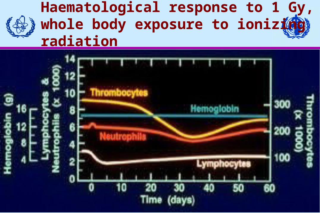
Haematological response to 1 Gy, whole body exposure to ionizing radiation

11
Haematological response to 3 Gy, whole body exposure

12
Phases of haematopoietic syndrome (HPS)
Prodromal phase symptoms nausea and vomiting lasts only a few hours, with time of onset from later than one hour to about 24 hours after exposure
Latent phase lasts up to a month. Relatively asymptomatic except for some fatigue and weakness
Manifest illness phase, characterized by neutropenic fevers, systemic and localized infections, sepsis, and haemorrhage

13
Gastrointestinal (GI) syndrome (8-30 Gy)
Depletion of the epithelial cells lining lumen of gastrointestinal tract
Intestinal bacteria gain free access to body
Haemorrhage through denuded areas
Loss of absorptive capacity
Irradiated GI Mucosa
Pathophysiology of the GI Syndrome

14
Phases of Gl syndrome
Prodromal period: Severe nausea and vomiting, watery diarrhoea and cramps. Occurs within hours after exposureLatent (subacute) phase: Asymptomatic for hours to days, severe tiredness, weaknessManifest illness: Return of severe diarrhoea, vomiting with fever; progression to bloody diarrhoea, shock and death without aggressive medical intervention

15
Systemic effects of GI syndrome
MalabsorptionmalnutritionFluid and electrolyte shiftsdehydration, acute renal failure, cardiovascular collapse GI bleedinganaemiaSepsisParalytic ileusvomiting, abdominal distention

16
Pulmonary effects
Irradiated lung tissue Pulmonary fibrosis

17
At 30 Gy and above
Endothelial cell damage
Neurovascular syndrome (NVS)
18
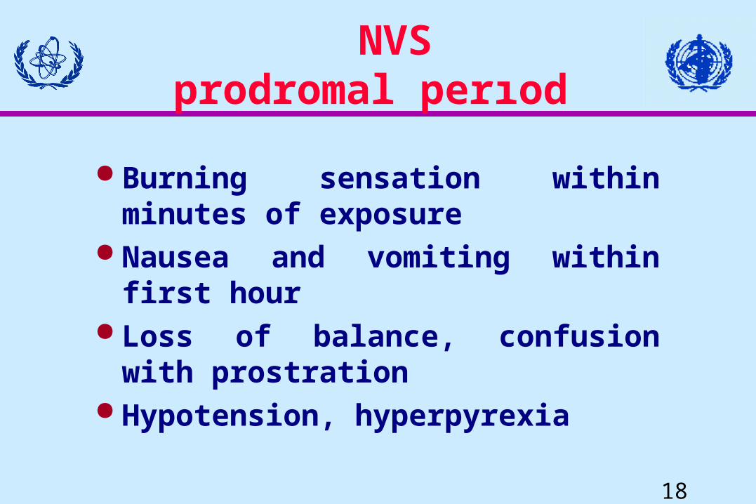
NVSprodromal perıod
Burning sensation within minutes of exposure
Nausea and vomiting within first hour Loss of balance, confusion with
prostration Hypotension, hyperpyrexia

19
NVSlatent period
Apparent improvement lasting several hours–May be lucid and in
no pain but weak

20
NVSovert clinical picture
Rapid onset Watery diarrhoea Respiratory distress Gross CNS signs Wide pulse pressure
Hypotension

21
ARSNeurovascular Syndrome
SymptomsRadiationdose (Gy)
Life threateninginjuries
Death of patients
16202530
Loss of consciousness
Neurovascular damage
5-12 days
2-5 days

22
Triage of injured persons (degrees )

23
Measurement of severity
Prodromal effects Time of onset Degree of symptoms
Haematological changes Lymphocyte counts Biological dosimetry
Physical dosimetry Attendant readable
24
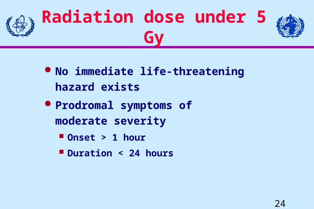
Radiation dose under 5 Gy
No immediate life-threatening hazard exists
Prodromal symptoms of moderate severity Onset > 1 hour Duration < 24 hours

25
Fatal radiation
Nausea and vomiting within minutes (during the first hour)
Within hours (on the first day):
Explosive bloody diarrhoea Hyperthermia Hypotension Erythema Neurological signs
26
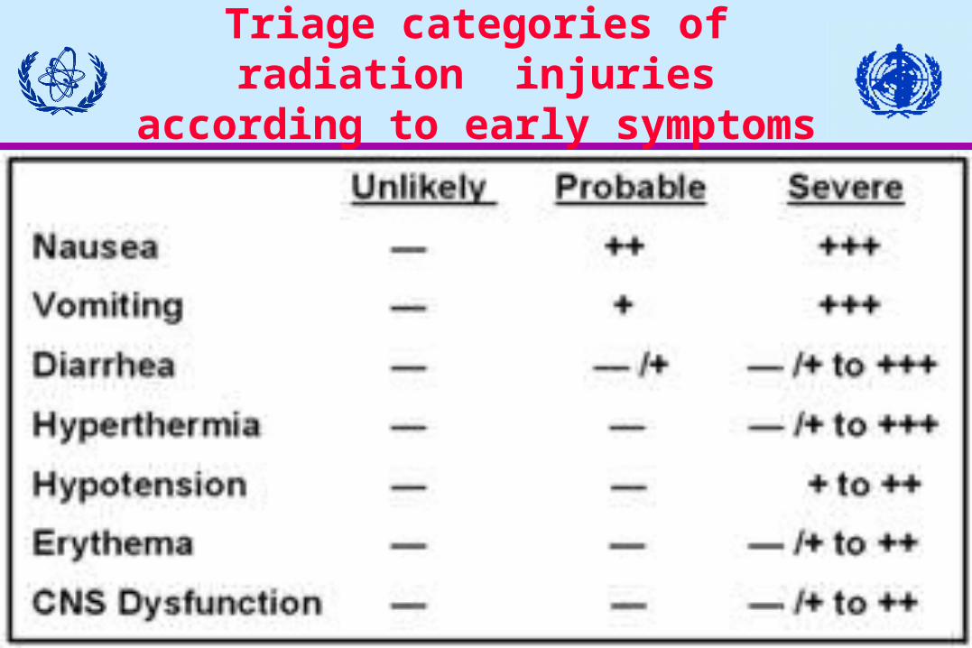
Triage categories of radiation injuries according to early
symptoms

27
Guide for management of radiation injuries on the basis of early symptoms
No vomiting Vomiting 2-3 hafter exposure Vomiting 1-2 hafter exposure Vomiting earlier than 1 h, other severe symptoms, like hypotensionhyperthermia,diarrhoea, oedema, erythema, CNS symptoms
< 1 Gy 1-2 Gy 2-4 Gy > 4 Gy
Outpatient with 5-week surveillance Surveillance in a general hospital (or outpatient for 3 weeks) followed by hospitalization Hospitalization in a haematological department Hospitalization in a well equipped haematological or surgical department with transfer to a specialized centre for radiopathology

28
Lymphocytes
3Gy
4-5Gy

29
Change of lymphocyte counts in initial days of ARS depending on dose of
acute WB exposureDegree of
ARSDose (Gy)
Lymphocyte counts (cells/L)6 days after first exposure
Preclinical phaseMildModerateSevereVery severeLethal
0.1-1.01.0-2.02.0-4.04.0-6.06.0-8.0
>8.0
1500-2500700-1500500-800300-500100-300 0-50
30
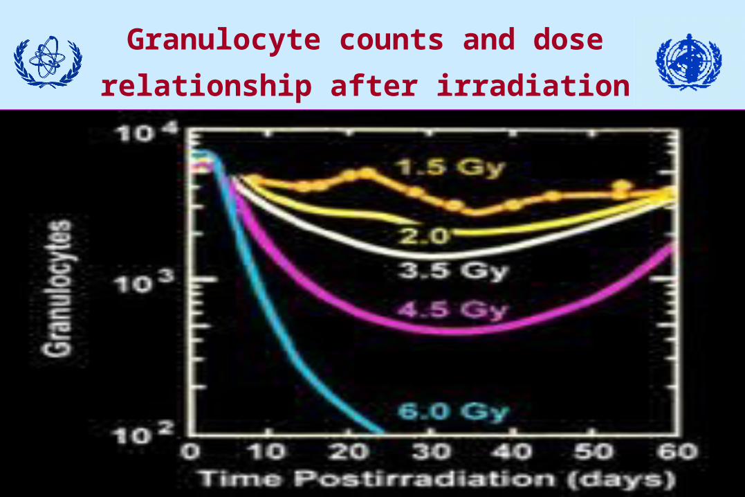
Granulocyte counts and dose relationship after irradiation

31
Medical management of
acute radiation syndrome

32
Therapeutic support for haematopoietic syndrome patientPrimary goal of haematopoietic support is reduction in both depth and duration of leukopenia
Prevention and management of infection is mainstay of therapy
Quantitative relationship between degree of neutropenia and increased risk of infectious complications. Absolute neutrophil count (ANC) < 100/L is greatest risk factor

33
Infection managmentGeneral principles
Prophylaxis Barrier/isolation Gut decontamination Antiviral agents Antifungal agents Pneumocystis prophylaxis Early cytokine therapy Close wounds Avoid invasive procedures
Direct therapy for infections
Culture specific antibiotics
Therapy for leukopenia
Cytokine administration

34
Isolation Treat ARS patients with estimated WB >2Gy in isolated rooms. Warn nursing personnel of the need for rigorous environmental control including:
laminar flow isolation strict hand washing before and after patient
care surgical scrubs for staff gowns, caps, gloves, masks for staff double bagging of all disposables

35
Prevention of infection Reduction of microbial acquisition
Contact control (e.g. careful, frequent hand washing)
Low-microbial content food Acceptable water supply Air filtration to reduce aspergillus infection
Reduction of invasive procedures (e.g. nasogastric tubes, catheters)

36
Approach to prevent infection in immunocompromised patients
Suppression of micro-organismsSelective gut decontamination
Administration of oral non-absorbable antibacterial drugs (e.g.,Quinolones) that preserve anaerobic bacteria
Awareness of resistant bacterial acquisition during clinical course
Antivirals (Acyclovir) as guided by positive anti-HSV (herpes simplex virus) antibody or empirically if test not available

37
Approach to prevent infection in immunocompromised patients
Suppression of micro-organisms Physiological interventions
Maintenance of gastric acidityAvoidance of antiacids and H2 blokersUse of sucralfate for stress ulcer prophylaxis when
indicated to reduce gastric colonization and pneumonia
Early oral enteral nutrition (when feasible) Adequate personal hygiene
Povidone-iodine (Betadine) or chlorhexidine for skin disinfection, shampoo
Oral hygiene (brushing and flossing)

38
Approach to prevent infection in immunocompromised patients
Improvement of host defencesActive vaccination for expected pathogens (e.g.
influenza)Passive immunization with immunoglobulins
(utility not yet established)Cytokine G-CSF administered prophylactically to
reduce duration of neutropenia and provide adequate numbers of functional neutrophils

39
Management of infection
Survey for possible source, pancultures Administer antibiotics for absolute neutrophil
count (ANC) <500/mm3 Use broad spectrum antibiotic coverage Add amphotericin for prolonged fever lasting 5-7
days after starting standard antibiotics Continue antibiotics for duration of ANC <1000

40
If there is evidence of resistant gram-positive infection, add vancomycin
If diarrhoea is present, examine stool cultures for salmonella, shigella, camphylobacter and yersinia
Oral and pharyngeal mucositis and oesophagitis suggest herpes simplex infection or candidiasis. Empiric acyclovir or antifungal therapy should be considered
Management of infection

41
Total parenteral nutrition vs enteral feeding
Premise for early enteral feeding
Early enteral feeding Parenteral feeding
Nutrients stimulate villi growth Villi atrophy from enteral starvation
Gut mucosal barrier remains intact
Gut mucosal barrier breaks down
Healthy mucosa limits translocation of bacteria
Unhealthy mucosal allows translocation of bacterial/endotoxin
Immune system clears limited volume of translocated bacteria
Complementary activation occurs
Results: lowered stress response and risk of sepsis
Results: increased stress response and risk of sepsis

42
Cytokines

43
Cytokines Granulocyte-macrophage stimulating
factor (GM-CSF) - Sargramostim(Leukine(R))
Macrophage colony stimulating factor (M-CSF)
Granulocyte colony stimulating factor (G-CSF) - Filgastrim (Neupogen(R))
Stem cell factor (SCF) Interleukin series (IL 1-16)

44
Selected cytokines
G-CSF and GM-CSF are potent stimulators of haematopoiesis and effective in reducing duration and degree of neutropenia
Additional benefit of CSFs ability to increase functional capacity of neutrophil and thereby contribute to prevention of infection as active part of cellular host defence
45
Advantages of cytokine therapy
Bone marrow increase production of white cells stimulate production of colony forming units decrease maturation time
Mature cells increase viability prime neutrophils/macrophages stimulate additional cytokine release
Many act in synergy to increase haematopoiesis

46
Results of cytokine therapyGM-CSF Sargramostim (LeukineR)
Proven efficacy for decreasing duration of absolute neutropenia
Decreased length of hospital stay Decreased need for antibiotics Fewer fever days

47
Use of cytokines for treatment of ARS
G-CSF and GM-CSF increase rate of hemopoietic recovery in patients after radiation exposure and may obviate need for BMT, when stem cells are still viable. Interleukins (IL-1 and IL-3) act in synergism with GM-CSF
Successfully used for radiation victims after Goiânia, San Salvador, and Belarus and Istanbul accidents

48
Initiation and duration of cytokine administration
Benchmark absolute lymphocyte count less than 500/l threshold for beginning cytokine therapy in first 2 days
Continue cytokine administration with daily injections to reach ANC of 1000/l

49
Cytokine dosage
G-CSF Filgrastim (NeupogenR)2.5-5.0 µg/kg/day (100-200 µg/m2/day)
GM-CSF Sagramostim (LeukineR)5.0-10.0 µg/kg/day (200-400
µg/m2/day) Begin therapy as early as practical
for maximum effect

50
Comparative toxicity of CSFs
Predominant side effect of G-CSF - medullary bone pain, observed shortly after initiation of G-CSF treatment and again just before onset of neutrophil recovery from nadir
G-CSF may exacerbate preexisting inflammatory conditions
Main side effects of GM-CSF - fever, nausea, fatigue, headache, bone pain, myalgia

51
Contraindication for cytokine treatment
Possibly in cases where radiation exposure is continuing (e.g. via internally deposited radionuclides, chronic external irradiation)

52
Conventional therapy for thrombocytopenia
Transfusion of platelets remains primary therapy to maintain adequate platelet counts
Requirement for platelet support depends on patient's condition. In irradiated patients with or without other major medical problems, platelets should be maintained at greater than 20 000/L. If surgery is needed, platelet count should be greater than 75 000/L

53
Conventional therapy for thrombocytopenia
All blood products should receive 15-20 Gy of radiation before infusion to prevent graft-vs-host disease through infusion of present mononuclear cells
If transplant performed, avoid use of platelets from related donors

54
Growth factor therapy for thrombocytopenia
Use of thrombopoietic agents immediately after radiation injury is not currently recommended
Consider use of thrombopoietic agents megakaryocyte growth and development factor/thrombopoietin (MGDF/Tpo) or synthetic IL-3 receptor agonist Synthokine in patient with neutrophil recovery but still platelet transfusion dependent after accidental irradiation
55
Therapy for anaemia
Transfusion of peripheral red blood cells (PRBCs) remains primary therapy to maintain haemoglobin above 8 g/dl; PRBC transfusions should be irradiated
Erythropoietin (Epo) anaemia therapy: Use of Epo after radiation injury is not recommended even though probably safe as anaemia is not generally life-threatening in this situation

56
Granulocyte transfusions (GTX) from CSF-stimulated donors
G-CSF, when administered to normal individuals, increases granulocytes collected, resulting in significant circulating levels of granulocytes in neutropenic patients. Use of HLA-compatible donors may avoid the problem of alloimmunization
G-CSF additional benefit, enhancing phagocytic and microbicidal activity of stimulated PMNs
GTX of G-CSF-stimulated PMNs could prove effective therapy for severely neutropenic patients with sepsis who have failed to respond to appropriate antibiotic therapy

57
Bone marrowtransplantation (BMT)
Indication for BMT following radiation accidents is probably limited
Following reversible BM injury BMT may have a negative effect, development of high risk graft rejection

58
Indications for BMT
Physicians should consider allo-BMT if: fully matched sibling donor available patient has absolute lymphocyte count
(ALC) <100/l radiation dose unknown or likely to be 8-12 Gy no other injuries preclude survival or
transplantation (e.g. severe burns) irradiation is not continuing from internal source
59
Timing of grafting
Timing is important - grafting in peak period of immunosuppression may reduce chance of graft rejection. Early marrow transplantation desirable, even in first week after exposure
Note importance of reliable clinical, biological and dosimetric findings to assess dose level and distribution in the body. Without reliable physical dosimetry and haematological parameters, allogenic bone marrow transplant unjustified
60
Limitations of BMT
Identification of histocompatible donors HLA typing in lymphogenic patients Need for additional immunosuppression Risk of graft versus host disease (GvHD)

61
Peripheral blood stem cell transplantation (PBSCT)
Increasing evidence that PBSCT of cells mobilized by growth factors enables reliable, rapid, and durable autologous haematopoietic engraftment
Autologous mobilized (primed) PBSCT offered more rapid recovery of granulocytes and platelets than BMTs derived from normal, resting marrow
Cautious use of allogeneic PBSCT based on unknown toxicities from cytokine administration in donors and increased risk of GVHD from the large number of t-cells infused

62
Combination of allo-BMT and allo-PBSCT
Potential advantage of dual engraftment properties available in PBSC grafts
1. Mobilized peripheral blood cells contain large quantities of committed progenitors in addition to haematopoietic stem cells. These committed progenitors would provide for an earlier, although unsustained, phase of engraftment
2. More primitive stem cells contained in both PB and BM graft would then provide for later, durable, long-term reconstitution

63
Criteria for choice of therapy-I
Therapeutic recommendations:If lymphocyte count during first week 200-
500 cells/µL, spontaneous recovery possible Therapy: Isolation, antibiotics, supportive therapy including platelet infusion. Growth factors (cytokines) can be used

64
Criteria for choice of therapy-II
If lymphocyte count in first week below 200 cells/µL, stem cells probably irreversibly damaged Therapy: Isolation, antibiotics, supportive therapy including platelet infusion. Additional growth factor therapy method of choice
65
Criteria for choice of therapy-III
If the lymphocyte count in first week below 100 cells/µL, consider treatment with growth factors and BMTObserve HLA compatibility at allogenic BMT. This therapy may be recommended for patients exposed to WB radiation doses exceeding 9 Gy

66
Therapeutic support for severely irradiated patient: gastrointestinal
syndrome Nausea,vomiting and diarrhoea associated with
prodromal effects of radiation exposure most likely related to neurohumoral factors. Nausea and vomiting can be prevented/ameliorated by new generation of 5-HT3-receptor antagonists such as ondansetron and granisetron
Diarrhoea associated with prodromal and subacute phases of gastrointestinal injury most likely affects gastrointestinal motility and transport. Anticholinergics, metamucil, amphogel, and loperamide can be used

67
Therapeutic support forseverely irradiated patient:gastrointestinal syndrome

68
Generally, exposure to dose range 8-30 Gy - causes reproductive death of mucosal crypt stem cell
In spite of considerable medical advances in treatment of radiation injury, no patient with full-scale gastrointestinal syndrome has survived !
GI system and possibly lungs can limit survival probability, assuming patient survives bone marrow damage
Gastrointestinal syndrome