acvd-979; no.of pages9 article in press - repub.eur.nl · primum and septum secundum, leaving a...
TRANSCRIPT
ARTICLE IN PRESS+ModelACVD-979; No. of Pages 9
Archives of Cardiovascular Disease (2017) xxx, xxx—xxx
Available online at
ScienceDirectwww.sciencedirect.com
REVIEW
The left atrial septal pouch as a risk factorfor stroke: A systematic reviewLa poche septale atriale gauche comme facteur de risque embolique : unerevue systématique
Mihai Strachinarua,∗, Jose Castro-Rodriguezb,Thierry Verbeetb, Marie-Dominique Gazagnesc
a Cardiology, Erasmus University Medical Centre, Rotterdam, The Netherlandsb Cardiology, Brugmann University Hospital, Brussels, Belgiumc Stroke Unit, Brugmann University Hospital, Brussels, Belgium
Received 27 October 2016; received in revised form 30 December 2016; accepted 4 January2017
KEYWORDSLeft atrial septalpouch;Ischaemic stroke;Cryptogenic stroke;Intra-atrial thrombus
Summary The left atrial septal pouch (LASP) is formed by incomplete fusion of the septumprimum and septum secundum, leaving a cavity open towards the left atrium, but withoutinteratrial shunting. There is no recommendation concerning strategy in the presence of aLASP, especially in the setting of stroke. The aim of this review was to determine whether theLASP could be incriminated as the aetiology of a stroke. We included all pertinent publica-tions on the subject, and calculated hazard ratios for ischaemic stroke and cryptogenic stroke.There were only five case—control studies concerning the LASP, involving 516 stroke patientsand 779 controls. Overall LASP prevalence was 21%, with a slightly higher prevalence in thecryptogenic stroke group (26%), but this difference was not statistically significant (P = 0.27). Ina random-effects meta-analysis, there was no difference between controls and patients withischaemic stroke (hazard ratio 1.20, 95% confidence interval 0.96—1.53; P = 0.14). Cryptogenic
Please cite this article in press as: Strachinaru M, et al. The left atrial septal pouch as a risk factor for stroke: A systematicreview. Arch Cardiovasc Dis (2017), http://dx.doi.org/10.1016/j.acvd.2017.01.001
stroke appeared more frequently in patients with LASP (hazard ratio 1.53, 95% confidenceinterval 1.07—2.24; P = 0.02), but this was driven by only one severely underpowered study.The published case reports demonstrated that thrombus formation inside the pouch can occurin the presence of major predisposing factors. The LASP can be a site for thrombus formation,leading to embolic events, but its presence does not correlate with an increased incidence of
Abbreviation: LASP, left atrial septal pouch.∗ Corresponding author at: Cardiology, Erasmus University Medical Centre, Room Ba 302, Westzeedijk 361, 3015 AA Rotterdam, The
Netherlands.E-mail address: [email protected] (M. Strachinaru).
http://dx.doi.org/10.1016/j.acvd.2017.01.0011875-2136/© 2017 Elsevier Masson SAS. All rights reserved.

ARTICLE IN PRESS+ModelACVD-979; No. of Pages 9
2 M. Strachinaru et al.
stroke. Associated factors should be taken into consideration in the setting of stroke. Furtherstudies are necessary to validate a possible relationship with cryptogenic stroke.© 2017 Elsevier Masson SAS. All rights reserved.
MOTS CLÉSPoche septale atrialegauche ;Accident vasculairecérébral ischémique ;Accident vasculairecérébralcryptogénique ;Thrombus intra atrial
Résumé La poche septale atriale gauche (LASP) est formée par une fusion incomplète du sep-tum primum et du septum secundum. Il n’y a pas de recommandation concernant la conduiteà tenir en présence d’un LASP dans le cadre d’une embolie. Le but de cette revue est dedéterminer s’il est possible de l’incriminer comme étiologie d’un accident vasculaire cérébralischémique (AIC). Nous avons inclus toutes les publications pertinentes sur le sujet et calculé lesrapports de risque d’AIC et AIC cryptogénique. Il y a seulement cinq études cas-témoins concer-nant le LASP, totalisant une population de 516 patients d’AIC et 779 contrôles. La prévalenceglobale du LASP était de 21 %, avec une prévalence légèrement plus élevée dans le groupe AICcryptogénique (26 %), mais cette différence était statistiquement non significative (P = 0,27).Selon notre méta-analyse il n’y avait pas de différence entre les témoins et les patients souf-frant d’AIC (HR 1,20, IC 95 % 0,96—1,53 ; P = 0,14). Les AIC cryptogéniques semblaient plusfréquentes chez les patient ayant un LASP (HR 1,53, IC 95 % 1,07—2,24 ; P = 0,02), mais cettetendance est influencée par une seule étude de très faible puissance statistique. Les cas déjàpubliées démontrent qu’un thrombus peut se former dans la poche en présence des facteursprédisposants. Le LASP pourrait être un site favorisant la thrombogenèse, mais n’est pas associéà l’augmentation du risque d’AIC. Les facteurs associés doivent être pris en considération dansle contexte d’embolie artérielle. D’autres études sont nécessaires pour valider une possiblerelation avec les AIC cryptogéniques.© 2017 Elsevier Masson SAS. Tous droits reserves.
B
Tbos[jsTbctLfnfipm
tcfisss
i
cwts
M
TM(rwa(soo
Lmu‘ct
ackground
he fusion of the interatrial septum was initially thought toe homogenous all along the coaptation line. The absencef fusion of the completely developed septum primum andecundum defines the presence of a patent foramen ovale1]. It was recognized surprisingly late that this fusion mayust be incomplete, leading to the appearance of pouch-liketructures on either side of the interatrial septum (Fig. 1).he left atrial septal pouch (LASP) was described in 2010y Krishnan and Salazar [2]. From the beginning, there wasoncern regarding the potential for thrombogenesis insidehis pouch [3]. It is possible that in certain conditions theASP behaves like the left atrial appendage, presenting a riskor embolic events. Unlike the appendage, this pouch haso contractility of its own, being formed almost exclusivelyrom fibrous structures [4]. Fortunately, its cranial openings directly in the way of the flow coming from the right upperulmonary vein (Fig. 1), which may facilitate a washingechanism, preventing local stasis and clot formation [5].Several reports have demonstrated the presence of
hrombi in the LASP, mostly in the setting of factors thatan be assimilated into the classical triad of Virchow: atrialbrillation, left ventricular dysfunction and procoagulabletate. Despite these findings, later in 2010, Tugcu et al.
Please cite this article in press as: Strachinaru M, et al. The leftreview. Arch Cardiovasc Dis (2017), http://dx.doi.org/10.1016
howed an absence of evidence for an association betweentroke and the presence of LASP [6].
There is no clear recommendation regarding strategyn the presence of a LASP, especially in the setting of a
teTt
ryptogenic stroke. The aim of this review was to determinehether we possess sufficient information to incriminate
his anatomical structure as the aetiology of a stroke, orhould simply discard it as an innocent bystander.
ethods
his systematic review was conducted in accordance witheta-analysis Of Observational Studies in Epidemiology
MOOSE) guidelines [7]. We planned to include studies of theelationship between the LASP and stroke in adults. Studiesere included if they reported at least one of the following:
relationship between a septal pouch and embolic eventsstroke), a septal pouch and local thrombus formation or aeptal pouch and cryptogenic stroke. We excluded studiesnly published as abstracts, reviews and duplicate reportsf the same study.
We searched the following databases: PubMed (Nationalibrary of Medicine, National Center for Biotechnology Infor-ation); and EMBASE. The following search terms were
sed: ‘‘atrial pouch’’; ‘‘septal pouch’’; ‘‘atrial thrombus’’;‘septal thrombus’’; and ‘‘septal pouch stroke’’. We alsohecked manually the reference lists of all relevant paperso identify any studies that might have been overlooked by
atrial septal pouch as a risk factor for stroke: A systematic/j.acvd.2017.01.001
he automated search. Only studies in English were consid-red. The last search was performed on 25 October 2016.he studies were screened independently by two investiga-ors (M. S. and J. C.-R.), based on title and abstract. The
ARTICLE IN PRESS+ModelACVD-979; No. of Pages 9
The left atrial septal pouch: A systematic review 3
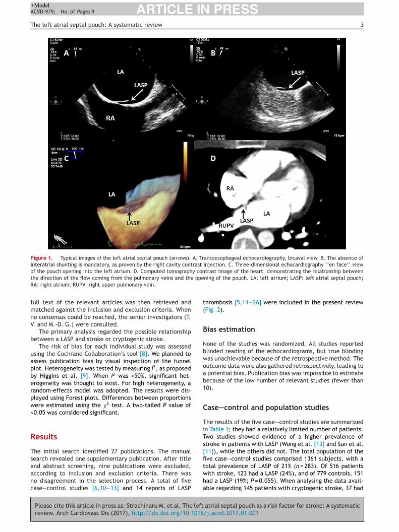
Figure 1. Typical images of the left atrial septal pouch (arrows). A. Transoesophageal echocardiography, bicaval view. B. The absence ofinteratrial shunting is mandatory, as proven by the right cavity contrast injection. C. Three-dimensional echocardiography ‘‘en face’’ viewof the pouch opening into the left atrium. D. Computed tomography contrast image of the heart, demonstrating the relationship between
ope
t(
B
Nbwoab1
C
TiTs[fi
the direction of the flow coming from the pulmonary veins and theRA: right atrium; RUPV: right upper pulmonary vein.
full text of the relevant articles was then retrieved andmatched against the inclusion and exclusion criteria. Whenno consensus could be reached, the senior investigators (T.V. and M.-D. G.) were consulted.
The primary analysis regarded the possible relationshipbetween a LASP and stroke or cryptogenic stroke.
The risk of bias for each individual study was assessedusing the Cochrane Collaboration’s tool [8]. We planned toassess publication bias by visual inspection of the funnelplot. Heterogeneity was tested by measuring I2, as proposedby Higgins et al. [9]. When I2 was >50%, significant het-erogeneity was thought to exist. For high heterogeneity, arandom-effects model was adopted. The results were dis-played using Forest plots. Differences between proportionswere estimated using the �2 test. A two-tailed P value of<0.05 was considered significant.
Results
The initial search identified 27 publications. The manualsearch revealed one supplementary publication. After title
Please cite this article in press as: Strachinaru M, et al. The leftreview. Arch Cardiovasc Dis (2017), http://dx.doi.org/10.1016
and abstract screening, nine publications were excluded,according to inclusion and exclusion criteria. There wasno disagreement in the selection process. A total of fivecase—control studies [6,10—13] and 14 reports of LASP
twha
ning of the pouch. LA: left atrium; LASP: left atrial septal pouch;
hrombosis [5,14—26] were included in the present reviewFig. 2).
ias estimation
one of the studies was randomized. All studies reportedlinded reading of the echocardiograms, but true blindingas unachievable because of the retrospective method. Theutcome data were also gathered retrospectively, leading to
potential bias. Publication bias was impossible to estimateecause of the low number of relevant studies (fewer than0).
ase—control and population studies
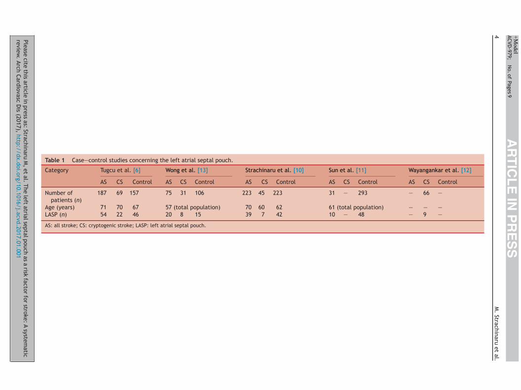
he results of the five case—control studies are summarizedn Table 1; they had a relatively limited number of patients.wo studies showed evidence of a higher prevalence oftroke in patients with LASP (Wong et al. [13] and Sun et al.11]), while the others did not. The total population of theve case—control studies comprised 1361 subjects, with a
atrial septal pouch as a risk factor for stroke: A systematic/j.acvd.2017.01.001
otal prevalence of LASP of 21% (n = 283). Of 516 patientsith stroke, 123 had a LASP (24%), and of 779 controls, 151ad a LASP (19%; P = 0.055). When analysing the data avail-ble regarding 145 patients with cryptogenic stroke, 37 had
Please cite
this article
in press
as: Strachinaru
M,
et al.
The left
atrial septal
pouch as
a risk
factor for
stroke: A
systematic
review.
Arch Cardiovasc
Dis
(2017), http://dx.doi.org/10.1016/j.acvd.2017.01.001
AR
TIC
LE
IN P
RE
SS
+Model
ACVD-979;
N
o. of
Pages 9
4
M.
Strachinaru et
al.
Table 1 Case—control studies concerning the left atrial septal pouch.
Category Tugcu et al. [6] Wong et al. [13] Strachinaru et al. [10] Sun et al. [11] Wayangankar et al. [12]
AS CS Control AS CS Control AS CS Control AS CS Control AS CS Control
Number ofpatients (n)
187 69 157 75 31 106 223 45 223 31 — 293 — 66 —
Age (years) 71 70 67 57 (total population) 70 60 62 61 (total population) — — —LASP (n) 54 22 46 20 8 15 39 7 42 10 — 48 — 9 —
AS: all stroke; CS: cryptogenic stroke; LASP: left atrial septal pouch.

ARTICLE IN PRESS+ModelACVD-979; No. of Pages 9
The left atrial septal pouch: A systematic review 5
y. LAS
e[swia
ato[figspssp(etc
ctrltwatiawpt
Figure 2. Flowchart displaying the selection process for our stud
a LASP (26%), and of the 486 controls, 103 had a LASP (22%;P = 0.27).
The overall relationship of a LASP with ischaemic stroke(Fig. 3) displayed a hazard ratio of 1.2 (95% confidenceinterval 0.96—1.53; I2 = 10%; P = 0.14), and with cryptogenicstroke (Fig. 4) displayed a hazard ratio of 1.5 (95% confi-dence interval 1.07—2.24; I2 = 0%; P = 0.02).
Case reports
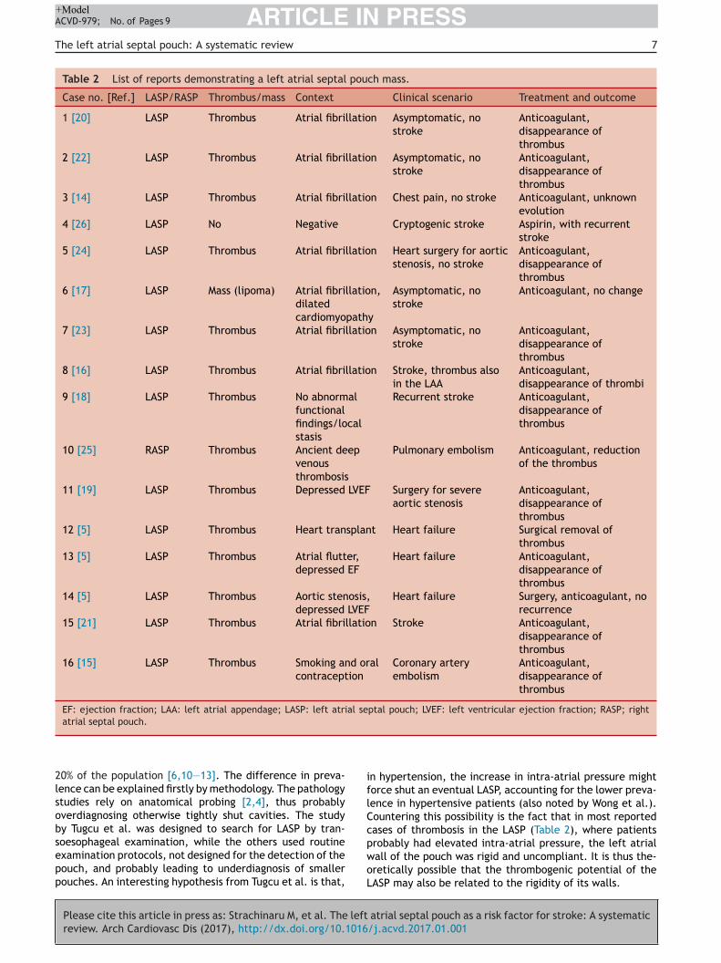
A careful internet search for reliable sources of informationled to the discovery of only 14 reports describing 16 casesof thrombi or other masses in or originating from an atrialseptal pouch thrombosis [5,14—26]. An overview of thesereports is depicted in Table 2. We noticed that only one caseproved to be a lipoma in the pouch [17]. In almost all casesthere was at least one clear identifiable factor leading tothrombosis: atrial fibrillation/flutter (10 cases [63%]); leftventricular dysfunction (three cases [19%]); local stasis orendothelial lesion (one case with a giant pouch [18] and onecase after heart transplantation [5]); or procoagulant state(one case). One case (6%) had no obvious cause of throm-bosis, and resulted in repetitive stroke. In all other cases,anticoagulant treatment was indicated based on the clini-cal context (such as atrial fibrillation or mechanical heartvalve), with good results. Six of the cases (38%) presentedwith embolic events probably related to the thrombus for-mation (four strokes and one coronary embolism for theLASP; one pulmonary embolism for the right atrial septalpouch).
Summarizing the available data
The first description [2] and the most recent pathologicalstudy [4] found a prevalence of around 40% of septal pouchesopening towards the left atrium and 4% of pouches openingtowards the right atrium.
Please cite this article in press as: Strachinaru M, et al. The leftreview. Arch Cardiovasc Dis (2017), http://dx.doi.org/10.1016
The first case—control study was published in 2010 byTugcu et al., who found that the prevalence of LASP wasonly 30%, and that no association could be established withischaemic stroke in univariate or multivariable analyses [6].
tgwm
P: left atrial septal pouch.
In 2013, the largest study was published by Wayangankart al., on 566 transoesophageal studies for all indications12]. The overall prevalence of LASP was low (11%), and notatistical association was noted with cryptogenic stroke orith any other clinical or echocardiographic variable. No
nformation was given, however, on the relationship withtrial fibrillation.
Three other case—control studies were published in 2015nd 2016. The designs were similar, relying on retrospec-ive analysis of transoesophageal echographies performedn stroke patients or patients with other pathologies10,11,13]. The study by Wong et al. demonstrated a signi-cantly higher prevalence of LASP in the cryptogenic strokeroup [13], but the stroke population was too small to betatistically strong in a multivariable analysis (75 strokeatients with 31 cryptogenic strokes). The study by Sun et al.howed that LASP is a significant risk factor for ischaemictroke [11], also in a very limited stroke population (31atients). The two studies with larger stroke populations187 patients in Tugcu et al. [6]; 223 patients in Strachinarut al. [10]) provided the same conclusion: no significant sta-istical association between LASP and stroke in general orryptogenic stroke.
Numerical analysis of the pooled data seemed to indi-ate a tendency towards higher prevalence of LASP inhe stroke group (24% vs. 19%; P = 0.055), but the P valueemained non-significant. Our meta-analysis confirmed theack of a statistically significant association between thewo (Fig. 3). Concerning cryptogenic stroke, numerical dataere non-significant (26% vs. 22%; P = 0.27), but the meta-nalysis demonstrated that cryptogenic stroke occurred 1.5imes more frequently in patients with a LASP. By look-ng at Fig. 4, you can see that this trend is driven by
single study, which has the largest weight. However,hen it comes to cryptogenic stroke population, this isrecisely the study that is the most underpowered amonghose analyzed (only 31 patients). It is a well-known facthat by using the Mantel-Haenszel method, there is a dan-
atrial septal pouch as a risk factor for stroke: A systematic/j.acvd.2017.01.001
er of overestimating the weights. In such a situation, theeights cannot be interpreted as the amounts of infor-ation contributed by each study. However, studies with

ARTICLE IN PRESS+ModelACVD-979; No. of Pages 9
6 M. Strachinaru et al.
Figure 3. Forest plot representation of the results from the significant case—control studies comparing ischaemic stroke with controls.Studies are displayed on the vertical, and are marked with a square of proportional size to the study’s calculated weight. The overall effectis lowermost, and marked with a rhombus. CI: confidence interval; HR: hazard ratio; LASP: left atrial septal pouch.
Figure 4. Forest plot representation of the results from the significant case—control studies comparing cryptogenic stroke with controls.S of pri HR: h
sm
idtopidt
ata
usps
tudies are displayed on the vertical, and are marked with a squares lowermost, and marked with a rhombus. CI: confidence interval;
mall numbers of events are better analyzed using thisethod.With respect to the most important question (i.e. is
t dangerous or not to have a LASP?), the cases alreadyescribed had a number of embolic events. However,hrombi were also found in situations where no stroke hadccurred. In the majority of cases when a thrombus wasresent, a classical factor from the Virchow triad could be
Please cite this article in press as: Strachinaru M, et al. The leftreview. Arch Cardiovasc Dis (2017), http://dx.doi.org/10.1016
dentified (blood stasis in atrial fibrillation or left ventricularysfunction, procoagulable state or endothelial injury). Fur-hermore, treatment guidelines for the underlying pathology
e
a
oportional size to the study’s calculated weight. The overall effectazard ratio; LASP: left atrial septal pouch.
nd for the respective factor were sufficient for a therapeu-ic decision, independent of the presence of the LASP, with
favourable clinical outcome (Table 2).This ambiguity is sustained by our analysis, which was
nable to find a statistical association between ischaemictroke and LASP. The slight tendency towards a largerrevalence in the cryptogenic stroke group was statisticallyignificant, but was driven by only one severely underpow-
atrial septal pouch as a risk factor for stroke: A systematic/j.acvd.2017.01.001
red study.The LASP is an anatomical variant of fusion of the inter-
trial septum, with an echography prevalence of around
ARTICLE IN PRESS+ModelACVD-979; No. of Pages 9
The left atrial septal pouch: A systematic review 7
Table 2 List of reports demonstrating a left atrial septal pouch mass.
Case no. [Ref.] LASP/RASP Thrombus/mass Context Clinical scenario Treatment and outcome
1 [20] LASP Thrombus Atrial fibrillation Asymptomatic, nostroke
Anticoagulant,disappearance ofthrombus
2 [22] LASP Thrombus Atrial fibrillation Asymptomatic, nostroke
Anticoagulant,disappearance ofthrombus
3 [14] LASP Thrombus Atrial fibrillation Chest pain, no stroke Anticoagulant, unknownevolution
4 [26] LASP No Negative Cryptogenic stroke Aspirin, with recurrentstroke
5 [24] LASP Thrombus Atrial fibrillation Heart surgery for aorticstenosis, no stroke
Anticoagulant,disappearance ofthrombus
6 [17] LASP Mass (lipoma) Atrial fibrillation,dilatedcardiomyopathy
Asymptomatic, nostroke
Anticoagulant, no change
7 [23] LASP Thrombus Atrial fibrillation Asymptomatic, nostroke
Anticoagulant,disappearance ofthrombus
8 [16] LASP Thrombus Atrial fibrillation Stroke, thrombus alsoin the LAA
Anticoagulant,disappearance of thrombi
9 [18] LASP Thrombus No abnormalfunctionalfindings/localstasis
Recurrent stroke Anticoagulant,disappearance ofthrombus
10 [25] RASP Thrombus Ancient deepvenousthrombosis
Pulmonary embolism Anticoagulant, reductionof the thrombus
11 [19] LASP Thrombus Depressed LVEF Surgery for severeaortic stenosis
Anticoagulant,disappearance ofthrombus
12 [5] LASP Thrombus Heart transplant Heart failure Surgical removal ofthrombus
13 [5] LASP Thrombus Atrial flutter,depressed EF
Heart failure Anticoagulant,disappearance ofthrombus
14 [5] LASP Thrombus Aortic stenosis,depressed LVEF
Heart failure Surgery, anticoagulant, norecurrence
15 [21] LASP Thrombus Atrial fibrillation Stroke Anticoagulant,disappearance ofthrombus
16 [15] LASP Thrombus Smoking and oralcontraception
Coronary arteryembolism
Anticoagulant,disappearance ofthrombus
EF: ejection fraction; LAA: left atrial appendage; LASP: left atrial septal pouch; LVEF: left ventricular ejection fraction; RASP; rightatrial septal pouch.
iflCc
20% of the population [6,10—13]. The difference in preva-lence can be explained firstly by methodology. The pathologystudies rely on anatomical probing [2,4], thus probablyoverdiagnosing otherwise tightly shut cavities. The studyby Tugcu et al. was designed to search for LASP by tran-
Please cite this article in press as: Strachinaru M, et al. The leftreview. Arch Cardiovasc Dis (2017), http://dx.doi.org/10.1016
soesophageal examination, while the others used routineexamination protocols, not designed for the detection of thepouch, and probably leading to underdiagnosis of smallerpouches. An interesting hypothesis from Tugcu et al. is that,
pwoL
n hypertension, the increase in intra-atrial pressure mightorce shut an eventual LASP, accounting for the lower preva-ence in hypertensive patients (also noted by Wong et al.).ountering this possibility is the fact that in most reportedases of thrombosis in the LASP (Table 2), where patientsrobably had elevated intra-atrial pressure, the left atrialall of the pouch was rigid and uncompliant. It is thus the-retically possible that the thrombogenic potential of the
atrial septal pouch as a risk factor for stroke: A systematic/j.acvd.2017.01.001
ASP may also be related to the rigidity of its walls.

IN+ModelA
8
D
P
Ttafs
tccgoas(lt
sfiualemteibst
C
TetAsa
S
N
A
Mdsrc
D
T
R
[
[
[
[
[
[
[
[
[
[
ARTICLECVD-979; No. of Pages 9
iscussion
ractical and future considerations
he LASP is a normal variant of the interatrial septum, andhe evidence presented so far cannot incriminate it as theetiology of a stroke, in the absence of major predisposingactors for thrombosis that in themselves require treatmentufficient to prevent thrombosis recurrence.
The presence of a LASP should be noted during anyransoesophageal echocardiography, but without any clini-al implication. In the setting of stroke, especially withoutlear aetiology, this pouch should be thoroughly investi-ated, ideally with multiplane/tridimensional imaging, inrder to exclude a thrombus. No clear consensus existsbout whether the presence of a LASP alone in cryptogenictroke is an indication for anticoagulation or other treatmentocclusion or surgical removal). In atrial fibrillation, whenooking to exclude an intra-atrial thrombus, care should beaken to examine an existing LASP.
The difference between a LASP and a double interatrialeptum with persistent interatrial cavity is sometimes dif-cult to distinguish [27]. In the latter case, the cavity issually bigger, there is often a communication with the righttrium and the opening in the septum primum towards theeft atrium is rather a fenestration in a membrane. Thembryogenesis might be different, involving an accessoryembrane, presumably a remnant of the superior part of
he septum primum or a persistent left venous valve of thembryologic sinus venosus [28]. This structure has also beenmplicated in the appearance of embolic events [29,30]. Weelieve that a case presented in 2006 as a double interatrialeptum was, in fact, a LASP (but not described as such at thatime) [15].
onclusions
he LASP can be a site for thrombus formation, leading tombolic events, but the presence of this anatomical fea-ure is not correlated with an increased incidence of stroke.ssociated factors should be taken into consideration in theetting of stroke. Further studies are necessary to validate
possible relationship with cryptogenic stroke.
ources of funding
one.
uthors contribution
. S. collected the data, did the statistical analysis and
Please cite this article in press as: Strachinaru M, et al. The leftreview. Arch Cardiovasc Dis (2017), http://dx.doi.org/10.1016
rafted the article. J. C.-R. collected the data and did thetatistical analysis. T. V. and M.-D. G. handled supervision,eviewed the manuscript for key intellectual content andontributed to the writing of the manuscript.
[
PRESSM. Strachinaru et al.
isclosure of interest
he authors declare that they have no competing interest.
eferences
[1] Hara H, Virmani R, Ladich E, et al. Patent foramen ovale: cur-rent pathology, pathophysiology, and clinical status. J Am CollCardiol 2005;46:1768—76.
[2] Krishnan SC, Salazar M. Septal pouch in the left atrium: a newanatomical entity with potential for embolic complications.JACC Cardiovasc Interv 2010;3:98—104.
[3] Chandrashekhar Y, Narula J. La septal pouch as a source ofthromboembolism: innocent until proven guilty? JACC Cardio-vasc Imaging 2010;3:1296—8.
[4] Holda MK, Koziej M, Holda J, et al. Atrial septal pouch — mor-phological features and clinical considerations. Int J Cardiol2016;220:337—42.
[5] Gurudevan SV, Shah H, Tolstrup K, Siegel R, Krishnan SC. Septalthrombus in the left atrium: is the left atrial septal pouch theculprit? JACC Cardiovasc Imaging 2010;3:1284—6.
[6] Tugcu A, Okajima K, Jin Z, et al. Septal pouch in the leftatrium and risk of ischemic stroke. JACC Cardiovasc Imaging2010;3:1276—83.
[7] Stroup DF, Berlin JA, Morton SC, et al. Meta-analysis ofobservational studies in epidemiology: a proposal for report-ing. Meta-analysis Of Observational Studies in Epidemiology(MOOSE) group. JAMA 2000;283:2008—12.
[8] Higgins JP, Altman DG, Gotzsche PC, et al. The Cochrane Col-laboration’s tool for assessing risk of bias in randomised trials.BMJ 2011;343:d5928.
[9] Higgins JP, Thompson SG, Deeks JJ, Altman DG. Measuringinconsistency in meta-analyses. BMJ 2003;327:557—60.
10] Strachinaru M, Catez E, Jousten I, et al. The left atrial septalpouch as a possible risk factor for stroke. Echocardiography2016;33:1016—23.
11] Sun JP, Meng F, Yang XS, et al. Prevalence of atrial septal pouchand risk of ischemic stroke. Int J Cardiol 2016;214:37—40.
12] Wayangankar SA, Patel JH, Patel B, Stavrakis S, Sivaram CA.Clinical and echocardiographic variables associated with LAseptal pouch. JACC Cardiovasc Imaging 2013;6:833—5.
13] Wong JM, Lombardo DM, Barseghian A, et al. Left atrial septalpouch in cryptogenic stroke. Front Neurol 2015;6:57.
14] Aggarwal S, Kalavakunta J, Gupta V. Left atrial septal pouchthrombus: a common pathology in an uncommon location. IntJ Cardiol 2016;212:369—70.
15] Breithardt OA, Papavassiliu T, Borggrefe M. A coronary embo-lus originating from the interatrial septum. Eur Heart J2006;27:2745.
16] Buchholz S, Robaei D, Jacobs NH, O’Rourke M, Fene-ley MP. Thromboembolic stroke with concurrent left atrialappendage and left atrial septal pouch thrombus. Int J Cardiol2012;162:e16—7.
17] Cresti A, Capati E, Picchi A, Guerrini F, Severi S. [Atrial septalpouch: not always a thrombus. A case report and literaturereview]. G Ital Cardiol (Rome) 2012;13:622—4.
18] Elsokkari I, Reyneke E, William M. Left atrial septal pouch caus-ing an ischaemic stroke in association with aortic coarctation.Eur J Echocardiogr 2011;12:916.
19] Kuwaki H, Takeuchi M, Kaku K, et al. Thrombus attached tothe left atrial septal pouch assessed on 3-dimensional trans-esophageal echocardiography. Circ J 2011;75:2280—1.
atrial septal pouch as a risk factor for stroke: A systematic/j.acvd.2017.01.001
20] Padilla Pérez M, Almagro Torres F, Sanchez de Castro M, et al.Unusual presentation of a left atrial thrombus mimicking acardiac myxoma. Thromb Res 2015;133(Suppl 3):S56 [abstractC0128].
IN+Model
[
[
[
[Cardiovasc Imaging 2012;28:685—6.
ARTICLEACVD-979; No. of Pages 9
The left atrial septal pouch: A systematic review
[21] Palinkas A, Nagy E, Czako L. Deviation of the atrial septumprimum predisposing to local thrombus formation. J Am SocEchocardiogr 2011;24(935):e3—5.
[22] Shimamoto K, Kawagoe T, Dai K, Inoue I. Thrombus in theleft atrial septal pouch mimicking myxoma. J Clin Ultrasound2014;42:185—8.
[23] Strachinaru M, Morissens M, Latifyan S, Costescu I. Left atrialseptal pouch thrombus assessed on three-dimensional transoe-sophageal echocardiography. Eur Heart J Cardiovasc Imaging2012;13:967.
[24] Strachinaru M, Wauthy P, Sanoussi A, Morissens M, Costescu I,Catez E. The left atrial septal pouch as a possible location forthrombus formation. J Cardiovasc Med (Hagerstown) 2013;14,
Please cite this article in press as: Strachinaru M, et al. The leftreview. Arch Cardiovasc Dis (2017), http://dx.doi.org/10.1016
http://dx.doi.org/10.2459/JCM.0b013e328360297e.[25] Wayangankar SA, Patel J, Latif F, Sivaram C. Right atrial sep-
tal pouch—–a potential nidus for thrombosis. Echocardiography2012;29:E1—4.
[
PRESS9
26] Wong JM, Lombardo D, Handwerker J, Fisher M. Cryptogenicstroke and the left atrial septal pouch: a case report. J StrokeCerebrovasc Dis 2014;23:564—5.
27] Roberson DA, Javois AJ, Cui W, Madronero LF, CuneoBF, Muangmingsuk S. Double atrial septum with persis-tent interatrial space: echocardiographic features of a rareatrial septal malformation. J Am Soc Echocardiogr 2006;19:1175—81.
28] Javois AJ, Roberson DA. Unusual atrial septal anatomy resul-ting in an interatrial chamber: the true triatrial heart? PediatrCardiol 2007;28:224—8.
29] Martin M, Rios E, Garcia-Ruiz JM, et al. Double trouble. Int J
atrial septal pouch as a risk factor for stroke: A systematic/j.acvd.2017.01.001
30] Seyfert H, Bohlscheid V, Bauer B. Double atrial septum withpersistent interatrial space and transient ischaemic attack. EurJ Echocardiogr 2008;9:707—8.