adenylyl cyclase: mediator of the intraocular … michiel... · 2016-03-10 · chapter 6 aqueous...
TRANSCRIPT

ADENYLYL CYCLASE: MEDIATOR OF THE INTRAOCULAR
PRESSURE RESPONSE TO ADRENERGIC AGENTS
Experimental studies with a phosphodiesterase-inhibitor

Promotie commissie
Promotor:Prof. Dr. P.T.V.M. de Jong Co-promotor:Dr. Ph.F.J. Hoyng
Overige leden:Prof. Dr. I.L. Bonta Prof. Dr. E.L. Greve Prof. Th.W. Mittag, PhD

ADENYLYL CYCLASE: MEDIATOR OF THE INTRAOCULAR
PRESSURE RESPONSE TO ADRENERGIC AGENTS
Experimental studies with a phosphodiesterase-inhibitor
(ADENYLAAT CYCLASE: MEDIATOR VAN INTRAOCULAIRE DRUK EFFECTEN VAN ADRENERGE STOFFEN
Experimentele studies met een fosfodiesterase-remmer)
PROEFSCHRIFT
ter verlcrijging van de graad van doctor aan de Erasmus U niversiteit Rotterdam
op gezag van de rector magnificus Prof. Dr. C.J. Rijnvos
en volgens beslnit van het college van dekanen. De openbare verdediging zal plaatsvinden op
woensdag 16 september 1992 te 15.45 uur
door
MICIDEL JACOBUS WILHELMUS MARIE BUSCH
geboren te Nijmegen
1992

Publication of this thesis is supported by grants
from Kabi Pharmacia, Uppsala, Sweden,
and "Stichting Blindenhulp", 's-Gravenhage.
ISBN: 90-9005265-8
Druk SSN, Nijmegen
Part of this thesis was supported by a travelgrant from the Netherlands Organization for Scientific Research (Nederlandse organisatie voor Wetenschappelijk Onderzoek (NWO)).
Cover: Schematic representation for the proposed mechanism by which adrenaline and combinations between a selective "' and .fl:, -adrenergic agonist reduce the intaocular pressure (see epilogue of this thesis, p 117).

Aan mijn ouders


VOORWOORD
Voor bet tot standkomen van dit proefschrift ben ik velen erkentelijk. Jacques de Feiler (Interuniversitair Oogbeelkundig Instituut, Amsterdam), Tony van Hamersveld en Gemma Rigter (proefdierenhuis van bet Centraal Laboratorium van de Bloedtransfusie dienst, Amsterdam; boofd P van Mourik) voor tecbische bulp en verzorging van proefdieren; Nico Bakker voor vee! fotografisch werk; Mrs BiegerSmith voor correctie van het Engels; Merrill N Kramp, secretaresse; Josanna van Workum en Gabrielle JF Thijssen voor redactie en lay-out advies; Valerie M Bodelier en Dr Nico van Haeringen voor de uitvoering van biochemische bepalingen; Crujoanne M Groeneboer, oogarts, voor het leggen van de basis voor de experimenten en Aad vd Ende .voor kritische kanttekeningen in manuscripten; Dr J van Best en J van Everdingen, afdeling Oogheelkunde Academisch Ziekenhuis, Leiden, voor de geboden gelegenheid en de uitvoering van fluorofotometrie-experimenten; het Interuniversitair Oogheelkundig Instituut, Amsterdam (hoofd Dr AJ Otto), voor de faciliteiten en mogelijkheden die het mij de afgelopen jaren geboden heeft. Onvermeld laat ik hen die belangrijk waren voor het creeren van een goede werk- en onderzoekssfeer.
Parts of this work have been performed in Uppsala and New York in collaboration with J S\iernschantz MD PhD, Pharmacia Laboratory of Glaucoma Research, Uppsala, Sweden, and with Prof Th W Mittag PhD, Dept of Ophthalmology and Pharmacology, Mount Sinai Medical Center, New York, U.S.A .. I am grateful to J ohan S\iernschantz for the Lab facilities; to him, to Giiran Selen PhD and Maria Astin for the assistence and the teaching of physiological techniques. I am grateful to Thorn Mittag for the facilities at his Lab and his teaching about basic pharmacology and experimental design; to Kaori Kobayashi MD and to Wenbin Guo MD for their cooperation in the lab.
In het bijzonder Dr Philip FJ Hoyng, co-promotor; zijn creatieve ideeen, loyaliteit en energiek stimuleren vormen de basis voor dit proefschrift. Voor mij is het een samenwerking geweest die geleid heeft tot de meest goede ervaringen, ook buiten het terrein van dit onderzoek.


CONTENTS
Abbreviations 12
Chapter 1 General introduction 13 Intraocular pressure 14 Adrenergic receptors 14 Adeny1yl cyclase 16 Adrenergic agents, adenylyl cyclase, and intraocular pressure 18
ciliary processes and aqueous flow 19 trabecular meshwork and outflow facility 21
Inhibition of the phosphodiesterase enzyme as pharmacological tool 24 Aim of the thesis 25
Chapter 2 Materials and methods Experimental animals and tissue Intraocular pressure Outflow facility Aqueous flow Ocular blood flow Aqueous cyclic AMP, protein and prostaglandins Adenylyl cyclase activity Statistics
Chapter 3 Isobutylmethylxanthine enhances adrenergic-induced ocular,
27 27 27 29 30 30 31 31 32
hypotension in rabbits and beagles 33 Abstract 33 Introduction Materials and methods Results Discussion
Chapter 4 Soluble forms of isobutylmethylxanthine enhance the ocular
33 34 37 41
hypotension induced by catecholamines 45 Abstract 45 Introduction 45

Materials and methods Results Discussion
46 47 49
Chapter 5 Increase in ocular blood flow induced by isobntylmethy!xantbine and epinephrine 53 Abstract 53 Introduction Materials and methods Results Discussion
54 54 57 59
Chapter 6 Aqueous humour dynamics after topical isobutylmethylxanthine and epinephrine in :rabbits 63 Abstract 63 Introduction Materials and methods Results Discussion
Chapter 7 Potentiation of ocular hypotensive response to catecholamines by isobutylmethy!xantbine: interaction between alfa,- and
64 64 66 68
beta,-adrenergic stimulation 71 Abstract 71 Introduction Materials and methods Results Discussion
Chapter 8 Interaction between selective «,- and 62-adrenergic agonists: reduction of intraocular pressure and increase of outflow
72 72 74 77
facility 79 Abstract 79 Introduction 80 Materials and methods 80 Results Discussion
83 87

Chapter 9 Effects of cyclic nucleotide analogs on intraocular pressure and trauma-induced inflammation in the rabbit eye 89 Abstract 89 Introduction 89 Materials and methods 90 Results Discussion
Cbapter 10 Adenylyl cyclase in human and bovine trabecular meshwork
Summary Epilogue
References
Abstract Introduction Materials and methods Results Discussion
Sam en vatting Epiloog
Curriculum vitae
92 97
101 101 102 103 105 109
113 116
121
133 136
139

Abbreviations AC AH ARVO AMP ATP BAB cAMP cGMP CGRP C-value DAG DMSO DPE EDTA EGTA EPI EP-receptor FSK GDP GDPBS G, G, GTP HSA IBMX IBMX-AT
IBMX-CD
IBMX-ED IOP IP, ISO MABP NE NPE NPY PBA Po PG(s) SDS SEM TM TML VIP
adenylyl cyclase aqueous humour Association for Research on Vision and Ophthalmology adenosine 5' -monophosphate adenosine 5' -triphosphate blood aqueous barrier adenosine 3' ,5'- cyclisch monophosphate guanosine 3' ,5' -cyclisch monophosphate calcitonin-gene-related-peptide coefficient of outflow facility (expressed in pl.min·'.mmHg·') diacylglycerol dimethylsulfoxide dipivalyl epinephrine ethylenediaminetetraacetic acid ethy!eneglycol-bis(ll-aminoethylether)-N ,N ,N' ,N' -tetraaceticacid epinephrine prostaglandin E receptor forskolin guanosine 5' -diphosphate guanosine 5' -0-(2-thiodiphosphate) inhibitory G-protein stimulatory G-protein guanosine 5' -triphosphate human serum albumin 3-isobutyl-l-methylxanthine 3-isobutyl-1-methylxanthine dissolved in 0.5% hydroxypropylmethylcellulose (artificial tears) 3-isobutyl-1-methyixanthine dissolved in saline containing 2% B-cyclodextrin 3-isobutyl-l-methylxanthine-ethylenediamine intraocular pressure inositol triphosphate isoproterenol mean arterial blood pressure norepinephrine non-pigmented epithelium (of ciliary processes) neuropeptide Y phenoxybenzamine episcleral venous pressure prostaglandin(s) sodium dodecyl sulphate standard error of the mean trabecular meshwork timolol vasoactive intestinal peptide

CHAPTER 1
GENERAL INTRODUCTION
Treatment of glaucoma focusses on reducing intraocular pressure (IOP) to prevent the loss of neuroretinal nerve fibres. The conservative management of glaucoma is directed toward pharmacological manipulation of the mechanisms that regulate intraocular pressure (IOP). The adrenergic nervous system is believed to play a major role in the maintenance of IOP homeostasis and agents that influence this system have been the subject of investigation for decades. Adrenergic antagonists, such as timolol, levobunolol and betaxolol, and adrenergic agonists, such as epinephrine (DiopineR), have become pillars of glaucoma treatment. The receptor concept is fundamental to adrenergic pharmacology. The binding site of a receptor recognizes a specific endogenous hormone that initiates a sequence of intracellular events, i.e. signal transduction. The signal transduction system, which is directly coupled to the receptor, can generate a "second messenger" molecule that evokes specific cellular responses. A few distinct second messenger systems have been proposed. Adenylyl cyclase (A C) is the most well-known second messenger system, which generates cyclic adenosine monophosphate (cAMP) as second messenger moiecule. Adrenergic receptors have been subdivided into alpha and beta subclasses by Ahlquist (1948), and each subclass in turn is divided into two subtypes. AC has been found to be directly coupled to the J\-adrenergic complex in a variety of tissues and species. The AC second messenger system has long been considered to play an impotant role in the regulation of aqueous humour secretion by ciliary processes. Increasing evidence suggests that AC is also important for regulation of the outflow facility. Several drugs and agents that act on receptors coupled to AC in various tissues, e.g. Jl-adrenergic agents such as epinephrine, affect the IOP, aqueous humour production and outflow facility. This thesis focusses on the role of the AC/cAMP system in the response to adrenergic agents that reduce IOP. The principal approach was to amplify the AC/cAMP signal of adrenergic agents pharmacologically by combination with the phosphodiesterase-inhibitor 3-isobutyl-1-methylxanthine (IBMX). IBMX is known to inhibit the activity of the enzyme
13

phosphodiesterase that metabolizes the second messenger molecule cAMP into inactive AMP. The following physiological and biochemical parameters were studied in vivo: intraocular pressure. outflow facility. aqueous humour production, regional ocular blood flow; their correlation with aqueous cAMP levels was also assessed. In vitro, AC characteristics in trabecular meshwork membrane preparations were examined.
Intraocular pressure
The principal factors determining intraocular pressure are the rate of aqueous humour formation and resistence encountered in the outflow pathway. Aqueous humour from ciliary processes enters the posterior chamber via secretion, as a consequence of active ionic transport by non-pigmented epithelial cells (!Gnsey, 1971; Maren, 1974), and by passive ultrafiltration which is determined by hydrostatic and colloid osmotic gradients (Barany, 1963; Macri and Cevario, 1974; Macri and Cevario, 1975; Bill, 1975; Sears et al, 1981). Aqueous humour passes from the posterior chamber through the pupil into the anterior chamber and then leaves the eye by two major routes, the trabecular meshwork a.'ld the uveoscleral pathway. The trabecular route is characterized by drainage of the aqueous humour across the inner wall of Schlemm's canal into collector channels, aqueous veins and finally the venous circulation. The bulk of the flow is through the trabecular meshwork. Uveoscleral drainage passes through the connective tissues between the muscle bundles of the iris root and ciliary muscle into the suprachoroidal space, then through the sclera and finally into the circulation (Bill, 1975). The uveoscleral pathway accounts for 25-60% of aqueous drainage in the monkey and man (Bill, 1989) and, under physiological conditions, is considerably lower in cats (20%)(Bill, 1966a) and rabbits (2%)(Bill, 1966b).
Adrenergic receptors
Adrenergic receptors are membrane-bound, localized postsynaptically and/or presynaptically and classified as a and 8-adrenoceptors (Ahlquist, 1948); a and 8-receptors are each divided into two subtypes, i.e. a 1 (postsynaptic), a2 (presynaptic and postsynaptical) (Langer, 1974; Wikberg, 1978), Jl1 and Bz adrenoceptors (Lands, 1967). These receptors are bifunctional with a binding component and an effector component which induce a biological response. Basically, receptors can be studied in
14

two ways. The biological effect of the administration of agonists and antagonists on an intact (isolated) organ indicates the functional activity and efficacy of the receptor. Interpretation of the results. however, is complicated by transport and distribution of the drug as well as its interaction with surrounding tissues before it reaches the receptor and by biological effector responses, which consist of an unknown number of steps. The second approach is measurement of radioactive ligand binding to a homogenate or slice preparation. Ligands (agonists) are molecules that bind to receptor binding sites. In this way the affinity for and localization of receptors in whole tissue, on cell membranes, in specific regions of cell membranes and subcellularly are investigated. Ideally, both approaches should be used to study properties such as saturability,
Table 1.1 Adrenergic agonists and antagonists used in this thesis and their relative selectivity for adrenergic receptors.
Adrenoceptor selectivity
a, a, 6, 6,
Agonists norepinephrine ++ (+) + epinephrine + (+) ++ isoproterenol (+) (+) +++ salbutamol +++ terbutaline +++
dobutamine +++ +
phenylephrine +++
B-HT920 +++
Antagonists timolol ++ +++ betaxolol +++ + phenoxybenzamine +++ ++
An empty space indicates little or no affinity; ( + ), +, + + or + + + indicates moderate to high affinity.
15

specifity, and reversibility. This thesis deals with biological responses and refers to the literature on ligand binding. In vivo IOP, aqueous inflow, facility of outflow, and aqueous cAMP levels were studied together with in vitro A C responses in membrane fractions of trabecular meshwork. Adrenergic drugs specificallly bind directly to adrenergic receptors and can be divided into agonists and antagonists. Agonists and antagonists are subdivided according to their selectivity for a(a1 and/or a,) and/or 8(81 and/or Jl.,) adrenergic receptors. The selectivity of the adrenergic agonists and antagonists used in this thesis is reviewed in Table LL
Adenylyl cyclase
The effector component transduces the signal initiated by the ligand bound to a receptor to the cell via a second messenger system. A large variety of receptors are coupled to relatively few signal transduction systems. A few distinct systems have been described (Cooper et al, 1986). Schematic representations of the two major systems involved in adrenergic receptor responses are depicted in Figure L L The AC system is shown in the left-hand paneL The binding of an agonist switches the receptor to an activated state whereas antagonists stabilize the ground state. Binding of antagonists is slower but stronger and often irreversible. Once a blockade is achieved, it is long-lasting. Activation of the receptor leads to activation of the coupling G-proteins which have stimulating (GJ or inhibitory (G,) properties. The catalytic unit of AC is thus activated or inactivated, respectively, to modulate production of second messenger cyclic AMP using A TP as the substrate. Cyclic AMP activates phosphorylation of proteins but is metabolized intracellularly by the enzyme phosphodiesterase to inactive AMP. A variety of receptors couple to this AC system: B, and 8:,- adrenoceptors but also non-adrenergic receptors, such as those for vasoactive intestinal peptide (VIP), prostaglandin E (PGE) and adenosine, stimulate AC via G,; ""'-adrenoceptors inhibit AC via G;. These (non-)adrenergic receptors bave been demonstrated in ciliary processes and trabecular tissue; their presence, location and influence on IOP will be discussed below. The second messenger system which leads to protein phosphorylation by phospholipase C is depicted in Figure l.l in the right-hand paneL Receptor binding activates phospholipase C, which in tum produces the second messenger molecules inositol triphosphate (IP3) and diacylglycerol (DAG). IP3 mobilizes intracellular ca++, a third second messenger molecule. IP,, DAG and ca++ activate protein kinase C and induce protein phosphorylation. Of the adrenergic receptors acreceptors activate this system.
16

Interactions between the cAMP and calcium/protein kinase C systems have been observed in a variety of tissues. Nishizuka (1986) has classified these interactions as monodirectional and bi-directional, indicating positive and negative feed-back of the response. In the eye several examples have been described. In the bovine iridic sphincter, cAMP may act as a regulator of responses to neurotransmitters that exert their action through the IP3-Ca2+ system (Tachado et a!, 1989). In rabbit ciliary prooesses protein kinase C activation has been linked to AC (Mittag et a!, 1987c; Yoshimura eta!, 1989), and a calcium/calmodulin-sensitive AC has been demonstrated in rabbit ciliary prooesses (Tormay et al, 1987). How the second messenger molecules cAMP, ca++, IP3 and DAG exert their intracellular and biological effects is not known in detail. Protein phosphorylation, a
agonist agonist agonist + + +
receptor receptor receptor
t t t R·, Ri R .. i
G'/ \• G; G·,.~ \ G, G·, "I \ G1
GTP GDP ~ 1 GDP GTP
J GDP GTP
ADENYLYL CYCLASE
rATP
Phospholipase C
t Ca 2
'" ~ IP3 + DAG phospho-AMP ~ I cAfM p diesterase
' protein kinase 1\ Ca2• + CaM kinase protein kinase C
v """
/ protein phosphorylation protein phosphorylation
t + biological response bio!ogica! response
Figure 1.1 Schematic representation of the adenylyl cyclase/cAMP and calcium/protein kinase C second messenger systems. R• == agonist bound to receptor; G. = GTP bound to G-protein; G. = stimulatory and G, = inhibitory G-protein.
17

common phenomenon, may ultimately lead to
1. metabolic changes, enzyme activation or inactivation; 2. changes in receptor affinity, e.g. desensitization; 3. changes in ion conductance and 4. protein synthesis.
In conclusion, adrenergic agents exert their effect by binding to adrenergic receptors. Adrenergic receptors can be classified as a 1, a,, Jl1 and 81-subtypes. A second messenger system is coupled to the receptor and transduces the receptor signal to the cell by formation of second messenger molecules. Beta,(and 81)-adrenoceptors are coupled directly to AC. Activation of these £,-receptors stimulates AC via G., leading to synthesis of the second messenger molecule cAMP. Alpha2-adrenoceptors are also coupled directly to AC but receptor activation inhibits AC via the inhibitory G-protein. In contrast, a 1-adrenoceptor activation initiates intracellular events directed by a different second messenger system, generating calcium, IP3, and DAG as second messenger molecules.
Adrenergic agents, adenylyl cyclase and intraocular pressure
Adrenergic drug therapy is directed toward reducing IOP by manipulating adrenergic receptors pharmacologically. The gross non-visual innervation of the anterior eye of vertebrates consists of sensory, parasympathetic, and sympathetic fibres. The sympathetic nervous system is presumed to play an important role in maintaining IOP homeostasis and to be responsible for diurnal variations in intraocular pressure. Sympathetic innervation originates from thoracic root fibres and runs via the superior cervical ganglion and carotid plexus through the short and long ciliary nerves to the eye. Norepinephrine is the classical neurotransmitter of the effector organ. The trabecular meshwork contains nerve fibres in all regions, including both walls of Schlemm's canal (Holland et al, 1956); they consist of sympathetic (Nomura and Smelser, 1974) as well as parasympathetic and sensory fibres (Holland et al, 1957; Ruskell, 1976). The density of sympathetic fibres, however, is very low in comparison to the number of adrenergic receptors and adrenergic responses. Kaufman (1989) therefore suggested the presence of extrajunctional, non-innervated Jl-receptors that respond to ambient free catecholamines, as demonstrated in various other tissues. Furthermore, paracrine cell clusters produce and release neurotransmitters or a local hormone that affects neighbouring cells, while the non-synaptic contact of nerves may
18

affect neighbouring cells by means of a neurosecretory mechanism (Stone et al, 1984; Mittag, 1989). The limbal vasculature, including the episcleral vascular system which receives aqueous humour from Schlemm's canal, contains adrenergic nerves but little is known about their function (Ehinger, 1964; Ehinger, 1966). In ciliary processes abundant adrenergic fibres innervate the uveal blood vessels and ciliary processes (Stone et al, 1989); intraepithelial nerve fibres have been demonstrated (Yamada, 1988; Yamada, 1989) in ciliary processes. The presence of adrenergic receptors in ciliary processes and trabecular meshwork has been demonstrated by radiolabeled ligand binding techniques. These data will be discussed in the text below. The functional effects of adrenergic receptor stimulation and some non-adrenergic agents on J>,.C activity in ciliary processes and trabecular meshwork in vitro are reviewed in Table 1.2. The physiological significance of the stimulation (or inhibition) of AC by adrenergic and some non-adrenergic agents for the regulation of IOP homeostasis is reviewed in Table 1.3. (Several authors have reviewed the pharmacology of the role of adrenergic agents in aqueous humour dynamics: Potter, 1981; Mishima, 1982; Sears, 1984; Mittag, 1989; Polansky, 1990).
Ciliary processes and aqueous flow
Ligand binding studies have provided ample evidence that ciliary processes contain 11-adrenoceptors. The rabbit iris and ciliary body (Neufeld et a!, 1978) and ciliary processes (Bromberg et al, 1980) contain 11-receptors. About 20-25% of all adrenergic receptors in the iris and ciliary body are of the liz subtype (Mittag and Tormay, 1985b). In human iris-ciliary body membrane preparations 90% of the 11-receptors were of the liz-subtype, as demonstrated by displacement studies of the high affinity binding of [125I]iodopindolol C:Wax and Molinoff, 1987). Beta,-adrenoceptors were found only in small amou.nts around the blood vessels in sheep ciliary bodies (Trope and Clarke, 1982). In rabbit ciliary processes a 1 and a,-receptors have been identified, the majority being of the <>,-subtype (Mittag and Tormay, 1985; Mittag et al, 1985; Mallorga et al, 1988; Jumblatt et al, 1987). The discovery of AC in rabbit ciliary processes and its activation by catecholarnines was the first indication that AC may play a role in aqueous humour formation !Y'/aitzman and Woods, 1971) This was followed by many studies, in vitro as well in vivo, focussing on the role of AC (Tables 1.2 and 1.3, respectively). One consistent finding is that liz-receptors stimulate AC in ciliary processes, particularly in nonpigmented ciliary epithelial cells which secrete aqueous humour. AC stimulation was also obtained with non-adrenergic drugs, such as VIP, forskolin (FSK), fluoride
19

(F) and choleratoxin. It has been suggested that B and VIP receptors are coupled to the same AC system in non-pigmented epithelial cells (Mittag, 1987b). However, the role of 8:,-receptor agonists and other cAMP stimulators in aqueous humour production is more complicated, as indicated by contradictory results. On the one hand 8:,selective agonists and VIP increase the flow in primates (Nilsson et al, 1990); this £stimulated flow can be blocked by timolol. Comparable findings have been reported for rabbits. Timolol reduces aqueous flow during the night when the sympathetic tone is supposed to be high (Gregory, 1990; Yoshitomi and Gregory, 1991). The enhanced sympathetic tone was deduced from high aqueous cAMP (Rowland et al, 1986) and elevated endogenous norepinephrine levels (Liu, 199la and b). In contrast to the 8:,adrenergic-stimulated increase in flow, direct stimulators of AC, such as cholera toxin (Gregory et al, 198lb) and FSK (Caprioli, 1984a), have been reported to reduce aqueous flow in primates. Thus, opposite physiological effects have been reported for various stimulators of (the same?) AC in ciliary processes. Several explanations have been suggested. For example, Mittag and Tormay (1981) showed that the desensitization and uncoupling of Jl-receptors could be attributed to high doses of Jlagonists; functionally, this implies that a Jl-adrenergic agonist may have the same effect as a Jl-blocker. The flow-decreasing effect ofFSK has been questioned but may be due to a slight breakdown of the blood aqueous barrier (BAB) (Bartels et al, 1987), indicating that it can be a non-specific effect. Alpha,-receptors inhibit AC (see Table 1.3); they are located postjunctionally, as can be deduced from cervical sympathectomy experiments which show that denervation does not reduce the number of az receptors in ciliary processes. This has led to the concept of dual control of AC by a 2 and 8:,-receptors (Mittag and Tormay, l985a), whereby 8:,-stimulated AC is counterbalanced by inhibitory az-receptors. This is supported by the finding that both the 8:, and YIP-receptor-stimulated formation of cAMP in ciliary bodies are both inhibited by the az-agonist clonidine (Bausher et al, 1989; Cepelic and Hynie, 1990a). Postsynaptic az-receptors and the negative coupling to AC might play an important role in aqueous production; for example, clonidine has been shown to decrease flow (Chiou, 1983). The significance of the large number of az-adrenoceptors in the ciliary body remains unclear (Mittag, personal communication). Somewhat inconclusive is the role of phenylephrine/a1 in ciliary processes; a 1-
stimulation evokes primarily a calcium signal, which essentially is a different second messenger system from the AC/cAMP system. Physiologically, both stimulation and inhibition of aqueous inflow have been reported. The immediate effect may be an increase in flow accompanied by an initial hypertensive response, possibly due to BAB breakdown. The late effect is flow reduction and a drop in the IOP. Among the non-adrenergic stimulators of AC in ciliary processes are PGE, calcitonin-
20

gene-related peptide (CGRP), and calcium calmodulin, PGE:, might inhibit prejunctional norepinephrine release (Neufeld, 1976); the presence of calcium/ calmodulin-sensitive AC in rabbit ciliary processes provides evidence for an interaction between the a 1-adrenoceptor/calcium and the adeny1yl cyclase/cAMP second messenger systems, Other non-adrenergic inhibitors of AC in ciliary processes are neuropeptide Y (NPY) (Bausher and Horio, 1990; Cepe1ic and Hynie, 1990b; Gooch et al, 1989), somatostatin in non-pigmented epithelial cells (NPE cells), and acetyl choline binding to muscarinic1 receptors, Their physiological effects on IOP have not yet been clarified. In conclusion, ciliary processes contain B-adrenergic receptors predominantly of the Jl,-subtype, which are located in epithelial cells. They are directly coupled to AC; stimulation (terbutaline) seems to increase and inhibition (timolol) to decrease aqueous flow. Alpha2-adrenoceptors are present in large quantities in ciliary processes, inhibit AC and may decrease flow. A dual control of AC by a, and Jl,-receptors has been proposed. Alphacadrenoceptors are also present in ciliary processes, are not coupled to AC but to the calcium/IP3/DAG second messenger system and may inhibit aqueous humour formation.
Trabecular meshwork and outflow faciliJy
The Jl,-adrenergic receptors in human trabecular meshwork (Jampel et al, 1987; Wax et al, 1989; Elena et al, 1990) and in cultured human trabecular meshwork cells (Jarnpel et al, 1987; Wax et al, 1989) have been characterized by radioactive ligand binding techniques. In trabecular tissue, trabecular endothelium, and cultured human trabecular cells Jl,-receptor stimulation consistently activates AC (see table 1.2). Outflow facility increases after Jl-adrenoceptor stimulation (see table 1.3); many authors report an increase in outflow facility after administration of epinephrine and isoproterenol, whereas timolol antagonizes this increase. Alpha-adrenoceptors in trabecular meshwork have not been studied by ligand binding techniques to our knowledge. A role for acreceptors, however, has long been suggested but is not yet firmly established. Neufeld and Sears (1974) reported that AC in rabbit trabecular tissue is activated by a-adrenergic stimulation since epinephrine stimulated AC activity could be antagonized by the a-adrenergic antagonist phenoxybenzamine. Furthermore, PGE and VIP are non-adrenergic stimulators of AC in trabecular meshwork. PGs may be involved in the effect of epinephrine on outflow facility (Hoyng et al, 1982); in support of this is the finding that indomethacin partially inhibited the increase in outflow facility induced by topical epinephrine (Anderson and
21

Table 1.2 Adenylyl cyclase responses in vitro and in vivo
species ocular stimulus effect on references tissue adenylyl cyclase
Ciliary body rabbit cil.body 6 t Neufeld and Sears, 1974 rabbit iris~cb 6,; "'' t;+ Mittag and Tormay, 1985a rabbit iris-cb PGE, t Banacherjee et al, 1991 rabbit cil. proc 13:, t Cepelic and Cernohorsky, 1981 rabbit cil. proc "'' • Kintz et al, 1988 rabbit cil. proc o:2 postsyn. • Bausher et al, 1987 rabbit cil. proc VIP t Mittag and Tormay, 1987b rabbit cil. proc NPY; Som.st <;• Bausher and Horio,1990 rabbit ciL proc NPY • Cepelic and Hynie, 1990b rabbit cil. proc et2;1l:,, VIP ,PGE, <;t;t;< Jumblatt et al, 1990 bovine cil. proc 13:, t Elena et al, 1984 monkey cil. proc 13:, t Crawford et al, 1991 human cil.proc 13:, t Nathanson, 1981 human npe-cells CGRP t Yabu et al, 1991 human npe-cells PGE, t Neltner et al, 1991 Trabecular meshwork rabbit scl.trab. 6; a; PGEt t;t;t Neufeld and Sears. 1974 bovine trab.tissue 6 0 Bartels, 1988b monkey scl.trab. 6 t Neufeld and Sears, 1974 monkey trab.cells ll:,;VIP;PGE, t;'t;t Koh and Yue, 1988 monkey trab.membr 13:, t Crawford et al, 1991 human scl.trab. B t Neufeld and Sears, 1974 human trab.cells B; (a) t Tripathi and Tripathi, 1984 human trab.cells 6; (et) t Polansky and Alvaredo, 1985 Aqueous humour rabbit AH 6 t Boas et al, 1981; Radius and Langham, 1973 rabbit AH B; a 1 t·• , ' Rowland and Potter. 1979 rabbit AH PGF,., PGI2 t;'t Groeneboer et al, 1989
Effect on AC, i.e. stimulation ( t ), inhibition ( • ), or no effect (0) of adrenergic and some nonadrenergic agents in various species. AC activity was determined in vitro by the adenylyl cyclase assay and/or by measurement of cAMP synthesis in ocular tissue preparations by radioimmunoassay. Tissue preparations included from whole iris/ciliary body (iris-cb), ciliary body (cil.body), ciliary processes (cil.proc), non-pigmented epithelial cells (npe-cells), scleraltrabecular rings (scl.trab.), trabecular meshwork explants (trab.tissue), trabecular meshwork membrane preparations (trab.membr) and cultured trabecular endothelial cells (trab.cells). AC activity was determined in vivo by measuring cAMP levels in aqueous humour (AH) by radioimmunoassay. Adrenergic receptor stimuli (a, a 1, a2 , B, Bh ~ were studied with adrenergic agonists and/or antagonists (see table 1.1), and non-adrenergic receptor stimuli with prostaglandin E (PGE), PGF,., PGl2, vasoactive intestinal peptide (VIP), neuropeptide Y (NPY) and somatostatin (Som. st).
22

Table 1.3 Effect of adrenergic and some non-adrenergic agents on intraocular pressure and aqueous humour dynamics
drug lOP species aqueous outflow flow facility references
Adrener~c a2onists ll, (epinephrine, • rabbit •It t 1-5 isoproterenol, salbutamol, • primate t t 6-17 or terbutaline)
a, (phenylephrine) t/+ rabbit • t 2,4 t!+ primate t!+ ? 18
a, (B-HT920) • primate • 19
Adrenerl'ric anta::onists ll,,, (timolol) Of+ rabbit OJ+ • 4, 20
• primate • • 21, 12 a, (corynanthine) • primate 0 0 (uv.scl.flowt) 2
"" • rabbit
• primate
Non-adrenergic agents forskolin • rabbit • 0 22 cholera toxin • rabbit • 23 cAMP • primate ? t 16,17,24,25
Effect of adrenergic and some non-adrenergic agents on intraocular pressure (lOP), aqueous humour production as measured by fluorophotometry and trabecular outflow facility as measured by tonography or two-level constant pressure perfusion, in rabbits and primates (including man). References: (1) Eakins, 1963; (2) Sears and Sherk, 1964; (3) Lambie, 1977; (4) Araie, 1985; (5) Anderson and Williams, 1990; (6) Townsend and Brubaker, 1980; (7) Schenker et al, 1981; (8) Coakes and Siab, 1984; (9) Nilsson et al, 1990; (10) Bill, 1969; (11) Bill, 1970; (12) Miichi and Nagataki, 1983; (13) Gharagozloo et al, 1988; (14) Higgins and Brubaker, 1980; (15) Kaufman, 1985; (16) Neufeld et al, 1975; (17) Neufeld et al, 1978; (18) v Genderen, 1988; (19) Chiou, 1983; (20) Gregory, 1990; (21) Bartels, 1988a; (22) Serle et al, 1984; (22) Caprioli, 1984a; (23) Gregory, 1981b; (24) Kaufman, 1986; (25) Kaufman, 1987.
23

Williams, 1990). A direct role for cAMP in increasing outflow facility has been suggested but has not yet been confirmed; perfusion of the anterior chamber with cyclic nucleotide analogues increased outflow facility (Neufeld, 1978; Kaufman, 1987), but a direct temporal relationship between aqueous cAMP levels and the drop in IOP could not be demonstrated (Boas et al, 1981; see summary of this thesis). In conclusion, increasing evidence indicates that the trabecular endothelium contains £:,-adrenergic receptors which are coupled to AC. Beta,-adrenergic stimulation (e.g. epinephrine, isoproterenol) increases outflow facility. Alpha,-adrenoceptor stimulation may increase outflow facility, although this is more li.1<:ely to occur in rabbits than in primates. An overview of the various stimuli of aqueous humour production and outflow facility is given in table L 4.
Table 1.4: Schematic overview of the presence of adrenergic receptors and the effects of various stimuli on adenylyl cyclase activity
stimulus presence of receptors in: Effects on:
ciliary trabecular second aqueous humour outflow processes tissue messenger formation facility
ll, +++ + ACt t t VIP ACt t ?
Forskolin AC 1' • 0 Cholera toxin ACt • ?
"'' +++ ? AC • • t
"'' + ? IP, t ,(ACt) t and • t
The presence of adrenergic receptors bas been demonstrated by ligand binding techniques in ciliary processes and trabecular meshwork; the effects of selective adrenergic agonists and some non-adrenergic agents on the adenylyl cyclase (AC) or IP3 second messenger system and on aqueous humour dynamics are shown.
Inhibition of the phosphodiesterase enzyme as pbannacologica! tool
In this thesis a phosphodiesterase-inhibitor was combined with adrenergic agents to
24

augment the AC/cAMP-mediated effects of these agents. The enzyme phosphodiesterase (see Figure 1.1) metabolizes active cAMP to inactive 5'AMP. For this purpose 3-isobutyl-1-methylxanthine (IBMX), a methylated xanthine derivative related structurally to caffein and theophylline, was used. Three basic cellular effects of the methylxanthines have been described (Rail, 1985):
L increased accumulation of cyclic nucleotides, particularly cAMP, induced by non-specific inhibition of phosphodiesterases, 2. translocation of intracellular calcium; alterations in the cellular metabolism of calcium may occur in the presence of high levels of methy1xanthines, 3. blockade of receptors for adenosine.
Other types of activity that have received relatively little attention include reduction of there-uptake and/or metabolism of catecholamines in non-neural tissues (Kalsner, 1971; Kalsner et al, 1975). The inhibition of cyclic nucleotide-phosphodiesterases and antagonism for adenosine receptors are the best known cellular activities. IBMX is a potent inhibitor of phosphodiesterases, 15 times stronger than theophylline (Beavo et al, 1970). Methylxanthines, especially IBMX and theophylline, have been shown to potentiate both the effects of neurotransmitters or hormones and the accumulation of cAMP or cGMP. In this thesis it was found that the potentiating effects of IBMX on IOP were accompanied by markedly elevated/potentiated levels of aqueous cAMP; this is considered indicative of the pronounced phosphodiesterase-inhibiting properties of IBMX. The solubility of methylxanthines, which is low, can be greatly enhanced by the formation of either a salt or complexes with, for example, cyclodextrins. A wellknown example is theophylline which is solubilized with ethylenediamine to form the salt aminophylline. In chapter 4 the ethylenediamine salt of IBMX and inclusion complexes of IBMX with cyclodextrin are investigated.
Aim of the thesis
This study was designed to gain more knowledge about the role of the AC/cyclic AMP second messenger system in the reduction of IOP, particularly in response to adrenergic agents. The principle approach was to augment pharmacologically the AC/cAMP signals of adrenergic agonists in vivo by combination with the phosphodiesterase inhibitor
25

IBMX.
More specifically the aims were to:
Chapter 3
Chapter 4 Chapter 5
Chapter 6 Chapter 7
Chapter 8
Chapter 9 Chapter 10
26
increase the ocular hypotensive effect of catecholamines by administering JBMX; test soluble forms of IBMX; study the effects on regional ocular blood flow of the administration of epinephrine and IBMX; study aqueous humour dynamics in the set-up of chapter 5; determine the adrenoceptor selectivity of catecholamines to obtain potentiation of the ocular hypotensive effect by IBMX; study the interaction between a 1-/calcium and Jl,/cAMP signals, as observed in chapter 7, by measuring the parameters: lOP, outflow facility, and aqueous cAMP levels; study the direct effects of cyclic nucleotide analogs on lOP and study the basic characteristics of the AC enzyme in preparations of membranes of trabecular meshwork.

CHAPTER2
MATERIALS AND METHODS
An introduction to the materials and methods used is given below. For a detailed description the reader is referred to the individual chapters and literature references.
Experimental animals and tissues
Measurement of intraocular pressure, analysis of aqueous humour composition and assessment of aqueous humour dynamics were performed in adult pigmented dutch rabbits of both sexes (chapter 3,4, 6-9). The IOP was also measured in normotensive beagle dogs (chapter 3). Effects on ocular blood flow were assessed in albino rabbits of either sex (chapter 5). Bovine eyes and human donor eyes, provided by the Cornea Bank of the Netherlands Ophthalmic Research Institute, were used to prepare trabecular meshwork membrane fractions to measure adenylyl cyclase activity (chapter 10).
Intraocular pressure
The relationship between factors determining the IOP may be described under steady state conditions as (Kaufman and Crawford, 1989):
Inflow = Outflow = c_ X (IOP - p J + uveoscleral flow,
whereby C..., is the trabecular outflow facility and Po the episcleral venous pressure. Intraocular pressure was measured non-invasively by applanation tonography using an Alcon pneumatonograph. The pneumatonometer had been calibrated for the human eye at delivery. Figure 2.1 shows the calibration curve for rabbit eyes.
27

Artificial intraocular pressure levels were induced manometrically by a cannula connected to a water column and inserted into the anterior chamber. Pneumatonometer values were recorded in triplicate with the stopcock in closed position and then averaged. Step-wise 2.5 mmHg increments or drops in IOP levels were used to determine the pneumatonometric values which correspond to manometric IOP values in the range 7.5 to 40 mmHg. Tlris procedure was repeated five times (3 times for increasing and twice for decreasing steps) in five eyes from different rabbits. The standard error of the mean of these five values for each pressure level in individual eyes ranged between 0 and 0.61 mmHg (mean 0.21 mmHg). This is indicative of a high reproducibility and low variation in the results of the pneumatonometer and/or the examiner. By interpolation conversion values were calculated for pneumatonometrically recorded lOP values (Figure 2.1). Near linearity was observed between 10 and 25 mmHg. An Alcon calibrator was used before and after each IOP experiment, and one person measured the lOP. The biological variability, as indicated
28
40
35 o! a;
,fo
<loP 0
~ 30 §. ;fo D., 25 ~0 Q
~ .g o-
" 20 ~
E 0" 0 0
0
" 15 ~ £ "' 0
E 0
" 10 il " 0
" :0: c. "§? 5
0 0 5 10 15 20 25 30 35 40
manometric lOP (mmHg)
Figure 2.1 Calibration curve for the pneumatonometer for rabbit eyes. Closed circles indicate the mean pneumatonometric lOP values ± s.e.m., corresponding to the manometrically induced IOP levels. Open circles indicate the manometric lOP values corresponding to pneumatonometric IOP values calculated by interpolation. Five eyes from different rabbits were used.

in Figure 2.1 by error bars, is probably due to differences in corneal curvature and/or corneal elasticity. The effect of biological variation was ntinimized by using the contralateral eye as a control eye and/or by using the experimental eye as control eye on another day. Local anaesthesia was achieved with 301'1 0.2% oxybuprocain.
Outflow facility (see chapters 6 and 8)
The outflow facility consist of trabecular outflow facility and uveoscleral outflow facility (chapter 1). The outflow facility was measured with a Berkely electronic Schiotz indentation tonograph. A known weight was placed on the cornea; this raises the IOP. During the next 4 minutes aqueous is squeezed out of the anterior chamber and the IOP decreases. The rate of decrease in IOP is a measure of the pressure-dependent outflow facility. The flow across the trabecular meshwork is lOP-dependent, but uveoscleral flow is virtually independent of IOP (Bill, 1989; Kaufman and Crawford, 1989). Moreover, in rabbits uveoscleral flow is low (less than 2%). The ultrafiltration, which is the passive component of aqueous humour production, is also lOP-dependent; when the outflow facility is assessed with tonography this pressure sensitive decrease in aqueous flow will be falsely measured as outflow facility; this is termed pseudofacility. Pseudofacility is believed to account for 5-10% of the total facility (Kaufman, 1989). The contribution of this pseudofacility component, however, is approximately constant in ·an· measurements. Pseudofacility may increase due to breakdown of the blood aqueous barrier (BAB), particularly in rabbits since they have a fragile BAB which is easily disrupted by drugs or trauma. In this thesis assessment of the aqueous protein content was always included as a control of the effect of drugs on the BAB and, as wili be demonstrated, it did not increase. Furthermore, only one eye was used to exclude consensual effects and baseline C-values were assessed one or two days beforehand in the same experimental eye. Outflow facility determined after low dose drug therapy and baseline values from different days did not differ (Table 6.1). This indicates that variation of the baseline C-values on different days is small and neglectible. The Friedenwald tables, modified for humans by Moses (1958), were used for conversion to C-values, in I'Lntin·1.mmHg"1
• Data obtained should therefore be considered as qualitative changes in trabecular outflow facility rather than as quantitative changes. A major advantage of tonography is its non-invasiveness; e.g. indomethacin pretreatment, which is required for rabbits to prevent BAB breakdown
29

when invasive techniques are used, was not needed. Indomethacin may interfere with potential PG-mediated effects of catecholarnines. Therefore, tonography may provide important qualitative information about changes in/effects on outflow facility.
Aqueous flow (see chapter 6)
Aqueous humour production was measured by fluorophotometry. Techniques for measuring the rate of aqueous humour formation were based on the timed dilution of exogenous tracer in tl:te anterior chamber (Gaul and Brubaker, 1985). For the noninvasive approach, topical fluorescein is used as tracer while a fluorophotometer in front of the cornea measures the decrease in the fluorescence of an excitation light bundle. For the invasive technique, radioiodonated albumin is infused by cannulation into the anterior chamber and samples are withdrawn by a push-pull system to analyse activity (Bill, 1989). The non-invasive fluorophotometric technique employed in this thesis (v Genderen et al, 1988) was adapted for rabbits. Neither indomethacin nor sedatives were required. Advantages and limitations of the techniques for measuring aqueous humour dynamics were reviewed by Kaufman and Crawford (1989).
Ocular blood flow (see chapter 5)
Regional ocular blood flow was measured by means of the radiolabeled microsphere technique (Buckberg et al, 1971; Almand Bill, 1972; S\jernschantz et al, 1976) which is based on the following principle. Radioactive microspheres, 15,um in diameter, are injected into the circulation. The microspheres travel to the small peripheral vessels where they are trapped; next, the organ is removed and the radioactivity is measured. A known quantity of arterial blood, the reference sample, is withdrawn to measure its activity. The radioactivity of the tissue samples tzken for assessment of the flow is divided by that of the reference sample and multiplied by the weight of the reference blood sample. The resulting blood flow is expressed as mg.min·1tissue·1
•
Albino rabbits were anaesthetized with i.v. sodium pentobarbital and a catheter for microsphere injection was inserted into the left heart ventricle through the left brachial artery. The reference sample was tzken from a cannulated femoral artery. The eyes were enucleated and dissected into the choroid, ciliary processes, iris and sclera to calculate blood flow as described above.
30

Aqueous cyclic Al\1P, protein and prostaglandins
Aqueous humour samples were aspirated with a tuberculin syringe and 27-gauge needle after topical anaesthesia was achieved with 301'1 2-4% tetracain. The needle was inserted into the anterior chamber near the limbus, avoiding the lens and iris; 100-200 1'1 were taken, divided into aliquots and stored at -80 'C. Samples were thawed only once. The protein content was assessed by the Bradford (1976) dye-binding method using human serum albumin as the reference protein. Cyclic AMP levels were determined in duplicate by radioimmunoassay (Rianen, Dupont, NEN) under nonacetylated assay conditions, using a lO 1'1 sample. PGE:,-levels were assessed in duplicate by radioimmunoassay (Rianen, Dupont, NEN), again using a 10 !'I sample.
Adenylyl cyclase activity (see chapter 10)
Most of the AC enzyme is bound to plasma membranes. Trabecular tissue was prepared surgically from isolated trabecular meshwork from bovine (Anderson et al, 1980) and human (Tripathi and Tripathi, 1982) donor eyes. The human preparation was checked by histology (not shown). Membrane fractions were obtained by standardized procedures for homogenization, centrifugation and resuspension of the pellet in buffer solution. Adenylx1 cyclase catalyzes the conversion of ATP to 3'5'-cyclic AMP and pyrophosphate in the presence of GTP and magnesium ions. In the enzyme assay radiolabeled ATP was the substrate. The rate of conversion was determined by isolating and measuring the amount of 32P-cyclic AMP formed from c?-P-ATP.
Adenosine 5'-32P-P-P --------------------~ (orP-ATP) GTP, Mg2+
Adenosine 3'5' -32P + P-P ('
2P-cyclic AMP)
This was achieved by sequential chromatography on a Dowex 50 cation exchanger and on neutral alumina, as originally described by Salomon (1974). This AC assay has become a well-established method for measuring AC activity. Interference due to degradation of A TP by various A TPases and nucleotidases was reduced by including an ATP-regenerating system (creatine kinase and creatine phosphate). Degradation of labeled cAMP by phosphodiesterases was reduced by including theophylline, a phosphodiesterase inhibitor, together with unlabeled cAMP. The activity of adenylyl cyclase was stimnlated at various levels. A variety of
31

drugs/agents that stimulate receptors coupled to AC in various tissues was tested: for example, epinephrine, isoproterenol, PGE and VIP. Fluoride ions, which by-pass the receptor level, are known to stimulate G-proteins directly; the balance between G, and G, determines the final stimulatory or inhibitory response. Forskolin, an organic compound, was used to stimulate the catalytic unit of AC directly.
Statistics
All data are given as means ± SEM unless otherwise stated. Comparisons of means were based on the Student's t-test. Aqueous humour production data (chapter 6) are ananlyzed by the Wilcoxon rank sum test. Curve fitting of dose-response effects (chapter 3 and 10) was performed by means of non-linear regression analysis (proc. NUN. SAS Institute Inc.)
32

CHAPTER3
ISOBUTYLMETHYLXANTillNE ENHANCES ADRENERGICINDUCED OCULAR HYPOTENSION IN RABBITS AND BEAGLES
Philip FJ Hoyng, Caljoanne Groeneboer, and Michie! JWM Busch; published in Exp Eye Res 52, 511-517, 1991
Abstract
Isobutylmethylxanthine (IBMX), a strong phosphodiesterase/adenosine-inhibitor, was combined with norepinephrine (nE), epinephrine (Epi) and isoproterenol, respectively to evaluate their effect on intraocular pressure. Application of topical IBMX alone had no measurable effect on IOP. When IBMX was combined with nE or Epi the ocular hypotension in rabbits and beagles increased. TheE~ for nE alone was 2.9 ± 0.4 mmHg, for Epi alone 7.3 ± 0.5 mmHg and for isoproterenol alone 5.1 ± 0.3 mmHg. The ECso was 0.2 ± 0.05% (nE), 0.05 ± 0.01% (Epi) and 0.003 ± 0.001% for isoproterenoL When given in combination with 1% IBMX the Em~ for nE was 7.4 ± 1.7 mmHg, for Epi 9.0 ± 0.8 mmHg and for isoproterenol 6.1 ± 0.3 mmHg. The corresponding values for ECso were 0.07 ± 0.03% (nE), 0.02 ± 0.006% (Epi) and 0.002 ± 0.001% for isoproterenoL Combining 1% IBMX with 0.1% Epi did increase the aqueous humour cyclic AMPlevels at 1, 3 and 5 hr in rabbits. The results of this study demonstrate that a strong phosphodiesterase/ adenosineinhibitor such as IBMX enhances the reduction in lOP induced by adrenergic agonists.
Introduction
Methylxanthines are phosphodiesterase-inhibitors and antagonists of the receptor-mediated activity of adenosine. Methylxanthines enhance bot.'> the contractions induced by epinephrine (Epi) in aortic strips (Kalsner, 1971) and the relaxation that follows B-adrenoceptor stimulation in bovine coronary arteries (Kalsner eta!, 1975).
33

At therapeutic plasma concentrations methylxanthines cause an increase in the level of circulating catecholamines (Robertson et al, 1978; Vestal et al, 1983). Of the three substituted xanthine derivatives 3-isobutyl-methylxanthine (IBMX) is one of the more potent phosphodiesterase-inhibitors (Beavo et al, 1970; Beavo et al, 1971). lBMX enhanced the increase in cyclic-AMP induced by biogenic amines and histamine in the gninea pig cerebral cortex in vitro (Schultz and Daly, 1973a, 1973b). Topical ocular therapy with Epi or dipivalylepinephrine (DPE) is one of the pillars of glaucoma therapy at the present time. The reduction in lOP caused by these agents is attributable to both a- and Jl,-adrenoceptor stimulation in tissues of the anterior segment of the eye. The reduction in intraocular pressure (lOP) is accompanied by an increase in cyclic-AMP in the aqueous humour (Neufeld, Jampol and Sears, 1972a; Radius and Langham, 1973; Rowland and Potter, 1979; Boas et al, 1981). When applied intracamerally cyclic-AMP increases the outflow facility in both rabbit (Neufeld et al, 1975) and primate eyes (Neufeld and Sears, 1975). In two studies forskolin (Caprioli and Sears, 1983) and cholera toxin (Gregory et al, 1981), both stimulators of the adenylyl cyclase enzyme, were found to decrease IOP by inhibiting aqueous humour flow by approximately 50%; but neither of these studies provided evidence of increased cyclic-AMP levels in vivo. However, since the catecholamineinduced decrease in IOP seems to be at least in part cyclic-AMP-mediated, and since phosphodiesterase-inhibitors prevent the intracellular degradation of cyclic-AMP, we became interested in determining the effect of a combination of a phosphodiesterase-inhibitor with catecholamines on the reduction in IOP in normotensive rabbits and beagles.
Materials and methods
Young adult pigmented rabbits (2.5-4 kg, 6-12 months old) accustomed to IOP measurements were used. Six normotensive beagles (7.6-12.3 kg, 1-4 years old) were trained for at least 2 weeks to undergo tonometry while awake. The eyes of all animals were examined with the slit-lamp for ocular inflammation and cataracts before the experiments started. After local anesthesia was induced with 30 ;.<I of 0.2% (rabbits) or 0.4% (beagles) oxybuprocaine (Novesine, Chibret, Riom, France), the lOP was measured with an Alcon Pneumatonograph while the animals were awake. The pneumatonometer was manometrically calibrated for rabbit and beagle eyes. Once included in an experiment an animal was not used again for at least 3 weeks to allow the effect of the drug to wear off. One day before the experiments, baseline lOP diurnal curves were made.
34
5.---------------------------------,
"' I E
0 5 e c 0 * u
Q_
g 1
o_ -5 X
"' Q_
g
-I OL------;\-------+---;!;-----!;------!;------/o-----!:-1 0 2 3 4 5 6
Time (hr)
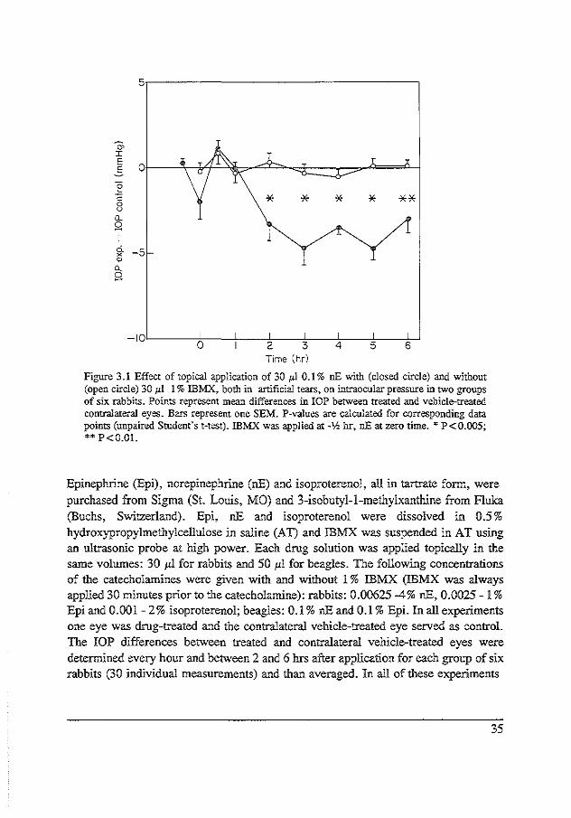
Figure 3.1 Effect of topical application of 30 p.l 0.1% nE with (closed circle) and without (open circle) 30 J.d. 1% IBMX, both in artificial tears, on intraocular pressure in two groups of six rabbits. Points represent mean differences in IOP between treated and vehicle-treated contralateral eyes. Bars represent one SEM. P-values are calculated for corresponding data points (unpaired Student's t-test). IBMX was applied at -lh hr, nEat zero time. * P<0.005; •• P<O.OI.
Epinephrine (Epi), norepinephrine (nE) and isoproterenol, all in tartrate fonn, were purchased from Sigma (St. Louis, MO) and 3-isobutyl-1-methylxanthine from fluka (Buchs, Switzerland). Epi, nE and isoproterenol were dissolved in 0.5% hydroxypropylmethylcellulose in saline (AT) and IBMX was suspended in AT using an ultrasonic probe at high power. Each drug solution was applied topically in the same volumes: 30 p.l for rabbits and 50 1'1 for beagles. The following concentrations of the catecholamines were given with and without 1 % IBMX (IBMX was always applied 30 minutes prior to the catecholamine): rabbits: 0.00625-4% nE, 0.0025- 1% Epi and 0.001-2% isoproterenol; beagles: 0.1% nE and 0.1% Epi. In all experiments one eye was drug-treated and the contralateral vehicle-treated eye served as control. The IOP differences between treated and contralateral vehicle-treated eyes were determined every hour and between 2 and 6 hrs after application for each group of six rabbits (30 individual measurements) and than averaged. In all of these experiments
35

"' I
5,-----------------------------~
~ Dt-----?<------"'<+-- ---------t
:g c 8 [L
g I c5.. -5 ~ [L
g 0 6 v
-]Qc_ __ _j_ _ __,__-;~--';--~----!c------J,,.---J 0 2 3 4 5 6
Time (hr)
Figure 3.2 Effect of topical application of30 JL] 0.2% nEat zero time with (closed circle) and without (open) 30 pl 1% IB:M::X at 2 hr on intraocular pressure in two groups of six rabbits. Points represent mean values ± SEM. P-values calculated for corresponding data points.
no significant effect on lOP was observed in the contralateral vehicle-treated eyes compared to the baseline values made on the previous day. The averaged lOP-differences (a IOP) found for the various concentrations of the catecholamines in the presence or absence of I % IBMX were fitted to a log doseresponse curve with the aid of a computer program. Non-linear regression analysis (proc. NLIN, SAS Institute Inc.) was applied, using the formula:
IOP = 1 + eA (log EC
50-log c)
where E_ is the maximum difference in lOP, EC50 the catecholamine concentration
36

that yields half of the maximum effect and A is a factor determining the slope of the curve. E_, EC50 and A are calculated parameters. In 9 groups of 8 rabbits aqueous humour protein and cyclic-AMP levels were determined 1, 3 and 5 hrs after topical application of 30 1'1 0.1% Epi or 30 !'11% IBMX or the two combined. Under local anesthesia, induced by two doses of 50 l'l of 4% topical tetracaine, 200 l'l of aqueous humour were carefully aspirated from restrained wrapped rabbits with a 27-gauge tuberculin needle, avoiding the lens and iris. The protein concentration of all aqueous humour samples was determined in duplicate in appropriate volumes acccrding to Bradford (1976), using human serum albumin as a standard. Cyclic-AMP levels were determined in !0 !'I of aqueous humour in duplicate by radioimmunoassay (New England Nuclear, Boston, MA). A non-acetylated assay condition was used. Since there was a considerable variation in aqueous humour cyclic-AMP in the individual vehicle treated control eyes and between the values of the vehicle treated control eyes of the different radioimmunoassay kits, aqueous humour cyclic-AMP was expressed as a ratio of the experimental to the control eye. (The values in untreated control eyes ranged from 2.5 p moles/ml to 41.0 p moles/mi.) The significances of the cyclic-AMP data were calculated from the means and all values are expressed ± SEM. Differences between IOP values were tested for significance with the two-tailed Student's t-test for unpaired data; aqueous humour protein and cyclic-AMP differences were tested with the two-tailed Student's t-test for paired data.
Results
Intraocular pressure
Rabbits. Baseline IOPs ranged between 20 and 25 mmHg. One percent IBMX (30 1'1 in AT) had no effect on lOP. Figure 3.1 shows the effect of30 1'1 ofO.l% nE alone and in combination with 30 1'1 of 1% IBMX applied topically 30 min prior to the start of the experiment. It is clear that the effect of nE on IOP is enhanced by prior treatment with IBMX. Thirty microliters of 1% IBMX was also applied topically 2 hr after 0.2% nE administration (Figure 3 .2). It can be seen that the hypotension induced in this case by 0.2% nE is also increased when IBMX is given after the topical nE. In Figure 3.3 a log dose-response curve for nE alone (open symbols) and nE in combination with 1% IBMX (closed symbols) is shown. The difference in IOP is obtained by averaging all differences between drug-treated and contralateral vehicle-treated eyes in the period between 2 and 6 hrs. This was done because the
37

8r--------------------------------------,
6
:'i" E 4 s
o._ Q
<I 2
-3
•
. . -2
..
~ s:
I 5' 2 2
2
. . : . . . . -I 0
log (cone 0/<;>)
Figure 3.3 Effect of several concentrations of 30 ,ul nE with (closed circle) and without (open circle) 30 JLl I% !BMX, both in hydroxypropylmethylcellulose. The ordinate represents the mean of individual differences in lOP ± SEM between drug-treated and contralateral vehicle-treated eyes in the period between 2 and 6 hrs. The abcissa represents the log concentration of nE in per cent. Asterisks indicate P-value (* P < 0.01; ** P < 0.005; unpaired Student's t-test). Several groups of six rabbits were used. No effect was noted in the contralateral vehicle-treated eyes.
primary aim of glaucoma treatment is sustained ocular hypotension and not a maximum effect at a certain point in time. TheE_ for nE was 2.9 ± 0,4 mmHg and EC50 was 0.2 ± 0.05%. The combination ofnE with 1% IBMX gave an Emu of7A ± L7 mmHg and an EC50 of0.07 ± 0.03%. Figure 3,4 shows the log dose-response curve for Epi with (closed symbols) and without (open symbols) 1% IBMX, obtained in the same way as that for nE. E_ was 7.3 ± 05 mmHg for Epi alone and 9.0 ± 0.8 mmHg for the combination. EC50 was 0.05 ± 0.01% for Epi alone and 0.02 ± 0.006% for Epi combined with 1% IBMX. Figure 3 5 shows the effects of the concentrations of isoproterenol alone (open symbols) and in combination with 1% IBMX (closed symbols). The effect of isoproterenol is enhanced when the combination is given. TheE= was 5.1 ± 0.2 mmHg for isoproterenol alone and 6.1 ± 0.3 mmHg for the combination. EC50 was 0.0033 ± 0.001% for isoproterenol and 0.0022 ± 0.001% for the combination. It is interesting to observe that low doses of isoproterenol (0. 01, 0. 025 and 0. 05%) in AT at pH 6.0 have a marked effect on IOP both when administered alone and in Combination with IBMX.
38

10
8
"' :r: E 5 5 £1_ g
4 <l
2
0 •o -3 -2 -I 0
log (cone%)
Figure 3.4 Effect of 30 ,ul of several concentrations of Epi with (closed circle) and without (open circle) 30 pl 1% !BMX both in 0.5% hydroxypropylmethylcellulose. The ordinate represents the mean of individual differences in IOP ± SEM between treated and contralateral vehicle-treated eyes in the period between 2 and 6 hrs. The abcissa represents the log concentration ofEpi in percent. Asterisks indicate P-values (*' P < 0.05; *'* P < 0.001). No consensual effect was noted in the vehicle-treated eyes. Several groups of six rabbits were used.
8
l .. J
6 l l
"' I E .5 £1_
g <l
: -2 -I 0
log {cone%)
Figure 3.5 Effect of 30 ,ul of several concentrations of isoproterenol with (open) and without (closed circle) 30 1'1 1% IBMX, both in 0.5% hydroxypropylmethylcellulose. The ordinate represents the mean of individual differences in IOP ± SEM between treated and contralateral vehicle-treated eyes in the period between 2 and 6 hrs. The abcissa represents the log concentration of isoproterenol in per cent. Significance: see Figure 3.4. No consensual effect on IOP was noted. Several groups of six rabbits were used in this experiment.
39

ci. X
"' 0.
5,-----------------------------,
8 -5
-10'----:!::-------!-----:',---~---+---+---+---_j 0 2 3 4 5 6
Time (hr)
Figure 3.6 Effect of30 p.l 0.1% nE and 0.1% Epi with and without 1% IBMX on intraocular pressure in six normo-tensive beagles. The contralateral eye was vehicle-treated. Maximum SEM was 0.9 mmHg. Determination of significance see in Results. Open circles 0.1% nE; open squares 0.1% Epi; closed circles 0.1% nE in combination with 1% IBMX; closed squares 0.1% Epi with 1% IBMX.
Beagles. IBMX alone did not affect IOP. Figure 3.6 shows that the effects of 50 ;.<I nE and Epi on IOP were significantly enhanced by 50 1'1 of 1% IBMX in normotensive beagles. The reduction in IOP in the period between 2 and 6 brs was 0.1 ± 0.3 mmHg for 0.1% nE versus 3.9 ± 0.3 mmHg for the combination (P < 0.005) and 1.3 ± 0.3 mmHg for 0.1% Epi versus 4.6 ± 0.4 mmHg for the combination (P < 0.005).
Aqueous humour protein and cyclic-AMP
One, 3 and 5 brs after 0.1 % Epi, 1 % IBMX or 0.1 % Epi combined with 1 % IBMX was applied, no significant effect on aqueous humour protein levels was observed (fable 3.1). At 1 br the aqueous humour cyclic-AMP level was slightly elevated after adntinistration of 1% IBMX and markedly elevated after the combination. At 3 and 5 hrs only the combination induced a marked increase in the cyclic-AMP content in the aqueous humour. The effect ofO.l% Epi alone at 5 brs was significant but modest (fable 3.2).
40

Table 3.1 Aqueous humour protein
Protein (g.l"')
Treated eyes Contralateral
1 hr after 30 pl 0.1% Epi I hr after 30 p.l I% !BMX I hr after 30 1-'l I% !BMX and 0.1% Epi 3 hrs after 30 p.l 0.1% Epi 3 hrs after 30 1-'l I % !BMX 3 hrs after 30 1-'1 I% !BMX and 0.1% Epi 5 hrs after 30 1-'l 0.1% Epi 5 hrs after 30 pl 1 % IBMX 5 hrs after 30 1-'l I% !BMX and 0.1% Epi
0.3 ± 0.04 0.2 ± 0.03 0.3 ± 0.08 0.4 ± 0.08 0.4 ± 0.04 0.5 ± 0.03 0.3 ± 0.06 0.3 ± 0.03 0.4 ± 0.1
control eyes
0.3 ± 0.03 0.3 ± 0.04 0.3 ± 0.08 0.4 ± 0.05 0.4 ± 0.04 0.5 ± 0.03 0.3 ± 0.06 0.3 ± 0.04 0.4 ± 0.04
Aqueous humour protein in 9 groups of 8 rabbits. All data are mean values ± SEM.
Table 3.2 Aqueous humour cyclic AMP
Time after treatment (hours)
With 30 pl 0.1% of Epi With 30 l'l of I% !BMX With the combination of 30 ;.tl of I% !BMX and 0.1% Epi
3
1.3 ± 0.3 1.6 ± 0.4 2.4 ± 0.4' 2.5 ± 1.2
5
1.3 ± 0.1' 1.0 ± 0.1
24.5 ± 3.9** 4.9 ± 1.2'* 2.0 ± 0.2'*
Aqueous humour cAMP ratio exp/contr ± SEM in 9 groups of 8 rabbits. Asterisks indicate: • p < 0.05; ** p < 0.005.
Discussion
The results presented in this study provide the first evidence, to our knowledge, that a strong phosphodiesterase/adenosine-inhibitor enhances the pressure reduction
41

produced by catecholamines in rabbit and beagle eyes. IBMX alone has no effect on IOP. It is suggested that although catecholamines are continuously released from the sympathetic nerves in the iris-ciliary body, the level of intracellular cyclic-AMP is so low that IBMX is not able to increase it to the level needed for a reduction in IOP. Combining IBMX with nE results in an increase in the nE-induced reduction in IOP, and it was shown that this increase is independent of whether IBMX is given before or after nE. The effect is most pronounced at concentrations of nE ranging from 0.025 to 0.5%. Similar results were found for Epi. The effects of Epi and IBMX are greatest when the concentrations of Epi range between 0.025 and 0.1 %. The log dose-response curves of Epi and isoproterenol reveal an enhanced maximal response after combination with IBMX. This effect of IBMX is consistent with it having an effect primarily by preventing metabolism of cAMP at the intracellular level, thereby increasing the maximal reponse. Low concentrations of isproterenol alone in AT induce a remarkable reduction in IOP, probably because the time of contact of the drug with the cornea is prolonged by hydroxypropylmethylcellulose and the pH is neutral. The maximal effect, i.e. efficacy, of nE is also enhanced if given in combination with IBMX, and it is more pronounced than that of Epi or isoproterenol. It was not expected that the effect of IBMX on nE-induced ocular hypotension would be more pronounced than that of isoproterenol. One would expect the reverse, if the effect of IBMX on catecholamine-induced hypotension was merely due to 8,-adrenoceptor-stimulated adenylyl cyclase. However, methylxanthines are also competitive inhibitors of adenosine. In several studies it was shown that some of the effects of methylxanthines, which were attributed to adenylyl cyclase, are due to inhibition of adenosine (Fredholm, 1980; Evoniuk et al, 1986). Endogenous adenosine antagonizes some of the effects of catecholarnines (e.g. by by inhibition of adenylyl cyclase), and adenosine receptor agonists have been shown to increase IOP in rabbits (Hirschfield et al, l986).In the heart adenosine production is enhanced by exogenous catecholamines (Schrader et al, 1977). If IBMX also acts as an adenosine receptor antagonist, then the depressive effect of adenosine is inhibited, which could explain why the effect of IBMX and catecholamines on IOP is greater than with catecholamines alone. The flat shape of the nE response curve (0.01-0.1 %) may reflect antagonism by adenosine which is relieved by IBMX, acting additionally as a competitive antagonist at adenosine receptors. The shift to the left which seems to be present after combination of nE with IBMX may indicate changes in drug-receptor interaction. The catecholamine levels in the aqueous humour after combination with IBMX are not known. A major role for IBMX causing inhibition of catecholamine metabolism or increased corneal penetration is not likely since a recent study on effects on ocular
42

blood flow after Epi, IBMX and combined treatment revealed an increased blood flow after combined treatment, whereas Epi alone caused vasoconstriction (Busch et a!, 1991c). An increased Epi content in the aqueous humour could not account for this change in ocular blood flow when the combination was used. In addition, in the present study changes primarily of the Em~· were observed while an increased corneal penetration or decreased catecholamine metabolism would more likely have affected the EC50 •
The role of cyclic-AMP in mediating the effect on IOP of catecholamines is debatable. The second messenger cyclic-AMP is known to mediate the effects of B-adrenergic stimulation. Cyclic-AMP given intracamerally decreases IOP in rabbits (Neufeld eta!, 1972a), and causes an increase in outflow facility (Neufeld and Sears, 1975; Neufeld et al, 1975). Furthermore aqueous humour inflow is reduced after stimulation of the adenylyl cyclase system of the ciliary body by Jl-adrenergic agonists (Gregory et a!, 1981a), cholera toxin (Gregory et al, 1981b) and forsko1in (Caprioli et al, 1984a). These studies suggest an important role of cyclic-AMP in reducing IOP by either direct or indirect stimulation of adenylyl cyclase. Several studies showed an increase in the aqueous humour cyclic-AMP level after nE (Radius and Langham, 1973) and Epi (Rowland et a!, 1979; Boas et a!, 1981). However, no direct time-relationship between the increase in aqueous humour cyclic-AMP and the reduction in IOP could be observed. It is known that Epi and nE, in the doses used in these studies, can produce an ocular hypertensive response (Norton and Vierstein, 1972; Lambie, 1973; Langham and Krieglstein, 1976; Rowland and Potter, 1980) which is a-adrenergic-induced (Rowland and Potter, 1980; Langham and Krieglstein, 1976). In rabbits the direct relation between aqueous humour cyclic-AMP and the response ofiOP may be obscured by an initial increase in the IOP when higher doses of a-adrenergic agonists are given. This is not the case in our study. Administration of 0.1% Epi does not increase the cyclic-AMP level of the aqueous humour at 1 and 3 hrs, although there is a significant reduction in IOP at these points in time. Additionally, the combination of 0.1% Epi with IBMX results in a marked elevation of the aqueous humour cyclic-AMP level at 1 hr although there is not yet a reduction in IOP; the latter does occur, however, at 3 and 5 hrs. At those points in time there is still a five and two-fold increase, respectively, of aqueous humour cyclic-AMP levels. Although aqueous humour cyclic-AMP is suggested to be only a weak extracellular parameter of what is occurring at the intracellular level in the tissues surrounding the aqueous humour, our results show that during the ocular hypotension with the combination there is a sustained increase of aqueous humour cyclic-AMP. In conclusion, the effect of IBMX on catecholamine-induced ocular hypotension may be due to enhancement of cyclic-AMP induced events by inhibition of the enzymatic
43

degradation, to antagoruzmg adenosine receptors, or to both acting together. Summarizing: this study shows that IBMX significantly enhances the reduction in IOP caused by catecholamines in rabbits and beagles. This finding may be of value in the treatment of glaucoma.
44

CHAPTER4
SOLUBLE FORMS OF ISOBUTYLMETHYLXANTHINE ENHANCE Tiffi OCULAR HYPOTENSION INDUCED BY
CATECHOLAMINES
Michie/ JWM Busch, and Philip FJ Hoyng; published in Graefe's Arch Clin Exp Ophthalmol 229, 583-586, 1991
Abstract
Isobutylmethylxanthine (IBMX) suspended in 0.5% hydroxypropyl methylcellulose has been shown to enhance the reduction in intraocular pressure (lOP) induced by epinephrine and norepinephrine in rabbits and beagles. In the present study, the effects of two soluble forms of IBMX, i.e. the ethylenediamine salt (IBMX-ED) and IBMX bound to cyclodextrin in saline (IBMX -CD), were studied in rabbits. When used alone, l% IBMX-ED did not affect the lOP, but the hypotensive response to 0.1% epinephrine was enhanced dose-dependently by combination of the catecholamine with IBMX-ED. Although l% IBMX-CD applied alone also failed to influence the lOP, the hypotensive response to epinephrine was enhanced by combination of these substances with IBMX-CD. Soluble IBMX-ED as well as soluble IBMX-CD and the IBMX suspension enhanced, to a similar extent and in a des-dependent manner, the IOP reduction induced by catecholamines. IBMX-CD, which has a neutral pH, may be a suitable form of IBMX for future long-term experiments.
Introduction
Epinephrine and dipivalyl epinephrine are important in the management of ocular hypertension and primary open-angle glaucoma. The ability of epinephrine (Becker and Morton, 1966; Becker et al, 1961; Harris et al, 1970; and Obstbaum et al, 1974)
45

and dipivalyl epinephrine (Kass et al, 1979; Kohn et al, 1979; Krieglestein and Leydhecker, 1978) to lower IOP during long-term treatment has been demonstrated conclusively. In recent studies (Busch et al, 1991c; Hoyng et al, 1991/chapter 3) it was shown that isobutylmethylxanthine (IBMX), a phosphodieterase inhibitor/adenosine receptor antagonist (Daly, 1985; Fredholm, 1980; Rail, 1985), markedly enhanced the reduction of IOP induced by topical epinephrine, norepinephrine and isoproterenol in rabbits and beagles. IBMX alone did not affect the IOP. Thus, the results suggest that an interaction between IBMX and catecholarnines reduces IOP, indicating that in single-drop experiments, lower doses of the catecholamines can induce marked ocular hypotension when combined with IBMX. Long-term experiments are hampered by the instability of the IBMX suspension. Therefore, two soluble forms ofiBMX, i.e. the ethylenediamine salt and IBMX bound to cyclodextrin in the form of inclusion complexes, were developed and their ability to enhance epinephrine- and norepinephrine-induced ocular hypotension in rabbits was tested and compared with the effect of the IBMX suspension. In addition, the effect of IBMX on the IOP reduction induced by dipivalyl epinephrine was studied.
Materials and methods
All studies were performed in the animal laboratory under standard light and atmospheric conditions using conscious adult pigmented rabbits (2.5-4 kg) restrained in cloth wrappers. The cornea was anaesthetized with 30 1'1 0.2% oxybuprocaine (Novesine, Chibret, Riom, France), and the IOP was measured with an Alcon pneumatonometer. The pneumatonometer was manometrically calibrated for rabbit eyes and the digital readings were checked with an Alcon calibrator before and after each experiment. Three different forms of methylxanthine were tested: 3-isobutyl-1-methylxanthine (Fiuka, Buchs, Switzerland) suspended in 0.5% hydroxypropyl methylcellulose in saline (AT) using an ultrasonic probe at high power, pH 6 (IBMX-AT); 3-isobutyl-1-methylxanthine-ethylenediamine in saline, pH 9.7 (IBMX-ED); and 3-isobutyl-1-methylxanthine dissolved in saline containing 2% betacyclodextrin, pH 7 (IBMX-CD). The suspension of IBMX in AT was instilled in rabbit eyes within 15 min of its preparation. Epinephrine and norepinephrine (Sigma, St. Louis Mo.) were dissolved in AT and dipivalyl epinephrine eyedrops (PropineR, Allergan, Irvine, Calif.) were diluted in saline; the catecholamine solutions were freschly prepared every day. The drug was applied topically to one eye in a volume of 30 JLI; the contralateral
46

control eye received the appropriate vehicle. IBMX-AT, IBMX -ED or IBMX -CD was applied at 30 min prior to catecholamine administration; a possible effect of cyclodextrin was tested by comparing the effect of 0.1% epinephrine dissolved in saline with that of 0.1% epinephrine dissolved in saline containing 2% betacyclodextrin. The same group of seven rabbits were used for an experiment carried out with and without an IBMX test substance; a 2-week interval between studies was established to ensure adequate washout. The effect on lOP is expressed as the mean difference between treated and contralateral control eyes ± the standard error of the mean (LlJOP ± SEM). The sustained ocular hypotension at 2-6 h after drug administration was calculated by averaging the differences in IOP ± the standard error of the mean (Ll.IOP 2-6 h ± SEM). Statistical evaluation was done using Student's t-test for group means, whereby P < 0. 05 was considered to indicate statistical significance.
Results
Baseline IOP values ranged between 20 and 25 mmHg; an effect on the contralateral control eye was not observed. When used alone, 1% IBMX-ED did not affect the IOP
5 ®
0.
~ "-Q
o 1% IBMX-ED o 0.1% EP! • 1% IBMX-ED + 0.1% EP! A 1% IBMX-AT + 0.1% EPI
c 0.1% EPI o 0.1% IBMX-ED + 0.1% EP! c 0.25% !BMX-ED + 0.1% EPI A 0.5% I BMX-ED + 0.1% EPI 111 1% IBMX-ED + 0.1% EPI
·10f-~~~-~-~-~-~-~ -1 0 2 3 4 5 6 -1 0 2 3 4 5 6
hours hours Fig. 4.1 A Effect of topical 1% !BMX-ED and 0.1% epinephrine (EPI) alone as well as topical 0.1% epinephrine in combination with either 1% IB:MX-ED or 1% IBMX suspended in 0.5% hydroxypropylmethylcellulose (!BMX-AT) on the lOP in 7 rabbits. B Effect of topically applied 0.1% epinephrine alone (EPI) and 0.1% epinephrine in combination with 0.1 %, 0.25% and 1% topical !BMX-ED on the lOP in 7 rabbits. The effect on lOP is expressed as the mean difference between treated and contralateral control eyes (lOP exp- lOP control, in mmHg); statistically significant differences between epinephrine alone and epinephrine in combination with IBMX-ED or IBMX-AT are indicated by asterisks. * P<O.OS
47

a. ~ 0.. 0 --10
-1
05 15 :r: E .s .c "' 10
"' 0.. Q <I
5
0
-2
o 1% lBMX-CD .D. 0.05% EPI CJ 0.1% EPI A 1% lBMX-CD + 0.05% EPI a 1% IBMX-CD + 0.1% EPI
@ ~ 0.05% EPI A 0.1% IBMX-CD + 0.05% EPI 111 0.5% IBMX-CD + 0.05% EP! o 1% lBMX-CD + 0.05% EPl
2 3 4 5 6 -1 0 2 3 4 5 6 hours
o 1% IBMX-CD + EPI
o EPl
* *
-1 log (cone%)
hours
Fig. 4.2 A Effect of topical I% IBMXCD alone, 0.05% and 0.1% epinephrine (EPI) alone and the combination of topical I% IBMX-CD with 0.05% and 0.1% epinephrine on the lOP in 7 rabbits. B Effect of 0.01 %, 0.02%, 0.05% and 0.1% topical epinephrine alone and in combination with topical l% IBMX-CD on the mean (±SEM, bars) reduction in lOP at 2-<5 hr (L>!OP 2-6 hr~ in mmHg) after drug administration. C Effect of 0.05% topical epinephrine alone and in combination with 0.1 %, 0.5% and 1% topical IBMX-CD on the lOP in 7 rabbits.* P<0.05
(Fig. 4.1 A), but it enhanced the ocular hypotension induced by 0.1% epinephrine to the same extent as did 1% IBMX-AT (Fig. 4.1 A). Application of 1% and 0.5% IBMX-ED increased the mean IOP reduction observed at 2-6 hr after the administration of0.1% epinephrine by 3.6 ± 0.6 and 3.4 ± 0.6 mmHg, respectively (P<0.05). The effect ofiBMX-ED on 0.1% epinephrine-induced ocular hypotension was dose-dependent (Fig. 4.1 B). When applied alone, 1% IBMX-CD had no effect on the IOP (Fig. 4.2A); 0.1% epinephrine dissolved in saline reduced the IOP to the same extent as did 0.1% epinephrine dissolved in saline containing 2% beta-cyclodextrin (data not shown). IBMX -CD enhanced the ocular hypotension induced by 0. 01 %-0.1 % epinephrine (Fig. 4.2 A, B), with the mean IOP reduction at 2-6 induced by 0.01 %, 0.02%, 0.05% and
48

0.1% epinephrine being increased by 2.1 ± 0.4, 3.2 ± 0.8, 5.6 ± 0.6 and 5 ± 0.7 mmHg, respectively (all, P<O.OOl). The dose dependence of this effect is shown in Fig. 4.2C. In addition, 1% IBMX-CD enhanced the lOP reduction iduced by 0.05% and 0.1% dipivalyl epinephrine (Fig. 4.3A) by 3.3. ± 0.6 and 3.9 ± 0.8 mmHg, respectively (P<0.05); when this test substance was combined with 0.1% norepinephrine, the mean enhancement of this effect at 2-6 h after catecholamine administration was 1.6 ± 0.5 mmHg (P<0.05, Fig. 4.3B).
o; I
5
E 5 2 0 c 0 u
D.. Q
·5 0. X
"' D.. 0
® o 0.05% OPE o 0.1%DPE = 1% IBMX-CD + 0.05% OPE • 1% IBMX-CD + 0.1% OPE
@ T o 0.1% NE o 0.5% NE II 1% lBMX-CD + 0.1% NE • 1% IBMX-CD + 0.5% NE
* --10
-1 0 2 4 5 6 -1 0 2 3 4 5 6 hours hours
Fig. 4.3 A Effect of 0.05% and 0.1% topical dipivalyl epinephrine (DPE) alone and in combination with 1% topical IBMX CD on the lOP in 7 rabbits. B Effect of 0.1% and 0.5% topical norepinephrine (NE) alone and in combination with 1% topical IBMX-CD on the IOP in 7 rabbits.* P<0.05.
Discussion
In a previous study (Hoyng et al, 1991), it was demonstrated by means of doseresponse curves that the ocular hypotension induced by 0.002%-1% epinephrine and 0.005%-1% norepinephrine was enhanced by combination of these catecholamines with a suspension of IBMX in 0.5% hydroxypropyl methylcellulose. The present investigation showed that IBMX-ED and IBMX-CD have a similar effect on epinephrine- and norepinephrine-induced ocular hypotension. The effect reached a maximum at 3 h after drug administration and the IOP reduction was sustained at a low level. Dose responsiveness was demonstrated for both IBMX-ED and IBMX-CD.
49

The formation of ethylenediamine salts of methylxanthines is a well-known method for improving the aqueous solubility of methylxantines (Rail, 1985). Cyclodextrins in general are used to enhance the solubility and/or stability of a variety of compounds through the formation of inclusion complexes (Cohen and Lach, 1963; Lach and Chin, 1964; Szejtli, 1982). Both soluble derivatives of IBMX seem to be suitable for longterm experiments to evaluate tachyphylaxis and tolerance, since IBMX-ED as well as IBMX in a cyclodextrin solution yield stable and soluble forms. In contrast, IBMX suspended in AT rapidly precipitates within 5-10 min. It can be argued that IBMX and/or cyclodextrin may enhance corneal penetration or decrease the metabolism of catecholamines. However, the addition of cyclodextrin (2%) to the 0.1% epinephrine solution did not alter the pressure response induced by 0.1% epinephrine alone. Therefore, it is conceivable that the effect of IBMX-CD in combination with catecholamines is attributable to IBMX itself. Moreover, physiological experiments on regional ocular blood flow using the radioactively labelled microsphere technique (Busch eta!, l99lc/chapter 5) showed that epinephrine alone induced slight vasodilation, whereas the combination of IBMX-CD and epinephrine induced a strong and long-lasting increase in ocular blood flow in the iris and ciliary processes. Increased corneal penetration or decreased metabolism of catecholamines cannot explain this qualitative change in response. Furthermore, doseresponse curves illustrating the pressure response after the administration of epinephrine and norepinephrine showed an increased maximal effect (E...J rather than a shift to the left following combination of these catecholamines with the 1% IBMX suspension (hoyng eta!, 1991). Finally, a pronounced and consistent increase in the initial pressure response was not demonstrated in the present study, whereas high doses of epinephrine, dipivalyl epinephrine and norepinephrine are known to induce marked initial hypertensive responses. The experiments carried out with and without IBMX always used the same group of rabbits, since variations occur in the response to catecholamines were noted among different groups of rabbits; a strong hypotensive response to 0.1% and 0.05% epinephrine (Fig. 4.2A) was found in the group treated with IBMX-CD as compared with the weaker response to 0.1% epinephrine seen in the group treated with IBMXED (Fig. 4.1A). However, when the effects ofiBMX-ED and IBMX-CD were studied using equieffective doses of catecholamines, they proved to be very similar. The effect of IBMX-CD in combination with norepinephrine was less pronounced. However, 0.5% norepinephrine induced an initial hypertensive response that was responsible for the late onset of the reduction in IOP, clearly present at 6 hr after catecholamine administration. Dipivalyl epinephrine (DPE) is widely used for the treatment of glaucoma. This prodrug of epinephrine is known to facilitate the corneal penetration of this
50

catecholamine. It is interesting that IBMX-CD also increased the ocular hypotensive response to DPE. IBMX-CD was studied more extensively than IBMX-ED; the pH of the solutions was 7 and 9.7, respectively; the IBMX-AT suspension had a pH of6. Ophthalmic solutions with a buffered pH between 7 and 9 can generally be applied without causing pain in humans. For this reason, IBMX-CD should preferably be used in further experiments. We conclude that a suspension of IBMX in 0.5% hydroxypropyl methylcellulose as well as IBMX-ED and IBMX-CD similarly enhance the reduction in IOP induced by catecholamines, the effect being maximal at 2-6 h after drug administration. In our opinion, IBMX-CD, which has a neutral pH and good solubility, is the preferred form of IBMX for further research on the mechanism(s) of its interaction with catecholamines in reducing IOP, and it may be of value in the treatment of glaucoma.
51


CHAPTERS
INCREASE IN OCULAR BLOOD FLOW INDUCED BY ISOBUTYLMETHYLXMlHINE AND EPINEPHRINE
Michie/ JWM Buscll·2, Johan Stjenschantz!, and Philip FJ Hoyn:f; published in
Exp Eye Res 52, 199-204, 1991
1 Glaucoma Research Laboratory, Pharmacia AB, Uppsala, Sweden 2 Department of Experimental Ophthalmology, Netherlands Ophthalmic Research
Institute, Amsterdam
Abstract
Alterations in regional blood flow of the eye were studied in rabbits using the radioactively labelled microsphere technique. The animals were topically treated with I% ffiMX, 0.1% epinephrine in combination with 1% ffiMX or with 0. 1% epinephrine only, After ffiMX there was a tendency towards an increase in blood flow in the iris, the ciliary processes and the sclera whereas the choroidal blood flow tended to decrease. After epinephrine the iridial blood flow increased about 50% at 6 hours. After ffiMX combined with epinephrine a biphasic response was obtained; an initial decrease in the blood flow of the iris, the ciliary processes and to some extent the choroid was followed by a marked and long lasting increase in blood flow from 2.5 up to 75 hours in the iris (up to 220%), the ciliary processes (123%) and the sclera (115%). The choroidal blood flow was not increased. The marked increase in blood flow of the iris and the ciliary processes indicates that the reduction of intraocular pressure seen after topical treatment with a combination of epinephrine and ffiMX, is not based on a vascular mechanism. The increase of ocular blood flow after a combination of ffiMX and epinephrine is probably based on increased cAMP secondary to inhibition of phosphodiesterases. This may indicate the presence of£adrenergic receptors in the ocular vasculature.
53

Introduction
Topical epinephrine is widely used to reduce intraocular pressure (lOP) in the treatment of glaucoma. A study of the combination of 3-isobutyl-1-methylxanthine (IBMX) and epinephrine in rabbits demonstrated that topically applied IBMX, a phosphodiesterase-inhibitor, enhanced the ocular hypotension induced by epinephrine (Hoyng and Groeneboer, 1987/chapter 3). IBMX alone did not affect IOP, but the ocular hypotensive effect of norepinephrine, dipivalylepinephrine and isoproterenol was also enhanced by prior treatment with IBMX. Thus, an interaction between catecholamines and IBMX to reduce intraocular pressure was shown. The purpose of the present study was to investigate whether the epinephrine induced reduction ofiOP, enhanced by IBMX, is based on a vascular mechanism. In addition, since very little is known about methylxanthines in the eye in general, it was thought to be of interest to study the vascular effect of IBMX in the eye.
Materials and methods
Albino rabbits of either sex, weighing 2.1-4.5 kg, were used. The animals were anaesthetized with sodium pentobarbital administered intravenously, 40-60 mg/kg b. w. Additional doses of the same anaesthetic were administered when necessary to maintain adequate anaesthesia. The animals were tracheotomized, and both the femoral arteries were cannulated, one for blood sampling and the other for blood pressure registration. The arterial blood pressure was continuously recorded during the entire experiment with a PDCR 75/1 pressure transducer (Druck Ltd) connected to a recorder (BBC, model SE 460). In two animals the blood pressure was sustained by giving 15-30 ml 3% macrodex (Pharmacia, Uppsala) intravenously. The pH, Pea, and Po, of arterial blood were analyzed in an ABL300 (Radiometer, Copenhagen) acid-base analyzer. When necessary the animal was ventilated artificially. Ventilation was adjusted to give normal values of arterial Po,, Pea, and pH. If the animal suffered from marked metabolic acidosis 5-l 0 ml 5% sodium bicarbonate was administered intravenously. A heating pad was used to keep the animals at normal body temperature. A catheter, for the microsphere injections, was inserted into the left heart ventricle through the left brachial artery. The position of the tip of the catheter was controlled by recording the intraventricular blood pressure. All cannulas contained heparinized isotonic saline to prevent clotting. Regional blood flow was determined with the reference flow method (Aim and Bill,
54

1972; S\jemschantz et al, 1976). Radioactively labelled microspheres with a size of 15 p.m were obtained from New England Nuclear (Boston, Mass.). By using differently labelled microspheres it was possible to determine the blood flow at different stages of the experiment in each animal. The microspheres were labelled with 141-Ce, 113-Sn, 103-Ru or 95-Nb. Each injection contained l-2.106 microspheres, suspended in 2 ml isotonic saline. The microspheres were injected within 5-10 sec. into the left heart ventricle and from the start of the injection blood samples were collected from the femoral artery during the following 60 sec. In each animal three measurements were performed. The times of measurements were randomized between 0 and 7.5 hours after administration of the test substance. The time interval between the first and last injection was usually less than three hours. After the third sphere injection the animal was sacrificed with an overdose of pentobarbital, the eyes were enucleated and dissected and lung samples were collected as a control of microsphere recirculation. The eye was opened from the posterior pole, and the different ocular tissues were collected. The ciliary and iridial processes were scraped from the back of the iris-ciliary body preparation. Tne iris preparation thus contained some of the ciliary body and remnants of the ciliary and iridial processes. The radioactivity of the blood and tissue samples was determined in a multi-channel gamma spectrometer (1282 Compugamma Wallac LKB-Pharmacia). The blood flow in the different tissues of the eye was calculated by dividing the radioactivity of the tissue sample with that of the reference blood sample obtained during a one minute period, multiplied with the weight of the blood sample. The resulting blood flow was expressed as mg/min/tissue. Three different drug regimens were tested; in group l consisting of l3 animals only l% IBMX was tested; in group 2 consisting of 14 animals the combination of 1% IBMX and 0.1% epinephrine was tested. IBMX was administered 30 min prior to epinephrine and the time was set to zero at the IBMX administration; in group 3 consisting of 6 animals 0.1% epinephrine was tested. One eye, randomly left or right, was treated and the contralateral eye received only the vehicle. In group 3 with administration of epinephrine only, the IBMX vehicle was administrated 30 min prior to epinephrine in both eyes. IBMX (Fluka, Buchs, Switzerland) was dissolved in a phosphate buffered solution containing 2% betacyclodextrin (Avebe, Groningen, the Netherlands). Epinephrine eye drops (l %) (EppyR, Pharmacia, Uppsala, Sweden) were freshly diluted with isotonic saline to 0.1%. All test substances were applied topically in a volume of 30 p.l.
The results are expressed as the arithmetical mean value ± standard error of mean (Mean ± SEM). The change in blood flow i.e. differences between the experimental eye and contralateral control eye was expressed as percentage of the blood flow in the
55
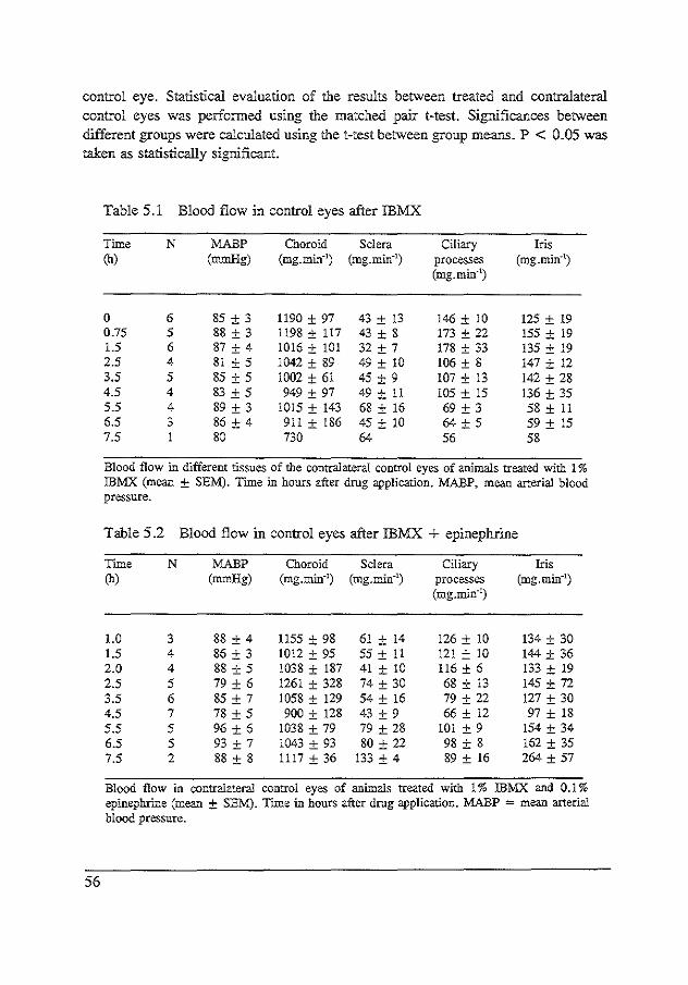
control eye. Statistical evaluation of t.lJe results between treated and contralateral control eyes was performed using the matched pair t-test. Significances between different groups were calculated using the !-test between group means. P < 0.05 was taken as statistically significant.
Table 5.1 Blood flow in control eyes after lBMX
Time N MABP Choroid Sclera Ciliary Iris (h) (mmHg) (mg.min-1
) (mg.min-1) processes (mg.min·')
(mg. min"')
0 6 85 ± 3 1190 ± 97 43 ± 13 146 ± 10 125 ± 19 0.75 5 88 ± 3 1198 ± !17 43 ± 8 173 ± 22 155 ± 19 1.5 6 87 ± 4 1016 ± 101 32 ± 7 178 ± 33 135 ± 19 2.5 4 81 ± 5 1042 ± 89 49 ± 10 106 ± 8 147 ± 12 3.5 5 85 ± 5 1002 ± 61 45 ± 9 107 ± 13 142 ± 28 4.5 4 83 ± 5 949 ± 97 49 ± II 105 ± 15 136 ± 35 5.5 4 89 ± 3 1015 ± 143 68 ± 16 69 ± 3 58± !! 6.5 3 86 ± 4 9!1 ± 186 45 ± 10 64 ± 5 59± 15 7.5 1 80 730 64 56 58
Blood flow in different tissues of the contralateral control eyes of animals treated with 1% IBMX (mean ± SEM). Time in hours after drug application. MABP, mean arterial. blood pressure.
Table 5.2 Blood flow in control eyes after lBMX + epinephrine
Time N MABP Choroid Sclera Ciliary Iris (h) (mmHg) (mg.rnin-1) (mg.min·') processes (mg.rnin-1
)
(mg.mi.n-1)
!.0 3 88 ± 4 1155 ± 98 61 ± 14 126 ± 10 134 ± 30 1.5 4 86 ± 3 1012 ± 95 55± II 121 ± 10 144 ± 36 2.0 4 88 ± 5 1038 ± 187 41 ± !0 !16 ± 6 133 ± 19 2.5 5 79 ± 6 1261 ± 328 74 ± 30 68 ± 13 145 ± 72 3.5 6 85 ± 7 1058 ± 129 54± 16 79 ± 22 127 ± 30 4.5 7 78 ± 5 900 ± 128 43 ± 9 66 ± 12 97 ± 18 5.5 5 96 ± 6 1038 ± 79 79 ± 28 101 ± 9 !54± 34 6.5 5 93 ± 7 1043 ± 93 80 ± 22 98 ± 8 162 ± 35 7.5 2 88 ± 8 1117 ± 36 133 ± 4 89 ± 16 264 ±57
Blood flow in contralateral control eyes of animals treated with 1% IBMX and 0.1% epinephrine (mean ± SEM). Time in hours after drug application. MABP = mean arterial blood pressure.
56

Results
The blood flow values in the different tissues of the contralateral control eyes in the IBMX, IBMX-epinephrine and epinephrine groups are presented in Tables 5.1, 5.2 and 5.3, respectively. The percentual changes in blood flow of the iris, the ciliary processes, the sclera and the choroid in the experimental eyes compared with the contralateral control eyes are presented in the Figures 5.1, 5 .2, 5.3 and 5 .4. IBMX alone showed a tendency towards increased blood flow in the ciliary processes, the iris and the sclera. Pooled data from the increased level at 1.5-4.5 hours showed a significant change of 63 ± !9% (P < 0.005) in the iris, 24 ± 8% (P < 0.01) in the ciliary processes and 50 ± 18% (P < 0.05) in the sclera. After 4.5 hours no effect was seen. On the contrary the choroidal blood flow tended to decrease slightly. Combined treatment with IBMX and epinephrine induced an initial decrease in blood flow, followed by a marked increase ra11ging from 3.5 to 7.5 hours in the iris, the ciliary processes and the sclera. The maximal increase was 220 ± 50% (P < 0.05), 123 ± 32% (P < 0.01) and 1!5 ± 29% (P < 0.01), respectively. The choroidal blood flow was only slightly affected with a small, but statistically significant, decrease at 1.5 hour. The blood flow a.c4:er epinephrine alone was studied at 4.5, 5.5 and 6.5 hours after administration of the drug. During this time period there was only an increase in the iridial blood flow which was 50 ± l3% (P < 0.05) 6.5 hours after administration of epinephrine.
Table 5.3 Blood flow in control eyes after epinephrine
Time N MABP Choroid Sclera Ciliary Iris (h) (mmHg) (mg. min·') (mg.min-1) processes (mg.min-1
)
(mg.min-1)
4 6 93 ± 2 1048 ± 140 61 ± 13 92 ± 9 97 ± 20 5 6 91 ± 2 885 ± 106 52± 12 92 ± 5 82 ± 18 6 6 88 ± 2 842 ± 131 43 ± 10 93 ± 10 91 ± 21
Blood flow hi contralateral control eyes of animals treated with 0.1% epinephrine (mean ± SEM). Time in hours after drug application. MABP, mean arterial blood pressure.
57

58
300 ~ IBMX +epinephrine * IRIS 250
,_...., IBMX * " epinephrine "' --"' ~ 200 " * .!: ;, 150 ,g -o 100 0 .9. .0 50 <fl.
0
-50
0 2 4 6 8 hours
Figure 5.1 Effect of 1% IB:MX~ 0.1% epinephrine, and the combination of 1% IB11X and 0.1% epinephrine on blood flow in the iris. The change in blood flow is expressed as percentage of the blood flow in the contralateral control eye (mean ± SEM) * P < 0.05, ** P < 0.0!. Significantly different effects between 0.1% epinephrine or 1% !BMX and the combination of 1% IBMX and 0.1% epinephrine are indicated in the former curves with open symbols (P < 0.05). The increase in blood flow after combination of IBMX and epinephrine was statistically significantly different from that of IBMX alone at various points in time as seen in Figures 1~3 ; the difference compared to epinephrine alone was also statistically significant in the iris, the ciliary processes and the sclera (Figures 1-3).
250 .,_.. lBMX +epinephrine CILIARY PROCESSES - IBMX
" 200 ---... epinephrine "' "' ** ~ 150 " .s
* ;, 100 ,g -o 50 0 0 :0 0 <f.
-50
0 2 4 6 8 hours
Figure 5.2 Effect on blood flow in the ciliary processes of 1% IBMX~ 0.1% epinephrine and the combination of both. For legends see Figure 5.1.
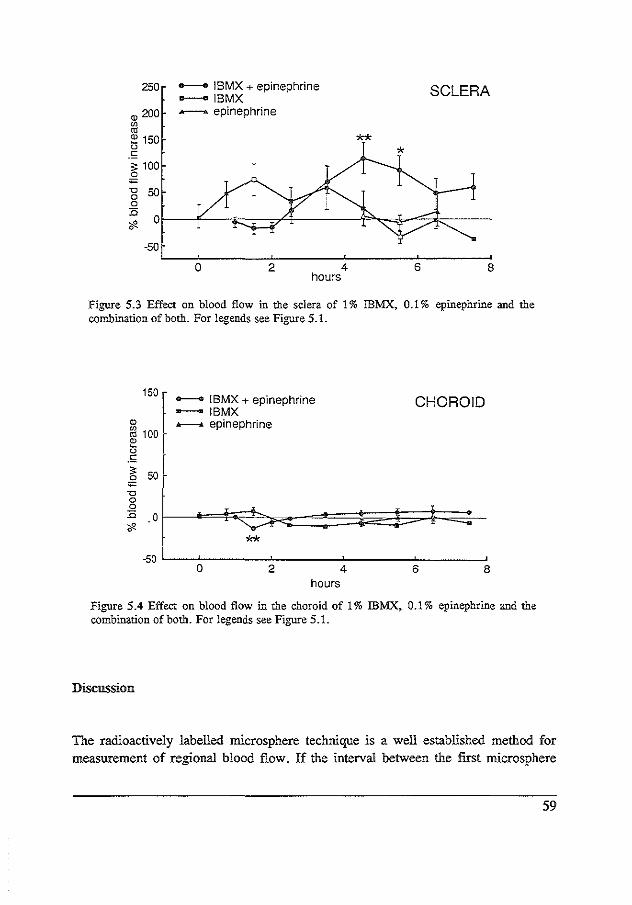
250
<ll 200
"' "' 1: 150 " .s ;: 100 0
;
-gsa 0 :0 <f.
-50
___. IBMX +epinephrine -IBMX - epinephrine
0 2 4 hours
SCLERA
**
6 8
Figure 5.3 Effect on blood flow in the sclera of 1% IBMX~ 0.1% epinephrine and the combination of both. For legends see Figure 5.1.
150 --. tBMX +epinephrine - IBMX
CHOROID <ll Jr---..-£ epinephrine ~ 100 1: " .s ;:
50 0 ;
"' 0 0 :0 .0 <f.
** -50
0 2 4 6 8 hours
Figure 5.4 Effect on blood flow in the choroid of 1% IBMX~ 0.1% epinephrine and the combination of both. For legends see Figure 5.1.
Discussion
The radioactively labelled microsphere technique is a well established method for measurement of regional blood flow. If the interval between the first microsphere
59

injection and the third microsphere injection is long, a recirculation of microspheres from the peripheral tissues could theoretically occur. Therefore, lung samples were obtained from each animal and analysis of these confirmed that practically no recirculation had occurred, when comparing the values obtained with the first and the last microsphere injection. This indicates that a three hour time interval between the first and last microsphere injection was acceptable. Moreover, any recirculation can be expected to affect both eyes to a similar extent and should therefore not influence the results with respect to differences in blood flow between the eyes. In the rabbit the retinal circulation is very poorly developed. Therefore, the blood flow of the retina was not determined. The main objective of this study was to determine whether changes in the haemodynamics could explain the IBMX-enhanced reduction of intraocular pressure compared to epinephrine alone. Epinephrine is known to induce marked vasoconstriction in the anterior uvea of monkeys (Aim, 1980; Caprioli eta!, 1984) and rabbits (Morgan et al, 1981), and a marked vasoconstriction would tend to lower the lOP. Vasodilation, however, cannot reduce the IOP. Indeed, 1% IBMX alone induces a moderate increase in blood flow but is not capable of lowering the lOP (Hoyng and Groeneboer, 1987). The moderate decrease and following increase in blood flow of the anterior uvea after IBMX in combination with epinephrine indicate that changes in haemodynamics do not play a causative role in the IBMX enhanced reduction of the IOP to epinephrine. Control experiments with indomethacin pretreated rabbits (not included in Results) showed that the vasodilatory response of epinephrine and IBMX could not be prevented by cyclooxygenase inhibition, thus making an involvement of prostaglandins in this response uulikely. Increased uveal blood flow concomitant with a reduction in the IOP may be explained on the basis of an increased perfusion .pressure (Bill, 1975), but cannot account for the marked increase in blood flow seen in the iris and the ciliary processes since similar and significant choroidal vasodilation is not observed. Epinephrine has been shown to induce vasodilation in the anterior uvea of rabbits after an initial vasoconstriction (Morgon et al, 1981). A 60 times higher dose of epinephrine was used however compared with the present study, and a 2.5 fold increase in blood flow was measured only in the iris, 3 hours after administration. In the group treated with 0.1% epinephrine alone in this study a moderate vasodilation was seen at 6 hrs in the iris only. Early measurements were not performed in the epinephrine group since the vasoconstrictive response has been well documented. It is most likely that the initial decrease in blood flow after IBMX combined with epinephrine was due to the effect of epinephrine. Analysis of the results shows that the vasodilation after IBMX alone was not statistically sigrrificant at any individual time point, due to a fairly large variation in results and the small number of animals tested. Some animals
60

showed a marked vasodilation, some responded less. The tendency, however, of IBMX to induce vasodilation in the eye is demonstrated. In contrast, all animals treated with IBMX combined with epinephrine responded clearly with vasodilation, 3.5 up to 7.5 hours after treatment. Thus, it appears to be a very constant phenomenon. The marked increase in blood flow after IBMX with epinephrine cannot be explained on the basis of an additive effect of IBMX to epinephrine because the individual treatment responses to IBMX alone and epinephrine alone are too weak and the onset of the vasodilation after the combination is too early. Except for the cerebral circulation, the effect of methylxanthines on the peripheral vasculature generally is vasodilation (Rail, 1982; Rutherford et a!, 1981). Thus, methylxanthines seem to have a similar effect in the anterior uvea of the eye, although relatively weak. Methylxanthines are known to have two major cellular effects. They inhibit intracellular phosphodiesterases and they antagonize adenosine receptors (Rail, 1982; Rail, 1985). The effect of adenosine on the peripheral vasculature is vasodilation (Rail, 1982; Daly, 1985) and data supporting a similar effect in the eye have recently been presented (Braunagel et a!, 1988; Campochiaro and Sen, 1989). 8-Phenyltheophylline, an adenosine receptor antagonist, blocked vasodilation in the anterior uvea after combined treatment with adenosine and dipyridamol applied intravitreally. Dipyrimadol is an adenosine-re-uptake inhibitor (Braunagel et al, 1988). Thus, it seems less likely that the mechanism responsible for the increased blood flow in the anterior uvea after IBMX and IBMX in combination with epinephrine is based on antagonism of adenosine receptors. One would therefore be inclined to interpret the mechanism of IBMX-epinephrine induced blood flow increase as being based on an adenylate cyclase mediated process. Furthermore, we have found that the cAMP content in the aqueous humor after topical treatment with IBMX and epinephrine is markedly increased compared to IBMX and epinephrine alone (unpublished data). Our data suggest potentiation of a beta adrenergic vasodilation by IBMX. So far, however, evidence for beta adrenergic receptors in the intraocular vasculature is very sparse (Bill, 1975; Nathanson, 1981; Trope, 1982; Ferrari-Dileo, 1988; Martin and Rabineau, !989; Denis, 1989) and a systematic study to investigate this aspect seems warranted. The slight increase in the choroidal blood flow seen after combination of IBMX and epinephrine may be explained by a reduction in the lOP resulting in an increased perfusion pressure. A reduction in the IOP of 3-4 mmHg can be anticipated to increase the choroidal blood flow with 5-10% since autoregulation of blood flow is essentially lacking in the choroid (Bill, 1975). The cause of the tendency towards a slight decrease in the choroidal blood flow after IBMX alone remains unclear. One may speculate that a contributing factor could be shunting of blood from the anterior part of the choroid to the anterior uvea. In fact, a 30-60% increase in the anterior uveal
61

blood flow could be anticipated to reduce total choroidal blood flow with around 5-10%. In conclusion, the results of the present study indicate that the epinephrine induced reduction of the lOP, enbanced by IBMX, is not based on a vascular mechanism. The methylxanthine IBMX in combination with epinephrine induces a strong and long lasting increase in blood flow of the iris, the ciliary processes and the sclera, a phenomenon that best can be characterized as a potentiation probably based on increased cAMP due to inbibition of phosphodiesterases.
62

CHAPTER6
AQUEOUS HUMOUR DYNAMICS AFTER TOPICAL ISOBUTYLMETHYLXANTHINE AND EPINEPHRINE IN RABBITS
Michie! JWM Busclt, Judith v Everdingen2, Jaap v Besf, and Philip FJ Hoynfl;
submitted
1 Department of Experimental Ophthalmology, The Netherlands Ophthalmic Research Institute, Amsterdam, The Netherlands.
2 Laboratory of the department of Ophthalmology, University Hospital, Leiden, The Netherlands.
Abstract
Combination drug treatment of topical isobutylmethylxanthine (IBMX), a phosphodiesterase inhibitor, with topical-epinephrine has demonstrated that IBMX enhances the ocular hypotensive response induced by epinephrine in rabbits. The present study investigates the site of action for this effect of IBMX by measuring outflow facility by indentation tonography and aqueous humour production by fluorophotometry. It was observed that topical 1% IBMX, l% epinephrine, and 0.05% dipivalyl epinephrine alone did not individually affect outflow facility; however, combined treatment of l% IBMX with 1% epinephrine, and 1% IBMX with 0.05% dipivalyl epinephrine increased outflow facility by 43% (P < 0.01) and 32% (P < 0.05), respectively. Topical 0.05% dipivalyl epinephrine alone induced a reduction of flow between 3 and 6 hours after medication, from 3.0 ± 0.3 to 2.6 ± 0.4 pl.min·1 (P < 0.05). Flow after 1% IBMX in combination with 0.05% dipivalyl epinephrine was 3.2 ± 0.1 I'Lmin·'. This was not different from baseline flow but higher than after dipivalyl epinephrine alone (P < 0.05).
63

It is concluded that IBMX potentiates the ocular hypotensive effect of epinephrine by increasing the effect of epinephrine on outflow facility and not by decreasing aqueous humour flow. These in vivo findings lend support to a role for adenylyl cyclase in controlling outfiow facility in rabbits, particularly in response to epinephrine.
Introduction
Topical epinephrine is widely used to reduce intraocular pressure (IOP) in the treatment of glaucoma. Combination drug studies with topical 3-isobutyl-1-methylxanthine (IBMX), a phosphodiesterase inhibitor, and topical epinephrine in rabbits have shown that IBMX enhances the ocular hypotensive response elicited by epinephrine (Hoyng eta!, 1991; Busch and Hoyng, 199lb). IBMX itself did not affect IOP. This effect of IBMX was also found for combination of IBMX with dipivaly1 epinephrine, norepinephrine, and isoproterenoL Thus, an interaction between catecholamines and IBMX to reduce lOP was demonstrated. Adenyly1 cyclase/cyclic AMP is thought to play an important role as second messenger system in the regulation of aqueous humour secretion by ciliary processes \W aitzman and Woods, 1971; Sears et a!, 1981; Neufeld et al, 1972a; and Cepelic and Cemohorsky, 1981). Increasing evidence accumulates that cAMP is also important in controlling outflow mechanisms in the trabecular meshwork, particularly in response to adrenergic drugs such as epinephrine (Neufeld eta!, l972a, Neufeld et a!, 1975). IBMX prevents metabolism of cAMP (Rall,l982), and may conceivably increase effects of epinephrine in trabecular meshwork as well as in ciliary processes. The purpose of the present study was to investigate whether IBMX potentiates the ocular hypotensive response of epinephrine by increasing outflow facility and/or by reducing aqueous humour formation.
Materials and methods
Drug preparation
3-Isobutyl-1-methylxanthine (IBMX) (Fluka, Buchs, Switzerland) was dissolved in saline containing 2% beta-cyclodextrin. A 1% IBMX solution was thus prepared. Lepinephrine bitartrate (Sigma) and dipivalyl epinephrine (PropineR, Allergan) were dissolved or diluted in 0.5% hydroxypropyl methylcellulose. Drugs were topically
64

administered in a 30JLl volume. IBMX was applied 30 minutes prior to (dipivalyl) epinephrine. The contralateral eyes received the appropriate vehicle.
Measuremenr of outflow facility by indemarion tonography
Outflow facility was non-invasively measured by indentation Schiotz tonography using an electronic Schiotz tonograph (Berkeley, CA). Animals were anaesthetized with l5mg.m1'1 thiopental sodium, slowly infused i.v. into a cannulated marginal ear vein. Additionally, one drop 0.2% oxybuprocaine was administered topically to the eye. The tonograph was placed on !he corneal surface during a 4 minute period. The tonograph was calibrated before and after each individual measurement. The C-value was estimated using the 1955 Friedenwald Tables, modified by Moses. C-values were determined in three groups of 8 rabbits, in one eye, 4 hours after topical administration of 0.2%, l% epinephrine or 0.05% dipivalyl epinephrine. One or several days prior to the experiment baseline C-values were determined in the same eyes. After a 2 week wash-out period the experiments were repeated applying topically 1% IBMX 30 minutes prior to 0.2%, 1% epinephrine or 0.05% dipivalyl epinephrine. The same experimental eyes were used. In 8 rabbits the effect of 1% IBMX alone was measured.
Measuremenr of aqueous flow by jluorophotometry
Aqueous flow was determined with a computer fluorophotometer (Fluorotron Master, Coherent Radiation Inc; Palo Alto, CA), equipped with an anterior segment adaptor for detailed scanning of !he anterior segment of !he eye. The fluorescence excitation wavelength was 430nm to 490nm, and the fluorescence range was 510nm. Ten drops of a 2% fluorescein solution in saline were instilled into the lower conjunctival sac of both eyes at a rate of l drop every two minutes. Ten minutes after instillation of the last drop both eyes were flushed 5 times witl1. saline. Flow measurements were performed starting 4 hours after fluorescein instillation to obtaln homogenous fluorescein distributution in the cornea, Four consecutive measurements of the fluorescein concentration in the cornea and anterior chamber were performed at half hour intervals during a 3 hours period. Measurements were corrected for autofluorescence and spatial resolution. The corneal thickness of each rabbit was measured by pachymetry and the corneal volume was calculated using pachymetric measurements at the limbus and at the center of the cornea. The anterior chamber volume was calculated using the anterior chamber depth and the inner corneal radius
65

(v Genderen eta!, 1988). Both values were estimated from histologic sections of a rabbit eye. The average aqueous flow values between 4 and 5.5 hours and between 5.5 and 7 hours after fluorescein instillation were calculated from the decay of fluorescein concentration in the cornea and the anterior chamber according to a method described previously (v Genderen eta!, 1988). This method is based on the assumption that all fluorescein in the corneal stroma diffuses homogeneously into the anterior chamber, from which it vanishes by aqueous flow only (Gaul and Brubaker, 1985). It was further assumed that a steady state of fluorescein transfer did occur 4 hours after fluorescein instillation, resulting in an identical slope of fluorescein concentration decay in the cornea and anterior chamber. A group of 7 rabbits was used for the flow experiments. One eye received topically 30J41 0.05% dipivalyl epinephrine or 30J41 1% IBMX combined with 301'1 0.05% dipivaly! epinephrine. The contralateral eye received the vehicle(s). This medication was administered 1 hour after fluorescein instillation, i.e. 3 hours before starting with flow measurements. Flow values were assessed in both eyes. Additionally, baseline flow values were determined in the same eyes that were used for testing drug effects. An interval of 14 days was allowed to permit adequate washout between experiments.
Statistics
Tonographic data were subjected to a two-tailed Student's t-test for paired observations; the non-parametric Wilcoxon paired test was used for flow data. A P-value less than 0.05 was considered statistically significant. The investigations adhered to the the ARVO resolution on the Use of Animals in Research.
Results
Tonography
Values for tonographic outflow facility are presented in Table 6.1. Topical!% IBMX alone, topical 0.2 and 1% epinephrine alone, and topical 0.1% dipivalyl epinephrine alone did not affect the C-value individually, relative to their baseline values. However, combination treatment of 1% epinephrine with 1% IBMX, and 0.05% dipivaly1 epinephrine with 1% IBMX increased the C-value by 43% (P < 0.01) and
66

32% (P < 0.05), respectively. This effect can be characterized as potentiation since these drugs had no effect individually at the doses used. The effect of 0.2% epinephrine on the C-value was not significantly affected by prior treatment with I% IBMX. Only a tendency was observed reflected by the relative large s.e.m. value; some animals responded with an increase while others had no effect.
Table 6.1 Tonographic facility of outflow
Facility of outflow in ,ul.min·1.mm.Hg"1
baseline treated eye difference
1% IBMX 0.21 ± 0.01 0.22 ± 0.02 0.01 ± 0.02 0.2% epinephrine 0.20 ± 0.02 0.22 ± 0.03 0.02 ± 0.03
1% IBMX + 0.2% epinephrine 0.20 ± 0.02 0.23 ± 0.03 0.03 ± 0.04 1% epinephrine 0.21 ± 0.01 0.20 ± 0.01 -0.02 ± 0.01 1% IBMX + 1% epinephrine 0.21 ± 0.01 0.30 ± 0.02 0.09- ± 0.02
0.05% dipivalyl epinephrine 0.21 ± 0.01 0.22 ± 0.01 0.01 ± 0.01 1% IBMX + 0.05%
0.05% dipivalylepinephrine 0.21 ± 0.01 0.28 ± 0.03 0.07' ± 0.03
Mean facility of outflow ± s.e.m., measured by indentation tonography 4 hour after topical drug treatment in three groups of eight rabbits (n=S). The same rabbits were used for measurements in the presence and absence of 1% IBMX. · P < 0.05, - P < 0.001; two tailed t-test for paired observations.
Fluorophotometry
On examination with blue light no corneal lesions were observed. The mean corneal thickness measured in all rabbits by pachymetry was 0.348 mm. The anterior chamber depth estimated from histological sections of one rabbit eye amounted to 3.42 mm. The anterior chamber volume was calculated to be 2101'1 (v Genderen et al, 1988). Values for aqueous flow are presented in Table 6.2. Aqueous flow slightly decreased after topical 0.05% dipivalyl epinephrine administration as compared to the contralateral vehicle treated eye; the mean ·decrease was 11 ± 2.7 % (P<0.05). Combined treatment of dipivalyl epinephrine and IBMX had no net effect on flow; in fact, the flow reducing effect of dipivalyl epinephrine was abolished. Flow in the contralateral control eyes was not different from baseline flow values, indicating that no significant contralateral drug induced effects were present.
67

Table 6.2 Aqueous flow assessed by fluorophotometry
average aqueous flow in ,ul.min"1
experimental eye contralateral eye difference
vehicle (n=7) 3.0 ± 0.3 0.05% dipivalyl epinephrine (n=6) 2.6 ± 0.4 3.0 ± 0.4 0.4' ± 0.1
1% IBMX + 0.05% dipival yl epinephrine (n=6) 3.2 ± 0.1 3.2 ± 0.2 0.02 ± 0.2
Average aqueous flow ± s.e.m. between 3 and 6 hours after topical drug treatment to one eye in JLI.min·1• measured by fluorophotometry in one group of 7 rabbits. A two week washout period was allowed between experiments. • P < 0.05, Wilcoxon test for paired measurements.
Discussion
The present results demonstrate that IBMX enhances the ocular hypotensive effect of epinephrine by increasing the effect of epinephrine on outflow facility and not by decreasing aqueous humour production. The increase of tonographic outflow facility may conceivably be interpreted as a qualitative increase of trabecular outflow facility. Tonography is known to measure pressure dependent changes of outflow while uveoscleral flow is probably not pressure sensitive. Moreover, uveoscleral flow in rabbits has been reported to be less then 2% under physiologic conditions (Bill, 1989). An increase of outflow facility is therefore not likely due to an enhanced uveoscleral outflow. Major changes due to pseudofacility are also not likely, since neither epinephrine, IBMX, nor the combination of both disrupted the blood-aqueous barrier as reflected by normal aqueous protein levels, observed in a previous study (Hoyng et al, 1991). Epinephrine has been demonstrated to reduce lOP in rabbits mainly by increasing outflow facility (Eakins, 1963; Lambie, 1974). Evidence from various sources indicates that cAMP plays an important role as a second messenger mediating the increase of outflow facility (Neufeld et al, 1972a, 1975; Potter and Rowland, 1978). The major cellnlar action ofiBMX is the inhibition of phosphodiesterases, which leads to an inhibition of cAMP degradation. Moreover, a variety of B:,-adrenoceptor/cAMP mediated physiological responses has been reported to be increased by methylxanthines
68

(Rall, 1982). The current results lend support to the preferred physiological role of adenylyl cyclase in regulating outflow facility, particularly in response to epinephrine. IBMX alone had no effect on intraocular pressure (Hoyng et al, 1991) and also did not affect outflow facility in the present experiments. Comparable findings have also been observed for forskolin, a direct stimulator of adenylyl cyclase by activating the catalytic unit. Forskolin had no effect on outflow facility by itself, but potentiated the outflow increasing effect of isoproterenol (Caprioli, 1984a). Aqueous humour cAMP levels have been considered to play a role in affecting outflow facility (Neufeld et al, 1975). Kaufman (1987) demonstrated in monkeys that infusion of exogenous cAMP into the anterior chamber increased outflow facility. On the other hand, a close time relationship between aqueous cAMP levels and the intraocular pressure response could not be demonstrated (Boas et al, 1981). Previous observations (Hoyng et al, 1991) showed slightly raised aqueous cAMP levels after epinephrine and IBMX alone. However, combination treatment markedly enhanced the aqueous cAMP levels one hour after medication whereas after 3 hours this effect was levelled off. At that time point the lOP effect was maximal. This suggests that a close timerelationship between aqueous cAMP levels and the outflow facility induced intraocular hypotensive response is not likely. In rabbit ciliary processes AC stimulation by epinephrine has been well documented (Waitzman and Woods, 1971; Neufeld eta!, 1972a; Cepelic and Cemohorsky, 1981). However, contradictory effects on aqueous flow have been reported for epinephrine, and also for other 8-selective adrenergic agonists and non-adrenergic stimulators of adenylyl cyclase. Employing an invasive inu1ine diluting method (Lambie, 1977) an initial decrease of flow was observed after topical epinephrine. Some studies using fluorophotometric techniques report an increase, others a decrease of aqueous flow due to stimulation of 8-adrenergic receptors. For example, 8-blockade with timolol reduces intraocular pressure and aqueous flow only during the dark (Gregory, 1990; Y oshitomi and Gregory, 1991) when aqueous levels of cAMP (Rowland et al, 1986) and endogenous norepinephrine are high (Liu a11d Dacus, 1991, Liu et a1 1991). From these findings it was concluded that8-adrenoceptor stimulation increases aqueous flow. On the other hand, Araie (1985) found that salbutamol reduced aqueous flow 1 to 4 hours after drug instillation. In humans and primates 8-adrenergic stimulation increases flow (Townsend and Brubaker, 1980; Schenker et al, 1981; Coakes and Siah, 1984; Nilsson et al, 1990); but direct activators of AC such as cholera toxin (Gregory et al, 1981 b) and forskolin (Caprioli, 1984a) can reduce flow. The present experiments show that IBMX abolished an 11 % flow reduction after epinephrine. This tendency to increase flow cannot account for the potentiation of the ocular hypotensive effect of epinephrine by IBMX. Epinephrine is predominantly a 8-selective adrenergic agonist. Theoretically, IBMX can be expected to increase this
69

flow reducing effect, but an adverse response was observed. Several explanations may be profferred. Desensitization of B-receptors has been described in rabbit ciliary processes when using high doses of epinephrine (Neufeld et a!, 1978; Mittag and Tormay, 1981). IBMX could conceivebly accelerate this process of desensitization. Another factor may be found in adverse effects of epinephrine alone and in combination with IBMX on local haemodynamics. A 2-fold increase of basal blood flow in ciliary processes was observed after combination of epinephrine with IBMX; epinephrine alone however, induced vasoconstriction. This was previously found using the radioactively labelled microsphere technique (Busch et a!, 199lc). The marked increase of blood flow in ciliary processes, probably due to beta-mediated relaxation of the vascular bed, could well increase the ciliary perfusion rate which may in tum increase the aqueous humour formation rate. This would oppose the slight flow reducing effect of epinephrine alone. Summarizing, IBMX potentiates the ocular hypotensive effect of epinephrine by increasing the effect of epinephrine on outflow facility and not by further inhibition of aqueous humour formation. The present results provide in vivo evidence for a role for AC in controlling outflow facility, particularly in response to epinephrine.
70

CHAPTER 7
POTENTIATION OF OCULAR HYPOTENSIVE RESPONSE OF CATECHOLAMINES BY ISOBUTYLMETHYLXANTHINE:
INTERACTION BETWEEN a 1 AND 62-ADRENERGIC STIMULATION
Michie! JWM Busch, and Philip FJ Hoyng; submitted
Abstract
Combination drug treatment of IBMX, a phosphodiesterase inhibitor, with catecholamines has demonstrated that IBMX potentiates the ocular hypotensive response of catecholamines. Potentiation by IBMX, however, was observed to be better for less selective or non-selective agonists nor- and epinephrine as compared to the B-selective agonist isoproterenol. The present study investigates in rabbits the role of the a-signal of catecholamines for potentiation by IBMX. Combination drug experiments were performed with phenylephrine (a,), salbutamol and IBMX (Jl,/ cAMP). Topical I% IBMX increased the ocular hypotension after topical salbutamol; IBMX alone does not affect lOP. In contrast, IBMX had no effect on topical phenylephrine; however, additional treatment of a sub-threshold dose (0.1 %) phenylephrine further potentiated the ocular hypotensive response to IBMX with salbutamol. The results indicate that phosphodiesterase inhibition with IBMX potentiates the ocular hypotensive effect induced by catecholamines not only due to 82-adrenoceptor/cAMP stimulation but also to a 1-adrenergic receptor stimulation.
71

Introduction
In a recent study topically applied 3-isobutyl-l-methylxanthine (IBMX), a phosphodiesterase inhibitor, has been shown to enhance the ocular hypotensive response induced by norepinephrine, epinephrine, and isoproterenol in rabbits (Hoyng et al, 1991; Busch and Hoyng, 199lb). Topical IBMX alone had no effect on IOP. Thus an interaction between catecholamines and IBMX to reduce intraocular pressure was shown. The site of action for this effect of IBMX has been localized in the outflow pathway since IBMX increased the effect of epinephrine on outflow facility, whereas a slight flow reduction induced by epinephrine was abolished by combination with IBMX (Hoyng and Busch, 1992). Potentiation of the intraocular pressure response by topical IBMX was more pronounced for epinephrine and norepinephrine as compared to isoproterenol. This was an unexpected finding since IBMX could be expected to increase primarily responses mediated by adenylyl cyclase/cAMP pathway. IBMX is a non-specific inhibitor of phosphodiesterases which prevents metabolism of cAMP (Rail, 1982) and isoproterenol is a highly selective Jl-agonist which mediates its effect by activating adenylyl cyclase (AC). Norepinephrine, however, is a non-selective adrenergic agonist with high affinity for acadrenoceptor and moderate affinity for Jl-adrenoceptors; and acadrenoceptors primarily evoke a calcium-signal which is generally not directly related to AC!cAMP. The purpose of ti1e present study was to investigate systematically the adrenoceptor selectivity of catecholamines for potentiation of the intraocular pressure response by IBMX, particularly regarding the role of, additional, a 1-stimulation. Combination-drug experiments were performed with a selective a,-or <>,-adrenergic agonist (phenylephrine!B-HT920) in combination with a selective 13,-or 1l1-agonist (salbutamol/dobutamine) in t':!e presence and absence of IBMX.
Materials and methods
Drug preparation
3-Isobutyl-1-methylxanthine (IBMX) (Fluka, Buchs, Switzerland) was dissolved in saline containing 2% beta-cyclodextrin to prepare a l% solution. The following adrenergic agonists were used: phenylephrine HCL (Sigma), B-HT920 (Thomae), dobutamine HCL (Lilly), terbutaline sulphate (Astra), salbutamol sulphate (Sigma),
72

L-epinephrine bitartrate (Sigma), L-norepinephrine bita.-trate (Sigma). Adrenergic antagonists: phenoxybenzamine (Smith, Kline and French), timolol maleate (TimoloiR, Chibret). Drug solutions were daily prepared in 0.5% hydroxypropyl methylcellulose and administered topically in a 30;.d volume. Phenoxybenzamine was dissolved in saline. The contralateral eyes received the appropriate vehicle.
Measuremem of imraocular pressure
The experiments were performed under standard light and atmospheric conditions on conscious 2.5-4 kg pigmented rabbits of either sex. The rabbits had free acces to water and food. Intraocular pressure (IOP) was measured tonometrically using an Alcon applanation pneumatonograph, calibrated for rabbit eyes. Digital readings were standardized with a11 Alcon Calibrator before and after each experiment. Local anaesthesia was obtained with topical administration of 301'1 0.2% oxybuprocain (NovesineR, Chibret). One eye was drug treated unless indicated otherwise; the contralateral control eye received the appropriate vehicle. Adrenergic drugs were topically applied at zero-time. 1% IBMX was topically administered at t=-0.5 hr. Groups of 6 to 10 rabbits were used. The same group of rabbits was used for treatment with adrenergic drugs alone and in combination with IBMX to minimize variability due to different responsiveness to catecholamines. A 7-10 day interval was allowed to permit adequate washout a.'1d 14 days if adrenergic antagonists were used. IOP was measured prior to drug administration followed by hourly measurements up to 6 or 8 hours. Results were expressed as the mean of the difference between treated and control eye ± S.E.M. determined every hour and then averaged between 3 and 6 or 8 hrs. The control eye was the contralateral, vehicle treated eye. Since B-HT920 is known to reduce IOP in the contralateral eye, the IOP values were recalculated to a baseline curve made the day before if treatment included B-HT920.
Statistics
Data were subjected to two-tailed Student's t-test for paired or unpaired observations. A p-value less than 0.05 was considered statistically significant.
73

6.---~----------------, J.,.-..A. IBMX + sa!b + 0.1% phen ............. lBMX + salbutamol
5
lr-ll salb + 0.1% phen o--o salbutamol
* *
o+-~--~--r--r--~-.~ -3.5 -3.0 -2.5 -2.0 -1.5 -1.0 -0.5 0.0
log (cone.%)
Figure 7.1 Dose-response curves for the ocular hypotensive effect of salbutamol with and without 1% ffi:MX (one eye drug-treated); and for the effect of adding a subthreshold dose of 0.1% phenylephrine to salbutamol or to salbutamol with I% IBMX (both eyes treated respectively with salbutamol or salbutamol with IBMX, and one eye additionally with 0.1% phenylephrine). The latter differences between contralateral eyes are superimposed on the curves for salbutamol and IBMX + salbutamol, respectively; • P < 0.05,- P <0.01 paired t-test. The ordinate represents the mean difference ± s.e.m. from 3-8 hours in groups of 8-10 rabbits, and the abcissa the log concentration of salbutamol in %.
Results
A dose-response curve for the hypotensive effect of the £-selective adrenergic agonist salbutamol is demonstrated in Figure 7 .1. 1% IBMX alone does not affect IOP (Hoyng et al, 1991). 1% IBMX pretreatment significantly potentiated the lOPresponses of salbutamol; the upward shift of the dose-response curve indicates that IBMX increased the efficacy of salbutamol. Topical 1 % IBMX had no effect on moderate and high effect doses of phenylephrine (0.1, 1 and 2%; Table 7.1). Topical 0.1% phenylephrine alone did not reveal a detectible effect on IOP; this sub-threshold
74

dose was used for combination experiments with either salbutamol alone or with salbutamol in combination with IBMX (Figure 7.1). Phenylephrine, 0.1 %, did not alter the hypotensive response of salbutamol alone. However, addition of this subthreshold dose phenylephrine to 0.03, 0.2 and 0.5% salbutamol in I% IBMX pretreated eyes elicited significantly increased hypotensive responses; this effect of phenylephrine can be characterized as synergism, and indicates that the a 1-signal potentiates the £,/cAMP stimulated ocular hypotensive response of salbutamol with IBMX. Similar combination experiments were performed with terbutaline, another £,-selective agonist, in stead of salbutamol. IBMX enhanced the response of high but not low dose terbutaline alone (Table 7.1); the combined effect of low dose terbutaline with phenylephrine was potentiated by IBMX (Figure 7.2a), and confirms the observations
5 ® .c. 0.01% Terb + 2%Phen • 1%1BMX + 0.01%Terb +2"/oPhen
L:::. 3%Dob + 2%Phen A. 1 %1BMX + 3%Dob + 2%Phen
@
0 2 3 4 5 6 ·1 hours
@
0
o 0.01%Terb + 0.2"/oB-HT920 • 1 %1BMX + 0.01% Terb + O.ZO/oB-HT920
D 3%Dob + 0.2%B-HT920 11 1%1BMX + 3%Dob + 0.2%B-HT920
2 3 4 5 6 hours
Figure 7.2 Effect of topical 1% IBM:X pretreatment on the ocular hypotensive response to combination treatment of (a) terbutaline with phenylephrine (J3z+a1), (b)terbutaline with BHT920 (fi,+a,). (c)dobutamine with phenylephrine (fi, +a,), and (d) dobutamine with BHT920 (fi, +a,)( d) in 4 groups of 8 rabbits. • P<0.05.
75

found with salbutamoL Control experiments were performed to evaluate involvement of selectivity for 012- and Jlcadrenoceptors for potentiation by IBMX. Threshold doses of 3% dobutamine (Jl1)
and 0.2% B-HT920 (a:J were used, and IBMX did not affect the hypotensive responses to B-HT920 alone and dobutamine alone (Table 7.1). The moderate hypotensive responses to combination treatment of 0.01% terbutaline and 0.2% BHT920 (/32 + az), 3% dobutamine and 2% phenylephrine (/31 + a 1), 3% dobutamine and 0.2% B-HT920 and (a2 + /31), were not enhanced by IBMX (Figure 7.2b, c, and d respectively). This indicates that phosphodiesterase inhibition with IBMX enhances the ocular hypotensive responses after combined Jl, and a 1 adrenoceptor stimulation. Finally, two sets of experiments (Figure 7.3) show that both a- and B-adrenoceptors are involved in the potentiation of the ocular hypotensive response of epinephrine and norepinephrine by IBMX. Beta-blockade with topical timolol, but also a-blockade with topical phenoxybenzamine, inhibited the markedly increased IOP responses to IBMX with either norepinephrine or epinephrine. Timolol 0.5% and 1% phenoxybenzamine, alone and in combination with 1% IBMX, did not affect lOP (not shown).
76
Table 7.1 Selective adrenergic agonists with and without IBMX
0.1% phenylephrine 1% phenylephrine 2% phenylephrine
0.2% B·HT920
3% dobutamine 10% dobutamine
0.01% terbutaline 1% terbutaline
Mean ocular hypotensive response between 3 and 6 hours in mmHg
without IBMX with IBMX
0.1 ± 0.2 0.2 ± 0.2 1.4 ± 0.2 0.8 ± 0.2 1.9 ± 0.4 1.4 ± 0.7
2.9 ± 0.4 3.2 ± 0.5
2.5 ± 0.4 2.9 ± 0.5 4.9 ± 0.5 4.7 ± 0.3
2.1 ± 0.4 2.6 ± 0.4 3.0 ± 0.3 4.7* ± 0.3
Ocular hypotensive effect of topical selective adrenergic agonists with and without topical 1% IBMX ± s.e.m .. Significant enhancement induced by pretreatment with IBMX is indicated by * P < 0.05, two tailed t-test for paired observations.

5 @
a; I
o 0.2%nE • 1%1BMX + 0.2%nE
o 0.1%Epi • 1%1BMX + 0.1%Epi
1%PBA + 1%1BMX + 0.1%Epi E
= 1%PBA+1%1BMX+0.2%nE ... 0.5% TML + 1 %lBMX + 0.2''/cnE ... 0.5%TML+1%1BMX+0.1%Epi
E :-o E 0 u
o._ Q ~-5 X
"' * o._ • Q •
• • • • -10
-1 0 2 3 4 5 6 hours
-1 0 3 4 5 6 hours
Figure 7_3 Effect of topical 0.5% timolol (TML) and 1% phenoxybenzamine (PBA) pretreatment on the 1% IBMX-enhanced ocular hypotensive response to (a) 0.2% norepinephrine (nE) and (b) 0.1% epinephrine (Epi) in two groups of 7 rabbits. Significant enhancement of the effect of nE/Epi by IB:r-.1X is indicated by a bold *; significant inhibition of these IBMX -enhanced responses of nE/Epi by TI\.11. or PBA are indicated by a small * . • p < 0.05.
Discussion
The present study demonstrates that phosphodiesterase inhibition with IBMX enhances the ocular hypotensive response of catecholamines which are selective for Jl,adrenoceptors. However, additional subthreshold a,-adrenoceptor stimulation further potentiates this effect of IBMX. These results support and explain our previous observation that the ocular hypotensive response of the B-selective agonists isoproterenol was less potentiated by combination with IBMX as compared to combination treatment with non-selective agonists epinephrine and norepinephrine (Hoyng et al, 1991). The order of responsiveness of catecholamines for potentiation by IBMX can be summarized as:
norepinephrine = epinephrine > isoproterenol = salbutamol > terbutaline.
Recent experiments on aqueous humour dynamics showed that potentiation of the ocular hypotensive response to epinephrine by IBMX was due to an increase of outflow facility since IBMX increased epinephrine's effect on outflow facility (Hoyng and Busch, 1992; chapter 6 this thesis) whereas a slight flow reduction after
77

epinephrine was abolished by a combination with IBMX which could thus not account for an IOP fall. It may therefore be hypothesized that in this study the IBMX-effects on IOP are mainly due to affecting outflow mechanisms and that combination treatment of salbutamol and phenylephrine mimics the properties of epinephrine. Both a- and ,6-adrenoceptors have been reported to be involved in decreasing IOP and increasing outflow facility in rabbits (Sears and Barany, 1960; Eakins, 1963; Langham and Diggs, 1974; Potter and Rowland, 1978). Adenylyl cyclase has been postulated to play an important role in controlling outflow mechanisms, particularly after Jl-receptor stimulation (Neufeld et a!, l972a; Neufeld et a!, 1973). Biochemically, the question arises how a 1-receptor stimulation may increase the ,62-adrenergic/and cAMP mediated ocular hypotensive response, since phenylephrine evokes a calcium-calmodulin signal whereas IBMX/Jl2-selective agonists generate increased cAMP responses. Neufeld and Sears (1974), however, demonstrated in scleral trabecular tissue that a-antagonism with phenoxybenzamine could inhibit the epinephrine stimulated production of cAMP, suggesting also amediated stimulation of AC. Other studies suggested and showed involvement of prostaglandins in the IOP decreasing and outflow increasing effect of epinephrine (Hoyng et a!, 1982; Abdel Latiff, 1989; Camras, 1989; Anderson and Williams, 1990), and prostaglandins in turn may activate AC. It is concluded that IBMX enhances Jl,- adrenoceptor/cAMP mediated ocular hypotensive responses; additional acadrenergic receptor stimulation significantly potentiates this effect. For this reason potentiation of the ocular hypotensive response of catecholamines by IBMX is better for non-selective agonists (nor)epinephrine than for selective Jl,-agonists terbutaline and salbutamol. Since the site of action for the IBMX effect is on trabecular outflow facility, these results may indicate interaction between the <>c and Jl,/cAMP signalling systems in the outflow pathway.
78

CHAPTERS
INTERACTION BETWEEN SELECTIVE a 1 AND 132-ADRENERGIC AGONISTS: REDUCTION OF INTRAOCULAR PRESSURE AND
INCREASE IN Ol.JTFLOW FACILITY
Michie! JWM Busch, and Philip FJ Hoyng; submitted
Abstract
Combination drug studies of phenylephrine with either salbutamol or terbutaline were performed, measuring intraocular pressure (IOP), outflow facility, and correlations with aqueous cAMP levels in rabbits. Additive ocular hypotensive responses were observed after combining topical phenylephrine, a selective a,-agonist, with either salbutamol or terbutaline, selective Jl,-agonists. In contrast no additivity was found after combining terbutaline with BHT920 (a,), or after combining dobutamine (B,) with either phenylephrine or BHT920. Tonographic outflow facility was neither affected by 4% phenylephrine nor by I% salbutamol alone; combination treatment, however, increased C-value by 32%. Aqueous humour cAMP levels increased after salbutamoL A high dose of phenylephrine increased aqueous cAMP only if administered in combination with a phosphodiesterase inhibitor. The effects on aqueous cAMP levels after combination treatment of salbutamol with phenylephrine were not additive. It is concluded that there is an interaction between selective a,- and selective llzadrenoceptor stimulation which mediates a reduction ofiOP and an increase of outflow facility. The results suggest that the IOP reduction and increase of outflow facility after non-selective agonists (nor)epinephrine depends on a 1- and Jlz-adrenergic stimulation, probably by interaction and not merely addition. Adenylyl cyclase may be involved after both the llz- and a,-signal.
79
Introduction
There is convincing evidence that Jl-adrenergic stimulation by topical application of catecholamines reduces intraocular pressure by increasing outflow fac'Jity in rabbits (Eakins, 1963; Sears and Sherk, 1964; Lambie, 1977; Potter, 1981). The role of aadrenergic stimulation in regulating outflow facility is less clear but various studies performed in rabbits have indicated a role for a-adrenergic receptors in increasing outflow facility, particularly in response to non-selective drugs such as (nor)epinephrine (Potter, 1981). For example, the increase in outflow facility after intravitreous norepinephrine, a non-selective agonist with strong a-adrenergic properties, was substantially inhibited by prior administration of the a-adrenergic antagonist phentolamine (Eakins and Ryan, 1964). Also a-adrenergic antagonists have been shown to antagonize the increase of outflow facility during denervation (Sears and Barany, 1960), which was supposed to be induced by endogenous norepinephrine release. Recently, it has been shown that a topical phosphodiesterase inhibitor, 3-isobutyl-1-methylxanthine (IBMX), potentiated the outflow facility increase after epinephrine (Hoyng and Busch, 1992). This was probably due to both a 1- and 8;,-adrenoceptor stimulation since the enhancement of the ocular hypotensive response by IBMX (Hoyng et al, 1991; Busch and Hoyng, 199lb) was mediated by selective 8;,- as well as selective acadrenoceptor stimulation (Busch and Hoyng, 199la). Furthermore, some studies suggest that the reduction of IOP and increase of outflow facility after (nor)epinephrine may be, partially, mediated by endogenous prostaglandin synthesis in response to a-adrenergic stimulation (Anderson and Williams, 1990; Camras, 1989). The present study investigates interactions between ac and 8;,-adrenergic stimulation in reducing IOP and in increasing outflow facility. Combination drug experiments were performed with a selective eel- (phenylephrine) and a selective (32- (salbutamol or terbutaline) adrenergic agonist. Additionally, correlations with aqueous cAMP levels were evaluated since cyclic AMP is considered to play an important role as second messenger in controlling outflow facility (Neufeld et al, 1975; Potter, 1981; Caprioli et al, !984a; and Kaufman, 1987).
Materials and methods
The experiments were performed in the animal facilities under standard light and
80

atmosphere conditions on restrained, conscious 2.5- 4 kg pigmented rabbits of either sex. The rabbits had free access to water and food. The following selective adrenergic drugs were used: phenylephrine HCL (Sigma), BHT920 (Thomae), salbutamolsulphate (Sigma), terbutalinesulphate (Sigma), and dobutamine HCL (Lilly); 3-isobutyl- 1-methylxanthine (lBMX) was from Fluka (Buchs, Switzerland). All drugs were daily freshly prepared in 0.5% hydroxypropylmethyl-cellulose. Each drug was administered topically in a volume of 301'!. One eye was drug-treated and the contralateral control eye received the appropriate vehicle. Adrenergic agonists were applied at zero-time. The experiments were performed in groups consisting of 6-8 rabbits, and the same group of rabbits was used for each set of combination experiments. Between drug treated experiments a period of at least 14 days was allowed to permit adequate washout.
lruraocular pressure
Intraocular pressure (IOP) was measured tonometrically using an Alcon applanation pneumatonograph, calibrated for rabbit eyes (Chapter 2 of this thesis). Digital readings were standardized with an Alcon Calibrator before and after each experiment. Local anaesthesia was obtained with topical 301'1 0.2% oxybuprocain (Novesine, Chibret, RIOM, France). lOP was measured before drug administration and afterwards hourly up to 8 hours. Results are expressed as mean differences between treated and control eye ± S.E.M. at every hour, and were averaged between 3 and 6 or 8hrs as indicated. The control eye was the contralateral, vehicle treated eye. When using BHT920 a baseline curve was made the day before because of effects of B-HT920 on the contralateral eye; IOP values were compared with these baseline values.
OUJjlow facility
Outflow facility was measured by indentation tonography, using an electronic Schiiitz tonograph (Berkely, CA). General anaesthesia was induced with i.v. thiopental sodium, 40-60 mg.kg bodyweighr1
, via a cannulated marginal ear vein. Tonographic measurements were made 4 hrs after topical drug administration. Baseline control Cvalues were determined one day in advance in the same, experimental, eyes. Three groups of 6-8 rabbits were used, and only one eye was drug treated. Measurements were made during a 4 minute period and the C-value was approximated from the 1955 Friedenwald Tables, modified by Moses (1958).
81

Aqueous humour analysis
Effects of selective adrenergic agonists on aqueous humour cAMP levels were determined in the presence and absence of 1 % IBMX pretreatment. IBMX was given topically at t=-0.5 hr. Aqueous humour, 100-200 JLl, was carefully aspirated from restrained wrapped rabbits with a 27 -gauge tuberculin needle after local anaesthesia (50JL14% tetracaine), aliquotted, and immediately stored at 80°C. Cyclic AMP levels were determined in duplicate in 101'1 of aqueous humour by radioimmunoassay (New England Nuclear, Boston, MA) under non-acetylated assay conditions. Aqueous humour protein was determined in duplicate by the Bradford (1976) Dyebinding method using human serum albumin as the reference protein.
Statistical analysis
Statistical significance was determined based on conventional two-tailed Student t-test for unpaired, or paired if appropriate, observations. A P-value less than 0.05 was considered statistically significant.
82
Table 8.1 Intraocular pressure
mean lOP fall between 3 and 6 hours in mmHg
0.01% terbutaline 2.2 ± 0.5 2% phenylephrine 1.9 ± 0.6
0.01% terbutaline + 2% phenylephrine 4.5 ± 0.5
0.01% terbutalin 2.7 ± 0.6 0.2% B-HT920 0.9 ± 0.2
0.01% terbutaline + 0.2% B-HT920 2.3 ± 0.5
3% dobutamine 1.2 ± 0.1 2% phenylephrine 2.9 ± 0.3 3% dobutamine+ 2% phenylephrine 3.4 ± 0.5
3% dobutamine 1.2 ± 0.1 0.2% B-HT920 LO ± 0.3
3% dobutamine+ 0.2% B-HT920 1.6 ± 0.3
Ocular hypotensive effect after topical combination treatment of two selective adrenergic agonists. Drugs were applied in a 30J,Ll volume to one eye in groups of 6-8 rabbits.

Results
Imraocular pressure
Baseline IOP values ranged between 20-25mmHg. Fig. 8.1 shows the ocular hypotensive responses induced by salbutamol, phenylephrine, and by combined treatment of salbutamol and phenylephrine. Weak dose-response characteristics were
8r-------------------------------, D 1% phenylephrine
7 EZl salbutamol
6
Cis I E §.4 0.. Q <] 3
2
1
!lffiilll 1% phen + sa!b
*
0.005 0.01 0.02 0.05 0.2 0.5 1salb (%) -Figure 8.1 Mean ocular hypotensive effect between 3 and 8 hours after topical treatment of one eye with salbutamol alone, 1% phenylephrine alone, and after combination treatment of salbutamol with 1% phenylephrine. The concentration salbutamol in a group with and without phenylephrine remained the same. Significant difference between the combination treatment versus salbutamol alone is indicated by asterisk. • P< 0.05, - P< 0.005. unpaired t-test.
present for salbutamol, a selective £,-agonist, alone. A near maximal response was noted at low doses. The selective a 1-agonist phenylephrine (1%) alone yielded a 1.3 mmHg IOP fall. Combination treatment of salbutamol with this threshold dose phenylephrine induced additive responses. This additive effect was more pronounced if 1% phenylephrine was combined with relatively high doses of salbutamol.
83

Figure 8.2 demonstrates that a 1 to 4 ratio between the doses salbutamol and phenylephrine was more effective in showing additivity; e.g. treatment of 0.05% salbutamol in combination with 0.2% phenylephrine (Figure 8.2) induced a higher response compared to combination of 0.05% salbutamol with 1% phenylephrine (Figure 8.1). The former response may be indicative for potentiation. Table 8.1 shows that combination of phenylephrine with another fi,-selective adrenergic agonist, terbutaline, also induoed an additive response. This is consistent with the data obtained with salbutamol. Control combination experiments were performed using small effect doses of dobutamine (mainly 81) and B-HT920 (nz). No additivity was found for combination treatment of either terbutaline with B-HT920, or
8
*** D phenylephrine(%)
7 12:3 salbutamol (%) llll phen + salb
6 ** ***
-!f's E .s4 a. Q
3 <l
2
1
0 0.2 0.05 1 0.2 2 0.5 4 1 (%)
Figure 8.2 Mean ocular hypotensive effect between 3 and 8 hour after topical treatment of one eye with salbutamol alone, phenylephrine alone, or combination of salbutamol with phenylephrine. The ratio between [salbutamol]:[phenylephrine] = !:4. Significance for the smaller difference between combination treatment versus salbutamol alone or phenylephrine alone is indicated by asterisks.- P< 0.01, -· P< 0.005, unpaired t-test.
for dobutamine in combination with either phenylephrine or B-HT920. Thus, additivity of the ocular hypotensive responses was observed only when combining a selective fi,and a 1-adrenergic agonist.
84

Outflow facility
Outflow facility was measured 4 hours after topical treatment with either 1 % salbutamol or 4% phenylephrine alone and after a combination of both. Phenylephrine 4% alone and 1% salbutamol alone did not significantly alter C-value as compared to the respective baseline values performed the day before in the same eyes. However, combination treatment of 4% phenylephrine and 1% salbutamol significantly increased C-value by 32%, ie from 0.22 ± 0.02 to 0.29 ± 0.03 (P < 0.05) (Table 8.2). This effect is indicative for potentiation.
Table 8.2 Tonographic facility of outflow
Facility of outflow in p.l.min·1.mmHg·1
4% 1% 4%
phenylephrine(n=7) salbutamol(n=6) phenylephrine + 1% salbutamol (n=8)
control eye
0.22 ± 0.03 0.22 ± 0.02 0.22 ± 0.02
treated eye
0.21 ± 0.01 0.19 ± 0.01 0.29 ± 0.03
difference
-O.QI ± 0.03 -0.03 ± 0.03 0.07' ± 0.03
Outflow facility (pl/minlrnmHg), measured by indentation tonography, in three groups of rabbits after topical application of 4% phenylephrine,!% salbutamol and combined treatment of 4% phenylephrine and 1% salbutamol. Measurements are performed 4 hours after drug administration and control baseline facility measurements were performed one day prior in the same, experimental, eye. Data are mean values ± SEM. * P < 0.05, paired t-test.
Aqueous humour parameters
Aqueous humour cyclic AMP levels (Figure 8.3) were assessed one hour after topical adrenergic drug treatment. IBMX, a non specific phosphodiesterase inhibitor which prevents intracellular metabolism of cyclic AMP to AMP (Rail, 1982), was used to increase effects on aqueous humour cAMP levels. Baseline cAMP in the aqueous humour were 19.5 ± 1.3 pg.ml·' without IBMX, and 29.4 ± 2.6 pg.ml·' in the presence of IBMX. Low and high doses salbutamol moderately increased aqueous cAMP levels. These responses were markedly increased by IBMX pretreatment. Phenylephrine alone, 0.2 and 4%, did not reveal detectible effects on aqueous cAMP. However, in combination with IBXM a significant effect was found with 4%
85

86
without IBMX ® @
withiBMX
Figure 8.3 Aqueous cyclic AMP levels, in pmol.ml-1, one hour after topical adrenergic drug
treatment. These experiments were done in the absence (a) and in the presence (b) of topical 1% IBMX, administered at t=-(),5 hr, in groups of7-9 rabbits. Potentiation by IBMX in the right panel is indicated by blank parts of the bars, • P < 0.05, paired t-test.
Table 8.3 Aqueous humour protein
basal
0.1% phenylephrine 0.025% salbutamol
0.1% phen + 0.025% salb
4% phenylephrine 1% salbutamol 4% phen + 1% salb
Protein in g.I-1
0.61 ± 0.11
0.73 ± 0.17 0.34 ± 0.03 0.60 ± 0.22
0.64 ± 0.05 0,54 ± 0.05 0,56 ± 0.05
Aqueous humour protein in mg.ml\ determined one hour after topical drug treatment in groups of 8 rabbits.

phenylephrine. Combining salbutamol with phenylephrine did not induce a higher aqueous cAMP levels than with salbutamol alone; pretreatment with IBMX did not change this response, indicating that additivity of aqueous cAMP responses was not present. Aqueous humour protein (table 8.3) was determined one hour after topical drug treatment. No effects on aqueous humour protein were present. This indicates that neither high and low doses of salbutamol and phenylephrine, nor combinations of both, affected the blood aqueous barrier.
DISCUSSION
This study was performed to analyze interactions between selective a- and selective 8-adrenergic stimulation in reducing IOP and increasing outflow facility. Additivity of hypotensive responses can only be obtained using phenylephrine in combination with either terbutaline or salbutamol. Tonographic measurements indicate that the mechanism for this fall in IOP is, at least partially, by increasing outflow facility. This effect can be characterized as potentiation since neither phenylephrine nor salbutamol alone affect outflow facility and provides direct evidence for an interaction between a 1- and 82-adrenergic stimulation in mediating an increase in outflow facility. Aqueous humour protein levels were not affected and this excludes major bias due to increased pseudofacility because of blood aqueous barrier breakdown. Adenylyl cyclase (AC) is thought to play an important role in controlling outflow facility. Many in vitro studies indicate the presence of 8,-adrenoceptor stimulated AC in trabecular tissue in rabbits (Neufeld and Sears, 1974) and other species CW'ax et al, 1989; Crawford et al, 1991; Busch et al, 1992). The effect of salbutamol in the present study is likely mediated by 8,-adrenoceptors coupled to AC. Alphac adrenoceptor stimulation is known to generate primarily a calcium-calmodulin signal but a high dose phenylephrine in the present study markedly increased thelevel of aqueous cAMP, if combined with IBMX. This is suggestive for interaction of the Casignalling system with AC/cAMP. Other studies have also indicated that a 1-
adrenoceptor stimulation may lead to increases of cAMP. Neufeld (!974) has demonstrated in rabbit comeoscleral rings that the epinephrine-stimulated cAMP synthesis could be partially blocked by the a-adrenergic antagonist phenoxybenzamine. Rowland and Potter (1979) have observed previously similar findings with phenylephrine on aqueous cAMP levels since topical 1001'1 2% phenylephrine alone raised the aqueous cAMP level. Various biochemical mechanisms may be responsible for the rise of aqueous cAMP
87

after phenylephrine. For example, it is observed that rabbit ciliary processes contain AC that could be activated by Ca-calmodulin (Lind et al, 1989). Alpha-stimulation has also been demonstrated to induce prostaglandin synthesis in iris and ciliary body and to increase PG release in the aqueous humour (Hoyng et al, 1982; Abdel-Latif, 1989; Camras, 1989); and PGs in turn may stimulate AC. In fact, this mechanism may be present in trabecular meshwork as well, since the increase of outflow facility by epinephrine could be blocked in rabbits by indomethacin and piroxicam, both inhibitors ofPG synthesis (Anderson and Williams, 1990). Furthermore, AC in rabbit corneoscleral rings could be stimulated by PGE (Neufeld, 1974); and in trabecular tissue membrane preparation of bovine and human trabecular meshwork, PGE, and PGE, have been shown to stimulate cAMP synthesis mediated by EP-receptors linked to AC (Busch et al, 1992). Additivity of raised aqueous cAMP responses after combining high doses phenylephrine and salbutamol was not present. AC seemed to be maximally stimulated by phenylephrine as well as by salbutamol. It can be speculated that both a,- and llzadrenergic stimulation have the same AC as target for activation. On the other hand, one would expect an additive response using low doses but this was neither present. The use ofiBMX, however, raises in our opinion the value of aqueous humour cAMP levels since metabolism of cAMP is inhibited, resulting in increased aqueous humour responses. It is concluded that an interaction between a 1- and ll:,-adrenoceptor stimulation mediates an increase of outflow facility and reduction of IOP. The mechanisms by which phenylephrine and salbutamol interact and increase outflow facility likely involve the AC pathway. The results suggests that the IOP reduction and increase of outflow facility seen after non-selective agonists (nor)epinephrine depends on ac and ll:,-adrenergic stimulation, probably by interaction and not merely addition. Adenylyl cyclase may be involved after both the ll:,- and acadrenergic stimulation.
88

CHAPTER9
EFFECTS OF CYCLIC NUCLEOTIDE ANALOGS ON INTRAOCULAR PRESSURE AND TRAUMA-Il'l"DUCED
INFLAMMATION IN THE RABBIT EYE
Michie! JWM Busch, Eric JGM van Oosterhout, and Philip FJ Hoyng; pUblished in Cw-rem Eye Research 11, 5-13, 1992
Abstract
In this study the effects of cell-permeable 8-bromo-cAMP and 8-bromo-cGMP on intraocular pressure (IOP) and puncture-induced inflammatory response were investigated. Both 8-bromo-cAMP and 8-Bromo-cGMP reduced IOP when given subconjunctivally, but not topically. Subconjunctival administration of8-bromo-cAMP induced a moderate disruption of the blood-aqueous barrier (BAB); in addition, subconjunctival 8-bromo-cAMP, but not topical 8-bromo-cAMP or subconjunctival 8-bromo-cGMP, reduced the disruption of the BAB and elevation of the aqueous PGE, level after puncture trauma. It is concluded that the effects of 8-bromo-cAMP depend on the mode of administration, since this determines the concentration of 8-bromo-cAMP reached in the aqueous humour. It is suggested that 8-bromo-cAMP can partially suppress a slight inflammatory response by interference with the release of arachidonic acid from the tissues surrounding the aqueous humour.
Introduction
Corneal puncture has been shown to be a traumatic procedure which provokes a mild
89

inflammation in the rabbit eye (Hoyng eta!, 1986). The mechanism for this model of inflammation is, amongst others, presumed to be that puncture liberates endogenous prostaglandins (PGs) into the aqueous humour (Neufeld et a!, 1972b, Neufeld and Sears, !973). The released PGs are to a large extent responsible for the breakdown of the blood aqueous barrier (BAB), slight cellular infiltration of the aqueous humour (AH), conjunctival hyperemia and the biphasic intraocular pressure (lOP) response (Hoyng et a!, 1986). Exogenous, topically administered, PGs can partially inhibit the inflammatory response after trauma (Hoyng et a!, 1986) and after bacterial endotoxin (Wong and Howes, 1983) in the rabbit eye. The underlying mechanism for this this effect of exogenous PGs is not clear. Weissmann eta! (Weissman eta!, 1980) suggested that a secondary rise in intracellular cAMP mediates anti-inflammatory effects of PGs. Other studies have shown that topical isobutylmethylxanthine (IBMX), a phosphodiesterase inhibitor, raises cAMP levels in the aqueous humour (Hoyng et al, 1991) and protects the BAB in human serum albumin (HSA)-induced uveitis (Verhey et a!, 1987). Thus, topical exogenous PGs as well as topical lBMX modulate endogenous PG formation after corneal puncture and HSA-induced uveitis, respectively. These and other studies (Weissman eta!, 1980; Bourne eta!, 1974; Deporter, et a!, 1976, Deporter, 1977; and Seo and Saeki, 1980) suggest involvement of intracellular cAMP in modulation of the inflammatory response. However, it is not clear whether this inhibitory effect can be specifically related to cAMP. Therefore, this study investigates the direct effect of a permeable c-AMP analog, i.e. 8-bromo-cAMP, on trauma-induced inflammation in the rabbit eye and a comparison was made with the effects after 8-bromo-cGMP. In addition, effects of 8-bromo-cAMP and 8-bromo-cGMP on lOP were evaluated.
Materials and methods
Young adult pigmented rabbits (2.0-3.5 kg), aged 6-12 months, were used. They received food and water ad libitum. Before entering an experiment the eyes were examined for the absence of inflammatory signs and cataract with a Zeiss hand slit lamp. IOP was measured in conscious animals after local anesthesia (30 1'1 0.2% oxybuprocaine; Novesine, Chibret, RIOM, France) using a pneumotonograph (Alcon, Fort Worth, Texas), standardized with an Alcon calibrator. The day before testing drug effects, a baseline lOP-curve was obtained over a 6-hour period. On the experimental day, the drug was applied after the first pressure reading, and the lOP
90
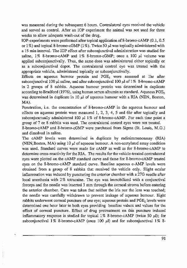
was measured during the subsequent 6 hours. Contralateral eyes received the vehicle and served as control. After an lOP experiment the animal was not used for three weeks to allow adequate wash-out of the drug. lOP-experiments were performed after topical application of 8-bromo-cAMP (0 .I, 0.5 or 1 %) and topical8-bromo-cGMP (1 %). Twice 50 JLI was topically administered with a 15 min interval. The lOP effect after subconjunctival administration was studied for saline, I% 8-bromo-cAMP and 1% 8-bromo-cGMP; once a 100 JLI volume was applied subconjunctivally. Thus, the same dose was administered either topically or as a subconjunctival depot. The contralatenal control eye was treated with the appropriate vehicle, administered topically or subconjunctivally. Effects on aqueous humour protein and PGE:, were assessed at 1hr after subconjunctivallOO JLl saline, and after subconjunctival100 JLI of 1% 8-bromo-cAMP in 2 groups of 8 rabbits. Aqueous humour protein was determined in duplicate according to Bradford (1976), using human serum albumin as standard. Aqueous PGE:, was determined in duplicate in 10 JLl of aqueous humour with a RIA (NEN, Boston, MA). Penetration, i.e. the concentration of 8-bromo-cAMP in the aqueous humour and effects on aqueous protein were measured 1, 2, 3, 4, 5 and 6hr after topically and subconjunctually administered 100 JLl 1% of 8-bromo-cAMP. For each time point a group of 7 or 8 rabbits was used. The contralatenal control eyes were not treated. 8-bromo-cAMP and 8-bromo-cGMP were purchased from Sigma (St. Louis, M.O.) and dissolved in saline. The cAMP levels were determined in duplicate by radioimmunoassay (RIA) (NEN,Boston, MA) using 10 JLl of aqueous humour. A non-acetylated assay condition was used. Standard curves were made for cAMP as well as for 8-bromo-cAMP to determine cross-reactivity for the RIA. The results for the vehicle-treated contralateral eyes were plotted on the cAMP standard curve and those for 8-bromo-cAMP treated eyes on the 8-bromo-cAMP standard curve. Baseline aqueous c-AMP levels were obtained from a group of 8 rabbits that received the vehicle only. Slight ocular inflammation was induced by puncturing the anterior chamber with a 27G needle after local anesthesia with 2% tetracaine. The eye was immobilized with a conjunctival forceps and the needle was inserted 3 mm through the corneal stroma before entering the anterior chamber. Care was taken that neither the iris nor the lens was touched; the needle was carefully withdrawn to prevent leakage of aqueous humour. Eight rabbits underwent corneal puncture of one eye; aqueous protein and PGE:, levels were determined one hour later in both eyes providing baseline values and values for the effect of corneal puncture. Effect of drug pretreatment on this puncture induced inflammatory response is studied for topical 1% 8-bromo-cAMP (twice 50 JLI); for subconjunctival 1% 8-bromo-cAMP (once 100 JLI) and for subconjunctival I% 8-
91

bromo-cGMP (once 100 ;.<I). The drug was administered to the experimental eye and the vehicle to the contralateral control eye one hr before corneal puncture of both eyes. Groups of 7-8 rabbits were used. Samples of 100 ;.<I of aqueous humour were obtained at 1, 2, 3 or 5 hr after corneal puncture and stored at -70°C to determine aqueous humour protein and PGE, levels. IOP values and concentrations are presented as means ± SEM. Differences between values were tested for significance with Student's t-test. These experiments adhered to the ARVO resolution on the use of animals in experimental research.
RESULTS
Intraocular pressure was not affected by topical 0.1 %, 0.5% and 1% 8-bromo-cAMP or 1% 8-bromo-cGMP (data not shown). Subconjunctival application 1% 8-bromo-cAMP (Figure 9.1) or 8-bromo-cGMP (Figure 9.2), however, decreased
92
OJ 5 I E .s
0. X
" Q.
Q -5
-10 0
100111 1% 8-bromo-c AMP subconjunctivally
* * * * * * * * * * * * * * 2 3 4 5 6
hours
Figure 9.1 Effect on intraocular pressure of subconjunctival administration of 100 JLI of saline (open circles), the contralateral eyes untreated, and of subconjunctival administration of 100 p.l 1% 8-bromo-cA!vfP {closed circles). the contralateral eyes received the vehicle subconjunctivally (both n=7). The points represent means ± 1 SEM. Asterisks in the figure represent p-values for the difference lOPexp -!OPcontr (•• P ( 0.001; ••• P ( 0.005, twotailed paired t-test).

IOP. No effect in the contralateral vehicle-treated eyes was observed. 8-bromo-cAMP reduced the IOP between 1 and 6 hours after application, with a maximum of 5.5 ± 0.8 mmHg (P < 0.005) at 4 and 5 hrs; 8-bromo-cGMP reduced IOP between 2 to 6 hrs after application, the maximum being 3.4 ± 0.8 mmHg at 4 hrs. (P < 0.005). Subconjunctivally administered 100 1-'l saline alone did not affect IOP (Figure 9.1).
1 OO_ul1% 8-bromo-c GMP subconjunctivally
c. X
" 0.. Q -5
* * * * * * * * * -10 0 2 3 4 5 6
hours
Figure 9.2 Effect of subconjunctival application of 100 ,ul 1% 8-bromo-cG:MP (contralateral eyes received the vehicle subconjunctivally) on intraocular pressure (n=7). The points represent means ± l SEM. Asterisks indicate: * P < 0.05; ** P < 0.01; *** P < 0.005, two-tailed paired t-test.
Effect on aqueous humour protein and PGE, of subconjunctival injection of 100 1-'1 saline and 100 1-'11% 8-bromo-cAMP is illustrated in Table 9.1. A slight increase of aqueous protein and aqueous PGE, was observed both with saline and 8-bromo-cAMP; the increased aqueous PGE, level after subconjunctivally injected 8-bromo-cAMP tended to be lower than after saline, the difference being 52.3 + 27.6 pg/ml (P < 0.1, unpaired two-tailed t-test). The baseline c-AMP level in aqueous humour was 19.8 ± 1.4 p moles/ml. Table 9.2 demonstrates the cross-reactivity of 8-bromo-cAMP for the adenosine 3' ,5' cyclic monophosphate radioimmuno assay.
93

Table 9.1 Aqueous humour protein and PGE, levels
contralateral untreated eye saline subconjunctival 1% 8-bromo-cAMP subconjunctival
Protein in gJ t-
0.4 ± 0.05 1.4 ± 0.4* 2.8 ± 0.7*
PGE, in pg.ml"'
32.8 ± 5.7 119.6 ± 24.5* 67.3 ± 12.6*
A volume of 100 ,ul saline or 8-bromo-cAMP was administered subconjunctivally.* indicates: significantly different from values in the contralateral untreated eye, P < 0.05 two-tailed paired t-test. Two groups of 8 rabbits were used.
Table 9.2 Cross-reactivity of8-bromo-cAMP for the cAMP radioimmunoassay
Standard concentration cyclic AMP and 8-bromo-cAMP in pmol.m.I-1
0 0.5 1.0 2.5 5.0
10.0 25.0 50.0
Percentage bound labeled tracer to antiserum
cyclic AMP
100 86.3 82.2 75 69.2 52.4 32.6 20.4
8-bromo-cyclic AMP
100 82.4 75.2 59.5 47.7 33.5 19.9 11.8
Percent bound labeled tracer to antiserum for standard concentrations cAMP and &-bromocAMP, calculated by dividing the average net counts of the standard with the average net counts of the zero standard* 100%. The cross-reactivity for 8-bromo-cAMP, calculated at the 50% point by interpolation, is 2.6.
Aqueous 8-bromo-cAMP levels after topical and subconjunctival administration are presented in Table 9.3 and 9.4, respectively. Topical 100 JLI 1% 8-bromo-cAMP induced a maximum concentration of 290 ± 82 p moles/ml (P < 0.01) at 1 hour; subsequently, the level gradually decreased. No effect on aqueous humour protein levels in either treated or vehicle-treated contralateral eyes was present (Table 9.3).
94
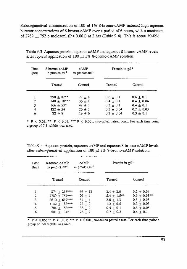
Subconjunctival administration of 100 1'1 1% 8-bromo-cAMP induced high aqueous humour concentrations of 8-bromo-cAMP over a period of 6 hours, with a maximum of2789 + 752 p moles/ml (P<0.001) at 2 hrs (Table 9.4). This is about 10-fold
Table 9.3 Aqueous protein, aqueous cAMP and aqueous 8-bromo-cAMP levels after topical application of 100 1'1 1% 8-bromo-cAMP solution.
Time 8-bromo-cAMP (hrs) in pmoles.rn.I·1
Treated
1 290 ± 82** 2 148 ± 18*** 3 166 ± 53* 4 122 ±54 6 32 ± 8
cAMP in pmoles.rnl·1
Control
29 ± 8 36 ± 8 48 ± 7 26 ± 2 19 ± 6
Protein in gf1·1
Treated
0.6 ± 0.1 0.4 ± 0.1 0.3 ± 0.1 0.3 ± 0.04 0.3 ± 0.04
Control
0.6 ± 0.1 0.4 ± 0.04 0.4 ± 0.1 0.2 ± 0.03 0.3 ± 0.1
* P < 0.05; ** P < 0.01; *** P < 0.001, two-tailed paired t-test. For each time point a group of 7-8 rabbits was used.
Table 9 A Aqueous protein, aqueous cAMP and aqueous 8-bromo-cAMP levels after subconjunctival application of 100 1'1 1% 8-bromo-cAMP solution.
Time (hrs)
I 2 3 4 5 6
8-bromo-cAMP cAMP in pmoles.m1·1 in pmoles.ml-1
Treated Control
874 ± 218*** 66±13 2789 ± 752*** 29 ± 4 2610 ± 619*** 34 ± 4 1142 ± 183*** 23 ± 2 704 ± 152*** 36 ± 9 501 ± 134* 26 ± 7
Protein in g/1·1
Treated Control
3.4 ± 2.0 0.2 ± 0.04 5.4 ± 1.5** 0.9 ± 0.03** 2.0 ± 1.3 0.3 ± 0.03 1.2 ± 0.5 0.3 ± 0.03 0.5 ± 0.1 0.3 ± 0.05 0.7 ± 0.2 0.4 ± 0.1
* P < 0.05; ** P < 0.01; *** P < 0.001, two-tailed paired t-test. For each time point a group of 7-8 rabbits was used.
95
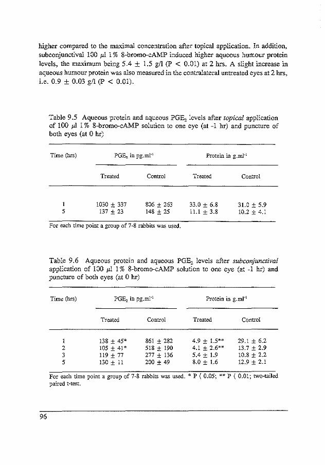
higher compared to the maximal concentration after topical application. In addition, subconjunctival 100 ;.tl 1% 8-bromo-cAMP induced higher aqueous humour protein levels, the maximum being 5.4 ± 1.5 gil (P < 0.01) at 2 hrs. A slight increase in aqueous humour protein was also measured in the contralateral untreated eyes at 2 hrs, i.e. 0.9 ± 0.03 gil (P < 0.01).
96
Table 9.5 Aqueous protein and aqueous PGE, levels after topical application of 100 pl1% 8-bromo-cAMP solution to one eye (at -1 hr) and puncture of both eyes (at 0 hr)
Time (hrs)
5
PGEz in pg.ml-1
Treated
1030 ± 337 !37 ± 23
Control
806 ± 263 148 ± 25
For each time point a group of 7-8 rabbits was used.
Protein in g.rnJ-1
Treated
33.0 ± 6.8 11.1 ± 3.8
Control
31.0 ± 5.9 10.2 ± 4.1
Table 9.6 Aqueous protein and aqueous PGE, levels after subconjunctival application of 100 ;.tl 1% 8-bromo-cAMP solution to one eye (at -1 hr) and puncture of both eyes (at 0 hr)
Time (hrs)
1 2 3 5
PGEz in pg.ml-1
Treated
138 ± 45* 105 ± 41* 119 ± 77 130 ± 11
Control
861 ± 282 518 ± 190 277 ± !36 200 ± 49
Protein in g.ml-1
Treated
4.9 ± 1.5** 4.1 ± 2.6** 5.4 ± 1.9 8.0 ± 1.6
Control
29.1 ± 6.2 13.7 ± 2.9 10.8 ± 2.2 12.9 ± 2.1
For each time point a group of 7-8 rabbits was used. • P ( 0.05; •• P ( 0.01; two-tailed paired t-test.

Corneal puncture of t.'le rabbit eye increased aqueous humour protein and PGE:, levels to 33.5 ± 5.6 gil and 1600 ± 400 pg/ml, respectively, at 1 hr. In the contralateral non-punctured eyes protein was 0.6 ± 0.1 gil and PGE:, was 30 ± 10 pg/ml. Topical application of l 00 ;.d l % 8-bromo-cAMP had no effect on the increased aqueous humour PGE:, and protein levels in the eyes, 1 and 5 hrs after puncture (table 9.5). Subconjunctival administration of !00 ;<I of 1% 8-bromo-cAMP, however, significantly inhibited the increased aqueous PGE:, and protein levels 1 and 2 hr after puncture; a time course is presented in table 9.6.
Table 9. 7 Aqueous protein and aqueous PGE:, levels after subconjunctival administration of l 00 ;<I 1% 8-bromo-cGMP to one eye (at -l hr) and puncture of both eyes (t=O hr)
Time (hrs)
I 5
PGE, in pg.mJ·'
Treated
931.2 ± 265 162 ± 23.4
Control
1390 ± 298 227 ± 47.2
For each time point a group of 7-8 rabbits was used.
Protein i."l. g.ml-1
Treated
31.4 ± 5.5 6.9 ± 2.0
Control
28.8 ± 5.6 10.8 ± 3.5
Subconjunctival administration of 100 ;<I 1% 8-bromo-cGMP showed to have no inhibitory effect on aqueous humour protein and PGE:, levels after corneal puncture (Table 9.7).
DISCUSSION
This study shows that 8-bromo-cAMP and 8-bromo-cGMP reduce IOP and that 8-bromo-cAMP but not 8-bromo-cGMP inhibit trauma-induced ocular inflammation. These effects of exogenously administered cyclic nucleotide analogs depend on the mode of administration. Differences in pharmacokinetics for subconjunctival and topical administration (Maurice and Mishima, 1989) are clearly demonstrated. Subconjunctival application of 8-bromo-cAMP induced 3 to 15-fold higher 8-bromo-cAMP concentrations in the aqueous humour compared to topical administration of the same dose. A plateau
97

maximal concentration at 2 and 3 hr shows the long duration achieved after giving a subconjunctival depot. Topical treatment rapidly reached a 10-fold lower maximal concentration at one hour. These results are in agreement with literature on ocular pharmacokinetics (Maurice and Mishima, 1989). In our opinion the different penetration into the anterior chamber is the reason why subconjunctival but not topical administration of 8-bromo-cAMP induced a sustained reduction of IOP. High levels of 8-bromo-cAMP are needed to achieve an IOP reduction; these levels correlated well in time with the hypotensive effect. Intracamerally administered cAMP has been shown to induce a two-fold increase in outflow facility in rabbits (Neufeld et al, 1975). In another study (Wong and Howes, 1983), intravitreal injections of cAMP did not cause elevation of the intracellular cAMP levels. In the current study, 8-bromo-cAMP was used instead of cAMP for mainly two reasons. Firstly, cyclic AMP is rapidly degraded by cAMP dependent phosphodiesterases, while 8-bromo-cAMP is a poor substrate for this enzyme (Beebe et al, 1988). Secondly, 8-bromo-cAMP is cell-permeable whereas cAMP itself does not easily pass cell membranes. Many studies have indicated that endogenous cAMP is involved in the reduction of lOP achieved with various stimulators of adenylate cyclase such as catecholamines (Hoyng et al, 1991; Neufeld et al, l972a, 1973; Radius and Langham, 1973; Rowland and Potter, 1979; and Boas et al, 1981), cholera toxin (Gregory, 198lb; Sears et al, 1981) or forskolin (Caprioli et al, 1983; Caprioli et al, 1984a). In this study we provide evidence that also the cAMP analog 8-bromo-cAMP can lower IOP. It was not expected that subconjunctival administration of only saline induced a slight increase of aqueous humour PGE, and protein; the PGE, probably derives from conjunctival tissue. However, the raised level of aqueous humour PGE, was insufficient to have an effect on lOP. Moreover, it should be noted that, when treating one eye subconjunctivally, the contralateral control eye received the vehicle subconjunctivally too, so that the effect on IOP can only be attributed to 8-bromocAMP itself. Li...lce 8-bromo-cAMP, 8-bromo-cGMP also induced a reduction of IOP when given subconjunctivally but not when given topically. Recently, it has been reported that atrial natriuretic factor (ANF) (Sugrue and Viader, 1986; Mittag et al, 1987a; Nathanson, 1988; Becker, 1990; and Korenfeld and Becker, 1989) reduced IOP in rabbits and that a simultaneous elevation of cGMP in the ciliary processes was observed (Mittag, 1987a; Becker, 1990; Korenfeld, 1989; and Nathanson, 1987). The 1% topical application of 8-bromo-cGMP in our study had no hypotensive effect. This is slightly different from Becker (Becker, 1990) who demonstrated a shortlasting effect with this topical dose in NZW rabbits. Our finding that 8-bromo-cGMP reduces IOP when given subconjunctivally may support the hypothesis that the peptide ANF
98

reduces IOP via a cGMP regulating mechanism in pigmented rabbits. There is evidence that drugs that activate intracellular adenylate cyclase can modulate the inflammatory response after bacterial endotoxin (Wong and Howes, 1983) or immunogenic provocation (Verhey et al, 1987; Deporter eta!, 1976, Deporter, 1977; and Seo and Saeki, 1980). Therefore we were interested in the effect of the cellpermeable c-AMP analog 8-bromo-cAMP on protein and PG levels in aqueous humour after a slight trauma such as induced by corneal puncture. Subconjunctival but not topical 8-bromo-cAMP partially protected the breakdown of the BAB. It reduced the endogenous formation of PGs as indicated by the PGE, content in aqueous humour 1, 2 and 3 hr after corneal puncture. In the contralateral eyes saline was given subconjunctivally. It can be argued, that the effect of 8-bromo-cAMP on aqueous humour PGE, after corneal puncture is due to the effect of subconjunctival injection of saline inducing PGE,-formation. However, the eyes pre-treated with subconjunctival saline followed by puncture showed an increase in aqueous humour PGE,-Ievel not different from that after corneal puncture only. The breakdown of the BAB after puncture with subconjunctival administration of 8-bromo-cAMP did not exceed the breakdown of the BAB after 8-bromo-cAMP alone. Similar to effects of 8-bromocAMP on IOP, the puncture experiments also reveal that higher levels of 8-bromocAMP, up to 3 nmoles/ml, are needed in the aqueous humour to be effective, i.e. inhibit endogenous PGE-production. It has been reported that intracellular c-AMP elevation reduces phosphoinositide breakdown in platelets (Nishizuka, 1984) and inhibits the formation of inositol 1,4,5,-triphosphate in bovine iris sphincter muscle (Tachado eta!, 1989). It is suggested that in this study 8-bromo-cAMP may suppress the pathways by which arachidonic acid is released from phosphoinositides after a slight trauma. The protective effect on the BAB seems to be a secondary effect due to inhibition of PG release. In control experiments, neither topical or subconjunctival application of 8-bromocGMP had influence on the breakdown of the BAB or PG formation in aqueous humour after puncture. This is supportive for a specific anti-inflammatory effect of 8-bromo-cAMP. In conclusion: subconjunctival administration of 8-bromo-cAMP as well as 8-bromocGMP reduces IOP, although probably by different mechanisms. Subconjunctival application of 8-bromo-cAMP but not 8-bromo-cGMP reduces the breakdown of the BAB and formation of PGE, after puncture trauma of the cornea. A role for c-AMP in modulation of the arachidonic acid cascade during inflammation is suggested.
99


CHAPTER 10
ADENYLYL CYCLASE IN HUMAN AND BOVINE TRABECULAR MESHWORK
Michie! JWM Busch1 •2, Kami Kobayashi1 , Philip FJ Hoynff, and Thomas W
Mittaf!; submitted.
1 Deparment of Ophthalmology and Pharmacology, Mount Sinal' Medical School of Medicin, New York, U.S.A.
2 DeptTTtment of Experimental Ophthalmology, The Netherlands Ophthalmic Research Institute, Amsterdam, The Netherlands.
Abstract
Adenylyl cyclase activity was compared in bovine and human trabecular meshwork membrane fractions and in whole tissue homogenate (bovine ouly). Basic characteristics and responses to various drugs/agents were tested including forskolin (FSK), manganese (Mn2+), fluoroaluminate (AIF;l, isoproterenol (ISO), prostaglandins (PGE1, PGE,, PGF,J and vasoactive intestinal peptide (VIP). In bovine trabecular meshwork particulate fraction was stimulated 3.3- and 2.6-fold (x) basal by 60 and 21'M FSK respectively, 2.2 x by ALF4, 1.5 x by PGE1 and PGE,, while no or very weak response was obtained with PGF,., ISO, norepinephrine, phenylephrine, p-amino clonidine, adenosine and VIP. The PGE1 stimulation was dose- and G-protein dependent, evidence for prostaglandin E receptor (EP-receptor) mediated activation. In human trabecular meshwork membrane fractions AC stimulation was more pronounced, being 12.4 and 5.5 x for 60 and 21'M FSK, and 3 x for PGE, and PGE,. PGF,. had no effect. Significant AC stimulation was found with ISO, 2.8 x, and with VIP, 1.8 X.
101

The results indicate EP-receptor mediated activation of adenylyl cyclase in bovlne and human trabecular meshwork. Bovine trabecular meshwork may lack or have few llzadrenoceptors, or if present they are not coupled to adenylyl cyclase, and had no receptor-mediated responses for the drugs tested except for PGE, and PGE:,. The findings indicate significant species differences in receptors coupled to AC in TM tissue. The results also suggest that AC may have a regulatory role in human trabecular meshwork controlling outflow facility acting vla Jl,-adrenoceptors, PGE:, and VIP, all of which could have a local physiologic function in this tissue.
Introduction
Adenylyl cyclase/cyclic AMP (cAMP) is thought to play a role as second messenger in the regulation of aqueous humour secretion by ciliary processes. Increasing evidence accumulates that cAMP is also important in regulating outflow mechanisms in the trabecular meshwork. Several drugs or agents that act on receptors coupled to adenylyl cyclase in various tissues affect outflow facility; particularly adrenergic drugs such as epinephrine (Neufeld et al, 1972a, Neufeld et al, 1973). The importance of cAMP as second messenger in response to in vivo treatment with catecholamines is further demonstrated by combination drug studies with IBMX, a phosphodiesterase inhibitor. IBMX potentiated the hypotensive effect of epinephrine (Hoyng et al, 1991; Busch and Hoyng, 1991b) by increasing its effect on outflow facility (Hoyng and Busch, 1992) in rabbits. Other agents like prostaglandins have been suggested, but also questioned (Kaufman, 1989), to play a role in controlling outflow facility if topically applied (Camras et al, 1977), or as endogenous intermediate in response to epinephrine (Anderson and Williams, 1990). In vltro studies report the presence of adenylyl cyclase activity and some drug responses in trabecular meshwork explants in bovine (Bartels, 1988b), in monkey (Crawford et al, 1991) and human (Wax et al, 1989) trabecular meshwork membrane fractions and in cultured human trabecular meshwork cells (Wax et al, 1989; Polansky and Alvaredo, 1985). Jl,-Adrenoceptors have been localized and characterized in human trabecular meshwork (Wax eta!, 1989; Jampel eta!, 1987; Elena, 1990). In the present study trabecular meshwork membrane fractions were prepared from bovlne (also tissue homogenate) and from donor bank human eyes. Basic AC characteristics and stimulatory responses to various drugs/agents were investigated and demonstrated for catecholamines, forskolin, prostaglandins, and the peptide VIP.
102

Materials and methods
Chemicals and reagents
Biochemical reagents were purchased from Sigma Chemical Company (St.Louis, MO). 'H-e-AMP was obtained from New England Nuclear Corp. (Boston, MA) and a-32PA TP from New England Nuclear or from Amersham (Arlington Heights,IL). Synthetic porcine VIP was from Cambridge Research Biochemials (Atlantic Beach NY11509). All other reagents and drugs were obtained from Sigma or from Fisher Scientific (Pittsburg, PA). Forskolin (FSK) was made up as 10 mM stock in dimethyl sulfoxide. The assay kit for protein determination by dye-binding was purchased from BioRad (Richmond,CA 94804).
Preparation of the tissues
Fresh bovine eyes were delivered on ice from a nearby slaughterhouse and, upon arrival, immediately dissected on ice. Trabecular meshwork explants were prepared according to a method described by Anderson, Wang and Epstein (!980). The anterior segment of the eye was cut off between the limbus and the equator. Choroidal tissue was severed from the sclera together with the lens and anterior vitreous. The irisciliary body was gently lifted from the sclera. The meshwork could be identified as a grey band encircling the cornea; posteriorly adjacent nontrabecular grey tissue was removed. Trabecular tissue was lifted from the sclera by using a small blunt gouge and immediately stored at -80°C. Thawed tissues were hand homogenized in isotonic divalent ion chelating buffer (0.3 M sucrose, 20 mM Tris pH 7.6, 5 mM EDTA, l mM EGTA, 5 mM dithiothreitol) in a teflon/glass homogenizer using 25 strokes of the pestle. The centrifuged particulate fraction (15 min, 27000 g) was washed once in the homogenizing buffer (6ml), recentrifuged (15 min, 27000 g), decanted and resuspended in the same buffer (3m!), containing 0.1 mM indomethacin and !OI'g/mlleupeptin, and passed through a 0.5mm nylon mesh. If whole tissue homogenate was used then the thawed tissue was homogenized in 3 ml of the homogenizing buffer containing 0.1 mM indomethacin and !OI'g/ml leupeptin with 25 strokes of the pestle and passed through the mesh. The protein content of the preparations usually ranged between 1-!.5 mg/ml. Human donor eyes were provided by the donor eye bank of the Netherlands Ophthalmic Research Institute. In the procedure for preserving corneas for transplantation purposes the eyes were enucleated as soon as possible and delivered
103
on ice by courier. The donors' ages ranged between 20-80 years. The anterior part of the eyes was cut off at the ora serrata, quickly frozen at -800C and stored up to 4 weeks before use. The post-mortem time before freezing the tissues was usually less than 8 hours. The tissue was thawed to 4'C and the trabecular meshwork was surgically prepared on ice under an operating microscope according to a procedure described by Tripathi and Tripathi (1982). The lens was removed after being severed from the zonular attachment. The iris-ciliary body was pulled away from the sclera; the remaining comeo-scleral ring was dissected in 8-10 equal radial segments; the scleral spur was identified by its ridge-like configuration. In each segment a partial thickness cut was made anteriorly in front of the scleral spur and a second parallel cut posterior to the line of Schwalbe. Trabecular meshwork tissue was lifted away with a sharp forceps. Histological sections confirmed the presence of predominantly trabecular tissue (not shown). Trabecular tissue from 4 human eyes was prepared for one experiment, hand homogenized in 1.2 ml of the same isotonic divalent ion chelating buffer as described above containing 0.1 mM indomethacin and lOI'g/ml leupeptin in a small Dounce glass/glass homogenizer using 35 strokes of the pestle, passed tbrough a mesh and centrifuged (15 min, 27000 g). The supernatant was removed by decanting and the particulate fraction resuspended in 1.2ml homogenizing buffer containing 0.1 mM indomethacin and lOI'g/ml leupeptin. The resuspended particulate fraction had a protein content of approximately 0.10- 0.30 mg/ml. Ouly once frozen and thawed tissue was used.
Adenylyl cyclase assay VIC)
Enzyme activity was determined in glass test tubes in a total volume of 250 1'1 for bovine tissue and !251'1 for human tissue, containing 60 mM sucrose, 80 mM Tris buffer pH 7.6, 3 mM MgCI2 (or MnCI2 as indicated), 1 mM EDTA, 0.2 mM EGTA, 5 mM creatinine phosphate (CP), 125 l'g creatinine phosphokinase, 20 I'M GTP (or 0.1 mM GDPBS as indicated), 4 mM theophylline, 20 I'M indomethacin, 0.2 mM ATP, a-32P-ATP (1-2 * 10' cpm for bovine and 4-6 * 10' for human tissue), 1 mM cAMP, and 3H-cAMP (1-3 * 10" cpm). The tubes were preincubated at 300C to equilibrate. To start the assay fifty l'l aliquots of the membrane or full tissue suspension were added to triplicate (bovine) or quadruplicate/quintuplicate (human) tubes containing a premix of all the other incubation ingredients and drugs as indicated. The assay was terminated after 5 (bovine) or 10 (human) minutes by addition of SDS, followed by placing the tubes in boiling water (3 min) and isolation of the 32P-cAMP with added 3H-cAMP tracer by the double column method of Salomon et al (1974). When FSK was used, the control assay tubes contained
104

DMSO( < 1%) equivalent to the amount used to dissolve FSK. GDPJlS, if used, was added to the bovine cell homogenate prior to the assay to allow 10 min preequilibration with the tissue.
Data analysis
Adenylyl cyclase stimulation was expressed as the ratio of corrected 32P-cAMP cpm for a drug effect relative to the corrected cpm for the control (baseline) assay tubes. Correction was based on recovery of the 'H-cAMP tracer, which was in the range of 55 to 80%. This yielded a relative value, i.e. stimulation index. Mean basal AC specific activities were calculated for bovine and human tissue preparations in tri/quadruplicates and were based on membrane protein solubilized with NaOH determined by the Bradford Dye-binding method with bovine gamma globulin as the reference protein. Stimulation indices multiplied by the basal specific activity approximated specific activities for drug effects. Experiments illustrated by bar graphs were done at least three (up to 8) times using different membrane/full tissue preparations. Error bars represent S.E.M. values, and significance (discriminating at P value 0.05) were determined by paired !-test. The AC activation dose-response curve depicted in figure 10.2 was fitted with noulinear regression analysis using the formula:
Eml!X x c'~
Effect= EC50' + c'
Em~ is maximal response, EC50 is drug concentration which yields 50% of the maximal effect, and ' is Hill slope coefficient, all being calculated values.
Results
Basal AC specific activity in bovine trabecular meshwork preparations was calculated to be 31.1 ± 10.7 pmol cAMP/min/mg protein (n=4). Routine assay conditions included Mg"+ as divalent ion regulator required for catalytic activity. The 3mM concentration used yielded approximately 1.8 mM free Mg"+ ion concentration, calculated by correcting for the presence of total EDTA + EGTA chelators of divalent ions. Stimulation of AC in bovine trabecular meshwork .
105

Sr--------------------,---------------, membranes ® tissue homogenate @
*
Figure 10.1 Adenylyl cyclase stimulation in membrane and tissue homogenate preparations of bovine trabecular meshwork in response to 60 and 2 J.f-M forskolin (FSK), 3 mM Mn2 +,
2 mM fluoride + 0.1 mM Afl+ (AlF4·), 10-5 M isoproterenol (ISO), 10-6 M vasoactive intestinal peptide (VIP), and 10-5 M PGE, (and 10-4M), PGE,, and PGF,., See text (data analysis) for definition of the stimulation index.
particulate with forskolin, AJF4- and Mn2+ is depicted in Figure IO.la and in bovine whole tissue homogenate in Figure IO.lb. FSK sensitive AC was present as well as G-protein dependent activity that can be activated by F- (Gs) which in turn stimulates AC. Mn-ATP and free Mn2+ are known to be better substrate and activator of the catalytic unit relative to M!f+ and amplify all AC-activities; Mn2+ increased basal activity in trabecular meshwork homogenate (Figure lO.la and lO.lb). The presence of stimulatory G-protein is indicative for some receptor(s) linked to AC. Surprisingly, ISO (lO-'M) did not appreciably stimulate the bovine trabecular meshwork AC (Figure lO.la). Further attempts to augment potential small effects ofiSO by combining ISO with 2i<M FSK and/or by using Mn2+ instead ofMg"+ were negative data (not shown). Positive controls using ISO were obtained with bovine ciliary processes particulate fraction from the same eyes (not shown) and with human trabecular meshwork tissue
106

1.3~---------------,
~ 1.2 "0 .s c: 0
~ 3 E ~ 1.1
Emax = 1.252
EC50 = 2.3pM
'\"--_./ '1 = 0.92
Figure 10.2. Dose-response curve for PGEcstimulated adenylyl cyclase in bovine trabecular tissue homogenate (five replicates per point). Em:;»;, EC!'h and '~(Hill coefficient) are calculated parameters from the fitted curve.
particulate preparation (see below). Stimulation or inhibition of AC was not found with norepinephrine, phenylephrine, p-aminoclonidine on bovine trabecular meshwork (not shown). Also no response was seen with the peptide VIP on this tissue preparation (Figure lO.la). Prostaglandins of theE-series stimulated bovine trabecular meshwork AC. Activation in the particulate fraction with PGE1 is demonstrated in Figure lO.la. Similar PGEc activation was found relative to its control baseline in whole tissue homogenate (Figure lO.lb), which includes the cytosol. Some AC acivity was also found in the collected supernatant, i.e. 0.75 ± 0.07 (n=4) compared to the basal activity of the corresponding, centrifuged, membranes. PGE,-(JO·'M) stimulation was 1.24 ± 0.02 in the membranes and 1.25 ± 0.06 in the supernatant (relative to their control basals). This finding probably indicates that small particulate membrane fractions remain in the 27000 x g supernate. Among other PGs tested, PGE:, activated bovine trabecular meshwork-AC to a similar extent as PGE1 whereas PGF,. had no effect (see Figure lO.lb). Dose-dependent stimulation with PGE1 is depicted in figure 10.2, suggesting receptor mediated activation. GTP-dependency for the PGE, response is demonstrated by adding GDPJlS to the tissue homogenate and leaving GTP out of the buffer. GDPJlS
107

14
12
X 10 " "0 .!:
" 8 0
:w s 6 .s '" 4
2
0
Figure 10.3 Adenylyl cyclase stimulation in membrane preparations of human trabecular meshwork in response to 2 mM fluoride + 0.1 mM Al3+ (AIF ol:), 60 and 2 .uM forskolin (FSK), 10-5 M isoproterenol (ISO), combination of 2 I'M FSK with 10-5 M ISO, 10-{) M vasoactive intestinal peptide (VIP), 10-5M PGEI (and 10-4), PGE,, and PGF,..
irreversibly binds to and blocks activation of G-proteins. In two experiments (n=8) GDPBS significantly reduced basal activity to a stimulation index less than 1, i.e. 0.58± 0.04 (p< 0.05); PGE,-stimulation index was reduced from 1.5 ± 0.1 in the presence of GTP to 0.68 ± 0.02 with GDPBS (p < 0.05). This further supports (EP-) receptor mediated AC activation. One time-course experiment (n =4) done with full tissue homogenate showed near linearity for the basal AC activity and the PGE1-response using assay times ranging from 5 up to 30 minutes. Basal activity was l, 0.99, 1.00 and 1.04 in 5-, 10-, 20-, and 30-minute assays. Respective PGE1 (lO·'M) stimulation was 1.31, 1.26, 1.27 and 1.22, indicating minimal loss of stimulatory capacity. Dopamine, adenosine, cyclohexal adenosine nor 5' -N-ethylcarboxamidoadenosine (NECA) (dose IO''M) did not affect bovine AC activity. Results of experiments with human trabecular tissue are shown in Figure 10.3. Basal specific activity of human trabecular meshwork particulate was 38.8 ± 8.8 pmol/min/mg protein (n=4). This is similar to the basal activity found for bovine trabecular meshwork preparations (see above). AC stimulation of human trabecular meshwork was evaluated in response to FSK, ISO, VIP and PGs. In contrast to bovine
108

trabecular meshwork a significant ISO as well as VIP response was present. Forskolin 2JLM, however, did not potentiate the effect of ISO. With respect to PGs a similar, quantatively more pronounced, stimulation was found. The order of responsiveness was PGE, = PGE1 > PGFz., similar as observed for bovine trabecular meshwork tissue.
Discussion
Values for basal AC specific acl:lv1ty obtained in bovine and human trabecular meshwork tissue preparations were similar. Higher stimulation indices were seen in response to drugs or agents in human tissue as compared to bovine tissue, also implicating higher specific activities. The results demonstrate the presence of PGEcsensitive AC in bovine and huma."l trabecular meshwork particulate fractions. Human trabecular meshwork particulate revealed ISO- and YIP-sensitive AC, whereas these responses were not significant in bovine trabecular meshwork. Activation with ISO is commonly observed most tissues containing AC but was notably absent in bovine trabecular meshwork particulate. Bartels (1988b) earlier reported that epinephrine in calf trabecular meshwork tissue incubations did not stimulate AC and suggested that Jl-adrenoceptors were not present, but could not exclude a-adrenergic inhibition by G,-protein. P-aminoclonidine and phenylephrine but were tested but neither a stimulatory nor inhibitory effect was observed. The lack of stimulatory responses to ISO, which is highly selective for Jl-adrenoceptors, confirms that either few Jl,-receptors are present or that they are not coupled to AC. In contrast, bovine ciliary processes do have high levels Jl,-adrenoceptor stimulated AC (Elena et a!, 1984). In human trabecular meshwork membranes ISO did stimulate AC. Previously, ISO and epinephrine have been shown to stimulate cAMP production in human scleratrabecular rings (Neufeld and Sears, 1974); human trabecular meshwork cells grown in primary culture, have been demonstrated to increase cAMP synthesis in response to Jl-receptor agonists (Polansky and Alvaredo, 1985), and selective Jl,-adrenoceptors have been characterized by autoradiography (Jampelet a!, 1987; Elena eta!, 1990) in sections of human trabecular meshwork, as well as by radiolabeled ligand binding techniques for human cultured trabecular meshwork cells and human trabecular tissue (Wax et a!, 1989). Recently, FSK-, ISO- and BPI-stimulated AC was reported in monkey trabecular meshwork membranes (Crawford eta!, 1991). The present results reveal an ISO response in human trabecular meshwork membranes which we interpret
109

as stimulation by binding to 8,-receptors coupled to AC. Combining FSK with ISO induced an additive response without further potentiation. FSK is known to elicit greater activation of the AC catalytic unit if it is complexed with activated G, alpha subunit than it does with the catalytic unit alone (Seaman, 1985). One may speculate whether the additivity and absence of a FSK-potentiated ISO response are indicative for the presence of different enzyme types of AC. A variety of compounds which bind to receptors coupled to AC in various tissues was tested since no ISO response was present in bovine tissue, while the FSK as well as AIF; stimulated AC response was indicative for stimulatory G-potein. For example VIP has been demonstrated to bind to VIP binding sites in rabbit ciliary epithelial cells and stimulates AC activity (Mittag et al, l987b). In monkey cultured trabecular meshwork cells VIP increased dose-dependently cAMP production (Koh and Yue, 1988). In this study we demonstrate VIP sensitive AC in human trabecular meshwork particulate whereas no response was seen in bovine trabecular meshwork particulate. This accords with commonly observed co-existence on cells of VIP- and 8,adrenoceptors. Dopamine- or adenosine-receptor agonists had no effect in bovine trabecular meshwork. A significant finding was the AC activation with PGE1 in bovine trabecular meshwork tissue. The dose-response relationship and GTP-dependency give evidence for a receptor mediated effect. The Hill coefficient of 0.92 is indicative for a single set of non-interactive ligand binding sites. The order of stimulation by the different PGs, PGE, = PGE:, > PGF2a, is suggestive for an EP-receptor. Discrimination between three currently distinguished EP-receptor subtypes remains to be established with sui prostone and EP !-antagonists. It seems reasonable to interprete the mechanism of PGE stimulation in human trabecular meshwork tissue also as EP-receptor mediated. This is based on analogy with the bovine results. Moreover, much of immediate post-receptor consequences of PGs is not known except for PGs of the Eseries which in most tissues stimulate (but can sometimes inhibit) AC (Robertson, 1986). In vitro studies on cultured human trabecular meshwork cells have shown that the human trabecular meshwork cells are capable of synthesizing PGs; the order of magnitude was PGE:, > PGF"' > 6-keto-PGF,. (Weinreb et al, 1988). Thus, together with the current results, a local physiological role for PGE and EP-receptors stimulating AC in trabecular meshwork seems likely. This may well contribute to controlling outflow facility. Earlier in vivo studies suggested that PGE, reduces intraocular pressure by increasing outflow facility (Camras, 1989). In a recent study we reported that phosphodiesterase inhibition with topical IBMX increased epinephrine's effect on C-value and that this effect was not only 8,- but also <>c adrenoceptor dependent (Busch and Hoyng, 1990). It may be hypothesized that theCasignal (a-adrenergic stimulation with phenylephrine) generates PG (PGE:,) production
l!O

via phospholipase A2 , which activates AC. Various adrenergic agents stimulate prostaglandin synthesis in ocular tissues (Abdei-Latif, 1989) and endogenous PGs have been suggested to be involved in the ocular hypotensive response to catecholamines (Hoyng eta!, 1982; Camras, 1989). Indomethacin pretreatment antagonized the effect of epinephrine to increase trabecular outflow facility (Anderson and Williams, 1990). PGE has been demonstrated to be a potent activator of AC in rabbit cameo-scleral rings (Neufeld and Sears, 1974). It is concluded that both in bovine and human trabecular meshwork membranes AC is stimulated by PGE, and PGE,, mediated by a prostaglandin E (EP-) receptor. In bovine trabecular meshwork ll:,-adrenoceptors may not be present or, if so, are not linked to AC. Human trabecular meshwork membranes contain AC which is responsive to ISO, and VIP. The findings indicate significant species differences in receptors coupled to AC in TM tissue. The results also suggest that AC may have a regulatory role in human trabecular meshwork controlling outflow facility by 82-adrenoceptors, PGE, and VIP, all of which could have a local physiologic function in this tissue.
111


CHAPTER 11
StJMMARY
The bifunctional properties of adrenergic receptors include a binding site, and an effector component which transduces the signal to the cell via a second messenger system. The adenylyl cyc!ase(AC)/cyclic AMP(cAMP) second messenger system is thought to play an important role in the regulation of aqueous humour secretion by ciliary processes as well as the control of outflow mechanisms in the trabecular meshwork, particularly in response to adrenergic agents such as epinephrine. The purpose of the present study was to investigate in vivo in rabbits the role of AC/cAMP by augmenting the AC/cAMP signals of epinephrine and other adrenergic agents pharmacologically with isobutylmethylxanthine (IBMX). IBMX is a phosphodiesterase inhibitor which inhibits the metabolism of active cAMP to inactive AMP; theoretically, IBMX can be expected to increase £,-adrenergic responses since 13,adrenoceptors are coupled directly to AC. Chapter 3. Topical IBMX alone had no effect on intraocular pressure (IOP), but IBMX enhanced the ocular hypotensive effect of the non-selective agonists epinephrine and norepinephrine and the selective 8:,-agonist isoproterenol. The main effect of IBMX was to increase the efficacy of these catecholamines. Thus an interaction between catecholamines and IBMX that reduces IOP has been demonstrated. Chapter 4. The IBMX is not soluble but was suspended in hydroxypropyl methylcellulose for the experiments described in chapter 3. Two soluble forms of isobutylmethylxanthine were tested, i.e. the ethylenediamine salt of IBMX and complexes of IBMX and 2% cyclodextrin prepared in saline. Both soluble formulations increased the hypotensive effect of epinephrine in a dose.<Jependent manner and on the same order of magnitude as a suspension of IBMX. This indicated that the bioavailability of IBMX was not substantially improved by either of these soluble forms. It was concluded that the IBMX complex with cyclodextrin, having a neutral pH, was a suitable formulation for future experiments.
113

Chapter 5. Alterations in regional blood flow in the eye were studied in rabbits using the radiolabeled microsphere technique. The purpose was to investigate whether the epinephrine induced reduction of IOP, enhanced by IBMX, is based on a vascular mechanism since epinephrine is known to induce marked vasoconstriction in the anterior uvea and a marked vasoconstriction would tend to lower the lOP. However, application of IBMX in combination with epinephrine induced an increase in ocular blood flow, lasting up to 7.5 hours, in both the iris (3-fold) and the ciliary processes (2-fold). IBMX alone tended to increase the blood flow in the iris and ciliary body, and epinephrine had no effect on the ciliary processes. This indicated that the reduction of intraocular pressure seen after topical treatment with a combination of IBMX and epinephrine was not based on a vascular mechanism. The increase in ocular blood flow after application of a combination of IBMX and epinephrine may be indicative of the presence of vasodilative B-adrenergic receptors in the ocular vasculature. Chapter 6. Aqueous humour dynamics were studied to determine whether the mechanism by which IBMX potentiates the ocular hypotensive response to (dipivalyl) epinephrine consisted of increasing the outflow facility in the trabecular meshwork or reducing aqueous humour secretion in the ciliary processes. Outflow facility was measured by means of Schi6tz indentation tonography and aqueous humour production by fluorophotometry in rabbits. Uveoscleral flow in rabbits is very low/neglible and was therefore not measured. Neither IBMX nor epinephrine affected the outflow facility; combination treatment with IBMX and epinephrine, however, increased the outflow facility by 43%. The slight reduction in aqueous humour formation after topical dipivalyl epinephrine was abolished by combination treatment with IBMX. The results indicate that phosphodiesterase inhibition with IBMX potentiates the ocular hypotensive effect of epinephrine by increasing the outflow facility and not by reducing aqueous humour secretion. These data support the accumulating evidence that AC plays an important role in the regulation of outflow facility. Chapter 7. Potentiation of the ocular hypotensive response to catecholamines by IBMX was more pronounced for the non-selective adrenergic agonists epinephrine and norepinephrine than for the selective ~-agonist isoproterenol (chapter 3). This was a surprising finding since IBMX is expected to increase primarily the responses mediated by ~-adrenoceptors coupled to AC. The adrenoceptor selectivity of catecholarnines for potentiation by IBMX was systematically investigated in this chapter by combination drug experiments using selective a,(phenylephrine), ~(BHT920), and 81 ( dobutamine ), ~(salbutamol, terbutaline )-adrenergic agonists. The order of responsiveness of the catecholarnines to potentiation by IBMX can be summarized as: norepinephrine (a,B) = epinephrine (B,a) > isoproterenol (maiuly ~ = salbutamol(~ > terbutaline (~. IBMX had no effect on the ocular hypotensive
114

responses to dobutamine, phenylephrine and B-HT920. However, the addition of a subthreshold dose of phenylephrine to the salbutamol regimen further potentiated the hypotensive response to IBMX achieved with salbutamol. It is concluded that IBMX primarily enhances 13:,- adrenoceptor/cAMP-mediated ocular hypotensive responses, but additional a,-adrenergic receptor stimulation potentiates this effect. This is of interest since it may indicate that a 1-adrenoceptor stimulation, which evokes primarily a calcium second messenger response, induces an interaction with the 13:,/AC/cAMP pathway. Chapter 8. Further experiments (in the absence of!BMX) focussed on this interaction between a, and 13:,-adrenergic signals by studying the combination of two selective adrenergic agonists. Enhanced ocular hypotensive responses were only found for the combination salbutamol (B:,) and phenylephrine (a,). The ouLilow facility measured by tonography was not affected by either salbutamol or phenylephrine alone. Combination treatment, however, increased the C-value by 32%. These results provided confirmation of an interaction between a 1 and Jl2-signalling systems that reduces IOP and increases outflow facility. Chapter 9. It was shown that administration of exogenous cAMP, in the form of the cell permeable analogue 8-bromo-cAMP, reduced IOP. Analysis of the penetration of 8-bromo-cAMP into the aqueous humour indicated that the hypotensive effect depends on the mode of administration; subconjunctival, but not topical, application raised the level of 8-bromo-cAMP in the aqueous humour sufficiently to reduce IOP. A close temporal relationship with the effects on IOP could not be demonstrated since the maximum aqueous 8-bromo-cAMP levels occurred at two hours whereas the IOP response was seen between 2 and 5 hours. The biochemical analysis of aqueous humour described in chapter 3 also focussed on the correlation between aqueous humour levels of endogenous cAMP and the IOP response. The aqueous levels of endogenous cyclic AMP were markedly elevated after combined epinephrine and IBMX treatment, indicating that adenylyl cyclase is stimulated by epinephrine whereas cAMP degradation is inhibited by the phosphodiesterase inhibition achieved with IBMX. A temporal relationship, however, was not present since potentiation of the pressure response occurred between 3 and 6 hours, whereas aqueous cAMP elevation was most pronounced 1 hour after medication. The absence of a close time relationship between cAMP levels and IOP response after epinephrine had already been mentioned by Boas eta! (1981). This may indicate that initial AC activation is followed by a period in which AC is refractory whereas a sequence of events after initial AC stimulation leads to a delayed IOP response. The correlations between effects on IOP and those on aqueous cAMP responses suggest a role for cAMP in the reduction of IOP. The finding that salbutamol, but also high doses of phenylephrine increased aqueous
ll5

cAMP levels (chapter 8) demonstrates that not only a Jl2 but also an a,! calcium signal stimulates AC. However, a combination of salbutamol and phenylephrine did not have a synergistic effect on cAMP responses which may be indicative of the presence of common AC. It is concluded that the results lend support to an interaction between a, and Bz-signals at the level of AC. Chapter 10. Increased outflow facility has been demonstrated to play an important role in the reduction of IOP by both IBMX (chapter 6) and the interaction of a 1 and Bz-adrenergic stimulation (chapters 7,8). This suggests an important role for AC in the trabecular meshwork. Therefore. in vitro characteristics of the AC enzyme in trabecular meshwork membrane preparations were studied in chapter 10. Bovine and human trabecular meshwork membrane preparations were used. Bovine trabecular meshwork AC could be stimulated directly (forskolin) as well as by G-proteins (alumina fluoride) and prostaglandin E receptors. Little or no response was obtained with isoproterenol, which indicates L'>at bovine trabecular meshwork may lack or have only a small number of 8,-adrenoceptors. Human trabecular meshwork AC stimulation was more pronounced. Human trabecular meshwork AC could be activated directly (forskolin) or by G-proteins (alumina fluoride), PGE,. isoproterenol and VIP. This is indicative of the presence of 8,, VIP, and prostaglandin E- receptors coupled to AC. Betq, VIP, and prostaglandin E- receptors may play a local physiological role in the regttlation of outflow facility in human trabecular meshwork with mediation of their effect by AC.
Epilogue
This thesis questions whether AC is a mediator of the ocular hypotensive response to adrenergic agents. Several arguments support a positive and others an inconclusive answer. The hypotensive effect ofiBMX is interpreted to be mainly due to phosphodiesteraseinhibition on the basis of the known properties of IBMX as well as the markedly enhanced aqueous cAMP levels and their correlation with the decrease in IOP found in the present studies. Antagonism for adenosine receptors is not likely to be an important pharmacological mechanism since IBMX had vasodilative effects whereas adenosine receptor antagonists are known to decrease ocular blood flow. Thus, IBMX proved to be a useful tool for t.'>e study of the role of AC/cAMP, providing evidence that AC is involved in mediation of the ocular hypotensive responses to epinephrine.
116

However, a more detailed answer is required regarding the observed differences in the effects on inflow and outflow facility. A proposed mechanism is presented in Figure 11.1. Outflow facility can be increased after strong AC/cAMP signals. This was demonstrated in this t.fJesis consistently for IBMX and epinephrine as well as salbutarnol and high doses of phenylephrine, and by others for isoproterenol, epinephrine and exogenous cAMP. Thus, it may be concluded that AC stimulation plays a role as mediator of the increase in outflow facility. Interpretation of the role of AC stimulation in ciliary processes and its effect on aqueous humour formation remains complex. It may well be t.'lat effects on active
cal~ium ... (~,&s~!.... s1gnal '.::!:1 ~/
0
trabecular outflow
a.1 ~ ------.
t2 -----------8_
---------~
cAMp --e--. blood flow ciliary processes
w active passive
secretion ultrafiltration """-((??)II AA ··
' HB inflow ----, ,,+, +
--------, :t intraocular pressure
reduction
Figure 11.1 Schematic representation for the proposed mechanism by whic.'1 adrenaline and combinations of a selective a:1 and Jl::-adrenergic agonist reduce the intraocular pressure. Effects induced by fi:-adrenoceptor stimulation are indicated by straight lines~ a:1-adrenoceptor dependent events by dotted lines. A stimulatory effect is marked by e and inhibitory by e.
117

secretory processes in the ciliary epithelium are masked by concomitant effecta on local haemodynamics. For example, the increase in blood flow in the iris and ciliary processes could well increase the ciliary perfusion rate which may in turn increase the rate of ultrafiltration. However, it is not really possible to determine whether a strong AC/cAMP signal inhibits active secretion in the ciliary epithelium and is counteracted by an increase in ultrafiltration due to vasodilation (IBMX with epinephrine) or that the reverse process occurs: the AC/cAMP signal increases active secretion but vasoconstriction (epinephrine) antagonizes this effect. The question of whether AC stimulation is involved in the reduction of the aqueous flow cannot therefore be answered conclusively. Whatever the real mechanism may be, a strong AC/cAMP signal did not reduce but tended to increase aqueous flow. The present studies may contribute to our understanding of the ocular hypotensive effect of epinephrine. Combination drug studies of highly selective adrenoceptor agonists indicate that L'le hypotensive effect of epinephrine probably depends on its selective a 1 and 8:,-adrenergic properties. The increase in outflow facility induced by epinephrine may not merely depend on addition but instead on interaction after simultaneous 8:, and a 1-adrenergic stimulation. The a/calcium signal could possibly stimulate AC in trabecular meshwork. Thus far, direct evidence for a 1-adrenergic stimulated AC in trabecular meshwork membrane preparations has not been found, but one may postulate that a calcium signal (a1) generates prostaglandins and that PGE, in turn directly activate AC via EP-receptors. Other possible mechanisms may include the presence of calmodulin-sensitive AC, as in rabbit ciliary processes (Tormay, 1987), or protein kinase C that activates AC. Additional studies seem to be warranted.
On the basis of the animal studies presented topical treatment with a phosphodiesterase inhibitor may be useful in glaucoma if combined, for example, with epinephrine. Additional long-term studies are needed to determine whether tachyphylaxis and desensitization occur. Anticipated major advantages are: 1. IBMX will enhance the reduction of !OP by epinephrine (via its 8:, and ac
adrenoceptor-mediated ocular hypotensive effects) 2. the commonly accepted dose of epinephrine (l%) can be reduced. This could
reduce the side effects of epinephrine, such as an irritating reactive hyperaemia. Irritation was never observed in the rabbit eyes treated with IBMX and epinephrine.
118


REFERENCES
Ahlquist RP (1948). A study of the adrenotropic receptors. Amer J Physic! 152, 586. Abdel-Latif AA (1989). Regulation of arachidonate release, prostaglandin synthesis, and sphincter
constriction in the mammalian iris-ciliary body. In: Bito LZ, and Stjernschantz J (Eds) "The ocular effects of prostaglandins and other eicosanoids", New York: Alan R Liss, Inc, pp 53-72.
Aim A and Bill A (1972). The oxygen supply to the retina. ll. Effects of high intraocular pressure and of increased arterial carbon dioxide tension on uveal and retinal blood flow in cats:a study with labelled microspheres including flow determinations in brain and some other tissues. Acta Physic! Scand 84, 306-319.
Aim A (1980). The effect of topical !-epinephrine on regional ocular blood flow in monkeys. Invest Ophthalmol Vis Sci 19, 487-491.
Anderson L, and Williams W (1990). Inhibition by indomethacin of the increased facility of outflow induced by adrenaline. Exp Eye Res 50, 119-126.
Anderson PJ, Wang J, and Epstein DL (1980). Metabolism of calf trabecular (reticular) meshwork. Invest Ophthalmol Vis Sci 19, 13.
Araie M (1985). Time change of rabbit aqueous flow under influence of adrenergic drugs. Exp Eye Res 40, 391-403.
Barany EH (1963). A rnathemetical formulation of intraocular pressure as dependent on secretion, ultrafiltration, bulk flow, and osmotic reabsorption of fluid. Invest Ophthalmol Vis Sci 2, 584-590.
Bartels SP, Lee SR, and Neufeld AH (1987). The effect of forskolin on cyclic AMP, intraocular pressure and aqueous humour formation in rabbits. Curr Eye Res 6, 307-320.
Bartels SP (1988a). Aqueous humour flow measured with fluorophotometry in timolol-treated primates. Invest Ophthalmol Vis Sci 29, 1498-1504.
Bartels SP (1988b) Effects of epinephrine and forskolin on cyclic AMP formation in calf trabecular meshwork. Invest Ophthalmol Vis Sci 29 (suppl), 88.
Bausher LP, Gregory DS, and Sears ML (1987). Interaction between alpha2- and beta2-adrenergic receptors in rabbit ciliary processes. Curr Eye Res 6, 497-505.
Bausher LP, Gregory DS, and Sears ML (1989). Alpha2-adrenergic and VJP receptors in rabbit ciliary processes interact. Curr Eye Res 8. 47-54.
Bausher LP, and Horio B (1990). Neuropeptide Y and somatostatin stimulated cyclic AMP production in rabbit ciliary processes. Curr Eye Res 9, 371-378.
Beavo JA, Rogers N!, Crofford OB, Hardman JG, Sutherland EW, and Newman EV (1970). Effects of xanthine derivates on lipolysis and adenosine 3'-5'-monophosphate phosphodiesterase activity. Mol Pharmacal 6, 597-603.
Beavo JA, Rogers NI, Crofford OB, Baird CE, Hardman JG, Sutherland EW, and Newman EV
121

(1971). Effect of phosphodiesterase inhibitors on cyclic-AMP levels and on lipolysis. Ann NY Acad Sci 185, 129-136.
Becker B, PetitTI, Gay AJ (1961). Topical epinephrine therapy of open-angle glaucoma. Arch Ophthalmol 66, 219-225.
Becker B, Morton W (1966). Topical epinephrine in glaucoma suspects. Am J Ophthalmol 62, 272-277.
Becker B (1990). Topical 8-bromo-cyclic GMP lowers intraocular pressure in rabbits. Invest Ophthalmol Vis Sci 31, 1647-1649.
Beebe SJ, Beasly-Leach A, and Corbin JD (1988). cAMP analogs used to study low-Km hormone-sensitive phosphodiesterase. Methods Enzymol159, 531-543.
Bhattacherjee P, Rhodes L, He Z, and Paterson CA (1991). Invest Ophthalmol Vis Sci (suppl) 32, 833.
Bill A (l966a). Formation and drainage of aqueous humour in cats. Exp Eye Res 5, 185-190. Bill A (1966b). The routes of bulk drainage of aqueous humour in rabbits with and without
cyclodialysis. Doc Ophthalmol 20, 157-169. Bill A (1969). Early effects of epinephrine on aqueous humour dynamics in vervet monkeys. Exp
Eye Res 8, 35-43. Bill A (1970). Effects of norepinephrine, isoproterenol and sympathetic stimulation on aqueous
humour dynamics in vervet monkeys. Exp Eye Res 10, 31-46. Bill A (1975). Blood circulation and fluid dynamics in the eye. Pharmacal Rev 55, 383-417. Bill A (1989). Uveoscleral drainage of aqueous humor: physiology and pharmacology. In: Bito
LZ, and Sljemschantz J (Eds) "The ocular effects of prostaglandins and other eicosanoids", New York: Alan R Liss, Inc, pp 417-427.
Boas RS, Messenger MJ, Mittag ThW, and Podos SM (1981). The effects of topically applied epinephrine and timolol on intraocular pressure and aqueous humour cyclic-AMP in the rabbit. Exp Eye Res 32, 681-690.
Bourne HR, Lichtenstein LM, Melmon KL (1974). Modulation of inflammation and innnunity by cyclic AMP. Science 184, 19-28.
Bradford MM (1976). A rapid and sensitive method for the quantitation of microgram quantities of protein utilizing the principle of protein-dye binding. Anal Biochem 72, 248-254.
Braunagel SC, Xiao JG, and Chiou GCY (1988). Tne potential role of adenosine in regulating blood flow in the eye. JOcular Pharmacal 4, 61-73.
Bromberg BB, Gregory DS, and Sears ML (1980). Beta-adrenergic receptors in ciliary processes of the rabbit. Invest Ophthalmol Vis Sci, 19, 203-207.
Buckberg GD, Luck JC, Payne DB, Hoffman JIE, Archie JP, and Fixler DE (1971). Some sources of error in measuring regional blood flow with radioactive microspheres. J Appl Physiol 31, 598-604.
Busch MJWM, and Hoyng PhFJ (1990). Interaction between isobutylmethylxanthine (!BMX) and epinephrine (Epi): reduction of intraocular pressure and increase of ocular blood flow. Invest Ophthalmol Vis Sci (suppl) 31, 232.
Busch MJWM, and Hoyng PhFJ (1991a). Selective a,- and 13:,-adrenoceptor stimulation is additive in reducing intraocular pressure. Invest Ophthalmol Vis Sci (suppl) 32, 1256.
Busch MJWM, and Hoyng PhFJ (199lb). Soluble forms of isobutylmethylxanthine enhance the ocular hypotension induced by catecholantines. Graefe's Arch Ophthalmol 229, 583-586.
Busch MJWM, Stjemschantz J, and Hoyng PhFJ (1991c). Increase in ocular blood flow induced by isobutylmethylxanthine and epinephrine. Exp Eye Res 52, 199-204.
Busch MJWM, Kobayashi K, and Mittag (1992). Adenylyl cyclase in human and trabecular meshwork. Invest Ophthalmol Vis Sci (suppl) 33, 902.
122

Campochiaro PA, Sen HA (1989). Adenosine and its agonists cause retinal vasodilation and hemorrhages. Arch Ophthalmoll07, 412-416.
Camras CB, Bito LZ, and Eakins KE (1977). Reduction of intraocular pressure by prostaglandins applied topically to the eyes of conscious rabbits. Invest Ophthalmol Vis Sci 16, 1125.
Camras CB, and Podos SM (1989). The role of endogenous prostaglandins in clinically-used and investigational glaucoma therapy. In: Bito LZ, and Stjernschantz J (Eds) "The ocular effects of prostaglandins and other eicosanoids", New York: Alan R Liss, Inc, pp 459-475.
Caprioli J, and Sears ML (1983). Forskolin lowers intraocular pressure in rabbits, monkeys and man. Lancet l, 958-960.
Caprioli J, Sears M, Bausher L, Gregory D, and Mead A (1984a). Forskolin lowers intraocular pressure by reducing aqueous inflow. Invest Ophthalmol Vis Sci 25, 268-277.
Caprioli J, Sears M and Mead A (1984b). Ocular blood flow in phakic and aphakic monkey eyes. Exp Eye Res 39, 1-7.
Cepelic J, and Cernohorsky M (1981). The effects of adrenergic agonists and antagonists on the adenylyl cyclase in albino rabbit ciliary process. Exp Eye Res 32, 291-299.
Cepelic J, and Hynie S (1990a). Inhibitory effects of clonidine and dopamine on adenylyl cyclase of rabbit ciliary pocesses. Curr Eye Res 9, 111-120.
Cepelic J, and Hynie S (1990b). Inhibitory effects of neuropeptide Yon adenylyl cyclase of rabbit ciliary pocesses. Curr Eye Res 9, 121-127.
Chiou GCY (1983). Effects of a 1 and a 2 activation on adrenergic receptors on aqueous humour dynamics. Life Sci 32, 1699.
Coakes RL, and Sinh PB (1984). Effects of adrenergic drugs on aqueous humour dynamics in the normal eye. 1. Salbutamol. Brit J Ophthalmol 68, 393-397.
Cohen J, Lach JL (1963). Interaction of pharmaceuticals with Schardinger dextrins 1. J Pharm Sci 52, 132-136.
Colossanti BK. and Barany E (1978). Reduction of the degeneration increase in outflow facility after superior cervical ganglionectomy in the rabbit. Invest Ophthalmol Vis Sci 17,802-805.
Cooper JR, Bloom FE. and Roth RH (1986). Modulation of synaptic transmission. In: Cooper JR, Bloom FE, and Roth RH (Eds) "The biochemical basis of neuropharmacology", New York Oxford: Oxford University Press, pp 106-124.
Crawford KS, Heideman W, Robinson JC, and Kaufman PL (1991). Stimulation of cyclic AMP formation in monkey iris, ciliary processes, ciliary muscle and trabecular meshwork. Invest Ophthalmol Vis Sci 32 (Suppl), 756.
Daly JW (1985). Adenosine receptors. Adv Cycl Nucl and Prot Phosph Res 19, 29-46. Denis Ph (1989). Recepteurs beta-adrenergiques vasculaires retiniens chez l'homme.
Ophtalmologie 3, 62-64. Deponer DA, Capasso F, and Willoughby GA (1976). Effects of modification of intracellular
cyclic AMP levels on the immediate hypersensitive reaction in vivo. J Pathol119, 147-151. Deponer DA (1977). Cyclic AMP and the mechanism of leukocyte lysosomal enzyme release
during an immediate hypersensitivity reaction in vivo. J Pathol 123, 129-133.
Eakins KE, and Barany EH (1960). Outflow resistence and adrenergic mechanism. Effects of sympathectomy, N-(2-chlorethyl) dibenzylamine hydrochloride (dibenamine), and dichloroisoproterenol on the outflow resistence of the rabbit eye. Arch Ophthalmol 64, 839-848.
Eakins KE (1963). The effect of intravitreous injections of norepinephrine, epinephrine and isoproterenol on the intraocular pressure and aqueous humour dynamics of rabbit eyes. J
!23

Pharmacal Exp Ther 140, 79-84. Eakins KE, and Ryan SJ (1964). The action of sympathomimetic amines on the outflow of
aqueous humor from the rabbit eye. Brit J Pharmacal 23, 374-82. Ehinger B (1964). Adrenergic nerves to the eye and its adnexa in rabbit and guina-pig. Acta Univ
Lund, section !1, No 20:1. Ehinger B (1966). Ocular and orbital vegatative nerves. Acta Physic! Scand 67, 1. Elena PP, Fredj-Reygrobellet D, Moulin G, and Lapalus Ph (1984). Pharmacological
characteristics of B-adrenergic-sensitive adenylyl cyclase in non pigmented and in pigmented cells of bovine ciliary process. Curr Eye Res 12, 1383-1388.
Elena PP, Denis Ph, Kosina-Boix M, Saraux H, and Lapalus Ph (1990). Beta adrenergic binding sites in the human eye: an autoradiographic study. J Ocul Pharmacal 6, 143-149.
Evoniuk GE, von Borstel RW, and Wurtman RJ (1986). Adenosine affects sympathetic neurotransmission at multiple sites in vivo. J Pharmacal Exp Ther 236, 350-55.
Ferrari-Dileo, G (1988). Beta-1 and beta-2 adrenergic binding sites in bovine retina and retinal blood vessels. Invest Ophthalmol Vis Sci 29, 695-699.
Fredbolm BB (1980). Are methylxanthine effects due to antagonism of endogenous adenosine? Trends Pharmacal Sci 1. 129-132.
Fredholm BB, and Hedqvist P (1980). Modulation of neurotransmission by purine nucleotides and nucleosides. Biochem Pharmacol29, 1635-1643.
Gaul GL, and Brubaker RF (1985). Measurement of aqueous flow in rabbits with corneal and vitreous depots of fluorescent dye. Invest Ophthalmol Vis Sci 27, 1331.
Genderen van MM, Best van JA, and Oosterhuis JA (1988). The immediate effect of phenylephrine on aqueous flow in man. Invest Ophthalmol Vis Sci 29, 1469.
Gharagozloo NZ, Larson RS, Kullerstrand U, and Brubaker RF (1988). Terbutaline stimulates aqueous humour flow in humans during sleep. Arch Ophthalmol !06, 12!8-1220.
Gooch JM, Hackmiller RC, and Jumblatt JE (1989). Neuropeptide Y modulates adenylyl cyclase in the rabbit iris and ciliary epithelium. Invest Ophthalmol Vis Sci 30 (suppl), 21.
Gregory DS, Bauscher LP, Bromberg BB, and Sears ML (1981a). The beta-adrenergic receptor and adenyl cyclase of rabbit ciliary processes. In: Sears ML (ed) "New Directions in Ophthalmic Research", New Haven: Yale University Press, pp 127-145.
Gregory D, Sears M, Bausher L, Mishima H, and Mead A (198lb). Intraocular pressure and aqueous flow are decreased by cholera toxin. Invest Ophthalmol Vis Sci 20, 371-381.
Gregory DS (1990). Timolol reduces lOP in normal NZW rabbits during the dark only. Invest Ophthalmol Vis Sci 31, 715-721.
Groeneboer MC, Hoyng PhFJ, and Kuizinga A (1989). Prostaglandin F,. isopropyl ester versus lloprost phenacyl ester in rabbit and beagle eyes. Curr Eye Res 8, 131-138.
Harris LS, Galin MA, Lerner R (1970). The influence of low-dose !-epinephrine on aqueous outflow facility. Ann Ophthalmol 2(5), 455-458.
Hedqvist P, Fredbolm BB, and Olundh S (1978). Antagonistic effect of theophylline and adenosine and adrenergic neuroeffector transmission in the rabbit kidney. Circ Res 43. 592-598.
Higgins RG, and Brubaker RF (1980). Acute effect of epinephrine on aqueous humour formation in the timolol-treated normal eye as measured by fluorophotometry. Invest Ophthalmol Vis Sci !9, 420.
124

Holland MG, von Sallman L, and Collins EM (1956). A study of the innervation of the chamber angle. Am J Ophthalmol 44, 148.
Holland MG, von Sallman L, and Collins EM (1957). A study of the innervation of the chamber angle. H. The origin of trabecular axons revealed by degenration experiments. Am J Ophthalmol 44, 206.
Hoyng PhFJ, Alphen van GWHM, and Haddeman E (1982). Does prostacyclin mediate alphaadrenergic induced hypotension? Doc Ophthalmol 53, !59.
Hoyng PhFJ, Verbey N, Thorig L, and Haeringen van NJ (1986). Topical prostaglandins inhibit trauma-induced inflanunation in the rabbit eye. Invest Ophthalrnol Vis Sci 27, 1217-1225.
Hoyng PhFJ, and Groeneboer C (1987). Isobutylmethylxanthine (!BMX), a phosphodiesteraseinhibitor, enhances ocular hypotension, induced by adrenergic agents in the pigemented rabbit. Invest Ophthalmol Vis Sci (suppl) 28, 12.
Hoyng PhFJ, Groeneboer MC, and Busch MJWM (1991). Isobutyhnethylxanthine enhances adrenergic-induced ocular hypotension in rabbits and beagles. Exp Eye Res 52, 5!1-517.
Hoyng PhFJ and Busch (1992). Alpha-! adrenoceptor stimulation potentiates IBMX-enhanced beta-2 adrenoceptor ocular hypotensive responses. Invest Ophthalmol Vis Sci (suppl). 1179.
Jampel HD, Lynch MG, Brown RH, Kuhar MJ, and De Souza EB (1987). B-adrenergic receptors in human trabecular meshwork. Invest Ophthalmol Vis Sci 28, 772-779.
Jay WM, Aziz MZ, and Green K (1984). Effect of topical epinephrine and timolol on ocular and optic nerve blood flow in phakic and aphakic rabbit eyes. Curr Eye Res 3, 1!99-1202.
Jumblatt JE, Liu JGH, and North GT (1987). Alpha, adrenergic modulation of norepinephrine secretion in the perfused rabbit iris-ciliary body. Curr Eye Res 6, 767-777.
JurnblattJE, North GT, and Hackmiller RC (1990). Muscarinic cholinergic inhibition of adenylyl cyclase in the rabbit iris-ciliary body and ciliary epithelium. Invest Ophthalmol Vis Sci 6, !103-1108.
Kalsner S (1971). Mechanism of potentiation of contractor responses to catecholamines by methylxanthines in aortic strips. Br J Pharmacal 43, 379-388.
Kalsner S, Frew RD, and Smith GM (1975). Mechanism of methylxanthine sensitization of norepinephrine responses in a coronary artery. Amer J Physiol 228, 1702-1707.
Kass MA, Goldberg I, Mandell AJ, Becker B (1979). Comparison of dipivefrin and epinephrine in the treattnent of elevated intraocular pressure. Invest Ophthalmol Vis Sci (Suppl), 165.
Kaufman PL (1985). Isoproterenol dose-outflow facility response relationships in the vervet monkey. Current Eye Res 4, 877-883.
Kaufman PL (1986). Total iridectomy does not alter outflow facility responses to cyclic AMP in cynomolgus monkey. Exp Eye Res 43, 441.
Kaufman PL (1987). cAMP and outflow facility in monkey eyes with intact and retrodisplaced ciliary muscle. Exp Eye Res 44, 415.
Kaufman PL (1989). Pressure dependent outflow. In: Ritch R, Shields MB, and K.-upin T (Eds) "The glaucomas", StLouis: The CV Mosby Comp, pp 219-240.
Kaufman PL, and Crawford (1989). Aqueous humour dynamics: how PGF,. lowers intraocular pressure. In: Bito LZ, and Stjernschantz J (Eds) "The ocular effects of prostaglandins and other eicosanoids", New York: Alan R Liss, Inc, pp 387-416.
Kinsey VE (1971). !on movement in ciliary processes. In: Bittar EE (Ed) "Membranes and ion transport", Vol 3, New York, Wiley & sons, pp 185-209.
Kintz P, Himber J, de Burlet G, and Anderman G (1988). Characterization of alpha,-adrenergic receptors~ negatively coupled to adenylyl cyclase~ in rabbit ciliary processes. Curr Eye Res
125

7, 287-292. Koh SWM, and Yue BYJT (1988) Effects of agonists on the intracellular cyclic AMP
concentration in monkey trabecular meshwork cells. Curr Eye Res 7, 75-80. Kohn AN, Moss AP, Harget NE, Ritch R, Smith H, and Podos H (1979). Dipivalyl epinephrine
and epinephrine. Am J Ophthalmol 87, 196-201. Korenfeld MS, and Becker B (1989). Atrial natriuretic peptides. Invest Ophthalmol Vis Sci 30,
2385-2392. Krieglstein GK, and Leydhecker W (1978). The dose-response relationship of dipivalyl
epinephrine in open angle glaucoma. Graefe's Arch Clin Exp Ophthalmol 205, 141-146.
Lach JL, Cohen J (1963). Interaction of pharmaceuticals with Schardinger dextrins 2. I Pharm Sci 52, 137-142.
Lach JL, Chin T (1964). Schardinger dextrin interaction 3. I Pharm Sci 53, 924-927. Lambie JW (1973). Some factors affecting the ocular hypotensive and mydriatic responses of the
rabbit to topically applied !-adrenaline bitartrate. Exp Eye Res 17, 473-479. Lambie JW (1974). Some responses of the the rabbit eye to topca!ly administered(-) adrenaline
bitartrate. Exp Eye Res 19, 419. Larnble JW (1977). Some efects of topically applied adrenaline bitartrate on the rabbit eye. Exp
Eye Res 24, 129-143. Lands AM, Arnold A, McAuliff JP, Luduena FP, and Brown TG (1967). Differentiation of
receptor systems activated by sympaticornimetic arnines. Nature 214, 597. Langer SZ (1974). Presynaptic regulation of catecholamine release. Biochem Pharmac 23, 1793. Langham ME, and Diggs E (1974). !3-Adrenergic responses in the eyes of rabbits, primates, and
man. Exp Eye Res 19, 281-295. Langham ME, and Krieglstein GK (1976). The biphasic intraocular pressure response of rabbits
to epinephrine. Invest Ophthalmol 15, 119-127. Lind GJ, Torrnay A, and Mittag ThW (1989). Ca2• /calmodulin activated phosphodiesterase(PDE)
from ciliairy processes. Invest Ophthalmol Vis Sci(suppl), 270. Liu JHK, and Dacus AC (1991). Endogenous hormonal changes and cicadian elevation of
intraocular pressure. Invest Ophthalmol Vis Sci 32, 496. Liu JHK, Dacus AC, and Bartels SP (1991). Adrenergic mechanism in circadian elevation of
intraocular pressure in rabbits. Invest Ophthalmol Vis Sci 32, 2178.
Macri FJ, and Cevario J (1974). A pharmacodynamic study of the inhibitory effects of !norepinephrine. 1-epinephrine. and d,l-isoproterenol on aqueous humor formation in the enucleated, arterially perfused cat eye. Invest Ophthalmol Vis Sci 13, 392-395.
Macri FJ, and Cevario J (1975). Ciliary ganglion stimulation. l. Effects on aqueous humor inflow and outflow. Invest Ophthalmol Vis Sci 14, 28-33.
Mallorga P, Buisson S, and Sugrue MF (1988). Alpha1-adrenoceptors in the albino rabbit ciliary process. I Ocul Pharmacol 4, 203-214.
Maren TH (1974). HCO; formation in aqueous humor: mechanisms and relation to the treatment of glaucoma. Invest Ophthalmol 13, 479-484.
Martin YD, and Rabineau PA (1989). Vasoconstrictive effect of topical timolol on human retinal arteries. Graefe's Arch Ophthalmol227, 526-530.
Maurice DM, and Mishima S (1989). Ocular pharmacokinetics. ln: Sears ML (Ed) "Pharmacology of the Eye", Berlin: Springer Verlag, pp 19-116.
Miichi H, and Nagataki S (1983). Effects of pilocarpine, salbutamol and timolol on aqueous humour formation in cynomolgus monkeys. Invest Ophthalmol Vis Sci 24, 1269-1275.
126

Mishima S (!982). Ocular effects of beta-adrenergic agents. Surv Ophtloalmol27, 187. Mittag ThW, and Tormay A (1981). Desensitization of the B-adrenergic-adenylate cyclase
complex in rabbit iris-ciliary body induced by topical epinephrine. Exp Eye Res 33, 497. Mircag ThW, Tormay A (!985a). Drug responses of adenylyl cyclase in iris-ciiiary body
determined by adenine labeling. Invest Ophthalmol Vis Sci 26, 396-399. Mir-..ag ThW, and Tormay A (1985b). Adrenergic receptor subtypes in rabbit iris-ciliary body
membranes: Classification by radioligand studies. Exp Eye Res 33, 239-249. Mittag ThW, Tonnay A, Severi.1. C, and Podos S (1985). Alpha-adrenergic antagonists:
correlation of the effect on intraocular pressure and on aradrenergic receptor binding specificity in the rabbit eye. Exp Eye Res 40, 591-599.
Mittag ThW, Tormay A, Ortega M, and Severin C (1987a). Atrial natriuretic peptide, guanylate cyclase and intraocular pressure. Curr Eye Res 6, 1189-1195.
Mittag ThW, Tormay A, a.."1.d Podos S (1987b). Vasoactive intest£nal peptide and intraocular pressure: Adenylyl cyclase activation and blnding sites for vasoactJve intestir..al peptide in membranes of ocular ciliary processes. J Pharmacal Exp Ther 241, 230-235.
Mittag ThW, Yoshimura N, and Podos SM (1987c). Phorbol ester: effect on intraocular pressure, adenylyl cyclase, a."d protein kinase in the rabbit eye. Invest Ophthalmol Vis Sci 28, 2057-2066.
Mittag ThW (1989). Adrenergic and dopa..uinergic drugs in glaucoma. In: Ritch R, Shields MB, and Krupin T (Eds) "The glaucomas", StLouis: The CV Mosby Comp, pp 523-537.
Morgan ThR, Green K, and BoW'Illan K (1981). Effects of adrenergic agoni.sts upon regional ocular blood flow in normal and ga..i.glionectomized rabbits. Exp Eye Res, 32, 691-697.
Moses RA, and Becker B (1958). Clinical tonography: the scleral rigidity correction. Am J Ophthalmol 45, 196-208.
Moses RA (1987). Intraocular pressure. In: Moses RA (Ed) "Adler's Physiology of the eye. Clinical Application", StLouis: CV Mosby Co, pp 223-245.
NathansonJ (1981). Human ciliary process adrenergic receptor: pharmacological characterization. Invest Ophthalmol Vis Sci 21, 798-804.
Nathanson JA (1987). Atriopeptin activated guanylate cyclase in the anterior segment. Invest Ophtcialmol Vis Sci 28, 1357-1364.
Nathanson JA (1988). Direct application of a guanylate cyclase activator lowers intraocular pressure. Eur J Pharmacal 147, 155.
Nelmer AM, Jumblatt MM, Coca-Prados M, and Paterson CA (1991). Human non-pigmented epithelial cells express an EP2 receptor in culture. Invest Ophthalmol Vis Sci (suppl) 32, 833.
Neufeld AH, Jampol LM, a,,d Sears ML (1972a). Cyclic-AMP in the aqueous humor: the effects of adrenergic agents. Exp Eye Res 14, 242-250.
Neufeld AH, Jampol LM, Sears ML (1972b). Aspirin prevents the disruption of the blood aqueous barrier in the rabbit eye. Nature 238, 158-159.
Neufeld AH, Chavis RM, and Sears ML (1973). Cyclic-AMP ;., the aqueous humor: the effect of repeated topical epinephrine administration &.i.d sympat.ietic denervation. Exp Eye Res 16, 265-72.
Neufeld AH, Sears ML (1973). Prostaglandines and eye. Prostagla."dines 4, 157-175. Neufeld AH, and Sears ML (1974). Cyclic AMP in ocular tissues of the rabbit, monkey, a.~d
human. Invest Opht.loalmol Vis Sci 13, 475-477. Neufeld AH, Dueker DK, Vegge T, and Sears ML (1975). Adenosine 3',5'-monophosphate
increases the outflow of aqueous humour from the rabbit eye. Invest Ophthalmol Vis Sci 14, 40-42.
127

Neufeld AH and Sears ML (1975). Adenosine 3'-5'-monophosphate analogue increases the outflow facility of primate eye. Invest Ophthalmol 14, 688-692.
Neufeld AH (1976). Regulation of adrenergic neuromuscular transmission in the rabbit iris. Exp Eye Res 20, 549-561.
Neufeld AH (1978). Influences of cyclic nucleotides on outflow facility in the vervet monkey. Exp Eye Res 27, 387-397.
Neufeld AH, Zawistowsky KA, Page ED, and Bromberg BB (1978). Influences on the density of £-adrenergic receptors in the cornea and iris-ciliary body of the rabbit. Invest Ophthalmol Vis Sci 17, 1069-1075.
Nilsson SFE, Maepea 0, Samueisson M, and Bill A (1990). Effects of timolol on terbutalineand YIP-stimulated aqueous humour flow in the cynomolgus monkey. Curr Eye Res 9, 863-872.
Nishizuka Y (1984). The role of protein kinase C in cell surface signal transduction and tumor promotion. Nature 308, 693-695.
Nishizuka Y (1986). Studies and perspectives of protein kinase C. Science 233, 305-312. Nomura T, and Smelser G (1974). The identification of adrenergic and cholinergic nerve endings
in the trabecular meshwork. Invest Ophthalmol Vis Sci 13, 525. Norton AL, a..J.d Vierstein U (1972). The effect of adrenergic agents on ocular dynamics as a
func<~on of administration site. Exp Eye Res 14, 154-61.
Obstbaum SA, Kolker AE, Phelps ChD (1974). Low-dose epinephrine. Effect on intraocular pressure. Arch Ophthalmol 92, 118-120.
Polansky JR, and Alvaredo JA (1985) Isolation and evaluation of target cells in glaucoma research: honnone receptors and drug responses. Curr Eye Res, 267-279.
Polar1.sky JR (1990). Beta-adrenergic therapy for glaucoma. lnt Ophthalmo1 Clin 30, 219-229. Potter DE, and Rowland JM (!978). Adrenergic drugs and intraocular pressure: effects of
selective ~-adrenergic agonists. Exp Eye Res 27, 615-625. Potter DE (1981). Adrenergic pharmacology of aqueous humour dynamics. Pharmacal Rev 33,
133-153.
Radius R, and Langham ME (1973). Cyclic-AMP and the ocular responses to norepinephrine. Exp Eye Res 17, 219-229.
Rall ThW (1982). Evolution of the mechanism of action of methylxanthines: from calcium mobilizers to antagonists of adenosine receptors. Pharrnoclogist 24, 277-287.
Rall TW (1985). Central nervous system stimulants. The xanthines. ln Goodman Gilman A, Goodman LS, Rall TW, Murad F (Eds): "The pharmacological basis of therapeutics", N::w
York, MacMillan, pp 587-603. Robertson D, Jiirgen C, Frolich MD, Carr RK, Watson JT, Hollifield JW, Shand DG, and Oates
JA (1978). Effects of caffeine on plasma renin ac<Jvity, catecbolamines and blood pressure. N Eng! J Med 298, 181-86.
Robertson RP (1986). Characterization and regulation of prostaglandin and leukotriene receptors:an overview. Prostaglandins 31, 395.
Rowi&,d JM, and Potter DE (1979). Effects of adrenergic drugs on aqueous cAMP and cGMP and intraocular pressure. Albrecht v Graefes Arch Klin Ophthalmol 212, 65-75.
Rowland JW, and Potter DE (1980). Adrenergic drugs and intraocular pressure: The hypertensive effect of epinephrine. Ophthalmol Res 12, 221-229.
Rowland JM, Sawyer WK, Tittel J, and Ford CJ (1986). Studies on the circadian rytJun of lOP in rabbits: Correlation with aqueous inflow and cAl\1P content. Curr Eye Res 5, 201.
128

Ruskell GL (1976). The source of nerve fibres of the trabeculae and adjacent structures in monkey eyes. Exp Eye Res 23, 449.
Rutherford JD, Varner SF and Braunwald E (1981). Effects and mechanisms of action of aminophylline on cardiac function and regional blood flow distribution in conscious dogs. Circulation 63, 378-87.
Salomon Y, Londos C, and Rodbell M (1974). A highly sensitive adenylyl cyclase assay. Anal Biochem 58, 541.
Schenker HI, Yablonsky ME, Podos SM, and Linder L (1981). Fluorophotometric study of epinephrine and timolol in human subjects. Arch Ophthalmol 99, 1212.
Schrader J, Baumann G, and Gerlach E (1977). Adenosine as in.'oibitor of myocardial effects of catecholamines. Pfluger's Arch 372, 29-35.
Schultz J, and Daly JW (1973a). Cyclic adenosine 3'-5'-monophosphate in guinea pig cerebral cortical slices H. The role of phosphodiesterase activity in the regulation of levels of cyclic adenosine 3'-5'-monophosphate. J Biol Chern 248, 853-867.
Schultz J, and Daly JW (1973b). Cyclic adenosine 3'-5'-monophosphate in guinea pig cerebral conical slices III: Formation, degradation and reformation of cyclic adenosine 3'-5'-monophosphate during sequential stimulations by biogenic ami.11es and adenosine. J Bioi Chern 248, 860-871.
Seaman KB (1985). Activation of hormone-sensitive adenylate cyclase by forskolin. Drug Development Research 6, 181.
Sears ML, and Barany EH (1960). Outflow resistence and adrenergic mechanisms. Effects of sympathectomy, N-(2-chlroethyl) dibenzylamine hydrochloride (dibenamine), and dichloroisoproterenol on the outflow resistence of the rabbit eye. Arch Ophthalmol 64, 839-848.
Sears ML, and Sherk TE (1964). The trabecular effect of noradrenaline in the rabbit eye. Invest Ophthalmol Vis Sci 3. 157.
Sears ML, Gregory D, Bausher L, Mishima H, and Stjernschantz J (1981). A receptor for aqueous humor formation. In: Sears ML (Ed) "New Directions in Ophthalmic Research", New Haven: Yale University press, pp 163-183.
Sears ML (1984). Autonomic nervous system: adrenergic agonists. In Sears (Ed) "Pharmacology of the eye", Berlin/Heidelberg: Springer-Verlag, pp 193-233.
Schenker HI, Yablonsky ME, Podos SM, and Linder L (1981). Fluorophotometric study of epinephrine and timolol in human subjects. Arch Ophthalmol 99, 1212-1216.
Sea S, and Saeki K (1980). Inhibition of adjuvant arthritis by salbutamol and aminophylline. Europ J Pharmacal 63, 267-273.
Serle JB, Stein AJ, Podos SM, Severin CH (1984). CorynanL'l1ine and aqueos humour dynarrjcs in rabbits and monkeys. Arch. Ophthalmol. 102, 1385-!388.
Stjernschantz J, Aim A, and Bill A (1976). Effects of intracranial oculomotor nerve stimulation on ocular blood flow in rabbits: modification by indomethacin. Exp Eye Res 23, 461- 469.
Stone RA, Kuwayama Y, Laties AM, and Marangos PJ (1984). Neuron-specific enolasecontaining cells in the rhesus monkey trabecular meshwork. Invest Ophthalmol Vis Sci 25, 1332.
Stone RA, a.'ld Kuwayama Y (1989). The nervous system and intraocular pressure. ln: Ritch R, Shields MB, and Krupin T (Eds) "The glaucomas" StLouis: The CV Mosby Camp, pp 257-279.
Sugrue MF, and Viader MP (1986). Synthetic natriuretic factor lowers rabbit intraocular pressure. Eur J Pharmacal 130, 349-354.
129

Szejtli DSc (1982). Cyclodextrins a.~d drugs. In: Szejtli J (ed) "Cyclodextrins and their inclusion complexes", Budapest: Akademlai Kiado, pp 204-235.
Tachado SD, Akhta RA, Z.'ld Abdel-Latif AA (1989). Activation of beta-adrenergic receptors causes stimulation of cyclic AMP, i.....ihibition of inositol triphosphate, a.11.d relaxation of bovine iris sphincter smooth muscle. Invest Ophthalmol Vis Sci 30, 2232-2239.
Tonnay A, Ortega M, and Mittag ThW (1987). Calciurnlcalmodulin-activated adenylyl cyclase in rabbit ciiary process membranes. Invest Ophthalmol Vis Sci (suppl) 28, 285.
Townsend DJ, and Brubaker RF (1980). Immediate effect of epi.~ephrine on aqueous formation in the normal human eye as measured by fluorophotometry. Invest Ophthalmol Vis Sci, 19, 256-266.
Tripathi RC, and Tripat.'li BJ (1982). Huma.~ trabecular endothelium, corneal endothelium, keratocytes, a..11d scleral fibroblasts in pri.."'nary ceH culture. A comparitive study of grovvth characteristics, morphology, and phagocytic activity by light and scanning electron microscopy. Exp Eye Res 35, 611.
Tripathi BJ, and Tripauii R (1984). Effect of epinephrine in vitro on t.'le morphology, phagocytosis, and miotic activity og human trabecular endothelium. Exp Eye Res 39, 731-744.
Trope GE, and Clark B (1982). Beta adrenergic receptors in pigmented ciliary processes. Brit J Ophthalmol 66, 788-92.
Verbey NL, Hoyng PhFJ, and De Jong PTVM (1987). Tne effect of a phosphodiesterase inhibitor (isobutylmethylxanthine, IBMX) on a HSA-induced uveitis in the rabbit eye. Ophthalrnic Res 19, 30-31.
Vestal RE, Eriksson CE, Musser B, OzakJ LK, and Halter JB (1983). Effect of intravenous aminophylline on plasma levels of catecholamines and related cardiovascular and metabolic responses in man. CirC'J.lation 67, 162-171.
Waitzman MB, and Woods WD (1971). Some characteristics of an adenyl cyclase preparation from rabbit ciliary process tissue. Exp Eye Res 12, 99-111.
Wax MB, and MolL~off PB (1987). Distribution and properties of B-adrenergic receptors in human iris-ciliary body. Invest Ophthalmol Vis Sci 28, 420-430.
Wax MB, Molinoff PB, Alvaredo J, and Polansky J (1989). Charac"cerization of B-adrenergic receptors in cultured human trabecular cells and in human trabecular meshwork. Invest Ophthalmol Vis Sci 30, 5!-57.
Weinreb RN, Polansky JR, Alvarado JA, and Mitchell MD (1988). Arachidonic acid metabolism in huma.~ trabecular meshwork cells. lnvest Ophthalmol Vis Sci 29, 1708.
Weissmann G, Smolen JE. a.~d Korchak H (1980). Prostaglandins and inflammation: Receptor/cyclase coupling and explanation of why PGE's and PGI2 inhibit functions of inflammatory cells. In: Samuelson B, Ramwell PW, and Paoletti R (Eds) "Advances in Prostaglandin and Thromboxane Research", vol.8, New York: Raven Press, pp 1637-1653.
Wikberg JES (1978). Pharmacological classification of adrenergic o:-receptors in the guinea pig. Nature 273, 164.
Wong KL, and Howes EL (1983). Intraocular injection of prostaglandins. Modification of response to circulating bacterial endotoxin. Arch OphthalmollOl, 275-279.
Yabu JM, Polansky JR. and Crook RB (1991). Calcitonin gene-related peptide stimulates adenylyl cyclase via a protein kinase C-controlled mechanism in fetal human NPE cells. Invest Ophthalmol Vis Sci (suppl) 32, 833.
130

Yamada E (1988) lntraepithelial nerve fibres in the rabbit ocular ciliary epithelium. Arch Hist Cytol 51, 43.
Yamada E (1989) Further observations on the intraepithelial nerve fibres in the rabbit ocular ciliary epithelium. Arch Hist Cytol 52, 191.
Yoshimura N, Mittag ThW, and Podos SM (1989). Cyclic nucleotide dependent phosphorylation of proteins in rabbit ciliary processes. Invest Ophthalmol Vis Sci, 30, 875-881.
Yoshitomi T, and Gregory DS (1991). Ocular adrenergic nerves contribute to control of the circadian rythm of aqueous flow in rabbits. Invest Ophthalmol Vis Sci 32, 523.
131


SAMENVATIING
Adrenerge stoffen hebben in de medicamenteuze behandeling van glaucoom een belangrijke plaats. Aangrijpingspunten voor deze stoffen zijn adrenerge receptoren, die onderscheiden worden in 4 subtypes: a 1, a2, .l'i1, en .l'iz-receptoren. Zowel selectieve en non-selectieve adrenerge agonisten en antagonisten verlagen de oogdruk. Zo is adrenaline (o.a. DiopineR) een adrenerge agonist die voornamelijk bind! aan .l'iz en in geringe mate aan a 1-receptoren. Ook adrenerge antagonisten worden op grate schaal gebruikt om de oogdruk te verlagen bij glaucoom. Voorbeelden hiervan zijn timolol, levobunolol (beide .l'i1_J, en betaxolol (.l'i1). De pharmacologie en physiologie van deze adrenerge receptoren en oogdruk regulatie wordt echter maar ten dele begrepen. Dit proefschrift richt zich op de betekenis en functie van adeny!aat cyclase a!s mediator voor oogdruk effecten van adrenerge stoffen. Adenylaat cyclase (AC) is een, a!gemeen voorkomend, intracellulair enzym systeem dat aan receptoren gekoppeld is en een agonist/receptor signaal doorgeeft in de eel. Receptoren zijn intracellulair gekoppeld aan een "second messenger" systeem. Binding van een agonist aan de receptor activeert de receptor en dit leidt vervolgens tot activering van het second messenger systeem. Een paar, algemeen voorkomende second messenger systemen zijn beschreven. Adenylaat cyclase (AC) is zo'n systeem en is voor wat betreft de adrenerge receptoren gekoppeld aan .l'i1, .l'iz en a,-receptoren; AC gebruikt ATP als substraat om dit om te zetten in het biologisch actieve produkt cyclisch AMP (cAMP), i.e. het second messenger molecuul. Receptor activatie via .l'i, en .l'iz-receptoren activeert het AC terwijl a 2-stimulatie de activiteit van AC remt. ac Receptoren activeren een geheel ander systeem, het calcium/protein kinase C second messenger systeem. Dit genereert calcium/inositol trifosfaat (JP3)/diacyl glycerol (DAG) a!s second messenger moleculen. AC/cAMP lijkt daarom een essentiele rol te spelen in zowel de physiologische als medicamenteus adrenerge oogdruk regulatie. Ook niet-adrenerge stoffen waarvan bekend is dat zij AC stimuleren (forskolin, vasoactive intestinal peptide, prostaglandine E) hebben sterke effecten op de oogdruk en bevestigen het belang van AC/cAMP. Dit leidde tot de probleemstelling voor dit proefschrift: medieert AC/cAMP het oogdruk verlagend effect van adrenerge stoffen? Met adrenaline in het
!33

bijzonder is dit in konijnen bestudeerd door rniddel van combinatie-experimenten met isobutylmethylxanthine (IBMX). IBMX remt de afbraak van cAMP tot de inactieve metaboliet AMP door rniddel van remrning vall het enzym phosphodiesterase. IBMX versterkt daardoor cAMP afhankelijke effecten; in het algemeen zijn dat vooral de 8:,adrenerg afbankelijke effecten. IBMX is chemisch en pharmacologisch verwant aan theophylline. Hoofdstuk 1 en 2 zijn respectievelijk de algemene inleiding en beschrijving van de gebruikte materialen en methodes. Hoofdstuk 3 laat in dose-response curves zien dat IBMX het oogdr-uk verlagende effect van adrenaline (B,a), noradrenaline (a,Jl) en isoproterenol (JJ;-selectief) sterk potentieert. IBMX zelf heeft geen effect. Hiermee is een interactie tussen catecholamines en IBMX aangetoond die de oogdruk verlaagt. In hoofdstuk 4 worden twee water-oplosbare vormen van IBMX getest omdat IBMX niet oplosbaar is en in hoofdstuk 3 gebruikt is als suspensie. Getest worden het ethyleendiarnine zout van IBMX en IBMX in een 2% cyclodextraan oplossing dat leidt tot vorrning van "inclusion complexes" . Beide oplossingen potentieren het oogdruk verlagende effect van adrenaline dosis-afbankelijk en in dezelfde mate als de IBMXsuspensie. Geconcludeerd wordt dat de biologische beschikbaarheid niet wezenlijk verbetert. IBMX in een 2% cyclodextraan oplossing, dat een neutrale pH heeft, is geschikt voor toekomstige experimenten. Hoofdstuk 5 bestudeert lokale hemodynarnische effecten in de ciliaire processen met behulp van radioactief gelabelde microsferen. Het doe! was om na te gaan of het potentierende effect van IBMX op de oogdrukdaling na adrenaline toediening verklaard kon worden op grand van lokaal hemodynarnische veranderingen. Adrenaline werkt vasoconstrictief, en vasoconstrictie zou kunnen leiden tot een verminderde kamerwater productie. Er werd echter een sterke en langdurige (lot 7.5 uur na medicatie) toename van de doorbloeding waargenomen in de ciliaire processen (2-voudig) en iris (3-voudig) na combinatie behandeling van IBMX mel adrenaline; IBMX en adrenaline afzonderlijk hadden weinig effect. Dit betekenl dal het oogdrukdaling potentierende effect van IBMX niet verklaard kan worden op grond van hemodynarnische veranderingen. De resultaten leveren een aanwijzing voor het bestaan van vasodilatoire 8-receptoren. Hoofdstuk 6 laat zien dat het oogdrukdaling potentierende effect van IBMX op adrenaline teweeg wordt gebracht door een afname van de weerstand in bet trabekelsysteem en niet door verminderde kamerwater productie. De uitstroomcapaciteit die door middel van tonografie gemeten was, narn 30-40% toe, terwijl een bescheiden inhibitie van kamerwater productie na adrenaline aileen juist werd opgeheven door toevoeging van IBMX (gemeten met fluorophotometrie). Hoofdstuk 7 bestudeert systematisch de adrenoceptor selectiviteit voor het oogdruk
134

verlagende effect van IBMX. Een met begrepen bevinding in hoofdstuk 3 was narnelijk dat IBMX meer effect had met (nor)adrenaline (a,Jl) dan met isoproterenol (B:,); met IBMX werd vooral een versterkend effect op selectief llz-adrenerge stimuli verwacht. Combinatie experimenten met selectieve a,, a,, Jl,, en llz- agomsten Iaten zien dat IBMX de Jlz-adrenerg geinduceerde oogdrukdaling (terbutaline, salbutamol) versterkt. Echter, een additionele a,-stimulus (fenylephrine) potentieert dit effect van IBMX nog meer. Dit verklaart dus waarom IBMX een beter effect heeft met metselectieve agomsten (nor)adrenaline (Jl,a) dan met isoproterenol (Jl.,). Het suggereert bovendien dat een interactie tussen twee wezenlijk verschillende "second messenger" signalen (Jl,/AC/cAMP en a,/calcium/IP3/CAM) optreedt die de oogdruk verlaagt. Hoofdstuk 8 gaat nader in op deze interactie tussen het 1lz en a,-signaal. Individueel geven zowel een selectief a,(fenylephrine), a,(B-HT920), Jl1(dobutamine), als een Jl,(salbutamol, terbutaline)-stimulus oogdrukdaling. Bij combinatie van twee van deze stoffenfsignalen werd een additief oogdruk verlagend effect aileen gevonden voor salbutamol (B:,) met fenylephrine (a,). Bovendien werd een potentierend effect op de afvoerweerstand in de karnerhoek aangetoond omdat salbutamol en fenylephrine aileen geen effect hadden maar in combinatie de weerstand met 32% verlaagden. Dit bevestigt het bestaan van een specifieke interactie tussen 1lz en a,-adrenerge stimulatie die de oogdruk en de weerstand in de kamerhoek verlaagt (zie Figuur 11.1). Hoofdstuk 9 laat zien dat een exogeen cel-permeabel cyclisch AMP derivaat, 8-bromo-cAMP, de oogdruk verlaagt. Dit geldt voor toediemng als subconjunctivaal depot en met in de vorm van lokaal druppelen. De reden hiervoor is dat met een subconjunctivaal depot tienvoudig hogere 8-bromo-cAMP concentraties in het karnerwater bereikt worden, en blijkbaar nodig zijn. Een sterke tijd-effect relatie was met aanwezig omdat de piek concentratie op 2 uur gevolgd wordt door een oogdrukverlaging tussen 2 en 5 uur. Zo was ook in hoofdstuk 3 de piek concentratie van endogeen cAMP in het kamerwater (na IBMX met adrenaline) maximaal op een uur terwij1 het oogdruk verlagend effect tussen 3 en 6 uur optrad. Het ontbreken van een sterke tijd-effect relatie is eerder door Boas (1981) voor adrenaline beschreven. Desondanks is een correlatie tussen kamerwater cAMP concentratie en oogdruk effect dmdelijk aanwezig. Een verklarend mechamsme kan bijvoorbeeld zijn dat imtiele AC stimulatie gevolgd word! door een refractaire periode (receptor ontkoppe1ing), en dat intracellulaire processen leiden tot een latere oogdrukdaling.
Het effect op de uitstroom capaciteit vormde het belangrijkste fysio1ogisch mechamsme voor het oogdruk dalend effect van IBMX en de beschreven interactie tussen 1lz en a,-stimulatie. Hoofdstuk 10 probeert hiervoor een biochemisch substraat te vinden. In vitro werd het AC enzym systeem in membraan fracties van humaan en runder trabekulair kamerhoekweefsel bestudeerd. Het runder AC enzym werd gestimuleerd rechtstreeks aangrijpend op de katalytische plaats (met forsko1in), via G-
135

eiwitten (met fluoride ionen) en via prostaglandine E receptoren. De geringe of afwezige stimulatie via Jl,-receptoren gaf aan dat mogelijk weinig of geen Jl,receptoren aanwezig zijn. AC stimulatie in humaa."l trabekel weefsel was sterker; niet aileen met PGE maar ook met Jl2, en vasoactive intestinal peptide(VIP)-stimulatie. Deze resultaten Iaten zien dat Jl,-adrenerge, maar ook prostaglandine E, en VIPreceptoren gekoppeld zijn aan AC in trabekel weefseL Zij kunnen van fysiologische betekenis zijn voor het reguleren van de weerstand in de kamerhoek via cAMP stimulatie. Een direct verband tussen a,-stimulatie en AC stimulatie werd niet aangetoond; echter, i::>direct zou a,-stimulatie via phospholipase A2 activatie en PGEz productie AC kunnen stimuleren (zie Figuur 1 L 1).
Epiloog
De werkingsmechanismen via welke medicamenteuze glaucoombehandeling met adrenerge stoffen de oogdruk verlaagt, zijn voor een groot dee! onbekend. In dit proefschrift wordt de vraag gesteld in hoeverre het adenylaat cyclase/cAMP systeem een rol speelt als tussenschakel in het verlagen van de oogdruk met adrenerge stoffen. Hiertoe word! gebruik gemaakt van de stof IBMX die AC/cAMP-signalen versterkt door middel van het remmen van de afbraak van cAMP. De uitgevoerde dierexperimentele studies bevestigen dat AC/cAMP een belangrijke rol speelt in de overdracht van adrenerge effecten op vooral de uitstroom capaciteit in de kamerhoek. Het combineren van IBMX met adrenerge stoffen is een bruikbare methode om AC/cAMP signalen van adrenaline en andere Jl-adrenerge stoffen te versterken. Eiochemisch blijkt dit uit sterk gestegen kamerwaterconcentraties van cAMP en physiologisch uit potentiering van oogdrukverlagende effecten van vooral Jl,-adrenerge stoffen. Onderscheid moet echter worden gemaakt tussen effecten op het trabekel systeem (verantwoordelijk voor de weerstand in het afvoersysteem van kamerwater) en de ciliaire processen (verantwoordelijk voor productie van kamerwater). IBMX heeft een duidelijk potentierend effect op de afvoerweerstand verlagende eigenschappen van £, (en zelfs a 1)-adrenerge stimulatie (zie Figuur 11.1). Eeduidend moeilijker is de interpretatie van effecten op kamerwater productie. Enerzijds geeft adrenaline afzonderlijk een bescheiden remming van kamerwater productie; in combinatie met IBMX echter wordt deze afname opgeheven en is het netto effect nul. Dit kan veroorzaakt worden doordat de kamerwaterproductie de resultante is van een bala..'ls tussen actieve en passieve secretie. Dit zijn achtereenvolgens actieve secretie van kamerwater uit ciliair epitheelcellen en ultrafiltratie dat waarschijnlijk afbankelijk is van bloeddoorstroming en colloid
!36

osmotische gradienten in de ciliaire processen. Een toegenomen perfusie zou kunnen leiden tot toename van de ultrafiltratie. Twee interpretaties zijn mogelijk op grond van de experimenten waarin de doorbloeding van de ciliaire processen en de kamerwaterproductie gemeten zijn (zie Figuur 11.1): L [),/ AC/cAMP stimulatie remt actieve secretie (adrenaline), maar de 2-3 voudig
toegenomen perfusie va11 de ciliaire processen bij zeer sterke f),-stimulatie (IBMX + adrenaline) vergroot de ultrafiltratie. Of:
2. {),lAC/cAMP stimulatie versterkt actieve secretie van het ciliaire epitheel (IBMX + adrenaline), maar adrenaline remt de kamerwater productie door vasoconstrictie in ciliaire processen en vermindering van de ultrafiltratie.
De rol van Ac/ cAMP in de overdracht van adrenerge effecten op de kamerwaterproductie kan op grond hiervan rdet eenduidig beantwoord worden. We! ktm geconcludeerd worden dat een sterk [),/ AC/cAMP-siguaal in ieder geval rdet leidt tot remming van de kamerwater productie. De studies in dit proefschrift geven meer inzicht in de werking van adrenaline als oogdruk verlagend medicament (zie Figure 11.1). Specifiek de{),- en a,-adrenerge eigenschappen lijken verantwoordelijk te zijn voor het verlagen van de oogdruk door vergroting van van de uitstroomcapaciteit in de kamerhoek. Combinatie experimenten met selectieve adrenerge agordsten geven aan dat dit rdet alleen berust op additie, maar juist op interactie tussen beide receptor signalen. Een hypothetisch biochemisch mechardsme hiervoor kan zijn dat adrenaline AC direct activeert via{), receptoren die direct aan AC koppelen en indirect via a,-stimulatie gevolgd door prostaglandine E synthese. PGE,_ stimuleert dan AC direct via PGE,_-receptoren, zoals werd aangetoond in membraan fracties van humaa.q kamerhoek weefsel. Er zijn geen aanwijzingen gevonden dat de «z-adrenoreceptor stimulerende eigenschap van adrenaline een rol van betekerds speelt in het verlagen van de oogdruk.
Bij medicamenteuze behandeling van glaucoom ktm het zinvol zijn, gelet op de beschreven dierexperimentele resuitaten, om adrenaline te combineren met een fosfodiesterase remmer zoals IBMX. Te verwachten voordelen van deze combinatie zijn: L IBMX versterkt de oogdruk verlagende eigenschappen van adrenaline en 2. de klirdsch gebruikelijke adrenaline dosering van 1% kan verlaagd worden.
Bijwerkingen als reactieve hyperemie kunnen daardoor wellicht afnemen. Gebleken is dat toevoegen van IBMX het vasoconstrictieve effect van adrenaline opheft en verandert in een toeneming van de doorbloeding in intraoculaire weefsels met het vermogen tot autoregulatie. Deze toeneming van de intraoculaire doorbloeding kan een voordeel zijn bij glaucoom patienten met normale oogdruk en een kritische papillaire doorbloeding.
137


CURRICULUM VITAE
Geboren in 1959 te Nijmegen werd in 1978 het eindexamen V.W.O. (Canisius College, Nijmegen) behaald, in !984 het doctoraal geneeslamde en in 1986 het artsexamen (R.U. Groningen). Als artsassistent algemene chirurgie, niet in opleiding, gedurende 1 jaar werkzaam in het R.K. Ziekenhuis Groningen (Dr L Vos, chirurg). Van april 1988 tot april 1992 werkzaam als wetenschappelijk medewerker aan dit proefschrift bij Dr PhFJ Hoyng, oogarts, op de afdeling Experimentele Oogheelkunde van het Interuniversitair Oogheelkundig Instituut (directeur Dr AJ Otto, oogarts), waarvan 5 maanden (!988) op het Pharmacia Laboratorium voor glaucoom onderzoek (J Stjemschantz MD PhD, Uppsala, Zweden) en 5 maanden (1991) op de afdeling Oogheelkunde en Farmacologie van bet Mount Sinai Medical Center (hoofd Prof ThW Mittag PhD, New York, V.S.). Van januari !990 tot maart 1992 tevens werkzaam (deeltijd) als arts-assistent EHBO/oogheelkunde in bet Leyenburg en Oogziekenhuis 's Gravenhage (Dr La Lau, oogarts). Met ingang van april 1992 in opleiding tot oogarts in Rotterdam (ProfPTVM de Jong, oogarts). Toekenning van de eerste prijs (met Dr PhFJ Hoyng) voor glaucoomonderzoek, gepresenteerd op poster, door de European Glaucoma Society, Amsterdam mei 1992.