adrenal function in humans - immunotech
TRANSCRIPT

- 1 - 2011-05-02
Adrenal Function in Humans

- 2 - 2011-05-02
Adrenal function in humans
Introduction
The adrenal glands are small, bilateral structures that weigh approximately 4-5 g, regardless of age, weight or gender. Each gland consists of a yellow outer cortex, and a grey inner medulla (see Fig.1).
Fig.1: Structure of adrenal gland8
The medulla or inner portion of the gland secretes catecholamines, such as dopamine, epinephrine and norepinephrine, and is part of the sympathetic nervous system. The cortex forms the bulk of the adrenal gland and is responsible for the secretion of three types of hormones that possess a wide range of biological functions: the glucocorticoids, the mineralocorticoids and the adrenal sex hormones.
- 3 - 2011-05-02
Adrenal cortical function is essential for life; total loss of adrenal cortical function is fatal within 4 to 14 days if left untreated. This is not the case of medullary function, as dopamine, epinephrine and norepinephrine are also secreted by sympathetic nervous system.
The following text will focus on adrenal cortex function and hormone production, particularly glucocorticoids and mineralocorticoids.
Table 1: Adrenal synthetic products and primary functions in the body
Zona glomerulosa
(outer)
Mineralocorticoids, mainly aldosterone
Long-term regulation of blood pressure
Zona fasciculata
Glucocorticoids, mainly cortisol
Response to stress, influence on metabolism of proteins,
carbohydrates and fats Cortex
Zona reticularis
(inner)
Androgens, mainly DHEA and DHEA-S
Androgen effects
Medulla
Catecholamines adrenaline (epinephrine)
and noradrenaline (norepinephrine)
Response to stress
(fight-or-flight response)
- 4 - 2011-05-02
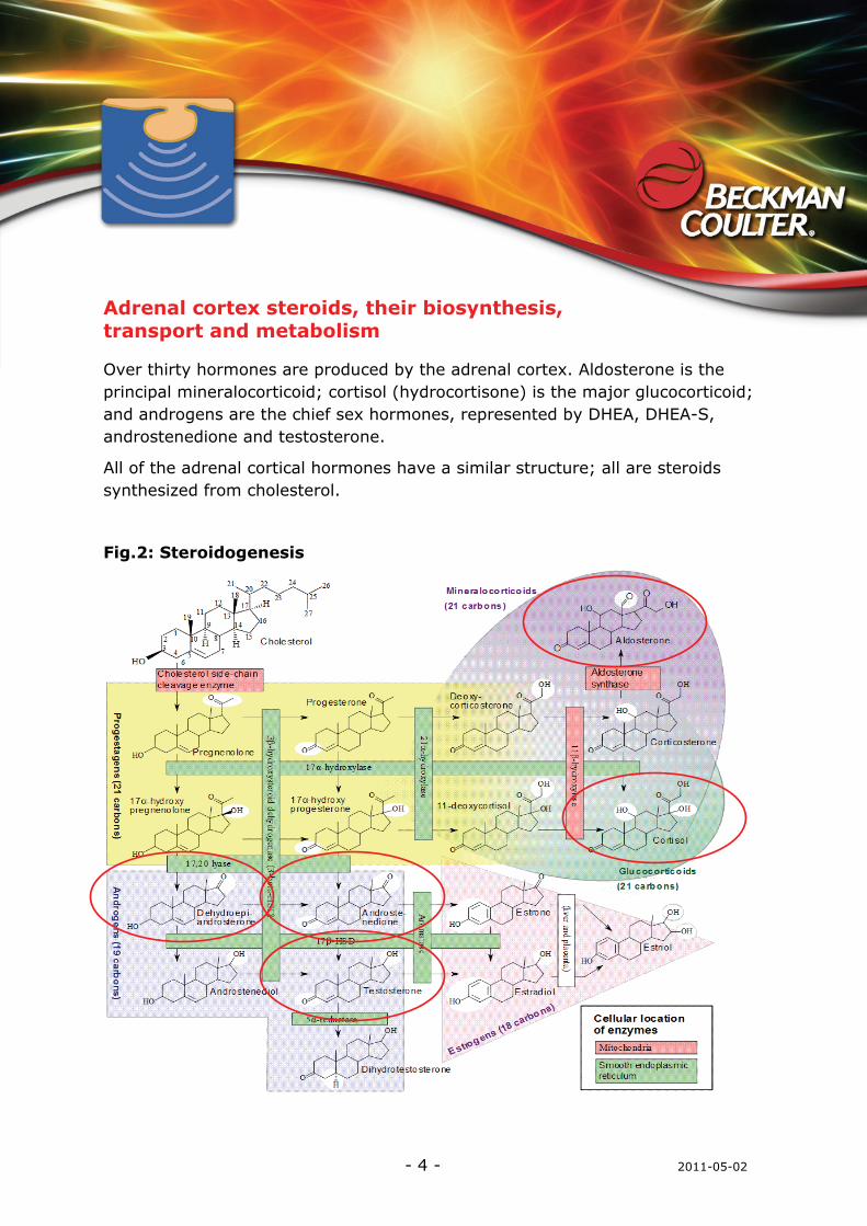
Adrenal cortex steroids, their biosynthesis, transport and metabolism
Over thirty hormones are produced by the adrenal cortex. Aldosterone is the principal mineralocorticoid; cortisol (hydrocortisone) is the major glucocorticoid; and androgens are the chief sex hormones, represented by DHEA, DHEA-S, androstenedione and testosterone.
All of the adrenal cortical hormones have a similar structure; all are steroids synthesized from cholesterol.
Fig.2: Steroidogenesis
- 5 - 2011-05-02
Each of the steps involved in the synthesis of the various hormones requires a specific enzyme.
The secretion of the glucocorticoids and the adrenal androgens is controlled by ACTH secreted by the anterior pituitary gland. To a certain extent, ACTH also regulates the synthesis of mineralocorticoids, but the primary regulation mechanism involves the renin-angiotensin-aldosterone system (RAS or RAAS). Regulation mechanisms of mineralocorticoid and glucocorticoid synthesis are described in detail below (Fig.3, 4, 5).
Adrenal hormones are secreted in an unbound state and bind to plasma proteins for transport in the circulatory system. Cortisol binds largely to corticosteroid-binding globulin and to a lesser extent to albumin.
Aldosterone circulates mostly bound to albumin.
The main site for metabolism of adrenal cortical hormones is the liver, where they undergo a number of metabolic conversions before being conjugated and made water-soluble. They are then eliminated in either urine or bile.
Adrenal hormones, regulation of their synthesis and biological function
Glucocorticoid hormones
The glucocorticoid hormones are synthesized in the zona fasciculate and the zona reticularis of the adrenal gland. The most important is cortisol.
Hormone blood levels are regulated by the negative feedback mechanisms of the hypothalamic-pituitary-adrenal (HPA) system (Fig.3). Cortisol is secreted in response to adrenocorticotropic hormone (ACTH) from the anterior pituitary. ACTH is itself secreted under the control of the hypothalamic peptide corticotropin-releasing hormone (CRH).
Cortisol levels increase as ACTH levels rise and decrease as ACTH levels fall.

- 6 - 2011-05-02
Fig.3: Hypothalamic-pituitary-adrenal (HPA) system2
There is considerable diurnal variation in ACTH levels, which reach their peak in the early morning (around 6:00 to 8:00 AM) and decline as the day progresses. This appears to be due to rhythmic activity in the central nervous system (CNS), which causes bursts of CRH secretion and, in turn, ACTH secretion. This diurnal pattern is reversed in people who work during the night and sleep during the day.
- 7 - 2011-05-02
The rhythm also may be changed by physical and psychological stresses, endogenous depression, manic-depressive psychosis, liver disease or other conditions that affect cortisol metabolism.
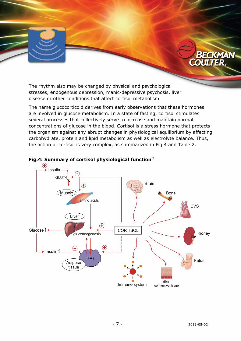
The name glucocorticoid derives from early observations that these hormones are involved in glucose metabolism. In a state of fasting, cortisol stimulates several processes that collectively serve to increase and maintain normal concentrations of glucose in the blood. Cortisol is a stress hormone that protects the organism against any abrupt changes in physiological equilibrium by affecting carbohydrate, protein and lipid metabolism as well as electrolyte balance. Thus, the action of cortisol is very complex, as summarized in Fig.4 and Table 2.
Fig.4: Summary of cortisol physiological function 2

- 8 - 2011-05-02
Table 2: Actions of cortisol
Major Influence Effect on Body
Stimulates gluconeogenesis Glucose metabolism
Decreases glucose use by tissues
Increases breakdown of proteins Protein metabolism
Increases plasma protein levels
Increases mobilization of fatty acids Fat metabolism
Increases use of fatty acids
Stabilizes lysosomal membranes of inflammatory cells, preventing the release of inflammatory mediators
Decreases capillary permeability to prevent inflammatory edema
Depresses phagocytosis by white blood cells to reduce the release of inflammatory mediators
Suppresses immune response
Causes atrophy of lymphoid tissue
Decreases eosinophils
Decreases antibody formation
Decreases development of cell-mediated immunity
Reduces fever
Anti-inflammatory action (pharmacologic levels)
Inhibits fibroblast activity
Psychic effect May contribute to emotional instability
Permissive effect Facilitates response of tissues to humoral and
neural influences, such as that of catecholamines, during trauma and extreme stress
- 9 - 2011-05-02
Mineralocorticoid hormones
The mineralocorticoids are produced in the zona glomerulosa, the outer layer of cells of the adrenal cortex.
The most potent naturally occurring mineralocorticoid is aldosterone.
Aldosterone secretion is stimulated primarily via the RAS, but it increases also in response to elevated level of potassium and, to lesser extent, also after the stimulation by ACTH.
The RAS plays an important role in regulating blood volume and systemic vascular resistance, which together influence cardiac output and arterial pressure.
As the name implies, there are three important components in this system:
• Renin • Angiotensin • Aldosterone
Sodium excretion and extracellular fluid volume are inversely related to the enzymatic activity of renin (plasma renin activity - PRA) and plasma aldosterone concentrations3.
When blood volume is low or when a drop in blood pressure occurs, the kidneys secrete renin. The enzyme circulates in the bloodstream and cleaves (hydrolyzes) angiotensinogen secreted from the liver into the peptide angiotensin I.
Angiotensin I is further cleaved in the lungs by endothelial-bound angiotensin converting enzyme (ACE) into angiotensin II, the most vasoactive peptide4.
Angiotensin II is a potent constrictor of all blood vessels. It acts on the musculature and thereby raises the resistance posed by these arteries to the heart. The heart, trying to overcome this increase in ‘load’, works more vigorously, causing blood pressure to rise. Angiotensin II also acts on the adrenal glands and releases aldosterone. Aldosterone’s main function is to stimulate the epithelial cells of the kidneys to increase re-absorption of sodium, chloride and water, increasing blood volume and blood pressure.
- 10 - 2011-05-02
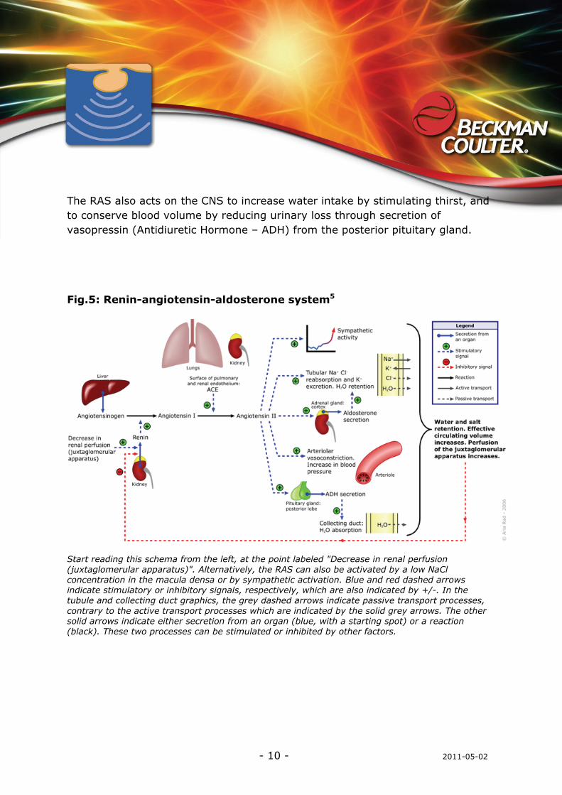
The RAS also acts on the CNS to increase water intake by stimulating thirst, and to conserve blood volume by reducing urinary loss through secretion of vasopressin (Antidiuretic Hormone – ADH) from the posterior pituitary gland.
Fig.5: Renin-angiotensin-aldosterone system5
Start reading this schema from the left, at the point labeled "Decrease in renal perfusion (juxtaglomerular apparatus)". Alternatively, the RAS can also be activated by a low NaCl concentration in the macula densa or by sympathetic activation. Blue and red dashed arrows indicate stimulatory or inhibitory signals, respectively, which are also indicated by +/-. In the tubule and collecting duct graphics, the grey dashed arrows indicate passive transport processes, contrary to the active transport processes which are indicated by the solid grey arrows. The other solid arrows indicate either secretion from an organ (blue, with a starting spot) or a reaction (black). These two processes can be stimulated or inhibited by other factors.

- 11 - 2011-05-02
Fig.6: The renin-angiotensin system,
showing the role of aldosterone in the adrenal glands and kidneys6
- 12 - 2011-05-02
Adrenal sex hormones
The adrenal sex hormones are synthesized primarily in the zona reticularis and the zona fasciculata of the cortex. Their production is stimulated by the HPA system, similarly to cortisol (Fig.3).
These sex hormones have androgenic effects, but their effect on normal sexual function is limited. Nevertheless, they at least contribute to the pubertal growth of body hair, particularly pubic and axillary hair in women. They may also play a role in the steroid hormone economy of the pregnant woman and the fetal-placental unit. In men, their effect is primarily the testicular synthesis of androgens.
The most important adrenal sex hormone is dehydroepiandrosterone (DHEA) and its sulfated derivative - dehydroepiandrosterone sulfate (DHEA-S). Levels of DHEA-S decline throughout life; the level at age 60 is approximately six times lower than that at age 20. The significance of this is unknown, but hormone replacement might improve general well-being and sexuality.
- 13 - 2011-05-02
Altered function of the adrenal glands
Adrenal insufficiency
There are three forms of adrenal insufficiency: primary, secondary and tertiary.
Primary insufficiency, also called as Addison’s disease, is caused by destruction of the adrenal gland. Secondary adrenal insufficiency results from a disorder of pituitary gland, and tertiary insufficiency is of hypothalamic origin.
Primary adrenal insufficiency is characterized by a deficiency of adrenal hormones and a simultaneous elevation of ACTH levels. Increase of ACTH is caused by a lack of feedback inhibition. Addison’s disease is a relatively rare disorder in which all layers of the adrenal cortex are destroyed. Infections are the most common cause worldwide; autoimmunity is the leading cause in western countries.
Addison’s disease is a chronic metabolic disorder that requires lifetime hormone replacement therapy. The adrenal cortex has a large reserve capacity, and the manifestations of adrenal insufficiency do not usually become apparent until approximately 90% of the gland has been destroyed. These manifestations are related primarily to mineralocorticoid deficiency, glucocorticoid deficiency, and hyperpigmentation resulting from elevated ACTH levels. As hyperpigmentation results from increased ACTH levels, it is helpful in distinguishing the primary and secondary forms of adrenal insufficiency.
Lack of adrenal androgens has few effects in men as the testes produce sufficient amounts of these hormones. Women have sparser-than-normal axillary and pubic hair.
Mineralocorticoid deficiency causes increased urinary losses of sodium, chloride and water, along with decreased excretion of potassium. The result is hyponatremia, loss of extracellular fluid, decreased cardiac output and hyperkalemia. There may be an abnormal appetite for salt. Orthostatic hypotension is common. Dehydration, weakness and fatigue are common early symptoms. If loss of sodium and water is extreme, cardiovascular collapse and shock will ensue.

- 14 - 2011-05-02
Because of a lack of glucocorticoids, a person with Addison’s disease has poor tolerance of stress. This deficiency causes hypoglycemia, lethargy, weakness, fever, and gastrointestinal symptoms such as anorexia, nausea, vomiting and weight loss.
Persons with Addison’s disease also have limited ability to respond to infections, trauma and other stresses. List of clinical findings in Addison’s disease is present in table 3.
Table 3: Clinical findings in adrenal insufficiency Finding Primary Secondary/Tertiary
Anorexia and weight loss Yes
(100%) Yes
(100%) Fatigue and weakness Yes
(100%) Yes
(100%) Gastrointestinal symptoms, nausea, diarrhea
Yes (50%)
Yes (50%)
Myalgia, arthralgia, abdominal pain Yes (10%)
Yes (10%)
Orthostatic hypotension Yes Yes Hyponatremia Yes
(85%–90%) Yes
(60%) Hyperkalemia Yes
(60%–65%) No
Hyperpigmentation Yes (>90%) No
Secondary deficiencies of testosterone, growth hormone, thyroxine, antidiuretic hormone
No Yes
Associated autoimmune conditions Yes No Secondary adrenal insufficiency may occur as a result of hypopituitarism or due to surgical removal of pituitary gland.
- 15 - 2011-05-02
Tertiary adrenal insufficiency results from a hypothalamic defect. The most common cause is the rapid withdrawal of glucocorticoids that have been administered therapeutically. These drugs suppress CRH synthesis, which leads to a decrease in ACTH, adrenal cortical atrophy and loss of cortisol production. This suppression continues long after drug therapy has been discontinued and can be critical during periods of stress or when surgery is performed.
The clinical features of secondary and tertiary adrenal insufficiency are similar to those of primary insufficiency, however hypotension is less severe in the absence of mineralocorticoid deficiency. Also, hyperpigmentation does not occur as ACTH levels are not elevated. Acute adrenal crisis is a acute, life-threatening condition that occurs when there is not enough cortisol in the body. The onset of an adrenal crisis may be sudden, or it may progress over a period of several days. An acute adrenal crisis requires rapid medical intervention, including administration of glucocorticoids and mineralocorticoids.
Hypoaldosteronism. Deficient aldosterone production also occurs in conditions other than Addison’s disease. Isolated aldosterone deficiency associated with normal cortisol production is seen in conditions such as:
• Inadequate production of renin by the kidneys
• Inherited enzyme defects in aldosterone biosynthesis
• Acquired forms of primary aldosterone deficiency (heparin therapy or postsurgery)
Adrenal excess
Glucocorticoid hormone excess – Cushing’s syndrome
The term Cushing’s syndrome refers to the manifestations of hypercortisolism of any cause. There are three main forms:
• Cushing’s disease, caused by a tumor of the pituitary gland
• Ectopic form, caused by a non-pituitary ACTH-secreting tumor
• Adrenal form, caused by a benign or malignant tumor

- 16 - 2011-05-02
The first two forms are classified as ACTH dependent, while the adrenal form is classified as ACTH-independent.
ACTH levels are normal or elevated in ACTH-dependent Cushing’s syndrome (Cushing’s disease and ectopic ACTH) and low in non-ACTH-dependent Cushing’s syndrome (adrenal tumors).
The most common signs of hypercortisolism are obesity, moon face, high blood pressure, increased blood glucose concentrations, fragile skin, acne, osteoporosis and hirsutism.
Cushing’s disease is the most common of the three forms and accounts for approximately 70% of Cushing’s syndrome cases. Pituitary microadenoma causes bilateral adrenal hyperplasia and cortisol overproduction.
Ectopic tumours secreting ACTH cause adrenal hyperplasia as well. ACTH is produced by non-endocrine tumours of, e.g., lung, gut or ovarian origin, and pituitary secretion of ACTH is suppressed.
The adrenal form is caused by a benign or malignant adrenal tumor. In these cases, increased secretion of cortisol inhibits secretion of both CRH and ACTH and, consequently, leads to the atrophy of non-tumorous adrenal tissue.
Adrenal androgen excess - Congenital adrenal hyperplasia and adrenal tumours
Congenital adrenal hyperplasia (CAH), or adrenogenital syndrome, is a congenital disorder caused by a deficiency of any of the enzymes necessary for the synthesis of cortisol (see Fig.1). While partial enzyme blockage may lead to only subtle clinical manifestations, complete blockage can be fatal.
A defect in the synthesis of cortisol results in increased levels of ACTH, and consequently to adrenal hyperplasia and increased levels of cortisol precursors. Increased levels of ACTH overstimulate the pathways for production of adrenal androgens. Mineralocorticoids may be produced in excessive or insufficient amounts.
- 17 - 2011-05-02
This disorder affects both sexes, but it is more easily diagnosed in girls. The increase in androgen levels leads to presence of ambiguous genitalia in newborn girls. In boys, abnormality may not be present until signs of precocious puberty or accelerated growth are present.
21α-hydroxylase deficiency is the most common form, responsible for more than 90% of cases. It is connected with increased values of 17α-hydroxyprogesterone. There are several variants:
• Simple virilizing form, with an increase of adrenal androgen secretion and virilization
• Salt-wasting form, which includes deficiency of aldosterone with salt wasting and hypotension
• Late-onset form, with mild to moderate hirsutism in women
11β-hydroxylase deficiency associated with excessive androgen production (androstenedione and DHEA-S) and impaired conversion of 11-deoxycorticosterone to corticosterone. The overproduction of 11-deoxycorticosterone, which has mineralocorticoid activity, is responsible for the hypertension that accompanies this deficiency.
Other deficiencies, e.g., 3β-hydroxysteroid dehydrogenase-isomerase and C-17,20-lyase/17α-hydroxylase, are much less frequent.
Adrenal tumors may be another cause of increased adrenal androgen levels. Plasma DHEA-S, DHEA and androstenedione levels are elevated in patients with virilizing adrenal adenomas and Cushing’s syndrome, but also in rarely-occurring adrenal carcinomas. Some rare tumors produce estrogens, either alone or along with androgens and other adrenal steroids.

- 18 - 2011-05-02
Mineralocorticoid hormone excess – hyperaldosteronism
Hyperaldosteronism is associated with increased levels of aldosterone.
Primary aldosteronism is characterized by excessive aldosterone production in the adrenal glands. Secondary aldosteronism is caused by extra-adrenal stimuli that activate the RAS. As described above, the interaction of renin, angiotensin and aldosterone is important to the regulation of extracellular fluid volume, blood pressure and the balance of sodium and potassium ions. A change in one component of this system causes changes in all the others.
Primary aldosteronism is characterized by elevated aldosterone concentration, with hypertension, RAS suppression and (frequently) hypokalemia. Recent data show that this cause of hypertension is much more frequent than was previously thought, with prevalence ranging from 5 to 25%7.
There are several types of aldosteronism:
• Aldosterone-producing adrenal adenoma (Conn’s syndrome) of one adrenal gland
• Hyperplasia of aldosterone-producing cells in both glands (idiopathic adrenal hyperplasia)
• Hyperplasia of aldosterone-producing cells in one gland
• Aldosterone-producing adrenal carcinoma
• Familial hyperaldosteronism (glucocorticoid-susceptible aldosteronism)
Clinical features are increased retention of sodium, expansion of extracellular fluid volume, hypokalemia and hypertension. Low renin/plasma renin activity is typical.
Pseudohyperaldosteronism (Liddle’s syndrome) resembles primary hyperaldosteronism clinically, but aldosterone production is low and hypertension is absent.

- 19 - 2011-05-02
Secondary hyperaldosteronism is associated with various states characterized by aldosterone overproduction. It may appear as a result of RAS stimulation (e.g., in cases of renovascular hypertension, malign hypertension, renin-secreting tumour, hyperplasy or hypertrophy of the juxtaglomeral apparatus), tubulopathy (Batter’s syndrome), hyponatremia or hypovolemia, excess potassium, overproduction of ACTH in Sutherland’s syndrome, or overproduction of progesterone in some pregnant women due to prolonged metabolic clearance of aldosterone.
It may be also associated with some organ disorders like liver cirrhosis with ascitus, nephritic syndrome, idiopathic edema or heart failure.
References
1 Wu Alan HB Wu:General Clinical Tests. In Tietz Clinical Guide to Laboratory Tests. St.Louis, 2006, WB Saunders Company, 1967-2002
2 http://www.ncbi.nlm.nih.gov/books/NBK26
3 Demers L.M., Sampson E.J., Hayes A.H.: Plasma rennin activity and plasma and urine aldosterone: A normal range study by radioimmunoassay, Clin. Biochem., 1976, 9, 243-246
4 Fujino T., Nakagawa N., Yuhki K., et al.: Decreased susceptibility to renovascular hypertension in mice lacking the prostaglandin I2 receptor IP. J. Clin. Invest. 2004, 114 (6), 805–812
5 A.Rad, Wikipedia, 2006
6 High Blood Pressure: Heart and Blood Vessel Disorders. Merck Manual Home Edition., Solomon, Scott D; Anavekar, Nagesh: A Brief Overview of Inhibition of the Renin-Angiotensin System: Emphasis on Blockade of the Angiotensin II Type-1 Receptor. Medscape Cardiology 2005, 9 (2)
7 Strauch B, Zelinka T., Hampl M., Bernhardt R., Widimsky J. Jr: Prevalence of primarty hyperaldosteronism in moderate to severe hypertension in the Central Europe region. Journal of Human Hypertension 2003, 17, 349-352
8 http://www.autismpedia.org
9 Starka L. et al.:Endokrinologie. Praha, Maxdorf, 1997