functioning adrenal tumors - scbtmr.org · • thin section ct detects many adrenal nodules •...
TRANSCRIPT

FUNCTIONING ADRENAL TUMORS
SCBTMR, San Diego, CaMarch 2010
Isaac R Francis, MDUniversity of Michigan
Ann Arbor, Michigan

Consultant- Research Contract General Electric HealthCare
DISCLOSURES

FUNCTIONING ADRENAL TUMORSDx HYPERCORTISOLISM
• 24 hr. screening with urinary free cortisol or 17-hydroxysteroid estimation
• Single dose-dexamethasone suppression test
• If both negative- Cushing’s excluded

FUNCTIONING ADRENAL TUMORSCUSHING’S SYNDROME
ACTH Dependent causesACTH-independent causes

FUNCTIONING ADRENAL TUMORSCUSHING’S
ACTH-dependent causes:• Pituitary causes (Cushing’s
disease)• Ectopic ACTH: • 80% located in thorax
* Bronchial carcinoid (common)* Thymic carcinoid* Medullary thyroid cancer
* Pancreatic islet-cell tumors* Phoechromocytoma
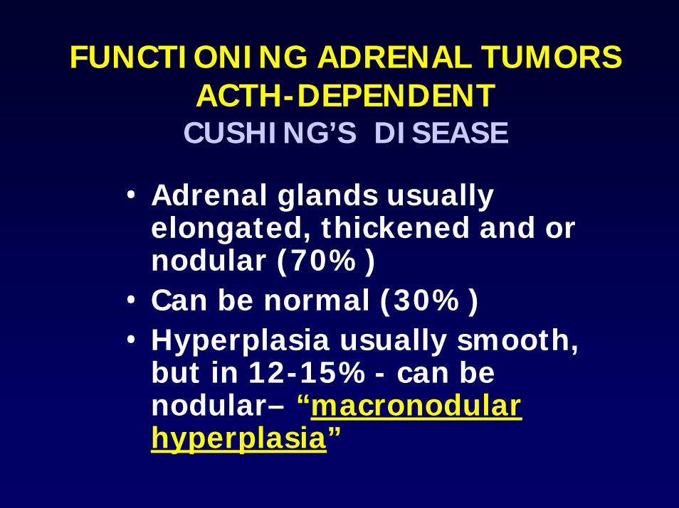
FUNCTIONING ADRENAL TUMORSACTH-DEPENDENT
CUSHING’S DISEASE
• Adrenal glands usually elongated, thickened and or nodular (70%)
• Can be normal (30%)• Hyperplasia usually smooth,
but in 12-15%- can be nodular– “macronodular hyperplasia”

FUNCTIONING ADRENAL TUMORSACTH-DEPENDENT
CUSHING’S DISEASE
• Accuracy for pituitary adenoma detection-CT/MRI-75%
• Inferior Petrosal Sinus Sampling [IPSS]- 90%
• IPSS +CRH stimulation-95%

FUNCTIONING ADRENAL TUMORSACTH-DEPENDENT
CUSHING’S SYNDROMEECTOPIC ACTH
• Rare cause of ACTH-dependent Cushing’s syndrome Accounts for small number of cases-approx. 5%
• Most common site- THORAX (80%)-anterior mediastinum, bronchial, lung parenchyma
• Bronchial carcinoid small tumors-hard to detect and indistinguishable from other lung nodules

FUNCTIONING ADRENAL TUMORSACTH-DEPENDENT
CUSHING’S SYNDROMEECTOPIC ACTH
• Small cell (oat cell) carcinoma• Carcinoid – thymus and
bronchial/lung• Medullary thyroid cancer• Pancreatic neuroendocrine• Pheochromocytoma* If no cause is found, systemic
venous sampling may be employed

FUNCTIONING ADRENAL TUMORSACTH-DEPENDENT
CUSHING’ SYNDROMEECTOPIC ACTH
• Adrenal glands are markedly thickened and nodular
• Thicker and larger than seen in pituitary- dependent Cushing’s
• 19% had nodules larger than 10 mm in size
* Sohaib SA et al AJR 99

FUNCTIONING ADRENAL TUMORSCUSHING’S
ACTH-independent causes:• Adrenal cortical adenoma• Adrenal cortical carcinoma• Account for 90% of cases
Additional rare causes:• ACTH-independent macronodular
adrenal hyperplasia (AIMAH)• Primary Pigmented nodular
hyperplasia (PPNAD)

FUNCTIONING ADRENAL TUMORSACTH-INDEPENDENTCUSHING’S SYNDROME
ADRENAL CORTICAL ADENOMA
IMAGING FINDINGS
• Account for 10-15% of Cushing’s patients
• Range 2-7 cm- Mean size 2-2.5 cm• Thin sections not necessary• Low density – due to lipid content• Contralateral gland maybe atrophic
(due to suppression), but usually appears normal

FUNCTIONING ADRENAL TUMORSACTH-INDEPENDENTCUSHING’S SYNDROME
ADRENAL CORTICAL CARCINOMA
• Tumors usually large (mean > 6cms)
• Often have areas of hemorrhage, necrosis and calcification
• May have IVC involvement

ADRENAL CORTICAL NEOPLASMSCUSHING’S SYNDROME
ADRENAL CORTICAL CARCINOMA
MALIGNANT TUMORS• Direct invasion of adjacent
organs- liver, kidneys• Lymph node metastases• Distant metastases

ADRENAL CORTICAL TUMORSADRENAL CORTICAL CARCINOMA
• 53 proven tumors• 36% with overt hyperfunction• 76% presented as Stage III and
IV• Common sites of metastases
were: Liver, lymph nodes, bone and lungs*Zografos GC, et al J of Surg Oncol 1994

ADRENAL CORTICAL TUMORSADRENAL CORTICAL CARCINOMA
Treatment• Surgical• Laparascopical removal : Only if
there are no imaging features suggestive of malignancy and tumor is under 6 cms
• CT and MR can evaluate for these features prior to surgery

FUNCTIONING ADRENAL TUMORSPRIMARY HYPERALDOSTERONISM
• Hypertension-accts. for 0.1-0.5% of hypertensive cases
• Hypokalemia• Metabolic acidosis• Low plasma renin• Elevated serum/urinary
aldosterone levels

FUNCTIONING ADRENAL TUMORSPRIMARY HYPERALDOSTERONISM
• Adenoma accounts for 75% of cases ( contrast to Cushing’s)
• Adrenal hyperplasia – 25%• Adrenal cortical carcinoma-
rare cause for Conn’s syndrome

FUNCTIONING ADRENAL TUMORSCONN’S ADENOMA OR
ALDOSTERONOMA
IMAGING FINDINGS• Mean size for adenoma 1-1.5
cm• Approx. 20% < 1 cm in size• Thin sections are mandatory
(in contrast to Cushing’s)• Variable appearances

FUNCTIONING ADRENAL TUMORSPRIMARY HYPERALDOSTERONISM
• CT adenoma diagnosis is fairly reliable• But CT diagnosis of adrenal hyperplasia
may be unreliable• Thin section CT detects many adrenal
nodules• Adrenal venous sampling essential in
patients:- with normal adrenal glands- bilateral nodules or thickening- Equivocal CT or MR findings or
discordancy between imaging and lab data

FUNCTIONING ADRENAL TUMORSMEDULLARY HYPERFUNCTION
• Hypertension• Elevated catecholamines-
adrenaline and noradrenaline• May also secrete dopamine,
ACTH, gastrin, serotonin, calcitonin

ADRENAL MEDULLARY NEOPLASMS
PHEOCHROMOCYTOMAS
• Majority of tumors located in adrenal gland (90%)
• Remainder are mostly in abdomen and pelvis
• Small % in neck and chest• Often referred to as the
“10% tumor”• 10% extra-adrenal,
malignant and metastasize

FUNCTIONING ADRENAL TUMORSPHEOCHROMOCYTOMA
EXTRA-ADRENAL LOCATIONS• Retroperitoneal (46%)• Organs of Zuckerkandl(29%)• Pelvis/Urinary bladder (10%)• Thorax Mediastinum/intrapericardial
(10%)• Head and neck (2-4%)

ADRENAL MEDULLARY NEOPLASMS
PHEOCHROMOCYTOMAS- ASSOC. SYNDROMES
• Von Hippel-Lindau disease [vHL]
• Neurofibromatosis• MEA (N) II a and b syndromes• Tuberous sclerosis• Carney’s triad - [GI stromal
tumors, pulmonary chondromas, and pheochromocytomas]
• Familial

ADRENAL MEDULLARY NEOPLASMS
PHEOCHROMOCYTOMAS-CT• Catecholamine response to ionic
iodinated contrast media unpredictable
• Study with small number of cases shown that non-ionic contrast can be given safely
* Raisanen J et al AJR 1984* Mukherjee JJ et al. Radiology 1997

ADRENAL MEDULLARY NEOPLASMS
PHEOCHROMOCYTOMAS-CT
• Tumors are large ( 4-5 cm)- except in MEA[N]
• Can have areas of hemorrhage, necrosis, fluid-fluid levels
• Can show areas of brisk enhancement with IV contrast
• Non-ionic contrast media can be safely used

ADRENAL MEDULLARY NEOPLASMS
PHEOCHROMOCYTOMAS-MRI• Areas of high SI on T1-W images
- Hemorrhage• Can be very hyperintense- “light
bulb” bright on T2-W images- this appearance is not pathognomonic
• Approx. 25% of tumors show areas of heterogenous contrast enhancement
• Cannot be readily differentiated from adrenal cortical carcinoma

FUNCTIONING ADRENAL TUMORSEXTRA-ADRENAL TUMORS
(PARAGANGLIOMAS)
• May be less hormonally active• More likely to be malignant
(40%)• Can be associated with MEN IIA,
MEN IIB, neurofibromatosis, vHL, Carney’s triad and familial
* Shadev A et al. Eur Radiol 2005

ADRENAL MEDULLARY NEOPLASMS
MIBG-SCINTIGRAPHY• Accuracy for pheochromocytoma detection
around 90%- less than that for CT and MR• Whole body imaging capability makes it
suitable to screen for extra-adrenal sites and for metastases
• MIBG may not be needed in patients biochemical evidence of pheo and adrenal mass seen on CT or MRI
* Miskulin J et al Surgery 2003* Greenblatt DY Ann Surg Oncol 2008
ADRENAL MEDULLARY NEOPLASMS
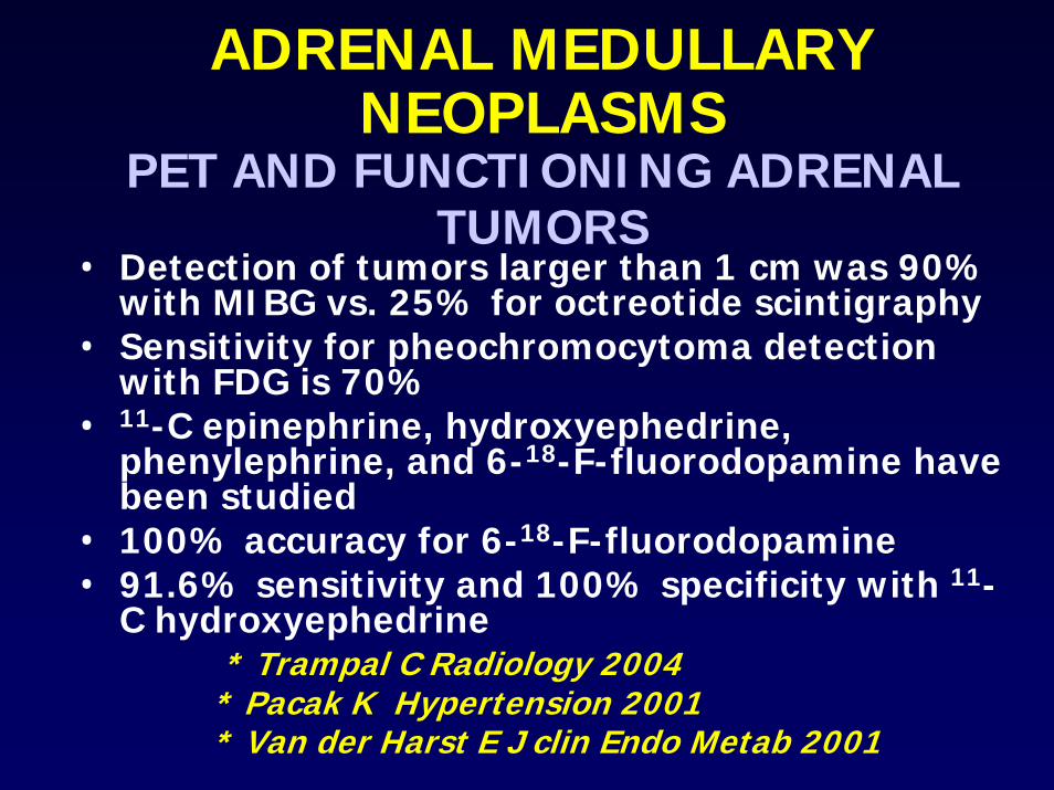
PET AND FUNCTIONING ADRENAL TUMORS
• Detection of tumors larger than 1 cm was 90% with MIBG vs. 25% for octreotide scintigraphy
• Sensitivity for pheochromocytoma detection with FDG is 70%
• 11-C epinephrine, hydroxyephedrine, phenylephrine, and 6-18-F-fluorodopamine have been studied
• 100% accuracy for 6-18-F-fluorodopamine• 91.6% sensitivity and 100% specificity with 11-
C hydroxyephedrine* Trampal C Radiology 2004
* Pacak K Hypertension 2001* Van der Harst E J clin Endo Metab 2001