advances in fecal microbiota transplantation · fecal microbiota transplant ... a fecal suspension...
TRANSCRIPT
READ REVIEWS
WRITE A REVIEW
CORRESPONDENCE:[email protected]
DATE RECEIVED:December 31, 2016
DOI:10.15200/winn.148321.15013
ARCHIVED:December 31, 2016
KEYWORDS:Fecal MicrobiotaTransplantation, Clostridiumdifficile
CITATION:Seema Gollamudi, Advances inFecal MicrobiotaTransplantation , TheWinnower 4:e148321.15013 ,2016 , DOI:10.15200/winn.148321.15013
© Gollamudi This article isdistributed under the terms ofthe Creative CommonsAttribution 4.0 InternationalLicense, which permitsunrestricted use, distribution,and redistribution in anymedium, provided that theoriginal author and source arecredited.
Advances in Fecal Microbiota Transplantation
Seema Gollamudi
Surgical Intensive Care Unit,
Mount Sinai Hospital,
No 1 Gustave Levy Place,
New York, New York-10029
Phone: 212-241-8667
Fax: 212-860-3669
Keywords: Fecal Microbiota Transplantation, Clostridium difficile
[abstract] Intestinal microbiota has an important role in our health. The resident
microbiota of the human intestine in an undisturbed state provides protection against
bacterial infections. The microbiota is influenced by our diet and environment. Dysbiosis
is associated with a range of gastrointestinal and non-gastrointestinal diseases including
Clostridium difficile infection (CDI). Fecal microbiota transplant (FMT) is a process used
when stool is taken from a healthy individual and instilled into a sick person to cure a
certain disease. FMT is now an emerging treatment for a wide range of disorders. Recent
clinical trials have shown that FMT has become an exciting avenue for the treatment for
MEDICINE
Advances in Fecal Microbiota TransplantationSEEMA GOLLAMUDI
Available from:Tennant, J. P., Poisot, T., Kubke, M. F., Michonneau, F., Taylor, M. P., Steel, G., … McKiernan, E. C. (2014). Open Letter to TheAmerican Association for the Advancement of Science. The Winnower. doi:10.15200/winn.140813.35294
✎
GOLLAMUDI The Winnower DECEMBER 21 2016 1
CDI and related illnesses. This review highlights advances in FMT for treating CDI in light
of advances in genomics, animal models of CDI, increased understanding the
microbiome, gut biochemistry and the relationship of gut with other body regions.
[\abstract]
[manuscript]
INTRODUCTION
The term FMT has replaced preceding name of ‘bacteriotherapy because fecal
preparations contain organisms belonging to all three domains of life, including archaea,
eukaryota and viruses (Bakken et al. 2011, Floch 2010). The importance of FMT has risen in the
wake of CDI epidemic although it was practiced since early 4th century AD (Lessa et al. 2015).
Application of FMT for the treatment of severe CDI has been found to have a success rate of
over 90% (van Nood et al. 2013) and is also effective in preventing later recurrence (Aroniadis et
al. 2016, Lagier, Delord, et al. 2015). In a long term follow-up of recurrent CDI (RCDI), FMT was
found to have an efficacy of 91 % primary cure rate, 98 % secondary cure rate and that 97 % of
patients expressed willingness to undergo another FMT in the future, and 53 % stated that they
would choose FMT as a first-line treatment before antibiotics (Brandt et al. 2012). The first
reports of the administration of human fecal suspension by mouth for patients with food
poisoning or severe diarrhea dates back to the 4th century in China (Zhang et al. 2012). The use
of a variety of stool products for treatment of diarrhea, fever, pain, vomiting and constipation
have been reported as early as the 16th century (Zhang et al. 2012). In the 17th century, FMT
was used in veterinary medicine and later termed ‘transfaunation’ (Borody et al. 2004). In 1958
the first use of fecal enemas for the treatment of pseudomembranous colitis in four human
patients exhibited dramatic resolution 24-48h post-treatment (Eiseman et al. 1958).
A fecal suspension can be administered by nasogastric or nasoduodenal tube,
colonoscope, enema, or capsule. The high success rate and safety in the short term reported for
RCDI has elevated FMT as an emerging treatment for a wide range of disorders, including
Parkinson’s disease, fibromyalgia, chronic fatigue syndrome, myoclonus dystopia, multiple
sclerosis, obesity, insulin resistance, metabolic syndrome, and autism (Choi and Cho 2016). The
resident microflora can stay up to 70 days after FMT (Fuentes et al. 2014). The transplanted
feces from a healthy donor can possibly preserve 1,000–1,150 functional bacteria species and
can eventually reestablish a “ healthy ” functional microbiota in the recipient (Qin et al. 2010). A
number of agents have been used for treatment and prevention of CDI in clinical trials (Table I).
Availability of orally deliverable FMT products, such as capsules containing lyophilized fecal
ADVANCES IN FECAL MICROBIOTA TRANSPLANTATION : MEDICINE
GOLLAMUDI The Winnower DECEMBER 21 2016 2
microbiota, has simplified CDI treatment in both CDI and non-CDI diseases. FMT can also be
used to intentionally eradicate colonization with antibiotic-resistant bacteria from the GI tract
(Bilinski et al. 2016, Crum-Cianflone, Sullivan, and Ballon-Landa 2015, Lagier, Million, et al.
2015, Singh et al. 2014).
To characterize the microbiome from Intensive Care Unit (ICU), sequential microbiome
sampling from ICU patients was performed, (Wischmeyer, McDonald, and Knight 2016). The
fecal, oral, and skin samples from 115 mixed ICU patients across four centers in the United
States and Canada were collected at two time points: within 48h of ICU admission, and ICU
discharge or ICU day 10 and compared to the large control group. Critical illness showed a rapid
and distinct change from a ‘healthy’ fecal and oral microbiome. Fecal ICU samples tended to
have a lower relative abundance of Firmicutes and increased relative abundance of
Proteobacteria. Organisms such as Faecali-bacterium shown to confer anti-inflammatory
benefits and which produce short-chain fatty acids that are vital to the gut were found to be
depleted (Sokol et al. 2008). The study concluded that severe dysbiosis occurred in a broad,
larger population of critically ill study participants. They proposed targeted microbial therapies
using specific probiotics or targeted, multimicrobe ‘stool pills’ to restore a healthy microbiome
and improve outcomes in critical illness (Wischmeyer, McDonald, and Knight 2016).
CLOSTRIDIUM DIFFICILE INFECTION
CDI has become one of the most prevalent hospital-acquired infections in recent years.
C. difficile is known to cause severe disease and death (McDonald et al. 2005). The risk factors
for CDI are previous hospitalization, underlying disease, advanced age (>65 years), use of
antibiotics, impairment in humoral immunity, renal disease and hypoalbuminemia (Smits et al.
2016, Lutynski and Kuratowska 1977, Miller et al. 2013, Islam et al. 2014, Di Bella et al. 2015).
Certain antibiotic treatments have also been associated with higher recurrence of CDI (Abou
Chakra et al. 2014). In one study the expression of at least a subset of colonization factors by
the bacterium such as cell surface protein Cwp84 and surface layer protein A (SlpA) was found
to be stimulated in the presence of antibiotics ampicillin and clindamycin (Deneve et al. 2008).
C . difficile causes a spectrum of clinical diseases ranging from mild diarrhea to toxic
megacolon, colonic perforation and death. Systemic complications in life threatening CDI include
cardiopulmonary arrest (Johnson et al. 2001), acute respiratory distress syndrome (Jacob et al.
2004), multiple organ failure (Dobson, Hickey, and Trinder 2003), renal failure (Cunney et al.
1998) and liver damage (Sakurai et al. 2001). However, this bacterium might also be carried
asymptomatically in the gut, potentially leading to 'silent' onward transmission. The period from
spore ingestion to symptom onset is variable but typically short. One study reported 82% of
CDIs occurred within 4 weeks of a potential donor infection (Walker et al. 2012). At least four
ADVANCES IN FECAL MICROBIOTA TRANSPLANTATION : MEDICINE
GOLLAMUDI The Winnower DECEMBER 21 2016 3
events are integral to C. difficile pathogenesis before the development of symptomatic infection.
i. Transmission of spores via the fecal-oral route.
ii. Exposure to antibiotics (or immunosuppressant’s) establishes susceptibility to
infection through perturbation of the intestinal microbiota.
iii. Transformation of spores into vegetative bacteria through interaction with small
molecular germinants, such as bile acids.
iv. Release of C. difficile toxins, which are essential for the disease manifestations.
It is important to diagnose symptomatic and asymptomatic carriers of C . difficile. The
diagnosis of CDI includes presence of clinical symptoms and laboratory assays (Smits et al.
2016). The three types of assays are
i) Testing for C. difficile products such as glutamate dehydrogenase (GDH), aromatic fatty
acids, and the two major toxins (TcdA/TcdB)
ii) Culture methods for detecting toxin producing C. difficile
iii) Nucleic acid amplification for C. difficile genes such as 16sRNA, toxin genes or GDH
gene
The first nucleic acid amplification test to receive Food and Drug Administration (FDA)
approval was the BD GeneOhmTM Cdiff assay in 2009. Tests which detect toxins are specific to
subjects with symptomatic CDI whereas tests which detect parts of the bacterium indicate
asymptomatic carriers (Planche and Wilcox 2015). Cytotoxigenic culture can detect toxigenic C.
difficile and gives a positive result more frequently because of colonization, which means that
individuals can have the bacterium but no free toxin compared to the cytotoxin assay, which
detects preformed toxin in feces (Planche et al. 2013, Planche and Wilcox 2015).
Rapid diagnosis of CDI is desirable to allow early isolation and treatment of patients,
reducing potential patient-to-patient transmission and length of hospital stay for those affected.
In addition, C . difficile strain typing can identify outbreaks within a hospital or the wider
community.
CLOSTRIDIUM DIFFICILE STRAIN TYPING
There are several methods for typing C. difficile (Collins, Elliott, and Riley 2015, Rupnik
2010). The most active polymerase chain reaction (PCR) ribotyping program is in the United
Kingdom available on request via the C . difficile Ribotyping Network (CDRN)
(https://www.gov.uk/guidance/clostridium-difficile-ribotyping-network-cdrn-guide-to-services).
ADVANCES IN FECAL MICROBIOTA TRANSPLANTATION : MEDICINE
GOLLAMUDI The Winnower DECEMBER 21 2016 4
The variant C. difficile strains differ from the notoriously virulent reference strain (VPI 10463) in
restriction sites and length of toxin producing genes tcdA and tcdB, and other pathogenicity
locus (PaLoc) regions (Merrigan et al. 2010). These variant strains have been identified and can
be defined by a series of overlapping PCRs spanning the PaLoc, allowing strains to be assigned
to different toxinotypes I-XXXI (Rupnik 2010). PCR ribotyping is the most frequently (n=49, 89%)
performed method to type isolates at the laboratories. Other methods include Restriction
Enzyme Analysis (REA), Arbitrarily Primed PCR (AP-PCR), Pulsed-Field Gel Electrophoresis
(PFGE), Amplified Fragment Length Polymorphism, Toxinotyping (PCR-RFLP), SlpA PCR-
RFLP (S-layer precursor protein), Multi-Locus Sequence Typing (MLST, 7-12 genes), Multi-
Locus Variable number of tandem repeat Analysis (MLVA), Tandem repeat sequence analysis.
The Clostridium ribotype doesn’t predict severity of the disease. The molecular typing methods
are described in Table II.
PFGE types represent different ribotypes on PCR assay, according to an analysis that is
performed on a random sample of the most prevalent NAP (North American PFGE) types:
NAP1, 027; NAP4, 020; NAP6, 002; NAP7, 078; and NAP11, 106. The characteristics of NAP1
are positive for toxins A and B and C. difficile binary toxin with an 18-bp deletion in tcdC) but
does not meet the 80% cutoff for relatedness on PFGE. Characteristics of NAP7 are positive for
toxins A and B and C. difficile binary toxin with a 39-bp deletion in tcdC) but does not meet the
80% cutoff for relatedness. The strains in the unnamed category include 80 PFGE types that do
not fall within NAP1 through NAP12.
Amongst these the virulence pattern of C. difficile ribotype BI/NAP1/027 is high because it
is unable to down regulate toxin production, with consequent high levels of toxin synthesis.
Additionally in vitro studies with ribotype BI/NAP1/027 strains have demonstrated 16- and 23-
fold higher levels of TcdA and TcdB production, respectively as well as greater numbers of
spores compared to other ribotypes. This increased toxin and spore production capability
enables ribotype BI/NAP1/027 to compete and become the dominant strain within any
environment to which it is introduced (Warny et al. 2005). A range of disease severity was found
in mouse models infected with C. difficile clinical isolates (Lewis, Carter, and Pamer 2016). The
most virulent strains were members of clade 2 (MLST1/ribotype 027) and clade 5 (MLST11, also
classified as ribotype 078). These strains approached and/or surpassed the disease score of
their reference strain, VPI10463 (Merrigan et al. 2010). Members of clade 4 (1 isolate) and clade
1 (16 isolates) had heterogeneous disease scores but consistently produced lower morbidity
than VPI10463.
CLOSTRIDIUM DIFFICILE TOXINS
The 19.6 Kb chromosomal pathogenicity locus (PaLoc) of C. difficile not only carries the
ADVANCES IN FECAL MICROBIOTA TRANSPLANTATION : MEDICINE
GOLLAMUDI The Winnower DECEMBER 21 2016 5
toxin producing tcdA and tcdB genes (Hammond and Johnson 1995, Hundsberger et al. 1997)
but also carries the tcdR gene encoding an alternative RNA polymerase sigma factor
responsible for tcdA and tcdB expression (Mani and Dupuy 2001), the tcdE gene encoding a
putative holing necessary for extracellular release of both toxins (Tan, Wee, and Song 2001)
and the tcdC which negatively regulates TcdA and TcdB synthesis. These three genes regulate
toxin expression and promote toxin release (Matamouros, England, and Dupuy 2007, Heinlen
and Ballard 2010, Solomon 2013). The two protein exotoxins toxin A and toxin B are responsible
for the symptoms of CDI. The severity of the diseases caused by C. difficile has been correlated
to the levels of toxins that are produced during host infection. This observation has strengthened
the idea that the regulation of toxin synthesis is an important part of C. difficile pathogenesis
(Martin-Verstraete, Peltier, and Dupuy 2016). The disease severity is higher when infected by
strains C. difficile BI/NAP1/027 which produce toxin A and toxin B in addition to the binary toxin
(known as C . difficile transferase (CDT)). In addition C . difficile 027 ribotype has been
associated with higher rates of recurrence at some institutions (Loo et al. 2005). The CDT toxin
consists of two polypeptides: a binding component, CDTb, responsible for attachment of the
toxin complex to the host cell surface and the active component, CDTa that displays an actin-
specific ADP-ribosyltransferase activity (Papatheodorou et al. 2011). It has been shown that
CDT depolymerizes the actin cytoskeleton and at low doses, enhances the adhesion of C.
difficile cells to the gastrointestinal epithelium by inducing the formation of microtubule-based
protrusions in the host cell membranes (Schwan et al. 2009). Therefore, CDT might potentialize
the toxicity of TcdA and TcdB and lead to more severe disease, which would be consistent with
the correlation between the presence of binary toxin and the severe outcomes of CDI leading to
higher mortality rate (Gerding et al. 2014). Non-toxigenic strains do not produce toxin A or B
(Heinlen and Ballard 2010). The creation of C . difficile strains with mutations in the genes
encoding toxin A and B indicate that toxin B plays a major role in overall CDI pathogenesis (Di
Bella et al. 2016).
TREATMENT OF CDI
During the last 15 years, CDI has become epidemic and continues to gain momentum. As
the C difficile epidemic continues to grow, the numbers of failed treatments and rates of
relapses or recurrences also are increasing. Metronidazole and vancomycin are the first-line
agents for C difficile treatment; however, recent data suggest that metronidazole is losing its
efficacy, and expert opinion is shifting toward the use of vancomycin as first-line therapy (Zar et
al. 2007).
In 2011, the first drug from a new class of antibacterial, fidaxomicin (FDX), was approved
by the U.S. Food and Drug Administration (FDA) for the treatment of C . difficile associated
ADVANCES IN FECAL MICROBIOTA TRANSPLANTATION : MEDICINE
GOLLAMUDI The Winnower DECEMBER 21 2016 6
diarrhea in adults ≥18 years of age (Optimer Pharmaceuticals, Dificid Package Insert, San
Diego, CA, 2011[www.accessdata.fda.gov/drugsatfda_docs/label/2011/201699s000lbl.pdf]).
FDX inhibits the growth of C. difficile, and other susceptible organisms, by inhibiting the clinically
validated target bacterial RNA polymerase (Coronelli et al. 1975). The mechanism of inhibition
and the predicted binding site for FDX on RNA polymerase (RNAP) is distinct from that of the
rifamycins (Srivastava et al. 2011). Fidaxomicin had a lower rate of recurrences compared with
vancomycin in 2 studies, but its role in the therapy of RCDI has not been established. The only
currently available immunologic approach to treat CDI is administration of pooled intravenous
immunoglobulin (IVIG). However, clinical trials assessing the efficacy of IVIG have been less
than ideal in treating RCDI (O'Horo and Safdar 2009). In another study no statistical significance
in mortality rate was found between IVIG versus non IVIG group treated for severe CDI (Shahani
and Koirala 2015).
The FDA also regulates the FMT process. The FDA issued guidance in 2013 to exercise
enforcement discretion regarding the investigational new drug (IND) requirements for using of
FMT to treat C . difficile infection not responding to standard therapies
(https://www.advancingbio.org/Portals/0/Documents/FDA%20Guidelines%202013.pdf). In the
United States there are two not-for-profit stool banks - OpenBiome, in Medford, Massachusetts,
and AdvancingBio, in Mather, California. OpenBiome was founded in 2012 and is the older of
the two, serving some 500 hospitals with ready-to-use fecal transplants. AdvancingBio, which
opened in February 2015, has about 15 hospital customers. OpenBiome already has submitted
a drug master file to the FDA whereas AdvancingBio proposes to submit a drug master file to
the FDA. This document describes how a company manufactures, processes, packages, and
stores its products.
FMT PROCEDURE AND FOLLOW-UP STUDIES
The current protocol for FMT involves fairly intense screening followed by simple
techniques (Bakken et al. 2011, Kelly et al. 2015). The FMT recipient ceases antibiotics 2-3 days
prior to FMT and an FMT donor is selected. A bowel preparation is administered to all patients
on the day before FMT regardless of route. A donor not only completes a questionnaire but is
also screened for different pathogens. The screening of bacterial pathogens includes C. difficile,
Listeria monocytogenes, Vibrio cholera, Helicobacter pylori, Treponema pallidum, the parasites
include Giardia and Cryptosporidium and viruses include rotavirus, hepatitis A/B/C, Creutzfeldt-
Jakob, and human immunodeficiency virus (Paramsothy et al. 2015, Vestal 2016). Donors can
be excluded if they have had recent antibiotics or tattoos, or a history of gastrointestinal disease.
Although donor stool is used within 8 hours of passage, frozen stool samples have been
ADVANCES IN FECAL MICROBIOTA TRANSPLANTATION : MEDICINE
GOLLAMUDI The Winnower DECEMBER 21 2016 7

administered 1-8 weeks after passage with similar success rates (Costello et al. 2015, Hamilton
et al. 2012). Stool is then collected and prepared for transplant (e.g., diluted and homogenized
before filtration through gauze pads to remove large particulate matter). A recent study has
demonstrated that frozen/thawed stool works as well as fresh stool (Costello et al. 2015,
Hamilton et al. 2012). The routes for FMT until 1989 was retention enema (Bakken et al. 2011),
however, alternative methods have been used subsequently, including nasogastric tube in 1991
(Aas, Gessert, and Bakken 2003), colonoscopy (2000), (Persky and Brandt 2000) and self-
administered enemas (Silverman, Davis, and Pillai 2010).
In one study subjects with relapsing CDI were treated by FMT using frozen encapsulated
inoculum from unrelated donors and had a positive outcome (Youngster et al. 2014). In this
study twenty patients were enrolled with at least 3 episodes of mild to moderate CDI and failure
of a 6- to 8-week taper with vancomycin or at least 2 episodes of severe CDI requiring
hospitalization (Youngster et al. 2014). In another study on two subjects, a mixture of 33 bacteria
was effective in two patients with CDI (Petrof et al. 2013). In a study in Denmark, rectal
bacteriotherapy with a mixture of 12 bacteria resolved CDI in 64% of 55 subjects in 30 days
suggesting that rectal bacteriotherapy is a viable alternative to FMT in patients with relapsing C.
difficile-associated diarrhea (Tvede, Tinggaard, and Helms 2015).
Long term follow-up of subjects who received FMT suggest that both bacterial and viral
genomes need to be sequenced. Bacterial and viral microbiota in the feces at various time in a
long term follow-up for 4.5 years in a patient who recovered CDI post FMT was done and
compared with the stool donor (Broecker et al. 2016). DNA sequencing to characterize bacteria
and double-stranded DNA (dsDNA) viruses including phages were carried out in the feces. Until
7 months post-FMT the patient's microbial communities showed little overall similarity to the
donor, but after 4.5 years, the patient's bacteria attained donor-like compositions at phylum,
class, and order levels with similar bacterial diversity. Unexpectedly they also identified viruses
such as Caudovirales phages and sequences related to giant algae-infecting Chlorella viruses
suggesting that virome analysis should be included in gut microbiota studies (Broecker et al.
2016). In another study three pediatric ulcerative colitis patients received FMT from a single
healthy human donor and transfer of multiple viral lineages between human individuals through
FMT was identified after a course of 22 to 30 FMT treatments (Chehoud et al. 2016).
In an unblinded randomized control trial of FMT on 43 patients, because of widely
different response rates in the control and FMT arms the study was stopped early (van Nood et
al. 2013). The study compared FMT administered via nasoduodenal tube to oral vancomycin for
14 days or vancomycin administered for 14 days plus gastrointestinal lavage in patients with 1–9
prior CDI recurrences. The FMT group received 4 days of vancomycin followed by bowel lavage
ADVANCES IN FECAL MICROBIOTA TRANSPLANTATION : MEDICINE
GOLLAMUDI The Winnower DECEMBER 21 2016 8
before nasoduodenal FMT. 13 of 16 (81%) patients in the FMT arm sustained resolution of
diarrhea after the first fecal infusion compared with 4 of 13 (31%) patients who were treated with
vancomycin and 3 of 13 (23%) who were treated with vancomycin plus bowel lavage (P = 0.008
and 0.003, respectively). Repeat FMT in patients who failed their first FMT resulted in success in
2 of 3 patients, raising FMT response rate to 94% (van Nood et al. 2013).
In another trial the authors compared vancomycin alone with vancomycin+FMT to treat
RCDI (Cammarota et al. 2015). In the FMT+vancomycin group, eighteen of the twenty patients
(90%) exhibited resolution of C. difficile-associated diarrhea. In FMT group, five of the seven
patients with pseudomembranous colitis reported a resolution of diarrhea, whereas in the
vancomycin group resolution of CDI occurred in 5 of the 19 (26%) patients (P < 0.0001). Future
studies should be targeted on a larger population size with diverse disease conditions and
longer term follow-up time to evaluate the safety and efficacy of FMT.
BIOCHEMISTRY OF GUT MICROBIOTA
Before C. difficile can colonize a susceptible host, its highly resistant, metabolically
dormant spore form must germinate in response to specific bile salts in the gastrointestinal tract
(Kevorkian, Shirley, and Shen 2016, Olguin-Araneda et al. 2015, Lewis, Carter, and Pamer
2016). Spore germination begins when germinant receptors bind specific germination-inducing
small molecules known as germinants (Koenigsknecht et al. 2015). The relative concentrations
of bile acids are especially important in C. difficile infections. The primary bile acid taurocholic
acid induces germination of metabolically latent spores (Sorg and Sonenshein 2008), while
secondary bile acids such as lithocholate serve as potent inhibitors for spore germination
(Francis, Allen, and Sorg 2013, Theriot et al. 2014, Lewis, Carter, and Pamer 2016). Francis et al
identified the germination-specific protease, CspC, as the C. difficile bile acid germinant receptor
and showed that bile acid-mediated germination is important for establishing C. difficile disease
in a hamster model of infection (Francis et al. 2013). Only a select group of bacteria produce the
enzymes necessary to dehydroxylate primary bile acids, and they are sensitive to killing by
many commonly prescribed antibiotics (Ridlon, Kang, and Hylemon 2006, Weingarden et al.
2016).
Pseudoproteases play a critical role in regulating the signaling pathway during C. difficile
spore germination (Kevorkian, Shirley, and Shen 2016). Glycine can also act as a germinant
through an uncharacterized mechanism (Sorg and Sonenshein 2008). Interestingly, the gut
microbiota of a healthy gut possesses 7α-dehydroxylation activity which allows them to convert
primary bile acids to secondary bile acids (Stellwag and Hylemon 1978, Sorg and Sonenshein
2008, Ridlon, Kang, and Hylemon 2006). This correlates with high levels of primary bile acids
and low levels of secondary bile acids observed in rCDI patients pre-FMT with levels post-FMT
ADVANCES IN FECAL MICROBIOTA TRANSPLANTATION : MEDICINE
GOLLAMUDI The Winnower DECEMBER 21 2016 9
resembling that from a healthy gut (Weingarden et al. 2014).
Weingarden et al, 2016 analyzed spore germination of 10 clinical C . difficile isolates
exposed to combinations of bile acids present in patient feces before and after FMT
(Weingarden et al. 2016). They found that the concentration of bile acids found in patients' feces
prior to FMT induced germination of C. difficile; however, bile acids at concentrations found in
patients after FMT did not induce germination and inhibited vegetative growth of all C. difficile
strains. In addition upon sequencing they found a correspondence of variation in germination
responses across isolates with mutations in CspC the germinant receptor of C. difficile. Their
results suggest the idea that intra-colonic bile acids play a key mechanistic role in the success of
FMT. Future studies should aim at novel therapeutic alternatives for treatment of R-CDI by
targeting manipulation of bile acid composition in the colon (Weingarden et al. 2016, Smits et al.
2016).
It was observed in mouse models that antibiotic administration not only caused dysbiosis
but also increased free mucosal sialic acid (a carbohydrate energy source for C. difficile) which
inadvertently caused an expansion of C. difficile in the gut (Ng et al. 2013). These results came
from experiments involving colonization of gnotobiotic mice with a sialidase-deficient mutant of
Bacteroides thetaiotaomicron, a model gut symbiont, that reduced free sialic acid levels resulting
in C. difficile down regulating its sialic acid catabolic pathway and exhibiting impaired expansion
(Ng et al. 2013).
GENOMICS OF CDI
The efforts of the The Human Microbiome Project and the European based MetaHit
project followed with a large scale multicenter effort have comprehensively characterized the
human microorganisms found on and in our bodies to determine their various roles (Human
Microbiome Project 2012, Human Microbiome Jumpstart Reference Strains et al. 2010). The
complete genome of the Clostridium difficile type strain DSM 1296T was sequenced using a
combination of single-molecule real-time (SMRT) and Illumina sequencing technology (Riedel et
al. 2015). It revealed the presence of one chromosome and two extrachromosomal elements,
the bacteriophage phiCDIF1296T and a putative plasmid-like structure harboring genes of
another bacteriophage (Riedel et al. 2015). The chromosome of C. difficile DSM 1296T has a
size of 4,109,692 bp and contains 3,596 predicted coding sequences, 35 rRNAs, and 90 tRNAs.
In addition to the genes in the paLoc locus, a Wood-Ljungdahl pathway cluster, genes coding for
carbon monoxide dehydrogenase, a ferredoxin:NAD -oxidoreductase (RNF) complex, formate
dehydrogenases, and hydrogenases; mobile elements such as transposons and prophages were
also identified (Riedel et al. 2015).
16S rRNA-encoding gene sequence analysis was used to compare the fecal microbiota
ADVANCES IN FECAL MICROBIOTA TRANSPLANTATION : MEDICINE
GOLLAMUDI The Winnower DECEMBER 21 2016 10
of patients with RCDI and non-RCDI (Seekatz et al. 2016). Distinct differences in microbiota
diversity of patients that did or did not develop RCDI was identified by 16s rRNA sequencing
(Seekatz et al. 2016). Their results implied that patients with a more dynamic fecal microbiota
were less likely to develop recurrence (Seekatz et al. 2016). Using shotgun metagenomics data
L i et al, (2016) quantified and described the extent of changes to population structure of gut
microbiome after FMT at species and strain level (Li et al. 2016). They observed extensive
coexistence of donor and recipient strains of the same species with considerable amount of
strain replacement over a 3-month observational period. This raises the possibility of using well
characterized and/or customized strains to modulate the microbiome for example outcompeting
undesirable strains (de Vos 2013).
Metagenomic shotgun sequencing (MGS) revealed significant association between gut
microbiome and various intrinsic, environmental, dietary and medication parameters and
disease phenotype with a high replication rate between MGS and 16s rRNA gene sequencing
data from same individuals (Zhernakova et al. 2016). The study showed a relationship between
the microbiome and 126 exogenous and intrinsic host factors, including 31 intrinsic factors, 12
diseases, 19 drug groups, 4 smoking categories, and 60 dietary factors. The study associated
110 factors to 125 species and observed that fecal chromogranin (CgA), a protein secreted by
enteroendocrine cells was exclusively associated with 61 microbial species whose abundance
collectively accounted for 53% of microbial composition. Hence, CgA showed a high potential as
a biomarker for gut health (Zhernakova et al. 2016).
In another study, the structure, function and diversity of the healthy human microbiome
was analyzed from a total of 4,788 specimens from 242 screened and phenotyped adults (129
males, 113 females) representing the majority of the target Human Microbiome Project (HMP)
cohort of 300 individuals (2012). Microbiome samples were collected from up to 18 body sites at
one or two time points from the 242 individuals clinically screened for absence of disease.
Samples were subjected to 16S rRNA gene pyrosequencing (454 Life Sciences), and a subset
were shotgun sequenced for metagenomics using the Illumina GAIIx platform. The large sample
size and consistent sampling of many sites from the same individuals allowed for an
understanding of the relationships among microbes, and between the microbiome and clinical
parameters which may ultimately be critical for understanding microbiome-based disorders.
Considerable progress has been made in recent years to describe the structure and
function of the intestinal microbiota that belong to the major phyla of the Firmicutes,
Actinobacteria, Bacteroidetes, Proteobacteria and Verrucomicrobia (Rajilic-Stojanovic, Smidt,
and de Vos 2007). Significant attention has been given to culture-independent and high-
throughput approaches that generated important baseline information on the intestinal
ADVANCES IN FECAL MICROBIOTA TRANSPLANTATION : MEDICINE
GOLLAMUDI The Winnower DECEMBER 21 2016 11
microbiota composition, the description of a reference metagenome of 3.3 Mb, and its structuring
into clusters, termed enterotypes (Qin et al. 2010, Arumugam et al. 2011, Human Microbiome
Project 2012). In a prospective study, changes in 16s rRNA and bacterial and fungal microbiota
sequencing from stool were able to distinguish C. difficile infection from other forms of diarrhea
in 12 out of 24 enrolled patients who had CDI (Sangster et al. 2016). An increased numbers of
Akkermansia muciniphila in CDI patients was observed which is known to degrade mucin
thereby providing a selective advantage toward CDI. Of the fungal elements, Penicillium was
predominant in CDI; these organisms produce antibacterial chemicals which may resist recovery
of healthy microbiota. The most frequent CDI microbial community networks involved
Peptostreptococcaceae and Enterococcus, with decreased population density of Bacteroides
(Sangster et al. 2016).
A comparative genomic analysis of five Australian toxin-negative isolates of C . difficile
that lack tcdA , tcdB and both binary toxin genes cdtA and cdtB that were recovered from
humans and farm animals with symptoms of gastrointestinal disease was accomplished by 16s
rRNA PCR, whole genome next generation sequencing (Roy Chowdhury et al. 2016). It revealed
that five C. difficile isolates cluster closely with virulent toxigenic strains of C. difficile belonging
to the same sequence type (ST) and have virulence gene profiles akin to those in toxigenic
strains (Roy Chowdhury et al. 2016). They used the genome of the epidemic C. difficile CD630
strain in analysis with the test C. difficile genomes to identify genes that have been correlated
with pathogenicity (Roy Chowdhury et al. 2016).
ANIMAL MODELS
Animal models provide a good alternative to study the host factors, microbiome and
colonization during FMT. Although the microbiota composition at the phylum level generally
appeared to be similar between humans and other animals, at the species and strain level there
is considerable divergence, likely due to underlying differences in host anatomy/physiology, and
dietary regimes (Nguyen et al. 2015). However, recent work has shown that it may be possible to
mitigate this issue somewhat as a significant proportion of human-associated bacterial species
appear to be able to successfully colonize the intestines of animal models following FMT
(Ellekilde et al. 2014). In another study it was found that not only the obese microbiome had an
increased capacity to harvest energy from the diet, this trait was transmissible: colonization of
germ-free mice with an 'obese microbiota' resulted in a significantly greater increase in total
body fat than colonization with a 'lean microbiota'(Turnbaugh et al. 2006). The obese (ob/ob)
phenotype had been shown to be transmissible and is adopted in germ- free mice, infused with
intestinal microbiota from conventionally raised, genetically obese mice [58].
FMT has also been shown to influence behavior as observed from experiments where GF
ADVANCES IN FECAL MICROBIOTA TRANSPLANTATION : MEDICINE
GOLLAMUDI The Winnower DECEMBER 21 2016 12
BALB/c mice, a strain with known deficits in sociality and diligent risk assessment of the
environment received stool from explorative NIH Swiss mice and vice versa (Jacome et al. 2011,
Brinks et al. 2007). The BALB/c mouse recipients exhibited more explorative behavior in the
weeks following FMT. On the other hand, when NIH Swiss mice were colonized with the BALB/c
microbiota, they displayed greater hesitancy (Bercik et al. 2011).
The role of antibiotics as a risk factor for CDI was also explored in mice. Mice treated with
antibiotics were more susceptible to C . difficile infections due to gut dysbiosis (Theriot et al.
2014). Antibiotic treated mice had substantial changes in the gut microbial community and
metabolome which made them susceptible to CDI. They had a decrease in the levels of
secondary bile acids such as glucose, free fatty acids and dipeptides, while the primary bile
acids and sugar alcohols increased. In vitro and ex vivo analyses demonstrated that C. difficile
can exploit specific metabolites that became more abundant in the mouse gut after antibiotics,
including the primary bile acid taurocholate for germination, and carbon sources such as
mannitol, fructose, sorbitol, raffinose and stachyose for growth (Theriot et al. 2014).
Bacterial consortia transplantation (BCT) for targeted restoration of the intestinal
ecosystem is considered a relatively safe and simple procedure and has recently been found to
have effects comparable to FMT in reestablishment of mucosal barrier function in mice with
intestinal dysbiosis (Li et al. 2015). To establish the dysbiosis model, male BALB/c mice were
treated with ceftriaxone intra-gastrically for 7 days. After that, FMT and BCT were performed on
ceftriaxone-treated mice for 3 consecutive days to rebuild the intestinal ecosystem. The effects
of BCT were comparable to that of FMT, especially in normalizing the intestinal levels of
oligomeric mucus/gel-forming (Muc 2), secretory immunoglobulin A (SIgA), and defensins (Li et
al. 2015). Muc2 is the major component of the mucus layer in the small and large intestines. An
increase in intestinal mucus production in mice causes dysbiosis. This is one of the causes of
antibiotic-associated diarrhea because hypersecretion of glycoproteins by the intestinal mucosa
is observed during acute infection (Hasnain, Thornton, and Grencis 2011). Muc2 helps the
disassociation of pathogenic and normal microbiota from the intestinal mucosa to prevent
infectious colitis (Bergstrom et al. 2010). Muc2-deficient mice developed spontaneous colitis
(Wenzel et al. 2014). Intestinal mucus provides a large matrix for a rich array of antimicrobial
molecules such as SIgA and defensins. They are essential components of the innate immune
system and contribute greatly to intestinal barrier function. SIgA is the most abundant
immunoglobulin found in intestinal mucus (Corthesy and Spertini 1999).
Using mouse model of CDI, the interactions between C. difficile cells and other bacteria
and with host mucosa during CDI was investigated (Semenyuk et al. 2015). The GI tracts of
infected mice were sectioned at various days post infection and probed with 16S rRNA
ADVANCES IN FECAL MICROBIOTA TRANSPLANTATION : MEDICINE
GOLLAMUDI The Winnower DECEMBER 21 2016 13
fluorescent in situ hybridization (FISH) probes targeting most bacteria as well as C . difficile
specifically. By using FISH and 16S rRNA gene sequence analysis they drew four major
conclusions about CDI in the mouse: (i) during infection, C. difficile is found in communities in
the cecum and colon, starting at day 1 post infection.; (ii) these communities are associated with
the loose, outer layer of the mucus; (iii) C. difficile is a minority member of these communities;
and (iv) the communities contain bacteria of several families of Bacteroidetes and Firmicutes.
Animal models have provided novel insights into the successes of FMT and how FMT
can influence behavior, metabolism, microbiome and metabolome.
FUTURE DIRECTIONS
As each individual’s microbiome is unique, blinded matches of donor FMT to recipient are
likely to be met with limited success in treating many complex diseases; besides the host factors
cannot be changed and extraneous factors cannot be controlled. Another relevant point to
consider is the metabolome which includes both the host and the microbial derived metabolites
(McHardy et al. 2013, Theriot et al. 2014). It is of significance to point that specific microbiota-
mediated metabolite profiles can be associated with the predisposition to metabolic
impairments, such as impaired glucose homeostasis and non-alcoholic fatty liver disease
(NAFLD) (Dumas et al. 2006). It has also been postulated that the gut microbiota by means of
the associated metabolome may influence the host’s long-term physiology via modulating its
epigenome (Mischke and Plosch 2013). There is evidence that susceptibility to CDI following
antibiotic administration is associated with distinct shifts in gastrointestinal microbiome and
metabolome (Theriot et al. 2014). Future research should be focused on these factors
influencing the microbiota and the metabolome and how these influence CDI and FMT.
A drawback maybe that PaLoc can be horizontally transferred to non-pathogenic strains
characterized by the lack of tcdA and tcdB, converting them in pathogenic strains producer
(Braun et al. 1996, Brouwer et al. 2013). Although PaLoc possesses some characteristics of a
mobile genetic element, it does not appear to be intrinsically mobile and is located at the same
site in all toxigenic C . difficile strains (Braun et al. 1996). Genetic engineering C . difficile to
generate TcdA-\- and TcdB-/- mutant strains or strains overexpressing TcdC being a negative
regulator of TcdA and TcdB should be considered (Heinlen and Ballard 2010). Hence long term
effects of FMT are unknown. Some of the other drawbacks of FMT are cases of peripheral
neuropathy, microscopic colitis, contact dermatitis, Sjögren’s disease, idiopathic
thrombocytopenic purpura, rheumatoid arthritis, weight gain, bacteremia, and ulcerative colitis
flare (Brandt et al. 2012, De Leon, Watson, and Kelly 2013, Quera et al. 2014, Alang and Kelly
2015). The most frequent adverse events caused by FMT are fever, abdominal pain, diarrhea,
increase of C reactive protein which are transient and self-limiting, even if long-term
ADVANCES IN FECAL MICROBIOTA TRANSPLANTATION : MEDICINE
GOLLAMUDI The Winnower DECEMBER 21 2016 14
immunological or infectious effects have not yet been evaluated due to short-term follow-up of
patients. FMT is somewhat less effective in clearing RCDI from patients with Inflammatory Bowel
Disease (IBD), compared with patients without IBD, based on an analysis of 272 patients,
regardless of immunosuppressive therapy (Khoruts et al. 2016).
Fischer et al., 2016 found that severe and severe-complicated indications, inpatient status
during FMT, and the number of previous CDI-related hospitalizations were strongly associated
with early failure of a single FMT for CDI on the basis of a multivariable logistic regression model
(Fischer et al. 2016). In the univariate analysis some of the variables associated with early FMT
failure were the use of non-CDI antibiotics within 8 weeks of FMT, a history of CDI-related
hospitalization, a number of CDI-related hospitalizations, severe or severe-complicated CDI,
pseudomembranous colitis, serum albumin concentration, and inpatient FMT (Fischer et al.
2016).
In another study effectiveness of FMT was evaluated on gastroenterological diseases
based on 45 studies; 34 on CDI, 7 on IBD, 1 on -metabolic syndrome, 1 on constipation, 1 on
pouchitis and 1 on irritable bowel syndrome (IBS) (Rossen et al. 2015). The study found that
FMT is highly effective in CDI, and holds promise in ulcerative colitis. As for Crohn’s Disease,
chronic constipation, pouchitis and IBS data was too limited to draw conclusions. In CDI, 90%
resolution of diarrhea in 33 case series (n = 867) was found, and 94% resolution of diarrhea
after repeated FMT in a randomized controlled trial (RCT) (n = 16). In ulcerative colitis remission
rates of 0% to 68% were found (n = 106). In Crohn’s disease (CD) (n = 6), no benefit was
observed. In IBS, 70% improvement of symptoms was found (n = 13). Reversal of symptoms
was observed in 100% of constipation (n = 3) cases. In pouchitis, none of the patients (n = 8)
achieved remission. One RCT showed significant improvement of insulin sensitivity in metabolic
syndrome (n = 10) (Rossen et al. 2015).
FMT can also be used to decolonize the gut from multi drug–resistant (MDR) bacterial
infections (Manges, Steiner, and Wright 2016). Observations from eight case reports illustrated
the potential effectiveness and safety of FMT for MDR bacterial decolonization. FMT therapy
involved the replacement of a patient’s existing dysfunctional microbiota, containing MDR
opportunistic pathogens, with a healthy microbiota, characterized by high levels of beneficial
microorganisms, exhibiting lower levels of bacterial drug resistance.
A link between antibiotic exposure and altered brain function is well evidenced by the
psychiatric side-effects of antibiotics, which range from anxiety and panic to major depression,
psychosis and delirium (Sternbach and State 1997). The gut microbiome has been shown to
influence mental illnesses (Rogers et al. 2016). The delicate balance between the human
microbiome and the development of psychopathologies is significant given the ease with which
ADVANCES IN FECAL MICROBIOTA TRANSPLANTATION : MEDICINE
GOLLAMUDI The Winnower DECEMBER 21 2016 15
the microbiome can be altered by external factors, such as diet (Gohir et al. 2015), exposure to
antimicrobials (Russell et al. 2012, Ma et al. 2014),or disrupted sleep patterns (Thaiss et al.
2014). Future studies should be targeted to i) identify links between dysbiosis and other human
diseases, ii) develop sensitive diagnostic assays for detecting C. difficile in body fluids and iii)
identify biomarkers which can detect susceptibility prior to CDI.
Manges et al proposed that ideally, whole stool FMT could be replaced by a clearly
defined and regulated complex mixture of functional micro-biota organisms (defined microbiota
transplant) (Manges, Steiner, and Wright 2016). Future work may involve use of FMT for not
only curing CDI but a whole array of human diseases based on the discovery of the relationship
between the gut-brain axis (Carabotti et al. 2015) and brain-gut-bone marrow axis (Santisteban
et al. 2016) and others.
REFERENCES
2012. "Structure, function and diversity of the healthy human microbiome." Nature 4\n86 (7402):207-214. doi: http://www.nature.com/nature/journal/v486/n7402/abs/nature11234.html#supplementary-inf\normation.
Aas, J., C. E. Gessert, and J. S. Bakken. 2003. "Recurrent Clostridium difficile col\nitis: case seriesinvolving 18 patients treated with donor stool administered via a nasogastric tube." Clin Infect Dis 36(5):580-5. doi: 10.1086/367657.
Abou Chakra, C. N., J. Pepin, S. Sirard, and L. Valiquette. 2014. "Risk factors for \nrecurrence,complications and mortality in Clostridium difficile infection: a systematic review." PLoS One 9(6):e98400. doi: 10.1371/journal.pone.0098400.
Alang, N., and C. R. Kelly. 2015. "Weight gain after fecal microbiota transplantation\n." Open ForumInfect Dis 2 (1):ofv004. doi: 10.1093/ofid/ofv004.
Aroniadis, O. C., L. J. Brandt, A. Greenberg, T. Borody, C. R. Kelly, M. Mellow, C. \nSurawicz, L.Cagle, L. Neshatian, N. Stollman, A. Giovanelli, A. Ray, and R. Smith. 2016. "Long-term Follow-upStudy of Fecal Microbiota Transplantation for Severe and/or Complicated Clostridium difficile Infection:A Multicenter Experience." J Clin Gastroenterol 50 (5):398-402. doi:10.1097/MCG.0000000000000374.
Arumugam, M., J. Raes, E. Pelletier, D. Le Paslier, T. Yamada, D. R. Mende, G. R. Fe\nrnandes, J.Tap, T. Bruls, J. M. Batto, M. Bertalan, N. Borruel, F. Casellas, L. Fernandez, L. Gautier, T. Hansen, M.Hattori, T. Hayashi, M. Kleerebezem, K. Kurokawa, M. Leclerc, F. Levenez, C. Manichanh, H. B.Nielsen, T. Nielsen, N. Pons, J. Poulain, J. Qin, T. Sicheritz-Ponten, S. Tims, D. Torrents, E. Ugarte, E.G. Zoetendal, J. Wang, F. Guarner, O. Pedersen, W. M. de Vos, S. Brunak, J. Dore, H. I. T.Consortium Meta, M. Antolin, F. Artiguenave, H. M. Blottiere, M. Almeida, C. Brechot, C. Cara, C.Chervaux, A. Cultrone, C. Delorme, G. Denariaz, R. Dervyn, K. U. Foerstner, C. Friss, M. van deGuchte, E. Guedon, F. Haimet, W. Huber, J. van Hylckama-Vlieg, A. Jamet, C. Juste, G. Kaci, J. Knol,O. Lakhdari, S. Layec, K. Le Roux, E. Maguin, A. Merieux, R. Melo Minardi, C. M'Rini, J. Muller, R.Oozeer, J. Parkhill, P. Renault, M. Rescigno, N. Sanchez, S. Sunagawa, A. Torrejon, K. Turner, G.Vandemeulebrouck, E. Varela, Y. Winogradsky, G. Zeller, J. Weissenbach, S. D. Ehrlich, and P. Bork.2011. "Enterotypes of the human gut microbiome." Nature 473 (7346):174-80. doi:10.1038/nature09944.
Bakken, J. S., T. Borody, L. J. Brandt, J. V. Brill, D. C. Demarco, M. A. Franzos, C.\n Kelly, A. Khoruts,
ADVANCES IN FECAL MICROBIOTA TRANSPLANTATION : MEDICINE
GOLLAMUDI The Winnower DECEMBER 21 2016 16
T. Louie, L. P. Martinelli, T. A. Moore, G. Russell, C. Surawicz, and Workgroup Fecal MicrobiotaTransplantation. 2011. "Treating Clostridium difficile infection with fecal microbiota transplantation." Clin Gastroenterol Hepatol 9 (12):1044-9. doi: 10.1016/j.cgh.2011.08.014.
Bercik, P., E. Denou, J. Collins, W. Jackson, J. Lu, J. Jury, Y. Deng, P. Blennerhas\nsett, J. Macri, K.D. McCoy, E. F. Verdu, and S. M. Collins. 2011. "The intestinal microbiota affect central levels of brain-derived neurotropic factor and behavior in mice." Gastroenterology 141 (2):599-609, 609 e1-3. doi:10.1053/j.gastro.2011.04.052.
Bergstrom, K. S., V. Kissoon-Singh, D. L. Gibson, C. Ma, M. Montero, H. P. Sham, N. R\nyz, T. Huang,A. Velcich, B. B. Finlay, K. Chadee, and B. A. Vallance. 2010. "Muc2 protects against lethal infectiouscolitis by disassociating pathogenic and commensal bacteria from the colonic mucosa." PLoS Pathog6 (5):e1000902. doi: 10.1371/journal.ppat.1000902.
Bilinski, J., P. Grzesiowski, J. Muszynski, M. Wroblewska, K. Madry, K. Robak, T. Dzi\neciatkowski, W.Wiktor-Jedrzejczak, and G. W. Basak. 2016. "Fecal Microbiota Transplantation Inhibits Multidrug-Resistant Gut Pathogens: Preliminary Report Performed in an Immunocompromised Host." ArchImmunol Ther Exp (Warsz) 64 (3):255-8. doi: 10.1007/s00005-016-0387-9.
Borody, T. J., E. F. Warren, S. M. Leis, R. Surace, O. Ashman, and S. Siarakas. 2004.\n"Bacteriotherapy using fecal flora: toying with human motions." J Clin Gastroenterol 38 (6):475-83.
Brandt, L. J., O. C. Aroniadis, M. Mellow, A. Kanatzar, C. Kelly, T. Park, N. Stollma\nn, F. Rohlke, andC. Surawicz. 2012. "Long-term follow-up of colonoscopic fecal microbiota transplant for recurrentClostridium difficile infection." Am J Gastroenterol 107 (7):1079-87. doi: 10.1038/ajg.2012.60.
Braun, V., T. Hundsberger, P. Leukel, M. Sauerborn, and C. von Eichel-Streiber. 1996.\n "Definition ofthe single integration site of the pathogenicity locus in Clostridium difficile." Gene 181 (1-2):29-38.
Brinks, V., M. van der Mark, R. de Kloet, and M. Oitzl. 2007. "Emotion and cognition \nin high and lowstress sensitive mouse strains: a combined neuroendocrine and behavioral study in BALB/c andC57BL/6J mice." Front Behav Neurosci 1:8. doi: 10.3389/neuro.08.008.2007.
Broecker, F., J. Klumpp, M. Schuppler, G. Russo, L. Biedermann, M. Hombach, G. Rogler\n, and K.Moelling. 2016. "Long-term changes of bacterial and viral compositions in the intestine of a recoveredClostridium difficile patient after fecal microbiota transplantation." Cold Spring Harb Mol Case Stud 2(1):a000448. doi: 10.1101/mcs.a000448.
Brouwer, M. S., A. P. Roberts, H. Hussain, R. J. Williams, E. Allan, and P. Mullany. \n2013. "Horizontalgene transfer converts non-toxigenic Clostridium difficile strains into toxin producers." Nat Commun4:2601. doi: 10.1038/ncomms3601.
Cammarota, G., L. Masucci, G. Ianiro, S. Bibbo, G. Dinoi, G. Costamagna, M. Sanguinet\nti, and A.Gasbarrini. 2015. "Randomised clinical trial: faecal microbiota transplantation by colonoscopy vs.vancomycin for the treatment of recurrent Clostridium difficile infection." Aliment Pharmacol Ther 41(9):835-43. doi: 10.1111/apt.13144.
Carabotti, M., A. Scirocco, M. A. Maselli, and C. Severi. 2015. "The gut-brain axis: \ninteractionsbetween enteric microbiota, central and enteric nervous systems." Ann Gastroenterol 28 (2):203-209.
Chehoud, C., A. Dryga, Y. Hwang, D. Nagy-Szakal, E. B. Hollister, R. A. Luna, J. Vers\nalovic, R.Kellermayer, and F. D. Bushman. 2016. "Transfer of Viral Communities between Human Individualsduring Fecal Microbiota Transplantation." MBio 7 (2). doi: 10.1128/mBio.00322-16.
Choi, H. H., and Y. S. Cho. 2016. "Fecal Microbiota Transplantation: Current Applicat\nions,Effectiveness, and Future Perspectives." Clin Endosc. doi: 10.5946/ce.2015.117.
ADVANCES IN FECAL MICROBIOTA TRANSPLANTATION : MEDICINE
GOLLAMUDI The Winnower DECEMBER 21 2016 17
Collins, D. A., B. Elliott, and T. V. Riley. 2015. "Molecular methods for detecting a\nnd typing ofClostridium difficile." Pathology 47 (3):211-8. doi: 10.1097/PAT.0000000000000238.
Coronelli, C., R. J. White, G. C. Lancini, and F. Parenti. 1975. "Lipiarmycin, a new \nantibiotic fromActinoplanes. II. Isolation, chemical, biological and biochemical characterization." J Antibiot (Tokyo)28 (4):253-9.
Corthesy, B., and F. Spertini. 1999. "Secretory immunoglobulin A: from mucosal protec\ntion to vaccinedevelopment." Biol Chem 380 (11):1251-62. doi: 10.1515/BC.1999.160.
Costello, S. P., M. A. Conlon, M. S. Vuaran, I. C. Roberts-Thomson, and J. M. Andrews\n. 2015."Faecal microbiota transplant for recurrent Clostridium difficile infection using long-term frozen stool iseffective: clinical efficacy and bacterial viability data." Aliment Pharmacol Ther 42 (8):1011-8. doi:10.1111/apt.13366.
Crum-Cianflone, N. F., E. Sullivan, and G. Ballon-Landa. 2015. "Fecal microbiota tran\nsplantation andsuccessful resolution of multidrug-resistant-organism colonization." J Clin Microbiol 53 (6):1986-9. doi:10.1128/JCM.00820-15.
Cunney, R. J., C. Magee, E. McNamara, E. G. Smyth, and J. Walshe. 1998. "Clostridium \ndifficilecolitis associated with chronic renal failure." Nephrol Dial Transplant 13 (11):2842-6.
De Leon, L. M., J. B. Watson, and C. R. Kelly. 2013. "Transient flare of ulcerative c\nolitis after fecalmicrobiota transplantation for recurrent Clostridium difficile infection." Clin Gastroenterol Hepatol 11(8):1036-8. doi: 10.1016/j.cgh.2013.04.045.
de Vos, W. M. 2013. "Fame and future of faecal transplantations--developing next-gen\nerationtherapies with synthetic microbiomes." Microb Biotechnol 6 (4):316-25. doi: 10.1111/1751-7915.12047.
Deneve, C., C. Delomenie, M. C. Barc, A. Collignon, and C. Janoir. 2008. "Antibiotics\n involved inClostridium difficile-associated disease increase colonization factor gene expression." J Med Microbiol57 (Pt 6):732-8. doi: 10.1099/jmm.0.47676-0.
Di Bella, S., P. Ascenzi, S. Siarakas, N. Petrosillo, and A. di Masi. 2016. "Clostrid\nium difficile ToxinsA and B: Insights into Pathogenic Properties and Extraintestinal Effects." Toxins (Basel) 8 (5). doi:10.3390/toxins8050134.
Di Bella, S., A. W. Friedrich, E. Garcia-Almodovar, M. S. Gallone, F. Taglietti, S. T\nopino, V. Galati, E.Johnson, S. D'Arezzo, and N. Petrosillo. 2015. "Clostridium difficile infection among hospitalized HIV-infected individuals: epidemiology and risk factors: results from a case-control study (2002-2013)." BMC Infect Dis 15:194. doi: 10.1186/s12879-015-0932-x.
Dobson, G., C. Hickey, and J. Trinder. 2003. "Clostridium difficile colitis causing t\noxic megacolon,severe sepsis and multiple organ dysfunction syndrome." Intensive Care Med 29 (6):1030. doi:10.1007/s00134-003-1754-7.
Dumas, M. E., R. H. Barton, A. Toye, O. Cloarec, C. Blancher, A. Rothwell, J. Fearnsi\nde, R. Tatoud,V. Blanc, J. C. Lindon, S. C. Mitchell, E. Holmes, M. I. McCarthy, J. Scott, D. Gauguier, and J. K.Nicholson. 2006. "Metabolic profiling reveals a contribution of gut microbiota to fatty liver phenotype ininsulin-resistant mice." Proc Natl Acad Sci U S A 103 (33):12511-6. doi: 10.1073/pnas.0601056103.
Eiseman, B., W. Silen, G. S. Bascom, and A. J. Kauvar. 1958. "Fecal enema as an adjun\nct in thetreatment of pseudomembranous enterocolitis." Surgery 44 (5):854-9.
Ellekilde, M., E. Selfjord, C. S. Larsen, M. Jakesevic, I. Rune, B. Tranberg, F. K. V\nogensen, D. S.Nielsen, M. I. Bahl, T. R. Licht, A. K. Hansen, and C. H. Hansen. 2014. "Transfer of gut microbiota from
ADVANCES IN FECAL MICROBIOTA TRANSPLANTATION : MEDICINE
GOLLAMUDI The Winnower DECEMBER 21 2016 18
lean and obese mice to antibiotic-treated mice." Sci Rep 4:5922. doi: 10.1038/srep05922.
Fischer, M., D. Kao, S. R. Mehta, T. Martin, J. Dimitry, A. H. Keshteli, G. K. Cook, \nE. Phelps, B. W.Sipe, H. Xu, and C. R. Kelly. 2016. "Predictors of Early Failure After Fecal Microbiota Transplantationfor the Therapy of Clostridium Difficile Infection: A Multicenter Study." Am J Gastroenterol. doi:10.1038/ajg.2016.180.
Floch, M. H. 2010. "Fecal bacteriotherapy, fecal transplant, and the microbiome." J \nClinGastroenterol 44 (8):529-30. doi: 10.1097/MCG.0b013e3181e1d6e2.
Francis, M. B., C. A. Allen, R. Shrestha, and J. A. Sorg. 2013. "Bile acid recognitio\nn by theClostridium difficile germinant receptor, CspC, is important for establishing infection." PLoS Pathog 9(5):e1003356. doi: 10.1371/journal.ppat.1003356.
Francis, M. B., C. A. Allen, and J. A. Sorg. 2013. "Muricholic acids inhibit Clostrid\nium difficile sporegermination and growth." PLoS One 8 (9):e73653. doi: 10.1371/journal.pone.0073653.
Fuentes, S., E. van Nood, S. Tims, I. Heikamp-de Jong, C. J. ter Braak, J. J. Keller,\n E. G. Zoetendal,and W. M. de Vos. 2014. "Reset of a critically disturbed microbial ecosystem: faecal transplant inrecurrent Clostridium difficile infection." ISME J 8 (8):1621-33. doi: 10.1038/ismej.2014.13.
Gerding, D. N., S. Johnson, M. Rupnik, and K. Aktories. 2014. "Clostridium difficile \nbinary toxin CDT:mechanism, epidemiology, and potential clinical importance." Gut Microbes 5 (1):15-27. doi:10.4161/gmic.26854.
Gohir, W., F. J. Whelan, M. G. Surette, C. Moore, J. D. Schertzer, and D. M. Sloboda.\n 2015."Pregnancy-related changes in the maternal gut microbiota are dependent upon the mother'spericonceptional diet." Gut Microbes 6 (5):310-20. doi: 10.1080/19490976.2015.1086056.
Hamilton, M. J., A. R. Weingarden, M. J. Sadowsky, and A. Khoruts. 2012. "Standardize\nd frozenpreparation for transplantation of fecal microbiota for recurrent Clostridium difficile infection." Am JGastroenterol 107 (5):761-7. doi: 10.1038/ajg.2011.482.
Hammond, G. A., and J. L. Johnson. 1995. "The toxigenic element of Clostridium diffic\nile strain VPI10463." Microb Pathog 19 (4):203-13.
Hasnain, S. Z., D. J. Thornton, and R. K. Grencis. 2011. "Changes in the mucosal barr\nier duringacute and chronic Trichuris muris infection." Parasite Immunol 33 (1):45-55. doi: 10.1111/j.1365-3024.2010.01258.x.
Heinlen, L., and J. D. Ballard. 2010. "Clostridium difficile infection." Am J Med Sc\ni 340 (3):247-52.doi: 10.1097/MAJ.0b013e3181e939d8.
Huber, C. A., N. F. Foster, T. V. Riley, and D. L. Paterson. 2013. "Challenges for st\nandardization ofClostridium difficile typing methods." J Clin Microbiol 51 (9):2810-4. doi: 10.1128/JCM.00143-13.
Human Microbiome Jumpstart Reference Strains, Consortium, K. E. Nelson, G. M. Weinsto\nck, S. K.Highlander, K. C. Worley, H. H. Creasy, J. R. Wortman, D. B. Rusch, M. Mitreva, E. Sodergren, A. T.Chinwalla, M. Feldgarden, D. Gevers, B. J. Haas, R. Madupu, D. V. Ward, B. W. Birren, R. A. Gibbs, B.Methe, J. F. Petrosino, R. L. Strausberg, G. G. Sutton, O. R. White, R. K. Wilson, S. Durkin, M. G.Giglio, S. Gujja, C. Howarth, C. D. Kodira, N. Kyrpides, T. Mehta, D. M. Muzny, M. Pearson, K. Pepin,A. Pati, X. Qin, C. Yandava, Q. Zeng, L. Zhang, A. M. Berlin, L. Chen, T. A. Hepburn, J. Johnson, J.McCorrison, J. Miller, P. Minx, C. Nusbaum, C. Russ, S. M. Sykes, C. M. Tomlinson, S. Young, W. C.Warren, J. Badger, J. Crabtree, V. M. Markowitz, J. Orvis, A. Cree, S. Ferriera, L. L. Fulton, R. S.Fulton, M. Gillis, L. D. Hemphill, V. Joshi, C. Kovar, M. Torralba, K. A. Wetterstrand, A. Abouellleil, A.M. Wollam, C. J. Buhay, Y. Ding, S. Dugan, M. G. FitzGerald, M. Holder, J. Hostetler, S. W. Clifton, E.Allen-Vercoe, A. M. Earl, C. N. Farmer, K. Liolios, M. G. Surette, Q. Xu, C. Pohl, K. Wilczek-Boney,
ADVANCES IN FECAL MICROBIOTA TRANSPLANTATION : MEDICINE
GOLLAMUDI The Winnower DECEMBER 21 2016 19
and D. Zhu. 2010. "A catalog of reference genomes from the human microbiome." Science 328(5981):994-9. doi: 10.1126/science.1183605.
Human Microbiome Project, Consortium. 2012. "Structure, function and diversity of the\n healthyhuman microbiome." Nature 486 (7402):207-14. doi: 10.1038/nature11234.
Hundsberger, T., V. Braun, M. Weidmann, P. Leukel, M. Sauerborn, and C. von Eichel-St\nreiber.1997. "Transcription analysis of the genes tcdA-E of the pathogenicity locus of Clostridium difficile." Eur J Biochem 244 (3):735-42.
Islam, J., A. L. Taylor, K. Rao, G. Huffnagle, V. B. Young, C. Rajkumar, J. Cohen, P.\n Papatheodorou,D. M. Aronoff, and M. J. Llewelyn. 2014. "The role of the humoral immune response to Clostridiumdifficile toxins A and B in susceptibility to C. difficile infection: a case-control study." Anaerobe 27:82-6.doi: 10.1016/j.anaerobe.2014.03.011.
Jacob, S. S., J. C. Sebastian, D. Hiorns, S. Jacob, and P. K. Mukerjee. 2004. "Clostr\nidium difficileand acute respiratory distress syndrome." Heart Lung 33 (4):265-8.
Jacome, L. F., J. A. Burket, A. L. Herndon, and S. I. Deutsch. 2011. "Genetically inb\nred Balb/c micediffer from outbred Swiss Webster mice on discrete measures of sociability: relevance to a geneticmouse model of autism spectrum disorders." Autism Res 4 (6):393-400. doi: 10.1002/aur.218.
Johnson, S., S. A. Kent, K. J. O'Leary, M. M. Merrigan, S. P. Sambol, L. R. Peterson,\n and D. N.Gerding. 2001. "Fatal pseudomembranous colitis associated with a variant clostridium difficile strainnot detected by toxin A immunoassay." Ann Intern Med 135 (6):434-8.
Kelly, C. R., S. Kahn, P. Kashyap, L. Laine, D. Rubin, A. Atreja, T. Moore, and G. Wu\n. 2015. "Updateon Fecal Microbiota Transplantation 2015: Indications, Methodologies, Mechanisms, and Outlook." Gastroenterology 149 (1):223-37. doi: 10.1053/j.gastro.2015.05.008.
Kevorkian, Y., D. J. Shirley, and A. Shen. 2016. "Regulation of Clostridium difficile\n spore germinationby the CspA pseudoprotease domain." Biochimie 122:243-54. doi: 10.1016/j.biochi.2015.07.023.
Khoruts, A., K. M. Rank, K. M. Newman, K. Viskocil, B. P. Vaughn, M. J. Hamilton, and\n M. J.Sadowsky. 2016. "Inflammatory Bowel Disease Affects the Outcome of Fecal MicrobiotaTransplantation for Recurrent Clostridium difficile Infection." Clin Gastroenterol Hepatol. doi:10.1016/j.cgh.2016.02.018.
Koenigsknecht, M. J., C. M. Theriot, I. L. Bergin, C. A. Schumacher, P. D. Schloss, a\nnd V. B. Young.2015. "Dynamics and establishment of Clostridium difficile infection in the murine gastrointestinaltract." Infect Immun 83 (3):934-41. doi: 10.1128/IAI.02768-14.
Lagier, J. C., M. Delord, M. Million, P. Parola, A. Stein, P. Brouqui, and D. Raoult.\n 2015. "Dramaticreduction in Clostridium difficile ribotype 027-associated mortality with early fecal transplantation by thenasogastric route: a preliminary report." Eur J Clin Microbiol Infect Dis 34 (8):1597-601. doi:10.1007/s10096-015-2394-x.
Lagier, J. C., M. Million, P. E. Fournier, P. Brouqui, and D. Raoult. 2015. "Faecal m\nicrobiotatransplantation for stool decolonization of OXA-48 carbapenemase-producing Klebsiella pneumoniae." J Hosp Infect 90 (2):173-4. doi: 10.1016/j.jhin.2015.02.013.
Lessa, F. C., Y. Mu, W. M. Bamberg, Z. G. Beldavs, G. K. Dumyati, J. R. Dunn, M. M. F\narley, S. M.Holzbauer, J. I. Meek, E. C. Phipps, L. E. Wilson, L. G. Winston, J. A. Cohen, B. M. Limbago, S. K.Fridkin, D. N. Gerding, and L. C. McDonald. 2015. "Burden of Clostridium difficile infection in theUnited States." N Engl J Med 372 (9):825-34. doi: 10.1056/NEJMoa1408913.
Lewis, B. B., R. A. Carter, and E. Pamer. 2016. "Bile acid sensitivity and in vivo vi\nrulence of clinical
ADVANCES IN FECAL MICROBIOTA TRANSPLANTATION : MEDICINE
GOLLAMUDI The Winnower DECEMBER 21 2016 20
Clostridium difficile isolates." Anaerobe. doi: 10.1016/j.anaerobe.2016.05.010.
Li, M., P. Liang, Z. Li, Y. Wang, G. Zhang, H. Gao, S. Wen, and L. Tang. 2015. "Fecal\n microbiotatransplantation and bacterial consortium transplantation have comparable effects on the re-establishment of mucosal barrier function in mice with intestinal dysbiosis." Front Microbiol 6:692. doi:10.3389/fmicb.2015.00692.
Li, S. S., A. Zhu, V. Benes, P. I. Costea, R. Hercog, F. Hildebrand, J. Huerta-Cepas,\n M. Nieuwdorp,J. Salojarvi, A. Y. Voigt, G. Zeller, S. Sunagawa, W. M. de Vos, and P. Bork. 2016. "Durablecoexistence of donor and recipient strains after fecal microbiota transplantation." Science 352(6285):586-9. doi: 10.1126/science.aad8852.
Loo, V. G., L. Poirier, M. A. Miller, M. Oughton, M. D. Libman, S. Michaud, A. M. Bou\nrgault, T.Nguyen, C. Frenette, M. Kelly, A. Vibien, P. Brassard, S. Fenn, K. Dewar, T. J. Hudson, R. Horn, P.Rene, Y. Monczak, and A. Dascal. 2005. "A predominantly clonal multi-institutional outbreak ofClostridium difficile-associated diarrhea with high morbidity and mortality." N Engl J Med 353(23):2442-9. doi: 10.1056/NEJMoa051639.
Lutynski, A., and Z. Kuratowska. 1977. "[Fetal hemoglobin in various pathological con\nditions inadults]." Acta Haematol Pol 8 (3):197-204.
Ma, J., A. L. Prince, D. Bader, M. Hu, R. Ganu, K. Baquero, P. Blundell, R. Alan Harr\nis, A. E. Frias,K. L. Grove, and K. M. Aagaard. 2014. "High-fat maternal diet during pregnancy persistently alters theoffspring microbiome in a primate model." Nat Commun 5:3889. doi: 10.1038/ncomms4889.
Manges, A. R., T. S. Steiner, and A. J. Wright. 2016. "Fecal microbiota transplantati\non for theintestinal decolonization of extensively antimicrobial-resistant opportunistic pathogens: a review." Infect Dis (Lond):1-6. doi: 10.1080/23744235.2016.1177199.
Mani, N., and B. Dupuy. 2001. "Regulation of toxin synthesis in Clostridium difficile\n by an alternativeRNA polymerase sigma factor." Proc Natl Acad Sci U S A 98 (10):5844-9. doi:10.1073/pnas.101126598.
Martin-Verstraete, I., J. Peltier, and B. Dupuy. 2016. "The Regulatory Networks That \nControlClostridium difficile Toxin Synthesis." Toxins (Basel) 8 (5). doi: 10.3390/toxins8050153.
Matamouros, S., P. England, and B. Dupuy. 2007. "Clostridium difficile toxin expressi\non is inhibitedby the novel regulator TcdC." Mol Microbiol 64 (5):1274-88. doi: 10.1111/j.1365-2958.2007.05739.x.
McDonald, L. C., G. E. Killgore, A. Thompson, R. C. Owens, Jr., S. V. Kazakova, S. P.\n Sambol, S.Johnson, and D. N. Gerding. 2005. "An epidemic, toxin gene-variant strain of Clostridium difficile." NEngl J Med 353 (23):2433-41. doi: 10.1056/NEJMoa051590.
McHardy, I. H., M. Goudarzi, M. Tong, P. M. Ruegger, E. Schwager, J. R. Weger, T. G. \nGraeber, J.L. Sonnenburg, S. Horvath, C. Huttenhower, D. P. McGovern, A. J. Fornace, Jr., J. Borneman, and J.Braun. 2013. "Integrative analysis of the microbiome and metabolome of the human intestinal mucosalsurface reveals exquisite inter-relationships." Microbiome 1 (1):17. doi: 10.1186/2049-2618-1-17.
Merrigan, M., A. Venugopal, M. Mallozzi, B. Roxas, V. K. Viswanathan, S. Johnson, D. \nN. Gerding,and G. Vedantam. 2010. "Human hypervirulent Clostridium difficile strains exhibit increased sporulationas well as robust toxin production." J Bacteriol 192 (19):4904-11. doi: 10.1128/JB.00445-10.
Miller, M. A., T. Louie, K. Mullane, K. Weiss, A. Lentnek, Y. Golan, Y. Kean, and P. \nSears. 2013."Derivation and validation of a simple clinical bedside score (ATLAS) for Clostridium difficile infectionwhich predicts response to therapy." BMC Infect Dis 13:148. doi: 10.1186/1471-2334-13-148.
Mischke, M., and T. Plosch. 2013. "More than just a gut instinct-the potential interp\nlay between a
ADVANCES IN FECAL MICROBIOTA TRANSPLANTATION : MEDICINE
GOLLAMUDI The Winnower DECEMBER 21 2016 21
baby's nutrition, its gut microbiome, and the epigenome." Am J Physiol Regul Integr Comp Physiol 304(12):R1065-9. doi: 10.1152/ajpregu.00551.2012.
Ng, K. M., J. A. Ferreyra, S. K. Higginbottom, J. B. Lynch, P. C. Kashyap, S. Gopinat\nh, N. Naidu, B.Choudhury, B. C. Weimer, D. M. Monack, and J. L. Sonnenburg. 2013. "Microbiota-liberated hostsugars facilitate post-antibiotic expansion of enteric pathogens." Nature 502 (7469):96-9. doi:10.1038/nature12503.
Nguyen, T. L., S. Vieira-Silva, A. Liston, and J. Raes. 2015. "How informative is the\n mouse for humangut microbiota research?" Dis Model Mech 8 (1):1-16. doi: 10.1242/dmm.017400.
O'Horo, J., and N. Safdar. 2009. "The role of immunoglobulin for the treatment of Clo\nstridium difficileinfection: a systematic review." Int J Infect Dis 13 (6):663-7. doi: 10.1016/j.ijid.2008.11.012.
Olguin-Araneda, V., S. Banawas, M. R. Sarker, and D. Paredes-Sabja. 2015. "Recent adv\nances ingermination of Clostridium spores." Res Microbiol 166 (4):236-43. doi: 10.1016/j.resmic.2014.07.017.
Papatheodorou, P., J. E. Carette, G. W. Bell, C. Schwan, G. Guttenberg, T. R. Brummel\nkamp, and K.Aktories. 2011. "Lipolysis-stimulated lipoprotein receptor (LSR) is the host receptor for the binary toxinClostridium difficile transferase (CDT)." Proc Natl Acad Sci U S A 108 (39):16422-7. doi:10.1073/pnas.1109772108.
Paramsothy, S., T. J. Borody, E. Lin, S. Finlayson, A. J. Walsh, D. Samuel, J. van de\nn Bogaerde, R.W. Leong, S. Connor, W. Ng, H. M. Mitchell, N. Kaakoush, and M. A. Kamm. 2015. "Donor Recruitmentfor Fecal Microbiota Transplantation." Inflamm Bowel Dis 21 (7):1600-6. doi:10.1097/MIB.0000000000000405.
Persky, S. E., and L. J. Brandt. 2000. "Treatment of recurrent Clostridium difficile-\nassociated diarrheaby administration of donated stool directly through a colonoscope." Am J Gastroenterol 95 (11):3283-5. doi: 10.1111/j.1572-0241.2000.03302.x.
Petrof, E. O., G. B. Gloor, S. J. Vanner, S. J. Weese, D. Carter, M. C. Daigneault, E\n. M. Brown, K.Schroeter, and E. Allen-Vercoe. 2013. "Stool substitute transplant therapy for the eradication ofClostridium difficile infection: 'RePOOPulating' the gut." Microbiome 1 (1):3. doi: 10.1186/2049-2618-1-3.
Planche, T. D., K. A. Davies, P. G. Coen, J. M. Finney, I. M. Monahan, K. A. Morris, \nL. O'Connor, S.J. Oakley, C. F. Pope, M. W. Wren, N. P. Shetty, D. W. Crook, and M. H. Wilcox. 2013. "Differences inoutcome according to Clostridium difficile testing method: a prospective multicentre diagnosticvalidation study of C difficile infection." Lancet Infect Dis 13 (11):936-45. doi: 10.1016/S1473-3099(13)70200-7.
Planche, T., and M. H. Wilcox. 2015. "Diagnostic pitfalls in Clostridium difficile in\nfection." Infect DisClin North Am 29 (1):63-82. doi: 10.1016/j.idc.2014.11.008.
Qin, J., R. Li, J. Raes, M. Arumugam, K. S. Burgdorf, C. Manichanh, T. Nielsen, N. Po\nns, F. Levenez,T. Yamada, D. R. Mende, J. Li, J. Xu, S. Li, D. Li, J. Cao, B. Wang, H. Liang, H. Zheng, Y. Xie, J. Tap,P. Lepage, M. Bertalan, J. M. Batto, T. Hansen, D. Le Paslier, A. Linneberg, H. B. Nielsen, E. Pelletier,P. Renault, T. Sicheritz-Ponten, K. Turner, H. Zhu, C. Yu, S. Li, M. Jian, Y. Zhou, Y. Li, X. Zhang, S. Li,N. Qin, H. Yang, J. Wang, S. Brunak, J. Dore, F. Guarner, K. Kristiansen, O. Pedersen, J. Parkhill, J.Weissenbach, H. I. T. Consortium Meta, P. Bork, S. D. Ehrlich, and J. Wang. 2010. "A human gutmicrobial gene catalogue established by metagenomic sequencing." Nature 464 (7285):59-65. doi:10.1038/nature08821.
Quera, R., R. Espinoza, C. Estay, and D. Rivera. 2014. "Bacteremia as an adverse even\nt of fecalmicrobiota transplantation in a patient with Crohn's disease and recurrent Clostridium difficileinfection." J Crohns Colitis 8 (3):252-3. doi: 10.1016/j.crohns.2013.10.002.
ADVANCES IN FECAL MICROBIOTA TRANSPLANTATION : MEDICINE
GOLLAMUDI The Winnower DECEMBER 21 2016 22
Rajilic-Stojanovic, M., H. Smidt, and W. M. de Vos. 2007. "Diversity of the human gas\ntrointestinaltract microbiota revisited." Environ Microbiol 9 (9):2125-36. doi: 10.1111/j.1462-2920.2007.01369.x.
Ridlon, J. M., D. J. Kang, and P. B. Hylemon. 2006. "Bile salt biotransformations by \nhuman intestinalbacteria." J Lipid Res 47 (2):241-59. doi: 10.1194/jlr.R500013-JLR200.
Riedel, T., B. Bunk, J. Wittmann, A. Thurmer, C. Sproer, S. Gronow, H. Liesegang, R. \nDaniel, and J.Overmann. 2015. "Complete Genome Sequence of the Clostridium difficile Type Strain DSM 1296T." Genome Announc 3 (5). doi: 10.1128/genomeA.01186-15.
Rogers, G. B., D. J. Keating, R. L. Young, M. L. Wong, J. Licinio, and S. Wesselingh.\n 2016. "Fromgut dysbiosis to altered brain function and mental illness: mechanisms and pathways." Mol Psychiatry.doi: 10.1038/mp.2016.50.
Rossen, N. G., J. K. MacDonald, E. M. de Vries, G. R. D'Haens, W. M. de Vos, E. G. Zo\netendal, andC. Y. Ponsioen. 2015. "Fecal microbiota transplantation as novel therapy in gastroenterology: Asystematic review." World J Gastroenterol 21 (17):5359-71. doi: 10.3748/wjg.v21.i17.5359.
Roy Chowdhury, P., M. DeMaere, T. Chapman, P. Worden, I. G. Charles, A. E. Darling, a\nnd S. P.Djordjevic. 2016. "Comparative genomic analysis of toxin-negative strains of Clostridium difficile fromhumans and animals with symptoms of gastrointestinal disease." BMC Microbiol 16:41. doi:10.1186/s12866-016-0653-3.
Rupnik, M. 2010. "Clostridium difficile toxinotyping." Methods Mol Biol 646:67-76. d\noi: 10.1007/978-1-60327-365-7_5.
Russell, S. L., M. J. Gold, M. Hartmann, B. P. Willing, L. Thorson, M. Wlodarska, N. \nGill, M. R.Blanchet, W. W. Mohn, K. M. McNagny, and B. B. Finlay. 2012. "Early life antibiotic-driven changes inmicrobiota enhance susceptibility to allergic asthma." EMBO Rep 13 (5):440-7. doi:10.1038/embor.2012.32.
Sakurai, T., K. Hajiro, H. Takakuwa, A. Nishi, M. Aihara, and T. Chiba. 2001. "Liver \nabscess causedby Clostridium difficile." Scand J Infect Dis 33 (1):69-70.
Sangster, W., J. P. Hegarty, K. M. Schieffer, J. R. Wright, J. Hackman, D. R. Toole, \nR. Lamendella,and D. B. Stewart, Sr. 2016. "Bacterial and Fungal Microbiota Changes Distinguish C. difficile Infectionfrom Other Forms of Diarrhea: Results of a Prospective Inpatient Study." Front Microbiol 7:789. doi:10.3389/fmicb.2016.00789.
Santisteban, M. M., S. Kim, C. J. Pepine, and M. K. Raizada. 2016. "Brain-Gut-Bone Ma\nrrow Axis:Implications for Hypertension and Related Therapeutics." Circ Res 118 (8):1327-36. doi:10.1161/CIRCRESAHA.116.307709.
Schwan, C., B. Stecher, T. Tzivelekidis, M. van Ham, M. Rohde, W. D. Hardt, J. Wehlan\nd, and K.Aktories. 2009. "Clostridium difficile toxin CDT induces formation of microtubule-based protrusions andincreases adherence of bacteria." PLoS Pathog 5 (10):e1000626. doi: 10.1371/journal.ppat.1000626.
Seekatz, A. M., K. Rao, K. Santhosh, and V. B. Young. 2016. "Dynamics of the fecal mi\ncrobiome inpatients with recurrent and nonrecurrent Clostridium difficile infection." Genome Med 8 (1):47. doi:10.1186/s13073-016-0298-8.
Semenyuk, E. G., V. A. Poroyko, P. F. Johnston, S. E. Jones, K. L. Knight, D. N. Gerd\ning, and A.Driks. 2015. "Analysis of Bacterial Communities during Clostridium difficile Infection in the Mouse." Infect Immun 83 (11):4383-91. doi: 10.1128/IAI.00145-15.
Shahani, L., and J. Koirala. 2015. "Use of intravenous immunoglobulin in severe Clost\nridium difficile-associated diarrhea." Hosp Pract (1995) 43 (3):154-7. doi: 10.1080/21548331.2015.1071636.
ADVANCES IN FECAL MICROBIOTA TRANSPLANTATION : MEDICINE
GOLLAMUDI The Winnower DECEMBER 21 2016 23
Silverman, M. S., I. Davis, and D. R. Pillai. 2010. "Success of self-administered hom\ne fecaltransplantation for chronic Clostridium difficile infection." Clin Gastroenterol Hepatol 8 (5):471-3. doi:10.1016/j.cgh.2010.01.007.
Singh, R., E. van Nood, M. Nieuwdorp, B. van Dam, I. J. ten Berge, S. E. Geerlings, a\nnd F. J.Bemelman. 2014. "Donor feces infusion for eradication of Extended Spectrum beta-Lactamaseproducing Escherichia coli in a patient with end stage renal disease." Clin Microbiol Infect 20(11):O977-8. doi: 10.1111/1469-0691.12683.
Smits, W. K., D. Lyras, D. B. Lacy, M. H. Wilcox, and E. J. Kuijper. 2016. "Clostridi\num difficileinfection." Nat Rev Dis Primers 2:16020. doi: 10.1038/nrdp.2016.20.
Sokol, H., B. Pigneur, L. Watterlot, O. Lakhdari, L. G. Bermudez-Humaran, J. J. Grata\ndoux, S.Blugeon, C. Bridonneau, J. P. Furet, G. Corthier, C. Grangette, N. Vasquez, P. Pochart, G. Trugnan,G. Thomas, H. M. Blottiere, J. Dore, P. Marteau, P. Seksik, and P. Langella. 2008. "Faecalibacteriumprausnitzii is an anti-inflammatory commensal bacterium identified by gut microbiota analysis of Crohndisease patients." Proc Natl Acad Sci U S A 105 (43):16731-6. doi: 10.1073/pnas.0804812105.
Solomon, K. 2013. "The host immune response to Clostridium difficile infection." The\nr Adv Infect Dis1 (1):19-35. doi: 10.1177/2049936112472173.
Sorg, J. A., and A. L. Sonenshein. 2008. "Bile salts and glycine as cogerminants for \nClostridiumdifficile spores." J Bacteriol 190 (7):2505-12. doi: 10.1128/JB.01765-07.
Srivastava, A., M. Talaue, S. Liu, D. Degen, R. Y. Ebright, E. Sineva, A. Chakraborty\n, S. Y. Druzhinin,S. Chatterjee, J. Mukhopadhyay, Y. W. Ebright, A. Zozula, J. Shen, S. Sengupta, R. R. Niedfeldt, C.Xin, T. Kaneko, H. Irschik, R. Jansen, S. Donadio, N. Connell, and R. H. Ebright. 2011. "New target forinhibition of bacterial RNA polymerase: 'switch region'." Curr Opin Microbiol 14 (5):532-43. doi:10.1016/j.mib.2011.07.030.
Stellwag, E. J., and P. B. Hylemon. 1978. "Characterization of 7-alpha-dehydroxylase \nin Clostridiumleptum." Am J Clin Nutr 31 (10 Suppl):S243-S247.
Sternbach, H., and R. State. 1997. "Antibiotics: neuropsychiatric effects and psychot\nropicinteractions." Harv Rev Psychiatry 5 (4):214-26.
Tan, K. S., B. Y. Wee, and K. P. Song. 2001. "Evidence for holin function of tcdE gen\ne in thepathogenicity of Clostridium difficile." J Med Microbiol 50 (7):613-9. doi: 10.1099/0022-1317-50-7-613.
Thaiss, C. A., D. Zeevi, M. Levy, G. Zilberman-Schapira, J. Suez, A. C. Tengeler, L. \nAbramson, M. N.Katz, T. Korem, N. Zmora, Y. Kuperman, I. Biton, S. Gilad, A. Harmelin, H. Shapiro, Z. Halpern, E.Segal, and E. Elinav. 2014. "Transkingdom control of microbiota diurnal oscillations promotesmetabolic homeostasis." Cell 159 (3):514-29. doi: 10.1016/j.cell.2014.09.048.
Theriot, C. M., M. J. Koenigsknecht, P. E. Carlson, Jr., G. E. Hatton, A. M. Nelson, \nB. Li, G. B.Huffnagle, Z. Li J, and V. B. Young. 2014. "Antibiotic-induced shifts in the mouse gut microbiome andmetabolome increase susceptibility to Clostridium difficile infection." Nat Commun 5:3114. doi:10.1038/ncomms4114.
Turnbaugh, P. J., R. E. Ley, M. A. Mahowald, V. Magrini, E. R. Mardis, and J. I. Gord\non. 2006. "Anobesity-associated gut microbiome with increased capacity for energy harvest." Nature 444(7122):1027-31. doi: 10.1038/nature05414.
Tvede, M., M. Tinggaard, and M. Helms. 2015. "Rectal bacteriotherapy for recurrent Cl\nostridiumdifficile-associated diarrhoea: results from a case series of 55 patients in Denmark 2000-2012." ClinMicrobiol Infect 21 (1):48-53. doi: 10.1016/j.cmi.2014.07.003.
ADVANCES IN FECAL MICROBIOTA TRANSPLANTATION : MEDICINE
GOLLAMUDI The Winnower DECEMBER 21 2016 24
van Nood, E., A. Vrieze, M. Nieuwdorp, S. Fuentes, E. G. Zoetendal, W. M. de Vos, C. \nE. Visser, E.J. Kuijper, J. F. Bartelsman, J. G. Tijssen, P. Speelman, M. G. Dijkgraaf, and J. J. Keller. 2013."Duodenal infusion of donor feces for recurrent Clostridium difficile." N Engl J Med 368 (5):407-15. doi:10.1056/NEJMoa1205037.
Vestal, Richard. 2016. "Fecal Microbiota Transplant." Hospital Medicine Clinics 5 (1\n):58-70. doi:http://dx.doi.org/10.1016/j.ehmc.2015.08.006.
Walker, A. S., D. W. Eyre, D. H. Wyllie, K. E. Dingle, R. M. Harding, L. O'Connor, D.\n Griffiths, A.Vaughan, J. Finney, M. H. Wilcox, D. W. Crook, and T. E. Peto. 2012. "Characterisation of Clostridiumdifficile hospital ward-based transmission using extensive epidemiological data and molecular typing." PLoS Med 9 (2):e1001172. doi: 10.1371/journal.pmed.1001172.
Warny, M., J. Pepin, A. Fang, G. Killgore, A. Thompson, J. Brazier, E. Frost, and L. \nC. McDonald.2005. "Toxin production by an emerging strain of Clostridium difficile associated with outbreaks ofsevere disease in North America and Europe." Lancet 366 (9491):1079-84. doi: 10.1016/S0140-6736(05)67420-X.
Weingarden, A. R., C. Chen, A. Bobr, D. Yao, Y. Lu, V. M. Nelson, M. J. Sadowsky, and\n A. Khoruts.2014. "Microbiota transplantation restores normal fecal bile acid composition in recurrent Clostridiumdifficile infection." Am J Physiol Gastrointest Liver Physiol 306 (4):G310-9. doi:10.1152/ajpgi.00282.2013.
Weingarden, A. R., P. I. Dosa, E. DeWinter, C. J. Steer, M. K. Shaughnessy, J. R. Joh\nnson, A.Khoruts, and M. J. Sadowsky. 2016. "Changes in Colonic Bile Acid Composition following FecalMicrobiota Transplantation Are Sufficient to Control Clostridium difficile Germination and Growth." PLoS One 11 (1):e0147210. doi: 10.1371/journal.pone.0147210.
Wenzel, U. A., M. K. Magnusson, A. Rydstrom, C. Jonstrand, J. Hengst, M. E. Johansson\n, A. Velcich,L. Ohman, H. Strid, H. Sjovall, G. C. Hansson, and M. J. Wick. 2014. "Spontaneous colitis in Muc2-deficient mice reflects clinical and cellular features of active ulcerative colitis." PLoS One 9(6):e100217. doi: 10.1371/journal.pone.0100217.
Wiep Klaas Smits, Dena Lyras, D. Borden Lacy, Mark H. Wilcox4 and Ed J. Kuijper. 2016\n."Clostridium difficile infection." Nat Rev Dis Primers 2:16021. doi: 10.1038/nrdp.2016.21.
Wischmeyer, P. E., D. McDonald, and R. Knight. 2016. "Role of the microbiome, probiot\nics, and'dysbiosis therapy' in critical illness." Curr Opin Crit Care 22 (4):347-53. doi:10.1097/MCC.0000000000000321.
Youngster, I., G. H. Russell, C. Pindar, T. Ziv-Baran, J. Sauk, and E. L. Hohmann. 20\n14. "Oral,capsulized, frozen fecal microbiota transplantation for relapsing Clostridium difficile infection." JAMA312 (17):1772-8. doi: 10.1001/jama.2014.13875.
Zar, F. A., S. R. Bakkanagari, K. M. Moorthi, and M. B. Davis. 2007. "A comparison of\n vancomycinand metronidazole for the treatment of Clostridium difficile-associated diarrhea, stratified by diseaseseverity." Clin Infect Dis 45 (3):302-7. doi: 10.1086/519265.
Zhang, Faming, Wensheng Luo, Yan Shi, Zhining Fan, and Guozhong Ji. 2012. "Should We\nStandardize the 1,700-Year-Old Fecal Microbiota Transplantation[quest]." Am J Gastroenterol 107(11):1755-1755.
Zhernakova, A., A. Kurilshikov, M. J. Bonder, E. F. Tigchelaar, M. Schirmer, T. Vatan\nen, Z. Mujagic,A. V. Vila, G. Falony, S. Vieira-Silva, J. Wang, F. Imhann, E. Brandsma, S. A. Jankipersadsing, M.Joossens, M. C. Cenit, P. Deelen, M. A. Swertz, study LifeLines cohort, R. K. Weersma, E. J. Feskens,M. G. Netea, D. Gevers, D. Jonkers, L. Franke, Y. S. Aulchenko, C. Huttenhower, J. Raes, M. H.Hofker, R. J. Xavier, C. Wijmenga, and J. Fu. 2016. "Population-based metagenomics analysis reveals
ADVANCES IN FECAL MICROBIOTA TRANSPLANTATION : MEDICINE
GOLLAMUDI The Winnower DECEMBER 21 2016 25
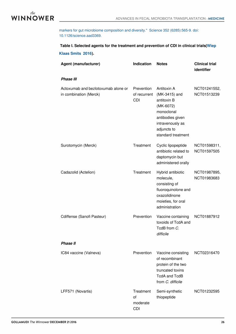
markers for gut microbiome composition and diversity." Science 352 (6285):565-9. doi:10.1126/science.aad3369.
Table I. Selected agents for the treatment and prevention of CDI in clinical trials (Wiep
Klaas Smits 2016).
Agent (manufacturer) Indication Notes Clinical trialidentifier
Phase III
Actoxumab and bezlotoxumab alone orin combination (Merck)
Preventionof recurrentCDI
Antitoxin A(MK‑3415) andantitoxin B(MK‑6072)monoclonalantibodies givenintravenously asadjuncts tostandard treatment
NCT01241552,NCT01513239
Surotomycin (Merck) Treatment Cyclic lipopeptideantibiotic related todaptomycin butadministered orally
NCT01598311,NCT01597505
Cadazolid (Actelion) Treatment Hybrid antibioticmolecule,consisting offluoroquinolone andoxazolidinonemoieties, for oraladministration
NCT01987895,NCT01983683
Cdiffense (Sanofi Pasteur) Prevention Vaccine containingtoxoids of TcdA andTcdB from C.difficile
NCT01887912
Phase II
IC84 vaccine (Valneva) Prevention Vaccine consistingof recombinantprotein of the twotruncated toxinsTcdA and TcdBfrom C. difficile
NCT02316470
LFF571 (Novartis) TreatmentofmoderateCDI
Semi-syntheticthiopeptide
NCT01232595
ADVANCES IN FECAL MICROBIOTA TRANSPLANTATION : MEDICINE
GOLLAMUDI The Winnower DECEMBER 21 2016 26
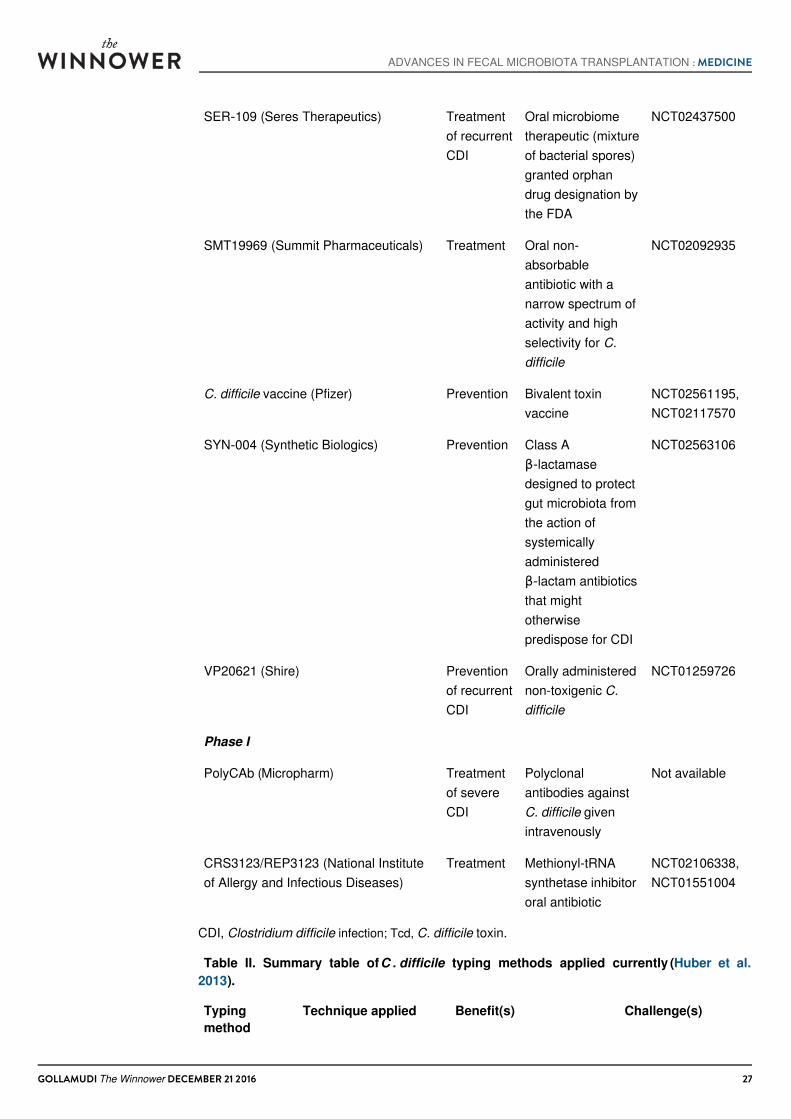
SER‑109 (Seres Therapeutics) Treatmentof recurrentCDI
Oral microbiometherapeutic (mixtureof bacterial spores)granted orphandrug designation bythe FDA
NCT02437500
SMT19969 (Summit Pharmaceuticals) Treatment Oral non-absorbableantibiotic with anarrow spectrum ofactivity and highselectivity for C.difficile
NCT02092935
C. difficile vaccine (Pfizer) Prevention Bivalent toxinvaccine
NCT02561195,NCT02117570
SYN‑004 (Synthetic Biologics) Prevention Class Aβ‑lactamasedesigned to protectgut microbiota fromthe action ofsystemicallyadministeredβ‑lactam antibioticsthat mightotherwisepredispose for CDI
NCT02563106
VP20621 (Shire) Preventionof recurrentCDI
Orally administerednon-toxigenic C.difficile
NCT01259726
Phase I
PolyCAb (Micropharm) Treatmentof severeCDI
Polyclonalantibodies againstC. difficile givenintravenously
Not available
CRS3123/REP3123 (National Instituteof Allergy and Infectious Diseases)
Treatment Methionyl-tRNAsynthetase inhibitororal antibiotic
NCT02106338,NCT01551004
CDI, Clostridium difficile infection; Tcd, C. difficile toxin.
Table II. Summary table of C . difficile typing methods applied currently (Huber et al.2013).
Typingmethod
Technique applied Benefit(s) Challenge(s)
ADVANCES IN FECAL MICROBIOTA TRANSPLANTATION : MEDICINE
GOLLAMUDI The Winnower DECEMBER 21 2016 27
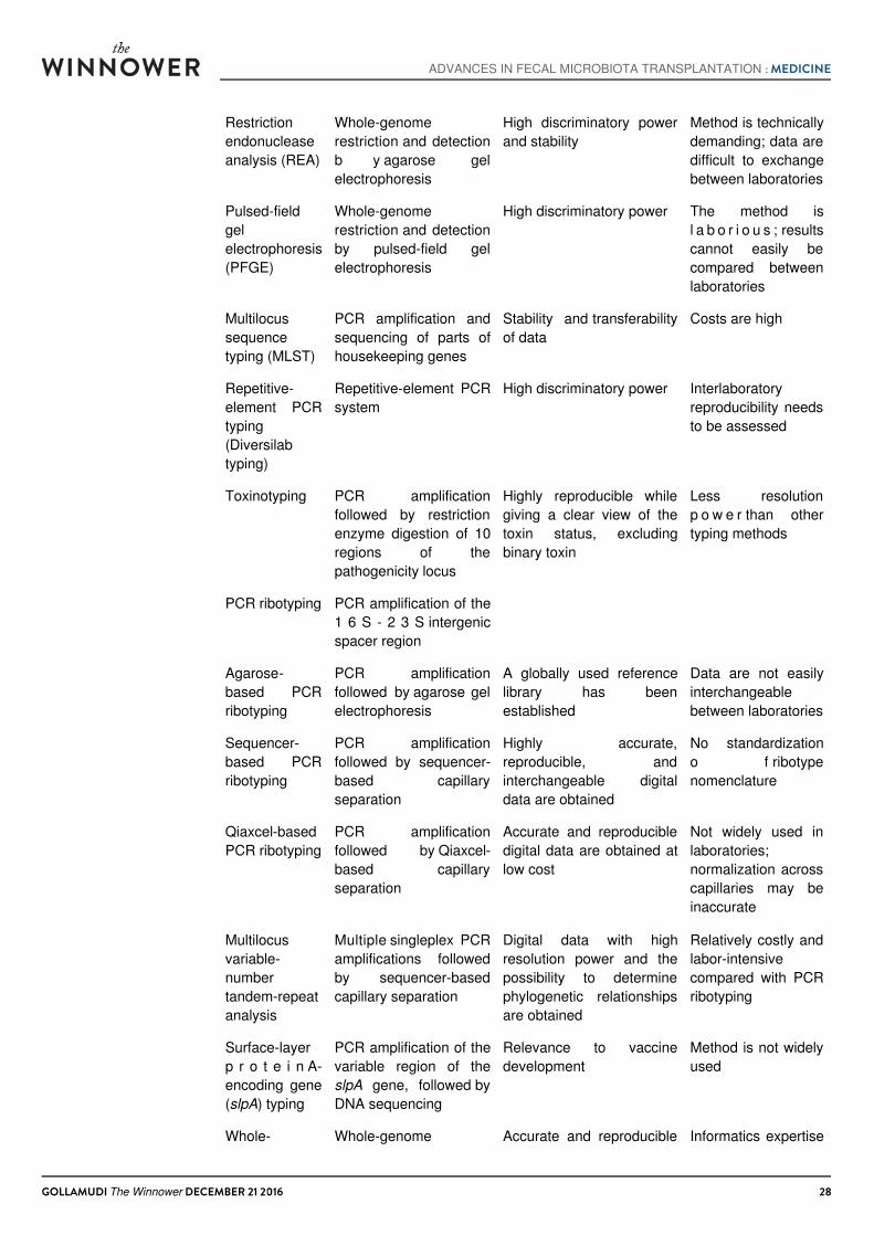
Restrictionendonucleaseanalysis (REA)
Whole-genomerestriction and detectionb y agarose gelelectrophoresis
High discriminatory powerand stability
Method is technicallydemanding; data aredifficult to exchangebetween laboratories
Pulsed-fieldgelelectrophoresis(PFGE)
Whole-genomerestriction and detectionby pulsed-field gelelectrophoresis
High discriminatory power The method isl a b o r i o u s ; resultscannot easily becompared betweenlaboratories
Multilocussequencetyping (MLST)
PCR amplification andsequencing of parts ofhousekeeping genes
Stability and transferabilityof data
Costs are high
Repetitive-element PCRtyping(Diversilabtyping)
Repetitive-element PCRsystem
High discriminatory power Interlaboratoryreproducibility needsto be assessed
Toxinotyping PCR amplificationfollowed by restrictionenzyme digestion of 10regions of thepathogenicity locus
Highly reproducible whilegiving a clear view of thetoxin status, excludingbinary toxin
Less resolutionp o w e r than othertyping methods
PCR ribotyping PCR amplification of the1 6 S - 2 3 S intergenicspacer region
Agarose-based PCRribotyping
PCR amplificationfollowed by agarose gelelectrophoresis
A globally used referencelibrary has beenestablished
Data are not easilyinterchangeablebetween laboratories
Sequencer-based PCRribotyping
PCR amplificationfollowed by sequencer-based capillaryseparation
Highly accurate,reproducible, andinterchangeable digitaldata are obtained
No standardizationo f ribotypenomenclature
Qiaxcel-basedPCR ribotyping
PCR amplificationfollowed by Qiaxcel-based capillaryseparation
Accurate and reproducibledigital data are obtained atlow cost
Not widely used inlaboratories;normalization acrosscapillaries may beinaccurate
Multilocusvariable-numbertandem-repeatanalysis
Multiple singleplex PCRamplifications followedby sequencer-basedcapillary separation
Digital data with highresolution power and thepossibility to determinephylogenetic relationshipsare obtained
Relatively costly andlabor-intensivecompared with PCRribotyping
Surface-layerp r o t e i n A-encoding gene(slpA) typing
PCR amplification of thevariable region of theslpA gene, followed byDNA sequencing
Relevance to vaccinedevelopment
Method is not widelyused
Whole- Whole-genome Accurate and reproducible Informatics expertise
ADVANCES IN FECAL MICROBIOTA TRANSPLANTATION : MEDICINE
GOLLAMUDI The Winnower DECEMBER 21 2016 28
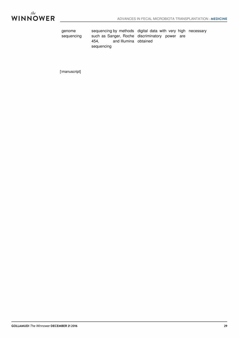
genomesequencing
sequencing by methodssuch as Sanger, Roche454, and Illuminasequencing
digital data with very highdiscriminatory power areobtained
necessary
[\manuscript]
ADVANCES IN FECAL MICROBIOTA TRANSPLANTATION : MEDICINE
GOLLAMUDI The Winnower DECEMBER 21 2016 29