advances in management of periprosthetic joint infections ... · table i. definition of pji...
TRANSCRIPT
129
Abstract. – OBJECTIVE: The purpose of our study is to assess the incidence of prosthetic joint infection (PJI) after total Knee arthroplas-ty (TKA), total Hip arthroplasty (THA) and to-tal Shoulder arthroplasty (TSA), to identify risk factors, determine the microbial spectrum and management’s outcome.
PATIENTS AND METHODS: A case-control, retrospective observational study was performed analyzing patients who developed a PJI after TKA, THA, and TSA from 2000 to 2017 at our hospital. The patient’s risk profile was defined extracting from clinical records the following data: sex, age, BMI, type of implant, comorbidity, year of surgery, year of infection, previous intra-articular injection, microbial isolation, medical and surgical manage-ment outcome. We include in the “control group” for each “case” at least 3 patients who didn’t have a PJI after TJA.
RESULTS: 28 patients met all inclusion and ex-clusion criteria. Comparing the “cases” with “con-trols” demographics parameters, medical comor-bidities and previous intra-articular injection were not associated with an increased risk of PJI. Com-paring the “early/delayed group” with “late group”, BMI was associated with an increased risk of ear-ly/delayed PJI, while demographics parameters, medical comorbidities, and previous intra-articular injection did not significantly increase the risk of PJI. Logistic regression showed that for each BMI unit there was a 20-fold increased risk of early prosthetic infection (OR 1.19, IC 1.03-1.38, p=0.01). Staphylococci were isolated most frequently from pre-operative and intra-operative cultures. Two-stage arthroplasty exchange and surgical debride-ment resulted in the most performed surgical treat-ment with a success rate of 88 and 87%.
CONCLUSIONS: Obesity is a risk factor for “ear-ly/delayed infection” of TJA. Two-stage arthro-plasty exchange, debridement, antibiotics, and implant retention in patients are treatments with a high rate of success in terms of reinfection.
Key WordsProsthetic joint infection (PJI), total Knee arthroplas-
ty (TKA), total Hip arthroplasty (THA), Risk factors, Ar-throplasty exchange, Antibiotics therapy.
Introduction
Total joint arthroplasty is a successful treat-ment that improves joint function, relieves pain, and increases the overall quality of life1. Due to the increase in the number of patients undergoing joint replacement procedure, a concomitant in-crease in the number of complications is expect-ed2. “Prosthetic Joint Infection” (PJI) is one of the most feared complications of arthroplasties that has been estimated to range from 2.0% to 2.4% of total hip and knee replacement3. Despite ad-vancement in surgical procedures and in antibi-otic prophylaxis, PJI remains the most important cause of implant failure and require for revision. Its consequences represent an impressive clinical and economic burden. It extends hospitalization by 12-20 days and doubles the re-hospitalization rate with a considerable impact on the patient quality of life. PJI often requires one or more complex surgical procedure, that increase the cost of care. Treatment’s cost of a PJI is 3 to 4 times the cost of a primary implantation and 2.8 times the cost of an aseptic revision arthroplasty3,4. Today, a gold-standard definition of PJI does not exist. To standardize the definition of PJI, espe-cially to avoid compromising the validity and comparability of study’s results, several medical societies and working groups have proposed dif-ferent definitions. In 2011, the Musculoskeletal Infectious Society (MSIS) proposed a group of criteria for the diagnosis of PJI5, that was later revised by the International Consensus Meeting on PJI6, providing the best available evidence regarding the prevention, diagnosis, and manage-ment of PJI (Table I). In 2018 Parvizi et al7 pub-lished an evidence-based and validated updated version of PJI diagnosis criteria. They assigned a weighted score to all minor criteria and divided preoperative from intraoperative diagnosis. The new criteria definition obtained a better sensi-
European Review for Medical and Pharmacological Sciences
R. PAPALIA1, U. VESPASIANI-GENTILUCCI2, U.G. LONGO1, C. ESPOSITO1, B. ZAMPOGNA1, R. ANTONELLI INCALZI3, V. DENARO1
1Department of Orthopedic and Trauma Surgery University Campus Bio-Medico of Rome, Rome, Italy2Department of Internal Medicine and Hepatology Unit, University Campus Bio-Medico, Rome, Italy3Department of Internal Medicine and Geriatric Unit, University Campus Bio-Medico, Rome, Italy
Corresponding Author: Biagio Zampogna, MD; e-mail: [email protected]
Advances in management of periprosthetic joint infections: an historical prospective study
2019; 23(2 Suppl.): 129-138
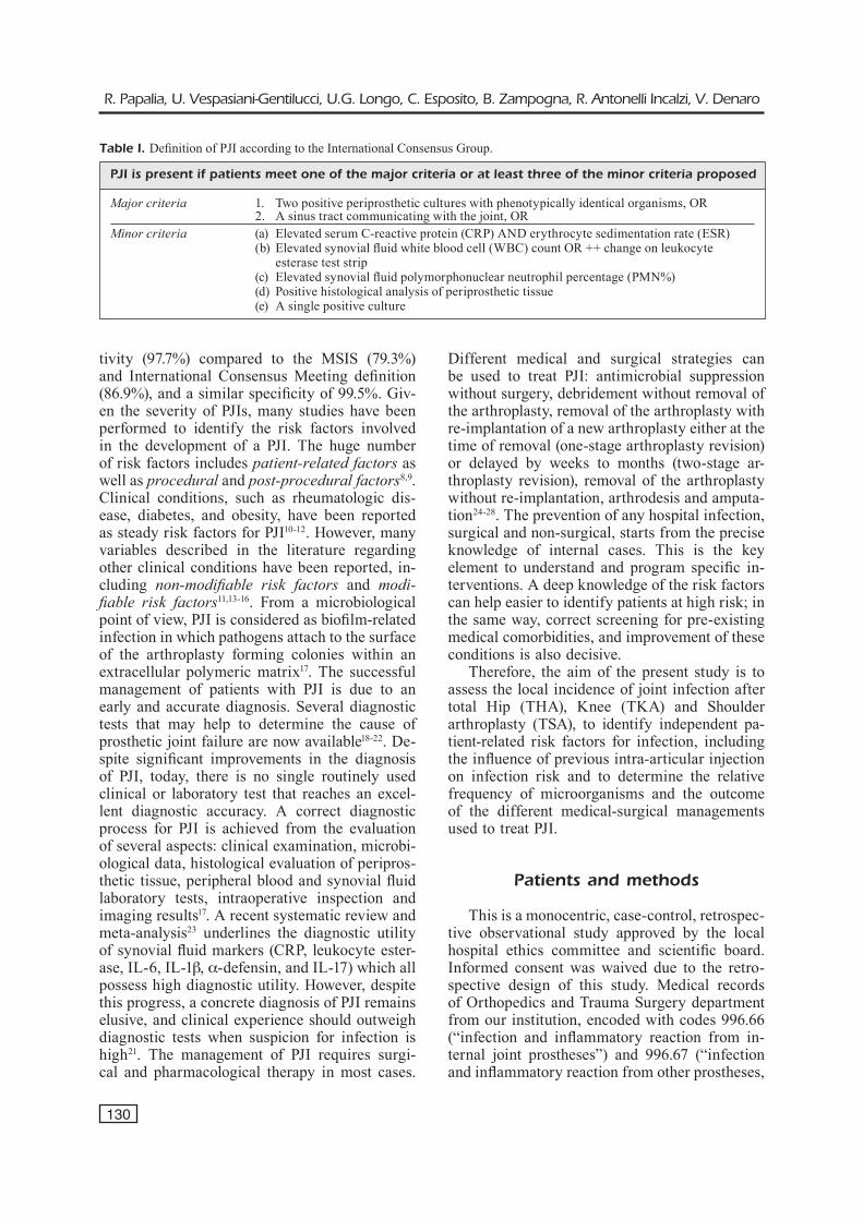
R. Papalia, U. Vespasiani-Gentilucci, U.G. Longo, C. Esposito, B. Zampogna, R. Antonelli Incalzi, V. Denaro
130
tivity (97.7%) compared to the MSIS (79.3%) and International Consensus Meeting definition (86.9%), and a similar specificity of 99.5%. Giv-en the severity of PJIs, many studies have been performed to identify the risk factors involved in the development of a PJI. The huge number of risk factors includes patient-related factors as well as procedural and post-procedural factors8,9. Clinical conditions, such as rheumatologic dis-ease, diabetes, and obesity, have been reported as steady risk factors for PJI10-12. However, many variables described in the literature regarding other clinical conditions have been reported, in-cluding non-modifiable risk factors and modi-fiable risk factors11,13-16. From a microbiological point of view, PJI is considered as biofilm-related infection in which pathogens attach to the surface of the arthroplasty forming colonies within an extracellular polymeric matrix17. The successful management of patients with PJI is due to an early and accurate diagnosis. Several diagnostic tests that may help to determine the cause of prosthetic joint failure are now available18-22. De-spite significant improvements in the diagnosis of PJI, today, there is no single routinely used clinical or laboratory test that reaches an excel-lent diagnostic accuracy. A correct diagnostic process for PJI is achieved from the evaluation of several aspects: clinical examination, microbi-ological data, histological evaluation of peripros-thetic tissue, peripheral blood and synovial fluid laboratory tests, intraoperative inspection and imaging results17. A recent systematic review and meta-analysis23 underlines the diagnostic utility of synovial fluid markers (CRP, leukocyte ester-ase, IL-6, IL-1b, a-defensin, and IL-17) which all possess high diagnostic utility. However, despite this progress, a concrete diagnosis of PJI remains elusive, and clinical experience should outweigh diagnostic tests when suspicion for infection is high21. The management of PJI requires surgi-cal and pharmacological therapy in most cases.
Different medical and surgical strategies can be used to treat PJI: antimicrobial suppression without surgery, debridement without removal of the arthroplasty, removal of the arthroplasty with re-implantation of a new arthroplasty either at the time of removal (one-stage arthroplasty revision) or delayed by weeks to months (two-stage ar-throplasty revision), removal of the arthroplasty without re-implantation, arthrodesis and amputa-tion24-28. The prevention of any hospital infection, surgical and non-surgical, starts from the precise knowledge of internal cases. This is the key element to understand and program specific in-terventions. A deep knowledge of the risk factors can help easier to identify patients at high risk; in the same way, correct screening for pre-existing medical comorbidities, and improvement of these conditions is also decisive.
Therefore, the aim of the present study is to assess the local incidence of joint infection after total Hip (THA), Knee (TKA) and Shoulder arthroplasty (TSA), to identify independent pa-tient-related risk factors for infection, including the influence of previous intra-articular injection on infection risk and to determine the relative frequency of microorganisms and the outcome of the different medical-surgical managements used to treat PJI.
Patients and methods
This is a monocentric, case-control, retrospec-tive observational study approved by the local hospital ethics committee and scientific board. Informed consent was waived due to the retro-spective design of this study. Medical records of Orthopedics and Trauma Surgery department from our institution, encoded with codes 996.66 (“infection and inflammatory reaction from in-ternal joint prostheses”) and 996.67 (“infection and inflammatory reaction from other prostheses,
Table I. Definition of PJI according to the International Consensus Group.
PJI is present if patients meet one of the major criteria or at least three of the minor criteria proposed
Major criteria 1. Two positive periprosthetic cultures with phenotypically identical organisms, OR 2. A sinus tract communicating with the joint, ORMinor criteria (a) Elevated serum C-reactive protein (CRP) AND erythrocyte sedimentation rate (ESR) (b) Elevated synovial fluid white blood cell (WBC) count OR ++ change on leukocyte esterase test strip (c) Elevated synovial fluid polymorphonuclear neutrophil percentage (PMN%) (d) Positive histological analysis of periprosthetic tissue (e) A single positive culture

Advances in management of periprosthetic joint infections: an historical prospective study
131
implants and internal orthopedic implants”) be-tween January 2000 and July 2017 were analyzed. We included in the “cases’ group” all patients who developed a PJI after a total hip/knee/shoul-der arthroplasty, which diagnosis was confirmed by the positive cultures exam and/or clinical ex-am’s positivity (i.e. a sinus tract communicating with the joint) according to the major criteria proposed by PJI Consensus Group. The following exclusion criteria were applied: prosthetic joint infections in other joints, non-prosthetic joint in-fections, prosthetic joint infection which doesn’t meet one of the major criteria or at least three of the minor criteria proposed by the International Consensus Meeting on PJI for PJI diagnosing, subcutaneous and soft tissues’ infection without involvement of prosthetic components, lack of de-mographic and anthropometric data, mobilization of arthroplasty caused by metastatic localization or different from the infectious one. From the application of above-mentioned criteria, 35 pa-tients were excluded from the 63 patients initially enrolled. To limit the bias related to the possible variations of operative/perioperative techniques and nosocomial infectious agents’ epidemiology during the seventeen years, we decided to include in the “control group” for each “case” at least 3 patients who did not have an infection after total joint arthroplasty, randomly drawn from homo-geneous type of implant and year of surgery. To define the patient’s risk profile and the variables related to the onset of infection, we reviewed clinical records extracting the following data: sex, age, BMI, type of implant, comorbidity, year of surgery, year of admission, microbial isolation, medical and surgical management, and relative outcomes. Previous ipsilateral intra-articular in-jection therapy, type of injection, and the timing of the last injection before the surgery were also recorded. Variables to be evaluated were selected according to data availability of our retrospective cohort and to literature current evidence for as-
sociation to PJI8,9. Patient follow-up was analyzed until the most recent outpatient examination or new hospitalization.
Statistical AnalysisStatistical data analysis was performed with
SPPS statistical software (version 21, Chicago, USA). The two groups were compared with Mann-Whitney U-test for continuous variables, and the X2 test for categorical variables. Binary logistic regression was used to analyze the asso-ciation between BMI (the only variable signifi-cantly different between the group “early” and “late-onset” or “controls”) and risk of PJI. Only p-values < 0.05 were considered to represent sta-tistical significance.
Results
According to our inclusion and exclusion cri-teria, 28 patients who developed a PJI infection in the investigation time-lapse (from 2000 to 2017) were enrolled in the study. Of these, 10 patients (35.72%) underwent to a total hip arthroplasty, 16 patients (57.14%) underwent a total knee arthro-plasty, 2 patients (7.14%) underwent a total shoul-der arthroplasty. The “cases’ group” consist of 28 patients, 12 women and 16 men, with an age of 73.32 ± 8.29 years, and a BMI of 28.97 ± 4.39 kg/m2. The “controls’ group” consist of 84 patients, 47 women and 37 men, with an age of 75.43 ± 9.10 years and a BMI of 28.29 ± 3.59. Therefore, the “control group” was composed of 84 patients, homogeneous for site and year of surgery. The prevalence of diabetes, dyslipidemia, and hyper-tension were, respectively, 14.3%, 21.4%, and 75% in the “cases’ group” and 8.3%, 20.2%, and 58.3% in the “controls’ group” (Table II). In the cases’ group, 9 patients received an intra-articular injec-tion before arthroplasty: in 8 patients hyaluronic acid was used while for 1 patient PRP was used.
Table II. Patients’ features: groups “cases” vs. “controls”.
Parameter Cases Controls p
Age 73.32 ± 8.29 75.43 ± 9.10 0.086Gender (M/F) 16/12 37/47 0.277BMI 28.97 ± 4.39 28.29 ± 3.59 0.256Diabetes 4/28 (14.3%) 7/84 (8.3%) 0.463Dyslipidemia 6/28 (21.4%) 17/84 (20.2%) 1Hypertension 21/28 (75%) 49/84 (58.3%) 0.176Intra-articular injection 9/28 (32.1%) 43/84 (51.2%) 0.080

R. Papalia, U. Vespasiani-Gentilucci, U.G. Longo, C. Esposito, B. Zampogna, R. Antonelli Incalzi, V. Denaro
132
All 9 patients received and completed the joint in-filtration cycle between one year and three months before surgery, according to the recommendations of the PJI Consensus Group. In the control’s group 43 patients received an intra-articular injection before arthroplasty: in 38 patients hyaluronic acid was used, in 4 patients was used PRP while 1 received a corticosteroid injection. Similarly, to cases’ group, all 43 patients received and complet-ed the joint infiltration cycle not later than three months before surgery. As can be seen, despite the multitude of factor risk for PJI recognized in liter-ature, there were no patient demographics (gender p=0.277, age p=0.086, BMI p=0.256) and medi-cal comorbidities (diabetes p=0.463, dyslipidemia p=1, hypertension p=0.176) that were associated with an increased risk of PJI after TJA, comparing the “cases’ group” with “controls’ group”. The risk of infection was not significantly increased for patients who received an intra-articular injection before the day of operation (p=0.080). Patients who developed a PJI were divided, in patients with “early infection” (within 3 months from the day of surgery), “delayed infection” (within 12 months) and “late infection” (more than 12 months) (Ta-ble III). Patients with an “early/delayed” infection were compared to patients with “late infection” for all the variables of interest. The “early/delayed infection” group consists of 16 patients, 5 women and 11 men, with an age of 73.25 ± 6.79 years and a BMI of 30.82 ± 3.94; the “late infection” group consists of 12 patients, 7 women and 5 men, with an age of 73.41 ± 10.27 years and a BMI of
26.5 ± 3.79. The prevalence of diabetes, dyslipid-emia and hypertension were, respectively, 18.7%, 18.7%, and 75% in the “early/delayed group” and 8.3%, 25%, and 75% in the “late group” (Table IV). In the early/delayed infection group 6 pa-tients received an intra-articular injection before arthroplasty: in 5 patients was used hyaluronic acid while for 1 patient was used PRP. In the late infection group 3 patients received an intra-ar-ticular injection of hyaluronic acid. The patient demographics (gender p=0.250, age p=0.802) and medical comorbidities (diabetes p=0.613, dyslipid-emia p=1, hypertension p=1) did not significantly increase the risk of post-operative infection. About intra-articular injection, the risk of infection was not increased (p=0.483). Comparing the “early/delayed group” with “late group”, BMI was the only factor significantly associated with an in-creased risk of early/delayed PJI (p=0.003). BMI index resulted statistically significant higher in the “early/delayed group” also comparing with “controls’ group” (p=0.008). Logistic regression showed that for each BMI unit there was a 20-fold increased risk of early prosthetic infection (OR 1.19, IC 1.03-1.38, p=0.01). The other factors, that did not significantly increase the risk of postoper-ative infection are shown in Table V. Regarding to microbial spectrum (Table VI), several micro-organisms were isolated from pre-operative and intra-operative cultures: S. aureus was isolated in 7 patients, S. epidermidis was isolated in 6 patients, polymicrobial infections were isolated in 8 patients and other coagulase-negative were isolated in 3 patients. Klebsiella pneumoniae or Propionibacterium acnes were isolated only in one case. In 2 cases the intraoperative culture failed to show growth; however, the diagnosis was confirmed by the presence of draining sinus tract, elevated CRP, and intraoperative positive histol-ogy, matching the PJI Consensus Group major and minor criteria. Several strategies were used
Table III. Timing of infection.
Timing of infection Number Percentage Early 10 35.71%Delayed 6 21.42%Late 12 42.85%
Table IV. Patients’ features: groups “early/delayed” vs. “late” infection.
Parameter Early/delayed infection Late infection p
Age 73.25 ± 6.79 73.41 ± 10.27 0.802Gender (M/F) 11/5 5/7 0.250BMI 30.82 ± 3.94 26.5 ± 3.79 0.003Diabetes 3/16 (18.7%) 1/12 (8.3%) 0.613Dyslipidemia 3/16 (18.7%) 3/12 (25%) 1Hypertension 12/16 (75%) 9/12 (75%) 1Intra-articular injection 6/16 (37.5%) 3/12 (25%) 0.483
Advances in management of periprosthetic joint infections: an historical prospective study
133
for surgical management of PJI (Table VII). Eight patients underwent to “debridement and surgical toilette”, and among these patients only one patient developed a reinfection. One patient underwent to “One-Stage Arthroplasty Exchange” who later developed a reinfection. Seventeen patients under-went to “Two-Stage Arthroplasty Exchange”, and among these patients only 2 developed a reinfec-tion. Two patients underwent to “Arthroplasty Re-section without Reimplantation” (i.e., Girdlestone procedure). The number of joint arthroplasties performed in our hospital has risen significantly during the study period: we moved from 14 TKA performed in 2000 to 467 in 2017, from 25 THA performed in 2000 to 331 in 2017, from 1 TSA performed in 2008 to 76 in 2017. In September of 2014 our University Hospital was accredited by the Joint Commission International (JCI) that is an independent international organization that eval-uates excellence within healthcare facilities. The
standards established by the Joint Commission International are objectives required to improve patient safety and the quality of patient care. Com-paring the incidence of PJI before and after JCI accreditation, we recorded a reduction in the rate of PJI from 1.16% to 0.21% for hip arthroplasty and from 1.7% to 0.63% for knee arthroplasty. About shoulder, we did not record any infection in the last 3 years.
Discussion
Though several prospective and retrospective cohort studies have been published is still ex-tremely difficult to predict the risk of post-oper-ative PJI. Our monocentric, case-control, retro-spective observational study tried to determine which factors put a patient a higher risk of PJI. We found a significant statistic correlation be-tween BMI and early PJI: for each BMI unit in-crease there was a 20-fold increased risk of early prosthetic infection (OR 1.19, IC 1.03-1.38, p=0.01). Wu et al29 found that patients with a BMI greater than 28 kg/m2 had a 2.77 -fold higher risk of PJI compared with patients with a BMI be-tween 18.5 and 28 kg/m2. Several other studies support our results regarding the effect of BMI on PJI risk11,13,16,30-35 but not all authors agree on that issue36,37. On the contrary, Berbari et al38 in-dicated that a low BMI (<25) was associated with an increased risk of PJI. They explained that patients with low BMI might have less nutrition-
Table V. Patients’ features: groups “early/delayed” infection vs. “controls”.
Parameter Early/delayed infection Controls p
Age 73.25 ± 6.79 75.43 ± 9.10 0.112Gender (M/F) 11/5 37/47 0.101BMI 30.82 ± 3.94 28.29 ± 3.59 0.008Diabetes 3/16 (18.7%) 7/84 (8.3%) 0.198Dyslipidemia 3/16 (18.7%) 17/84 (20.2%) 1Hypertension 12/16 (75%) 49/84 (58.3%) 0.270Intra-articular injection 6/16 (37.5%) 43/84 (51.2%) 0.315
Table VII. Surgical treatment used for patients with PJI
Surgical treatment Number Percentage Reinfection of infection
Debridement and toilette 8 28.57% 1One-Stage Arthroplasty Exchange 1 3.57% 1Two-Stage Arthroplasty Exchange 17 60.71% 2Arthroplasty Resection without Reimplantation 2 7.15% /
Table VI. Microorganism isolated from patients with PJI.
Microorganism Number Percentage Staphylococcus aureus 7 25%Staphylococcus epidermidis 6 21,4%Polymicrobial infection 8 28.6%Staphylococcus coagulase 3 10.7% negativeCulture-negative 2 7.1%Klebsiella Pneumoniae 1 3.6%Propionibacterium Acnes 1 3.6%
R. Papalia, U. Vespasiani-Gentilucci, U.G. Longo, C. Esposito, B. Zampogna, R. Antonelli Incalzi, V. Denaro
134
al reserve and multiple comorbidities, such as immunosuppression, rheumatoid arthritis, and nicotine dependency. The American Association of Hip and Knee Surgeons (AAHKS) recom-mends that arthroplasty is delayed in cases of morbid obesity (BMI > 40), especially in patients with multiple comorbidities39. Total joint arthro-plasty is one of the most common elective surger-ies performed in older adults. Older patient age often coincides with poorer nutritional status and immunity depression, thus resulting in a higher risk of infection. However, in our study, we didn’t find a significant correlation between age and risk of PJI. Probably, it is due to the small size and homogeneity of the “cases’ group”. Wu et al29 found that patients aged 65-75 years had 3.36-fold higher risk of PJI compared with patients aged 45-65 years. Similar results were reported by Ridgeway et al40. In an opposite way, in a sin-gle-center analysis of 8494 TJA, Malinzak et al32 found that younger age was associated with in-creased risk of infection. They supposed that younger patients are more active than elderly ones and that their implants undergo a greater number of use cycles, leading potential revision surgery, and possibility of infection. About gen-der, several studies10,33,37,41-45 suggest that males have a higher risk of PJI. Male and female skin colonization is different, and this may result in differences in skin pH, sebum production, or skin thickness46. Our data showed no significant cor-relation between gender and PJI. In our study, the only comorbidities affecting patients who devel-oped a PJI were diabetes, dyslipidemia and hy-pertension. None of these comorbidities was sta-tistically significant correlated with a high risk of PJI. However, several studies8,9,15,47,48 in the liter-ature found that diabetes mellitus, smoking, alco-holism, anemia, rheumatoid arthritis, immuno-suppressive medications, systemic infection, car-diology and gastroenterology disorders, liver and kidney disease, HIV infection are all related to an increased risk of infection. There are no clear conclusions regarding the relationship between preoperative intraarticular injections and postop-erative PJI after TJA, since existing studies have provided conflicting results. About hip, Werner et al49 reviewed a total of 34597 THA and found that incidence of PJI was significantly higher in the patients who underwent hip injection within 3 months before THA while this association was not noted when THA occurred more than 3 months after the injection. Other three studies have demonstrated higher rates of PJI in patients
who had an intraarticular steroid injection in the hip before THA50-53 while 4 prior studies have not demonstrated any association between preopera-tive intraarticular steroid injection and PJI after THA54-57. About knee, Cancienne et al50 found a significant higher risk of PJI in patients who un-derwent ipsilateral knee injections within three months prior to TKA, but not in patients who received the injection more than three months before TKA. Several studies have reported that previous steroid injections were not associated with an increased risk of PJI following TKA58-62. Kokubun et al63 found no relationship between timing and number of intra-articular injections with complication rate, infection, or poor short-term functional outcomes. In our study, we found no relationship between timing and type of injec-tions and increased risk of PJI. Most of the pa-tients, both in the cases’ and controls’ group, received an intra-articular injection with hyal-uronic acid; no patient who develops a PJI re-ceived a corticosteroid injection. Moreover, all patients received the last injection cycle within one year and not later than three months before surgery, according to the recommendations of the PJI Consensus Group. Regarding the microbial spectrum, Tande et al17 reported the microbiolog-ical results of 14 large studies including 2400 patients with hip or knee arthroplasty infection. Gram-positive cocci are involved in the majority of hip and knee PJIs in all the studies examined; infections by S. aureus and coagulase-negative staphylococci contribute to 50-60% of PJIs, while streptococci and enterococci together account for only 10% of cases. Aerobic Gram-negative bacil-li are involved in 10% of cases of knee and hip PJI. The percentage of culture-negative infections varied from 5 to 34%. In our study, we found similar data. The most frequently isolated patho-gens, S. aureus and/or S. epidermidis, and coag-ulase-negative staphylococci, were ensemble re-sponsible of PJI in 53% of cases. Similarly, the polymicrobial forms were isolated in 28.6% of patients while culture-negative infection resulted in 7.1% of cases. Surgical treatment success has been variably described in the literature; there are no randomized trials comparing the different approaches, and variability between hospitals that perform mainly one-stage compared to two-stage arthroplasty exchanges limits comparison across the studies. A systematic review of hip PJI analyzing 375 patients undergoing one stage ex-change reported an 87% success rate, compared with 90% for the 929 patients undergoing two-
Advances in management of periprosthetic joint infections: an historical prospective study
135
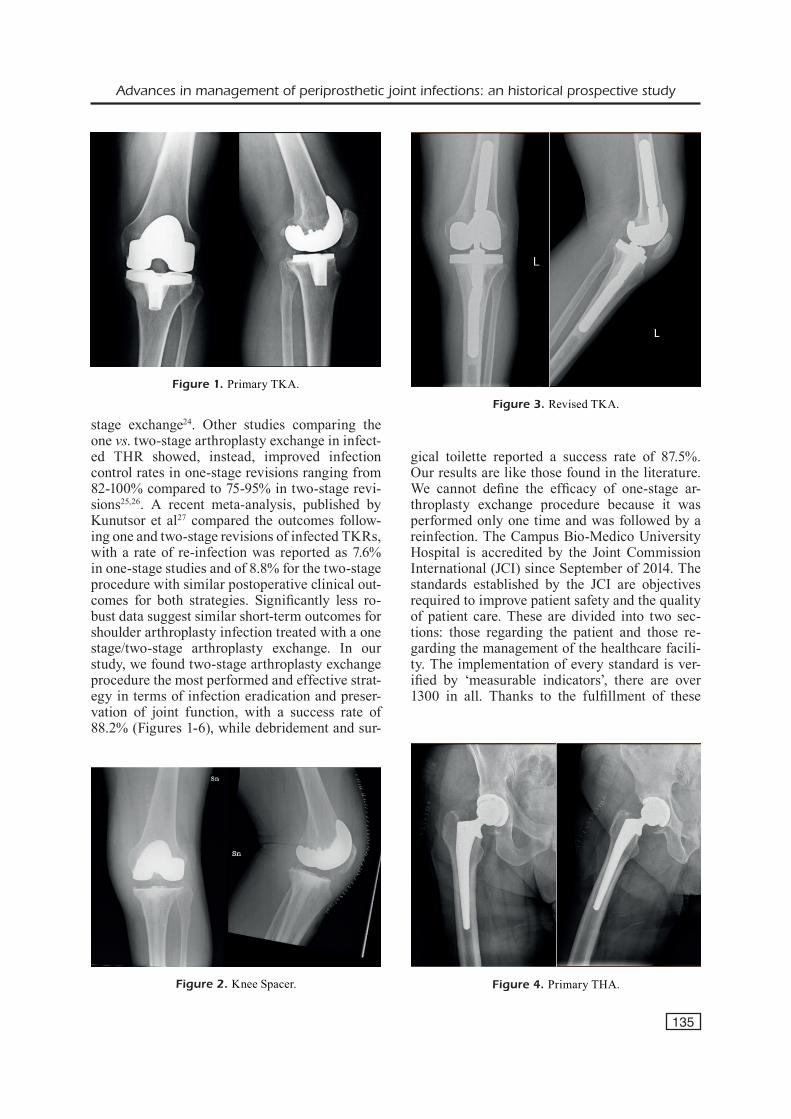
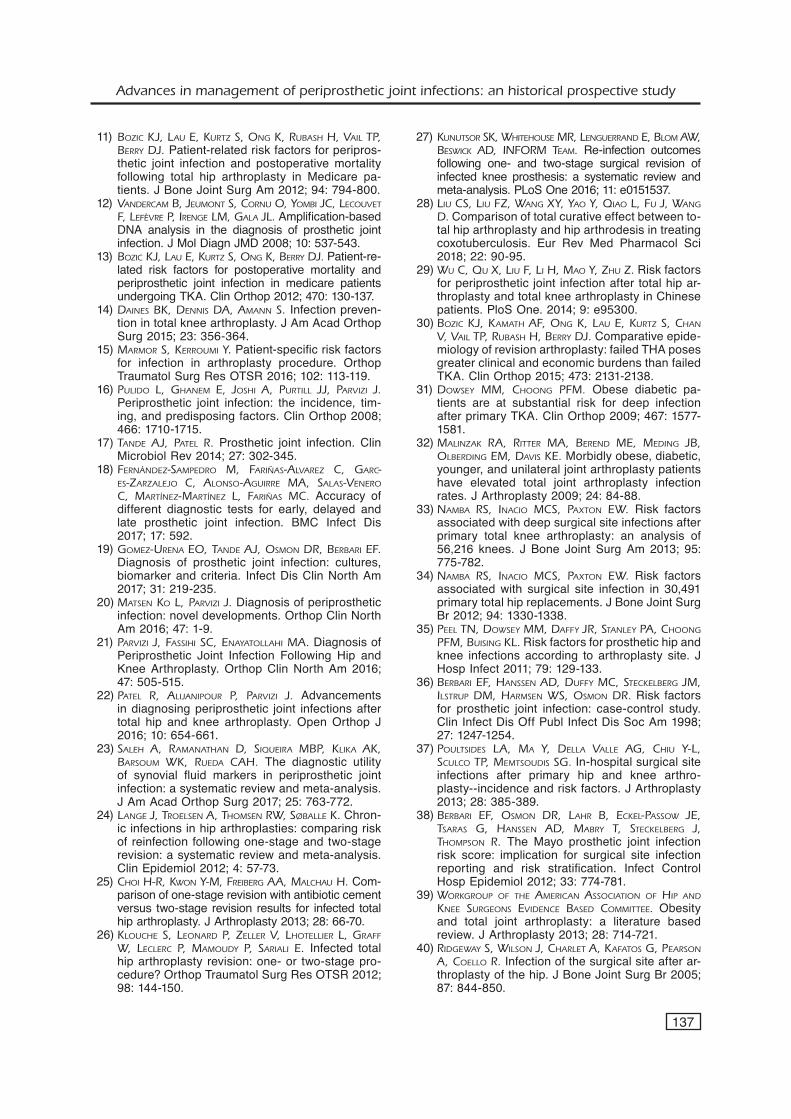
stage exchange24. Other studies comparing the one vs. two-stage arthroplasty exchange in infect-ed THR showed, instead, improved infection control rates in one-stage revisions ranging from 82-100% compared to 75-95% in two-stage revi-sions25,26. A recent meta-analysis, published by Kunutsor et al27 compared the outcomes follow-ing one and two-stage revisions of infected TKRs, with a rate of re-infection was reported as 7.6% in one-stage studies and of 8.8% for the two-stage procedure with similar postoperative clinical out-comes for both strategies. Significantly less ro-bust data suggest similar short-term outcomes for shoulder arthroplasty infection treated with a one stage/two-stage arthroplasty exchange. In our study, we found two-stage arthroplasty exchange procedure the most performed and effective strat-egy in terms of infection eradication and preser-vation of joint function, with a success rate of 88.2% (Figures 1-6), while debridement and sur-
gical toilette reported a success rate of 87.5%. Our results are like those found in the literature. We cannot define the efficacy of one-stage ar-throplasty exchange procedure because it was performed only one time and was followed by a reinfection. The Campus Bio-Medico University Hospital is accredited by the Joint Commission International (JCI) since September of 2014. The standards established by the JCI are objectives required to improve patient safety and the quality of patient care. These are divided into two sec-tions: those regarding the patient and those re-garding the management of the healthcare facili-ty. The implementation of every standard is ver-ified by ‘measurable indicators’, there are over 1300 in all. Thanks to the fulfillment of these
Figure 1. Primary TKA.
Figure 2. Knee Spacer.
Figure 3. Revised TKA.
Figure 4. Primary THA.

R. Papalia, U. Vespasiani-Gentilucci, U.G. Longo, C. Esposito, B. Zampogna, R. Antonelli Incalzi, V. Denaro
136
standards, we recorded in the last 3 years a re-duction of one percentage point in the incidence of PJI after hip, knee and shoulder arthroplasty, despite the increased number of arthroplasties.
Main weaknesses of the study are represented by a small patients’ cohort and a retrospective and monocentric investigation design. The ac-curacy of the data is dependent on the doctor and coders who are responsible for entering it. The possibility of miscoding certainly exists and some patients with PJI may not be considered. The infection rate was very low during the stud-ied period, but we could not evaluate the possible association between others PJI and other relevant risk factors widely recognized in literature be-cause not available in our retrospective cohort.
Conclusions
It is essential that orthopedic surgeons under-stand and identify risk factors before TJA so that they can optimize patients’ status and minimize their risk of developing a postoperative infection. Clarification of the most common risk factors is critical for taking further steps. Our study highlighted obesity as a risk factor especially for “early/delayed PJI” compared to “controls’ and “late infection” groups in the studied period. Two-stage arthroplasty exchange, debridement, antibiotics and implant retention are treatments with a high rate of success in terms of reinfec-tion. Waiting for further analysis, considering the growing epidemiology of obesity and associated dysmetabolic pathologies, it is essential to get weight loss before proceeding to implant a joint arthroplasty.
Conflict of InterestsThe Authors declare that they have no conflict of interests.
References
1) Jones CA, VoAklAnder dC, Johnston dW, suArez-Al-mAzor me. Health related quality of life outcomes after total hip and knee arthroplasties in a com-munity based population. J Rheumatol 2000; 27: 1745-1752.
2) kurtz s, ong k, lAu e, moWAt F, hAlpern m. Pro-jections of primary and revision hip and knee arthroplasty in the United States from 2005 to 2030. J Bone Joint Surg Am 2007; 89: 780-785.
3) kurtz sm, lAu e, WAtson h, sChmier Jk, pArVizi J. Economic burden of periprosthetic joint infection in the United States. J Arthroplasty 2012; 27: 61-65.
4) klouChe s, sAriAli e, mAmoudy p. Total hip arthro-plasty revision due to infection: a cost analysis approach. Orthop Traumatol Surg Res OTSR 2010; 96:124-132.
5) pArVizi J, zmistoWski B, BerBAri eF, BAuer tW, springer Bd, dellA VAlle CJ, gArVin kl, mont mA, Wong-WorAWAt md, zAlAVrAs Cg. New definition for periprosthetic joint infection: from the Workgroup of the Musculoskeletal Infection Society. Clin Orthop 2011; 469: 2992-2994.
6) pArVizi J, gehrke t. International Consensus Group on Periprosthetic Joint Infection. Definition of periprosthetic joint infection. J Arthroplasty 2014; 29: 1331.
7) pArVizi J, tAn tl, gosWAmi k, higuerA C, dellA VAlle C, Chen AF, shohAt n. The 2018 definition of periprosthetic hip and knee infection: an evi-dence-based and validated criteria. J Arthroplas-ty 2018; 33: 1309-1314.
8) ChirCA i, mArCulesCu C. Prevention of infection in orthopedic prosthetic surgery. Infect Dis Clin North Am 2017; 31: 253-263.
9) küçükdurmAz F, pArVizi J. The prevention of peri-prosthetic joint infections. Open Orthop J 2016; 10: 589-599.
10) BAek s-h. Identification and preoperative optimi-zation of risk factors to prevent periprosthetic joint infection. World J Orthop 2014; 5: 362-367.
Figure 5. Hip Spacer.
Figure 6. Revised THA
Advances in management of periprosthetic joint infections: an historical prospective study
137
11) BoziC kJ, lAu e, kurtz s, ong k, ruBAsh h, VAil tp, Berry dJ. Patient-related risk factors for peripros-thetic joint infection and postoperative mortality following total hip arthroplasty in Medicare pa-tients. J Bone Joint Surg Am 2012; 94: 794-800.
12) VAnderCAm B, Jeumont s, Cornu o, yomBi JC, leCouVet F, leFèVre p, irenge lm, gAlA Jl. Amplification-based DNA analysis in the diagnosis of prosthetic joint infection. J Mol Diagn JMD 2008; 10: 537-543.
13) BoziC kJ, lAu e, kurtz s, ong k, Berry dJ. Patient-re-lated risk factors for postoperative mortality and periprosthetic joint infection in medicare patients undergoing TKA. Clin Orthop 2012; 470: 130-137.
14) dAines Bk, dennis dA, AmAnn s. Infection preven-tion in total knee arthroplasty. J Am Acad Orthop Surg 2015; 23: 356-364.
15) mArmor s, kerroumi y. Patient-specific risk factors for infection in arthroplasty procedure. Orthop Traumatol Surg Res OTSR 2016; 102: 113-119.
16) pulido l, ghAnem e, Joshi A, purtill JJ, pArVizi J. Periprosthetic joint infection: the incidence, tim-ing, and predisposing factors. Clin Orthop 2008; 466: 1710-1715.
17) tAnde AJ, pAtel r. Prosthetic joint infection. Clin Microbiol Rev 2014; 27: 302-345.
18) Fernández-sAmpedro m, FAriñAs-AlVArez C, gArC-es-zArzAleJo C, Alonso-Aguirre mA, sAlAs-Venero C, mArtínez-mArtínez l, FAriñAs mC. Accuracy of different diagnostic tests for early, delayed and late prosthetic joint infection. BMC Infect Dis 2017; 17: 592.
19) gomez-urenA eo, tAnde AJ, osmon dr, BerBAri eF. Diagnosis of prosthetic joint infection: cultures, biomarker and criteria. Infect Dis Clin North Am 2017; 31: 219-235.
20) mAtsen ko l, pArVizi J. Diagnosis of periprosthetic infection: novel developments. Orthop Clin North Am 2016; 47: 1-9.
21) pArVizi J, FAssihi sC, enAyAtollAhi mA. Diagnosis of Periprosthetic Joint Infection Following Hip and Knee Arthroplasty. Orthop Clin North Am 2016; 47: 505-515.
22) pAtel r, AliJAnipour p, pArVizi J. Advancements in diagnosing periprosthetic joint infections after total hip and knee arthroplasty. Open Orthop J 2016; 10: 654-661.
23) sAleh A, rAmAnAthAn d, siqueirA mBp, klikA Ak, BArsoum Wk, ruedA CAh. The diagnostic utility of synovial fluid markers in periprosthetic joint infection: a systematic review and meta-analysis. J Am Acad Orthop Surg 2017; 25: 763-772.
24) lAnge J, troelsen A, thomsen rW, søBAlle k. Chron-ic infections in hip arthroplasties: comparing risk of reinfection following one-stage and two-stage revision: a systematic review and meta-analysis. Clin Epidemiol 2012; 4: 57-73.
25) Choi h-r, kWon y-m, FreiBerg AA, mAlChAu h. Com-parison of one-stage revision with antibiotic cement versus two-stage revision results for infected total hip arthroplasty. J Arthroplasty 2013; 28: 66-70.
26) klouChe s, leonArd p, zeller V, lhotellier l, grAFF W, leClerC p, mAmoudy p, sAriAli e. Infected total hip arthroplasty revision: one- or two-stage pro-cedure? Orthop Traumatol Surg Res OTSR 2012; 98: 144-150.
27) kunutsor sk, Whitehouse mr, lenguerrAnd e, Blom AW, BesWiCk Ad, inForm teAm. Re-infection outcomes following one- and two-stage surgical revision of infected knee prosthesis: a systematic review and meta-analysis. PLoS One 2016; 11: e0151537.
28) liu Cs, liu Fz, WAng Xy, yAo y, qiAo l, Fu J, WAng d. Comparison of total curative effect between to-tal hip arthroplasty and hip arthrodesis in treating coxotuberculosis. Eur Rev Med Pharmacol Sci 2018; 22: 90-95.
29) Wu C, qu X, liu F, li h, mAo y, zhu z. Risk factors for periprosthetic joint infection after total hip ar-throplasty and total knee arthroplasty in Chinese patients. PloS One. 2014; 9: e95300.
30) BoziC kJ, kAmAth AF, ong k, lAu e, kurtz s, ChAn V, VAil tp, ruBAsh h, Berry dJ. Comparative epide-miology of revision arthroplasty: failed THA poses greater clinical and economic burdens than failed TKA. Clin Orthop 2015; 473: 2131-2138.
31) doWsey mm, Choong pFm. Obese diabetic pa-tients are at substantial risk for deep infection after primary TKA. Clin Orthop 2009; 467: 1577-1581.
32) mAlinzAk rA, ritter mA, Berend me, meding JB, olBerding em, dAVis ke. Morbidly obese, diabetic, younger, and unilateral joint arthroplasty patients have elevated total joint arthroplasty infection rates. J Arthroplasty 2009; 24: 84-88.
33) nAmBA rs, inACio mCs, pAXton eW. Risk factors associated with deep surgical site infections after primary total knee arthroplasty: an analysis of 56,216 knees. J Bone Joint Surg Am 2013; 95: 775-782.
34) nAmBA rs, inACio mCs, pAXton eW. Risk factors associated with surgical site infection in 30,491 primary total hip replacements. J Bone Joint Surg Br 2012; 94: 1330-1338.
35) peel tn, doWsey mm, dAFFy Jr, stAnley pA, Choong pFm, Buising kl. Risk factors for prosthetic hip and knee infections according to arthroplasty site. J Hosp Infect 2011; 79: 129-133.
36) BerBAri eF, hAnssen Ad, duFFy mC, steCkelBerg Jm, ilstrup dm, hArmsen Ws, osmon dr. Risk factors for prosthetic joint infection: case-control study. Clin Infect Dis Off Publ Infect Dis Soc Am 1998; 27: 1247-1254.
37) poultsides lA, mA y, dellA VAlle Ag, Chiu y-l, sCulCo tp, memtsoudis sg. In-hospital surgical site infections after primary hip and knee arthro-plasty--incidence and risk factors. J Arthroplasty 2013; 28: 385-389.
38) BerBAri eF, osmon dr, lAhr B, eCkel-pAssoW Je, tsArAs g, hAnssen Ad, mABry t, steCkelBerg J, thompson r. The Mayo prosthetic joint infection risk score: implication for surgical site infection reporting and risk stratification. Infect Control Hosp Epidemiol 2012; 33: 774-781.
39) Workgroup oF the AmeriCAn AssoCiAtion oF hip And knee surgeons eVidenCe BAsed Committee. Obesity and total joint arthroplasty: a literature based review. J Arthroplasty 2013; 28: 714-721.
40) ridgeWAy s, Wilson J, ChArlet A, kAFAtos g, peArson A, Coello r. Infection of the surgical site after ar-throplasty of the hip. J Bone Joint Surg Br 2005; 87: 844-850.

R. Papalia, U. Vespasiani-Gentilucci, U.G. Longo, C. Esposito, B. Zampogna, R. Antonelli Incalzi, V. Denaro
138
41) CroWe B, pAyne A, eVAngelistA pJ, stAChel A, phillips ms, sloVer Jd, inneh iA, iorio r, BosCo JA. Risk fac-tors for infection following total knee arthroplasty: a series of 3836 cases from one institution. J Arthroplasty 2015; 30: 2275-2278.
42) dAle h, FenstAd Am, hAllAn g, hAVelin li, Furnes o, oVergAArd s, pedersen AB, kärrholm J, gArelliCk g, pulkkinen p, eskelinen A, mäkelä k, engesæter lB. Increasing risk of prosthetic joint infection after total hip arthroplasty. Acta Orthop 2012; 83: 449-458.
43) meehAn Jp, dAnielsen B, kim sh, JAmAli AA, White rh. Younger age is associated with a higher risk of early periprosthetic joint infection and aseptic mechanical failure after total knee ar-throplasty. J Bone Joint Surg Am 2014; 96: 529-535.
44) ong kl, kurtz sm, lAu e, BoziC kJ, Berry dJ, pArVizi J. Prosthetic joint infection risk after total hip arthroplasty in the Medicare population. J Arthroplasty 2009; 24:105-109.
45) Willis-oWen CA, konyVes A, mArtin dk. Factors af-fecting the incidence of infection in hip and knee replacement: an analysis of 5277 cases. J Bone Joint Surg Br 2010; 92: 1128-1133.
46) Fierer n, hAmAdy m, lAuBer Cl, knight r. The in-fluence of sex, handedness, and washing on the diversity of hand surface bacteria. Proc Natl Acad Sci U S A 2008; 105: 17994-17999.
47) ekA A, Chen AF. Patient-related medical risk fac-tors for periprosthetic joint infection of the hip and knee. Ann Transl Med 2015; 3: 233.
48) george dA, drAgo l, sCArponi s, gAllAzzi e, hAddAd Fs, romAno Cl. Predicting lower limb periprosthet-ic joint infections: a review of risk factors and their classification. World J Orthop 2017; 8: 400-411.
49) Werner BC, CAnCienne Jm, BroWne JA. The Timing of Total Hip Arthroplasty After Intraarticular Hip Injection Affects Postoperative Infection Risk. J Arthroplasty 2016; 31: 820-823.
50) CAnCienne Jm, Werner BC, luetkemeyer lm, BroWne JA. Does timing of previous intra-articular steroid injection affect the post-operative rate of infection in total knee arthroplasty? J Arthroplasty 2015; 30: 1879-1882.
51) kAspAr s, de V de Beer J. Infection in hip arthroplas-ty after previous injection of steroid. J Bone Joint Surg Br 2005; 87: 454-457.
52) mCintosh Al, hAnssen Ad, Wenger de, osmon dr. Recent intraarticular steroid injection may increase infection rates in primary THA. Clin Or-thop 2006; 451: 50-54.
53) rAVi B, esCott Bg, WAsserstein d, CroXFord r, hollAnds s, pAterson Jm, kreder hJ, hAWker gA. Intraarticular hip injection and early revision sur-gery following total hip arthroplasty: a retrospec-tive cohort study. Arthritis Rheumatol Hoboken NJ 2015; 67: 162-168.
54) Chitre Ar, Fehily mJ, BAmFord dJ. Total hip re-placement after intra-articular injection of local anaesthetic and steroid. J Bone Joint Surg Br 2007; 89: 166-168.
55) meermAns g, Corten k, simon J-p. Is the infection rate in primary THA increased after steroid injec-tion? Clin Orthop 2012; 470: 3213-3219.
56) sAnkAr B, seneVirAtne s, rAdhA s, rAJeeV A, BAnAsz-kieWiCz p. Safety of total hip replacement following an intra-articular steroid hip injection--an audit. Acta Orthop Belg 2012; 78: 183-186.
57) sreekumAr r, VenkitesWArAn r, rAut V. Infection in primary hip arthroplasty after previous steroid infiltration. Int Orthop 2007; 31: 125-128.
58) Amin nh, omiyi d, kuCzynski B, Cushner Fd, sCuderi gr. The risk of a deep infection associated with intraarticular injections before a total knee arthro-plasty. J Arthroplasty 2016; 31: 240-244.
59) ChArAlAmBous Cp, prodromidis Ad, kWAees tA. Do intra-articular steroid injections increase infection rates in subsequent arthroplasty? A systematic review and meta-analysis of comparative studies. J Arthroplasty 2014; 29: 2175-2180.
60) desAi A, rAmAnkutty s, BoArd t, rAut V. Does in-traarticular steroid infiltration increase the rate of infection in subsequent total knee replacements? Knee 2009; 16: 262-264.
61) horne g, deVAne p, dAVidson A, AdAms k, purdie g. The influence of steroid injections on the incidence of infection following total knee arthro-plasty. N Z Med J 2008; 121: U2896.
62) Joshy s, thomAs B, gogi n, modi A, singh Bk. Effect of intra-articular steroids on deep infections following total knee arthroplasty. Int Orthop 2006; 30: 91-93.
63) kokuBun BA, mAnistA gC, Courtney pm, keArns sm, leVine Br. Intra-articular knee injections before to-tal knee arthroplasty: outcomes and complication rates. J Arthroplasty 2017; 32: 1798-1802.