early diagnosis of periprosthetic joint infection of …...periprosthetic infection in the process...
TRANSCRIPT

Early Diagnosis of Periprosthetic Joint Infection of theHip—Current Status, Advances, and Perspectives�
Diagnóstico precoce da infecção articular periprotéticado quadril – situação atual, avanços e perspectivasLuiz Sérgio Marcelino Gomes1,2
1School of Medicine of Uberaba, Universidade Federal do TriânguloMineiro, Uberaba, MG, Brazil
2Center of Studies of the Service of Surgery and Orthopedic-Traumatological Rehabilitation of Batatais, Batatais , SP, Brazil
Rev Bras Ortop 2019;54:368–376.
Address for correspondence Luiz Sérgio Marcelino Gomes, MD, PhD,Rua 7 de Setembro 466, Centro, Batatais, SP, 14300-000, Brazil(e-mail: [email protected]).
Introduction
Periprosthetic joint infection (PJI) is one of the most fearedcomplications of total hip replacement (THR), both by sur-geons and patients, since its occurrence can result in perma-
nent joint functional deficits or even be life-threatening inmore extreme situations. Although its incidence is progres-sively reducing, from a rate of up to 10%, in the 1960s,1 to 0,5-2% , in the current days, there is a growing increase in theabsolute number of PJIs resulting from thehigher demand forhip arthroplasty procedures, to the point in which PJI hasbecome one of the 3 most frequent causes of revision THRsurgeries in many centers.2
Although significant advances have been achieved inantibiotic prophylaxis and in increased knowledge of the
Keywords
► hip prosthesis► biomarkers► microbiology► signs and symptoms
Abstract Periprosthetic joint infection (PJI) has devastating consequences on joint function andthe morbidity and mortality rate of patients who are victims of this serious complica-tion. Although early diagnosis is one of the consensuses with well-established impor-tance, routine workup is still conducted on an empirical, inconsistent, and nonobjectivebasis in many centers around the world. The present article seeks to contextualize thecurrent state of knowledge about the early diagnosis of PJIs, as well as to discuss theadvances and perspectives, within a scenario of its routine use by the healthcare teamresponsible for managing this dreaded complication.
Palavras-chave
► prótese de quadril► biomarcadores► microbiologia► sinais e sintomas
Resumo A infecção articular periprotética (IAP) tem consequências devastadoras sobre a funçãoarticular e sobre a taxa de morbimortalidade dos pacientes vitimados por esta gravecomplicação. Ainda que o diagnóstico precoce seja um dos consensos com importânciabem estabelecida, as rotinas de investigação são ainda conduzidas de forma empírica,inconsistente e pouco objetiva emmuitos centros de todo o mundo. O presente artigobusca contextualizar a situação atual dos conhecimentos sobre o diagnóstico precocedas infecções articulares periprotéticas, assim como discutir os avanços e perspectivas,dentro de um cenário de sua aplicabilidade rotineira pela equipe médica responsávelpelo manejo desta temida complicação.
� Work performed at the Pró-Reitoria de Pós-Graduação of theUniversidade Federal do Triângulo Mineiro, Uberaba, MG, Brazil.Luiz Sérgio Marcelino Gomes’s ORCID is https://orcid.org/0000-0003-1813-7171.
receivedMarch 29, 2018acceptedJuly 10, 2018
DOI https://doi.org/10.1055/s-0039-1693138.ISSN 0102-3616.
Copyright © 2019 by Sociedade Brasileirade Ortopedia e Traumatologia. Publishedby Thieme Revnter Publicações Ltda, Riode Janeiro, Brazil
Update Article | Artigo de AtualizaçãoTHIEME
368

risk factors, of the pathophysiology, and of the role ofbiofilms in PJIs, the evidence is not yet shared in a consensualway in different parts of the world, or even in differentregions of the same country. However, therapeutic contro-versies aside, there is a strong consensus regarding theabsolute requirement for an early diagnosis.
Early diagnosis and intervention may mitigate the needfor numerous repeated procedures, reduce functionalsequelae, and, most notably, contribute to lower morbidityand mortality rates. Due to the absence of a single, goldstandard test for the diagnosis of PJI, clinical findings, imag-ing, and combinations of various blood, synovial fluid (SF),and periprosthetic tissues biomarkers, as well as biomarkersfrom fluids obtained through the sonication of explants, and,more recently, genetic sequencing results, are considered.3
However, the principles of early diagnosis are not appliedconsistently, uniformly, and objectively in several centers,thus contributing to an unacceptable failure of the therapeu-tic procedures performed subsequently.
The present paper aims to evaluate the current state of theknowledge regarding early PJI diagnosis, as well as to discussthe advances and perspectives, within a scenario of routineapplicability, by the medical team responsible for managingthis serious complication.
Definition of Periprosthetic Joint Infection
Althoughwidely investigated today, there is still nouniversallyaccepted standard for thedefinitionof PJI and, therefore, for itsdiagnosis (►Table 1). This is a very relevant aspect, since it caninfluence the early identification and the reported prevalenceof PJI, as well as make it difficult to interpret and to comparefindings from different clinical researches.
Berbari et al4established theoccurrenceofcutaneousfistulawith the prosthetic joint and/or the presence of two positivecultures with the identification of identical microorganisms(MOs), either in the SF or in the periprosthetic tissue, asdefinitive (major) criteria for the diagnosis and/or the presenceofanacute inflammatoryprocess intheperiprosthetic tissue,aswell as the observation of accumulation of periprosthetic pus.Although the accumulation of periprosthetic pus is consideredamajor criterion for the diagnosis of PJI in the previous versionof the Musculoskeletal Infection Society (MSIS) document,5 aswell as by the Infectious Diseases Society of America (IDSA),6
more recently, the International Consensus on PeriprostheticJoint Infections (CIAP-2013)7 does not recognize this findingeither as a major criterion or even as a minor diagnosticcriterion. In fact, accumulation of periprosthetic pus can beobserved in other noninfectious hip arthroplasty complica-tions, such as in adverse local tissue reactions (ALTR), whetheras an osteolysis reaction to polyethylene particles (►Fig. 1) orto metallic particles from the metal-on-metal prosthesissurface. Nevertheless, this finding has been reconsidered bymany experts as a minor criterion to be evaluated.
The most common definition currently used is the oneproposed by the CIAP-2013,7 according towhich joint fistulaor two positive cultures with phenotypically identical MOsare considered as major criteria, that is, sufficient by them-selves to define and diagnose PJI. On the other hand, theremust be at least three of the following minor criteria:
• Erythrocyte sedimentation rate (ESR) > 30 mmfor chronicinfections, andC-reactive protein (CRP) level > 10 mg/L forchronic infections or > 100 mg/L for acute infections;
• Leukocytes in theSF > 3,000/μL or leukocyte esteraseþ/þþ• Percentage of neutrophils in the SF > 80%
Table 1 Periprosthetic Joint Infection (PJI) Diagnostic Criteria
Diagnostic Criteria Berbari et al(1998)4
MusculoskeletalInfection Society(MSIS)5
Infectious DiseasesSociety of America(IDSA)6
InternationalConsensus onPeriprosthetic JointInfection (I ICM)7
Majorcriteria
Minorcriteria
Majorcriteria
Minorcriteria
Majorcriteria
Minorcriteria
Majorcriteria
Minorcriteria
Joint sinus tract X X X X
� 2 positive culturesfrom SF and/or PPT(identical MO)
X X X X
Periprostheticpus accumulation
X X X
Increased ESR andCRP in blood
X X
Leukocytosis in SF X X
Neutrophilia in SF X X
Histology: PPTinflammation
X X X X
Single positiveculture (SF or PPT)
X X X
Abbreviations: CRP, C-reactive protein; ESR, erythrocyte sedimentation rate; MO, microorganism; PPT, periprosthetic tissue; SF, synovial fluid.
Rev Bras Ortop Vol. 54 No. 4/2019
Early Diagnosis of Periprosthetic Joint Infection of the Hip Gomes et al. 369

• Histology of the periprosthetic tissue with more than 5neutrophils in at least 5 fields at a magnification of 400x
• A positive culture
Regarding the minor criteria, considering the currentefforts to search for more sensitive and specific tests, it hasnot yet been possible to establish a gold standard. Thus, theclinical findings, as well as several different serum or SFmarkers have been proposed for the diagnosis of PJI. A firstfactor that may interfere with the results from these criteriais related to the clinical presentation and time of onset of PJI.8
This aspect highlights the importance of the classification ofperiprosthetic infection in the process of early diagnosis.
Classification of Periprosthetic JointInfection
Despite the numerous classifications described in the litera-ture since the initial proposal by Coventry,9 the Tsukayamaclassification10,11 is the most frequently used in clinicalresearch. This classification adds to the presentation (acuteor chronic) the period of development of symptoms after the
initial surgery (early or late), and it recognizes not only themode of infestation by distant foci (hematogenic or endoge-nous) or by perioperative MO inoculation (exogenous), butalso infections diagnosed by positive cultures from SF or fromperiprosthetic tissues in revision surgeries in patients withpresumably aseptic arthroplasty failure (►Table 2). However,therearecontroversies as to thetime interval for thedefinitionof prosthetic infections. Zimmerli et al12 proposed a classifi-cation considering the early type, such as during the 1st
3 months after surgery, a delayed type, between 3 monthsand 2 years, and a late type, 2 years after the index procedure.The authors argue that this distinction correlates with impor-tant differences observed in the etiological agent, since morevirulent microbes, such as Staphylococcus aureus, tend tocause earlier infections, whereas more indolent or fastidiousagents, such as coagulase-negative Staphylococci or Cutibac-terium acnes, account for delayed infections.
However, it is worth noting, that the Tsukayama classifica-tion fulfills several requirements for an adequate evaluation ofthemost important information for the characterization of PJI.At the same time, the 4-week limit for early acute infectionsdelimits a postoperative recovery period in which wound
Fig. 1 Asymptomatic patient submitted to total hip arthroplasty 4 years ago. Radiographically we observe the excessive wear of thepolyethylene, not compatible with the period of service of the implants. (Fig 1. A–C). We performed revision surgery despite the absence ofclinical manifestations or tests indicative of infection. Preoperative tests: ESR ¼ 19mm, CRP ¼ 29.2 mg/L, dimer D ¼ 530 ng/mL Intraoperativeaspiration of the hip revealed an abundant amount of purulent-looking liquid. (Fig. 1-D ) We could not observe any signs of infectedperiprosthetic tissues, acetabular loosening or third body abrasion. (Fig. 1-E ) Large area of osteolysis was observed in the posteromedial regionof the proximal femur, which extended to the trochanteric region. After curetting the whitish and friable tissue, an extensive area of bone losscould be seen in the proximal femur. (Fig. 1-F ) Intraoperative tests: Leukocyte esterase: þ; Synovial leukocytes: 52,800; % Neutrophils: 50%. Allcultures of periprosthetic tissue and 1 culture of synovial fluid (in blood culture medium) were negative after 8 days of incubation. Up to18 months postoperatively, the patient was asymptomatic and without any changes in the serum markers for infection.
Rev Bras Ortop Vol. 54 No. 4/2019
Early Diagnosis of Periprosthetic Joint Infection of the Hip Gomes et al.370
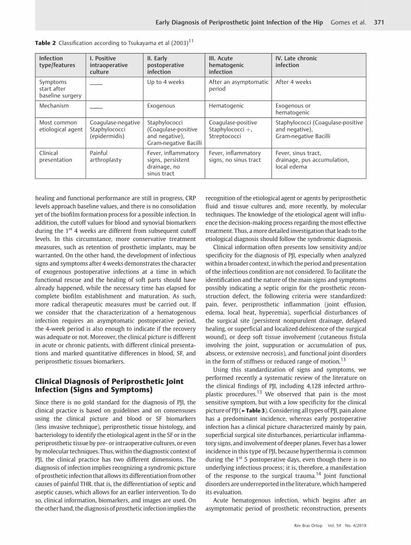
healing and functional performance are still in progress, CRPlevels approach baseline values, and there is no consolidationyet of the biofilm formation process for a possible infection. Inaddition, the cutoff values for blood and synovial biomarkersduring the 1st 4 weeks are different from subsequent cutofflevels. In this circumstance, more conservative treatmentmeasures, such as retention of prosthetic implants, may bewarranted. On the other hand, the development of infectioussigns and symptoms after 4weeks demonstrates the characterof exogenous postoperative infections at a time in whichfunctional rescue and the healing of soft parts should havealready happened, while the necessary time has elapsed forcomplete biofilm establishment and maturation. As such,more radical therapeutic measures must be carried out. Ifwe consider that the characterization of a hematogenousinfection requires an asymptomatic postoperative period,the 4-week period is also enough to indicate if the recoverywas adequate or not. Moreover, the clinical picture is differentin acute or chronic patients, with different clinical presenta-tions and marked quantitative differences in blood, SF, andperiprosthetic tissues biomarkers.
Clinical Diagnosis of Periprosthetic JointInfection (Signs and Symptoms)
Since there is no gold standard for the diagnosis of PJI, theclinical practice is based on guidelines and on consensusesusing the clinical picture and blood or SF biomarkers(less invasive technique), periprosthetic tissue histology, andbacteriology to identify the etiological agent in the SF or in theperiprosthetic tissuebypre- or intraoperative cultures, or evenbymolecular techniques. Thus,within thediagnosticcontextofPJI, the clinical practice has two different dimensions. Thediagnosis of infection implies recognizing a syndromic pictureofprosthetic infection that allows its differentiation fromothercauses of painful THR. that is, the differentiation of septic andaseptic causes, which allows for an earlier intervention. To doso, clinical information, biomarkers, and images are used. Ontheotherhand, thediagnosisofprosthetic infection implies the
recognition of the etiological agent or agents by periprostheticfluid and tissue cultures and, more recently, by moleculartechniques. The knowledge of the etiological agent will influ-ence the decision-making process regarding themost effectivetreatment. Thus, amoredetailed investigation that leads to theetiological diagnosis should follow the syndromic diagnosis.
Clinical information often presents low sensitivity and/orspecificity for the diagnosis of PJI, especially when analyzedwithin abroadercontext, inwhich theperiodandpresentationof the infectious condition are not considered. To facilitate theidentification and the nature of themain signs and symptomspossibly indicating a septic origin for the prosthetic recon-struction defect, the following criteria were standardized:pain, fever, periprosthetic inflammation (joint effusion,edema, local heat, hyperemia), superficial disturbances ofthe surgical site (persistent nonpurulent drainage, delayedhealing, or superficial and localized dehiscence of the surgicalwound), or deep soft tissue involvement (cutaneous fistulainvolving the joint, suppuration or accumulation of pus,abscess, or extensive necrosis), and functional joint disordersin the form of stiffness or reduced range of motion.13
Using this standardization of signs and symptoms, weperformed recently a systematic review of the literature onthe clinical findings of PJI, including 4,128 infected arthro-plastic procedures.13 We observed that pain is the mostsensitive symptom, but with a low specificity for the clinicalpictureof PJI (►Table 3). Considering all types of PJI, pain alonehas a predominant incidence, whereas early postoperativeinfection has a clinical picture characterized mainly by pain,superficial surgical site disturbances, periarticular inflamma-tory signs, and involvementofdeeper planes. Feverhas a lowerincidence in this type of PJI, because hyperthermia is commonduring the 1st 5 postoperative days, even though there is nounderlying infectious process; it is, therefore, a manifestationof the response to the surgical trauma.14 Joint functionaldisorders areunderreported in the literature,whichhamperedits evaluation.
Acute hematogenous infection, which begins after anasymptomatic period of prosthetic reconstruction, presents
Table 2 Classification according to Tsukayama et al (2003)11
Infectiontype/features
I. Positiveintraoperativeculture
II. Earlypostoperativeinfection
III. Acutehematogenicinfection
IV. Late chronicinfection
Symptomsstart afterbaseline surgery
____ Up to 4 weeks After an asymptomaticperiod
After 4 weeks
Mechanism ____ Exogenous Hematogenic Exogenous orhematogenic
Most commonetiological agent
Coagulase-negativeStaphylococci(epidermidis)
Staphylococci(Coagulase-positiveand negative),Gram-negative Bacilli
Coagulase-positiveStaphylococci þ,Streptococci
Staphylococci (Coagulase-positiveand negative),Gram-negative Bacilli
Clinicalpresentation
Painfularthroplasty
Fever, inflammatorysigns, persistentdrainage, nosinus tract
Fever, inflammatorysigns, no sinus tract
Fever, sinus tract,drainage, pus accumulation,local edema
Rev Bras Ortop Vol. 54 No. 4/2019
Early Diagnosis of Periprosthetic Joint Infection of the Hip Gomes et al. 371

Table
3Ana
lysisof
combine
dincide
ncean
dclinical
find
ings
forno
nspe
cifican
dspec
ificpe
riprosthe
ticjointinfections
rega
rdingpresen
tation
timean
dtype
13
(Com
bined
)Clin
ical
Find
ings
Ana
lysisof
theclinical
findingsfornon
spec
ific
PJIs
Inciden
ceoftheclinical
find
ingsforno
nspe
cifican
dSp
ecific
PJIs.
Ave
rage%
(95%
CI)
NoAC/Con
trols
(NoPJIs
THR/
TKR/others)
Sensitivity
%(95%
CI)
Spec
ificity%
(95%CI)
Accuracy
%(95%
CI)
Non
spec
ific
PJI
Early
postope
rative
PJI
Acu
tehem
atog
enic
PJI
Late
chronic
PJI
Pain
225/23
7(77/18
/0)
57.9
(47.3–
67.9)
28.3
(22.6–
34.5)
36.7
(31.5–
42.2)
77.2
(74.5–
79.9)
54.7
(51.3–
58.0)
92.8
(89.1–
96.6)
82.9
(77.5–
88.3)
Feve
r17
/205
(62/3/
0)13
.8(6.5–2
4.7)
96.1
(92.5–
98.3)
76.3
(70.7–
81.2)
31.2
(28.2–
34.1)
32.5
(29.4–
35.5)
75.5
(71.3–
79.7)
14.0
(10.4–
17.6)
Periarticu
lar
inflam
mationa
113/71
6(316
/220
/0)
14.9
(11.9–
18.4)
94.7
(92.8–
96.2)
61.8
(59.0–
64.6)
35.9
(33.2–
38.6)
49.0
(44.8–
53.3)
69.7
(60.1–
79.2)
18.4
(14.9–
22.0)
Supe
rficial
disturban
cesat
thesu
rgical
site
b
487/1,77
1(538
/392
/0)
23.6
(21.3–
26.1)
88.6
(87,0–
90.0)
61.8
(59.9–
63.5)
40.4
(37.5–
43.2)
46.8
(40.6–
53.0)
NR
24.1
(17.9–
30.2)
Dee
pso
ftpa
rts
invo
lvem
entc
24/62
(4/17/
3)43
.7(29.5–
58.8)
100.0
(97.0–
100.0)
84.3
(78.0–
89.4)
38.8
(35.4–
42.1)
44.0
(40.7–
47.4)
11.0
(8.1–1
4.0)
26.3
(21.8–
30.9)
Jointdysfunc
tiond
NR
NR
NR
NR
74.4
(69.9–
78.9)
NR
20.5
(1referenc
e)41
.7(1
referenc
e)
Totaln
umbe
rof
included
PJIs
(THR/
TKR/
othe
rs)
976
(561
/412
/3)
____
__25
23(142
1/10
71/31
)90
2(491
/40
8/3)
435(152
/27
4/9)
607(385
/18
1/41
)
Abbrev
iation
s:NoAC:ob
served
clinical
find
ings
-(True
þ)þ
(False
þ).CI,co
nfide
nceinterval;NR,
notreported;
PJI,pe
riprosthe
ticjointinfection;
THR;totalh
ipreplace
men
t;TK
R,totalk
neereplace
men
t.a Com
bine
dforjointeffusion
/edem
a,he
atan
d/orerythe
ma.
bCom
positeforde
laye
dhe
aling,pe
rsistent
non-purulen
tdraina
gean
d/or
supe
rficial
dehisce
nce.
c Com
pos
iteforde
epsinu
strac
t,pu
rulent
draina
ge,ab
scessan
d/or
extensivene
crosis.
dCom
pos
iteforrigidityan
d/orreduc
edrang
eof
jointmotion.
Rev Bras Ortop Vol. 54 No. 4/2019
Early Diagnosis of Periprosthetic Joint Infection of the Hip Gomes et al.372
mainlywith severepain, fever, andperiarticular inflammatorysigns. Concurrent bacteremia accounts for systemic signs/symptoms of infection.
On theotherhand, in late chronic infections,pain is themainsymptom, accompanied by periarticular soft tissue disordersand joint dysfunction. It is not uncommon that painful arthro-plasty, with no other marked manifestations, is the onlyindication of late chronic PJI caused by low virulence agents.This picture can be seen when positive intraoperative culturesare obtained in revision surgeries due to clinical suspicion ofasepticcauses, especially ifperformed < 5yearsafter the indexprocedure, and if mechanical or functional causes could not beidentified by conventional investigations.15
It is always very useful to evaluate the risk factors for infec-tion inpatientswith arthroplasticprocedurefailure, since thesefactors may be associated with a higher PJI prevalence; more-over, they are even used as scores for the construction of riskcalculators. More recently, an analysis of 43,253 total arthro-plasties, including 1,035 PJIs, identified as risk factors forinfection, in decreasing order of incidence: prior joint surgery,injectable drug abuse, revision surgery, acquired immunedeficiency syndrome (AIDS), coagulopathy, renal disease,congestive heart failure, psychoses, rheumatic diseases, kneearthroplasty, diabetes, anemia, male gender, liver disease,smoking, and body mass index (BMI).16 The authors acknowl-edged that these conditions are independent risk factors for PJI.
Thus, based on the clinical suspicion of a syndromiccondition, although not fully compatible with PJI, it isnecessary to investigate serum markers and images.
Serum Biomarkers in the Diagnosis ofPeriprosthetic Joint Infection
Blood biomarkers are intended to quantitatively portray theresponse of thebody to theunderlying inflammatory/infectiveprocess, using an increasing number of more specific antimi-crobial proteins and proinflammatory cytokines. Initially,serum markers are often preferable to SF markers becausethey are less invasive, less costly, and allow sequential meas-urements with no risk of iatrogenic joint infections.
The usefulness of leukocytes count as a PJI indicator isvery restricted because of its low sensitivity (45%), except foracute hematogenic PJI associated with bacteremia, in whichleukocytosis may be significant.
The serummarkers most used as adjuncts in the preoper-ative diagnosis of PJI are the ESR and the CRP levels.
Since ESR expresses only the rate of precipitation of redblood cells, its specificity is low. Considering a cutoff level of30 mm, it has 75% sensitivity and 70% specificity for chronicprosthetic infections. In acute infections, this test is consideredinadequatebecauseacutoff level cannotbereliablyassigned.17
On average, the kinetics of ESR after uncomplicated primaryelective arthroplasties show a return to baseline levels3 months after the index procedure.
C-reactive protein is an acute phase protein, synthesized byproinflammatory cytokines, such as interleukin 6 (IL-6); how-ever, its specificity is also restricted. When a cutoff value of10 mg/L is considered, its sensitivity ranges from68to82%, and
its specificity ranges from 71 to 80% for chronic infections.17,18
In acute infections, the cutoff level is 100 mg/L. On average, thekinetics of serum CRP levels after uncomplicated primaryelective arthroplasties show a return to baseline valuesbetween 3 and 4 weeks after the index procedure.
It should be noted that both ESR and CRP levels may beelevated in inflammatory diseases (false-positive finding forinfections) or normal (false-negative finding) in infectionscaused by low-virulenceMOs. Even so, their use in screeningto rule out PJI is mandatory, since the combined sensitivityand specificity of thesemarkers are reported between 84 and98% and between 47 and 96%, respectively.17–19
The use of other serumbiomarkers, such as IL-6 or interleu-kin4 (IL-4), isnot yet standardized. SerumIL-6kinetics showsapeakelevation in2 to3days, anda return tonormal levels in�5to 7 days after an uncomplicated prosthetic replacement, thusbeing an early indicator of PJI. Although its specificity rangesfrom 91 to 95% for a cutoff level of 10 pg/mL, its sensitivityvaries greatly in different studies (from 47 to 97%) and,therefore, its use is not routine due to these discrepancies;moreover, this is a more costly test when compared with ESRand CRP. The same applies to α1 acid glycoprotein.
Other cytokine groups, which also have numerous proin-flammatory actions, such as tumor necrosis factor alpha(TNF-α) and procalcitonin (PCT), which are secreted bymonocytes, have been identified as important markers ofinfection, but the reported results are still conflicting; there-fore, their routine clinical use has not yet been established. Inaddition, in the literature, there is no study with a level ofevidence I or II that indicates its diagnostic superiority.
More recently, Shahi et al have reported a promisingperspective on the use of D-dimer as a serum marker of theresponse to inflammatory stimuli, which promote elevatedlevels of fibrin degradation products in blood.17 In a studyevaluating 172 revision arthroplasty procedures (86 asepticconditions and 86 PJIs), the authors found 89% sensitivity and93% specificity for prosthetic infection diagnosiswhen a cutofflevel of 860 ng/mL was used.
Clinical Diagnosis of Periprosthetic JointInfection. Imaging Evaluation
Imaging tests usually have low specificity for the diagnosis of PJIand, therefore, their greatest utility is to rule out other nonin-fectious disorders as a cause of arthroplasty failure. Plain radio-graphs are usually normal and may eventually present somesigns suggestive of PJI, such as periprosthetic loosening and/orosteolysis in arthroplasties performed < 5 five years before,subperiosteal elevation or transcortical fistula. Even so, thespecificity of the radiological examination ranges from 50 to67%, with an accuracy rate of 64%.20 Similarly, ultrasound,computed tomography (CT) and magnetic resonance imaging(MRI) scans are not directly relevant to the diagnosis of PJI, buttheycan identifyothercauses forarthroplasticprocedurefailure.
Nuclear medicine scans, such as conventional bone scin-tigraphy, present high sensitivity, but very low specificity,which precludes its routine use in PJI investigation.21 Morerecently, promising results have been reported with
Rev Bras Ortop Vol. 54 No. 4/2019
Early Diagnosis of Periprosthetic Joint Infection of the Hip Gomes et al. 373

immunoscintigraphy using antigranulocyte antibodies andcombined to traced leukocytes and bone marrow scintigra-phy. Positron emission tomography (PET/CT) using fluoro-deoxyglucose (18F-FDG) is also indicated in suspected casesof infection, although its major indication is in the oncol-ogical area. However, these tests are time-consuming, notalways available, and costly.
Thus, the role of nuclear imaging in the diagnosis andtreatment of PJI is still uncertain,21 with more restricted use,sincethemethodsdescribedherefor thediagnosisof syndromicinfection are less invasive, inexpensive, and widely available.
Biomarkers from the Synovial Fluid andPeriprosthetic Tissue Histology
The evaluation of SF biomarkers requiresmore invasive collec-tion methods; moreover, it adds costs and risks of iatrogenicinfection. Inaddition,upto32%ofaspirationproceduresdonotprovide enough fluid for analysis.22 Therefore, a surgicalprocedure guided by imaging techniques is recommended.
The most commonly used biomarkers from the SF for thediagnosis of PJI are: total leukocytes count, percentage ofneutrophils (%N), leukocyte esterase (LE), CRP, and α-defen-sin. The following cutoff levels are considered: 3,000 leuko-cytes/μL, 80% N, þþ, 6.9 mg/L (specific cutoff level not yetvalidated), and positive for the lateral flow test, respectively.
Theα-defensin test, stillwith limitedavailability inBrazil, isvery expensive. Considering its positivity at the lateral flowtest, its sensitivity is 78%, and its specificity is 93%. Comparingit with the performance of total leukocyte count,% N, and EL,along with ESR and CRP, through the diagnostic odds ratio(DOR) method, which calculates the ratio between the chanceof positivity in PJI and the chance of positivity in noninfectedpatients, Shahi et al noted that, for standard cutoff values, LEhas the best performance (30.06 � 0.27), followed by leuko-cyte count (29.45 � 0.19), CRP (25.66 � 0.14), neutrophilspercentage (25.53 � 0.19), and by ESR and PCR combined(23.33 � 0.11).23 When analyzed along with ESR and CRP,LE, if concordant with these tests, has 95% accuracy in con-firming or ruling out the diagnosis of PJI.24
It is noteworthy that theLE testhas lowcost, sincedetectionuses urinalysis strips; for þ or þþ positivity, it presents 75%sensitivity and 91% specificity. On the other hand, CRP mea-surement in SF is less frequently used because the calibrationof the device for its dosage is different from the one employedfor serum values. Similarly, IL-6 dosage in SF has not yet beenroutinely incorporated into the clinical practice.
Periprosthetic tissue histology, considered significant if> 5 neutrophils per field are detected in > 5 fields (400xmagnification), requires specific collection and interpreta-tion; therefore, its routine use is still questioned by someauthors. However, in a recent study on the accuracy offreezing biopsy compared with permanent sections, itshowed a 97.6% concordance. When evaluated directly re-garding the diagnosis of PJI, freezing biopsy presented sen-sitivity, specificity and accuracy of 73.7% (95% confidenceinterval [CI]: 59.7–87.7%), of 98.8% (95%CI: 97.1–100.0%), andof 94.0% (95% CI: 90.7–97.3%), respectively.25
Etiological Diagnosis of Periprosthetic JointInfections
Culture and molecular techniquesIdentification of the infectious MO (etiological diagnosis) isundoubtedly the most important procedure for the institu-tion of the proper treatment and, consequently, for betteroutcomes. The performance of the conventional method ofculture of periprostheticfluids and tissues still does notmeetthe expectations of the medical team responsible for thetreatment of PJIs, with a high rate of false-negative results,which is easily demonstrated by the 5 to 12% rate of PJIsreferred to as culture-negative infections.26
However, infections associated with prosthetic implantscan be great diagnostic challenges, such as biofilm formation,defined byHall-Stoodley as infections caused by pathogenic oropportunistic MO aggregates, encapsulated in an exopolysac-charide matrix and protected from host defense mechanismsand from antibiotic treatment.27 As such, free (planktonic)bacteria in periprosthetic tissue are present in a smallernumber, whereas bacteria in a mildly active metabolic stateremain protected inside the biofilm. The explant sonicationtechnique was proposed to release these “dormant” bacteriafrom the biofilm throughmechanical agitation. More recently,seeding the sonicated liquid, or even periprosthetic tissuesamples, in bloodcultureflasks, has been shown tobeeffectivein increasing thesensitivityof thismicrobiological test byupto40% when compared with the conventional technique in agaror thioglycolate broth (from 44.4 to 60.7%),28 if the IDSAcriteria for the diagnosis of PJI were considered.6
Anothermajor challenge for traditional cultures is the lackof standardization of collection, packaging, transport, andmethod techniques, as well as of incubation time of biologi-cal materials. Now, it is known that more indolent bacteria(coagulase-negative Staphylococci or C. acnes, for instance)may require up to 15 days of culture, in contrast with thetraditional incubation of 3 to 5 days for more virulent MOs.
Nevertheless, the prevalence of culture-negative infec-tions remains significant and, therefore, there is a greatcommitment to improve molecular techniques, especiallythe next generation sequencing (NGS) procedures. In a recentprospective study including infected hip or knee revisions,while cultures were positive in 60.7% of the cases (95%CI:40.6–78.5%), NGS was positive in 89.3% of the cases (95%CI:71.8–97.7%).29 The authors conclude that NGS is useful inMOdetection in culture-negative, as well as in detecting con-comitant MOs that escape recognition in culture media.However, they point to the need for future studies to clarifythe presence of MOs isolated from patients undergoingpresumptive aseptic revision surgery.
Algorithm for the Diagnostic of Periprosthetic JointInfectionFrom the main tests presented here, the ICM-2013 proposedmodifications in the algorithm presented by the AmericanAcademy of Orthopedic Surgeons (AAOS),30 which guides,step-by-step, the indication and interpretation of the tests tobe performed for the identification of PJI (►Fig. 2).
Rev Bras Ortop Vol. 54 No. 4/2019
Early Diagnosis of Periprosthetic Joint Infection of the Hip Gomes et al.374

Final Considerations
The lack of a gold standard for the diagnosis of PJIs of the hipposes innumerous challenges to the early recognition of thisserious complication, which may result in a higher rate oftreatment failure. Advances in the knowledge about PJIsindicate the absolute necessity of an adequate and systemat-ic prevention and of an early diagnosis based on clinicalfindings, blood and SF biomarkers levels, periprosthetictissue histopathology findings, and the identification ofthe infectiveMO throughmolecular cultures and techniques.These procedures should be carried out sequentially, rou-tinely, and in a standardized form, in all centers dedicated tothe treatment of PJIs.
Conflicts of InterestThe author has no conflicts of interest to declare.
References1 Lidgren L. Joint prosthetic infections: a success story. Acta Orthop
Scand 2001;72(06):553–5562 Ulrich SD, Seyler TM, Bennett D, Delanois RE, Saleh KJ, Thong-
trangan I, et al. Total hip arthroplasties: what are the reasons forrevision? Int Orthop 2008;32(05):597–604
3 Esposito S, Leone S. Prosthetic joint infections: microbiology,diagnosis, management and prevention. Int J Antimicrob Agents2008;32(04):287–293
4 Berbari EF, Hanssen AD, Duffy MC, Steckelberg JM, Ilstrup DM,HarmsenWS, et al. Risk factors for prosthetic joint infection: case-control study. Clin Infect Dis 1998;27(05):1247–1254
5 Parvizi J, Zmistowski B, Berbari EF, Bauer TW, Springer BD, DellaValle CJ, et al. New definition for periprosthetic joint infection:from theWorkgroup of theMusculoskeletal Infection Society. ClinOrthop Relat Res 2011;469(11):2992–2994
6 OsmonDR, Berbari EF, Berendt AR, LewD, ZimmerliW, SteckelbergJM, et al. Infectious Diseases Society of America. Executive summa-ry: diagnosis andmanagement of prosthetic joint infection: clinicalpractice guidelines by the Infectious Diseases Society of America.Clin Infect Dis 2013;56(01):1–10
7 Parvizi J, Gehrke T, Chen AF. Proceedings of the internationalconsensus on periprosthetic joint infection. Bone Joint J 2013;95-B(11):1450–1452
8 Fernández-Sampedro M, Fariñas-Alvarez C, Garces-Zarzalejo C,Alonso-Aguirre MA, Salas-Venero C, Martínez-Martínez L,et al. Accuracy of different diagnostic tests for early, delayedand late prosthetic joint infection. BMC Infect Dis 2017;17(01):592
9 Coventry MB. Treatment of infections occurring in total hipsurgery. Orthop Clin North Am 1975;6(04):991–1003
10 Tsukayama DT, Estrada R, Gustilo RB. Infection after total hiparthroplasty. A study of the treatment of one hundred and sixinfections. J Bone Joint Surg Am 1996;78(04):512–523
Fig. 2 Modified American Academy of Orthopedic Surgeons algorithm (CIAP, 2013). Abbreviations: BAAR, Acid-alcohol-resistant bacilli, CRP, C-reactive protein; ESR, erythrocyte sedimentation rate; XR, X-ray; SF, synovial fluid.
Rev Bras Ortop Vol. 54 No. 4/2019
Early Diagnosis of Periprosthetic Joint Infection of the Hip Gomes et al. 375

11 Tsukayama DT, Goldberg VM, Kyle R. Diagnosis and managementof infection after total knee arthroplasty. J Bone Joint Surg Am2003;85-A(Suppl 1):S75–S80
12 Zimmerli W, Trampuz A, Ochsner PE. Prosthetic-joint infections.N Engl J Med 2004;351(16):1645–1654
13 Gomes LSM, Zullo SS, Pereira GA. Response to question Q118 (Hipand Knee): What clinical findings (e.g. fever, erythema, reducedROM) are most sensitive and specific for the diagnosis of PJI? In:Parvizi, J and Gherke Thorsten. Proceedings of the Second Inter-national Consensus Meeting on Musculoskeletal Infections. Broo-klandville, Maryland: Data Trace; 2018. p. 343–344
14 Ghosh S, Charity RM, Haidar SG, Singh BK. Pyrexia following totalknee replacement. Knee 2006;13(04):324–327
15 Jacobs AME, BénardM,Meis JF, vanHellemondtG, Goosen JHM. Theunsuspected prosthetic joint infection : incidence and consequen-ces of positive intra-operative cultures in presumed aseptic kneeand hip revisions. Bone Joint J 2017;99-B(11):1482–1489
16 Tan T, Maltenfort M, Chen A. Development and evaluation of apreoperative risk calculator for periprosthetic joint infection. In:Paper 171 presented at the 2016 Annual Meeting of the AAOS,Orlando, Florida, 2016
17 ShahiA, KheirMM,TarabichiM,HosseinzadehHRS, TanTL,Parvizi J.SerumD-DimerTest IsPromising for theDiagnosis ofPeriprostheticJoint Infection and Timing of Reimplantation. J Bone Joint Surg Am2017;99(17):1419–1427
18 Pérez-PrietoD, PortilloME, Puig-Verdié L, Alier A,Martínez S, SorlíL, et al. C-reactive protein may misdiagnose prosthetic jointinfections, particularly chronic and low-grade infections. IntOrthop 2017;41(07):1315–1319
19 Parvizi J, Fassihi SC, Enayatollahi MA. Diagnosis of periprostheticjoint infection following hip and knee arthroplasty. Orthop ClinNorth Am 2016;47(03):505–515
20 Aryana K, Hootkani A, Sadeghi R, Davoudi Y, Naderinasab M,Erfani M, et al. (99m) Tc-labeled ubiquicidin scintigraphy: apromising method in hip prosthesis infection diagnosis. NuclMed (Stuttg) 2012;51(04):133–139
21 Verberne SJ, Raijmakers PG, Temmerman OP. The accuracy ofimaging techniques in the assessment of periprosthetic hipinfection a systematic review and meta-analysis. J Bone JointSurg Am 2016;98(19):1638–1645
22 YeeDKH, Chiu KY, Yan CH, Ng FY. Reviewarticle: Joint aspiration fordiagnosis of periprosthetic infection. J Orthop Surg (Hong Kong)2013;21(02):236–240
23 Shahi A, Tan TL, Kheir MM, Tan DD, Parvizi J. Diagnosing Peripros-thetic Joint Infection: And the Winner Is? J Arthroplasty 2017;32(9S):S232–S235
24 Tarabichi M, Fleischman AN, Shahi A, Tian S, Parvizi J. Interpreta-tion of Leukocyte Esterase for the Detection of Periprosthetic JointInfection Based on Serologic Markers. J Arthroplasty 2017;32(9S):S97, 100.e1
25 Kwiecien G, George J, Klika AK, Zhang Y, Bauer TW, Rueda CA.Intraoperative Frozen Section Histology: Matched for Musculoskel-etal Infection Society Criteria. J Arthroplasty 2017;32(01):223–227
26 Peel TN, DowseyMM, Aboltins CA, Daffy JR, Stanley PA, Buising KL,et al. Culture negative prosthetic joint infection.- a description ofcurrent treatment and outcomes. Clin Microbiol 2013;2(02):106.Doi: 10.4172/2327-5073.1000106
27 Hall-Stoodley L, Stoodley P, Kathju S, Høiby N, Moser C, CostertonJW, et al. Towards diagnostic guidelines for biofilm-associatedinfections. FEMS Immunol Med Microbiol 2012;65(02):127–145
28 Peel TN, Dylla BL, Hughes JG, Lynch DT, Greenwood-QuaintanceKE, Cheng AC, et al. Improved Diagnosis of Prosthetic JointInfection by Culturing Periprosthetic Tissue Specimens in BloodCulture Bottles. MBio 2016;7(01):e01776–e15
29 Tarabichi M, Shohat N, Goswami K, Alvand A, Silibovsky R, BeldenK, et al. Diagnosis of periprosthetic joint infection: the potential ofnext-generation sequencing. J Bone Joint Surg Am 2018;100(02):147–154
30 Della Valle C, Parvizi J, Bauer TW, Dicesare PE, Evans RP, Segreti J,et al; American Academy of Orthopaedic Surgeons. Diagnosis ofperiprosthetic joint infections of the hip and knee. J Am AcadOrthop Surg 2010;18(12):760–770
Rev Bras Ortop Vol. 54 No. 4/2019
Early Diagnosis of Periprosthetic Joint Infection of the Hip Gomes et al.376