aging - theories and potential therapies
TRANSCRIPT


AGING


AGINGTheories and Potential Therapies
Joseph Panno, Ph.D.
T H E
newbiology

AGING: Theories and Potential Therapies
Copyright © 2005 by Joseph Panno, Ph.D.
All rights reserved. No part of this book may be reproduced or utilized in any form or by any means, electronic or mechanical, including photocopying, recording, or by any information storage or retrieval systems, without permission in writing from thepublisher. For information contact:
Facts On File, Inc.132 West 31st StreetNew York NY 10001
Library of Congress Cataloging-in-Publication DataPanno, Joseph.
Aging: theories and potential therapies / Joseph Panno.p. cm. — (The new biology)
Includes bibliographical references and index.ISBN 0-8160-4951-31. Aging. 2. Longevity. I. Title.
QP86.P33 2004612.6’7—dc22 2003025469
Facts On File books are available at special discounts when purchased in bulk quantitiesfor businesses, associations, institutions, or sales promotions. Please call our SpecialSales Department in New York at (212) 967-8800 or (800) 322-8755.
You can find Facts On File on the World Wide Web at http://www.factsonfile.com
Text design by Erika K. ArroyoCover design by Pehrsson DesignIllustrations by Richard Garratt and Joseph Panno
Printed in the United States of America
MP FOF 10 9 8 7 6 5 4 3 2 1
This book is printed on acid-free paper.

For my wife, Diana,
who worked with me in the lab for many years,
and for my daughter Eleanor,
who knew about cells before she could read or write.
V


CONTENTS
Preface xi
Acknowledgments xiii
Introduction xv
1 The Quest for Immortality 1One Hour upon the Stage 2
Growing Younger 7
The Road Ahead 7
2 Aging Theories 9Error Catastrophe Theory 9
Genes and Programmed Aging 10
Telomeres 11
Rate-of-Living Theory 13
Free Radicals 14
Hormonal Imbalance Theory 14
Concluding Remarks 21
3 Age-Related Diseases 24Alzheimer’s Disease 24
Arthritis 32
Cancer 33
Cardiovascular Disease 34
Diabetes 35
Osteoporosis 36
V

4 Antiaging Medicine 38Hormone Therapy 39
Antioxidants 41
Caloric Restriction 41
Gene Therapy 42
5 The History of Gerontology 43The Early Years 43
DNA Structure Inspires New Theories 45
Recombinant Technology Revolutionizes the Field 46
The Postgenomic Era 52
6 The Search for Longevity Genes 54Yeast 55
Nematode 58
Fruit Fly 59
Mouse 62
Human 62
Summary 63
7 Geriatrics 65Our Aging Society 66
Evaluating the Geriatric Patient 67
Managing Age-Related Disorders 68
Drug Therapy 71
Nursing Homes 72
Ethical Issues 73
8 Resource Center 75Eukaryote Cell Primer 75
Recombinant DNA Primer 95

Gene Therapy Primer 106
The Human Genome Project 110
Glossary 115
Further Reading 141
Index 147


xi
PREFACE
The New Biology set consists of the following six volumes: The Cell,Animal Cloning, Stem Cell Research, Gene Therapy, Cancer, and Aging.The set is intended primarily for middle and high school students, butit is also appropriate for first-year university students and the generalpublic. In writing this set, I have tried to balance the need for a com-prehensive presentation of the material, covering many complex fields,against the danger of burying—and thereby losing—young studentsunder a mountain of detail. Thus the use of lengthy discussions andprofessional jargon has been kept to a minimum, and every attempt hasbeen made to ensure that this be done without sacrificing the impor-tant elements of each topic. A large number of drawings are providedthroughout the series to illustrate the subject matter.
The term new biology was coined in the 1970s with the introductionof recombinant DNA technology (or biotechnology). At that time, biol-ogy was largely a descriptive science in danger of going adrift. Microbi-ologists at the turn of the century had found cures for a few diseases,and biologists in the 1960s had cracked the genetic code, but there wasstill no way to study the function of a gene or the cell as a whole.Biotechnology changed all that, and scientists of the period referred toit as the new technique or the new biology. However, since that time ithas become clear that the advent of biotechnology was only the firststep toward a new biology, a biology that now includes nuclear transfertechnology (animal cloning), gene therapy, and stem cell therapy. Allthese technologies are covered in the six volumes of this set.
The cell is at the very heart of the new biology and thus figuresprominently in this book series. Biotechnology was specifically designedfor studying cells, and using those techniques, scientists gained insightsinto cell structure and function that came with unprecedented detail.
V

As knowledge of the cell grew, the second wave of technologies—animal cloning, stem cell therapy, and gene therapy—began to appearthroughout the 1980s and 1990s. The technologies and therapies ofthe new biology are now being used to treat a wide variety of medical disorders, and someday they may be used to repair a damaged heart, asevered spinal cord, and perhaps even reverse the aging process. Theseprocedures are also being used to enhance food crops and the physicalcharacteristics of dairy cows and to create genetically modified sheepthat produce important pharmaceuticals. The last application alonecould save millions of lives every year.
While the technologies of the new biology have produced somewonderful results, some of the procedures are very controversial. Theability to clone an animal or genetically engineer a plant raises a host ofethical questions and environmental concerns. Is a cloned animal afreak that we are creating for our entertainment, or is there a valid med-ical reason for producing such animals? Should we clone ourselves, or usethe technology to re-create a loved one? Is the use of human embryonicstem cells to save a patient dying from leukemia a form of high-techcannibalism? These and many other questions are discussed through-out the series.
The New Biology set is laid out in a specific order, indicated previ-ously, that reflects the natural progression of the discipline. That is,knowledge of the cell came first, followed by animal cloning, stem celltherapy, and gene therapy. These technologies were then used to expandour knowledge of, and develop therapies for, cancer and aging.Although it is recommended that The Cell be read first, this is not essen-tial. Volumes 2 through 6 contain extensive background material,located in the final chapter, on the cell and other new biology topics.Consequently, the reader may read the set in the order he or she prefers.
xii Aging

ACKNOWLEDGMENTS
I would first like to thank my friend and mentor, the late Dr. KarunNair, for helping me understand some of the intricacies of the biologi-cal world and for encouraging me to seek that knowledge by lookingbeyond the narrow confines of any one discipline. The clarity and accu-racy of the initial manuscript for this book was greatly improved byreviews and comments from Diana Dowsley and Michael Panno, andlater by Frank Darmstadt, Executive Editor; Dorothy Cummings, ProjectEditor; and Anthony Sacramone, Copy Editor. I am also indebted toRay Spangenburg, Kit Moser, Sharon O’Brien, and Diana Dowsley fortheir help in locating photographs for the New Biology set. Finally, Iwould like to thank my wife and daughter, to whom this book is dedi-cated, for the support and encouragement that all writers need and areeternally grateful for.
xiii
V


INTRODUCTION
Everybody keeps getting older. It has been this way since multicellularcreatures crawled out of the oceans more than 500 million years ago.Indeed, mortality and multicellularity seem to go hand in hand, for ourunicellular ancestors, the protozoans and bacteria, have an indefinitelife span. If we think of those ancestors as being a single lineage, it is alife form that has been alive for 3 billion years. There are those whothink a human life span of 85 years is long enough, but compared with3 billion years, it truly is the short end of the stick. There are, to be sure,many animals that have a shorter life span than we do: A horse has 20years, a dog is lucky to see 15 summers, and the poor housefly is born anddead of old age in 30 days. On the other hand, a Galápagos tortoise anda sturgeon can live for 200 years.
On a cosmic scale, however, the difference in life span between ahousefly and a sturgeon is puny, and besides, the comparison begs thequestion of why we age in the first place. After all, we have a reasonablygood immune system; we heal well after being hurt; we have a group ofenzymes that monitor and repair our DNA; and, as long as we eat well,our cells have plenty of energy to take care of themselves from day today. Yet, despite all that, we get old with monotonous regularity. Thereappears to be neither rhyme nor reason to it. Some scientists thinkaging is due to evolutionary neglect: that natural selection was so busyfinding ways to make us successful in the short term that it forgot tocover us in our old age. It is almost as though Mother Nature is saying,“I will do what I can to get you up to your reproductive years, so youcan have offspring, but after that you are on your own.”
Being on our own has meant that our bodies begin to break downsoon after our peak reproductive years have past. The elderly cannotrun as far, think as fast, or fight off infectious diseases nearly as well asthey did when they were young. Moreover, one’s physical appearance
xv
V

changes dramatically with age: The hair turns gray, muscle massdeclines, the ears get bigger, and the skin becomes thin and wrinkled. Ata more subtle level, men and women approaching their 80s converge ona common physical appearance; men become more feminine andwomen become more masculine. In men, this trend becomes apparentas the shoulders get narrower, the hips broader, the beard thinner, andthe voice develops a higher pitch. In women, the shoulders becomebroader, the voice huskier, and hair begins to grow on the chin andupper lip. Gerontologists (scientists who study gerontology, or themechanisms involved in the aging process), in noting these changes,have pondered one of the most difficult questions pertaining to theaging process: Is aging caused by the degenerative changes in a singleorgan, which then acts like an aging-clock for the rest of the body, or areall organs breaking down simultaneously?
Answering this question has proved to be extremely difficult.Researchers have studied age-related changes in virtually all tissues,organs, and organ systems (e.g., the endocrine system, consisting ofmany hormone-producing glands) of the body. Some evidence suggeststhat the brain may be an aging-clock that determines the rate at whichthe whole body ages, but the results of many other studies suggests thatthe rate at which an animal ages may be the sum of age-related changesoccurring simultaneously in all parts of the body.
Consequently, the attempts to understand the aging process, involv-ing such a complex system, have generated a large number of theories butfew practical therapies. The therapies that are available are designed totreat diseases that are associated with aging, such as cancer and arthri-tis, but do not reverse the aging process itself. The recent trend in geron-tology, particularly since the completion of the human genome project,is to search for genes that have a demonstrable effect on life span, theso-called longevity genes. Many such genes have been identified, andalthough the manipulation of these genes does not stop the agingprocess, they are providing many valuable insights into the cellularmechanisms of aging that may lead to the development of truly effect-ing antiaging therapies.
This book, another in the New Biology set, describes the field ofgerontology and the many theories that scientists have developed overthe years to explain the age-related changes that occur in virtually all
xvi Aging

animals. The first four chapters discuss the range of animal life spansrelative to the human life span, aging theories, diseases that are associ-ated with the aging process, and aging therapies (antiaging medicine).Subsequent chapters discuss the history of gerontology, the ongoingeffort to find longevity genes, and the field of geriatrics, which uses ourgrowing knowledge of the aging process to improve the quality of lifefor the elderly. The final chapter provides background material on cellbiology, recombinant DNA technology, and other topics that are rele-vant to gerontology.
Introduction xvii


� 1 �
THE QUEST FORIMMORTALITY
Concerns about human mortality date back at least 20,000 years whenCro-Magnons, the first Homo sapiens, prepared one of their own forburial. Cro-Magnon funerals are taken as evidence by anthropologiststhat those people thought like us. They knew about death, and in theirsorrow, they adorned the corpse with prized possessions, possiblythinking they would be of use in a spiritual afterlife. In grieving for theirlost loved ones, Cro-Magnons were drawn to a quest for immortality,but one that dealt with the soul rather than the body.
Distant relatives of the Cro-Magnons, living 4,000 years ago in Egypt,carried on the same tradition but on a colossal scale. Egyptian pharaohswere buried with all their worldly possessions and even a little food to seethem on their way. According to their mythology, the dead, if accepted,could pass to the spirit world of the sun god, Amun-Ra, and his sister,Amunet, where they would live for eternity. The practice of burying thedead with all their belongings disappeared down through the millennia,but many people still believe in the eternal life of chosen spirits.
With the rise of science, and the appearance of powerful medicaltherapies, the quest for immortality has shifted from the spiritual to thephysical. Although the origins of the scientific method can be tracedback to the time of the Cro-Magnons, it did not assume its present formuntil the 20th century. Indeed, historians have noted that 90 percent ofall scientists who have ever lived are still alive today.
The accomplishments of Louis Pasteur and other microbiologists atthe turn of the last century and the explosive growth in biological
1

research since then have provided cures for many terrible diseases:diphtheria, polio, and smallpox, to name but a few. These triumphshave given us reason to hope that someday we will be able to reverse theeffects of age. If protozoans can live millions of years, why not thehuman body?
But so far, all attempts at physical rejuvenation have failed. Manysuch attempts date back to the turn of the early 1900s and involved theuse of concoctions, potions, and even radioactive cocktails, often withdisastrous results. One such concoction, popular in the 1920s, was Tho-Radia, a skin cream containing thorium and radium, two radioisotopesdiscovered by the great French physicist Marie Curie. The radioactivematerial was supposed to have an antiaging effect on the skin, but theiruse was abandoned when Curie and other scientists working withradioisotopes began having serious medical problems. Madame Curiedeveloped cataracts, kidney failure, and a fatal leukemia, all from over-exposure to radioactive materials.
More recently, a new wave of antiaging therapies have been devel-oped, employing everything from a shift in lifestyle to specific hormonesupplements. Antiaging creams are still with us, only now the activeingredient is retinoic acid (vitamin A) instead of radium. Whether anyof these treatments will be successful is in doubt, but the failures so farare like the first tentative steps of a toddler. We are only beginning tounderstand the tremendous complexities of the cell and the way anorganism changes with time. As our science matures, we may be able toreverse some effects of age, but whether this leads to physical immor-tality is a hotly debated topic at the present time.
One Hour upon the StageWhen William Shakespeare in his play Richard III compared the humanlife span to one hour on the stage, he was being very generous. If our lifespan were indeed 1/24 of the 3 billion years that microbes have beenalive, we would live 125 million years. As it is, our life span, on a 24-hour time scale, is but a wink of an eye.
Life spans vary considerably within the animal kingdom. In general,tiny animals bearing many offspring have short life spans, while largeanimals bearing few offspring live much longer. The fruit fly, Drosophila
2 Aging

melanogaster, is an example of asmall animal with a short lifespan. Drosophila have a maxi-mum life span of 40 days, butmost of them are dead in twoweeks. These animals are calledholometabolous insects becausethe eggs hatch into wormlike lar-vae that feed for a time beforepupating, during which time thelarvae metamorphose into theadult form. The newly emergedmales and females waste no timein producing the next genera-tion. The females mate within 24hours, and throughout theirshort lives produce tens of thou-sands of offspring. In a survivalstrategy such as this, all the bio-logical adaptations have focusedon preservation of the species atthe expense of the individual.Flies have many predators, andadaptations that could lengthentheir life span would be useless,since the flies would be eatenlong before their biological timewas up.
Elephants, on the other hand, are large animals with few predatorsthat produce a single offspring every five years. Elephants are mammalsand, like all mammals, spend a great deal of time rearing and caring fortheir young. In this case, adaptations to increase their life span make alot of sense. With reduced pressure from predators, the young canafford a leisurely developmental period, during which time the adultsteach them how best to deal with their environment. The longer theadults live, the more things they learn, and the more they can pass on totheir offspring. Consequently, these animals have a relatively long life
The Quest for Immortality 3
Scanning electron micrograph (SEM)of the fruit fly Drosophila melanogaster.This little insect is about 3 mm long, iscommonly found around spoiled fruit,and is an example of a very short-livedanimal. It is also much studied bygerontologists and geneticists aroundthe world. Mutant flies, with defects inany of several thousand genes, areavailable, and the entire genome hasbeen sequenced. (David M.Phillips/Photo Researchers, Inc.)

span of 75 to 100 years, similar to that of humans. In general, long-livedanimals tend to be rather intelligent, but there are some exceptions, themost notable of which are the sturgeon and the Galápagos tortoise.
The sturgeon is an extremely ancient fish that has existed for morethan 200,000 years, predating the rise of the dinosaurs during the Juras-sic period. They live in the oceans, seas, and rivers of North America,Europe, and Asia, where they often grow to a length of 20 feet and weigha ton or more. Sturgeon eggs, called caviar, are considered to be a greatdelicacy in many parts of the world. The sturgeon is possibly thelongest-lived animal that we know of, sometimes reaching 300 years ormore, and yet they are no more intelligent than any other fish. More-over, sturgeons, like most fish, have thousands of offspring each yearand spend no time taking care of them. The sturgeon’s strategy forlongevity is simply to keep growing. They have hit upon a rule of naturethat states that happy cells are dividing cells. As long as a sturgeon keepsgrowing, its longevity is regulated by external forces, such as accidents
4 Aging
African elephants (Loxodonta africana) in the Amboseli National Park, Kenya.These large, intelligent animals have a maximum life span of about 100 years,very similar to that of humans. (Martin Harvey/Photo Researchers, Inc.)

and predators, not by cellular senescence. Being a poikilotherm (cold-blooded animal) reinforces the sturgeon’s continuous-growth strategybecause it minimizes the growth rate and activity level of the animal.Continuous growth is a strategy that also explains the longevity of cer-tain plants, such as the California redwood or the oak tree, which can livea thousand years or more.
Galápagos tortoises live on a group of islands off the coast of SouthAmerica. They are large animals, sometimes weighing more than 500pounds that can live for 200 to 250 years and usually give birth to adozen offspring every year. They are not highly intelligent animals, atleast not as mammals understand intelligence; nor do they spend anytime taking care of their young. In fact, a tortoise never sees its young.The female lays the eggs in the sand, covers them over, and the rest isleft to Mother Nature and a bit of luck. When the young hatch, they digtheir way to the surface, a feat that takes a month to accomplish, and
The Quest for Immortality 5
Giant tortoise from the Galápagos Islands (Santa Cruz Island). These animalshave very long life spans that may exceed 200 years. ( Jeffrey Greenburg/PhotoResearchers, Inc.)

make straight for the water, which is usually 10 to 20 yards away. Thedash for the water is made through a predator gauntlet, and many of theyoung tortoises are caught by seagulls along the way. Those that make itto the water are preyed upon by fish in the sea, and the few that surviveto adulthood return to the beaches of their birth, where they live out therest of their lives. The tortoise, unlike the sturgeon, reaches a standardadult size, so most of the cells in the adult’s body become postmitotic,as occurs in mammals. The unusual longevity of this animal is believedto be due to its very low growth rate and, as it is a poikilotherm like thesturgeon, its low metabolic rate and activity level.
Humans have a maximum life span of more than 100 years. Thelongest-lived human on record was Jeanne Calment, a woman fromArles, France, who died in 1997 at the age of 122. As impressive as thisis, it is a short life span indeed when compared with the record holder
6 Aging
A sculpture of an elderly couple in their 80s showing the general effects of ageand the age-related convergence of physical characteristics described in theintroduction. (Dr. Joseph Panno)

from the plant kingdom. This goes to Methuselah, a 4,600-year-old pinethat lives on a mountainside in Arizona.
Growing YoungerMany gerontologists have claimed that it is impossible for individualhumans to grow younger, because it would be too difficult to rejuvenateall the cells and organs of the body. Such claims need to be taken with alarge grain of salt; it should be remembered that just five years beforethe first sheep was cloned, most scientists thought that cloning a mam-mal was biologically impossible. In addition, what we have learnedabout animal cloning and stem cells since 1996 suggests it may indeedbe possible to produce a therapy that will allow an individual to growyounger.
Growing younger, at the cellular level, is analogous to the dediffer-entiation of a cloned cell nucleus: Both are a matter of converting a cellfrom an aged phenotype (the physical expression of an organism’sgenes) to a youthful phenotype. In a sense, the cloning of a cell nucleusis the most successful attempt at rejuvenation that has yet been accom-plished. In a cloning experiment, the cytoplasm of the recipient oocyteconverts the donor nucleus from an aged phenotype to one that is capa-ble of supporting full embryonic development. At the organismic level,this is equivalent to converting an adult to an embryo. If it can be donein one cell, it could be done in many. And if all the nuclei in an old per-son’s body could be reprogrammed to a youthful phenotype, it wouldlead to the complete rejuvenation of all the cells in the body. If that hap-pened, the individual would grow younger.
The Road AheadIn 1900 life expectancy for the average North American was only 45years. This has increased to the current expectancy of 80 years primarilybecause of a dramatic reduction in infant mortality, cures for variousdiseases, better hygiene, and better living conditions. This increaseoccurred despite the enormous number of deaths per year from ciga-rette smoking. A further increase of 20 to 30 years is expected if cures arefound for cancer and cardiovascular disease. Beyond that, advances in life
The Quest for Immortality 7

expectancy will have to wait for an improvement in our understandingof the basic mechanisms of cellular senescence.
Developing therapies that will reverse the aging process, allowingindividuals to grow younger, is theoretically possible, but the realizationof that goal will likely turn out to be the most difficult challenge thatbiologists have ever faced. The production of aging therapies willrequire a fusion of animal cloning, gene therapy, and stem cell tech-nologies. But even these technologies, as powerful as they are, will not beenough. Gaining a deep understanding of the basic mechanisms ofaging will require detailed information about every gene in our bodiesand about what those genes are doing as we grow old. This informationis only now being made available, but over the next few years, we shouldsee real gains being made in the field of gerontology.
8 Aging

� 2 �
AGING THEORIES
Aging theories cover the genetic, biochemical, and physiological prop-erties of a typical organism, and the way these properties change withtime. Genetic theories deal with speculations regarding the identity ofaging genes, accumulation of errors in the genetic machinery, pro-grammed senescence, and telomeres. Biochemical theories are con-cerned with energy metabolism, generation of free radicals, the rate ofliving, and the health of mitochondria. Physiological theories dealalmost entirely with the endocrine system and the role of hormones inregulating the rate of cellular senescence.
Error Catastrophe TheoryRunning a cell is a complex affair. RNA and proteins have to be synthe-sized on a regular basis to maintain and run the cell’s machinery (seechapter 8 for a cell primer). Production of proteins, either for enzymesor structural materials, occurs in a two-step process: transcription ofthe gene to produce mRNA, followed by translation of the message toproduce the protein. For cells that are actively dividing, a third step,replication of the DNA, precedes the other two. Errors can occur allalong the way; when they do, defective genes, mRNA, and proteins areproduced. The error catastrophe theory, first proposed in the 1960s,suggests that over time, the number of errors build up to a catastrophiclevel leading to the death of the cell and, possibly, the entire organism.
Soon after this theory was proposed, many scientists conductedexperiments that attempted to force a buildup of errors to see how thecells would cope with it. Bacteria were grown on a medium containing
9

defective amino acids to maximize the error frequency of protein syn-thesis. Similar experiments were conducted on fruit flies (Drosophila)and mice, both of which were given food containing defective aminoacids. To everyone’s surprise, these experiments had no effect on thebacteria’s or animal’s health, vigor, or life span. Somehow the cells wereable to avoid an error catastrophe. Today we understand why thoseexperiments failed: Cells have elaborate repair systems and strategiesthat detect and destroy defective molecules. If a defective protein is syn-thesized, it is quickly broken down and replaced with a normal copy.Only in cases where the repair systems have been damaged would anerror catastrophe occur (see Werner’s syndrome in chapter 5).
In its original formulation, the error catastrophe theory focused onprotein synthesis, which apparently can tolerate a high error frequency.Consequently, many scientists began to wonder if errors in the genome,or possibly a defective regulation of the genes, might be responsible forthe aging process. After all, cells avoid an error catastrophe at the trans-lational level because they can always try again with a fresh mRNA froma good gene. But if the genes themselves are damaged, or programmedfor senescence, the outcome would be a gradual decline in cell vigor andthe eventual death of the organism.
Genes and Programmed AgingAre we programmed to get old? If we are, is it like the program thatguides our development from a single fertilized egg to a multicellularorganism? Or is aging the unfortunate side effect of adaptations thatmake it possible for us to have and protect our offspring? Many geron-tologists believe that aging is a matter of evolutionary neglect, ratherthan design.
However life spans evolved, it is clear that our genes have the finalsay in how long we are going to be on the stage. Even though flies andhumans are constructed from the same kinds of cells (eukaryotes), oneanimal lives two weeks, the other 80 years. If those eukaryotes hadremained free-living, as their protozoan ancestors have done, theywould live for millions of years.
The genes in a multicellular organism appear to be regulating lifespan for the good of the cell community as a whole. The size of the
10 Aging

community, the animal’s intelligence, the number of offspring, and thepressure the animal experiences from its predators, are all taken intoaccount. The final life span seems to be a balance of all these forcesand, given these forces, may be the best deal the organism can hope for.There would be no point to nature’s producing a fruit fly that couldlive a thousand years, because their predators eat them all in a matterof days. Scientists might try producing a fly that could live that long, butwhat in the world would an animal with that level of intelligence do forall that time? This is not just a whimsical point. There is a very strongcorrelation between longevity and the weight of the brain: Smart ani-mals live longer than dumb animals (with two exceptions, noted inchapter 1).
The goal of gerontologists is to try to get a better understanding ofthe covenant between the genes, the organism, and the environment.Whether intended by evolution or not, many genes are directly respon-sible for an animal’s life span. These genes may be exerting their effectsthrough inappropriate behavior (that is, they are turning on or off atthe wrong time) or through a mutation that eventually damages theprotein product.
Damage at the gene level re-invokes the error catastrophe theory,but many experiments have failed to establish a role for genetic (orsomatic) mutations in cell senescence. This is because the cell can detectand repair DNA damage as easily as it deals with errors in translation,and those repair systems remain intact long after the animal shows vis-ible signs of age.
The inappropriate expression of certain genes as a major cause ofaging is only now being addressed in a comprehensive way. With thegenome project now complete (see chapter 8), it will soon be possible toscreen for the expression of all human genes, in every tissue and organof the body. When this job is complete (and it will be as big a job as thegenome project itself) we will finally have an idea of which genes areresponsible for our life span.
TelomeresAlthough we have not identified the genes controlling our life span,there is a genetic element called a telomere that clearly regulates the
Aging Theories 11

replicative life span of human cells in culture. A telomere is a simpleDNA sequence that is repeated many times, located at the tips of eachchromosome. Telomeres are not genes, but they are needed for theproper duplication of the chromosomes in dividing cells. Each time thechromosomes are duplicated, the telomeres shrink a bit, until they getso short the DNA replication machinery can no longer work. Thisoccurs because the enzyme that duplicates the DNA (DNA polymerase)has to have some portion of the chromosome out ahead of it. Much likea train backing up on a track, DNA polymerase preserves a safe distancefrom the end of the DNA so it does not slip off the end. Telomeres alsoprovide a guarantee that genes close to the ends of the chromosomeshave been replicated. DNA polymerase stalls automatically whenever itgets too close to the end of the chromosome, permanently blocking theability of the cell to divide. When this happens, the cell is said to havereached replicative senescence.
The telomeres in human fibroblasts are long enough to permitabout 50 rounds of DNA replication. That is, the cell can divide about50 times in culture. This is often referred to as the Hayflick limit, afterLeonard Hayflick, the scientist who was the first to notice that normalcells cannot divide indefinitely in culture. Cancer cells, on the otherhand, can divide indefinitely, and from them scientists isolated anenzyme called telomerase that restores the telomeres after each cell divi-sion. If the telomerase gene is added to normal fibroblasts, they are nolonger bound by the Hayflick limit and can divide indefinitely, like animmortal cancer cell. The transformation of normal fibroblasts withthe telomerase gene was conducted for the first time in 1998 at theGeron Corporation, a biotechnology company. The results generated atremendous amount of excitement, for they seemed to imply that rever-sal of replicative senescence would be followed very quickly by thereversal of the aging process. Scientists at Geron began talking abouthuman life spans of several hundred years.
Experiments since have shown, however, that while telomerase canblock replicative senescence in cultured cells, it has little to do with thelife span of the animal as a whole. Indeed some animals with long lifespans have short telomeres and negligible telomerase activity, whileother animals with short life spans have long telomeres and activetelomerase. This is not surprising if we keep in mind that most cells in
12 Aging

an animal’s body are postmitotic; they stop dividing soon after the indi-vidual is born. So the life span of the individual made from those cellscannot be regulated by the length of the telomeres.
Rate-of-Living TheoryThis theory takes a pragmatic approach to the regulation of life span.Simply put, it claims that if you are going to live fast and hard, your lifewill be short. The engine in a race car, run at full throttle, is lucky to lasta full day. On the other hand, engines that are driven carefully, at mod-est RPMs, can last for 10 to 20 years and may even log 200,000 miles. Ofcourse, if you buy a new car, park it in a garage, and rarely drive it, it willlast even longer. This theory is not concerned with the underlyingmechanism of aging, but simply advocates repair or replacement ofbody parts as they wear out, much in the way we deal with a broken-down car.
Of course, some body parts, such as our brain and muscles, cannotbe replaced, and if anything serious happens to them, it would likely befatal. The rate-of-living theory tries to deal with this fatal scenario byadopting a preventive strategy, involving a reduction in activity leveland caloric intake. These strategies have been tested in houseflies, mice,and rats with some success.
Houseflies normally live one month in laboratory conditions, thatis, in a large cage where they are fed and protected from their predators.If they are kept in tiny cages, no bigger than a teacup, their flight activ-ity is severely restricted, and as a consequence, their life span is morethan doubled. Caloric restriction has the same effect, but is most likelydue to the forced reduction in flight activity, due to a lack of energy.Raising mice or rats in confined quarters to lower their activity level hasno effect and may even reduce the life span because of the stress itcauses in these animals. Caloric restriction, however, can increase a rat’slife span by 50 to 60 percent. While impressive, this is not a therapy thatis recommended for humans. Caloric restriction is really another way ofsaying starvation diet, and no one would opt for a therapy that involveseating so little food that the individual barely has strength to get out ofbed in the morning. However, a moderate limitation on caloric intakecould still add 10 to 20 years to one’s life span.
Aging Theories 13

Free RadicalsThe role of free radicals is closely related to the rate-of-living theory andwas originally proposed in the 1950s. Free radicals are molecules thathave an unpaired electron, which makes them very reactive. One of themost important, the oxygen free radical, is a toxic exhaust produced bymitochondria during the very important metabolic process of oxidativephosphorylation. This process produces the ATP that cells need to sur-vive. The oxygen free radical can remove an electron from virtually anymolecule in the cell, including DNA, RNA, proteins, and the lipids inthe cell membrane. When it does so, it triggers a chain reaction of desta-bilized molecules reacting with other molecules to form new free radi-cals and a variety of potentially dangerous compounds. Manygerontologists believe free radicals are directly responsible for cellularsenescence and the aging of the animal as a whole.
However, cells do not allow free radicals free rein. A special enzyme,called superoxide dismutase (SOD), neutralizes oxygen free radicals asthey are produced. Gerontologists in favor of the free radical theorymaintain that SOD does not neutralize all the free radicals and that thedamage is done by those that escape. Alternatively, aging may reduce theefficiency of SOD, such that the amount of free radicals increases grad-ually with age. An antiaging remedy, consisting of a regular diet ofantioxidants (chemicals that deactivate free radicals) such as vitamin Eor vitamin C, has been proposed. Many experiments have been con-ducted on mice and rats to test this remedy but with limited success.
Hormone Imbalance TheoryCoordination of an animal’s physiology is the job of the endocrine sys-tem. This system consists of a command center located in a part of thebrain called the hypothalamus; a master endocrine gland called thepituitary, which is connected directly to the hypothalamus; and a vari-ety of secondary endocrine glands located in various parts of the body.The hypothalamus controls the pituitary by releasing hormone mes-sengers that pass directly to the gland, where they stimulate or inhibit therelease of pituitary hormones. The pituitary hormones, released intothe blood, control the activity of other glands, such as the adrenal and
14 Aging

Cerebrum
Cerebellum
Hypothalamus
Pituitary gland
Thyroidgland
Adrenalgland
Uterus
Liver Ovaries
Testes
Mammaryglands
Immunesystem
The human endocrine system is controlled by the hypothalamus, whichregulates the production and release of various hormones from the pituitarygland. The pituitary hormones, in turn, regulate other glands, tissues, andorgans of the body.

thyroid glands, as well as organs such as the ovaries, testes, and liver. Allthe hypothalamic messengers and the pituitary hormones are smallproteins. Overall, the system is responsible for regulating reproductive
16 Aging
Kidney
LiverHeart
Muscle
Thyroid hormones
Thyroid gland
Pituitary gland
Hypothalamus
Thyrotropin
Regulation of the endocrine system. The hypothalamus instructs the pituitarygland to release thyroid-stimulating hormone (TSH), leading to secretion ofthyroid hormones, which stimulate the activity of several organs. Thyroidhormone levels are monitored by the hypothalamus. When they get too high,TSH release is reduced or stopped.

cycles, growth, energy metabolism, storage and mobilization of foodmolecules, and the fight-or-flight response (see table on pages 18–19).
The endocrine system is self-regulating, as illustrated by the controlof the thyroid gland. The hypothalamus releases a messenger moleculecalled thyrotropin-releasing hormone, which stimulates the release of
Aging Theories 17
Hypothalamus
Pituitary gland
Ovarian follicle
Follicle cell
Estrogen
Oocyte
FSH
Regulation of the ovarian cycle. The hypothalamus instructs the pituitary glandto release follicle-stimulating hormone (FSH), promoting maturation ofovarian follicle cells, which in turn begin synthesizing and releasing estrogen.Low estrogen levels stimulate FSH release. High levels of estrogen inhibit therelease of FSH but stimulate the release of a pituitary hormone (not shown)that initiates ovulation.

thyrotropin (also known as thyroid-stimulating hormone) from thepituitary. Thyrotropin, in turn, stimulates the thyroid gland to releasethyroid hormones, the most important of which is thyroxine, a hormonethat stimulates cell metabolism and growth. The self-regulating feature of this system is the ability of the hypothalamus to monitor thelevel of thyroxine in the blood. When it gets too high, the hypothalamussignals the pituitary to cut back on the release of thyrotropin, or to stopreleasing it altogether.
The regulation of the human female reproductive, or ovarian, cycleinvolves the same general scheme. In this case, the hypothalamusreleases a molecule called gonadotropin-releasing factor, which stimu-lates the pituitary to release follicle-stimulating hormone (FSH). FSH
18 Aging
HORMONES OF THE PITUITARY GLAND
Hormone Description
Adrenocorticotropin
(ACTH)
Antidiuretic hormone
(ADH)
Follicle-stimulating
hormone (FSH)
Growth hormone(GH)
This hormone stimulates release of adrenaline
and other steroids from the adrenal cortex. Adren-
aline is involved in the “flight or fight” response.
ACTH is controlled by a hypothalamic messenger
called corticotropin-releasing hormone.
ADH promotes water conservation by the kid-
neys. It is controlled by sensors that monitor the
degree of body dehydration.
This hormone promotes development of sperm
in the male and oocyte follicles in the female. Its
release is controlled by a hypothalamic messen-
ger called FSH-releasing hormone.
GH stimulates the uptake of glucose and amino
acids by all tissues (except neurons). Its release
is blocked by a hypothalamic messenger called
GH-inhibiting hormone.

stimulates growth and development of ovarian follicles, each of whichcontain an oocyte. As the follicle cells mature, they synthesize andrelease the female hormone estrogen into the blood. The hypothala-mus monitors the level of estrogen in the blood. Low levels of estrogenresult in continuous release of FSH from the pituitary gland, but highlevels, achieved when the follicle is mature, cause the hypothalamus toblock release of FSH from the pituitary and, at the same time, to stim-ulate release of luteinizing hormone (LH) to trigger ovulation. If themature oocyte is fertilized and successfully implants in the uterus, cellssurrounding the embryo produce large amounts of estrogen to preparethe mother’s body for the pregnancy and to block further release ofFSH. If the egg is not fertilized, estrogen levels drop, signaling the
Aging Theories 19
Hormone Description
Luteinizing hormone(LH)
Oxytocin
Prolactin
Thyrotropin
LH stimulates synthesis of testosterone by thetestes and ovulation in females. Its release is controlled by a hypothalamic messenger calledLH-releasing hormone.
Oxytocin stimulates uterine contractions duringchildbirth and the release of milk from mammaryglands. Its release is stimulated by cervical dis-tension and suckling.
Prolactin stimulates the growth of mammaryglands and milk production. Its release isblocked by a hypothalamic messenger called pro-lactin-inhibiting hormone.
This hormone initiates the release of thyroid hor-mones from the thyroid gland. Thyroid hor-mones are growth factors that stimulate cellularactivity and growth. The release of thyrotropin iscontrolled by a hypothalamic messenger calledthyrotropin-releasing hormone (TRH).

hypothalamus to stimulate renewed synthesis and release of FSH tocomplete the cycle. FSH also promotes development of sperm in themale (see the accompanying table).
Given its breadth of influence, it is no wonder the endocrine systemhas captured the attention of gerontologists, many of whom believe thataging of the organism as a whole begins with the senescence of thehypothalamus. In this sense, the hypothalamus is like a clock that regu-lates the rate at which the individual grows older. With the age-relatedfailure of the command center, hormonal levels of the body begin tochange, and this in turn produces the physical symptoms of age.
One of the most dramatic age-related changes in humans is the lossof the ovarian cycle in females, generally referred to as the onset ofmenopause. Menopause usually occurs as women reach 50 years of ageand is marked by a cessation in the development of ovarian folliclesand, as a consequence, a dramatic drop in estrogen levels. Estrogen,aside from its role in reproduction, is important to female physiologyfor the maintenance of secondary sexual characteristics, skin tone, andbone development. Female mice and rats also go through menopause,although in these animals it is called diestrous, or the cessation of theestrous cycle.
For gerontologists, the onset of menopause in mice and rats pro-vides an experimental system which can be used to test the idea that thehypothalamus is an aging-clock; that is, menopause or diestrous occursbecause the hypothalamus stops releasing the necessary messenger mol-ecules. When this happens, the reproductive system grinds to a halt.Many experiments were conducted in which pituitary glands or ovariesfrom old female rats were transplanted into young rats. In general, theyshowed that old pituitary glands functioned well in young bodies, andthat old ovaries regained their estrous cycle. When prepubertal ovarieswere transplanted to old female rats, the majority of them fail to regaintheir cycles. Similarly, when young pituitaries are transplanted into oldrats, they are usually unable to support a normal estrous cycle.
Additional evidence in support of the role of the hypothalamus inthe aging process comes from the observation that the levels of pituitaryhormones, with the exception of ACTH, gradually decrease with age.The overall effect of this change is believed to be the loss of vigor,physical strength, and endurance that is typical in an aging human.
20 Aging
Accordingly, many attempts have been made to reverse these effectswith hormone therapies that include GH, estrogen, or testosterone sup-plements. While these therapies have alleviated some of the symptomsof old age, they have not been able to reverse the aging process. Withour limited knowledge of the cell and the complexities of physiologicaland endocrinological systems, there are real dangers associated withhormone therapies. Estrogen supplements can minimize bone thinningin menopausal women, but constant exposure to this hormone can leadto breast cancer. Similarly, androgen supplements in men can increasevigor and physical strength, but constant exposure to testosterone, andestrogen, is known to be a leading cause of prostate cancer. Growth hor-mone supplements suffer from similar problems in that they can inducecancers; they can also lead to the development of bone deformations.
Despite its great promise and the fact that it has generated some use-ful geriatric therapies, the hormonal disregulation or imbalance theoryhas failed to produce a definitive model of the aging process; nor haveany of the hormonal therapies inspired by this theory been able toreverse the effects of age. Instead, the application of this theory, as withthe other theories already discussed, merely allows a somewhat health-ier old age, an effect which can also be obtained simply by eating well andgetting lots of fresh air and exercise.
Concluding RemarksWith the exception of the role of telomeres in aging, all the theories justdescribed have been with us for more than 40 years, and during thattime gerontologists have subjected those theories to thousands ofexperiments in the hope of gaining a better understanding of the agingprocess. But today we do not understand the underlying mechanism ofaging any better than we did when those theories were first formulated.This is not a criticism of the many outstanding scientists who devotedtheir research lives to this problem, but a recognition of the tremendouscomplexities involved in the aging process. Aging is the puzzle of allcenturies, and its resolution will take an effort that will dwarf all otherbiological research projects to date.
We simply do not know enough about the behavior of our genes aswe grow old, but the recent completion of the human genome project is
Aging Theories 21

providing the information nec-essary for a fresh start. Scientistswill soon be able to determine the behavior of all 30,000 human genes,in every organ and tissue of the body, throughout the human life span.The effort has already begun. Scientists at the University of Wisconsinhave recently reported the results of a study in which they evaluated theactivity of 20,000 genes in cells from the prostate gland, before and afterthe cells attained replicative senescence.
This type of study is made possible by the production of DNAmicroarrays. Based on information provided by the genome project, ashort piece of every available gene is spotted onto a solid support(usually, a specially treated glass microscope slide), then hybridizedwith labeled mRNA isolated from chosen cells. If a gene is active in thecell, its mRNA will bind to the piece of that gene attached to themicroarray, effectively labeling that particular point, or pixel, on thearray. Computers are used to compare the young and old cells, spot by spot, to gain a final estimate of expression for every gene representedon the array.
Microarray analysis provides an extremely powerful method foranalyzing the aging process in an unbiased manner. That is, until thegenome project was completed, gerontologists, using available theoriesas a guide, had to make an educated guess as to which genes might beinvolved in cellular senescence. Studies were then designed aroundthese genes in a few of the animal’s tissues or organs. It is clear now thatsuch a limited approach is doomed to failure. Aging is a highly inte-grated phenomenon, involving all the organs and tissues of the body.
22 Aging
Microarray analysis of geneexpression. Fragments of genes are
spotted onto a glass microscopeslide to produce a two-dimensionalarray. Labeled mRNA is hybridized
to the array to determine whichgenes are active (white spots) and
which are not (gray spots). Thissimulated array shows the
expression of 100 genes.

Some tissues or organs may age at their own rates, but they are all partof the same process.
Evaluating all the human genes will be an enormous job that will takemany years to complete. With more than 20 different organs and tissuesin the human body, representing 200 cell types, such an undertaking willtax the resources of the science community. There are many obstacles toovercome, such as acquisition of the tissue, most of which will have tobe obtained from deceased humans. Similar studies may also be con-ducted on flies, mice, and rats as their genomes are sequenced andmicroarrays become available. This is a big job, but one that will finallygive us an understanding of how and why animals grow old.
Aging Theories 23

� 3 �
AGE-RELATED DISEASES
Growing old holds many pleasures, but for someone with Alzheimer’sdisease (AD), it can be a confusing and often frightening experience.The image of an absentminded elderly man or woman has been with us for a long time. People today are in the habit of thinking that this isthe natural consequence of growing old, but gerontologists (scientistswho study the aging process) have taught us to be cautious of thisstereotype. Old people may be slower at certain tasks, but they are not necessarily senile or any more absentminded than a 20-year-old. Aging makes us more susceptible to certain diseases, but those diseases are not an inevitable consequence of growing old. Severalother age-related diseases are described in this chapter, but none are sodevastating as AD.
Alzheimer’s DiseaseAlzheimer’s disease is a neurological disorder affecting the centralnervous system (CNS) that leads to a progressive loss of memory,language, and the ability to recognize friends and family. The averagecourse of the disease, from early symptoms to complete loss ofcognitive ability, is 10 years. Alois Alzheimer first described AD in 1907, and it has since become the fourth-leading cause of deathamong the elderly. The incidence of this disease increases with age and is twice as common in women as in men. In 2004 more than 4.5 million Americans were suffering from AD. Worldwide, there areseveral million recorded cases, but because poor medical facilitiesand diagnostic procedures in many parts of the world result in
24

underreporting of the disease,the real number could be ashigh as 15 million cases.
The human CNS is dividedinto the cerebrum (the mainportion of the brain, includingthe cerebral cortex), the cerebel-lum, and the brain stem. Thecerebrum is the home of humanintellect and the source ofindividual personality. It alsoprocesses and analyzes informa-tion from all the sensory nervesof the body. A special area of thecerebrum called the hippocam-pus is important for processingmemories for long-term storagein other parts of the brain. Thecerebellum regulates fine motorcontrol over our muscles, mak-ing it possible for a person tolearn how to play the piano,manipulate fine objects withprecise control, and performother activities that require intricate coordination. The brain stem is incontrol of our automatic functions, such as the rate at which the heartbeats, the contraction of muscles of the digestive tract, and respiratoryrate. It also controls our ability to sleep and to stay awake.
AD begins in the hippocampus; during the early stages, known aspreclinical AD, some damage occurs to the brain, but not enough toproduce outward signs of the disease. Over a period of years, ADspreads to many areas of the cerebrum. Three genes have been identi-fied that are associated with the onset of AD. The first of these is tau,which codes for a protein needed for the construction of microtubules.The second gene, app (codes for amyloid precursor protein, APP),codes for a protein that is embedded in the cell membrane. The thirdgene, sen (senilin, also known as presenilin), codes for an enzyme that
Age-Related Diseases 25
Spinal cordCerebellum
Brain stem
Hippocampus
Cerebrum
The human central nervous system.The human brain consists of thecerebrum, the cerebellum, and thebrain stem, which is continuous withthe spinal cord. The brain and spinalcord are called the central nervoussystem (CNS). The hippocampus, lyingbeneath the surface of the brain,coordinates memory functions.

may be involved in processingAPP. Defects in any of thesegenes leads to the extensivedeath of neurons that is charac-teristic of AD.
Neurons are remarkable cells,specially designed for communi-cation. A signal, in the form ofan electrochemical jolt, enters aneuron at its dendrites and ispassed along to another neuronthrough the axon, a process thattakes less than a microsecond.Neural circuits are constructedwhen axons make contact withthe dendrites of other neurons.The connection between an axonand a dendrite is called asynapse. Circuits in the humanbrain consist of billions of neu-rons, each forming thousands ofsynaptic junctions with otherneurons. These circuits give usour intellect, emotions, vision,and the ability to recognize ourfriends and loved ones.
Although neurons commu-nicate through the synapse, theydo not actually touch oneanother. Close inspection of asynapse shows a small gap sepa-rating the axon from the den-drite. A signal is transmitted
across the gap by the release of small proteins called neurotransmitters,which are stored at the axon terminus in Golgi vesicles. The vesiclestravel to the axon terminus on a “railroad” constructed of micro-tubules. When a neuron receives a signal, the Golgi vesicles at the
26 Aging
Preclinical AD
Moderate AD
Severe AD
Progression of AD. Alzheimer’sdisease (black circles) begins in thehippocampus, spreading over a periodof years to affect several regions of thecerebrum.

terminus are released from the microtubules and fuse with the axonalmembrane, dumping their cargo into the synaptic gap. The neuro-transmitters quickly diffuse across the gap and bind to receptors on the dendrite membrane, triggering an electrochemical impulse inthe target neuron, thus completing transmission of the signal. Thismay seem like an awkward way for neurons to signal one another, butthe synaptic gap and the use of neurotransmitters are crucial formaintaining the strength of the signal over a network that consists of100 billion cells.
The tau gene and its product have a crucial role in the maintenanceof neuronal signal transmission. The tau protein is an important com-ponent of the microtubule railroad the Golgi vesicles use to reach theaxon terminus. A mutation in this gene produces a defective protein,leading to the breakdown of microtubules and a virtual collapse of the
Age-Related Diseases 27
Alzheimer’s disease. Sliced sections from two brains. On the left is a normalbrain of a 70-year-old. On the right is the brain of a 70-year-old withAlzheimer’s disease. The right brain is atrophied, with a loss of cortex andwhite matter. Alzheimer’s disease is a dementing disorder marked by certainbrain changes, regardless of the age of onset. (Biophoto Associates/PhotoResearchers, Inc.)

cell’s ability to pass on incoming signals. When a neuron loses its abilityto communicate, it is as though it loses its will to live. This phenomenonhas been observed in patients suffering from a damaged or severedspinal cord. Peripheral nerves, starved for signals from the CNS, degen-erate and die. Similarly, neurons in the brain of an AD patient degener-ate and die when signals stop coming in. In this case, however, the loss is
28 Aging
Nucleus
Neuron
Signal
Axon terminalsDendrites
Neural circuit
Axon
Neural signaling. A neuron receives signals at its dendrites and passes themon to other neurons through its axon. Circuits are constructed with axonterminals making connections with the dendrites of other neurons. Theconnection between an axon and a dendrite is called a synapse. Circuits in thebrain consist of billions of neurons, each forming thousands of synapticjunctions with other neurons. These circuits give us our intellect, ouremotions, our ability to see the world, and much more.

more than the movement of an arm or a leg; rather, it affects the core ofa person’s being.
A second route to the development of AD involves the app and sengenes. Neurons, like all cells, are covered in a molecular forest called the
Age-Related Diseases 29
Microtubule
Golgi vesicle
Neurotransmitters
Axon
Receptor
Dendrite
Synaptic junction. Axons and dendrites do not touch each other but areseparated by a small gap called the synapse or synaptic junction. A signal istransmitted by the release of small molecules called neurotransmitters thatare stored at the axon terminus in Golgi vesicles. Binding of theneurotransmitter to the receptor on the dendrite membrane completes thetransmission. The Golgi vesicles travel to the axon terminus on a railroadconstructed from microtubules.

glycocalyx. This forest consists of a wide variety of glycoproteins,resembling trees, that have many functions: Some are hormone or glu-cose receptors, others are involved in processing the electrochemicalsignals generated by neurotransmitters. An important member of aCNS neuron’s glycocalyx is the app protein (APP), which is believed to
30 Aging
Cell membrane Fusion of the Golgi vesiclewith the cell membrane
Vesicle membrane
Golgi vesicleAPP
Planting an APP forest. Vesicles from the cell’s Golgi complex carry amyloidprecursor protein (APP) to the cell surface. Fusion of the vesicle membranewith the cell membrane automatically plants APP in the cell membrane.

be involved in hormonal signal transduction. APP is processed throughthe Golgi complex and planted on the cell surface by fusion of the Golgivesicles with the cell membrane.
Neurons suffering from AD fail to process APP properly. This isbelieved to be due to a mutation in the sen gene, resulting in an enzymethat cuts APP in two, producing a truncated APP (tAPP) and a secondprotein called beta-amyloid. It is not clear whether this happens beforeor after APP is planted on the cell surface, but the final result is a defec-tive glycocalyx consisting of tAPP and the accumulation of beta-amyloid in the intercellular space. A normal glycocalyx is crucial for a
Age-Related Diseases 31
Normal glycocalyx
Cell membraneAPP
Alzheimer glycocalyx
Beta-amyloid plaques
Truncated APP
Normal versus Alzheimer-affected glycocalyx. A molecular forest called theglycocalyx covers all cells. An important member of a neuron’s glycocalyx is amolecule called amyloid precursor protein (APP). In Alzheimer’s disease, APPis cut into two pieces, forming truncated APP and beta-amyloid. The truncatedAPP (tAPP) remains in the membrane, while the beta-amyloid formsextracellular deposits known as plaques.

cell’s survival. In the case of AD, scientists believe the immune systemdetects the abnormal glycocalyx and orders the cell to commit suicide,in a process known as apoptosis. Thus, whether the onset of AD isthrough a defective tau or sen gene, the final outcome—extensive neu-ronal death—is the same.
At present, there is no way to cure AD, although treatments arebeing developed to reduce the accumulation of the beta-amyloid, whichcould be responsible for some of the neuron or circuit damage. Othertreatments being planned involve a combination of gene therapy andstem cell transplants to correct the mutated tau and sen genes and toreplace the damaged or dying neuronal population. Experiments showthat stem cells injected into damaged rat brains do differentiate intoappropriate neurons; whether they make the correct connections, how-ever, is yet to be determined. Given the delicacy of our CNS and thecomplexity of its circuits, it is likely that such therapies will beextremely difficult to develop.
ArthritisAlthough the term literally means “joint inflammation,” arthritis reallyrefers to a group of more than 100 rheumatic diseases and conditionsthat can cause pain, stiffness, and swelling in the joints. If left undiag-nosed and untreated, arthritis can cause irreversible damage to thejoints. There are two forms of this disease: osteoarthritis and rheuma-toid arthritis.
Osteoarthritis, previously known as degenerative joint disease,results from the wear and tear of life. The pressure of gravity and exten-sive use causes physical damage to the joints and surrounding tissues,leading to pain, tenderness, and swelling. Initially, osteoarthritis is non-inflammatory and its onset is subtle and gradual, usually involving oneor only a few joints. The joints most often affected are the knee, hip, andhand. Pain is the earliest symptom, usually made worse by repetitiveuse. Osteoarthritis affects 21 million people in the United States, andthe risk of getting it increases with age. Other risk factors include jointtrauma, obesity, and repetitive joint use; examples of the latter includepitcher’s elbow and the hip-joint difficulties that professional dancersdevelop as they grow old.
32 Aging

Rheumatoid arthritis is an autoimmune disease that occurs whenthe body’s own immune system mistakenly attacks the synovium (celllining inside the joint). This chronic, potentially disabling diseasecauses pain, stiffness, swelling, and loss of function in the joints. Thecauses of this disease are unclear but could involve a mutation thataffects the glycocalyx of the synovium, leading to an immune attack.This mechanism is similar to that proposed for the death of neurons inAD. Rheumatoid arthritis is much rarer than osteoarthritis, affectingabout 2 million people in the United States. This disease affects womenmuch more than men (the difference is twofold to threefold) and hasled many scientists to suggest it is related to the decline of estrogen lev-els that occurs in women after menopause. Current treatment involveshormone supplements, but this can place the patient at high risk ofdeveloping breast or uterine cancer.
CancerHuman cancer is primarily an age-related disease that strikes when anindividual is 50 years of age or older. The age of the individual and thetime element are important largely because the formation of a tumor isa multistep process that takes many years to complete. There are, how-ever, many exceptions to this relationship. Lung cancer, brought on bycigarette smoke, and childhood leukemias are the most notable examples.The chemicals in cigarette smoke are known to accelerate this process,but the factors responsible for cancer acceleration in children are stillunclear.
Cancers are age-related because our cells change with time, becom-ing more susceptible to genetic damage and less capable of dealing withthe damage when it does occur. Much of this problem is believed to bedue to a reduction in the ability of our immune system to track downand destroy abnormal cells as they appear, giving those cells time toevolve into a potentially lethal cancer.
The increased incidence of cancer as we approach our sixth decadeis also coincidental with the onset of sexual senescence in both men andwomen. It is possible that the hormonal changes that occur during thisperiod contribute to older adults’ increased susceptibility to cancer.Age-related hormonal changes include a shift in the ratio of estrogen to
Age-Related Diseases 33

testosterone (ET ratio) in both men and women. Young women naturallyhave a high estrogen/testosterone ratio (a lot of estrogen, very littletestosterone), whereas young men have a low estrogen/testosteroneratio (very little estrogen, a lot of testosterone). Estrogen levels dropdramatically in women after menopause, and men show a similardecline in the level of testosterone at a corresponding age. As a conse-quence, men and women approach a similar ET ratio throughout theirsixth to ninth decades, a condition that is thought to influence the rateat which genetic instability occurs. In addition, many scientists believethe shift in the ET ratio is largely responsible for the weakening of thehuman immune system, leading to the increased occurrence not only ofcancer but of many other diseases as well.
Cardiovascular DiseaseThe most common form of cardiovascular disease is called atheroscle-rosis, a disease of the arteries that can strike at any age, although it is nota serious threat until we reach our fifth or sixth decades. This is due inpart to cellular changes that make the blood vessels less elastic (harden-ing of the arteries) and weaken the heart muscles, but it is largely due topoor diet and lack of exercise. This disease is characterized by a nar-rowing of the arteries, caused by the formation of plaques (deposits)containing dead cells and cholesterol. Several factors influence theappearance of plaques, including high levels of cholesterol (and choles-terol precursors, such as triglyceride) in the blood, high blood pressure,and cigarette smoke. The body removes excess cholesterol from theblood using a protein called apolipoprotein E (ApoE). ApoE, encoded bya gene on chromosome 19, binds to cholesterol and delivers it to livercells, which store it for later use. Mutant ApoE loses the ability to bindto liver receptors, resulting in a buildup of cholesterol in the blood.
A second form of cardiovascular disease affects the coronary arter-ies, the blood vessels that carry blood to the cardiomyocytes, or heartmuscle cells. If coronary arteries become blocked or otherwise dam-aged, the cardiomyocytes die from lack of oxygen. In serious cases, thiscan lead to a massive heart attack and death of the patient. In mildercases, damage to the heart is minimal but coronary circulation is insuf-ficient to allow the patient a normal lifestyle. Many treatments are
34 Aging
available for cardiovascular disease, including surgical intervention,angioplasty, and gene therapy. But this disease, like diabetes, is largelythe result of lifestyle and is not an inevitable consequence of age. Acombination of adequate exercise and a healthy diet begun at an earlyage is the best treatment.
DiabetesThe appearance of life on Earth, more than 3.5 billion years ago, wasmade possible to a great extent by the presence of glucose in the oceansand the ability of the first cells to use this sugar as a source of energy. Tothis day, glucose is central to energy metabolism in animals, plants, andmicrobes. In mammals, defects in glucose metabolism and utilizationare caused by a disease known as diabetes.
For microbes, the process of acquiring glucose and extracting itsenergy is fairly straightforward. Each cell has receptors that import glu-cose from the environment, and biochemical pathways that break thesugar down to release the energy it contains. One of the pathways, con-sisting of a coordinated set of enzymes, is called glycolysis (meaning“sugar splitting”) and the other is called the Krebs cycle. These pathwaysconvert the sugar’s energy to ATP, which is used by all cells as an energysource (see chapter 8).
Glucose metabolism is more complex in humans and other mam-mals. In mammals, the uptake and utilization of glucose is coordinatedby the endocrine system and involves the overall physiological state of theanimal to ensure that the system as a whole has an adequate supply ofenergy. All cells in an animal’s body have glucose receptors, but cells donot import glucose unless their receptors are bound to a hormonecalled insulin, which is produced by the pancreas, a large gland locatedjust below the liver. The pancreas has two types of cells, called α (alpha)and β (beta). The α cells produce digestive enzymes that are secreteddirectly into the large intestine, and the β cells produce insulin. Glucosein the blood stimulates the β cells to make and release insulin; theamount of insulin released is directly proportional to the concentrationof glucose in the blood.
One might wonder why the body bothers with such an indirectmechanism: Why not let each cell take up glucose whenever it can? The
Age-Related Diseases 35

short answer to this question is that each cell would take up the glu-cose—a process that requires energy—whether it needed it or not.Dependence on insulin makes it possible for the endocrine system toregulate the uptake of glucose. For example, if the animal has a meal,but each cell already has plenty of ATP on hand, the endocrine systemblocks the uptake of glucose everywhere but the liver, which isinstructed to convert the glucose into glycogen, a molecule that serves asa storage depot.
Diabetes destroys the β cells’ ability to manufacture insulin, leadingto a buildup of glucose in the blood. A chronic elevation of blood glu-cose levels results in the inappropriate glycosylation (addition of sugarto proteins) of many proteins in the blood, including hemoglobin, theoxygen-carrying protein, as well as many other proteins associated withthe cells and tissues. Systemwide protein glycosylation can lead to blind-ness, heart disease, kidney failure, and neurological disease. This diseaseis a major health problem in North America, where it causes approxi-mately 500,000 deaths every year. Treatment is very expensive, amount-ing to about $98 billion annually.
There are two forms of this disease, known as type I and type II dia-betes. Type I diabetes is an autoimmune disease, in which the whiteblood cells attack and destroy the β cells of the pancreas. This form ofthe disease is sometimes called juvenile diabetes because it occurs pre-dominately in teenagers, although it can strike at any age. Type II diabetesaffects older people, usually beginning when they are 50 to 60 years ofage. In this case, the disease may be due to a genetic predisposition toshort-lived β cells, or it may be due to β cell burnout brought on by a lifelong preference for a diet that is heavy on sweets. This may accountfor the fact that nearly 80 percent of those suffering from type II dia-betes are overweight. At last count, 10 genetic loci were known to beassociated with the onset of both types of diabetes.
OsteoporosisOsteoporosis is a skeletal disorder characterized by weakened bonestrength leading to an increased risk of fracture. Bone strength is afunction of the mineral content, primarily calcium, and the health ofthe osteoblasts, the cells that produce the underlying bone matrix.
36 Aging

Bone mineral density (BMD) is a common criterion used to evaluatethe onset of this very common disease, which affects more than 20million people in North America alone. Women are four times morelikely to develop osteoporosis than men. One out of every two womenand one in eight men over 50 will have an osteoporosis-related fracturein her or his lifetime. Osteoporosis is caused primarily by hormonalchanges that affect women and men as they approach their sixthdecade. For women, this involves a dramatic drop in estrogen levels atmenopause, and for men, a reduction in the levels of testosterone at acomparable age.
Osteoporosis is responsible for more than 1.5 million fracturesannually, including 300,000 hip fractures, approximately 700,000 verte-bral (spinal) fractures, 250,000 wrist fractures, and more than 300,000fractures at other sites. In the presence of osteoporosis, fractures canoccur from normal lifting and bending, as well as from falls. Osteo-porotic fractures, particularly vertebral fractures, are usually associatedwith crippling pain. Hip fractures are by far the most serious and cer-tainly the most debilitating. One in five patients dies one year followingan osteoporotic hip fracture. Fifty percent of those people experiencinga hip fracture will be unable to walk without assistance, and 28 percentwill require long-term care.
Current treatments involve calcium and vitamin D supplements (atabout 400 to 1,000 IU/day for vitamin D). The preferred calcium sourceis milk, cheese, or yogurt. Hormone replacement therapy, involvingestrogen for women and testosterone for men, has proved to be veryeffective. However, the effective dose was once believed to be lowenough that cancer induction was not a serious concern, but studiesconcluded in 2003 indicate that this may not be so. Growth hormone isalso being tried as a therapy for both men and women to relieve thesymptoms of this disease, but the potential for cancer induction is aserious concern. In addition to drug therapies, regular exercise is rec-ommended as a way to prevent the onset of this disease or to minimizeits effects once it has started. A sedentary lifestyle has a devastatingeffect on bone mass because the induction of osteoblasts (bone-form-ing cells) is known to be dependent on physical activity. Consequently,a lifelong habit of avoiding exercise is believed to be a major risk factorin the onset of osteoporosis.
Age-Related Diseases 37

� 4 �
ANTIAGING MEDICINE
The treatment of the aging process and of the diseases associated withit is difficult and highly controversial. Many scientists believe there is nosuch thing as a treatment that will reverse the aging process. Indeed, in2002, a coalition of 51 gerontologists and biologists took an unprece-dented step of publishing a paper that was sharply critical of antiagingmedicines and the companies that market them. The following is anexcerpt from their position statement:
There has been a resurgence and proliferation of health care providers
and entrepreneurs who are promoting antiaging products and lifestyle
changes that they claim will slow, stop or reverse the processes of aging.
Even though in most cases there is little or no scientific basis for these
claims, the public is spending vast sums of money on these products and
lifestyle changes, some of which may be harmful. Scientists are unwit-
tingly contributing to the proliferation of these pseudoscientific antiaging
products by failing to participate in the public dialogue about the genuine
science of aging research. The purpose of this document is to warn the
public against the use of ineffective and potentially harmful antiaging
interventions.
The paper gives a thorough overview of what scientists have learnedabout the aging process and presents detailed arguments against the useof various therapies, particularly those involving hormone supple-ments, which can be dangerous if used without the supervision of aphysician. Most antiaging products, however, are no more dangerousthan sunscreens, skin creams, perfumes, and over-the-counter treat-
38

ments for athlete’s foot or acne. Indeed, it is the cosmetics industry thatis at the center of this controversy, for it is quick to market any com-pound that may be used as an antiwrinkle cream or skin exfoliant forwhat they call the vibrant, youthful look. Whether they have the rightto do so is up to governmental agencies, such as the Food and DrugAdministration (FDA) in the United States, which oversees the market-ing of any compound that claim to have medicinal properties.
To a great extent, the controversy between the gerontologists and thecosmetic industry is reminiscent of the general clash between the scien-tists who conduct basic biological research, which often discoverspotentially useful drugs or therapies, and biotech companies that wantto market those products. These two camps frequently have differentpriorities, but this fact should not interfere with the development ofuseful medical therapies. In the case of antiaging medicines, some of thedrugs, while not reversing the aging process, are effective at alleviatingsymptoms associated with aging, thus improving the quality of life formillions of people. These drugs, in addition to some of the more con-troversial therapies, are the subject of this chapter.
Hormone TherapyEndocrinologists (scientists who study hormones) have known for 30years that human hormone levels fluctuate with age. Several prominenthormones, such as estrogen, testosterone, thyroid hormone, and growthhormone (GH) decrease dramatically as we reach our sixth decade. Oneexample is the drop in estrogen levels when a women goes throughmenopause. Men experience a similar, though more gradual, decline intestosterone levels when they reach a comparable age (referred to asandropause). The effect of this decline on human physiology is pro-found. As described in chapter 3, the problem is not simply a drop inthe hormone level but a change in the estrogen/testosterone ratio thatoccurs in both sexes. A disturbance in this ratio weakens our bones andimmune system, and places us at an elevated risk of developing cancer,arthritis, osteoporosis, and other diseases.
Estrogen and testosterone supplementation, which has been a rou-tine medical procedure, is known to reverse the onset of osteoporosisand can alleviate the symptoms of osteoarthritis. The great concern
Antiaging Medicine 39

associated with the use of these steroids is their suspected role in cancerinduction. In July 2002, NIH released the results of a large hormonetherapy trial, involving 16,000 women. The study consisted of twogroups, one receiving a combination of estrogen and progesterone, theother a placebo. The results showed an increased risk in the groupreceiving hormone therapy for breast cancer, heart attacks, and stroke.However, the effect was relatively small. For example, the study sug-gested that of 10,000 women getting hormone therapy for a year, eightmore will develop invasive breast cancer and seven more will have heartattacks compared with a similar group not taking hormones. The ben-efits would be six fewer cases of colorectal cancer and five fewer hipfractures. The initial response to this study was that women should stophormone therapy, but many physicians and their patients believe therisks involved are worth it. Moreover, adjusting the hormone dose maymake it possible to retain the benefits while reducing the risks.
Human growth hormone (hGH) supplements, advertised by somecompanies as an antiaging miracle drug, are much more dangerous touse than are the sex steroids. This hormone, as its name implies, pro-motes growth in children and adolescents, but in a fully mature indi-vidual, GH takes care of many other physiological chores, including thedaily mobilization of energy reserves and amino acids. The adult choresrequire much less hGH than would be present in a child or an adolescent.Replacement therapies often produce dangerously high concentrationsof hGH in the blood, which can lead to a condition known asacromegaly. This disease, first described in the 1930s, is the result ofexcessive GH production in an adult, leading to severe disfigurement ofthe face, hands, and feet, as well as overgrowth of soft tissue, leading tothickening of the skin and visceral organs. In recent years, this disease hasoccurred in laboratory-bred transgenic salmon containing an extra GHgene. Initially, these fish simply grow faster than their cohorts, but asthey approach sexual maturity, they suffer extreme deformities of thehead and spine, making it difficult for them to feed and swim (these fishare produced for research purposes only). In 1999 the FDA laid chargesagainst an American company for selling hGH as a medicinal drug andfined the company $50 million.
The hormone thyroxine is also used to alleviate some of the symp-toms of old age. Thyroxine, produced by the thyroid gland located at
40 Aging

the base of the neck, is important for regulating the activity level of vir-tually every cell in the body and may be viewed as a tissue growth fac-tor. It is also important for regulating body temperature by stimulatingion pumps, located in cell membranes. The more these pumps work,the more heat they generate. Aging is associated with a decrease in theamount of thyroxine, also referred to as hypothyroidism. Reducedenergy level and difficulty keeping warm are the most prominent clini-cal symptoms of hypothyroidism. Doctors treating this condition rou-tinely prescribe thyroxin supplements, which alleviate some of thesymptoms, with few side effects.
AntioxidantsAccording to the free radical theory of the aging process, antioxidantsshould help reduce some of the symptoms associated with cellularsenescence. Experiments with mice and rats have failed to substantiatethis claim. However, there is some evidence suggesting that antioxidantssuch as vitamin C and E may reduce the threat of Alzheimer’s diseaseby dissolving beta-amyloid plaques (deposits). In addition, research onlongevity genes (described in chapter 6) has shown that some of thesegenes code for proteins that minimize oxidative damage to cells and tis-sues. This could mean that the failure of previous experiments to sub-stantiate the free radical theory may be the fault of the experimentalprocedure and, thus, not a true test of the theory.
Caloric RestrictionA starvation diet appears to be the only sure way to increase the meanlife span of a mammal. It has been shown to work in mice and rats, andthere is at present a very large caloric restriction (CR) study being con-ducted at the University of Wisconsin on 76 rhesus monkeys. This studywill not be concluded until 2005, but the research team released somepreliminary results in December 2000. The most notable effect in the CRgroup is a reduction in the amount of low density lipoprotein (LDL), amacromolecule that is associated with the onset of age-related cardio-vascular disease. The CR group has retained a lower body weight and amore youthful appearance but are less active than the control group.
Antiaging Medicine 41

Caloric restriction is important from a theoretical point of view, butit is never likely to form a practical therapy. North Americans in generalfind it difficult maintaining even modest shifts in eating habits. Thetypical CR diet, which reduces caloric intake to one-third of normallevels, is not likely to attract a large following. However, CR experimentsdo highlight the importance of diet on the rate of aging, and this couldat least encourage healthier, low-caloric eating habits.
Gene TherapyAntiaging medicines, such as estrogen or testosterone supplements, donot reverse the aging process, nor do they alleviate all symptoms asso-ciated with a loss of those hormones. This is due to the age-relatedchanges that occur in all the cells of the body. Old cells do not respondto hormones the same way they did when they were younger. Hormonereceptors in the membranes of every cell change with time, as does thetranslation machinery that uses mRNA to synthesize proteins. Successwith treating age-related diseases will always be limited until the healthof each cell in the body can be restored—a big job, but not inconceiv-able. Cloning technology has shown that it is possible to reprogram ahighly differentiated nucleus to assume the functional status of anundifferentiated embryonic cell. Nuclear transfer, the method used toclone Dolly the sheep, is really a form of cellular rejuvenation. When anucleus from an adult cell is placed inside an enucleated egg, the envi-ronment of the egg reprograms and rejuvenates the old nucleus. Factorsexist in the cytoplasm of the egg cell that are capable of doing this, andwith the human genome project complete, it will soon be possible toidentify them all.
The great challenge will be learning how to reset the clock for eachof the 200 cell types that make up the human body. Data from thegenome project, coupled with the expression arrays described in chap-ter 1, will make it possible to determine the expression profile for everyone of our 30,000 genes. Resetting the clock will involve adjusting theexpression profile for every cell with gene therapy. When this is accom-plished, true antiaging therapies will finally be available.
42 Aging

� 5 �
THE HISTORY OFGERONTOLOGY
Gerontology is a branch of the biological sciences devoted to the study ofthe aging process and its effects on cells and organisms. Philosophers andscientists have been interested in this subject for thousands of years, but thishistory will be confined to the modern era, extending back no furtherthan the late 1800s. The history of gerontology, like many other branchesof biological research, may be divided into four epochs. The first, coveringthe early years, began around 1870 with the invention of the compoundmicroscope and ended in the 1950s. The second epoch began with the dis-covery of the DNA double helix in 1952 and extended to the early 1970s.The third epoch began with the introduction of recombinant DNA tech-nology in 1973, ending in the early 1990s. The current epoch, known as thepostgenomic era, began with the formation of a genome sequencing con-sortium in 1990 and continues to the present day.
Gerontological research has always been driven by the same ques-tions: Why do we grow old? Why do we change with time? Can theeffects of age be reversed? Gerontologists have tried to answer thesequestions using a variety of techniques, but with the approach of thethird and fourth epochs, the questions became more numerous, morespecific, and much more complex.
The Early YearsIn 1868 the German physicist Ernst Abbe perfected the design of thecompound microscope and in so doing made it possible for scientists
43

to study the structure and function of individual cells in a way that wasnever before possible. While many microbiologists of the time concen-trated on studying the link between disease and microbes, many othersbegan studying the life cycle of bacteria and protozoa in the hope itwould shed some light on the aging process. These studies were descrip-tive in nature; that is, the researcher observed the behavior of the cellsand recorded it without subjecting the system to experimental proce-dures that would modulate the rate of the aging process.
In 1882, August Weismann, a German embryologist, proposed thefirst theory of senescence (the process of growing old) that tried to linklife span to natural selection. Weismann argued that the termination oflife may have a selective advantage, and that there is a connectionbetween a species’ life span and its ecological niche, body size, and intel-ligence. During this same period, German chemists were developing thefirst biochemical techniques that allowed Hans Krebs to work out thecyclic details of energy metabolism that now bear his name (the Krebscycle, also known as the citric acid cycle). The new biochemical tech-niques were used by chemists to begin cataloging the many molecules ofthe cell, and by the time the citric acid cycle had been worked out in1937, DNA had been identified and localized to the cell nucleus. Duringthe last three decades of the 1800s, European scientists, most notablyAnton Schneider, Paul Erlich, Santiago Ramón y Cajal, and CamilloGolgi, were developing special dyes and procedures that could be usedto stain cells, in order to study better the nucleus, cell division, and cyto-plasmic organelles, thus giving birth to histochemistry and histology.
Thus it was that light microscopy, biochemistry, histochemistry, andhistology became the basic tool kit for gerontologists during the earlyyears of scientific research in this field. Scientists at that time believedthey had all the techniques that were needed to fully understand thestructure and the function of cells and animals. They were only partlyright. The techniques of that day made it possible for scientists to gaina basic understanding of cell structure and, to some extent, how thatstructure changes with time, but they learned very little about the func-tional significance of those changes or how their knowledge could beused to form a physiological theory of the aging process. Elie Metch-nikoff, winner of the 1908 Nobel Prize for his work on the humanimmune system, attempted to form such a theory by suggesting that
44 Aging

lactic-acid bacteria (such as Bacillus acidophilus) in the digestive tractcould prolong life by preventing putrefaction (decay). He noted thatBulgarian villagers, who eat large quantities of curded milk and yogurt,were known for their longevity. Other scientists of the time believed thesecret of long life depended on hormones and, in particular, claimedthat an extract of dog endocrine glands could reverse the signs of age.Studies such as these make it clear that the early gerontologists had onlyvague notions about the mechanisms of cellular senescence.
DNA Structure Inspires New TheoriesOn April 25, 1953, James Watson and Francis Crick published their clas-sic paper on DNA in the journal Nature. Their paper, titled “A Structurefor Deoxyribose Nuclei Acid,” not only proposed a structural model forthe DNA molecule but also showed how DNA could store a geneticcode, specifying a unique protein, and how that code could be dupli-cated, in a process now known as DNA replication. Watson and Crickwere also the first to propose the existence of a molecular intermediary(messenger RNA) between DNA and protein synthesis, and specialadaptor molecules (transfer RNA) that were part of the protein synthe-sis machinery. By 1966, using synthetic messenger RNAs, other scien-tists had worked out the complete genetic code, thereby establishing theone-gene-one-protein hypothesis and describing the functional rela-tionships between replication, transcription, and translation.
Gerontologists of the second epoch quickly realized that the geneticcode and the events of protein synthesis gave them, for the first time,testable theories of the aging process. The first, proposed by DehamHarman in 1956, was the free radical theory, and the second, proposedby Leslie Orgel in 1963, was the error catastrophe theory. Both of thesetheories (described in chapter 2) suggest that aging is caused by errorsin biosynthesis, due either to free radicals or to inherent error frequen-cies associated with transcription and translation. In either case,according to the theories, the result is a buildup of dysfunctional proteinsthat damage normal cellular functions, thus reducing cell viability withtime. The error catastrophe theory was first tested on bacteria, experi-mental organisms introduced to gerontology during the early years. Tofurther test this theory and the free radical theory, gerontologists of the
The History of Gerontology 45
second epoch began using baker’s yeast (Saccharomyces cerevisiae), thehousefly (Musca domestica), the fruit fly (Drosophila melanogaster), therat, and the mouse (mus musculus). Experiments on all these organisms,though offering some support for the free radical theory, failed to sub-stantiate the original formulation of the error catastrophe theory.
However, many investigators were quick to realize that even thoughinduced errors in protein synthesis had no effect on the rate of aging,other errors, involving replication or the repair of the DNA molecule,could still be an important, if not primary, cause of the aging process.Testing the revised catastrophe theory required detailed informationabout the gene, but at the time, there was no way to sequence DNA orto infer the sequence of messenger RNA. Throughout the 1960s, physi-cists were busy perfecting the electron microscope, which offeredunparalleled resolution of cellular organelles and tissue ultrastructure.Consequently, many gerontologists turned their attention to refiningthe structural and biochemical analysis of age-related changes that wasbegun by scientists of the first epoch. These studies, carried out on thehousefly, Drosophila, and mouse, introduced methods for modulatingthe life span of the organism. The life span of houseflies, for example, wastripled when they were reared in tiny cages that minimized flight activ-ity. Caloric restriction was also introduced, which could extend the lifespan of a mouse by 30 to 40 percent. Finally, with extensive genetic dataavailable for Drosophila, many researchers conducted studies on long-lived or short-lived mutants in an attempt to correlate their life spanwith changes at the cellular or biochemical level. Although the researchin the second epoch used more powerful techniques than were availableduring the first epoch, the results were still largely descriptive in natureand generally fell far short of achieving a deeper understanding of theaging process.
Recombinant Technology Revolutionizes the FieldIn 1973, Paul Berg, a professor of biochemistry at Stanford University,produced the first recombinant DNA molecule, consisting of a piece ofmammalian DNA joined to a bacterial plasmid (a bacterial minichro-mosome). Bacteria have a natural tendency to take up plasmids from
46 Aging

the medium they are growing in; once they do, the plasmid DNA, withany insert it may contain, is replicated along with the bacterial chromo-some each time the cell divides. This proliferation of a segment of DNAis called amplification.
To amplify a mammalian gene, bacteria are coaxed to take up arecombinant plasmid in a small test tube containing a special medium,after which they are transferred to a large flask containing nutrientbroth and allowed to grow for 24 hours. By the end of the culturingperiod, the amount of cloned insert has increased more than a million-fold. In 1977, Fred Sanger, a professor at Cambridge University, andWalter Gilbert, a professor at Harvard, developed methods for sequenc-ing DNA. The production of recombinant clones, combined with thenew sequencing technology, made it possible to isolate any gene and toproduce enough of it for sequencing and expression studies (see chap-ter 8 for more details).
Expression studies observe the transcription of a gene to producemessenger RNA (mRNA) and the resulting translation of mRNA intoprotein. Because most mRNA is automatically translated into protein,conducting an expression study involves determining the amount ofmRNA being produced by a specific gene. The information gained bydoing so is extremely important because all cellular processes are ulti-mately controlled by the differential expression of various genes. Somegenes in some cells always stay off, whereas some are always on (consti-tutive expression), and some turn on or off as conditions demand (reg-ulative expression). One theory of aging suggests that the aging processis caused by subtle disruptions in the normal control of gene expres-sion. At first, gerontologists tried to test this assumption by examiningthe protein products of translation with two-dimensional (2-d) proteinelectrophoresis, a technology introduced in 1977. But 2-d elec-trophoresis can detect only a few hundred proteins; a typical cell iscapable of producing thousands of different proteins. Despite its limi-tations, many studies were conducted with this procedure throughoutthe 1980s on wild-type (normal) or mutant Drosophila. The hope wasthat electrophoresis would show that old animals were completely miss-ing a protein present in young animals or that a new protein wouldappear in old animals that might be responsible for the age-relatedchanges. But no such results were ever obtained, at least not on a con-
The History of Gerontology 47

sistent basis. The studies failed to show a consistent change in any of theproteins that could be visualized with this technique. The animals wereclearly aging, but they seemed to be making the same proteins whenthey were old as when they were young.
Still investigating whether the absence or presence of a given proteininfluenced aging, expression studies conducted in the third epoch ofgerontology research turned to recombinant technology, instead of pro-tein electrophoresis. With recombinant technology, it is theoreticallypossible to study the mRNA expression of every gene in the cell. Con-sequently, gerontologists of the third epoch conducted a large numberof expression studies involving genes coding for globin, actin, liverenzymes, microtubules, apolipoprotein (a protein that carries lipids inthe blood), brain- and kidney-specific proteins, and several oncogenes.In most cases the choice of which gene to study was an equal mix ofeducated guess and common practicality. If an investigator had a hunchthat a particular liver enzyme was responsible for some aspect of cellu-lar aging, the expression of the gene could be studied, but only if it hadalready been cloned (the clone, as explained in chapter 8, serves as aprobe to localize and quantify the mRNA). Since no one at the time hada clear idea of which genes were responsible for the aging process, vir-tually any gene for which a probe was available made a good candidatefor an expression study.
It was during this epoch that investigators demonstrated the strikingage-related decline in the expression of the growth hormone (GH) genein rats, which was later confirmed in the mouse and human. However,other expression studies, carried out primarily on rat, Drosophila, andhousefly tissues did not produce the striking results that most scientistswere expecting. The expression of some genes was shown to increasewith age while others decreased, but there was no obvious connection tocellular senescence. Even worse, the expression of some genes wasshown to decrease with age in the rat but not in Drosophila or themouse, and since the aging process should be similar for all animals,those genes could not be the cause of a universal aging mechanism.When all expression studies were taken together, there appeared to be ageneral decline in the rate of gene expression with age and, with theexception of GH expression, no clear support for any single theory ofthe aging process.
48 Aging

Those scientists interested inchromatin structure and the roleit plays in regulating gene expres-sion adopted a different approachto the study of the aging process.Eukaryote chromosomes are acomplex of DNA and proteins,called histones, that are arrangedon the DNA like beads on astring. Each bead, consisting ofseveral different kinds of histone,is called a nucleosome. This com-plex of DNA and histones isknown as chromatin. The his-tones are essential for packing upthe chromosomes in preparationfor cell division. Phosphorylatingthe nucleosomes (adding phos-phate groups to the proteins) islike releasing a stretched rubberband: The chromosome contractsto form a compact structure that is10,000 times shorter than thebare piece of DNA. Just as a suit-case makes it possible for us totake our clothes on a trip, his-tones and the chromatin struc-ture they produce make itpossible for the cell to package itsgenes in preparation for cell divi-sion (see the eukaryote cellprimer in chapter 8).
Chromatin compaction, orcondensation, is also used duringinterphase (the period between cell divisions) to help manage the chro-mosomes. It is also one mechanism for controlling gene expression. Thepacking ratio of interphase chromatin (condensed length divided by
The History of Gerontology 49
Protein electrophoresis. In thisprocedure, proteins are extracted fromcells of interest and then fractionated byelectrophoresis on a polyacrylamide gel.After the gel is stained, or exposed to X-ray film, the proteins appear as bands.In the example shown, approximately 30different proteins (bands) have beenidentified. Lanes 1 to 3 are proteinsextracted from housefly flight muscle at1, 4, and 8 days of age. Lanes 4 and 5are size markers that decrease in sizefrom top to bottom. In a different formof this procedure, called two-dimensional protein electrophoresis,the proteins appear as spots over theface of the gel. Two-dimensional proteingels have a higher resolution and candetect about 1,000 different proteins,but this is still much less than the morethan 20,000 proteins a typical animalcell can produce. (Dr. Joseph Panno)

relaxed length) is about 1:1,000 overall, but there are highly condensedregions where it can be as low as 1:10,000. This variation in the densityof the chromatin accounts for the blotchy appearance that most inter-phase nuclei have. Areas of the nucleus that are very dark representhighly compacted chromatin, whereas the lighter regions contain chro-matin in a more relaxed state. At the molecular level, chromatin con-densation is an extremely dynamic process that is used to close downsingle genes or whole neighborhoods consisting of hundreds of genes.The mechanism by which this occurs is fairly straightforward: Highlycondensed chromatin blocks the transcription machinery, so it cannotget access to the gene.
Many gerontologists of the third epoch studied chromatin conden-sation as a function of age. These studies were either biochemical orthey relied on computerized histochemistry. The biochemical analysisdepended on the fact that uncondensed chromatin is easy to dissociate(i.e., it is easy to separate the histones from the DNA) in certain buffers,whereas highly condensed chromatin is either very difficult to dissoci-ate or does not dissociate at all. Studies such as these invariably showedthat chromatin became more condensed with age. Consequently, con-densed chromatin was believed to be responsible for the age-relatedreduction in transcriptional activity. Computerized histochemicalanalysis of intact nuclei supported the biochemical results and, in addi-tion, provided a way to visualize the progressive condensation of cellnuclei. Scientists produced a model of this progressive condensation byanalyzing the condensation pattern over the surface of the nucleus andthen, with the aid of computer algorithms, selecting nuclei that bestrepresent the young and old groups.
In 1987, to better facilitate the interpretation of aging research data,the American National Institute of Aging (NIA) launched the Bio-markers of Aging Project to identify biological signs, or biomarkers, inhuman subjects that best characterize the aging process. Biomarkers,which include the performance of the cardiovascular system, bloodinsulin levels, blood pressure, and several other factors, are usually abetter measure of an organism’s aging rate than is chronological age(see the accompanying table).
The third epoch was a productive period for gerontologicalresearch that provided many insights into the mechanisms controlling
50 Aging

the aging process. However, most scientists began to realize that thefinal assault on this very difficult problem would require more DNAsequence data to expand the expression profile for the organismsunder study. Indeed, it became clear that what was needed was thecomplete genomic sequence for humans and for all organisms forwhich age-related studies were under way.
The History of Gerontology 51
BIOMARKERS OF NORMAL AGING1
Biomarker Change with Age
Arteries Flexibility decreases
Blood pressure Increases
Body fat Increases, but can be prevented with exercise
Brain Some neurons lost; basic functions remain intact, but short-term memory declines
Cholesterol Increases, but can be prevented with exercise and diet
Dehydroepiandrosterone (DHEA)2 Decreases
Hearing Detection of high frequencies is lost
Heart Thickness of ventricular wall increases
Insulin Blood concentration increases
Kidneys Reduction in urine output
Lungs Vital capacity3 declines by about 40 percent
Vision Ability to focus close up is lost, night vision becomes poor, and ability to detect moving objects is impaired.
1Normal, or successful, aging occurs in the absence of such diseases asAlzheimer’s or Parkinson’s.2DHEA is a precursor of the sex hormones estrogen and testosterone.3Vital capacity is the maximum amount of air inspired with each breath.

The Postgenomic EraAn international genome sequencing consortium was formed in 1990to sequence human, bacteria, yeast, nematode (Caenorhabditis elegans,or simply C. elegans), Drosophila, and mouse genomes. This project wasinitiated by the United States Department of Energy and the NationalResearch Council and is coordinated by the Human Genome Organiza-tion (HUGO). The principal consortium members include the UnitedStates, United Kingdom, France, Germany, Japan, and China. Sequenc-ing of the human genome was completed in early 2003, and it isexpected that the genomes of the other organisms will be completed by2005 (for additional information, see chapter 8).
In 1993 the American National Institute of Aging (NIA) started aprogram to identify longevity genes in yeast, nematode, Drosophila, andmice. This program provided research funding for scientists at NIA, aswell as other scientists working in university laboratories around thecountry. The main interest of this program is single-gene mutants thatmay be used to identify genes and physiological factors that favorlongevity in all animal species. These include the insulin-like signalingpathway, stress resistance, and most recently, chromosome and nucleararchitecture. The ultimate goal of this program is to use informationgathered from lower animals to identify longevity genes in humans.
In addition to financial support, the NIA program and the genomesequencing consortium provided encouragement and focus to thegerontological community. Research focus came in two forms. First, bysettling on just four research organisms, different research groups couldeasily compare results. Gerontology of previous epochs was carried outto a great extent on houseflies and rats, neither of which are geneticallydefined (i.e., mutants have not identified or characterized). The fourorganisms chosen by NIA are well characterized genetically, and there aremany long- and short-lived mutants available that greatly expediteaging research. Second, aging research shifted from projects aimed attesting one of the many theories of the aging process to a narrower, thusmore practical approach involving the search for longevity genes. Thiswas done by selecting for long-lived individuals or by searching for nat-urally occurring short-lived mutants. In some cases, exposing the animalsto chemical mutagens generated short-lived mutants. There is also a
52 Aging

great deal of interest in a human disease known as Werner’s syndromethat is characterized by a greatly accelerated rate of aging. Individualssuffering from this disease age so rapidly that they appear to be in their70s or 80s by the time they are 10 years old.
The great value of the sequencing consortium in the effort to iden-tify aging genes lies in the fact that all the organisms under study,including humans, share a common cellular and genetic heritage. Thus,if a longevity gene is discovered in Drosophila, its homolog (a gene hav-ing a similar or identical sequence) can be identified in humans simplyby searching the human database for a gene that matches the Drosophilasequence. Research of this kind (described more extensively in the nextchapter) is bringing us closer to identifying the physiological processesand molecular mechanisms that are important for longevity. Reversalof the aging process, and treatment of its clinical symptoms, willbecome a practical reality after all the genes controlling these processeshave been identified and their functions clearly defined.
The History of Gerontology 53

� 6 �
THE SEARCH FORLONGEVITY GENES
Aging research throughout the first three epochs of gerontology was pri-marily concerned with describing general aspects of the process coveringall levels of biological organization, from the molecular to the organismal.The data collected spawned a large number of theories (covered in chap-ter 2) touching on all aspects of cellular structure and function, as well aschanges that may occur at the physiological level. Although these theo-ries were crucial for producing advances in the discipline, they failed toproduce a clear picture of fundamental mechanisms responsible for theaging process. However, beginning in 1993 with the NIA program to iso-late aging genes, there has been a great surge in the genetic analysis ofaging and a better understanding of the molecular mechanisms, signal-ing pathways, and physiological processes that promote longevity.
Thus, with the beginning of the current epoch and the launching ofcomprehensive genome sequencing projects, the goal of gerontologyshifted to the identification and characterization of genes that promotelongevity. Despite their name, longevity genes were not always selectedby evolutionary forces to give an organism a long life span. Quite thecontrary, since some of these genes, when functioning normally, limitthe life span. It’s only after being mutated and made dysfunctional thatthey increase the organism’s life span. This type of longevity gene is saidto be a negative regulator of life span because their normal function isto limit an organism’s life span. Other longevity genes are said to bepositive regulators because expression (or overexpression) of thesegenes increases the life span.
54

The normal life span of an organism is produced by a complex mixof positive and negative regulator genes that seem to produce the opti-mum—not necessarily the longest—life span that best fits the organ-ism’s size, metabolic rate, and activity level, and its position in thegrander theater of predator-prey relationships. The search for longevitygenes in yeast, nematode, Drosophila, mice, and humans has lead to amuch clearer picture of the mechanisms controlling the aging process.It has also shed light on how those mechanisms can be modulated tofine-tune an organism’s life span to maximize the survival, not of theindividual, but of the species to which it belongs. Gerontologyresearchers expect, however, that a clear understanding of all longevitygenes will provide a way of reversing or stalling the signs of age inhumans.
YeastYeast are unicellular organisms that divide at regular intervals and, as apopulation, are immortal. Each cell begins as a mother cell that pro-
The Search for Longevity Genes 55
Researchers in the search for longevity genes have used yeast, such asSaccharomyces cerevisiae. This image shows several of the cells in the processof cell division by budding, which produces a daughter cell that is initiallysmaller than the mother cell. (SciMAT/Photo Researchers, Inc.)

duces a daughter cell each time it divides, but the mother cell ages witheach cell division; thus its life span is limited to a finite number of celldivisions, after which it dies, while the daughter cells continue on forthe same finite number cell divisions. The measure of the yeast life spanis thus the number of divisions of the mother cell before it dies, not theamount time it has lived. The identification of longevity genes in yeastprovided the first comprehensive list of physiological processes that arenow believed to control the aging process. These processes are meta-bolic control, resistance to stress, gene dysregulation, and the mainte-nance of genetic stability.
The first longevity gene, called lag-1 (longevity assurance gene num-ber 1), was isolated from yeast by Dr. S. Michel Jazwinski and his teamat Louisiana State University in 1994. Since that time, 14 additionallongevity genes have been identified in yeast (see the table for a partiallist). The lag-1 protein (LAG-1) is located in the membrane of theendoplasmic reticulum and is involved in the production of glycolipids(by convention, gene names are written in lowercase italic, whereas theprotein product is written in uppercase roman). Glycolipids are animportant component of the glycocalyx, a molecular “forest” that cov-ers the surface of all cells. The glycocalyx is essential for cell-to-cellcommunication and contains many receptors that regulate a host of cel-lular functions. Many glycolipids are involved in signaling pathwaysthat regulate growth, stress resistance, and apoptosis. LAG-1 is a positiveregulator of life span, and while the mechanism by which it influenceslife span is unclear, a mutation in this gene could reduce the cell’s abil-ity to cope with stress, to block proliferation, or to induce apoptosis.
All eukaryotes have an intracellular signaling pathway, known asthe retrograde response, that serves to coordinate mitochondrial func-tion with the expression of mitochondrial genes in the cell nucleus.Although mitochondria have their own genome, most of the Krebscycle enzymes (all of which function inside the mitochondrion) arecoded for by the cellular genome. The rate at which these genes aretranscribed depends on how badly the mitochondria need theenzymes. During periods of stress, caused by high temperatures or anunfavorable environment, mitochondria are extremely active. Allenzymes have a relatively short life span, and during periods ofextreme activity, they must be replaced more frequently. Ensuring that
56 Aging

the mitochondria always have enough Krebs cycle enzymes is the mainfunction of the retrograde response, a process that is regulated by twoother longevity genes, called ras-1 and ras-2. Mutations in either orboth of these genes eliminate the retrograde response, thus abolishingthe cell’s ability to deal with stress of the kind described. Consequently,the cell does not receive sufficient amounts of ATP, the main energysource, at a time when it needs it the most, resulting in cellular damageand early death. Overexpression of ras-2 can completely abolish thenegative effect on life span of chronic heat stress. Yeast demonstratingnatural thermotolerance early in life invariably have longer life spansthan is normal.
Genetic dysregulation with age has been observed in yeast that losetranscriptional silencing of genes in heterochromatic regions of thegenome (i.e., genes in highly condensed regions are supposed to beturned off). Active regions of the genome are associated with chromatinthat is acetylated; that is, the histones are modified with the addition ofacetyl groups, as though to mark the region as being transcriptionallyactive. Two yeast longevity genes, rpd-3 and hda-1, code for enzymescalled deacetylases that remove the acetyl groups, thus converting chro-matin from an active to an inactive configuration. Normal aging inyeast is associated with the continued expression of genes that are sup-posed to be silent, presumably because of a loss of rpd-3 and hda-1activity. A third gene, called sir-2, is also responsible for gene silencing,but its mechanism is not clear. Damage to any of these silencing genesis responsible for an age-related dysregulation of the ribosomal RNAgenes, resulting in an excessive production of ribosomal RNA that is notbalanced by the synthesis of ribosomal proteins. The consequence is theassembly of defective ribosomes and a reduction in the efficiency ofprotein synthesis.
Genetic stability, the fourth major process affected by the agingprocess, is maintained by a host of nuclear proteins and enzymes thatrepair DNA damage and by many other proteins that are needed foraccurate replication. One such enzyme, called a helicase, is encoded bythe sgs-1 gene. The function of a helicase is to unwind the DNA helix inpreparation for replication. Mutation of this gene leads to the corrup-tion of many genes during replication and is associated with acceleratedaging.
The Search for Longevity Genes 57
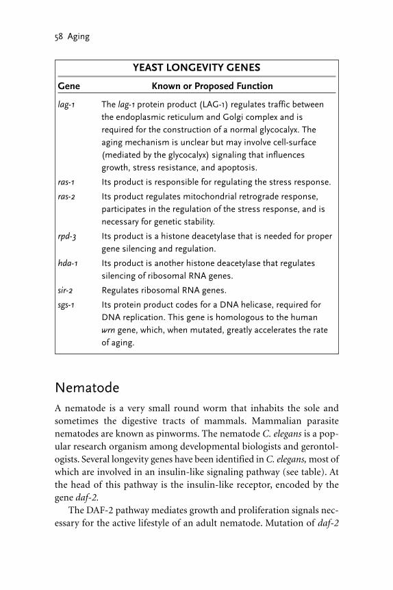
NematodeA nematode is a very small round worm that inhabits the sole andsometimes the digestive tracts of mammals. Mammalian parasitenematodes are known as pinworms. The nematode C. elegans is a pop-ular research organism among developmental biologists and gerontol-ogists. Several longevity genes have been identified in C. elegans, most ofwhich are involved in an insulin-like signaling pathway (see table). Atthe head of this pathway is the insulin-like receptor, encoded by thegene daf-2.
The DAF-2 pathway mediates growth and proliferation signals nec-essary for the active lifestyle of an adult nematode. Mutation of daf-2
58 Aging
YEAST LONGEVITY GENES
Gene Known or Proposed Function
lag-1 The lag-1 protein product (LAG-1) regulates traffic betweenthe endoplasmic reticulum and Golgi complex and isrequired for the construction of a normal glycocalyx. Theaging mechanism is unclear but may involve cell-surface(mediated by the glycocalyx) signaling that influencesgrowth, stress resistance, and apoptosis.
ras-1 Its product is responsible for regulating the stress response.
ras-2 Its product regulates mitochondrial retrograde response,participates in the regulation of the stress response, and isnecessary for genetic stability.
rpd-3 Its product is a histone deacetylase that is needed for propergene silencing and regulation.
hda-1 Its product is another histone deacetylase that regulatessilencing of ribosomal RNA genes.
sir-2 Regulates ribosomal RNA genes.
sgs-1 Its protein product codes for a DNA helicase, required forDNA replication. This gene is homologous to the human wrn gene, which, when mutated, greatly accelerates the rateof aging.

shifts the entire physiology of the animal from active behavior to some-thing resembling hibernation in mammals. Hibernation behavior innematodes is known as a diapause state. Nematode diapause is charac-terized by a shift from active glucose metabolism (i.e., burning calories)to storage functions, such as the deposit of fat. The animal’s activitylevel drops, and the life span is increased by nearly 80 percent. Thus daf-2 is a negative regulator of life span; it is an example of the kind of genethat limits life span as a result of maximizing activity level and meta-bolic performance. The effects observed in daf-2 mutants are very sim-ilar to the response of mammals to hibernation or caloric restriction.The products of other nematode longevity genes, such as age-1, daf-18,akt-1, and daf-16, transduce the signal received by the DAF-2 receptorprotein (e.g., the age-1 protein conveys the signal from the DAF-2receptor to the cell cytoplasm). Consequently, a mutation in any ofthese genes will lead to the diapause state and extended life span.
A second pathway has been identified that affects nematodelongevity. The daf-12 gene codes for a steroid hormone receptor that islinked to a pathway that appears to regulate the stress response. Indeed,this pathway specifies resistance to heat, ultraviolet radiation, andoxidative stress. Accordingly, daf-12 is a positive regulator of life span.A mutation in daf-12 or in ctl-1, a component of the pathway, shortenslife span.
Fruit FlyThe fruit fly Drosophila melanogaster is a popular research organism.During the 1980s, researchers managed to isolate long-lived Drosophilathrough selective breeding. These flies showed a greater metaboliccapacity and enhanced resistance to stress initiated by heat, desiccation,and ethanol vapors. In addition, they have higher activities of antiox-idative enzymes, they are more efficient at utilizing nutrients, and theyhave enhanced stores of lipid and glycogen. Many of these features areheld in common with long-lived nematodes and yeast.
Direct support for the free radical theory of the aging process camewith the isolation and characterization of sod-1, the gene coding forsuperoxide dismutase (see table). Transgenic fruit flies overexpressingsod-1 live longer than normal and suffer much less oxidative damage
The Search for Longevity Genes 59

induced by free radicals. Interestingly, overexpression of sod-1 inmotorneurons alone is sufficient to nearly double the mean life span ofthese animals. Overexpression of another gene, mth, also increases lifespan. The mth protein product, called methuselah, is a cell-surfacereceptor that is linked to a pathway that regulates the stress response.
The retrograde response (already described in the discussion onyeast longevity) involving traffic between the cell nucleus, cytoplasm,and the mitochondria is also involved in Drosophila aging. The indy(i’m not dead yet) gene codes for a mitochondrial membrane proteininvolved in transport of Krebs cycle intermediates. A mutation in theindy gene blocks import of these compounds, with an effect similar tocaloric restriction—a near doubling of life span. Insulin and insulin
60 Aging
CAENORHABDITIS ELEGANS LONGEVITY GENES
Gene Known or Proposed Function
daf-2 Its product codes for an insulin-like cell-membranereceptor (DAF-2). Disrupting this pathway extends lifespan.
age-1 / daf-23 These genes code for two kinases, directly linked to theDAF-2 signaling pathway.
daf-18 The protein product is on the DAF-2 pathway, down-stream from the age-1/daf-23 products.
akt-1 / akt-2 The products of these genes are on the DAF-2 pathway,downstream from daf-18.
daf-16 Its product, DAF-16, is a multifunction factor that isactivated by the DAF-2 and DAF-12 pathways. Loss offunction promotes a “hibernation” response, involvingthe storage of fat and glycogen that extends life span.
daf-12 Its product codes for a steroid hormone receptor (DAF-12) that is linked to a pathway that is important in stress resistance. A mutation in this gene shortenslife span.
ctl-1 The protein product is a cytoplasmic enzyme (catalase)on the DAF-12 stress-resistance pathway.

receptors modulate life span in Drosophila, much as they do in nema-todes. The Drosophila genes chico and inr (insulin receptor) encode aninsulin protein and insulin receptor very similar to those found innematodes and mammals. Mutations in chico or inr have the samephysiological effects as described for the daf-2 gene in nematodes. Thesugar baby gene achieves a similar though muted effect on life span.This gene codes for a maltose permease, an enzyme that enhances theuptake of maltose into cells. Overexpression of this gene shifts the ani-mal’s physiology away from glucose utilization, thus mimicking theeffects of caloric restriction. In this case, the increase in life span isabout 20 percent, compared with the more than 80 percent increaseobserved in inr mutants.
The Search for Longevity Genes 61
DROSOPHILA LONGEVITY GENES
Gene Known or Proposed Function
indy Its product codes for a mitochondrial membrane protein
involved in transport of Krebs cycle intermediates. The
loss of function increases life span by reducing the avail-
ability of nutrients (caloric restriction).
sod-1 The protein product is superoxide dismutase (SOD).
Overexpression increases life span by enhanced inactiva-
tion of free radicals.
mth Its product codes for a cell-membrane receptor called
methuselah, which enhances the stress response, thus
increasing life span.
chico The protein product, CHICO, is similar to mammalian
insulin. Loss of function increases life span through
caloric restriction.
inr Its product codes for the CHICO receptor. Lose of func-
tion has the same effect as a chico mutation. This recep-
tor is very similar to the nematode DAF-2 receptor.
sugar baby The protein product is a maltose permease. Overexpres-
sion increases life span by shifting metabolism away from
glucose, thus invoking partial caloric restriction.

MouseThe most consistent way to extend the life span of a mammal is bycaloric restriction. Such experiments (described in chapter 2) haveextended the life spans of mice and rats by up to 50 percent. Moreover,these calorie-restricted animals show similar metabolic responsesobserved in yeast, nematodes, and fruit flies, including resistance tostress. In addition, calorie-restricted rodents show a postponement ofage-related diseases, such as cancer, and have an increased lifetimemetabolic capacity. These changes, like the hibernation response innematodes and flies, are due to more efficient utilization of glucose anda shift toward deposit of fat and glycogen.
Three mouse genes have been identified that, when mutated, extendlife span in a manner similar to caloric restriction (see table). The geneprop-1 (“prophet of pit-1”) codes for a protein that regulates anothergene, pit-1, that codes for a pituitary-specific transcription factor.Mutation of prop-1 or pit-1 leads to developmental arrest of the pitu-itary gland, thus drastically reducing the normal levels of growth-inducing hormones such growth hormone (GH) and thyroid hormone(TH). In the absence of these hormones, cells cannot utilize glucose oramino acids to promote growth and maturation. Consequently, prop-1mutants are dwarves, but they have an extended life span. The conse-quence of this mutation is the same as if the animals were raised on acalorie-restricted diet even while in the womb.
A second type of longevity gene has been identified in mice. This isthe p66shc gene, which codes for a component of a signaling pathway thatregulates the stress response and apoptosis. As with the other positivelongevity genes already described, overexpression of this gene increaseslife span, while animals possessing a normally expressed p66shc haveshorter life spans.
HumanIdentification of longevity genes in lower organisms has stimulated asearch for similar genes in the human genome. The human homolog ofyeast lag-1 has already been cloned and is located on chromosome 19.Although the sequence homology is low, it can replace the yeast gene,
62 Aging

where it performs a longevity function. Consequently, human lag-1may be thought of as a human longevity gene, although much work isneeded to confirm its function in humans.
Perhaps the most striking similarity between longevity genes inhumans and lower organisms is the yeast sgs-1 gene and the human wrngene. The sgs-1 gene codes for a helicase and, when mutated, can accel-erate the aging process. Werner’s syndrome, described previously, is adisease in humans that is also associated with accelerated aging. Thegene responsible for this disease, called wrn, has been identified. Theprotein product of the wrn gene is a helicase, not the same helicaseencoded by the sgs-1 gene, but a member of the same family, possessinga similar function. Mutations in these two genes provide dramatic evi-dence in support of the connection between life span and genetic sta-bility.
SummaryThe search for longevity genes has identified four physiologicalprocesses that influence life span. They are metabolic control, resistanceto stress, gene dysregulation, and genetic stability. Evidence supportingthe involvement of metabolic control comes from the roles of lag-1 in
The Search for Longevity Genes 63
MOUSE LONGEVITY GENES
Gene Known or Proposed Function
prop-1 The protein product is regulator of a pituitary-specific tran-scription factor (PIT-1). Inactivation leads to poor develop-ment of the pituitary and production of pituitary hormones,particularly growth hormone. Mutated prop-1 increases lifespan by about 50 percent.
pit-1 This gene codes for PIT-1, a protein transcription factor. Theinactivation of pit-1 has the same effect as a prop-1 mutation.
p66shc The protein product is a component of a signal transductionpathway that makes cells resistant to apoptosis and oxidativestress.

yeast, daf-2 in nematodes, indy and sod-1 in Drosophila, and prop-1 inmice. Resistance to stress is a function of several longevity genes, such asras-2, daf-12, mth, and p66shc. Gene dysregulation, as a mechanism ofaging, has been clearly demonstrated in yeast with the isolation of threehistone deacytlase genes, rpd-3, hda-1, and sir-2. Finally, the relation-ship between genetic stability and life span is indicated by the effects ofsgs-1 mutants in yeast and the human disease known as Werner’s syn-drome, which is associated with accelerated aging and is caused by thegene wrn, a homolog of sgs-1.
This collection of genes, small though it is, has given a powerfulboost to gerontological research and provides an important conceptualframework that future research may follow. The goal is to isolate evenmore longevity genes from lower animals and then to find their coun-terparts in the human genome. This work has already begun with theisolation of human lag-1. The characterization of all longevity genes isexpected to revolutionize our understanding of the aging process, andit may provide the means by which the effects of age may be treated orreversed.
64 Aging

� 7 �
GERIATRICS
The World Health Organization (WHO) places health issues on a con-tinuum from disease to impairment to disability to handicap. Geriatricsis a branch of the biomedical sciences devoted to helping the elderly(65+ years old) deal with the effects of age. The geriatric approach doesnot try to reverse the aging process but rather to minimize its conse-quences by reducing or inhibiting the progression to disability. Thiseffort, conducted in hospitals, clinics, and nursing homes, is based on abroad range of therapies that are grounded in the biological, psycho-logical, and social sciences.
Treating, and caring for, the elderly is a complex endeavor. Olderpeople are usually suffering from several simultaneous disorders that,because of the patient’s age, cannot be treated with the drugs or therapiesthat are routine for younger individuals. Drug therapies assume a clear-ance time (physiological deactivation of the drug), made possible by ahealthy liver that may not exist in an older patient. Drugs that are safelyused to treat depression or cardiovascular disease in young patients canhave devastating effects on the elderly. Accurate medical histories areoften difficult to obtain from elderly patients, either because of poormemory or because of psychological compensation by which the patientignores and minimizes danger signs and symptoms. Growing old is oftena time of loss: lost physical abilities, lost friends, the patient’s spouse mayhave passed away, and the family home may have been given up for aroom in a nursing home or hospital ward. All these elements complicatethe diagnosis and prognosis for a geriatric patient.
The focus of this chapter is clinical geriatrics, which covers the manyproblems associated with the care and treatment of the elderly. We
65

begin with the demographics of North American society with respectto age distributions, epidemiology, and the capacity of health careproviders to deal with the ever-expanding geriatrics population.
Our Aging SocietyBetween 1900 and 1990, the total U.S. population increased threefold,while the number of elderly persons increased tenfold. In 1990 morethan 35 million Americans were over the age of 65, nearly twice as manyas in 1960. This number reached 35 million in 2000 (about 12 percentof the population) and is expected to increase further, to 53 million by2020 and 75 million by 2040 (20 percent of the population). One of thefastest growing segments of the population is the very old (personsolder than 85), who now account for 12 percent of all elderly personsand is expected to increase to 18 percent by 2040. In addition, the num-ber of persons reaching 100 years of age (centenarians) is expected toincrease from the current 60,000 to nearly 500,000 by 2040. In general,the mortality rate for elderly women has declined more quickly than ithas for older men. In 1950 there were 89 older men for every 100 olderwomen; in 2000 there were 70 men for every 100 women. Among thevery old, there are 41 men for every 100 women. Living arrangementsvary greatly for the elderly. At ages 65 to 74, one-third of women livealone, but after the age of 75, this proportion increases one-half. Beforethe age of 85, the elderly usually live with relatives or a spouse, but after85, 18 percent of men and 28 percent of women live in nursing homesor hospital wards.
Per capita costs for acute and long-term (chronic) health care serv-ices are highest for the very old, so the growth of this group will havea profound effect on health care costs. Persons over 65 currently rep-resent just over one in three of the patients seen by a primary carephysician; by 2040 this ratio is expected to increase to one in two.While the costs of caring for the elderly is expected to rise, this is duenot just to the patient’s age but also to a general increase in the com-plexity and expense of diagnostic procedures and equipment. It isexpected that as the proportion of older to younger persons increases,less financial and social support will be available for the elderly.Medicare and Medicaid cover much of the financial burden of caring
66 Aging

for the elderly in the United States. However, even with these publicservices, the elderly still bear a considerable share of the expenses.Currently, geriatric patients can expect to pay as much as 21 percent oftheir income for medical care.
Evaluating the Geriatric PatientEvaluation of a geriatric patient is much different from that of ayounger individual. Young patients generally have a single complaintthat the physician can focus on, and there is usually no reference to thepatient’s socioeconomic environment. The approach to a geriatricpatient, however, may begin with the physician asking the patient todescribe a typical day in his or her life. In this way, the physician canbest assess the elderly person’s overall quality of life, liveliness ofthought, and physical independence. This approach also helps developa good patient-physician rapport, something that is especially impor-tant to elderly patients, who often take longer to answer questions andmay be shy because of it. In the initial interview, geriatricians are espe-cially careful not to infantilize the patient by asking an attending relativequestions pertaining to the patient’s history or medical status. It is for thisreason that geriatric patients, unless suffering from dementia, are inter-viewed alone. During the initial evaluation and interview, the physicianattempts to gather information about the patient’s medical, drug, nutri-tion, and psychiatric histories.
MEDICAL HISTORY With an elderly patient, the medical historymay extend back to a time when society’s disease profile was differentthan it is today. For example, rheumatic fever and tuberculosis weremuch more common in the mid-1900s than they are today.Consequently, the physician will ask about diseases that were commonwhen the patient was young. The patient will also be asked aboutoutdated treatments, such as mercury for syphilis or pneumothoraxtherapy for tuberculosis. Many older persons tend to underreportsymptoms out of denial or a fear of illness, disability, and thedependence these conditions may bring. Aging can also alter theindividual’s response to certain diseases, such as a painless myocardialinfarction or pneumonia without a cough.
Geriatrics 67

DRUG HISTORY Although the physician will ask the patient, and thepatient’s relatives, about prescription drugs, some geriatricians havesuggested that the best approach is the “brown bag” technique, wherebythe patient is asked to empty his or her medicine cabinet into a brownpaper bag and then to bring it to the evaluation interview. Often thecomplaints of older patients are traced to a drug or combination ofdrugs they have been taking. The drug history includes determiningwhich drugs are used, at what dose, how often they are taken, whoprescribed them, and for what reason. Topical drugs are included, suchas eye drops for treating glaucoma, because there is the possibility thatsystemic absorption may cause unexpected side effects in the elderly.Over-the-counter drugs must be included because their overuse canhave serious consequences, such as constipation from laxative use orsalicylism from aspirin use. Patients are also asked to demonstrate theirability to read the labels (often printed in very small type) and to openthe container, which may contain a child-resistant lid. Because olderpatients are often treated with multiple medications, they are at risk ofnoncompliance and adverse effects.
NUTRITION HISTORY The physician tries to determine the type,quantity, and frequency of food eaten, including the number of hotmeals per week. Special diets, self-prescribed fad diets, alcoholconsumption, over-the-counter vitamins, and dietary fiber are alsodetermined. For the elderly, it is very important to determine theamount of money the patient has to spend on food each week andwhether suitable cooking facilities are available. The patient’s ability toeat is assessed by examining the mouth and the condition of the teethor dentures, if fitted. Poor vision, arthritis, immobility, or tremors mayaffect an old person’s ability to prepare food. A patient that suffers fromurinary incontinence may reduce fluid intake, which could also lead topoor food intake.
Managing Age-Related DisordersThe most common disorders of the elderly are dementia, cardiovascu-lar disease, osteoporosis, and incontinence. It is not unusual for elderlypatients to suffer from all these disorders simultaneously.
68 Aging

DEMENTIA Nearly half of all elderly patients suffer from variousdegrees of dementia. Two-thirds are caused by Alzheimer’s disease(AD), which is currently irreversible. Reversible dementias are causedby strokes, neoplasms, or toxins such as alcohol, or those produced byinfections. Although a complete cure for most dementias is not possible,optimal management can improve the ability of these patients to copewith basic tasks. In many cases, dementia is the result of one or moresmall strokes caused by hypertension. Thus the first step in managingdementia is aggressive treatment for high blood pressure. This isfollowed with pharmacological agents that enhance cognition andfunction, and treat associated problems such as depression, paranoia,delusions, agitation, and even psychoses.
Where AD is suspected, the patient may be treated with cholinesteraseinhibitors to maximize the half-life of brain neurotransmitters. There arethree such drugs available: donepezil, rivastigmine, and galantamine.Clinical trials have shown that these drugs can improve cognitive func-tion. However, side effects, including nausea, vomiting, and diarrhea, canlead to serious complications. Other drugs, such as estrogen (for women),vitamin E, ginkgo biloba, and nonsteroidal anti-inflammatory agents,are also used but their effectiveness is in doubt. However, while theseagents may be ineffective as a treatment for advanced dementia, theymay be useful in treating milder cases.
CARDIOVASCULAR DISEASE Cardiac output and the response ofthe heart to exercise decreases with age. Ventricular contractionsbecome weaker with each decade, a problem that is compounded by theage-related reduction in blood vessel elasticity. Hardening of the arteriesis the prime cause of hypertension in the elderly, but it is not anunavoidable consequence of aging. The first stage in managinghypertension and cardiovascular disease is a change in lifestyle. Clinicaltrials have shown that even the very old can benefit by this approach,which involves maintaining an ideal body weight, no smoking, regularaerobic exercises, and a diet consisting of fruits, vegetables, and low-fatdairy products (all of which are rich in essential potassium, calcium,and magnesium). If these procedures fail to reduce blood pressure,drugs such as thiazide, beta-blockers, or calcium channel blockers maybe used, but the diet and exercise regimen should be maintained.
Geriatrics 69

OSTEOPOROSIS Diminished bone mass can be determined mostconveniently with special X-ray machines (dual energy X-rayabsorptiometry) or with ultrasound densitometry. Both proceduresdetermine the density as g/cm2, which is compared to normal valuesfrom a younger population and is used to estimate the likelihood offracture. The first attempts to manage this disease involve a diet rich incalcium and vitamin D, along with regular weight-bearing exercises.Hormone replacement therapy has also been recommended, for menand women, but as discussed in a previous chapter, this approach canlead to dangerous side effects. An alternative drug therapy involves theuse of bisphosphonates, antiresorptive drugs that are known to increasebone mass. The bisphosphonate, alendronate, was shown to decreasethe incidence of vertebral and nonvertebral fractures by more than 50percent in postmenopausal women. The major side effects aregastrointestinal, and the drug must be taken on an empty stomach in anupright position.
INCONTINENCE Incontinence, or the involuntary loss of urine orstool, is very common in the geriatric population. About 33 percent ofelderly women and 20 percent of elderly men suffer from this disorder.The prevalence may be as high as 80 percent in nursing homes or long-term-care institutions. Incontinence may develop because ofneurological damage sustained after a stroke or it may be traced to age-related changes in the urinary system, in particular, the integrity of theurethra and the holding volume of the bladder, which decreases withage. Delirium and exposure to a new environment, such as recentadmission to hospital or nursing home, can also lead to incontinence inthe elderly. Simply modifying the patient’s fluid intake and eliminatingdiuretics such as coffee or tea can often treat transient incontinence.
Persistent or acute incontinence is managed initially by ensuringthat the patient can reach a toilet quickly. It may also be necessary toprovide the patient with incontinence undergarments and pads. Oftenwith special care and training, the problem can be resolved. In othercases, it may be necessary to resort to drug therapy. A commonly useddrug is a bladder relaxant, tolterodine, which is available in long-actingpreparations. In severe cases, surgery may be required to repair dam-aged sphincters that normally regulated urine flow through the urethra.
70 Aging

It may also be necessary to fit the patient with a catheter that continu-ally drains the bladder into a plastic bag. However, chronic indwellingcatherization is not advised, as it is associated with a high risk of devel-oping urinary system infections.
Drug TherapyGeriatric patients are often prescribed a large number of drugs to dealwith the many disorders they suffer from. However, as indicated above,there are usually effective nonpharmacological therapies available thatshould be attempted before resorting to drugs. All geriatric patientsneed a careful and thorough review of the drugs they are prescribed toensure they are necessary and that there is no change of potentially dan-gerous drug interactions. Effective drug therapy is often hampered byfaulty diagnosis. Older patients may underreport symptoms, or theircomplaints may be vague and multiple. In addition, symptoms of phys-ical diseases may overlap with psychological illness. Consequently, mak-ing the correct diagnosis and prescribing the appropriate drugs is a verydifficult task in geriatric medicine. Finally, the aging process alters theelderly patient’s ability to deal with drugs physiologically. This deficitoccurs primarily at the liver and at the kidneys.
The liver contains about 30 enzymes that are involved in the degra-dation of a wide variety of compounds that are consumed in an averagediet. These enzymes can also handle more exotic compounds such asalcohol or pharmaceutical drugs. Age-related or even alcohol-induceddeterioration of these enzymes make a safe drug dangerous when givento a geriatric patient. Potentially fatal ventricular arrhythmias have beencaused by certain antihistamines when given to older patients withdefective liver enzymes. However, the situation is too complex for aphysician to assume that an elderly patient with normal liver functiontests will be able to metabolize a given drug as efficiently as a youngerpatient.
The kidneys also play an important role in ridding the body of for-eign or unwanted chemicals and drugs. Drugs given to older patientsare cleared more slowly by the kidneys and thus have a tendency toaccumulate to high, possibly toxic, levels over the time-course of treat-ment. Thus drugs that have not been specifically tested for use on older
Geriatrics 71

subjects must be used with extreme caution. Medical servicing centersand pharmaceutical companies have developed computer algorithmsand databases to help evaluate drug usage and to detect possibly dan-gerous drug combinations that are prescribed for geriatric patients.
Nursing HomesThe poor quality of care provided in nursing homes has been knownfor decades. There has been some improvement since the Institute ofMedicine (IOM) released a critical report in 1986, but a more recentreport in 2000 indicated that serious problems still exist. One criticalpassage from the report indicates that “serious problems concerningquality of care apparently continue to affect residents of this country’snursing homes, and persistently poor providers of care are still in oper-ation. Pain, pressure sores, malnutrition, and urinary incontinence haveall been shown to be serious problems in recent studies of nursinghome residents.”
Nursing homes are intended as places where the elderly can be caredfor in their final years by a team of medical professionals who special-ize in geriatric medicine. In many cases, however, logistic and economicrestraints make this a very difficult goal to realize. Physician involve-ment in nursing home care is often limited to telephone conversationswith the nursing staff. Restrictive Medicare and Medicaid reimburse-ment policies do not encourage physicians to make more than therequired monthly or 60-day visits. Physician involvement in such essen-tial services as attendance at the medical team conferences, family meet-ings, and counseling residents and surrogate decision makers ontreatment plans in the event of terminal illness are usually not reim-bursable at all. In addition, most nursing homes lack expensive diag-nostic equipment, and thus many of the residents are sent to hospitalemergency rooms, where they are evaluated by staff who lack trainingand interest in the care of frail elderly patients.
Despite these many problems, the effectiveness of nursing homescan be improved with more attention paid to the documentation of theresident’s illness and treatment history, and the introduction of nursepractitioners and physician assistants. These medical practitionerscould be very helpful in implementing some of the screening and mon-
72 Aging

itoring that is needed to ensure proper care of the residents, and to thisextent would function as an independent patient advocate. They couldalso have an important role in communicating with the staff, residents,and families when the physician is not in the facility.
The problems facing nursing homes over the next 40 years aretremendous. In its report of 2000, the IOM noted the urgent need forresearch and data collection to obtain a better understanding anddescription of the various long-term-care arrangements throughout thecountry, including their size, the services provided, the staffing levelsand training, the characteristics of those receiving care, and the staffingand quality of care provided in the different settings and services. Theyalso called for increased funding, concluding that “the amounts andways we pay for long-term care are probably inadequate to support aworkforce sufficient in numbers, skills, stability, and commitment toprovide adequate clinical and personal services for the increasingly frailor complex populations using long-term care.”
Ethical IssuesThe basic ethical principle governing the care of the elderly were estab-lished in the 1970s in response to allegations that human subjects inbiomedical clinical trials were poorly treated. Principles of respect forpersons, beneficence, and informed consent apply equally well to eld-erly patients in a nursing home, or hospital ward, as they do to humansubjects involved in clinical trials.
RESPECT FOR PERSONS Respect for persons, in the context ofclinical trials, demands that subjects enter into research voluntarily andwith adequate information. This assumes the individuals areautonomous agents, that is, are competent to make up their own minds.However, there are many instances where potential research subjects arenot really autonomous; prisoners, patients in a mental institution,children, the elderly, and the infirm. All these people require specialprotection to ensure they are not being coerced or fooled intovolunteering as research subjects. Geriatric patients are especiallyvulnerable because of their many medical disorders, which often affecttheir ability to understand what is being done to them.
Geriatrics 73

BENEFICENCE It is not enough to respect a potential subject’sdecisions and to protect them from harm, but in addition, it is necessaryto do all that is possible to ensure their well-being. Beneficence isgenerally regarded as acts of kindness or charity, but in the case ofgeriatric patients, weakened by illness and age, it is an obligation. In thissense, it is the natural extension of the Hippocratic oath that allphysicians are expected to adhere by: I will give no deadly medicine toanyone if asked, nor suggest any such counsel. In other words, do noharm, and for those involved in biomedical research, never injure oneperson to benefit another. This is particularly relevant to prescribingdrugs for the elderly, who are especially sensitive to this type of therapy.
INFORMED CONSENT All participants in clinical trials mustprovide informed consent in writing. Moreover, steps must be taken toensure that the consent is, in fact, informed. This might involve anindependent assessment of the individual’s ability to understand thelanguage on the consent form and any instructions or explanations theinvestigators have given. Geriatric patients, many of whom suffer fromdementia, cannot be expected to give informed consent under manycircumstances. Consequently, it is necessary to proceed with extremecaution in such cases and to ensure that an action taken, such as movingan elderly person out of their home and into an institution, is really intheir best interest and not simply a convenience.
74 Aging

� 8 �
RESOURCE CENTER
Eukaryote Cell PrimerLife on Earth began 3.5 billion years ago in the form of single cells thatappeared in the oceans. These cells evolved into ancestral prokaryotesand, about 2 billion years ago, gave rise to Archaea, bacteria, and eukary-otes, the three major divisions of life in the world. Eukaryotes, in turn,gave rise to plants, animals, protozoans, and fungi. Each of these groupsrepresents a distinct phylogenetic kingdom. The Archaea and bacteriarepresent a fifth kingdom, known as the Monera, or prokaryotes.Archaea and bacteria are very similar anatomically, both lacking a truenucleus and internal organelles. A prokaryote genome is a single, circu-lar piece of naked DNA, called a chromosome, containing fewer than5,000 genes. Eukaryotes (meaning “true nucleus”) are much more com-plex, having many membrane-bounded organelles. These include anucleus, nucleolus, endoplasmic reticulum (ER), Golgi complex, mito-chondria, lysosomes, and peroxisomes.
The eukaryote nucleus, bounded by a double phospholipid mem-brane, contains a DNA (deoxyribonucleic acid) genome on two or morelinear chromosomes, each of which may contain up to 10,000 genes.The nucleus also contains an assembly plant for ribosomal subunits,called the nucleolus. The endoplasmic reticulum (ER) and the Golgicomplex work together to glycosylate proteins and lipids (attach sugarmolecules to the proteins and lipids producing glycoproteins and glycolipids), most of which are destined for the cell membrane to forma molecular “forest” known as the glycocalyx. The glycoproteins and
75

76 Aging
Centrosome
Cytoplasm
Mitochondrion
Endoplasmicreticulum
Nucleus
Lysosome
Golgiapparatus
Microtubule
Cell membrane
Golgi vesicle
Nucleolus Nuclear pore
Peroxisome
Ribosomes
Actin filaments
The eukaryote cell. The structural components shown here are present inorganisms as diverse as protozoans, plants, and animals. The nucleuscontains the DNA genome and an assembly plant for ribosomal subunits (thenucleolus). The endoplasmic reticulum (ER) and the Golgi work together tomodify proteins, most of which are destined for the cell membrane. Theseproteins are sent to the membrane in Golgi vesicles. Mitochondria provide thecell with energy in the form of ATP. Ribosomes, some of which are attached tothe ER, synthesize proteins. Lysosomes and peroxisomes recycle cellularmaterial and molecules. The microtubules and centrosome form the spindleapparatus for moving chromosomes to the daughter cells during cell division.Actin filaments and a weblike structure consisting of intermediate filaments(not shown) form the cytoskeleton.

glycolipids travel from the ER to the Golgi, and from the Golgi to thecell surface, in membrane-bounded vesicles that form by budding offthe organelle by exocytosis. Thus the cytoplasm contains many trans-port vesicles that originate from the ER and Golgi. The Golgi vesiclesbud off the outer chamber, or the one farthest from the ER. Mitochon-dria, once free-living prokaryotes, and the only other organelle with adouble membrane, provide the cell with energy in the form of adenosinetriphosphate (ATP). The production of ATP is carried out by an assem-bly of metal-containing proteins, called the electron transport chain,located in the mitochondrion inner membrane. Ribosomes, some ofwhich are attached to the ER, synthesize proteins. Lysosomes and per-oxisomes recycle cellular material and molecules. The microtubules andcentrosome form the spindle apparatus for moving chromosomes tothe daughter cells during cell division. Actin filaments and a weblikestructure consisting of intermediate filaments form the cytoskeleton.
MOLECULES OF THE CELLCells are biochemical entities that synthesize many thousands of mole-cules. Studying these chemicals, and the biochemistry of the cell, wouldbe a daunting task were it not for the fact that most of the chemicalvariation is based on six types of molecules that are assembled into justfour types of macromolecules. The six basic molecules are amino acids,phosphate, glycerol, sugars, fatty acids, and nucleotides. Amino acidshave a simple core structure consisting of an amino group, a carboxylgroup, and a variable R group attached to a carbon atom. There are 20different kinds of amino acids, each with a unique R group. Phosphatesare extremely important molecules that are used in the construction, ormodification, of many other molecules. They are also used to storechemical-bond energy. Glycerol is a simple, three-carbon alcohol thatis an important component of cell membranes and fat reservoirs. Sug-ars are extremely versatile molecules that are used as an energy sourceand for structural purposes. Glucose, a six-carbon sugar, is the primaryenergy source for most cells and it is the principle sugar used to glyco-sylate proteins and lipids for the production of the glycocalyx. Plantshave exploited the structural potential of sugars in their production ofcellulose, and thus wood, bark, grasses, and reeds are polymers ofglucose and other monosaccharides. Ribose, a five-carbon sugar, is a
Resource Center 77

component of nucleic acids as well as ATP. Ribose carbons are numbered as 1' (1 prime), 2', and so on. Consequently, references tonucleic acids, which include ribose, often refer to the 3' or 5' carbon.Fatty acids consist of a carboxyl group (when ionized it becomes a carboxylic acid) linked to a hydrophobic hydrocarbon tail. These molecules are used in the construction of cell membranes and fat.Nucleotides are building blocks for DNA and RNA (ribonucleic acid).Nucleotides consist of three components: a phosphate, a ribose sugar,and a nitrogenous (nitrogen containing) ring compound that behavesas a base in solution. Nucleotide bases appear in two forms: a single-ring nitrogenous base, called a pyrimidine, and a double-ringed base,called a purine. There are two kinds of purines (adenine and guanine),and three pyrimidines (uracil, cytosine, and thymine). Uracil is specificto RNA, substituting for thymine. In addition, RNA nucleotides containribose, whereas DNA nucleotides contain deoxyribose (hence the origin of their names). Ribose has a hydroxyl (OH) group attached to both the 2' and 3' carbons, whereas, deoxyribose is missing the 2' hydroxyl group. ATP, the molecule that is used by all cells as a sourceof energy, is a ribose nucleotide consisting of the purine base adenineand three phosphates attached to the 5' carbon of the ribose sugar. Thephosphates are labeled α (alpha), β (beta), and γ (gamma) and arelinked to the carbon in a tandem order, beginning with α. The energystored by this molecule is carried by the covalent bonds of the β and γphosphates. Breaking these bonds sequentially releases the energy theycontain while converting ATP to adenosine diphosphate (ADP) andthen to adenosine monophosphate (AMP). AMP is converted back toATP by mitochondria.
MACROMOLECULES OF THE CELLThe six basic molecules are used by all cells to construct five essentialmacromolecules. These include proteins, RNA, DNA, phospholipids,and sugar polymers, known as polysaccharides. Amino acids are linkedtogether by peptide bonds to construct a protein. A peptide bond isformed by linking the carboxyl end of one amino acid to the amino endof a second amino acid. Thus, once constructed, every protein has anamino end and a carboxyl end. An average protein may consist of 300 to400 amino acids. Nucleic acids are macromolecules constructed from
78 Aging

Resource Center 79
CH2P
Fatty acid
Aminogroup
Carboxylgroup
Sugars
Glucose Ribose
Carboxylic acid
Hydrocarbon tail
Nucleotides
CH2
CH2OH
H2N COOH
O-
O
R
H
O-PC HO
CH2OH
CH2 CH2CH
CH2
CH2 CH2 CH2 CH2 CH3O
O OOH OH
OH OH OH
OHHO
4 15
5'
4'
3' 2'
1'
32
6
O
P
OH OH OHH
N
N N N
NN
C
Pyrimidine base Purine base
RiboseDeoxyribosePhosphate
Amino acid Phosphate Glycerol
OH OH
OH
Molecules of the cell. Amino acids are the building blocks for proteins.Phosphate is an important component of many other molecules and is addedto proteins to modify their behavior. Glycerol is a three-carbon alcohol that isan important ingredient in cell membranes and fat. Sugars, like glucose, are aprimary energy source for most cells and also have many structural functions.Fatty acids are involved in the production of cell membranes and storage offat. Nucleotides are the building blocks for DNA and RNA.

80 Aging
Protein
RNA
DNA
Phospholipid
Polysaccharide
Carboxyl end
Amino endAmino acid
Nucleotide
Nucleotide
Phosphate Glycerol
Fatty acid tails
Head group
P
Monosaccharide
OO O O
O O O
Macromolecules of the cell. Protein is made from amino acids linked togetherto form a long chain that can fold up into a three-dimensional structure. RNAand DNA are long chains of nucleotides. RNA is generally single stranded butcan form localized double-stranded regions. DNA is a double-stranded helix,with one strand coiling around the other. A phospholipid is composed of ahydrophilic head-group, a phosphate, a glycerol molecule, and twohydrophobic fatty acid tails. Polysaccharides are sugar polymers.

nucleotides. The 5' phosphate ofone nucleotide is linked to the 3'OH of a second nucleotide. Addi-tional nucleotides are alwayslinked to the 3' OH of the lastnucleotide in the chain. Conse-quently, the growth of the chainis said to be in the 5' to 3' direction. RNA nucleotides areadenine, uracil, cytosine, andguanine. A typical RNA mole-cule consists of 2,000 to 3,000nucleotides; it is generally singlestranded but can form localizeddouble-stranded regions. RNA is involved in the synthesis ofproteins and is a structural andenzymatic component of ribo-somes. DNA, a double-strandednucleic acid, encodes cellulargenes and is constructed fromadenine, thymine, cytosine, andguanine deoxyribonucleotides(dATP, dTTP, dCTP, and dGTP,where “d” indicates deoxyribose). The two DNA strands coil aroundeach other like strands in a piece of rope, and for this reason the mol-ecule is known as the double helix. DNA is an extremely large macro-molecule, typically consisting of over a million nucleotide pairs (orbase pairs). Double-stranded DNA forms when two chains ofnucleotides interact through the formation of chemical bonds betweencomplementary base pairs. The chemistry of the bases is such that ade-nine pairs with thymine, and cytosine pairs with guanine. For stability,the two strands are antiparallel, that is, the orientation of one strand isin the 5' to 3' direction, while the complementary strand runs 3' to 5'.Phospholipids, the main component of cell membranes, are composedof a polar head-group (usually an alcohol), a phosphate, glycerol,and two hydrophobic fatty acid tails. Fat that is stored in the body as an
Resource Center 81
Molecule model of the 30S ribosomalsubunit, which consists of protein(light gray corkscrew structures) andRNA (coiled ladders). The RNA that isalso responsible for the catalyticfunction of the ribosome determinesthe overall shape of the molecule.(Courtesy of V. Ramakrishnan, MRCLaboratory of Molecular Biology,Cambridge).

energy reserve has a structure similar to a phospholipid, being com-posed of three fatty acid chains attached to a molecule of glycerol. Thethird fatty acid takes the place of the phosphate and head-group of a phospholipid. Sugars are polymerized to form chains of two ormore mono-saccharides. Disaccharides (two monosaccharides) and
82 Aging
Computer model of DNA. The two strands coil around each other to form ahelix that, when looking down on it from above, coils to the right. Thespherical structures in this image represent the various atoms in the sugarsand bases (dark gray) and phosphates (light gray). (Kenneth Eward/BioGrafx/Photo Researchers, Inc.)

oligosaccharides (three to 12monosaccharides) are attachedto proteins and lipids destinedfor the glycocalyx. Polysaccha-rides, such as glycogen andstarch, may contain several hundred monosaccharides andare stored in cells as an energyreserve.
THE CELL CYCLECells inherited the power ofreproduction from prebioticbubbles that split in half at regu-lar intervals under the influenceof the turbulent environmentthat characterized the Earthmore than 3 billion years ago.This pattern of turbulent frag-mentation followed by a briefperiod of calm is now a regularbehavior pattern of every cell.Even today, after 3 billion years,many cells still divide every 20minutes.
The regular alternation between division and calm has come to beknown as the cell cycle. In studying this cycle, scientists have recognizeddifferent states of calm and different ways in which a cell can divide.The calm state of the cell cycle, referred to as interphase, is divided intothree subphases, called Gap 1 (G1), S phase (a period of DNA synthe-sis), and Gap 2 (G2). The conclusion of interphase, and with it the ter-mination of G2, occurs with the division of the cell and a return to G1.Cells may leave the cycle by entering a special phase called G0. Somecells, such as postmitotic neurons in an animal’s brain, remain in G0 forthe life of the organism.
Although interphase is a period of relative calm, the cell grows continuously during this period, working hard to prepare for the next
Resource Center 83
Cell division
G1
G0
Sphase
G2
Interphase
The cell cycle. Most cells spend theirtime cycling between a state of calm(interphase) and cell division.Interphase is further divided into threesubphases: Gap 1 (G1), S phase (DNAsynthesis), and Gap 2 (G2). Cells mayexit the cycle by entering a specialphase called G0.

round of division. Two notable events are the duplication of the spindle(the centrosome and associated microtubules), a structure that is crucialfor the movement of the chromosomes during cell division, and theappearance of an enzyme called the maturation-promoting factor(MPF) at the end of G2. MPF phosphorylates histones. The histones areproteins that bind to the DNA, which when phosphorylated, compact (orcondense) the chromosomes in preparation for cell division. MPF isalso responsible for the breakdown of the nuclear membrane. When celldivision is complete, MPF disappears, allowing the chromosomes todecondense and the nuclear envelope to reform. Completion of a nor-mal cell cycle always involves the division of a cell into two daughtercells. This can occur by a process known as mitosis, which is intended forcell multiplication, and by second process known as meiosis, which isintended for sexual reproduction.
MITOSISMitosis is used by all free-living eukaryotes (protozoans) as a means ofasexual reproduction. The growth of a plant or an animal is also accom-plished with this form of cell division. Mitosis is divided into fourstages: prophase, metaphase, anaphase, and telophase. All these stagesare marked out in accordance with the behavior of the nucleus and thechromosomes. Prophase marks the period during which the duplicatedchromosomes begin condensation, and the two centrosomes beginmoving to opposite poles of the cell. Under the microscope, the chro-mosomes become visible as X-shaped structures, which are the twoduplicated chromosomes, often called sister chromatids. A specialregion of each chromosome, called a centromere, holds the chromatidstogether. Proteins bind to the centromere to form a structure called thekinetochore (see figure, for clarity, only two chromosomes are shown).Metaphase is a period during which the chromosomes are sorted outand aligned between the two centrosomes. By this time, the nuclearmembrane has completely broken down. The two centrosomes and themicrotubules fanning out between them form the mitotic spindle. Thearea in between the spindles, where the chromosomes are aligned, isoften referred to as the metaphase plate. Some of the microtubulesmake contact with the kinetochores, while others overlap, with motorproteins situated in between. Eukaryotes are normally diploid, so a cell
84 Aging

Prophase
Metaphase
Anaphase
Telophase
Identicaldaughter cells
Duplicated chromosome
Motor protein
Nuclear envelope reforming
Centrosome
Kinetochore
Mitoticspindle
Sister chromatidsseparate
Mitosis. Principal stages dealing with the movement and partitioning of thechromosomes between the future daughter cells. For clarity, only twochromosomes are shown.

would have two copies of each chromosome, one from the mother andone from the father. Anaphase is characterized by the movement of theduplicated chromosomes to opposite poles of the cell. The first step is therelease of an enzyme that breaks the bonds holding the kinetochorestogether, thus allowing the sister chromatids to separate from eachother while remaining bound to their respective microtubules. Motorproteins then move along the microtubule, dragging the chromosomesto opposite ends of the cell. Using energy supplied by ATP, the motorproteins break the microtubule down as it drags the chromosomealong, so that the microtubule is gone by the time the chromosomereaches the spindle pole. Throughout this process, the motor proteinsand the chromosome manage to stay one step ahead of the disintegrat-ing microtubule. The overlapping microtubules aid movement of thechromosomes toward the poles as another type of motor proteinpushes the microtubules in opposite directions, effectively forcing thecentrosomes toward the poles. This accounts for the greater overlap ofmicrotubules in metaphase as compared with anaphase. Duringtelophase, the daughter chromosomes arrive at the spindle poles anddecondense to form the relaxed chromatin characteristic of interphasenuclei. The nuclear envelope begins forming around the chromosomes,marking the end of mitosis. During the same period, a contractile ring,made of the proteins myosin and actin, begins pinching the parental cellin two. This stage, separate from mitosis, is called cytokinesis, and leadsto the formation of two daughter cells, each with one nucleus.
MEIOSISUnlike mitosis, which leads to the growth of an organism, meiosis isintended for sexual reproduction and occurs exclusively in ovaries andtestes. Eukaryotes, being diploid, receive chromosomes from both par-ents; if gametes were produced using mitosis, a catastrophic growth inthe number of chromosomes would occur each time a sperm fertilizedan egg. Meiosis is a special form of cell division that produces haploidgametes (eggs and sperm), each possessing half as many chromosomesas the diploid cell. When haploid gametes fuse, they produce an embryowith the correct number of chromosomes.
The existence of meiosis was first suggested 100 years ago, whenmicrobiologists counted the number of chromosomes in somatic and
86 Aging

Prophase
Metaphase
Anaphase
Telophase
Nonidenticaldaughter cells
Sister chromatidsdo not separate
Crossing over
Recombinantchromosome
Meiosis I. The most notable features include genetic recombination (crossingover) between the homologous chromosomes during prophase, comigrationof the sister chromatids during anaphase, and the production of nonidenticaldaughter cells. Only one homologous pair is shown.

germ cells. The roundworm, for example, was found to have four chro-mosomes in its somatic cells but only two in its gametes. Many otherstudies also compared the amount of DNA in nuclei from somatic cellsand gonads, always with the same result: The amount of DNA insomatic cells is exactly double the amount in fully mature gametes. Tounderstand how this could be, scientists studied cell division in thegonads and were able to show that meiosis occurs as two rounds of celldivision with only one round of DNA synthesis. The two rounds ofdivision were called meiosis I and meiosis II, and scientists observedthat both could be divided into the same four stages known to occur inmitosis. Indeed, meiosis II is virtually identical to a mitotic division.Meiosis I resembles mitosis, but close examination shows three impor-tant differences: gene swapping occurs between homologous chromo-somes in prophase; homologs (i.e., two homologous chromosomes)remain paired at metaphase, instead of lining up at the plate as is donein mitosis; and the kinetochores do not separate at anaphase.
Homologous chromosomes are two identical chromosomes thatcome from different parents. For example, humans have 23 chromo-somes from the father and the same 23 from the mother. We each havea maternal chromosome 1 and a paternal chromosome 1; they carry thesame genes but specify slightly different traits. Chromosome 1 maycarry the gene for eye color, but the maternal version, or allele, mayspecify blue eyes, whereas the paternal allele specifies brown. Duringprophase, homologous pairs exchange large numbers of genes by swap-ping whole pieces of chromosome. Thus one of the maternal chro-matids (gray in the figure on page 87) ends up with a piece of paternalchromosome, and a paternal chromatid receives the correspondingpiece of maternal chromosome. Mixing genetic material in this way isunique to meiosis, and it is one of the reasons sexual reproduction hasbeen such a powerful evolutionary force.
During anaphase of meiosis I, the kinetochores do not separate asthey do in mitosis. The effect of this is to separate the maternal andpaternal chromosomes by sending them to different daughter cells,although the segregation is random. That is, the daughter cells receive arandom assortment of maternal and paternal chromosomes, ratherthan one daughter cell receiving all paternal chromosomes and theother all maternal chromosomes. Random segregation, along with
88 Aging

genetic recombination, accounts for the fact that while children resem-ble their parents, they do not look or act exactly like them. These twomechanisms are responsible for the remarkable adaptability of alleukaryotes. Meiosis II begins immediately after the completion ofmeiosis I, which produces two daughter cells each containing a dupli-cated parent chromosome and a recombinant chromosome consistingof both paternal and maternal DNA. These two cells divide mitoticallyto produce four haploid cells, each of which is genetically unique, con-taining unaltered or recombinant maternal and paternal chromosomes.Meiosis produces haploid cells by passing through two rounds of celldivision with only one round of DNA synthesis. However, as we haveseen, the process is not just concerned with reducing the number ofchromosomes but is also involved in stirring up the genetic pot in orderto produce unique gametes that may someday give rise to an equallyunique individual.
DNA REPLICATIONDNA replication, which occurs during the S phase of the cell cycle,requires the coordinated effort of a team of enzymes, led by DNA heli-case and primase. The helicase is a remarkable enzyme that is responsi-ble for separating the two DNA strands, a feat that it accomplishes at anastonishing rate of 1,000 nucleotides every second. This enzyme gets itsname from the fact that it unwinds the DNA helix as it separates the twostrands. The enzyme responsible for reading the template strand, andfor synthesizing the new daughter strand, is called DNA polymerase.This enzyme reads the parental DNA in the 3' to 5' direction and createsa daughter strand that grows 5' to 3'. DNA polymerase also has an edi-torial function; in that it checks the preceding nucleotide to make sureit is correct before it adds a nucleotide to the growing chain. The editorfunction of this enzyme introduces an interesting problem: How canthe polymerase add the very first nucleotide when it has to check a pre-ceding nucleotide before adding a new one? A special enzyme, calledprimase, which is attached to the helicase, solves this problem. Primasesynthesizes short pieces of RNA that form a DNA-RNA double-stranded region. The RNA becomes a temporary part of the daughterstrand, thus priming the DNA polymerase by providing the crucial firstnucleotide in the new strand. Once the chromosome is duplicated,
Resource Center 89

90 Aging
3'
3'
3'
5'
5'5'
Template DNA
DNA polymerase
Clamp protein
Leading strand
RNA primer
Daughter strands
Lagging strand
Helicase
Primase
DNA replication. The helicase separates the two strands so the DNApolymerase can synthesize new strands. The primase provides replicationsignals for the polymerase, in the form of RNA primers, and the clamp proteinkeeps the polymerase from falling off the DNA. The leading strand requiresonly a single primer (not shown). The lagging strand requires many primers,and the daughter strand is synthesized as a series of DNA fragments that arelater joined into one continuous strand.

DNA repair enzymes remove the RNA primers and replace them withDNA nucleotides.
TRANSCRIPTION, TRANSLATION, AND THEGENETIC CODEGenes encode proteins and several kinds of RNA. Extracting the infor-mation from DNA requires the processes of transcription and transla-tion. Transcription, catalyzed by the enzyme RNA polymerase, copiesone strand of the DNA into a complementary strand of messengerRNA (mRNA) or ribosomal RNA (rRNA) that is used in the construc-tion of ribosomes. Messenger RNA translocates to the cytoplasm,where it is translated into a protein by ribosomes. Newly transcribedrRNA is sent to the nucleolus for ribosome assembly and is nevertranslated. Ribosomes are complex structures consisting of about 50proteins and four kinds of rRNA, known as 5S, 5.8S, 18S, and 28SrRNA (the “S” refers to a sedimentation coefficient that is proportionalto size). These RNAs range in size from about 500 bases up to 2,000bases for the 28S. The ribosome is assembled in the nucleolus as twononfunctional subunits before being sent out to the cytoplasm, wherethey combine, along with an mRNA, to form a fully functional unit.The production of ribosomes in this way ensures that translation neveroccurs in the nucleus.
The genetic code provides a way for the translation machinery tointerpret the sequence information stored in the DNA molecule, andrepresented by mRNA. DNA is a linear sequence of four different kindsof nucleotides, so the simplest code could be one in which eachnucleotide specifies a different amino acid. That is, adenine coding for the amino acid glycine, cytosine for lysine, and so on. The first cells may have used this coding system, but it is limited to the con-struction of proteins consisting of only four different kinds of aminoacids. Eventually, a more elaborate code evolved in which a combina-tion of three out of the four possible DNA nucleotides, called codons,specifies a single amino acid. With this scheme, it is possible to have aunique code for each of the 20 naturally occurring amino acids. Forexample, the codon AGC specifies the amino acid serine, whereas TGC specifies the amino acid cysteine. Thus a gene may be viewed as along continuous sequence of codons. However, not all codons specify
Resource Center 91

92 Aging
Codon Amino Acid Signal
noneSerine
Cysteine
Alanine
Methionine
none STOP
START
none
none
Methionine Serine Cysteine Alanine
STOP
Serine
DNA
mRNA
START
Transcription, translation, and the genetic code. Five codons are shown, fourspecifying amino acids (protein subunits) and two of the five serving as startand stop signals. The codons, including the start and stop signals, are linkedtogether to form a gene on the bottom, or coding, DNA strand. The codingstrand is copied into messenger RNA (mRNA), which is used to synthesizethe protein. Nucleotides appear as round beads: Adenine (A), Thymine (T),Cytosine (C), and Guanine (G). Amino acids appear as labeled elliptical beads.Note that in mRNA, uracil (U) replaces the thymine (T) found in DNA.

an amino acid. The sequence TGA signals the end of the gene, and aspecial codon, ATG, signals the start site, in addition to specifying theamino acid methionine. Consequently, all proteins begin with thisamino acid, although it is sometimes removed once construction ofthe protein is complete. As mentioned above, an average protein mayconsist of 300 to 400 amino acids; since the codon consists of threenucleotides for each amino acid, a typical gene may be 900 to 1,200nucleotides long.
POWER GENERATIONATP is produced in mitochondria from AMP, or ADP, and phosphate(PO4). This process involves a number of metal-binding proteins, calledthe respiratory chain (also known as the electron transport chain), anda special ion channel-enzyme called ATP synthetase. The respiratorychain consists of three major components: NADH dehydrogenase,cytochrome b, and cytochrome oxidase. All these components are pro-tein complexes that have an iron (NADH dehydrogenase, cytochromeb) or a copper core (cytochrome oxidase), and together with the ATPsynthetase are located in the inner membrane of the mitochondria. Therespiratory chain is analogous to an electric cable that transports elec-tricity from a hydroelectric dam to our homes, where it is used to turnon lights or run our stereos. The human body, like that of all animals,generates electricity by processing food molecules through a metabolicpathway, called the Krebs cycle. The electricity, or electrons generated,travel through the respiratory chain, and as they do, they power the syn-thesis of ATP. All electric circuits must have a ground, that is, the elec-trons need someplace to go once they have completed the circuit. In thecase of the respiratory chain, the ground is oxygen. After passingthrough the chain, the electrons are picked up by oxygen, which com-bines with hydrogen ions to form water.
THE GLYCOCALYXThis structure is an enormously diverse collection of glycoproteins andglycolipids that covers the surface of every cell, like trees on the surfaceof the Earth, and has many important functions. All eukaryotes origi-nated from free-living cells that hunted bacteria for food. The glycoca-lyx evolved to meet the demands of this kind of lifestyle, providing a
Resource Center 93

way for the cell to locate, capture, and ingest food molecules or preyorganisms. Cell-surface glycoproteins also form transporters and ionchannels that serve as gateways into the cell. Neurons have refined ion
94 Aging
Sugar Sugar
Glycocalyx
Membrane
Ceramide
Glycolipid Glycoprotein
Nucleus
Cytoplasm Membrane
Cell
The eukaryote glycocalyx. The eukaryote’s molecular forest consists ofglycoproteins and glycolipids. Two examples are shown at the top: aglycoprotein on the left and a glycolipid on the right. The glycoprotein treeshave “trunks” made of protein and “leaves” made of sugar molecules.Glycolipids also have “leaves” made of sugar molecules, but the “trunks” are afatty compound called ceramide that is completely submerged within theplane of the membrane. The glycocalyx has many jobs, including cell-to-cellcommunication and the transport and detection of food molecules. It alsoprovides recognition markers so the immune system can detect foreign cells.
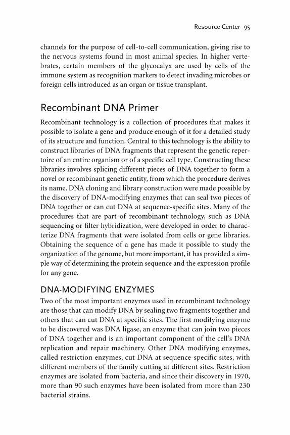
channels for the purpose of cell-to-cell communication, giving rise tothe nervous systems found in most animal species. In higher verte-brates, certain members of the glycocalyx are used by cells of theimmune system as recognition markers to detect invading microbes orforeign cells introduced as an organ or tissue transplant.
Recombinant DNA PrimerRecombinant technology is a collection of procedures that makes itpossible to isolate a gene and produce enough of it for a detailed studyof its structure and function. Central to this technology is the ability toconstruct libraries of DNA fragments that represent the genetic reper-toire of an entire organism or of a specific cell type. Constructing theselibraries involves splicing different pieces of DNA together to form anovel or recombinant genetic entity, from which the procedure derivesits name. DNA cloning and library construction were made possible bythe discovery of DNA-modifying enzymes that can seal two pieces ofDNA together or can cut DNA at sequence-specific sites. Many of theprocedures that are part of recombinant technology, such as DNAsequencing or filter hybridization, were developed in order to charac-terize DNA fragments that were isolated from cells or gene libraries.Obtaining the sequence of a gene has made it possible to study theorganization of the genome, but more important, it has provided a sim-ple way of determining the protein sequence and the expression profilefor any gene.
DNA-MODIFYING ENZYMESTwo of the most important enzymes used in recombinant technologyare those that can modify DNA by sealing two fragments together andothers that can cut DNA at specific sites. The first modifying enzymeto be discovered was DNA ligase, an enzyme that can join two piecesof DNA together and is an important component of the cell’s DNAreplication and repair machinery. Other DNA modifying enzymes,called restriction enzymes, cut DNA at sequence-specific sites, withdifferent members of the family cutting at different sites. Restrictionenzymes are isolated from bacteria, and since their discovery in 1970,more than 90 such enzymes have been isolated from more than 230bacterial strains.
Resource Center 95

96 Aging
Top View
Sample well Agarose gel
Direction of migration
Negative electricalterminal
Side ViewPositive electrical
terminal
Sample well Buffer solution
Bare electricalwire
Supportingplate
Agarosegel
Electrophoresistank
+
Agarose gel electrophoresis. An agarose gel is placed in an electrophoresistank and submerged in a buffer solution. The electric terminals are connectedto a power source, with the sample wells positioned near the negativeterminal. When the current is turned on, the negatively charged nucleic acidsmigrate toward the positive terminal. The migration rate is an inverse functionof molecular size. (Large molecules travel slower than small ones.)

The name “restriction enzyme” is cryptic and calls for an explana-tion. During the period when prokaryotes began to appear on Earth,their environment contained a wide assortment of molecules that werereleased into the soil or water by other cells, either deliberately or whenthe cells died. DNA of varying lengths was among these molecules andwas readily taken up by living cells. If the foreign DNA contained com-plete genes from a competing bacterial species, there was the real possi-bility that those genes could have been transcribed and translated by thehost cell with potentially fatal results. As a precaution, prokaryotesevolved a set of enzymes that would restrict the foreign DNA popula-tion by cutting it up into smaller pieces before being broken down com-pletely to individual nucleotides.
GEL ELECTROPHORESISThis procedure is used to separate different DNA and RNA fragments ina slab of agar or polyacrylamide subjected to an electric field. Nucleicacids carry a negative charge and thus will migrate toward a positivelycharged electrode. The gel acts as a sieving medium that impedes the movement of the molecules. Thus the rate at which the fragmentsmigrate is a function of their size; small fragments migrate more rap-idly than large fragments. The gel, containing the samples, is run sub-merged in a special pH-regulated solution, or buffer, containing anucleic-acid-specific dye, ethidium bromide. This dye produces astrong reddish-yellow fluorescence when exposed to ultraviolet (UV)radiation. Consequently, after electrophoresis, the nucleic acid can bedetected by photographing the gel under UV illumination.
DNA CLONINGIn 1973 scientists discovered that restriction enzymes, DNA ligase, andbacterial plasmids could be used to clone DNA molecules. Plasmids aresmall (about 4,000 base pairs, also expressed as 4.0 kilo base pairs or 4 Kbp) circular minichromosomes that occur naturally in bacteria andare often exchanged between cells by passive diffusion. When a bac-terium acquires a new plasmid, it is said to have been transfected. Forbacteria, the main advantage to swapping plasmids is that they oftencarry antibiotic resistance genes, so that a cell sensitive to ampicillin canbecome resistant simply by acquiring the right plasmid.
Resource Center 97

98 Aging
Foreign DNA
Streptomycin-resistance gene
Plasmid
Recombinantplasmid
Grow cells in selective media
Cloning DNA in a plasmid. The foreign DNA and the plasmid are cut with thesame restriction enzyme, allowed to fuse, and then sealed with DNA ligase.The recombinant plasmid is mixed with bacterial cells, some of which pick upthe plasmid, allowing them to grow in a culture medium containing theantibiotic streptomycin. The bacteria’s main chromosome is not shown.

The first cloning experiment used a plasmid from Escherichia colithat was cut with the restriction enzyme EcoRI. The plasmid had a sin-gle EcoRI site, so the restriction enzyme simply opened the circularmolecule, rather than cutting it up into many useless pieces. ForeignDNA, cut with the same restriction enzyme, was incubated with theplasmid. Because the plasmid and foreign DNA were both cut withEcoRI, the DNA could insert itself into the plasmid to form a hybrid, orrecombinant plasmid, after which DNA ligase sealed the two together.The reaction mixture was added to a small volume of E. coli so thatsome of the cells could take up the recombinant plasmid before beingtransferred to a nutrient broth containing streptomycin. Only thosecells carrying the recombinant plasmid, which contained an antistrep-tomycin gene, could grow in the presence of this antibiotic. Each time thecells divided, the plasmid DNA was duplicated along with the mainchromosome. After the cells had grown overnight, the foreign DNA hadbeen amplified, or cloned, billions of times and was easily isolated forsequencing or expression studies.
GENOMIC AND cDNA LIBRARIESThe basic cloning procedure described above not only provides a wayto amplify a specific piece of DNA, but it can also be used to constructgene libraries. In this case, however, the cloning vector is a bacterio-phage, called lambda. The lambda genome is double-stranded linearDNA of about 40 Kbp, much of which can be replaced by foreign DNAwithout sacrificing the ability of the virus to infect bacteria. This is thegreat advantage of lambda over a plasmid. Lambda can accommodatevery long pieces of DNA, often long enough to contain an entire gene,whereas a plasmid cannot accommodate foreign DNA that is largerthan four Kbp. Moreover, bacteriophage has the natural ability to infectbacteria, so the efficiency of transfection is 100 times greater than it isfor plasmids.
The construction of a gene library begins by isolating genomic DNAand digesting it with a restriction enzyme to produce fragments of 1,000to 10,000 base pairs. These fragments are ligated into lambda genomes,which are subjected to a packaging reaction to produce mature viral par-ticles, most of which carry a different piece of the genomic DNA. Thiscollection of viruses is called a genomic library and is used to study the
Resource Center 99

structure and organization of specific genes. Clones from a library such asthis contains the coding sequences, in addition to introns, interveningsequences, promoters, and enhancers. An alternative form of a genelibrary can be constructed by isolating mRNA from a specific cell type.This RNA is converted to the complementary DNA (cDNA) using an RNA-dependent DNA polymerase called reverse transcriptase. ThecDNA is ligated to lambda genomes and packaged for the genomiclibrary. This collection of recombinant viruses is a cDNA library and onlycontains genes that were being expressed by the cells when the RNA wasextracted. It does not include introns or controlling elements, as these arelost during transcription and the processing that occurs in the cell tomake mature mRNA. Thus a cDNA library is intended for the purpose ofstudying gene expression and the structure of the coding region only.
LABELING CLONED DNAMany of the procedures used in the area of recombinant technologywere inspired by the events that occur during DNA replication. Thisincludes the labeling of cloned DNA for use as probes in expressionstudies, DNA sequencing, and polymerase chain reaction (PCR,described in a following section). DNA replication involves duplicatingone of the strands (the parent or template strand) by linking nucleotidesin an order specified by the template and depends on a large number ofenzymes, the most important of which is DNA polymerase. Thisenzyme, guided by the template strand, constructs a daughter strand bylinking nucleotides together. One such nucleotide is deoxyadeninetriphosphate (dATP). Deoxyribonucleotides have a single hydroxylgroup located at the 3' carbon of the sugar group, while the triphos-phate is attached to the 5' carbon. The procedure for labeling DNAprobes, developed in 1983, introduces radioactive nucleotides into aDNA molecule. This method supplies DNA polymerase with a single-stranded DNA template, a primer, and the four nucleotides in abuffered solution to induce in vitro replication. The daughter strand,which becomes the probe, is labeled by including a nucleotide in thereaction mix that is linked to a radioactive isotope. The radioactivenucleotide is usually deoxycytosine triphosphate (dCTP) or dATP.
Single-stranded DNA hexamers (six bases long) are used as primers,and these are produced in such a way that they contain all possible per-
100 Aging

mutations of four bases taken six at a time. Randomizing the basesequence for the primers ensures there will be at least one primer site ina template that is only 50 bp long. Templates used in labeling reactionssuch as this are generally 100 to 800 bp long. This strategy of labelingDNA, known as random primer or oligo labeling, is widely used incloning and in DNA and RNA filter hybridizations (described in thefollowing sections).
DNA SEQUENCINGA sequencing reaction developed by the British biochemist Dr. FredSanger in 1976 is another technique that takes its inspiration from thenatural process of DNA replication. DNA polymerase requires a primerwith a free 3' hydroxyl group. The polymerase adds the first nucleotideto this group, and all subsequent bases are added to the 3' hydroxyl of the previous base. Sequencing by the Sanger method is usually
Resource Center 101
EXAMPLE OF A SEQUENCING REACTION
Tube Reaction Products
A G-C-A-T-C-G-T-C G-C-A-T-C-G-T-CC-G-T-A C-G-T-A-G-C-A
T G-C-A-T-C-G-T-CC-G-T
C G-C-A-T-C-G-T-C G-C-A-T-C-G-T-CC C-G-T-A-G-CG-C-A-T-C-G-T-C G-C-A-T-C-G-T-C
G C-G C-G-T-A-GG-C-A-T-C-G-T-CC-G-T-A-G-C-A-G
The Sanger sequencing reaction is set up in four separate tubes, each containinga different dideoxynucleotide (ddATP, ddTTP, ddCTP, and ddGTP). The reactionproducts are shown for each of the tubes: A (ddATP), T (ddTTP), C (ddCTP), andG (ddGTP). The template strand is GCATCGTC. Replication of the templatebegins after the primer binds to the primer site on the sequencing plasmid. Thedideoxynucleotide terminating the reaction is shown in bold. The daughterstrands, all of different lengths, are fractionated on a polyacrylamide gel.

performed with the DNA cloned into a plasmid. This simplifies thechoice of the initial primers because their sequence can be derived fromthe known plasmid sequence. An engineered plasmid primer site adja-cent to a cloned DNA fragment is shown in the figure. Once the primerbinds to the primer site, the cloned DNA may be replicated. Sanger’sinnovation involved the synthesis of artificial nucleotides lacking the 3'hydroxyl group, thus producing dideoxynucleotides (ddATP, ddCTP,ddGTP, and ddTTP). Incorporation of a dideoxynucleotide terminatesthe growth of the daughter strand at that point, and this can be used todetermine the size of each daughter strand. The shortest daughterstrand represents the complementary nucleotide at the beginning ofthe template, whereas the longest strand represents the complementarynucleotide at the end of the template (see table). The reaction products,consisting of all the daughter strands are fractionated on a polyacty-lamide gel. Polyacrylamide serves the same function as agarose. It has theadvantage of being a tougher material, essential for the large size of atypical sequencing gel. Some of the nucleotides included in the Sangerreaction are labeled with a radioactive isotope so the fractionateddaughter strands can be visualized by drying the gel and then exposingit to X-ray film. Thus the Sanger method uses the natural process of replication to mark the position of each nucleotide in the DNA fragment so the sequence of thefragment can be determined.
A representation of a sequen-cing gel is shown in the accom-
102 Aging
Primer site Primer
Cloned DNARecombinantplasmid
Plasmid primer site for DNAsequencing. The cloned DNA
is inserted into the plasmid near an engineered primer site.
Once the primer binds to the primer site, the cloned DNA may
be replicated, as part of asequencing reaction, in the
direction indicated by the arrow.Only one strand of the double-stranded plasmid, and cloned
DNA, is shown.

panying figure. The sequence ofthe daughter strand is readbeginning with the smallest frag-ment at the bottom of the geland ending with the largest frag-ment at the top. The sequence ofthe template strand (see table onpage 101) is obtained simply bytaking the complement of thesequence obtained from the gel(the daughter strand).
SOUTHERN ANDNORTHERN BLOTTINGOne of the most important tech-niques to be developed, as part ofrecombinant technology, is thetransfer of nucleic acids from anagarose gel to nylon filter paperthat can be hybridized to alabeled probe to detect specificgenes. This procedure was intro-duced by the Scottish scientist E. M. Southern in 1975 for trans-ferring DNA and is now knownas Southern blotting. Since theDNA is transferred to filter paper,the detection stage is known asfilter hybridization. In 1980 the procedure was modified to transfer RNAto nylon membranes for the study of gene expression and, in reference tothe original, is called northern blotting.
Northern blotting is used to study the expression of specific genesand is usually performed on messenger RNA (mRNA). Typical experi-ments may wish to determine the expression of specific genes in nor-mal, as opposed to cancerous, tissue or tissues obtained from groups ofdifferent ages. The RNA is fractionated on an agarose gel and thentransferred to a nylon membrane. The paper towels placed on top of the
Resource Center 103
A T C G
1
2
3
4
5
6
7
8
A representation of a sequencing gel.The reaction products run from thetop to the bottom, with the smallestfragment migrating at the highestrate. The sequence is read beginningwith the smallest fragment on the gel (band #1, in the “C” lane) andending with the largest fragment atthe top (band #8, in the “G” lane).The sequence is CGTAGCAG. Thecomplementary sequence isGCATCGTC. This is the templatestrand indicated in the table on page 101.

assembly pull the transfer buffer through the gel, eluting the RNA fromthe gel and trapping it on the membrane. The location of specific
mRNA can be determined byhybridizing the membrane to aradiolabeled cDNA or genomicclone. The hybridization proce-dure involves placing the filter ina buffer solution containing alabeled probe. During a longincubation period, the probebinds to the target sequenceimmobilized on the membrane.A-T and G-C base pairing medi-ate the binding between the probeand target. The double-strandedmolecule that is formed is ahybrid, being formed between theRNA target on the membraneand the DNA probe.
FLUORESCENT IN SITUHYBRIDIZATION (FISH)Studying gene expression doesnot always depend on northernblots and filter hybridization.In the 1980s, scientists found that cDNA probes could behybridized to DNA or mRNA insitu, that is, while located withincells or tissue sections fixed onmicroscope slide. In this case, theprobe is labeled with a fluores-cent dye molecule, rather than aradioactive isotope. The samplesare then examined and pho-tographed under a fluorescentmicroscope. FISH is an extremely
104 Aging
An autoradiogram of a portion of aDNA sequencing gel. A partialsequence (the first 20 bases) of the leftset, beginning at the bottom of the “T”lane, is TTTAGGATGACCACTTTGGC.(Dr. Joseph Panno)

Resource Center 105
Agarose gel
Weight
Paper towels
Transfer buffer
Transfer tank
Membrane
Agarose gel
Wick
Membrane replica Detection
Northern transfer and membrane hybridization. RNA is fractionated on anagarose gel and then placed face down on a paper wick in a transfer tank. Thegel is overlain with a piece of nylon membrane, paper towels, and weight. Thepaper towels draw the buffer through the gel and the membrane. The flow ofbuffer elutes the RNA from the gel, transferring it to the membrane. Aradiolabeled cDNA probe is hybridized to the membrane to detect specificmRNA transcripts. Note that the thickness of the membrane is exaggeratedfor clarity.

powerful variation on Southern and northern blots. This proceduregives precise information regarding the identity of a cell that expresses aspecific gene, information that usually cannot be obtained with filterhybridization. Organs and tissues are generally composed of many dif-ferent kinds of cells that cannot be separated from each other using stan-dard biochemical extraction procedures. Histological sections, however,show clearly the various cell types and, when subjected to FISH analysis,provide clear results as to which cells express specific genes. FISH is alsoused in clinical laboratories for the diagnosis of genetic abnormalities.
POLYMERASE CHAIN REACTION (PCR)PCR is simply repetitive DNA replication over a limited, primer-definedregion of a suitable template. The region defined by the primers is ampli-fied to such an extent that it can be easily isolated for further study. Thereaction exploits the fact that a DNA duplex in a low-salt buffer will melt(i.e., separate into two single strands) at 75°C, but will re-anneal (rehy-bridize) at 37°C. The reaction is initiated by melting the template in thepresence of primers and polymerase in a suitable buffer, cooling quicklyto 37°C, and allowing sufficient time for the polymerase to replicate bothstrands of the template. The temperature is then increased to 75°C tomelt the newly formed duplexes and then cooled to 37°C. At the lowertemperature, more primer will anneal to initiate another round of repli-cation. The heating-cooling cycle is repeated 20 to 30 times, after which thereaction products are fractionated on an agarose gel and photographed.The band containing the amplified fragment may be cut out of the geland purified for further study. The DNA polymerase used in these reac-tions is isolated from thermophilic bacteria that can withstand tempera-tures of 70°C to 80°C. PCR applications are nearly limitless. It is used toamplify DNA from samples containing, at times, no more than a few cells.It can be used to screen libraries and to identify genes that are turned onor off during embryonic development or during cellular transformation.
Gene Therapy PrimerWhen we get sick, it often is due to invading microbes that destroy ordamage cells and organs in our body. Cholera, smallpox, measles, diph-theria, AIDS, and the common cold are all examples of what we call an
106 Aging

infectious disease. If we catch any of these diseases, our doctor may pre-scribe a drug that will, in some cases, remove the microbe from ourbodies, thus curing the disease. Unfortunately, most of the diseases wefall prey to are not of the infectious kind. In such cases, there are nomicrobes to fight, no drugs to apply. Instead, we are faced with a farmore difficult problem, for this type of disease is an ailment that dam-ages a gene. Gene therapy attempts to cure these diseases by replacing orsupplementing the damaged gene.
When a gene is damaged, it usually is caused by a point mutation, achange that affects a single nucleotide. Sickle-cell anemia, a diseaseaffecting red blood cells, was the first genetic disorder of this kind to bedescribed. The mutation occurs in a gene that codes for the β (beta)chain of hemoglobin, converting the codon GAG to GTG, which sub-stitutes the amino acid valine at position 6 for glutamic acid. This sin-gle amino-acid substitution is enough to cripple the hemoglobinmolecule, making it impossible for it to carry enough oxygen to meetthe demands of a normal adult. Scientists have identified several thou-sand genetic disorders that are known to be responsible for diseasessuch as breast cancer, colon cancer, hemophilia, and two neurologicaldisorders, Alzheimer’s disease and Parkinson’s disease.
Gene therapy is made possible by recombinant DNA technology(biotechnology). Central to this technology is the use of viruses to clonespecific pieces of DNA. That is, the DNA is inserted into a viral chro-mosome and is amplified as the virus multiplies. Viruses are parasitesthat specialize in infecting bacterial and animal cells. Consequently, sci-entists realized that a therapeutic gene could be inserted into a patient’scells by first introducing it into a virus and then letting the virus carryit into the affected cells. In this context the virus is referred to as a genetherapy delivery vehicle or vector (in recombinant technology, it isreferred to as a cloning vector).
Commonly used viruses are the retrovirus and the adenovirus. Aretrovirus gets its name from the fact that it has an RNA genome thatis copied into DNA after it infects a cell. Corona viruses (which causethe common cold) and the AIDS virus are common examples ofretroviruses. The adenovirus (from “adenoid,” a gland from which thevirus was first isolated) normally infects the upper respiratory tract,causing colds and flulike symptoms. This virus, unlike the retrovirus,
Resource Center 107

has a DNA genome. Artificial vectors, called liposomes, have also beenused that consist of a phospholipid vesicle (bubble) containing thetherapeutic gene.
108 Aging
Adenovirus
Protein filament
Capsid
Capsid
Retrovirus
Envelope protein
Liposome
Phospholipid bilayer
Therapeutic gene
Envelope
RNA chromosome
DNA chromosome
Vectors used in gene therapy. Adenoviruses have a DNA genome contained ina crystalline protein capsid, and normally infect cells of the upper respiratorytract, causing colds and flulike symptoms. The protein filaments are used toinfect cells. Retroviruses have an RNA genome that is converted to DNA whena cell is infected. The capsid is enclosed in a phospholipid envelope, studdedwith proteins that are used to infect cells. The HIV (AIDS) virus is a commonexample of a retrovirus. Artificial vectors have also been used, consisting of aphospholipid bilayer enclosing the therapeutic gene.
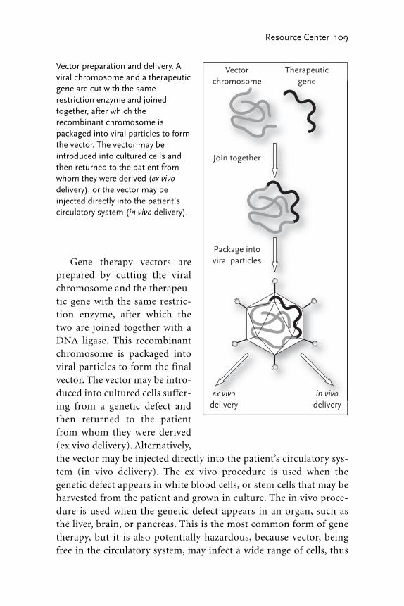
Gene therapy vectors areprepared by cutting the viralchromosome and the therapeu-tic gene with the same restric-tion enzyme, after which thetwo are joined together with aDNA ligase. This recombinantchromosome is packaged intoviral particles to form the finalvector. The vector may be intro-duced into cultured cells suffer-ing from a genetic defect andthen returned to the patientfrom whom they were derived(ex vivo delivery). Alternatively,the vector may be injected directly into the patient’s circulatory sys-tem (in vivo delivery). The ex vivo procedure is used when thegenetic defect appears in white blood cells, or stem cells that may beharvested from the patient and grown in culture. The in vivo proce-dure is used when the genetic defect appears in an organ, such as the liver, brain, or pancreas. This is the most common form of genetherapy, but it is also potentially hazardous, because vector, being free in the circulatory system, may infect a wide range of cells, thus
Resource Center 109
Vectorchromosome
Therapeuticgene
Package intoviral particles
Join together
in vivodelivery
ex vivodelivery
Vector preparation and delivery. Aviral chromosome and a therapeuticgene are cut with the samerestriction enzyme and joinedtogether, after which therecombinant chromosome ispackaged into viral particles to formthe vector. The vector may beintroduced into cultured cells andthen returned to the patient fromwhom they were derived (ex vivodelivery), or the vector may beinjected directly into the patient’scirculatory system (in vivo delivery).

activating an immune response that could lead to widespread tissueand organ damage.
The first gene therapy trial, conducted in 1990, used ex vivo delivery.This trail cured a young patient named Ashi deSilva of an immune defi-ciency (adenosine deaminase deficiency) that affects white blood cells.Other trials since then have either been ineffective or were devastatingfailures. Such a case occurred in 1999, when Jesse Gelsinger, an 18-year-old patient suffering from a liver disease, died while participating in agene therapy trial. His death was caused by multiorgan failure broughton by the viral vector. In 2002, two children being treated for anotherform of immune deficiency developed vector-induced leukemia (can-cer of the white blood cells). Despite these setbacks, gene therapy holdsgreat promise as a medical therapy, and there are currently more than 600trails in progress, in the United States alone, to treat a variety of geneticdisorders.
The Human Genome ProjectSequencing the entire human genome is an idea that grew over aperiod of 20 years, beginning in the early 1980s. At that time, theDNA-sequencing method invented by the British biochemist FredSanger, then at the University of Cambridge, was but a few years oldand had only been used to sequence viral or mitochondrial genomes(see chapter 8 for a description of sequencing methods). Indeed, oneof the first genomes to be sequenced was that of bacteriophage G4, avirus that infects the bacterium Escherichia coli (E. coli). The G4genome consists of 5,577 nucleotide pairs (or base pairs, abbreviatedbp) and was sequenced in Dr. Sanger’s laboratory in 1979. By 1982 theSanger protocol was used by others to sequence the genome of theanimal virus SV40 (5,224 bp), the human mitochondrion (16,569 bp),and bacteriophage lambda (48,502 bp). Besides providing invaluabledata, these projects demonstrated the feasibility of sequencing verylarge genomes.
The possibility of sequencing the entire human genome was firstdiscussed at scientific meetings organized by the United States Depart-ment of Energy (DOE) between 1984 and 1986. A committee appointedby the U.S. National Research Council endorsed the idea in 1988 but
110 Aging

recommended a broader program that would include the sequencing ofthe genes of humans, bacteria, yeast, worms, flies, and mice. They alsocalled for the establishment of research programs devoted to the ethical,legal, and social issues raised by human genome research. The programwas formally launched in late 1990 as a consortium consisting of coor-dinated sequencing projects in the United States, Britain, France, Ger-many, Japan, and China. At about the same time, the Human GenomeOrganization (HUGO) was founded to provide a forum for interna-tional coordination of genomic research.
By 1995 the consortium had established a strategy, called hierarchi-cal shotgun sequencing, which they applied to the human genome aswell as to the other organisms mentioned. With this strategy, genomicDNA is cut into one-megabase (Mb) fragments (i.e., each fragmentconsists of 1 million bases) that are cloned into bacterial artificial chro-mosomes (BACs) to form a library of DNA fragments. The BAC frag-ments are partially characterized, then organized into an overlappingassembly called a contig. Clones are selected from the contigs for shot-gun sequencing. That is, each shotgun clone is digested into small 1,000bp fragments, sequenced, and then assembled into the final sequencewith the aid of computers. Organizing the initial BAC fragments intocontigs greatly simplifies the final assembly stage.
Sequencing of the human genome was divided into two stages. Thefirst stage, completed in 2001, was a rough draft that covered about 80percent of the genome with an estimated size of more than 3 billionbases (also expressed as three gigabases, or three Gb). The final draft,completed in April 2003, covers the entire genome and refines the datafor areas of the genome that were difficult to sequence. It also filled inmany gaps that occurred in the rough draft. The final draft of thehuman genome gives us a great deal of information that may bedivided into three categories: gene content, gene origins, and geneorganization.
GENE CONTENTAnalysis of the final draft has shown that the human genome consists of3.2 Gb of DNA that encodes about 30,000 genes (estimates rangebetween 25,000 to 32,000). The estimated number of genes is surpris-ingly low; many scientists had believed the human genome contained
Resource Center 111

100,000 genes. By comparison, the fruit fly has 13,338 genes and thesimple roundworm, Caenorhabditis elegans (C. elegans), has 18,266. Thegenome data suggests that human complexity, as compared with thefruit fly or the worm, is not simply due to the absolute number of genesbut involves the complexity of the proteins that are encoded by thosegenes. In general, human proteins tend to be much more complex thanthose of lower organisms. Data from the final draft and other sourcesprovides a detailed overview of the functional profile of human cellularproteins.
GENE ORIGINSFully one-half of human genes originated as transposable elements, alsoknown as jumping genes. Equally surprising is the fact that 220 of ourgenes were obtained by horizontal transfer from bacteria, rather thanancestral, or vertical, inheritance. In other words, we obtained thesegenes directly from bacteria, probably during episodes of infection, in akind of natural gene therapy, or gene swapping. We know this to be thecase because, while these genes occur in bacteria, they are not presentin yeast, fruit flies, or any other eukaryotes that have been tested.
The function of most of the horizontally transferred genes isunclear, although a few may code for basic metabolic enzymes. Anotable exception is a gene that codes for an enzyme called monoamineoxidase (MAO). Monoamines are neurotransmitters, such asdopamine, norepinephrine, and serotonin, which are needed for neuralsignaling in the human central nervous system. Monoamine oxidaseplays a crucial role in the turnover of these neurotransmitters. HowMAO, obtained from bacteria, could have developed such an importantrole in human physiology is a great mystery.
GENE ORGANIZATIONIn prokaryotes, genes are simply arranged in tandem along the chro-mosome, with little if any DNA separating one gene from the other.Each gene is transcribed into messenger RNA (mRNA), which is trans-lated into protein. Indeed, in prokaryotes, which have no nucleus, trans-lation often begins even before transcription is complete. In eukaryotes,as we might expect, gene organization is more complex. Data from thegenome project shows clearly that eukaryote genes are split into sub-
112 Aging

units, called exons, and that each exon is separated by a length of DNA,called an intron. A gene, consisting of introns and exons, is separatedfrom other genes by long stretches of noncoding DNA called interven-ing sequences. Eukaryote genes are transcribed into a primary RNAmolecule that includes exon and intron sequences. The primary tran-script never leaves the nucleus and is never translated into protein.Nuclear enzymes remove the introns from the primary transcript, afterwhich the exons are joined together to form the mature mRNA. Thusonly the exons carry the necessary code to produce a protein.
Resource Center 113


115
GLOSSARY
acetyl A chemical group derived from acetic acid. Important inenergy metabolism and for the modification of proteins.
acetylcholine A neurotransmitter released at axonal terminals bycholinergic neurons. Found in the central and peripheral nervoussystem and released at the vertebrate neuromuscular junction.
acetyl CoA A water-soluble molecule, coenzyme A (CoA), that carries acetyl groups in cells.
acid A substance that releases protons when dissolved in water. Carriesa net negative charge.
actin filament A protein filament formed by the polymerization ofglobular actin molecules. Forms the cytoskeleton of all eukaryotesand part of the contractile apparatus of skeletal muscle.
action potential A self-propagating electrical impulse that occurs inthe membranes of neurons, muscles, photoreceptors, and hair cellsof the inner ear.
active transport Movement of molecules across the cell membrane,utilizing the energy stored in ATP.
adenylate cyclase A membrane-bound enzyme that catalyzes the conversion of ATP to cyclic AMP. An important component of cell-signaling pathways.
adherens junction A cell junction in which the cytoplasmic face ofthe membrane is attached to actin filaments.
adipocyte A fat cell.adrenaline (epinephrine) A hormone released by chromaffin cells in
the adrenal gland. Prepares an animal for extreme activity, increasesthe heart rate and blood-sugar levels.
adult stem cells Stem cells isolated from adult tissues, such as bonemarrow or epithelium.
V

aerobic Refers to a process that either requires oxygen or occurs in itspresence.
allele An alternate form of a gene. Diploid organisms have two allelesfor each gene, located at the same locus (position) on homologouschromosomes.
allogeneic transplant A patient receives a tissue or organ transplantfrom an unrelated individual.
alpha helix A common folding pattern of proteins in which a linearsequence of amino acids twists into a right-handed helix stabilizedby hydrogen bonds.
amino acid An organic molecule containing amino and carboxylgroups that is a building block of protein.
aminoacyl-tRNA An amino acid linked by its carboxyl group to ahydroxyl group on tRNA.
aminoacyl-tRNA synthetase An enzyme that attaches the correctamino acid to a tRNA.
amino terminus The end of a protein or polypeptide chain that car-ries a free amino group.
amphipathic Having both hydrophilic and hydrophobic regions, as ina phospholipid.
anabolism A collection of metabolic reactions in a cell whereby largemolecules are made from smaller ones.
anaerobic A cellular metabolism that does not depend on molecularoxygen.
anaphase A mitotic stage in which the two sets of chromosomesmove away from each other toward opposite spindle poles.
anchoring junction A cell junction that attaches cells to each other.angiogenesis Sprouting of new blood vessels from preexisting
ones.angstrom A unit of length, equal to 10-10 meter or 0.1 nanometer
(nm), that is used to measure molecules and atoms.anterior A position close to or at the head end of the body.antibiotic A substance made by bacteria, fungi, and plants that is
toxic to microorganisms. Common examples are penicillin andstreptomycin.
antibody A protein made by B cells of the immune system inresponse to invading microbes.
116 Aging

anticodon A sequence of three nucleotides in tRNA that is comple-mentary to a messenger RNA codon.
antigen A molecule that stimulates an immune response, leading tothe formation of antibodies.
antigen-presenting cell A cell of the immune system, such as amonocyte, that presents pieces of an invading microbe (the antigen)to lymphocytes.
antiparallel The relative orientation of the two strands in a DNAdouble helix; the polarity of one strand is oriented in the oppositedirection to the other.
antiporter A membrane carrier protein that transports two differentmolecules across a membrane in opposite directions.
apoptosis Regulated or programmed form of cell death that may beactivated by the cell itself or by the immune system to force cells tocommit suicide when they become infected with a virus.
asexual reproduction The process of forming new individuals with-out gametes or the fertilization of an egg by a sperm. Individuals pro-duced this way are identical to the parent and referred to as a clone.
aster The star-shaped arrangement of microtubules that is character-istic of a mitotic or meiotic spindle.
ATP (adenosine triphosphate) A nucleoside consisting of adenine,ribose, and three phosphate groups that is the main carrier of chem-ical energy in the cell.
ATPase Any enzyme that catalyzes a biochemical reaction by extract-ing the necessary energy from ATP.
ATP synthase A protein located in the inner membrane of the mito-chondrion that catalyzes the formation of ATP from ADP and inor-ganic phosphate using the energy supplied by the electron transportchain.
autogeneic transplant A patient receives a transplant of his or herown tissue.
autosome Any chromosome other than a sex chromosome.axon A long extension of a neuron’s cell body that transmits an elec-
trical signal to other neurons.axonal transport The transport of organelles, such as Golgi vesicles,
along an axon to the axonal terminus. Transport also flows from theterminus to the cell body.
Glossary 117
bacteria One of the most ancient forms of cellular life (the other isthe archaea). Bacteria are prokaryotes and some are known to causedisease.
bacterial artificial chromosome (BAC) A cloning vector that accom-modates DNA inserts of up to 1 million base pairs.
bacteriophage A virus that infects bacteria. Bacteriophages were usedto prove that DNA is the cell’s genetic material and are now used ascloning vectors.
base A substance that can accept a proton in solution. The purinesand pyrimidines in DNA and RNA are organic bases and are oftenreferred to simply as bases.
base pair Two nucleotides in RNA or DNA that are held together byhydrogen bonds. Adenine bound to thymine or guanine bound tocytosine are examples of base pairs.
B cell (B lymphocyte) A white blood cell that makes antibodies and ispart of the adaptive immune response.
benign Tumors that grow to a limited size and do not spread to otherparts of the body.
beta sheet Common structural motif in proteins in which differentstrands of the protein run alongside each other and are heldtogether by hydrogen bonds.
biopsy The removal of cells or tissues for examination under a micro-scope. When only a sample of tissue is removed, the procedure iscalled an incisional biopsy or core biopsy. When an entire lump orsuspicious area is removed, the procedure is called an excisionalbiopsy. When a sample of tissue or fluid is removed with a needle, theprocedure is called a needle biopsy or fine-needle aspiration.
biosphere The world of living organisms.bivalent A duplicated chromosome paired with its homologous
duplicated chromosome at the beginning of meiosis.blastomere A cell formed by the cleavage of a fertilized egg. Blas-
tomeres are the totipotent cells of the early embryo.blotting A technique for transferring DNA (Southern blotting), RNA
(northern blotting), or proteins (western blotting) from an agaroseor polyacrylamide gel to a nylon membrane.
BRCA1 (breast cancer gene 1) A gene on chromosome 17 that maybe involved in regulating the cell cycle. A person who inherits an
118 Aging

altered version of the BRCA1 gene has a higher risk of getting breast,ovarian, or prostate cancer.
BRCA2 (breast cancer gene 2) A gene on chromosome 13 that, whenmutated, increases the risk of getting breast, ovarian, or prostatecancer.
budding yeast The common name for the baker’s yeast Saccharomycescerevisiae, a popular experimental organism that reproduces bybudding off a parental cell.
cadherin Belongs to a family of proteins that mediates cell-to-celladhesion in animal tissues.
calorie A unit of heat. One calorie is the amount of heat needed toraise the temperature of one gram of water by 1°C. Kilocalories(1,000 calories) are used to describe the energy content of foods.
capsid The protein coat of a virus, formed by auto-assembly of oneor more proteins into a geometrically symmetrical structure.
carbohydrate A general class of compounds that includes sugars,containing carbon, hydrogen, and oxygen.
carboxyl group A carbon atom attached to an oxygen and a hydroxylgroup.
carboxyl terminus The end of a protein containing a carboxyl group.carcinogen A compound or form of radiation that can cause cancer.carcinogenesis The formation of a cancer.carcinoma Cancer of the epithelium, representing the majority of
human cancers.cardiac muscle Muscle of the heart. Composed of myocytes that are
linked together in a communication network based on free passageof small molecules through gap junctions.
caspase A protease involved in the initiation of apoptosis.catabolism Enzyme-regulated breakdown of large molecules for the
extraction of chemical-bond energy. Intermediate products arecalled catabolites.
catalyst A substance that lowers the activation energy of a reaction.CD28 Cell-surface protein located in T cell membranes, necessary for
the activation of T cells by foreign antigens.cDNA (complementary DNA) DNA that is synthesized from mRNA,
thus containing the complementary sequence. cDNA contains cod-ing sequence but not the regulatory sequences that are present in the
Glossary 119

genome. Labeled probes are made from cDNA for the study of geneexpression.
cell adhesion molecule (CAM) A cell surface protein that is used toconnect cells to each other.
cell body The main part of a cell containing the nucleus, Golgi com-plex, and endoplasmic reticulum. Used in reference to neurons thathave long processes (dendrites and axons) extending some distancefrom the nucleus and cytoplasmic machinery.
cell coat See glycocalyx.cell-cycle control system A team of regulatory proteins that governs
progression through the cell cycle.cell-division-cycle gene (cdc gene) A gene that controls a specific step
in the cell cycle.cell fate The final differentiated state that a pluripotent embryonic
cell is expected to attain.cell-medicated immune response Activation of specific cells to
launch an immune response against an invading microbe.cell nuclear replacement Animal-cloning technique whereby a
somatic cell nucleus is transferred to an enucleated oocyte. Syn-onomous with somatic-cell nuclear transfer.
central nervous system (CNS) That part of a nervous system thatanalyzes signals from the body and the environment. In animals, theCNS includes the brain and spinal cord.
centriole A cylindrical array of microtubules that is found at the center of a centrosome in animal cells.
centromere A region of a mitotic chromosome that holds sister chromatids together. Microtubules of the spindle fiber connect to anarea of the centromere called the kinetochore.
centrosome Organizes the mitotic spindle and the spindle poles. Inmost animal cells it contains a pair of centrioles.
chiasma (plural: chiasmata) An X-shaped connection betweenhomologous chromosomes that occurs during meiosis I, represent-ing a site of crossing-over, or genetic exchange between the twochromosomes.
chromatid A duplicate chromosome that is still connected to theoriginal at the centromere. The identical pair are called sister chromatids.
120 Aging

chromatin A complex of DNA and proteins (histones and nonhis-tones) that forms each chromosome and is found in the nucleus ofall eukaryotes. Decondensed and threadlike during interphase.
chromatin condensation Compaction of different regions of inter-phase chromosomes that is mediated by the histones.
chromosome One long molecule of DNA that contains the organ-ism’s genes. In prokaryotes, the chromosome is circular and naked;in eukaryotes, it is linear and complexed with histone and nonhistoneproteins.
chromosome condensation Compaction of entire chromosomes inpreparation for cell division.
clinical breast exam An exam of the breast performed by a physicianto check for lumps or other changes.
cyclic adenosine monophosphate (cAMP) A second messenger in acell-signaling pathway that is produced from ATP by the enzymeadenylate cyclase.
cyclin A protein that activates protein kinases (cyclin-dependent pro-tein kinases, or Cdk) that control progression from one state of thecell cycle to another.
cytochemistry The study of the intracellular distribution of chemicals.cytochrome Colored, iron-containing protein that is part of the elec-
tron transport chain.cytotoxic T cell A T lymphocyte that kills infected body cells.dendrite An extension of a nerve cell that receives signals from other
neurons.dexrazoxane A drug used to protect the heart from the toxic effects
of anthracycline drugs such as doxorubicin. It belongs to the familyof drugs called chemoprotective agents.
dideoxy sequencing A method for sequencing DNA that employs di-deoxyribose nucleotides.
diploid A genetic term meaning two sets of homologous chromo-somes, one set from the mother and the other from the father. Thusdiploid organisms have two versions (alleles) of each gene in thegenome.
DNA (deoxyribonucleic acid) A long polymer formed by linkingfour different kinds of nucleotides together like beads on a string.The sequence of nucleotides is used to encode an organism’s genes.
Glossary 121

DNA helicase An enzyme that separates and unwinds the two DNAstrands in preparation for replication or transcription.
DNA library A collection of DNA fragments that are cloned intoplasmids or viral genomes.
DNA ligase An enzyme that joins two DNA strands together to makea continuous DNA molecule.
DNA microarray A technique for studying the simultaneous expres-sion of a very large number of genes.
DNA polymerase An enzyme that synthesizes DNA using one strandas a template.
DNA primase An enzyme that synthesizes a short strand of RNA thatserves as a primer for DNA replication.
dorsal The backside of an animal. Also refers to the upper surface ofanatomical structures, such as arms or wings.
dorsoventral The body axis running from the backside to thefrontside or the upperside to the underside of a structure.
double helix The three-dimensional structure of DNA in which thetwo strands twist around each other to form a spiral.
doxorubicin An anticancer drug that belongs to a family of antitu-mor antibiotics.
Drosophila melanogaster Small species of fly, commonly called afruit fly, that is used as an experimental organism in genetics,embryology, and gerontology.
ductal carcinoma in situ (DCIS) Abnormal cells that involve only thelining of a breast duct. The cells have not spread outside the duct toother tissues in the breast. Also called intraductal carcinoma.
dynein A motor protein that is involved in chromosome movementsduring cell division.
dysplasia Disordered growth of cells in a tissue or organ, often lead-ing to the development of cancer.
ectoderm An embryonic tissue that is the precursor of the epidermisand the nervous system.
electrochemical gradient A differential concentration of an ion ormolecule across the cell membrane that serves as a source of poten-tial energy and may polarize the cell electrically.
electron microscope A microscope that uses electrons to produce ahigh-resolution image of the cell.
122 Aging

embryogensis The development of an embryo from a fertilized egg.embryonic stem cell (ES cell) A pluripotent cell derived from the
inner cell mass (the cells that give rise to the embryo instead of theplacenta) of a mammalian embryo.
endocrine cell A cell that is specialized for the production and releaseof hormones. Such cells make up hormone-producing tissue such asthe pituitary gland or gonads.
endocytosis Cellular uptake of material from the environment byinvagination of the cell membrane to form a vesicle called an endo-some. The endosome’s contents are made available to the cell after itfuses with a lysosome.
endoderm An embryonic tissue layer that gives rise to the gut.endoplasmic reticulum (ER) Membrane-bounded chambers that
are used to modify newly synthesized proteins with the addition of sugar molecules (glycosylation). When finished, the glycosy-lated proteins are sent to the Golgi apparatus in exocytotic vesicles.
endothelial cell A cell that forms the endothelium, a thin sheet ofcells lining the inner surface of all blood vessels.
enhancer A DNA regulatory sequence that provides a binding site fortranscription factors capable of increasing the rate of transcriptionfor a specific gene. Often located thousands of base pairs away fromthe gene it regulates.
enveloped virus A virus containing a capsid that is surrounded by alipid bilayer originally obtained from the membrane of a previouslyinfected cell.
enzyme A protein or RNA that catalyzes a specific chemical reaction.epidermis The epithelial layer, or skin, that covers the outer surface
of the body.ER signal sequence The amino terminal sequence that directs pro-
teins to enter the endoplasmic reticulum (ER). This sequence isremoved once the protein enters the ER.
erythrocyte A red blood cell that contains the oxygen-carrying pigment hemoglobin used to deliver oxygen to cells in the body.
Escherichia coli (E. coli) Rod shape, gram negative bacterium thatinhabits the intestinal tract of most animals and is used as an exper-imental organism by geneticists and biomedical researchers.
Glossary 123

euchromatin Lightly staining portion of interphase chromatin, incontrast to the darkly staining heterochromatin (condensed chro-matin). Euchromatin contains most, if not all, of the active genes.
eukaryote (eucaryote) A cell containing a nucleus and many mem-brane-bounded organelles. All life-forms, except bacteria andviruses, are composed of eukaryote cells.
exocytosis The process by which molecules are secreted from a cell.Molecules to be secreted are located in Golgi-derived vesicles thatfuse with the inner surface of the cell membrane, depositing thecontents into the intercellular space.
exon Coding region of a eukaryote gene that is represented in mes-senger RNA, and thus directs the synthesis of a specific protein.
expression studies Examination of the type and quantity of mRNAor protein that is produced by cells, tissues, or organs.
fat A lipid material, consisting of triglycerides (fatty acids bound toglycerol), that is stored adipocytes as an energy reserve.
fatty acid A compound that has a carboxylic acid attached to a longhydrocarbon chain. A major source of cellular energy and a compo-nent of phospholipids.
fertilization The fusion of haploid male and female gametes to forma diploid zygote.
fibroblast The cell type that, by secreting an extracellular matrix,gives rise to the connective tissue of the body.
filter hybridization The detection of specific DNA or RNA mole-cules, fixed on a nylon filter, by incubating the filter with a labeledprobe that hybridizes to the target sequence.
fixative A chemical that is used to preserve cells and tissues. Commonexamples are formaldehyde, methanol, and acetic acid.
flagellum (plural: flagella) Whiplike structure found in prokaryotesand eukaryotes that are used to propel cells through water.
fluorescein Fluorescent dye that produces a green light when illumi-nated with ultraviolet or blue light.
fluorescent dye A dye that absorbs UV or blue light and emits light ofa longer wavelength, usually as green or red light.
fluorescent microscope A microscope that is equipped with specialfilters and a beam splitter for the examination of tissues and cellsstained with a fluorescent dye.
124 Aging

follicle cell Cells that surround and help feed a developing oocyte.G0 G “zero” refers to a phase of the cell cycle. State of withdrawal
from the cycle as the cell enters a resting or quiescent stage. Occursin differentiated body cells as well as developing oocytes.
G1 Gap 1 refers to the phase of the cell cycle that occurs just aftermitosis and before the next round of DNA synthesis.
G2 Gap 2 refers to the phase of the cell cycle that follows DNA repli-cation and precedes mitosis.
gap junction A communication channel in the membranes ofadjacent cells that allows free passage of ions and small molecules.
gastrulation An embryological event in which a spherical embryo isconverted into an elongated structure with a head end, a tail end,and a gut (gastrula).
gene A region of the DNA that specifies a specific protein or RNAmolecule that is handed down from one generation to the next. Thisregion includes both the coding, noncoding, and regulatorysequences.
gene regulatory protein Any protein that binds to DNA and therebyaffects the expression of a specific gene.
gene repressor protein A protein that binds to DNA and blocks tran-scription of a specific gene.
gene therapy A method for treating disease whereby a defective gene,causing the disease, is either repaired, replaced, or supplementedwith a functional copy.
genetic code A set of rules that assigns a specific DNA or RNA triplet,consisting of a three-base sequence, to a specific amino acid.
genome All of the genes that belong to a cell or an organism.genomic library A collection of DNA fragments, obtained by digest-
ing genomic DNA with a restriction enzyme, that are cloned intoplasmid or viral vectors.
genomics The study of DNA sequences and their role in the functionand structure of an organism.
genotype The genetic composition of a cell or organism.germ cell Cells that develop into gametes, either sperm or oocytes.glucose Six-carbon monosaccharide (sugar) that is the principal
source of energy for many cells and organisms. Stored as glycogen
Glossary 125
in animal cells and as starch in plants. Wood is an elaborate polymerof glucose and other sugars.
glycerol A three-carbon alcohol that is an important component ofphospholipids.
glycocalyx A molecular “forest,” consisting of glycosylated proteinsand lipids, that covers the surface of every cell. The glycoproteinsand glycolipids, carried to the cell membrane by Golgi-derived vesi-cles, have many functions, including the formation of ion channels,cell-signaling receptors and transporters.
glycogen A polymer of glucose used to store energy in an animal cell.glycolysis The degradation of glucose with production of ATP.glycoprotein Any protein that has a chain of glucose molecules
(oligosaccharide) attached to some of the amino acid residues.glycosylation The process of adding one or more sugar molecules to
proteins or lipids.glycosyl transferase An enzyme in the Golgi complex that adds glu-
cose to proteins.Golgi complex (Golgi apparatus) Membrane-bounded organelle in
eukaryote cells that receives glycoproteins from the ER, which aremodified and sorted before being sent to their final destination. TheGolgi complex is also the source of glycolipids that are destined forthe cell membrane. The glycoproteins and glycolipids leave theGolgi by exocytosis. This organelle is named after the Italian histol-ogist Camillo Golgi, who discovered it in 1898.
granulocyte A type of white blood cell that includes the neutrophils,basophils, and eosinophils.
growth factor A small protein (polypeptide) that can stimulate cells togrow and proliferate.
haploid Having only one set of chromosomes. A condition that istypical in gametes, such as sperm and eggs.
HeLa cell A tumor-derived cell line, originally isolated from a cancerpatient in 1951. Currently used by many laboratories to study thecell biology of cancer and carcinogenesis.
helix-loop-helix A structural motif common to a group of gene reg-ulatory proteins.
helper T cell A type of T lymphocyte that helps stimulate B cells tomake antibodies directed against a specific microbe or antigen.
126 Aging

hemoglobin An iron-containing protein complex, located in redblood cells that picks up oxygen in the lungs and carries it to othertissues and cells of the body.
hemopoiesis Production of blood cells, occurring primarily in thebone marrow.
hepatocyte A liver cell.heterochromatin A region of a chromosome that is highly condensed
and transcriptionally inactive.histochemistry The study of chemical differentiation of tissues.histology The study of tissues.histone Small nuclear proteins, rich in the amino acids arginine and
lysine, that form the nucleosome in eukaryote nuclei, a beadlikestructure that is a major component of chromatin.
HIV The human immunodeficiency virus that is responsible forAIDS.
homolog One of two or more genes that have a similar sequence andare descended from a common ancestor gene.
homologous Organs or molecules that are similar in structurebecause they have descended from a common ancestor. Used pri-marily in reference to DNA and protein sequences.
homologous chromosomes Two copies of the same chromosome,one inherited from the mother and the other from the father.
hormone A signaling molecule, produced and secreted by endocrineglands. Usually released into general circulation for coordination ofan animal’s physiology.
housekeeping gene A gene that codes for a protein that is needed byall cells, regardless of the cell’s specialization. Genes encodingenzymes involved in glycolysis and the Krebs cycle are commonexamples.
hybridization A term used in molecular biology (recombinant DNAtechnology) meaning the formation of a double-stranded nucleicacid through complementary base-pairing. A property that isexploited in filter hybridization, a procedure that is used to screengene libraries and to study gene structure and expression.
hydrophilic A polar compound that mixes readily with water.hydrophobic A nonpolar molecule that dissolves in fat and lipid solu-
tions but not in water.
Glossary 127

hydroxyl group (-OH) Chemical group consisting of oxygen andhydrogen that is a prominent part of alcohol.
image analysis A computerized method for extracting informationfrom digitized microscopic images of cells or cell organelles.
immunofluorescence Detection of a specific cellular protein with theaid of a fluorescent dye that is coupled to an antibody.
immunoglobulin (Ig) An antibody made by B cells as part of theadaptive immune response.
incontinence Inability to control the flow of urine from the bladder(urinary incontinence) or the escape of stool from the rectum (fecalincontinence).
in situ hybridization A method for studying gene expression,whereby a labeled cDNA or RNA probe hybridizes to a specificmRNA in intact cells or tissues. The procedure is usually carried outon tissue sections or smears of individual cells.
insulin Polypeptide hormone secreted by β (beta) cells in the verte-brate pancreas. Production of this hormone is regulated directly bythe amount of glucose that is in the blood.
interleukin A small protein hormone, secreted by lymphocytes, toactivate and coordinate the adaptive immune response.
interphase The period between each cell division, which includes the G1, S, and G2 phases of the cell cycle.
intron A section of a eukaryotic gene that is noncoding. It is tran-scribed, but does not appear in the mature mRNA.
in vitro Refers to cells growing in culture, or a biochemical reactionoccurring in a test tube (Latin for “in glass”).
in vivo A biochemical reaction, or a process, occurring in living cellsor a living organism (Latin for “in life”).
ion An atom that has gained or lost electrons, thus acquiring a charge.Common examples are Na+ and Ca++ ions.
ion channel A transmembrane channel that allows ions to diffuseacross the membrane and down their electrochemical gradient.
Jak-STAT signaling pathway One of several cell-signaling pathwaysthat activates gene expression. The pathway is activated throughcell-surface receptors and cytoplasmic Janus kinases (Jaks), and sig-nal transducers and activators of transcription (STATs).
128 Aging

karyotype A pictorial catalog of a cell’s chromosomes, showing theirnumber, size, shape, and overall banding pattern.
keratin Proteins produced by specialized epithelial cells called ker-atinocytes. Keratin is found in hair, fingernails, and feathers.
kinesin A motor protein that uses energy obtained from the hydroly-sis of ATP to move along a microtubule.
kinetochore A complex of proteins that forms around the cen-tromere of mitotic or meiotic chromosomes, providing an attach-ment site for microtubules. The other end of each microtubule isattached to a chromosome.
Krebs cycle (citric acid cycle) The central metabolic pathway in alleukaryotes and aerobic prokaryotes, discovered by the Germanchemist Hans Krebs in 1937. The cycle oxidizes acetyl groupsderived from food molecules. The end products are CO2, H2O, andhigh-energy electrons, which pass via NADH and FADH2 to therespiratory chain. In eukaryotes, the Krebs cycle is located in themitochondria.
labeling reaction The addition of a radioactive atom or fluorescentdye to DNA or RNA for use as a probe in filter hybridization.
lagging strand One of the two newly synthesized DNA strands at areplication fork. The lagging strand is synthesized discontinuously,and therefore, its completion lags behind the second, or leading,strand.
lambda bacteriophage A viral parasite that infects bacteria. Widelyused as a DNA cloning vector.
leading strand One of the two newly synthesized DNA strands at areplication fork. The leading strand is made by continuous synthe-sis in the 5’ to 3’ direction.
leucine zipper A structural motif of DNA binding proteins, in whichtwo identical proteins are joined together at regularly spaced leucineresidues, much like a zipper, to form a dimer.
leukemia Cancer of white blood cells.lipid bilayer Two closely aligned sheets of phospholipids that form
the core structure of all cell membranes. The two layers are alignedsuch that the hydrophobic tails are interior, while the hydrophilichead groups are exterior on both surfaces.
Glossary 129

liposome An artificial lipid bilayer vesicle used in membrane studiesand as an artificial gene therapy vector.
locus A term from genetics that refers to the position of a gene alonga chromosome. Different alleles of the same gene occupy the samelocus.
long-term potentiation (LTP) A physical remodeling of synapticjunctions that receive continuous stimulation.
lymphocyte A type of white blood cell that is involved in the adaptiveimmune response. There are two kinds of lymphocytes: T lympho-cytes and B lymphocytes. T lymphocytes (T cells) mature in the thy-mus and attack invading microbes directly. B lymphocytes (B cells)mature in the bone marrow and make antibodies that are designedto immobilize or destroy specific microbes or antigens.
lysis The rupture of the cell membrane followed by death of the cell.lysosome Membrane-bounded organelle of eukaryotes that contains
powerful digestive enzymes.macromolecule A very large molecule that is built from smaller
molecular subunits. Common examples are DNA, proteins, andpolysaccharides.
magnetic resonance imaging (MRI) A procedure in which radiowaves and a powerful magnet linked to a computer are used to cre-ate detailed pictures of areas inside the body. These pictures canshow the difference between normal and diseased tissue. MRI makesbetter images of organs and soft tissue than other scanning tech-niques, such as CT or X-ray. MRI is especially useful for imaging thebrain, spine, the soft tissue of joints, and the inside of bones. Alsocalled nuclear magnetic resonance imaging.
major histocompatibility complex Vertebrate genes that code for alarge family of cell-surface glycoproteins that bind foreign antigensand present them to T cells to induce an immune response.
malignant Refers to the functional status of a cancer cell that growsaggressively and is able to metastasize, or colonize, other areas of thebody.
mammography The use of X-rays to create a picture of the breast.MAP-kinase (mitogen-activated protein kinase) A protein kinase
that is part of a cell proliferation–inducing signaling pathway.M-cyclin A eukaryote enzyme that regulates mitosis.
130 Aging

meiosis A special form of cell division by which haploid gametes areproduced. This is accomplished with two rounds of cell division butonly one round of DNA replication.
melanocyte A skin cell that produces the pigment melanin.membrane The lipid bilayer, and the associated glycocalyx, that sur-
rounds and encloses all cells.membrane channel A protein complex that forms a pore or channel
through the membrane for the free passage of ions and small molecules.
membrane potential A buildup of charged ions on one side of thecell membrane establishes an electrochemical gradient that is meas-ured in millivolts (mV). An important characteristic of neurons as itprovides the electric current, when ion channels open, that enablethese cells to communicate with each other.
mesoderm An embryonic germ layer that gives rise to muscle, con-nective tissue, bones, and many internal organs.
messenger RNA (mRNA) An RNA transcribed from a gene that isused as the gene template by the ribosomes, and other componentsof the translation machinery, to synthesize a protein.
metabolism The sum total of the chemical processes that occur in liv-ing cells.
metaphase The stage of mitosis at which the chromosomes areattached to the spindle but have not begun to move apart.
metaphase plate Refers to the imaginary plane established by thechromosomes as they line up at right angles to the spindle poles.
metaplasia A change in the pattern of cellular behavior that oftenprecedes the development of cancer.
metastasis Spread of cancer cells from the site of the original tumorto other parts of the body.
methyl group (-CH3) Hydrophobic chemical group derived frommethane. Occurs at the end of a fatty acid.
micrograph Photograph taken through a light, or electron, micro-scope.
micrometer (µm or micron) Equal to 10-6 meters.microtubule A fine cylindrical tube made of the protein tubulin,
forming a major component of the eukaryote cytoskeleton.millimeter (mm) Equal to 10-3 meters.
Glossary 131

mitochondrion (plural: mitochondria) Eukaryote organelle, for-merly free-living, that produces most of the cell’s ATP.
mitogen A hormone or signaling molecule that stimulates cells togrow and divide.
mitosis Division of a eukaryotic nucleus. From the Greek mitos,meaning “a thread,” in reference to the threadlike appearance ofinterphase chromosomes.
mitotic chromosome Highly condensed duplicated chromosomesheld together by the centromere. Each member of the pair is referredto as a sister chromatid.
mitotic spindle Array of microtubules, fanning out from the polarcentrioles and connecting to each of the chromosomes.
molecule Two or more atoms linked together by covalent bonds.monoclonal antibody An antibody produced from a B cell–derived
clonal line. Since all of the cells are clones of the original B cell, theantibodies produced are identical.
monocyte A type of white blood cell that is involved in the immuneresponse.
motif An element of structure or pattern that may be a recurringdomain in a variety of proteins.
M phase The period of the cell cycle (mitosis or meiosis) when thechromosomes separate and migrate to the opposite poles of thespindle.
multipass transmembrane protein A membrane protein that passesback and forth across the lipid bilayer.
mutant A genetic variation within a population.mutation A heritable change in the nucleotide sequence of a
chromosome.myelin sheath Insulation applied to the axons of neurons. The sheath
is produced by oligodendrocytes in the central nervous system andby Schwann cells in the peripheral nervous system.
myeloid cell White blood cells other than lymphocytes.myoblast Muscle precursor cell. Many myoblasts fuse into a syncytium,
containing many nuclei, to form a single muscle cell.myocyte A muscle cell.NAD (nicotine adenine dinucleotide) Accepts a hydride ion (H–),
produced by the Krebs cycle, forming NADH, the main carrier ofelectrons for oxidative phosphorylation.
132 Aging

NADH dehydrogenase Removes electrons from NADH and passesthem down the electron transport chain.
nanometer (nm) Equal to 10-9 meters or 10-3 microns.natural killer cell (NK cell) A lymphocyte that kills virus-infected
cells in the body. It also kills foreign cells associated with a tissue ororgan transplant.
neuromuscular junction A special form of synapse between a motorneuron and a skeletal muscle cell.
neuron A cell specially adapted for communication that forms thenervous system of all animals.
neurotransmitter A chemical released by neurons at a synapse thattransmits a signal to another neuron.
non-small-cell lung cancer A group of lung cancers that includessquamous cell carcinoma, adenocarcinoma, and large cell carci-noma. The small cells are endocrine cells.
northern blotting A technique for the study of gene expression. Mes-senger RNA (mRNA) is fractionated on an agarose gel and thentransferred to a piece of nylon filter paper (or membrane). A specificmRNA is detected by hybridization with a labeled DNA or RNAprobe. The original blotting technique invented by E. M. Southerninspired the name.
nuclear envelope The double membrane (two lipid bilayers) enclos-ing the cell nucleus.
nuclear localization signal (NLS) A short amino acid sequencelocated on proteins that are destined for the cell nucleus after they aretranslated in the cytoplasm.
nucleic acid DNA or RNA, a macromolecule consisting of a chain ofnucleotides.
nucleolar organizer Region of a chromosome containing a cluster ofribosomal RNA genes that gives rise to the nucleolus.
nucleolus A structure in the nucleus where ribosomal RNA is tran-scribed and ribosomal subunits are assembled.
nucleoside A purine or pyrimidine linked to a ribose or deoxyribosesugar.
nucleosome A beadlike structure, consisting of histone proteins.nucleotide A nucleoside containing one or more phosphate groups
linked to the 5’ carbon of the ribose sugar. DNA and RNA arenucleotide polymers.
Glossary 133

nucleus Eukaryote cell organelle that contains the DNA genome onone or more chromosomes.
oligodendrocyte A myelinating glia cell of the vertebrate centralnervous system.
oligo labeling A method for incorporating labeled nucleotides into ashort piece of DNA or RNA. Also known as the random-primerlabeling method.
oligomer A short polymer, usually consisting of amino acids (oligopep-tides), sugars (oligosaccharides), or nucleotides (oligonucleotides).Taken from the Greek word oligos, meaning “few” or “little.”
oncogene A mutant form of a normal cellular gene, known as aproto-oncogene, that can transform a cell to a cancerous phenotype.
oocyte A female gamete or egg cell.operator A region of a prokaryote chromosome that controls the
expression of adjacent genes.operon Two or more prokaryote genes that are transcribed into a sin-
gle mRNA.organelle A membrane-bounded structure, occurring in eukaryote
cells, that has a specialized function. Examples are the nucleus, Golgicomplex, and endoplasmic reticulum.
osmosis The movement of solvent across a semipermeable mem-brane that separates a solution with a high concentration of solutesfrom one with a low concentration of solutes. The membrane mustbe permeable to the solvent but not to the solutes. In the context ofcellular osmosis, the solvent is always water, the solutes are ions andmolecules, and the membrane is the cell membrane.
osteoblast Cells that form bones.ovulation Rupture of a mature follicle with subsequent release of a
mature oocyte from the ovary.oxidative phosphorylation Generation of high-energy electrons
from food molecules that are used to power the synthesis of ATPfrom ADP and inorganic phosphate. The electrons are eventuallytransferred to oxygen to complete the process. Occurs in bacteriaand mitochondria.
p53 A tumor-suppressor gene that is mutated in about half of allhuman cancers. The normal function of the p53 protein is to blockpassage through the cell cycle when DNA damage is detected.
134 Aging

parthenogenesis A natural form of animal cloning whereby an indi-vidual is produced without the formation of haploid gametes andthe fertilization of an egg.
pathogen An organism that causes disease.PCR (polymerase chain reaction) A method for amplifying specific
regions of DNA by temperature cycling a reaction mixture contain-ing the template, a heat-stable DNA polymerase, and replicationprimers.
peptide bond The chemical bond that links amino acids together toform a protein.
pH Measures the acidity of a solution as a negative logarithmic func-tion (p) of H+ concentration (H). Thus a pH of 2.0 (10-2 molar H+)is acidic, whereas a pH of 8.0 (10-8 molar H+) is basic.
phagocyte A cell that engulfs other cells or debris by phagocytosis.phagocytosis A process whereby cells engulf other cells or organic
material by endocytosis. A common practice among protozoans andcells of the vertebrate immune system. (Derived from the Greekword phagein, “to eat.”)
phenotype Physical characteristics of a cell or organism.phospholipid The kind of lipid molecule used to construct cell mem-
branes. Composed of a hydrophilic head-group, phosphate, glycerol,and two hydrophobic fatty acid tails.
phosphorylation A chemical reaction in which a phosphate is cova-lently bonded to another molecule.
photoreceptor A molecule or cell that responds to light.photosynthesis A biochemical process in which plants, algae, and
certain bacteria use energy obtained from sunlight to synthesizemacromolecules from CO2 and H2O.
phylogeny The evolutionary history of an organism, or group oforganisms, often represented diagrammatically as a phylogenetic tree.
pinocytosis A form of endocytosis whereby fluid is brought into thecell from the environment.
placebo An inactive substance that looks the same, and is adminis-tered in the same way, as a drug in a clinical trial.
plasmid A minichromosome, often carrying antibiotic-resistantgenes, that occurs naturally among prokaryotes. Used extensively asa DNA cloning vector.
Glossary 135

platelet A cell fragment, derived from megakaryocytes and lacking anucleus, that is present in the bloodstream and is involved in bloodcoagulation.
ploidy The total number of chromosomes (n) that a cell has. Ploidy isalso measured as the amount of DNA (C) in a given cell relative to ahaploid nucleus of the same organism. Most organisms are diploid,having two sets of chromosomes, one from each parent, but there is great variation among plants and animals. The silk gland ofthe moth Bombyx mori, for example, has cells that are extremelypolyploid, reaching values of 100,000C. Flowers are often highlypolyploid, and vertebrate hepatocytes may be 16C.
point mutation A change in DNA, particularly in a region containinga gene, that alters a single nucleotide.
polyploid Possessing more than two sets of homologous chromosomes.
portal system A system of liver vessels that carries liver enzymesdirectly to the digestive tract.
probe Usually a fragment of a cloned DNA molecule that is labeledwith a radioisotope or fluorescent dye and used to detect specificDNA or RNA molecules on Southern or northern blots.
promoter A DNA sequence to which RNA polymerase binds to initi-ate gene transcription.
prophase The first stage of mitosis. The chromosomes are duplicatedand beginning to condense but are attached to the spindle.
protein A major constituent of cells and organisms. Proteins, madeby linking amino acids together, are used for structural purposesand regulate many biochemical reactions in their alternative role asenzymes. Proteins range in size from just a few amino acids to morethan 200.
protein glycosylation The addition of sugar molecules to a protein.proto-oncogene A normal gene that can be converted to a cancer-
causing gene (oncogene) by a point mutation or through inappro-priate expression.
protozoa Free-living, single-cell eukaryotes that feed on bacteria andother microorganisms. Common examples are Paramecium andAmoeba. Parasitic forms are also known that inhabit the digestiveand urogenital tract of many animals, including humans.
136 Aging

purine A nitrogen-containing compound that is found in RNA andDNA. Two examples are adenine and guanine.
pyrimidine A nitrogen-containing compound found in RNA andDNA. Examples are cytosine, thymine, and uracil (RNA only).
radioactive isotope An atom with an unstable nucleus that emitsradiation as it decays.
randomized clinical trial A study in which the participants areassigned by chance to separate groups that compare different treat-ments; neither the researchers nor the participants can choosewhich group. Using chance to assign people to groups means thatthe groups will be similar and that the treatments they receive canbe compared objectively. At the time of the trial, it is not knownwhich treatment is best.
reagent A chemical solution designed for a specific biochemical orhistochemical procedure.
recombinant DNA A DNA molecule that has been formed by joiningtwo or more fragments from different sources.
regulatory sequence A DNA sequence to which proteins bind thatregulate the assembly of the transcriptional machinery.
replication bubble Local dissociation of the DNA double helix in prepa-ration for replication. Each bubble contains two replication forks.
replication fork The Y-shaped region of a replicating chromosome.Associated with replication bubbles.
replication origin (origin of replication, ORI) The location at whichDNA replication begins.
respiratory chain (electron transport chain) A collection of iron-and copper-containing proteins, located in the inner mitochondrionmembrane, that utilize the energy of electrons traveling down thechain to synthesize ATP.
restriction enzyme An enzyme that cuts DNA at specific sites.restriction map The size and number of DNA fragments obtained
after digesting with one or more restriction enzymes.retrovirus A virus that converts its RNA genome to DNA once it has
infected a cell.reverse transcriptase An RNA-dependent DNA polymerase. This
enzyme synthesizes DNA by using RNA as a template, the reverse ofthe usual flow of genetic information from DNA to RNA.
Glossary 137

ribosomal RNA (rRNA) RNA that is part of the ribosome and servesboth a structural and functional role, possibly by catalyzing some ofthe steps involved in protein synthesis.
ribosome A complex of protein and RNA that catalyzes the synthesisof proteins.
rough endoplasmic reticulum (rough ER) Endoplasmic reticulumthat has ribosomes bound to its outer surface.
Saccharomyces Genus of budding yeast that are frequently used in thestudy of eukaryote cell biology.
sarcoma Cancer of connective tissue.Schwann cell Glia cell that produces myelin in the peripheral nervous
system.screening Checking for disease when there are no symptoms.senescence Physical and biochemical changes that occur in cells and
organisms with age.signal transduction A process by which a signal is relayed to the
interior of a cell where it elicits a response at the cytoplasmic ornuclear level.
smooth muscle cell Muscles lining the intestinal tract and arteries. Lacks the striations typical of cardiac and skeletal muscle, giving it a smooth appearance when viewed under a microscope.
somatic cell Any cell in a plant or animal except those that producegametes (germ cells or germ cell precursors).
somatic cell nuclear transfer Animal cloning technique whereby a somatic cell nucleus is transferred to an enucleated oocyte.Synonomous with cell nuclear replacement.
Southern blotting The transfer of DNA fragments from an agarosegel to a piece of nylon filter paper. Specific fragments are identifiedby hybridizing the filter to a labeled probe. Invented by the Scottishscientist E. M. Southern in 1975.
stem cell Pluripotent progenitor cell, found in embryos and variousparts of the body, that can differentiate into a wide variety of celltypes.
steroid A hydrophobic molecule with a characteristic four-ringedstructure. Sex hormones, such as estrogen and testosterone, aresteroids.
138 Aging

structural gene A gene that codes for a protein or an RNA. Distin-guished from regions of the DNA that are involved in regulatinggene expression but are noncoding.
synapse A neural communication junction between an axon and adendrite. Signal transmission occurs when neurotransmitters,released into the junction by the axon of one neuron, stimulatereceptors on the dendrite of a second neuron.
syncytium A large multinucleated cell. Skeletal muscle cells are syn-cytiums produced by the fusion of many myoblasts.
syngeneic transplants A patient receives tissue or an organ from anidentical twin.
tamoxifen A drug that is used to treat breast cancer. Tamoxifen blocksthe effects of the hormone estrogen in the body. It belongs to thefamily of drugs called antiestrogens.
T cell (T lymphocyte) A white blood cell involved in activating andcoordinating the immune response.
telomere The end of a chromosome. Replaced by the enzyme telom-erase with each round of cell division to prevent shortening of thechromosomes.
telophase The final stage of mitosis in which the chromosomesdecondense and the nuclear envelope reforms.
template A single strand of DNA or RNA whose sequence serves as aguide for the synthesis of a complementary, or daughter, strand.
therapeutic cloning The cloning of a human embryo for the purposeof harvesting the inner cell mass (ES cells).
topoisomerase An enzyme that makes reversible cuts in DNA torelieve strain or to undo knots.
transcription The copying of a DNA sequence into RNA, catalyzedby RNA polymerase.
transcriptional factor A general term referring to a wide assortmentof proteins needed to initiate or regulate transcription.
transfection Introduction of a foreign gene into a eukaryote cell.transfer RNA (tRNA) A collection of small RNA molecules that
transfer an amino acid to a growing polypeptide chain on a ribo-some. There is a separate tRNA for amino acid.
transgenic organism A plant or animal that has been transfectedwith a foreign gene.
Glossary 139

trans-Golgi network The membrane surfaces where glycoproteinsand glycolipids exit the Golgi complex in transport vesicles.
translation A ribosome-catalyzed process whereby the nucleotidesequence of an mRNA is used as a template to direct the synthesis of a protein.
transposable element (transposon) A segment of DNA that canmove from one region of a genome to another.
ultrasound (ultrasonography) A procedure in which high-energysound waves (ultrasound) are bounced off internal tissues or organsproducing echoes that are used to form a picture of body tissues (a sonogram).
umbilical cord blood stem cells Stem cells, produced by a humanfetus and the placenta, that are found in the blood that passes fromthe placenta to the fetus.
vector A virus or plasmid used to carry a DNA fragment into a bacte-rial cell (for cloning) or into a eukaryote to produce a transgenicorganism.
vesicle A membrane-bounded bubble found in eukaryote cells. Vesi-cles carry material from the ER to the Golgi and from the Golgi to thecell membrane.
virus A particle containing an RNA or DNA genome surrounded by a protein coat. Viruses are cellular parasites that cause many diseases.
western blotting The transfer of protein from a polyacrylamide gel toa piece of nylon filter paper. Specific proteins are detected withlabeled antibodies. The name was inspired by the original blottingtechnique invented by E. M. Southern.
yeast Common term for unicellular eukaryotes that are used to brewbeer and make bread. Bakers yeast, Saccharomyces cerevisiae, is alsowidely used in studies on cell biology.
zygote A diploid cell produced by the fusion of a sperm and egg.
140 Aging

141
FURTHER READING
Alberts, Bruce. Essential Cell Biology. New York: Garland Publishing, 1998.Aldwin, Carolyn, and Diane Gilmer. Health, Illness and Optimal Aging:
Biological and Psychosocial Perspectives. London: Sage Publications,2003.
American Institute for Cancer Research. “Diets High in Fiber Cut Riskof Colon Cancer Nearly in Half.” Press Release, May 2, 2003. Availableonline. URL: http://www.aicr.org/presscorner/pubsearchdetail.lasso?index=1619. Accessed on March 5, 2004.
American Institute for Cancer Research. “New Scientific ThinkingImplicates Body Fat as Cancer Promoter.” Press Release, July 11,2002. Available online. URL: http://www.aicr.org/press/pressre-lease.lasso?index=1473. Accessed on March 5, 2004.
American Institute of Physics. “Madam Curie.” Available online. URL:http://www.aip.org/history/curie/radinst3.htm. Accessed on March5, 2004.
Beers, Mark H., and Robert Berkow, editors. The Merck Manual of Geri-atrics. New Jersey: Merck Research Laboratories, 2000.
Dr. Bob’s All Creature Site. “The Life Span of Animals.” This site ismaintained by a veterinary hospital in California. Available online.URL: http://www.sonic.net/~petdoc/lifespan.htm. Accessed onMarch 5, 2004.
Fish BC, British Columbia, Canada. “The White Surgeon.” Availableonline. URL: http://www.bcadventure.com/adventure/angling/game_fish/sturgeon.phtml. Accessed on March 5, 2004.
Hayflick, Leonard. “How and Why We Age.” Exp Gerontol Vol. 33(1998): 639–653.
Human Genome Program of the U.S. Department of Energy. “GeneTherapy.” Available online. URL: http://www.ornl.gov/TechRe-
V

sources/Human_Genome/medicine/genetherapy.html. Accessed onMarch 5, 2004.
Humber, James M., and Robert F. Almeder. Care of the Aged. New Jersey:Humana Press, 2003.
Institute of Medicine, Washington, DC.“Improving the Quality of LongTerm Care.” Available online. URL: http://www.iom.edu/file.asp?id=4136. Accessed on March 5, 2004.
Institute of Molecular Biotechnology. Jena, Germany. “Molecules oflife.” Available online. URL: http://www.imb-jena.de/IMAGE.html.Accessed on February 20, 2004.
Kane, Robert L., Joseph G. Ouslander, and Itamar B. Abrass. Essentials ofClinical Geriatrics. New York: McGraw-Hill, 2003.
Krstic, R. V. Illustrated Encyclopedia of Human Histology. New York:Springer-Verlag, 1984.
Lentz, Thomas L. Cell Fine Structure: An Atlas of Drawings of Whole-CellStructure. Philadelphia: Saunders, 1971.
Lovell-Badge, R. “The Future of Stem Cell Research.” Nature Vol. 414(2001): 88–91.
Mader, Sylvia S. Inquiry into Life. Boston: McGraw-Hill, 2003.Magalhães, João Pedro de, and Olivier Toussaint. “The Evolution of
Mammalian Aging.” Experimental Gerontology Volume 37 (2002):769–775.
National Center for Biotechnology Information. “Genes and Disease.”Chromosome maps of all genes known to cause human diseases.Available online. URL: http://www.ncbi.nlm.nih.gov:80/books/bv.fcgi?call=bv.View.ShowTOC&rid=gnd.TOC&depth=2. Accessed onMarch 5, 2004.
National Institute of Arthritis and Musculoskeletal and Skin Disease.“Handout on Health: Osteoarthritis.” Available online. URL:http://www.niams.nih.gov/hi/topics/arthritis/oahandout.htm.Accessed on March 5, 2004.
National Institute of Arthritis and Musculoskeletal and Skin Disease.“Handout on Health: Rheumatoid Arthritis.” Available online. URL:http://www.niams.nih.gov/hi/topics/arthritis/rahandout.html.Accessed on March 5, 2004.
National Institute of Arthritis and Musculoskeletal and Skin Disease.“Osteoporosis: Progress and Promise.” Available online. URL:
142 Aging
http://www.niams.nih.gov/hi/topics/osteoporosis/opbkgr.htm.Accessed on March 5, 2004.
National Institutes of Health. “Age and Preexisting Health ProblemsAffect the Prognosis and Treatment Options of Older Breast CancerPatients.” News release, February 20, 2001. Available online. URL:http://www.nia.nih.gov/news/pr/2001/0220.htm. Accessed on Octo-ber 25, 2003.
National Institutes of Health. “Findings Show Exceptional LongevityRuns in Families.” Available online. URL: http://www.nia.nih.gov/news/pr/2002/0610.htm. Accessed on March 5, 2004.
National Institutes of Health. “Folic Acid Possibly a Key Factor inAlzheimer’s Disease Prevention.” Available online. URL: http://www.nih.gov/news/pr/mar2002/nia-01.htm. Accessed on March 5, 2004.
National Institutes of Health.“Stem Cell Information.” Available online.URL: http://stemcells.nih.gov/index.asp. Accessed on March 5, 2004.
Nature. “Double Helix:50 Years of DNA.” Many articles assembled bythe journal to commemorate the 50th anniversary of James Watsonand Francis Crick’s classic paper describing the structure of DNA.Available online. URL: http://www.nature.com/nature/dna50/index.html. Accessed on March 5, 2004.
Nature Science Update. “Alzheimer’s Vaccine Setback Confirmed.”March 17, 2003. Available online. URL: http://www.nature.com/nsu/030310/030310-15.html. Accessed on March 5, 2004.
Nature Science Update. “Hope for Alzheimer’s Vaccine.” May 22, 2003.Available online. URL: http://www.nature.com/nsu/030519/030519-7.html. Accessed on March 5, 2004.
Nature Science Update. “Supplement Brakes Parkinson’s: CoenzymeQ10 May Delay Brain Degeneration.” October 15, 2002. Availableonline. URL: http://www.nature.com/nsu/021014/021014-3.html.Accessed on March 5, 2004.
Nature-Wildlife.com. “The Elephant, Loxodonta africana.” Availableonline. URL: http://nature-wildlife.com/eletxt.htm. Accessed onMarch 5, 2004.
The New York Times. “As More Live Past a Century, 100 Isn’t What ItUsed to Be.” January 20, 2003. Available online. URL: http://www.nytimes.com/2003/01/20/nyregion/20CENT.html. Accessed onMarch 5, 2004.
Further Reading 143

The New York Times. “At Age 4,600-Plus, Methuselah Pine Tree BegetsNew Offspring.” June 17, 2003. Available online. URL:http://www.nytimes.com/2003/06/17/science/17TREE.html. Accessedon March 5, 2004.
The New York Times. “Bone Marrow Found to Have Cells to Repair thePancreas.” March 15, 2003. Available online. URL: http://www.nytimes.com/2003/03/15/health/15STEM.html. Accessed on March5, 2004.
The New York Times. “Hormone Replacement Study a Shock to theMedical System.” July 10, 2002. Available online. URL: http://www.nytimes.com/2002/07/10/health/10HORM.html. Accessed onMarch 5, 2004.
The New York Times. “Hormone Use Found to Raise Dementia Risk.”May 28, 2003. Available online. URL: http://www.nytimes.com/2003/05/28/health/28HORM.html. Accessed on March 5, 2004.
The New York Times. “Low-Calorie-Diet Study Takes Scientists Aback.”September 19, 2003. Available online. URL: http://www.nytimes.com/2003/09/19/science/19DIET.html. Accessed on March 5, 2004.
The New York Times. “Male Hormone Therapy Popular but Untested.”August 19, 2002. Available online. URL: http://www.nytimes.com/ 2002/08/19/health/19HORM.html. Accessed on October 25,2003.
The New York Times. “New Ideas Energize Alzheimer’s Battle.” January14, 2003. Available online. URL: http://www.nytimes.com/2003/01/14/health/14DEME.html. Accessed on March 5, 2004.
The New York Times. “Politically Correct Stem Cell Is Licensed toBiotech Concern.” December 11, 2002. Available online. URL:http://www.nytimes.com/2002/12/11/business/11STEM.html.Accessed on March 5, 2004.
The New York Times. “Study Recommends Not Using Hormone Therapyfor Bone Loss.” October 1, 2003. Available online. URL:http://www.nytimes.com/2003/10/01/health/01HORM.html.Accessed on March 5, 2004.
Olshansky, S. J., Leonard Hayflick, and B. A. Carnes. “Position State-ment on Human Aging.” J Gerontol 57A (2002): B292–B297.
Scientific American. “Cell Communication: The Inside Story.” June 20,2000. Available online. URL: http://www.sciam.com/print_version.
144 Aging

cfm?articleID=0001998E-5F5A-1C74-9B81809EC588EF21. Accessedon March 5, 2004.
Whitbourne, Susan Krauss. The Aging Individual: Physical and Psycho-logical Perspectives. New York: Springer Publishing Company, 2002.
WEB SITES
The Department of Energy Human Genome Project (United States).Covers every aspect of the human genome project including appli-cations to gene therapy. http://www.ornl.gov/TechResources/Human_Genome. Accessed on October 25, 2003.
Gene Therapy Department, University of Southern California.http://www.humangenetherapy.com. Accessed on October 25,2003.
Genetic Science Learning Center at the Eccles Institute of HumanGenetics, University of Utah. An excellent resource for beginningstudents. This site contains information and illustrations coveringbasic cell biology, animal cloning, gene therapy, and stem cells.http://gslc.genetics.utah.edu. Accessed on October 25, 2003.
Institute of Molecular Biotechnology, Jena/Germany. Image Libraryof Biological Macromolecules. http://www.imb-jena.de/IMAGE.html. Accessed on October 25, 2003.
National Center for Biotechnology Information (NCBI). This site,established by the National Institutes of Health, is an excellentresource for anyone interested in biology. The NCBI providesaccess to GenBank (DNA sequences), literature databases (Med-line and others), molecular databases, and topics dealing withgenomic biology. With the literature database, for example, anyonecan access Medline’s 11 million biomedical journal citations toresearch biomedical questions. Many of these links provide freeaccess to full-length research papers. http://www.ncbi.nlm.nih.gov.Accessed on October 25, 2003.
The National Human Genome Research Institute (United States).The institute supports genetic and genomic research, including theethical, legal, and social implications of genetics research.http://www.genome.gov. Accessed on October 25, 2003.
Further Reading 145

National Institutes of Health (United States). Links to gerontologyand geriatrics articles. http://www.ncbi.nlm.nih.gov. Accessed onOctober 25, 2003.
National Institute on Aging. http://www.nia.nih.gov. Accessed onOctober 25, 2003.
Nature. The journal Nature has provided a comprehensive guide tothe human genome. This site provides links to the definitive his-torical record for the sequences and analyses of human chromo-somes. All papers, which can be downloaded for free, are based onthe final draft produced by the Human Genome Project.http://www. nature.com/nature/focus/humangenome/. Accessedon October 25, 2003.
The World Health Organization. Extensive coverage of age-relatedissues throughout the world. http://www.who.int/en. Accessed onOctober 25, 2003.
146 Aging

Italic page numbersindicate illustrations.
A
Abbe, Ernst 43–44acromegaly 40ACTH. See adrenocor-
ticotropinactin filaments 76, 77AD. See Alzheimer’s
diseaseadenine 78, 81, 91, 92adenosine deaminase
deficiency 110adenosine diphosphate
(ADP) 93adenosine monophos-
phate (AMP) 78, 93adenosine triphos-
phate (ATP) 77, 78,86, 93
adenoviruses 107, 108ADH. See antidiuretic
hormoneADP. See adenosine
diphosphateadrenal glands 14, 15adrenaline 18adrenocorticotropin
(ACTH) 18agarose gel elec-
trophoresis 96, 97
age-1 longevity gene59, 60
aging. See also antiag-ing medicine; elderly
programmed 10–11
reasons for xvreversing 7, 8signs of xv–xvi, 51,
65understanding
process of xviaging population
66–67aging theories 9–23
error catastrophetheory 9–10,45–46
free radical theory14, 45–46, 59–60
hormone imbalancetheory 14–21
programmed agingtheory 10–11
rate-of-living theory13
revised catastrophetheory 46
telomeres theory11–13
AIDS virus 107, 108akt-1 longevity gene
59, 60
akt-2 longevity gene 60
alendronate 70α cells 35Alzheimer, Alois 24Alzheimer’s disease
(AD) 24–32antioxidants reduc-
ing risk of 41brain in 27genes associated
with 25–26,27–32
glycocalyx in 31,31–32
incidence of 24–25management of 32,
69neural signaling in
27–28progression of 24,
25–26, 26American National
Institute of Aging(NIA) 50, 52
amino acidscoding for 91–93,
92in protein 78, 80structure of 77, 79
amino group 77AMP. See adenosine
monophosphate
INDEXV
147

amplification 47amyloid precursor
protein (APP) 25, 30,30–31
truncated 31, 31anaphase
in meiosis 87, 88in mitosis 85, 86
androgen supplements21
anemia, sickle-cell 107
antiaging medicine 2,38–42. See also hor-mone therapies
antioxidants in 14,41
caloric restriction in 13, 41–42, 46,62
gene therapy in 42radioactive 2retinoic acid in 2
antidiuretic hormone(ADH) 18
antioxidants 14, 41apolipoprotein E
(ApoE) 34apoptosis 32, 56, 58,
62, 63APP. See amyloid pre-
cursor proteinapp gene 25, 29–31Archaea 75arteries, as biomarkers
51arthritis 32–33asexual reproduction
84atherosclerosis 34ATP. See adenosine
triphosphateATP synthetase 93
autoimmune diseasesrheumatoid arthritis
as 33type I diabetes as 36
axon 26, 28, 29
B
bacteria 75bacteriophage 99, 110baker’s yeast 46beneficence 74Berg, Paul 46beta-amyloid 31, 31β cells 35, 36beta-blockers 69biochemical theories 9biochemistry of cell
77–78biomarkers 50, 51Biomarkers of Aging
Project 50biotechnology xi–xiibisphosphonates 70bladder relaxants 70–71blood pressure
as biomarker 51high. See hyperten-
sionBMD. See bone mineral
densitybody fat, as biomarker
51bone density evaluation
70bone health 36–37, 70bone mineral density
(BMD) 37brain
as biomarker 51normal vs. in
Alzheimer’s disease27
brain stem 25, 25breast cancer, estrogen
supplements and 21“brown bag” technique
68budding 55, 55–56
C
Caenorhabditis elegans58–59, 60, 112
calcium 37, 70calcium channel block-
ers 69Calment, Jeanne 6caloric restriction (CR)
13, 41–42, 46, 62Cambridge University
47cancer
age-related 33–34hormone therapies
and 21, 40cancer cells, replication
of 12capsid 108carboxyl group 77cardiovascular diseases
34–35, 69catheter 71caviar 4cDNA (complementary
DNA) 100, 104cDNA (complementary
DNA) library 100cell(s)
macromolecules of77, 78–83, 80
molecules of 77–78,79
cell cycle 83, 83–84cell division 83, 84. See
also meiosis; mitosis
148 Aging

cell-to-cell communi-cation 94, 95
cellular rejuvenation42
cellulose 77centenarians 66central nervous system
(CNS) 25, 25centromere 84centrosomes 76, 84, 85ceramide 94cerebellum 25, 25cerebral cortex 25cerebrum 25, 25chico longevity gene
61, 61child-resistant lid 68cholesterol
as biomarker 51and heart disease 34
cholinesteraseinhibitors 69
chromatids 84, 85, 87chromatin 49–50chromatin condensa-
tion (compaction)49–50
chromosome(s)during cell division
84–89, 85, 87in eukaryotes 75homologous 88maternal 88–89paternal 88–89in prokaryotes 75recombinant 87
cigarette smoking 33citric acid cycle. See
Krebs cycleclinical geriatrics. See
geriatricscloning 7, 42. See also
DNA cloning
CNS. See central nerv-ous system
codon 91–93, 92cognitive function,
cholinesteraseinhibitors and 69
common cold 107, 108complementary DNA.
See cDNAcompound microscope
43–44computerized histo-
chemistry 50consent, informed 74constitutive expression
47continuous-growth
strategy 5coronary arteries 34corona viruses 107corticotropin-releasing
hormone 18cosmetics industry
38–39CR. See caloric restric-
tionCrick, Francis 45Cro-Magnons 1crossing over 87ctl-1 longevity gene 59,
60Curie, Marie 2cysteine 91, 92cytochrome b 93cytochrome oxidase
93cytoplasm 77cytosine 78, 81, 91, 92
D
daf-2 longevity gene58–59, 60, 64
daf-12 longevity gene59, 60, 64
daf-16 longevity gene59, 60
daf-18 longevity gene59, 60
daf-23 longevity gene60
dATP. See deoxyadeninetriphosphate
daughter cells 84, 85,86, 87, 88, 89
dCTP. See deoxycyto-sine triphosphate
deacetylases 57, 58degenerative joint dis-
ease. See osteoarthritisdehydroepiandros-
terone (DHEA), asbiomarker 51
dementia 69. See alsoAlzheimer’s disease
irreversible 69management of 69reversible 69
demographics 66–67dendrite 26, 28, 29deoxyadenine
triphosphate (dATP)100
deoxycytosine triphosphate (dCTP)100
deoxyribonucleic acid.See DNA
deoxyribonucleotides81, 100
deoxyribose 78, 79Department of Energy
(DOE) 52, 110DeSilva, Ashi 110DHEA. See dehy-
droepiandrosterone
Index 149

diabetes 35–36type I 36type II 36
diapause state 59, 60dideoxynucleotides
101, 102diestrous animals 20diet
of elderly patient 68starvation 13, 41–42
diploid 84–86disaccharides 83DNA (deoxyribonucleic
acid)computer model of
82nucleotides in 78,
79replication of 45,
89–91, 90enzymes in
89–91, 100telomeres and 12
structure of 80, 81and gerontology
43, 45–46DNA cloning 97–99,
98, 100–101DNA helicase 57, 58,
89, 90DNA hexamers
100–101DNA labeling 100–101DNA ligase 95, 97DNA microarray 22DNA-modifying
enzymes 95–97DNA polymerase 12,
89, 90, 100, 101, 106DNA primase 89, 90DNA sequencing 101,
101–103, 102, 103,104
DOE. See Departmentof Energy
Dolly the sheep 42donepezil 69double helix 80, 81, 82Drosophila
melanogasterlife span of 2–3, 3longevity genes of
59–61, 61, 64research with 46
drug history, of patient68
drug labels 68drug therapy, for geri-
atric patients 71–72dual energy X-ray
absorptiometry 70
E
EcoRI 99eggs 86Egypt, ancient, funerals
in 1elderly. See also aging
diseases in 24–37,68–71. See alsospecific diseases
drug therapy for71–72
evaluation of67–68
gender of 66health care costs for
66–67living arrangements
of 66number of 66in nursing homes
72–73electricity 93electron microscope 46
electron transportchain 77, 93
electrophoresis 96, 97elephants, life span of
3–4, 4embryo 86endocrine glands 14endocrine system
14–21, 15, 16endocrinologists 39endoplasmic reticulum
(ER) 75–77, 76enhancer 100enzymes
DNA-modifying95–97
in DNA replication89–91, 100
of liver 71restriction 95–97
Erlich, Paul 44error catastrophe the-
ory 9–10, 45–46Escherichia coli (E. coli)
99, 110estrogen 17, 19–20, 39estrogen supplements
21, 37, 39–40estrogen to testosterone
ratio (ET ratio)33–34
estrous cycle 20ethics 73–74ethidium bromide 97eukaryote(s) 75–95
evolution of 75structure of 75–77,
76exercise
for bone health 38,70
for managing hypertension 69
150 Aging

exocytosis 77expression studies
47–48ex vivo delivery 109,
109, 110eye drops 68
F
fatty acids 77, 78, 79fatty acid tails 80, 81filter hybridization 103“flight or fight”
response 18fluorescent in situ
hybridization (FISH)104–106
follicle cell 17, 19follicle-stimulating
hormone (FSH) 17,18, 18–20
follicle-stimulatinghormone (FSH)-releasing hormone 18
Food and Drug Admin-istration (FDA) 39
fractures 36, 37, 70free radicals 14, 41,
45–46, 59–60fruit fly
life span of 2–3, 3longevity genes of
59–61, 61, 64research with 46
funeralsancient Egyptian 1Cro-Magnon 1
G
G0 83, 83G1 83, 83G2 83, 83
G4 genome 110galantamine 69Galápagos tortoise, life
span of xv, 5, 5–6gametes 86Gap 1. See G1Gap 2. See G2Gb. See gigabasegel electrophoresis 96,
97Gelsinger, Jesse 110gender, of elderly 66gene(s)
associated withAlzheimer’s disease25–26, 27–32
jumping 112longevity. See
longevity genesorganization of
112–113origin of 112regulating life span
10–11transcription of 47
gene silencing 57, 58gene therapy 42,
106–110, 108, 109genetic code 91–93,
92genetic disorders 107genetic dysregulation
57genetic recombination
88–89genetic stability 57, 58genetic theories 9genomic library
99–100geriatrics 65–74
definition of 65drug therapy in
71–72
ethical issues in73–74
managing age-related disorders in68–71
patient evaluation in67–68
germ cell 88Geron Corporation 12gerontologists xvi, 11gerontology xvi, 43–53
definition of 43DNA structure and
43, 45–46early years of 43–45postgenomic era 43,
52–53recombinant DNA
technology and43, 46–51
GH. See growth hor-mone
gigabase (Gb) 111Gilbert, Walter 47glucose 35–36, 77, 79glycerol 77, 79, 80, 81glycine 91glycocalyx 30–31,
75–77, 93–95evolution of 93–94function of 56, 94,
94–95lag-1 and 56, 58normal vs. in
Alzheimer’s disease31, 31–32
production of 77glycogen 36, 83glycolipids 56, 75, 93,
94glycolysis 35glycoproteins 30, 75,
93, 94, 94
Index 151

glycosylation 36Golgi, Camillo 44Golgi complex 75–77,
76Golgi vesicles 26–27,
27, 29, 77gonadotropin-releasing
factor 18growth hormone (GH)
18prop-1 and 62, 63supplements of 21,
37, 39, 40growth hormone
(GH)-inhibiting hor-mone 18
guanine 78, 81, 92
H
haploid 86, 89Harman, Deham 45Harvard University
47Hayflick, Leonard 12Hayflick limit 12hda-1 longevity gene
57, 58, 64health care costs 66–67hearing, as biomarker
51heart, as biomarker
51heart attack 34heart disease 34–35,
69helicase. See DNA
helicasehemoglobin 36, 107hGH. See human
growth hormonehibernation behavior
59, 60
hierarchical shotgunsequencing 111
hip fractures 37hippocampus 25, 25Hippocratic oath 74histochemistry, com-
puterized 50histone deacetylase 58histones 49, 84holometabolous insects
3homolog 88homologous chromo-
somes 88hormone imbalance
theory 14–21hormone therapies 21,
39–41adverse effects of
21, 40for osteoporosis 37,
39, 70housefly 46Human Genome
Organization(HUGO) 52, 111
human genome project21–22, 52, 110–113
human growth hormone (hGH) 40
human longevity genes62–63
hydrophobic hydrocar-bon tail 78
hydroxyl group (-OH)78, 79, 100, 101
hypertensioncause of 69management of 69and stroke 69
hypothalamus 14–20,15, 16
hypothyroidism 41
I
immune system. Seealso autoimmune dis-eases
and cancer 33regulation of 15
incontinence 70–71indy longevity gene
60–61, 61, 64informed consent 74inr longevity gene 61,
61Institute of Medicine
(IOM) 72, 73insulin 35–36
as biomarker 51and life span in fruit
flies 60–61intelligence, life span
and 4–5, 11interphase 83, 83–84intervening sequences
100intron 100in vivo delivery 109,
109IOM. See Institute of
Medicineirreversible dementia
69
J
Jazwinski, S. Michel 56joint diseases 32–33jumping genes 112juvenile diabetes 36
K
kidneyas biomarker 51
152 Aging

conservation by,antidiuretic hor-mone and 18
drugs and 71–72kinetochore 84, 85, 86,
88Krebs, Hans 44Krebs cycle 35, 44, 60,
61, 93
L
labels 68lactic-acid bacteria 45lag-1 (longevity assur-
ance gene number 1)56, 58, 62–63, 64
lag-1 protein (LAG-1)56
lambda bacteriophage99
LDL. See low densitylipoprotein
leukemia 33LH. See luteinizing hor-
monelife expectancy, human,
changes in 7–8life span xv, 2–7
of elephants 3–4, 4of fruit fly 2–3, 3of Galápagos tor-
toise xv, 5, 5–6human xv, 6, 6–7and intelligence
4–5, 11of sturgeon xv, 4–5
lifestyle changes 69liposomes 108, 108liver 15, 16, 71longevity assurance
gene number 1. Seelag-1
longevity genes xvi,54–64. See also spe-cific genes
fruit fly 59–61, 61,64
human 62–63mouse 62, 63, 64nematode 58–59,
60, 64yeast 56–57, 58,
63–64long-term care 72–73Louisiana State Univer-
sity 56low density lipoprotein
(LDL) 41Loxodonta africana 4lung cancer 33lungs, as biomarkers
51luteinizing hormone
(LH) 19, 19luteinizing hormone
(LH)-releasing hor-mone 19
lysine 91lysosomes 75, 76
M
macromolecules 77,78–83
maltose permease 61,61
mammary glands 15oxytocin and 19prolactin and 19
MAO. See monoamineoxidase
maternal chromosomes88–89
maturation-promotingfactor (MPF) 84
Mb. See megabaseMedicaid 66–67, 72medical history, of
patient 67Medicare 66–67, 72megabase (Mb) 111meiosis 86–89, 87meiosis I 87, 88meiosis II 88, 89membrane hybridiza-
tion 103–104, 105menopause 20, 39messenger RNA
(mRNA) 45, 47, 91,92, 103–104
metaphasein meiosis 87in mitosis 84–86, 85
metaphase plate 84Metchnikoff, Elie
44–45methionine 92, 93Methuselah (pine tree)
7methuselah (protein
product) 60, 61microarrray analysis
22, 22microscope
compound 43–44electron 46
microtubule 76, 86milk production, pro-
lactin and 19mitochondria 75, 76,
77mitosis 84–86, 85mitotic spindle 84, 85Monera 75monoamine oxidase
(MAO) 112monosaccharides 77,
80, 83
Index 153

mortality rate 66motor proteins 84, 85,
86mouse longevity genes
62, 63, 64MPF. See maturation-
promoting factormRNA. See messenger
RNAmth longevity gene 60,
61, 64Musca domestica 46mutation 107
N
NADH dehydrogenase93
National ResearchCouncil 52, 110–111
Nature (journal) 45nematode longevity
genes 58–59, 60, 64neural circuits 26, 28neural signaling 26–27,
28neurons 26, 28, 94–95neurotransmitters
26–27, 29maximizing half-life
of 69new biology xi–xiiNIA. See American
National Institute ofAging
Nobel Prize, in medi-cine, for Elie Metch-nikoff 44–45
nonvertebral fractures70
northern blotting103–104, 105
nuclear transfer 42
nucleic acids 78–80nucleolus 75, 76nucleosome 49nucleotides 77, 78, 79,
80–81nucleus 75, 76nursing homes 72–73nutrition history, of
patient 68
O
oligo labeling 101oocyte 17, 19Orgel, Leslie 45osteoarthritis 32osteoblasts 36, 37osteoporosis 36–37, 39,
70ovarian cycle 17, 18–20ovaries 15, 16overexpression 59–60over-the-counter drugs
68ovulation, luteinizing
hormone and 19, 19oxytocin 19
P
p66shc longevity gene62, 63, 64
pancreas 35, 36Pasteur, Louis 1paternal chromosomes
88–89patient evaluation
67–68patient-physician rap-
port 67PCR. See polymerase
chain reactionpeptide bond 78
peroxisomes 75, 76phenotype, converting
aged to young 7phosphates 77, 79, 80,
81, 93phospholipid bilayer
108, 108phospholipids, struc-
ture of 80, 81physician-patient rap-
port 67physiological theories
9pinworms 58–59, 60pit-1 longevity gene
62, 63pituitary gland 14, 15,
16pituitary hormones
14–21, 15, 18, 19function of 14–17prop-1 and 62, 63
pituitary-specific tran-scription factor (PIT-1) 62
plaques 34plasmids 97, 98poikilotherm 5, 6polyacrylamide gel 49,
97, 102polymerase chain reac-
tion (PCR) 106polysaccharides 78, 80,
83population, aging
66–67postgenomic era 43,
52–53presenilin 25–26primase. See DNA pri-
maseprogrammed aging
10–11
154 Aging

prokaryote(s)evolution of 75structure of 75
prolactin 19prolactin-inhibiting
hormone 19promoter 100prop-1 longevity gene
62, 63, 64prophase
in meiosis 87, 88in mitosis 84, 85
prostate cancer, andro-gen supplements and21
proteinin 30S ribosomal
subunit 81structure of 78, 80
protein electrophoresis47–48, 49
protein synthesis, errorfrequency of 10
purine 78, 79pyrimidine 78, 79
R
radioactive cocktails 2radium 2Ramón y Cajal, Santi-
ago 44random primer 101random segregation
88–89ras-1 longevity gene
57, 58ras-2 longevity gene
57, 58, 64rate-of-living theory
13recombinant chromo-
some 87
recombinant DNAtechnology xi–xii,95–106
DNA cloning in97–99, 98
DNA-modifyingenzymes in 95–97
DNA sequencing in101, 101–103, 102,103, 104
fluorescent in situhybridization in104–106
gel electrophoresisin 96, 97
gene therapy with106–110, 108, 109
genomic and cDNAlibraries in 99–100
and gerontology 43,46–51
labeling clonedDNA in 100–101
northern blotting in103–104, 105
polymerase chainreaction in 106
Southern blotting in103
recombinant plasmid99
regulative expression47
rejuvenation 7reproduction
asexual 84sexual 86, 88
respect for persons 73respiratory chain. See
electron transportchain
restriction enzymes95–97
retinoic acid (vitaminA) 2
retrograde response56–57, 58, 60
retroviruses 107–108,108
reverse transcriptase100
reversible dementia 69
revised catastrophe the-ory 46
R group 77rheumatoid arthritis
33ribonucleic acid. See
RNAribose 77–78, 79ribosomal RNA
(rRNA) 91ribosomes 76, 77, 91Richard III (Shake-
speare) 2rivastigmine 69RNA (ribonucleic acid)
in 30S ribosomalsubunit 81
nucleotides in 78,79
structure of 80, 81RNA polymerase 91roundworm 88rpd-3 longevity gene
57, 58, 64rRNA. See ribosomal
RNA
S
Saccharomyces cere-visiae 46, 55
Sanger, Fred 47, 101,110
Index 155

Sanger sequencingreaction 101,101–103, 110
Schneider, Anton 44sen gene 25–26, 31–32senilin 25–26serine 91, 92sexual reproduction
86, 88sgs-1 longevity gene
57, 58, 63, 64Shakespeare, William 2shotgun sequencing
111sickle-cell anemia 107sir-2 longevity gene 57,
58, 64sister chromatids 84,
85, 87smoking 33SOD. See superoxide
dismutasesod-1 longevity gene
59–60, 61, 64somatic cell 86–88Southern, E. M. 103Southern blotting 103sperm, production of
86follicle-stimulating
hormone and 18,20
S phase 89Stanford University 46starch 83starvation diet 13,
41–42steroid hormone recep-
tor 59, 60stroke
dementia after 69incontinence after
70
“A Structure forDeoxyribose NucleiAcid” (Watson andCrick) 45
sturgeon, life span ofxv, 4–5
sturgeon eggs 4sugar baby longevity
gene 61, 61sugar polymers 80,
81–83sugars 77–78, 79superoxide dismutase
(SOD) 14, 59–60, 61synapse 26, 28, 29synaptic junctions 26,
28, 29synovium 33
T
tAPP. See truncatedAPP
tau gene 25, 27–28telomeres 11–13telophase
in meiosis 87in mitosis 85, 86
testes 15, 16testosterone
decrease in levels of39
supplements of 21,37, 39–40
synthesis of,luteinizing hor-mone and 19
thiazide 6930S ribosomal subunit
81ThoRadia 2thorium 2thymine 78, 81, 92
thyroid glands 15, 16,16, 17–18, 19
thyroid hormones 16,19
prop-1 and 62supplements of 39,
40–41thyroid-stimulating
hormone (TSH) 16,18
thyrotropin 16, 18, 19thyrotropin-releasing
hormone (TRH)17–18, 19
thyroxine 18, 40–41tolterodine 70–71topical drugs 68transcription 47,
91–93, 92transfer RNA (tRNA)
45translation 47, 91–93,
92TRH. See thyrotropin-
releasing hormonetruncated APP (tAPP)
31, 31TSH. See thyroid-stim-
ulating hormonetype I diabetes 36type II diabetes 36
U
ultrasound densitome-try 70
University of Cam-bridge 110
University of Wisconsin22
uracil 78, 81, 92urinary system 70–71uterus 15, 19
156 Aging

V
vertebral fractures 37,70
viruses 107–109, 108vision, as biomarker
51vitamin A 2vitamin C 14, 41vitamin D 37, 70vitamin E 14, 41
W
Watson, James 45Weismann, August
44Werner’s syndrome
53, 64World Health
Organization (WHO)65
wrn gene 58, 63, 64
X
X-ray absorptiometry70
Y
yeast 55, 55–58baker’s 46longevity genes of
56–57, 58, 63–64
Index 157