a.k.jha 2010
TRANSCRIPT
This article was downloaded by: [182.185.246.196]On: 05 November 2013, At: 04:03Publisher: Taylor & FrancisInforma Ltd Registered in England and Wales Registered Number: 1072954 Registered office: Mortimer House,37-41 Mortimer Street, London W1T 3JH, UK
International Journal of Green Nanotechnology: Physicsand ChemistryPublication details, including instructions for authors and subscription information:http://www.tandfonline.com/loi/ugnp20
Green Synthesis of Silver Nanoparticles Using CycasLeafAnal K. Jha a & K. Prasad ba Department of Chemistry , T. M. Bhagalpur University , 812007, Indiab Department of Physics , T. M. Bhagalpur University , Bhagalpur, 812007, IndiaPublished online: 26 Mar 2010.
To cite this article: Anal K. Jha & K. Prasad (2010) Green Synthesis of Silver Nanoparticles Using Cycas Leaf, InternationalJournal of Green Nanotechnology: Physics and Chemistry, 1:2, P110-P117, DOI: 10.1080/19430871003684572
To link to this article: http://dx.doi.org/10.1080/19430871003684572
PLEASE SCROLL DOWN FOR ARTICLE
Taylor & Francis makes every effort to ensure the accuracy of all the information (the “Content”) containedin the publications on our platform. However, Taylor & Francis, our agents, and our licensors make norepresentations or warranties whatsoever as to the accuracy, completeness, or suitability for any purpose of theContent. Any opinions and views expressed in this publication are the opinions and views of the authors, andare not the views of or endorsed by Taylor & Francis. The accuracy of the Content should not be relied upon andshould be independently verified with primary sources of information. Taylor and Francis shall not be liable forany losses, actions, claims, proceedings, demands, costs, expenses, damages, and other liabilities whatsoeveror howsoever caused arising directly or indirectly in connection with, in relation to or arising out of the use ofthe Content.
This article may be used for research, teaching, and private study purposes. Any substantial or systematicreproduction, redistribution, reselling, loan, sub-licensing, systematic supply, or distribution in anyform to anyone is expressly forbidden. Terms & Conditions of access and use can be found at http://www.tandfonline.com/page/terms-and-conditions

International Journal of Green Nanotechnology: Physics and Chemistry, 1:P110–P117, 2010Copyright c© Taylor & Francis Group, LLCISSN: 1943-0876 print / 1943-0884 onlineDOI: 10.1080/19430871003684572
Green Synthesis of Silver Nanoparticles Using Cycas Leaf
Anal K. JhaK. Prasad
ABSTRACT. A green, low-cost, and reproducible Cycas leaf–negotiated synthesis of silver nanoparti-cles is reported. X-ray and transmission electron microscopy (TEM) analyses are performed to ascertainthe formation of Ag nanoparticles. Nanoparticles almost spherical in shape having the size of 2–6 nmare found. Rietveld analysis to the X-ray data indicated that Ag nanoparticles have fcc unit cell structure.Ultraviolet (UV)-visible study revealed the surface plasmon resonance at 449 nm. An effort has beenmade to understand the possible involved mechanism for the biosynthesis of Ag nanoparticles. Presentprocedure offers the benefit of eco-friendliness and amenability for large-scale production throughscaling up.
KEYWORDS. biosynthesis, green synthesis, nanobiotechnology, nanoparticle, nano silver
INTRODUCTION
Phytodiversity have not only been silent par-ticipants of the exponential process of evolution,rather have indeed survived the extremities of en-vironmental rigors themselves by the prodigallytreasuring different valued metabolites. Thesemetabolites (both primary and secondary) haveplayed an indispensable role for the survivinghumanity. Right from cyanobacteria to giant treeferns and pines are unique due to their wideyet well-organized treasures of metabolites, ca-pable of interacting with each other, obeyingthe principles of thermodynamics. Cycas, whichbelongs to the family Cycadaceae, is a com-mon gymnospermic plant and is the commer-cial source of sago. Along with fatty acids (likepalmitic, stearic, oleic, and behenic acids), they
Received 1 January 2010; accepted 4 February 2010.Anal K. Jha is affiliated with the Department of Chemistry, T. M. Bhagalpur University, 812007, IndiaK. Prasad is affiliated with the Department of Physics, T. M. Bhagalpur University, Bhagalpur 812007, IndiaThe authors gratefully acknowledge Dr. Kamlesh Prasad, SLIET, Longowal, India, for arranging the TEM
micrograph and K. AmarNath, HEG Ltd., Bhopal, India, for kindly providing XRD data.Address correspondence to K. Prasad,University Department of Physics, T. M. Bhagalpur University,
Bhagalpur 812007, India. E-mail: [email protected]
are rich in flavonoids broadly belonging to theclass of phenolic compounds.[1] The term phe-nolic compound embraces a wide range of plantsubstances that bear in common an aromaticring with one or more hydroxyl substituents.Some 10 classes of flavonoids are recognized butthey are mainly hydrophilic compounds and arepresent in all vascular plants. Whereas flavonesand flavonols are universal, isoflavones and espe-cially biflavonyls are of limited distribution andconfined to the gymnosperms only. Cycas leaveshave been found to contain Amentiflavone andHinokiflavone as characteristic biflavonyls.[2–5]
Synthesis of metallic and/or oxide nanopar-ticles taking assistance of plant extracts has at-tained an upsurge in the immediate past.[6–12]
Although a lot of works have been done in bi-ologically assisted synthesis of metal and oxide
P110
Dow
nloa
ded
by [
182.
185.
246.
196]
at 0
4:03
05
Nov
embe
r 20
13

A. K. Jha and K. Prasad P111
nanoparticles in the name of green synthesis inthe recent past, the actual issue has probablynot been addressed and that is the potential andpromises of plant systems. They are widely dis-tributed along the ecological boundaries, are eas-ily available and safe to handle, equipped with abroad weaponry of metabolites, and, above all,they are truly green while undertaking any chem-ical protocol. Such studies could prove to have anenormous impact in the immediate future if planttissue culture and downstream processing proce-dures are applied in order to synthesize metallicas well as oxide nanoparticles on industrial scale.The present investigation is an effort in this di-rection. In this work, Cycas-negotiated synthe-sis of silver nanoparticles (abbreviated Ag NPs)has been reported so that gymnosperms couldalso be taken as potential candidate plant speci-mens for the synthesis of metal as well as oxidenanoparticles. An effort has been also been madeto understand the possible involved mechanismfor the biosynthesis of Ag NPs.
MATERIALS AND METHODS
Synthesis of Silver Nanoparticles UsingCycas Leaf Broth
Known weight (5 g) of freshly collected, taxo-nomically authenticated healthy leaves of Cycas(inset Figure 1a) were taken and washed thor-oughly in flush of tap water in the laboratoryfor 10 min in order to remove the dust particles,cut into small pieces, and rinsed briefly in steriledistilled water. Then the sample was placed in a250-mL beaker containing 200 mL 50% ethanol(EtOH) and was placed on boiling steam bathfor 15 to 20 min until the color of the solventchanges to light brown. This solution was nottreated with activated charcoal in order to avoidadsorption of the probable candidate flavonoidpigments along with chlorophylls and their con-geners. The extract was cooled to room temper-ature, pressed, and filtered firstly through sterileserene cloth.
This solution was treated as source extractand was utilized in subsequent procedures. Next,40 mL of the source extract was doubled involume by adding 40 mL of sterile distilled
water. The extract solution was treated with 20mL of 0.25 M AgNO3 solution and warmedagain on the steam bath for 10 min until the colorof the solution changes to reddish brown andwas allowed to cool and incubate in the labora-tory ambience. Concurrently, ultraviolet-visible(UV-vis) spectrophotometric study was pursuedin which 50% EtOH extract of Cycas was takenas blank. The deposition gets distinctly visiblein the flask within 10 min, which was left for 4h (inset, Figure 1a) and subsequently filtered.
Characterization
The formation of single-phase compoundwas checked by X-ray diffraction (XRD) tech-nique. The XRD data were collected using X-ray diffractometer (XPERT-PRO, PW3050/60)at room temperature, with CuKα radiation (λ =1.5406 A) between 20◦ and 90◦. The XY (2θ ver-sus intensity) data obtained from this experimentwere plotted with the WinPLOTR program andthe angular positions of the peaks were obtainedwith the same program.[13] The dimensions ofthe unit cell, hkl values, and space group ofAg NPs were obtained using the TREOR pro-gram in the FullProf 2000 software package andthen refinement was carried out through the pro-file matching routine of FullProf.[14] The Braggpeaks were modeled with pseudo-Voigt functionand the background was estimated by linear in-terpolation between selected background points.The lattice strain of Ag NPs was estimated byanalyzing the broadening of X-ray diffractionpeaks, using Williamson-Hall approach[15]:
η cos θ = (Kλ/D) + 2(�ξ/ξ ) sin θ (1)
where η is diffraction full peak width at halfintensity (FWHM), �ξ/ξ is the lattice strain,and K is the Scherrer constant (0.89). The termsKλ/D and 2(�ξ/ξ ) sin θ , respectively, representthe Scherrer particle size distribution and thestrain broadening. A Lorentzian model was ap-plied to estimate the values of η. Transmissionelectron microscopy (TEM) analysis of Ag NPswas performed with Hitachi H-7500, operated at80 kV. The specimen was suspended in distilledwater, dispersed ultrasonically to separate indi-vidual particles, and two drops of the suspension
Dow
nloa
ded
by [
182.
185.
246.
196]
at 0
4:03
05
Nov
embe
r 20
13
P112 INTERNATIONAL JOURNAL OF GREEN NANOTECHNOLOGY: PHYSICS AND CHEMISTRY
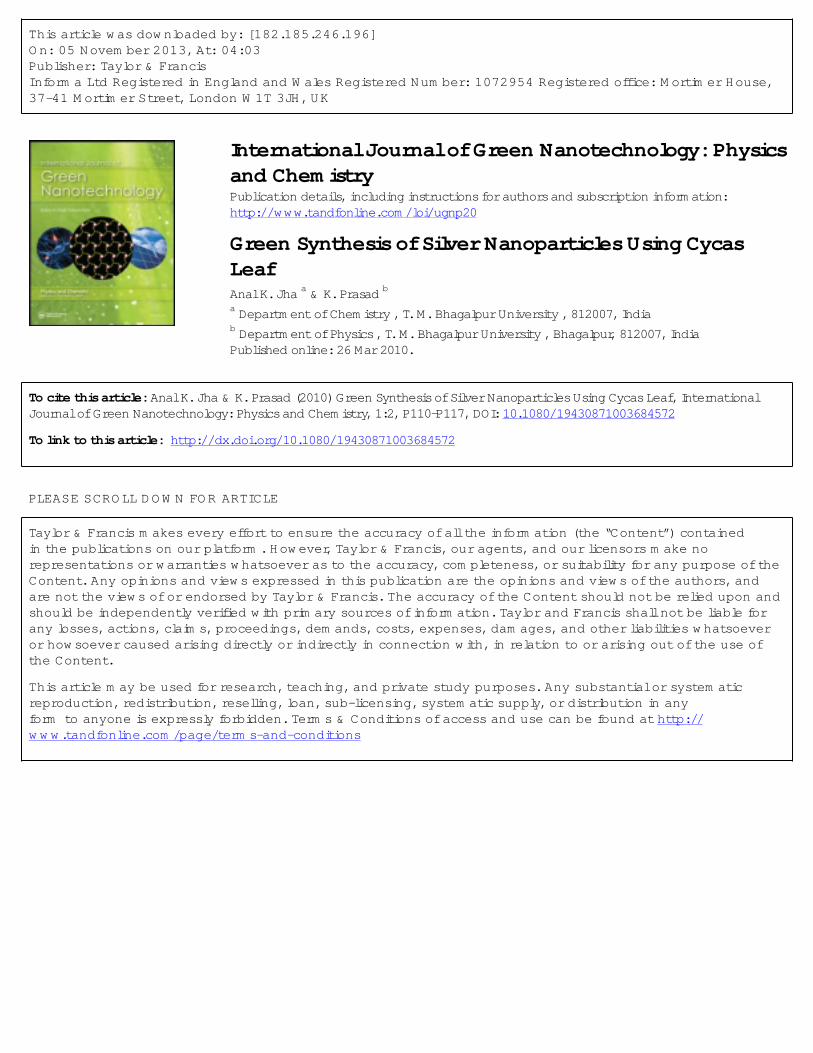
FIGURE 1. (a) TEM photograph, (b) SAED pattern, and (c) particle size distribution of Ag NPs.Inset: Photograph showing Cycas leaf, deposition of Ag NPs and its bottom view.
was deposited onto holey-carbon–coated coppergrids and dried under infrared lamp. The absorp-tion spectra of the sample were measured bya computer-controlled UV-visible spectropho-tometer (DR 5000; Hach, Germany).
RESULTS
Figure 1a shows the TEM image recordedfrom drop-coated film of Ag NPs synthesized bytreating AgNO3 solution with Cycas leaf brothfor 4 h. The micrograph clearly shows individ-ual nanoparticles, almost spherical in shape withdiameters in the range of 2–6 nm. The measure-ment of size was performed along the largestdiameter of the particles. Figure 1b shows theselected area electron diffraction (SAED) pat-tern obtained from Ag NPs. The Scherrer rings,
characteristic of fcc silver, are clearly seen, con-firming nanocrystalline nature of Ag NPs as ob-served in the TEM image (Figure 1a). The parti-cle size histogram of Ag NPs (Figure 1c) showsthe distribution of particle sizes, which followsa lognormal type of distribution:
D = Do + A/(√
2π.w.x)
· exp(− ln (x/xc)
2 / (/2.w))2
(2)
where A, w, and xc are respectively the area,full width at half height, and center of the curve.The values of the parameters A, Do, w, and xc
are respectively estimated to be 116.37, −1.941,0.40, and 3.29. The sizes of particles are rangedfrom 2 to 6 nm and the average particle sizecomes out to be 3.29 ± 0.22 nm. Also, it is
Dow
nloa
ded
by [
182.
185.
246.
196]
at 0
4:03
05
Nov
embe
r 20
13

A. K. Jha and K. Prasad P113
TABLE 1. The crystal data and refinement factors of Ag NPs obtained from X-ray powderdiffraction data
Parameters Results Description of parameters
Crystal system fcc Rp (profile factor) = 100[�|yi − yic|/�|yi|], where yi is the observed intensity and yic isSpace group Fm3m the calculated intensity at the ith step.
a (A) 4.0913 Rwp (weighted profile factor) = 100[�ωi|yi − yic|2/�ωi(yi)2]1/2, where ωi = 1/σ2i and σ2
iis variance of the observation.
V (A3) 68.4808 Rexp (expected weighted profile factor) = 100[(n − p)/�ωi(yi)2]1/2, where n and p arethe number of profile points and refined parameters, respectively.
Rp 45.3 RB (Bragg factor) = 100[�|I obs − I calc|/�|I obs|], where I obs is the observed integratedintensity and I calc is the calculated integrated intensity.
Rwp 41.5 RF (crystallographic RF factor) = 100[�|Fobs − Fcalc|/�|Fobs|], where F is the structurefactor, F = √
(I/L ), where L is Lorentz polarization factor.Rexp 28.1 χ2 = �ωi(yi − yic)2.RB 0.0278 d(Durbin–Watson statistics) = �{[ωi(yi − yic)-ωi−1(yi−1 − yic−1)]2}/�[ωi(yi − yic)]2.RF 0.0533 QD = expected d.χ2 2.177 S (goodness of fit) = (Rwp/Rexp).d 1.0885QD 1.8901S 1.612
observed that the Ag NPs are scattered over thesurface and no aggregates are noticed under theTEM. The difference in size is possibly due tothe fact that the nanoparticles are being formedat different times.[7]
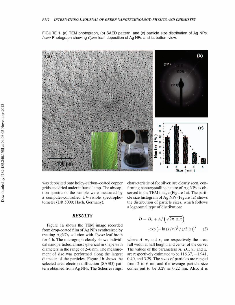
To ascertain the crystal structure of Ag NPs,XRD study was carried out. Rietveld refinementson the XRD data of Ag NPs were done, select-ing the space group Fm3m. Figure 2 depicts theobserved, calculated, and difference XRD pro-files for Ag NPs after final cycle of refinement.It can be seen that the profiles for observed andcalculated ones are perfectly matching, which iswell supported by the value of χ2 (= 2.18). Theprofile fitting procedure adopted was minimizingthe χ2 function.[16] The XRD analyses indicatedthat Ag NPs has a face-centered cubic (fcc) unitcell having the sets of lattice planes (111), (200),(220), (311), and (222). The crystal data and re-finement factors of Ag NPs obtained from XRDdata are summarized in Table 1. The lattice pa-rameter as obtained for Ag NPs is in good agree-ment with the literature report (JCPDS file no.04-0783). Also, it is important to note that the in-tensity ratio I111/I200 comes out to be 9, whichis much higher than the conventional value ‘4’.This indicates that the nanoparticles are abun-dant in (111) plane. Thus, diffraction intensity
of (111) plane should be greatly enhanced incomparison to that of other planes. This resultis in consistence with the earlier reports.[7,12,17]
The broadening of X-ray peaks observed is pri-marily due to the small particle size. Inset ofFigure 2 is the Williamson-Hall plot for Ag NPs.The lattice strain is estimated to be 0.0044 us-ing the linear least-square fitting of ηcosθ− sinθ
data. The low value of lattice strain might be duethe fact that the procedure adopted in the syn-thesis of nanoparticles is natural (biosynthetic)one. Also, the structure of Ag NPs as observedin SAED pattern resembles the XRD result.
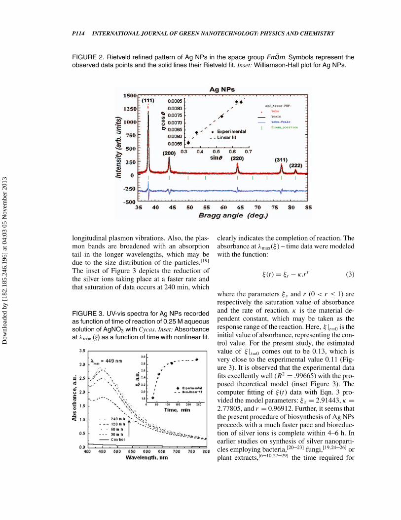
Figure 3 shows the UV-vis spectra recordedon 50% EtOH Cycas extract AgNO3 (0.25 M)solution as a function of time of reaction. Im-mediately after addition of AgNO3 solution tothe extract, the color of extract changes to yel-lowish brown. At this stage, formation of metalnanoparticles due to reduction was followed byUV-vis spectroscopy. The generation of coloris due to excitation of surface plasmons inmetal nanoparticles.[18] The silver surface plas-mon resonance was observed at 449 nm, whichsteadily increased in intensity as a function oftime of reaction (ranging from 30 to 240 min)(inset Figure 3) without showing any shift ofthe wavelength maximum. This simply indicates
Dow
nloa
ded
by [
182.
185.
246.
196]
at 0
4:03
05
Nov
embe
r 20
13
P114 INTERNATIONAL JOURNAL OF GREEN NANOTECHNOLOGY: PHYSICS AND CHEMISTRY
FIGURE 2. Rietveld refined pattern of Ag NPs in the space group Fm3m. Symbols represent theobserved data points and the solid lines their Rietveld fit. Inset: Williamson-Hall plot for Ag NPs.
longitudinal plasmon vibrations. Also, the plas-mon bands are broadened with an absorptiontail in the longer wavelengths, which may bedue to the size distribution of the particles.[19]
The inset of Figure 3 depicts the reduction ofthe silver ions taking place at a faster rate andthat saturation of data occurs at 240 min, which
FIGURE 3. UV-vis spectra for Ag NPs recordedas function of time of reaction of 0.25 M aqueoussolution of AgNO3 with Cycas. Inset: Absorbanceat λmax (ξ) as a function of time with nonlinear fit.
clearly indicates the completion of reaction. Theabsorbance at λmax(ξ ) – time data were modeledwith the function:
ξ (t) = ξs − κ.rt (3)
where the parameters ξ s and r (0 < r ≤ 1) arerespectively the saturation value of absorbanceand the rate of reaction. κ is the material de-pendent constant, which may be taken as theresponse range of the reaction. Here, ξ |t=0 is theinitial value of absorbance, representing the con-trol value. For the present study, the estimatedvalue of ξ |t=0 comes out to be 0.13, which isvery close to the experimental value 0.11 (Fig-ure 3). It is observed that the experimental datafits excellently well (R2 = .99665) with the pro-posed theoretical model (inset Figure 3). Thecomputer fitting of ξ (t) data with Eqn. 3 pro-vided the model parameters: ξ s = 2.91443, κ =2.77805, and r = 0.96912. Further, it seems thatthe present procedure of biosynthesis of Ag NPsproceeds with a much faster pace and bioreduc-tion of silver ions is complete within 4–6 h. Inearlier studies on synthesis of silver nanoparti-cles employing bacteria,[20–23] fungi,[19,24–26] orplant extracts,[6–10,27–29] the time required for
Dow
nloa
ded
by [
182.
185.
246.
196]
at 0
4:03
05
Nov
embe
r 20
13

A. K. Jha and K. Prasad P115
FIGURE 4. Mechanism for the biosynthesis of Ag NPs using Cycas leaf broth.
completion (i.e., complete reduction of Ag ions)ranged from 12 to 120, and are rather slow.
DISCUSSION
Salt/metal ion, as well as drought, chill-ing and extreme temperatures, increase the lev-els of reactive oxygen species (ROS). Exces-sive levels of ROS lead to oxidative damageto cellular molecules, aging, and cell death.The antioxidative system is important for themaintenance of intracellular ROS at appropriatelevels.[30] Mechanisms of metal detoxificationby biomolecules proceeds as cascade of events,such as induction of proteins such as metal-lothionein, heat-shock protein, phytochelatins,and ferritin, transferring; or by triggering an-tioxidant enzymes such as superoxide dismu-tase, catalase, glutathione, and peroxidase; orthrough high turnover of organic acids such asmalate, citrate, oxalate, succinate, aconitate, α-ketoglutarate, etc. Various types of heavy metal
adaptation strategies in plants have exhaustivelybeen studied.[31] Primarily two types of mech-anisms may explain the resistance to the tox-icity of metal ions in plants. The prominentmetal complexation processes are the synthesisof phytochelatins and of other metal-chelatingpeptides.[31,32] Phytochelatins were found to beinduced by Ag, Bi, Cd, Cu, Hg, Ni, Sn, W, andZn, whereas no induction was noticed in thecase of Na, Mg, Al, Ca, V, Cr, Sb, Te, Mn, Fe,Co, and Cs. Glutathione functions as a precursorof phytochelatin synthesis. Metal-induced phy-tochelatin production decreases cellular levelsof glutathione. Glutathione and its homologues,viz. homoglutathione and hydroxymethylglu-tathione, are the abundant low-molecular-weightthiols in plants. Glutathione was implicated toplay a major role in plants exposed to metalstress.[31]
Metallothionein (MT) is a family of cysteine-rich, low-molecular-weight (MW ranging from3500 to 14000 Da) proteins. MTs have the ca-pacity to bind both physiological (such as zinc,
Dow
nloa
ded
by [
182.
185.
246.
196]
at 0
4:03
05
Nov
embe
r 20
13

P116 INTERNATIONAL JOURNAL OF GREEN NANOTECHNOLOGY: PHYSICS AND CHEMISTRY
copper, selenium) and xenobiotic (such as cad-mium, mercury, silver, arsenic) heavy metalsthrough the thiol group of its cysteine residues,which represents nearly the 30% of its aminoacidic residues. The diversity of the plant MTgene family suggests that these may differ notonly in sequence but also in function. Thereis little information about MT genes in non-flowering plant species. However, genes encod-ing type 3 MTs have been cloned from severalgymnosperms.[33]
Antioxidant action of phenolic compounds isdue to their high tendency to chelate metals. Phe-nolics possess hydroxyl and carboxyl groups,able to bind particularly iron and copper. Theymay inactivate iron ions by chelating and addi-tionally suppressing the superoxide-driven Fen-ton reaction, which is believed to be the most im-portant source of ROS.[34,35] Tannin-rich plantssuch as tea, which are tolerant to Mn excess,are protected by the direct chelation of Mn.Plants contain two major types of peroxidases,which can be divided into two groups: perox-idases that use ascorbic acid as the preferen-tial electron donor, and those that use phenolics.Phenolics, especially flavonoids and phenylo-propanoids, are oxidized by peroxidase, and actin H2O2-scavenging system. Their antioxidantaction resides mainly in their chemical structure.There is some evidence of induction of phenolicmetabolism in plants as a response to multiplestresses (including heavy metal stress).[34]
Reduction is accomplished due to phy-tochemicals (flavonoids or other any otherpolyphenols) or phyotchelatins/glutathiones/metallothioneins present in Cycas leaves paren-chyma. Reduction of particle size by theleaf broth in a tick compared to bacteriaor fungi may be perceived as an apprecia-ble advancement. It is well established thatthe biflavanone, tetrahydrohinokiflavone, to-gether with amentoflavone, has been found inthe leaves of Cycas beddomei.[36] In Cycasleaf broth having well-defined metabolite trea-sure, the process of nano-transformation mighthave resulted due to redox activities of ascor-bic/dehydroascorbic acid and amenti/hinokiflavones and involvement of ascorbates/glutathi-ones/metallothioneins (Figure 4), leading to thereduction of silver ions present in the solution.
Therefore, compared to bacteria or fungi, plantcells are much suitable candidates for the syn-thesis of metallic nanoparticles. The significantreduction in reaction time with Cycas leaf isan important result and will enable nanoparti-cle biosynthesis methods to compete with otherplant-assisted biosynthesis routes for the forma-tion of silver nanoparticles that are currentlymuch more rapid and reproducible.
CONCLUSION
In conclusion, the present biotechnologicalmethod is capable of producing Ag nanoparti-cles. Also, it is a green, high-yield, fast, and low-cost approach. Reduction is accomplished prob-ably due to phytochemicals such as polyphenols,glutathiones, metallothioneins, and ascorbates,which may help in the production of Ag NPs asa result of the detoxification procedure.
REFERENCES
1. Chopra, R. N.; Nayar, S. L.; Chopra, I. C. Glossaryof Indian Medicinal Plants; CSIR Publication: New Delhi,1956, p. 86.
2. Harborne J. B.; Mabry, T. J.; Mabry, H., Eds. TheFlavonoids: Biflavonoids; Chapman and Hall: London,1975, p. 693.
3. Harborne, J. B. Phytochemical Methods; Chapmanand Hall: London, 1973. p. 52.
4. Mabry, T. J.; Markham, K. R.; Thomas, M. B. TheSystematic Identification of Flavonoids; Springer-Verlag:New York, 1970, p. 215.
5. Goodwin, T. W., Ed. Chemistry and Biochemistry ofPlant Pigments: Functions of Flavonoids in Plants; Aca-demic Press: New York, 1976, p. 736.
6. Jae, Y. S.; Beom, S. K. Bioprocess Biosyst. Eng.2009, 32, 79–84.
7. Jha, A. K.; Prasad, K.; Kumar, V.; Prasad, K.Biotechnol. Prog. 2009, 25, 1476.
8. Kumar, V.; Yadav, S. K. J. Chem. Technol. Biotech-nol. 2008, 84, 151.
9. Huang, J.; Li, Q.; Sun, D.; Lu, Y.; Su, Y.; Yang, X.;Wang, H.; Wang, Y.; Shao, W.; He, N.; Hong, J.; Chen, C.Nanotechnology 2008, 18, 105104–105115.
10. Li, S.; Shen, Y.; Xie, A.; Yu, X.; Qiu, L.; Li, Z.;Zhang, Q. Green Chem. 2007, 9, 852.
11. Arangasamy, L.; Munusamy, V. Afri. J. Biotechnol.2007, 7, 3162.
Dow
nloa
ded
by [
182.
185.
246.
196]
at 0
4:03
05
Nov
embe
r 20
13

A. K. Jha and K. Prasad P117
12. Jha, A. K.; Prasad, K.; Prasad, K.; Kulkarni, A. R.Colloids Surf. B Biointerfaces 2009, 73, 219.
13. Roisnel, J.; Rodrıguez-Carvajal, J. WinPLOTR.Laboratoire Leon Brillouin (CEA-CNRS) Centre d’Etudesde Saclay: Gif sur Yvette Cedex, France, 2000.
14. Rodriguez-Carvajal, J. FullProf 2000: A RietveldRefinement and Pattern Matching Analysis Program, Ver-sion: April 2008. Laboratoire Leon Brillouin (CEA-CNRS): France, 2008.
15. Williamson, G. K.; Hall, W. H. Acta Metall 1953, 1,22.
16. McCusker, L. B.; Von Dreele, R.B.; Cox, D.E.;Louer, D.; Scardi, P. J. Appl. Cryst. 1999, 32, 36.
17. Tian, X.; Wang, W.; Cao, G. Mater. Lett. 2007, 61,130.
18. Mulvaney, P. Langmuir, 1996, 12, 788.19. Vigneshwaran, N.; Ashtaputre, N. M.; Varadarajan,
P. V.; Nachane, R. P.; Paralikar, K. M.; Balasubramanya, R.H. Mater. Lett. 2007, 61, 1413.
20. Klaus, T.; Joerger, R.; Olsson, E.; Granqvist, C. G.Proc. Natl. Acad. Sci. U.S.A. 1999, 96, 13611.
21. Klaus, T.; Joerger, R.; Olsson, E.; Granqvist, C. G.Trends Biotechnol. 2001, 19, 15.
22. Joerger, R.; Klaus, T.; Granqvist, C. G. Adv. Mater.2001, 12, 407.
23. B. Nair, T. Pradeep. Cryst. Growth Des. 2002, 2,293.
24. Mukherjee, P.; Ahmad, A.; Mandal, D.; Senapati, S.;Sankar, S. R.; Khan, M. I.; Parischa, R.; Ajaykumar,
P. V.; Alam, M.; Kumar, R.; Sastry, M. Nano Lett. 2001, 1,515.
25. Ahmad, A.; Mukherjee, P.; Senapati, S.; Mandal,D.; Khan, M. I.; Kumar, R.; Sastry, M. Colloids Surf. BBiointerfaces 2003, 28, 313.
26. Sadowski, Z.; Maliszewska, I. H.; Grochowalska,B.; Polowczyk, I.; Kolecki, T. Mater. Sci. Poland 2008, 26,419.
27. Chandran, S. P.; Choudhary, M.; Pasricha, R.;Ahmad, A.; Sastry, M. Biotechnol. Prog. 2006, 22, 577.
28. Shankar, S. S.; Rai, A.; Ahmad, A.; Sastry, M. J.Colloid. Interface Sci. 2004, 275, 496.
29. Shankar, S. S.; Ahmad, A.; Sastry, M. Biotechnol.Prog. 2003, 19, 1627.
30. Amako, K.; Ushimaru, T. Nutr. Nat. Resour. 2009,4, 13.
31. Prasad, M. N. V., Ed. Plant Ecophysiology: TraceMetals; John Wiley and Sons: New York, 1977, p. 207.
32. Basra, A. S.; Basra, R. K., Eds. Mechanisms of En-vironmental Stress Resistance in Plants: Mechanisms ofPlant Resistance to Aluminum and Heavy Metals; HarwoodAcademic Publishers: Amsterdam, 1997, 241.
33. Chatthai, M.; Kaukinen, K. H.; Tranbarger, T. J.;Gupta, P. K.; Misra, S. Plant Mol. Biol. 1997, 34, 243.
34. Michalak,A. Polish J. Environ. Stud. 2006, 15, 523.35. Evans, C. A.; Miller, N. J.; Paganga, G. Trends Plant.
Sci. 1997, 2, 152.36. Rani, M. S.; Rao, C. V.; Gunasekar, D.; Blond, A.;
Bodo, B. Phytochemistry 1998, 47, 319.
Dow
nloa
ded
by [
182.
185.
246.
196]
at 0
4:03
05
Nov
embe
r 20
13