alaska department of fish and game juneau, … · viral diarrhea (bvd) virus, and parainfluenza iii...
TRANSCRIPT

ALASKA DEPARTMENT OF FISH AND GAME
JUNEAU, ALASKA
STATE OF ALASKA Bill Sheffield, Governor
DEPARTMENT OF FISH AND GAME Don w. Collinsworth, Acting Commissioner
DIVISION OF GAME Robert A. Hinman, Acting Director
Steven R. Peterson, Research Chief
SEROLOGIC SURVEYS FOR MICROBIAL PATHOGENS
By
Randal l L . Zarnke
Volume II I
Progress Report Federal Aid in Wildlife Restorat i on
Projects W-21-2 and W-22-1, Job 18.SR
Persons are free to use materia l in these reports for educational or informational purposes. However, since mos t reports treat only part o f continuing studies, persons intending to use this material i n scienti fic publications should obtain prior permission from the Department of Fish and Game. In all cases, tentative conclusions should be identif ied as such in quotation, --~ ~ .. ..._ credit would be appreciated.
SF (Printed January 1983) 997 I37 1981-82

SF 997 '7.,3']
11&-92.
PROGRESS REPORT (RESEARCH)
State: Alaska
Cooperator: Randall L. Zarnke
Project No. : W-21-2 Project Title: Big Game Investigations W-22-1
Job No.: 18.5R Job Title: Serologic Surveys for Microbial Pathogens
Period Covered: April 21, 1981 through April 20, 1982
SUMMARY
I. Sera from Dall sheep (Ovis dalli), caribou (Rangifer tarandus) , moose (Alces alces) , bison (Bison bison) , grizzly bear (Ursus arctos) , and black bear (Ursus americanus) collected in Alaska were tested for evidence of past infection by Brucella sp. and 12 serovarieties of Leptospira. Dall sheep, caribou, moose, and bison sera were also tested for evidence of contagious ecthyma (CE) virus, epizootic hemorrhagic disease (EHD) virus, bluetongue (BLU) virus, infectious bovine rhinotracheitis (IBR) virus, bovine viral diarrhea (BVD) virus, and parainfluenza III (PI3) virus. Dall sheep sera were also tested for Q fever antibody content.
Brucella spp. antibody was detected in 7 of 67 (10%) caribou, 1 of 39 (3%) moose, and 6 of 74 (8%) grizzly bear sera. Leptospira spp. antibody was found in 1 of 61 ( 2%)
L.: caribou, 1 of 37 (3%) moose, 6 of 74 (8%) grizzly bear, and 0 1 of 28 (4%) black bear sera. CE antibody was detected in 7 ..... of 17 (41%) Dall sheep and 5 of 53 (10%) caribou sera. EHD LO LO antibody was found in 8 of 17 (47%) Dall sheep and 2 of 39 CD ("") (6%) moose sera. IBR antibody was detected in 6 of 67 (9%) 0 caribou sera. BVD antibody was found in 2 of 67 (3%)0 caribou sera. PI3 antibody was detected in 14 of 21 (67%)0 LO bison sera. · Q fever antibody was found in 12 of 15 (80%)LO I'-- Dall sheep sera. Brucel"i a su:i,s IV, Leptospira interrogans ("") serovar ballum, and CE infections have been previously("") confirmed in Alaskan wildlife via isolation of the etiologic
agents.
II. Blood samples were collected from humans and several species of free-ranging wild mammals in Alaska. Sera were tested for antibody to Jamestown Canyon (JC) , snowshore hare (SSH), Northway (NOR) , Klamath (KLA) , Sakhalin (SAK) , Great Island (GI), and Silverwater (SIL) virus. JC antibody was found in 54% of 121 human, 89% of 97 bison, 51% of 84 Dall~2S 43% of 68 snowshoe hare (Lepus americanus), and 3% o~rctic fox (Alopex lagopus) sera. SSH antibodwa•a~es~&tt'tl
Library & Informatton Services Anch·Jtab'3 , A..:aska
i

in 42% of 121 human, 89% of 97 bison, 41% of 84 Dall sheep, and 65% of 68 snowshoe hare sera. NOR antibody was found in 14% of 121 human, 89% of 97 bison, 41% of 84 Dall sheep, and 65% of 68 snowshoe hare sera. NOR antibody was found in 14% of 121 human, 94% of 97 bison, 84% of 84 Dall sheep, 43% of 69 caribou, 3% of 68 snowshoe hare, 45% of 64 grizzly bear, 30% of 33 arctic fox, and 78% of 21 moose sera. KLA antibody was found in 5% of 121 human and 40% of 97 bison sera. SAK antibody was found in 2% of 97 bison and 3% of 33 arctic fox sera. GI antibody was found in 1% of 97 bison sera. No SIL antibody was found in any sera tested.
Thus, the natural host ranges of JC, SSH, NOR, and KLA viruses have been extended by inference from the occurrence of antibody. However, the results of this study have not revealed any direct threat to the health and welfare of Alaskan wildlife.
Key words: microbial, pathogens, serologic surveys.
ARLIS Alaska Resources
Library & Information Servtces ii Anch xag~ . A;.<iska

CONTENTS
I. Serologic Survey for Selected Microbial Pathogens in Alaskan Wildlife
Summary. . . . . . . . . . . . . . . . . . . . . . i Background . . . . . . . . . . . . . . . . . . • . 1 Procedures . . . . . . . . . . . . . . 2 Results. . . . . . . . . . . . . . . . 2 Discussion . . . . . . . . • . . . . . . . . . . . 4 Acknowledgments. . . . . . . . . . . . • . . . . · 7 Literature Cited . . . . . . . . . . . . • . . . . 7
II. Serologic Evidence of Arbovirus Infections in Humans and Wild Mammals of Alaska
Summary. . . . . . . . . . . . . . . . . . . . i Background . . . . . . . . . . . . . . . . . . . . . . . . 1 o Procedures . . . . . . . . . . . . . . . . . . • . . . 10
Serum Collections . . . . . . . . . . . . . • 10 Serologic Tests . . •............. •12 Viruses . . . . . . . . . . . . . . . . . . . . . 12
Re SU 1ts . . . . . . . . . . . . . . ... . . • • • • • 12 Discussion . . . . . . . . . . . . . . • . 12 Acknowledgments. . . . . . . . . . . . . . . . 17 Literature Cited. . . . . . . . . . . .... · ·17
I. Serologi! Survey for Selected Microbial Pathogens in Alaskan Wildlife .
BACKGROUND
There have been several documented instances in which infectious diseases have had an observed impact on wildlife populations in Alaska. Brucellosis in caribou and rabies in canids have been notable examples. One possible explanation for this apparent low number of severe disease problems in Alaskan wildlife may be the relative scarcity of trained observers. Another possible explanation is that many of the wildlife populations in the State have had little or no direct contact with domestic livestock. This isolation from domestic animals may have prevented spread of domestic animal pathogens to wildlife. The agricultural industry in Alaska is small but appears to be on the verge of major expansion. In an effort to determine which diseases are present in wildlife populations prior to this expected agricultural expansion, a serologic survey has been initiated. All of the etiologic agents which are included in this survey have been
1 Coathors with Zarnke are Charles H. Calisher and JoAnne Kerschner of the Division of Vector-Borne Viral Diseases, Centers for Disease Control, P.O. Box 2087, Fort Collins, CO. 80522.
l

detected in various North American wildlife species by means of isolation of the agent or by serologic tests (Neiland et al. 1968, Murray and Trainer 1970, Reilly et al. 1970, Rieman et al. 1979, Dieterich 198lb, Lance et al. 1981). Continuation of this survey may reveal changes in the epizootiology of seiected diseases in Alaska.
PROCEDURES
All of the black bear and most of the grjzzly bear blood samples were collected in southcentral Alaska. The remaining samples came from various areas in the northern half of the State. Bison samples were collected from animals killed by hunters during 1979-1980 near Delta ,Junction. All other blood samples were collected from 1978-1981 by Alaska Department of Fish and Game biologists during population studies which entailed capture of free-ranging animals. Blood samples were allowed to settle for 18-36 hours at ambient or refrigerated temperatures. Sera were collected by aspiration and frozen. All serologic tests were performed at the National Veterinary Services Laboratory (USDA, Ames, Iowa) .
Table 1 identifies the serologic test utilized for each etiologic agent. The following ~· interrogans serovarieties were included in the tests: pomona, ballum, canicola, icterohaemorrhagiae, wolffi, grippotyphosa, hardjo, autumnalis, bataviae, tarassovi, australis, and pyrogenes. For the card test and the immunodiffusion test, specimens are considered simply either positive or negative. For all other tests, minimum titers were established (Table 1) based upon natural or experimental infection of the host species in question or a selected domestic animal species. Sera which met or exceeded these titers (plus those designated "positive" in the card or immunodiffusion tests) were considered to contain evidence of past infection by the agent in question.
RESULTS
Results of the serologic tests are presented in Table 1. The single moose with leptospiral antibody had a titer of 3200 for serovar hardjo. One of the grizzly.bears had a titer of 400 for serovar canicola. The remaining 5 grizzly bears, the single black bear, and the lone caribou were positive for serovar grippot~phosa with titers ranging from 100 to 800. No statistically significant difference in antibody prevalence based upon age or gender of animals was found in any phase of this study except for leptospirosis in grizzly bears.
2
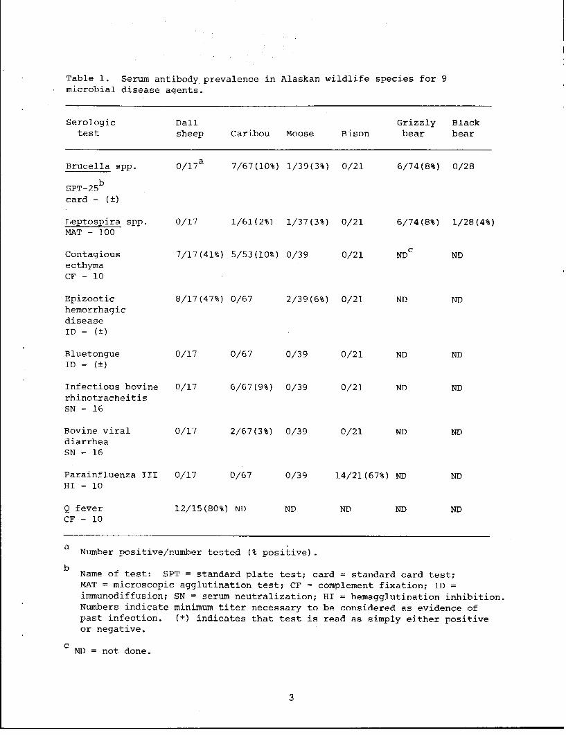
Table 1. Serum antibody prevalence in Alaskan wildlife species for 9 microbial disease agents.
Serologic Dall Grizzly Black test sheep Caribou Moose Bison bear bear
Brucella spp. 0/17a 7/67(10%} 1/39(3%} 0/21 6/74 (8%} 0/28
SPT-25b card - ( ±)
Leptos;eira spp. 0/17 1/61(2%) 1/37(3%} 0/21 6/74(8%} 1/28(4%) MAT - 100
Contagious 7/17 (41%) 5/53(10%} 0/39 0/21 NDc ND ecthyma CF - 10
Epizootic 8/17(47%} 0/67 2/39(6%) 0/21 ND ND hemorrhagic disease ID - (±)
Bluetongue 0/17 0/67 0/39 0/21 ND ND ID - (±)
Infectious bovine 0/17 6/67 (9%) 0/39 0/21 ND ND rhinotracheitis SN - 16
Bovine viral 0/17 2/67 (3%) 0/39 0/21 ND ND diarrhea SN - 16
Parainfluenza III 0/17 0/67 0/39 14/21(67%) ND ND HI - 10
Q fever 12/15(80%) ND ND ND ND ND CF - 10
a Number positive/number tested (% positive).
b Name of test: SPT = standard plate test; card = standard card test; MAT = microscopic agglutination test; CF = complement fixation; ID = immunodiffusion; SN = serum neutralization; HI = hemagglutination inhibition. Numbers indicate minimum titer necessary to be considered as evidence of past infection. (±) indicates that test is read as simply either positive or negative.
c ND = not done.
3

DISCUSSION
As with any serologic survey, these results should be considered as merely suggestive of the diseases which may be present in Alaskan wildlife. Isolation of the etiologic agents is the only conclusive evidence.
Evidence of CE infection in free-ranging Dall sheep has been reported twice previously (Dieterich et al. 198lb, Smith et al. 1982). The samples in the current survey were collected from a population which had serologic evidence of CE as early as 1971 (R. Zarnke, unpubl. data). The disease is believed to be enzootic in this population. Of the 8 samples with serologic evidence of past infection, 4 had titers of 10; 1 had a titer of 20; 1 had a titer of 40; and 2 had titers of 80. In domestic animals, CE infection is most severe in young animals. Recent evidence indicates that this is true for Dall sheep as well (Dieterich et al. 198lb, R. Zarnke, unpubl. data). The impact of this disease on free-ringing Dall sheep is unknown, but it may be capable of limiting population growth by causing mortality in the juvenile cohort.
EHD virus is closely related (antigenically and morphologically) to BLU virus (Hoff and Trainer 1978) . The former is primarily a disease of wild animals, whereas the latter is more common in domestic sheep and cattle (Hoff and Trainer 1978). BLU infection in bighorn sheep (Ovis canadensis) has been reported to produce a pneumonia-like syndrome (Marburger et al. 1970). This report is the 1st evidence of EHD infection in Dall sheep. There have been no reports in Dall sheep of massive die-offs or the dramatic symptoms which can accompany arthropod transmission of this disease during epizootics. During enzootic periods, overt symptoms may be absent and oral transmission may play an important role (Hoff and Trainer 1978). The significance of the current findings for the Dall sheep population is unknown. Presence of EHD in wild animal populations does provide the possibility for epizootic spread of the disease.
Q fever is a disease of domestic sheep, goats, and humans caused by the rickettsia Coxiella burnetti (Randhawa et al. 1977). In sheep, it can cause an influenza-like syndrome and rarely abortions (Enright et al. 1963). Seven sera had titers of 10, 1 was 20, and 4 were greater than 20. The high serum antibody prevalence reported here suggests that the disease may be widespread in the Dall sheep population of the central Alaska Range. Serologic tests of other populations are planned. Attempts will be made to isolate the causative agent of this disease. In the absence of obvious reproductive problems in the wild and the lack of experimental studies of the disease in captive sheep, it is difficult to assess the significance of this disease on Dall sheep populations.
4

Brucellosis is a common disease in caribou (Neiland et al. 1968). Serum antibody prevalence has ranged from 0-25% in various herds around Alaska (Neiland et al. 1968). The prevalence reported here falls between these 2 extremes and indicates continued transmission of the disease in Alaskan caribou. As measured by the standard plate test, Brucella spp. titers were equally distributed from 25 to 200 for the 7 "positive" caribou sera. All 7 of these sera gave positive card test results. There have been no massive die-offs directly attributable to this disease, but it almost certainly has a detrimentaJ. impact on natality and future productivity. A vaccine is being developed for use in reindeer on the Seward Peninsula (Dieterich et al. 198 la) , but logistical problems would probably preclude its use on a routine basis in free-ranging caribou.
Leptospirosis can cause chronic kidney infections and/or abortion. There have been numerous surveys of leptospirosis in wildlife (Reilly et al. 1970). Evidence of leptospirosis has been reported for northern fur seals (Callorhinus ursinus) collected in the Bering Sea of Alaska's west coast (Smith et al. 1977). A member of the ballum serogroup was isolated from the kidney of a tundra vole (Microtus oeconomus) captured on the Alaska Peninsula (157°W longitude, 57°30'N latitude) (Woods 1974). White-tailed deer (Odocoileus virginianus) are common hosts (Abdulla et al. 1962). This study is the 1st report of serological evidence of infection in caribou. In the absence of clinical studies of pathogenesis in caribou, the significance of the single positive sample is difficult to evaluate.
Of the 5 caribou samples with serologic evidence of past CE infection, 1 had a titer of 10; 2 had titers of 20; 1 had a titer of 40; and 1 had a titer of 80. Nat.urally occurring evidence of the CE in Alaskan wild animals has previously been limited to musk-ox (Ovibos moschatus), Dall sheep, and mountain goat (Oreamnos americanus) (Dieterich et al. 198lb). The disease is most common and severe in young animals. A-single caribou was shown to be susceptible to experimental CE infection, and its lesions were mild (R. Zarnke, unpubl. data) . On the basis of this limited experimental evidence, it appears as if CE does not pose a significant hazard to heal thy caribou. Animals under nutritional, reproductive, predatory, or other types of stress may be less capable of withstanding infection. Experimental studies of CE in caribou should be expanded to include a larger sample size. Serologic surveys will continue in order to assess the statewide distribution of this disease in caribou.
IBR, BVD, and PI3 constitute what is commonly known as the bovine respiratory virus group. Of the 6 caribou sera with evidence of previous exposure to IBR, 5 had titers of 16 and 1 had a titer of 32. Of the 2 specimens with antibody to BVD, 1 had a titer of 16 and the other was 32. These low titers may represent cross-reactive antibody or nonspecific neutralizing substances. Serologic evidence of IBR and BVD has been reported for a small
5

sample of caribou from Canada (Elazhary et al. 1981), and antibody to all 3 members of the group has been detected in reindeer from Alaska's Seward Peninsula (Dieterich 1981). Antibody to all 3 viruses has also been detected in moose sera (Thorsen and Henderson 1971, Thorsen et al. 1977). These viruses cause a multitude of respiratory and gastrointestinal ailments in domestic cattle. Infection can be seriously debilitating for short periods of time but js rarely chronic br fatal. Thus, in otherwise healthy, free-ranging animals, these infections could result in decreased weight gains and/or increased susceptibility to predation. When coupled with heavy parasite loads or other heal th-related complications, the outcome of respiratory virus infections may be more severe.
Brucellosis in moose is a rare occurrence (Jellison et al. 1953). Two common possible explanations for this situation are that moose are: (a) rarely exposed to the disease, and/or (b) so exquisitely susceptible to the disease that all individuals which are exposed inevitably die as a result of the infection and are rarely found by humans. The single animal in the current survey which was considered serologically positive had a very low serum antibody titer as measured by the plate test. It was negative on the card test. This may have represented cross-reactive antibody to some related agent or nonspecific agglutination. All indications are that brucellosis poses little threat to the moose population as a whole.
As in caribou, the significance of leptospirosis to the health of the moose population is largely unknown. Cattle can suffer abortions and/or chronic kidney dysfunction as a result of infection. Experimental studies would be needed to clarify the pathogenesis of this disease in moose. The low prevalence of infection in the current study docs not warrant such an experimental approach.
Serologic evidence of past bluetongue virus infection in moose has been reported (Hoff and Trainer 1978). There have been no similar reports for EHD. The effect of natural EHD infection on moose is unknown, but experimental infections have not caused mortality (Hoff and Trainer 1978). Presence of the agent in moose populations does provide for the possibility of epizootic spread of the disease.
Parainfluenza III is a member of the so-called bovine respiratory virus complex. As the name implies, these agents (and PI3, in particular) commonly cause respiratory tract infections. These agents have been detected in a variety of North American wildlife species (Kahrs et al. 1963, Thorsen and Henderson 1971, Thorsen et al. 1977) by means of serologic tests and/or isolation of the agent(s). There have been no previous reports of evidence of PI3 infection in bison. Results of a recent study indicate that in the absence of other infectious agents, PI3 causes little pathogenesis in domestic sheep (Davies et al. 1981). On the
6

other hand, in combination with Pasteurella hemolytica, PI3 was capable of producing a severe pneumonia which can be fatal (Davies et al. 1981). Whether a similar phenomenon pertains to bison is open to question. There are no data on the occurrence of this agent in domestic species in the vicinity where the bison are found. Without this information, no conclusions can be drawn regarding the source of infection for bison. The serum antibody titers in the current study were quite low and may represent cross-reactive antibody to some related agent or nonspecific inhibiting substances. However, the high prevalence (67%) warrants further surveillance. Tests of additional samples are planned for the near future.
Serological evidence of Brucella spp. infection in Alaskan grizzly bears has been reported previously (Neiland and Miller 1981) . Evidence of infection in black bears has been reported elsewhere in North America (Zarnke and Yuill 1981). Presumably, Alaskan bears are exposed to the disease when preying and/or scavenging on caribou infected with B. suis IV. The prevalence in this group of bears f ram southcentr~Alaska is lower than that for bears captured on the North Slope of Alaska's Brooks Range. Perhaps the opportunity for exposure in southern Alaska is not as great as in the northern portions of the State. As measured by the standard plate test, Brucella spp. titers ranged from 50 to 200 for the 6 "positive" grizzly bears. All 6 of these sera gave positive card test results. Signs and symptoms of the disease are probably similar to those which occur in other species, i.e., abortion and sterility. In this way, brucellosis could significantly affect the growth of a population.
Leptospirosis in bears is less well understood. The kidney and reproductive tract infections referred to above for moose and caribou could also pertain to bears. Prevalence rates in the current study agree with rates reported previously (Binninger et al. 1980). In the current study, the average age of grizzly bears with serologic evidence of previous exposure to Le~tospira spp. was 8.9 years. The average age for the entire 74 animals in the sample was 5.9 years. This suggests that the possibility of previous exposure is directly related to age.
ACKNOWLEDGMENTS
The author thanks W. E. Heimer, R~ D. Cameron, K. R. Whitten, J. L. Davis, P. Valkenburg, w. c. Gasaway, S. D. DuBois, D. M. Johnson, c. J. Champaine, ands. D. Miller for providing sera for this survey.
LITERATURE CITED
Abdulla, P. K., L. H. Karstad, and N. A. Fish. 1962. Cultural and serological evidence of leptospirosis in deer in Ontario. Can. Vet. J. 3:71-78.

Binninger, c. E., J. J. Beecham, L.A. Thomas, and L. D. Winward. 1980. A serologic survey for selected infectious diseases of black bears in Idaho. J. Wild!. Dis. 16:423-430.
Davies, D. H., M. Herceg, B. A. H. Jones, and D. C. Thurley. 1981. The pathogenesis of sequential infection with parainfluenza virus type 3 and Pasteurella haemolytica in sheep. Vet. Microbiol. 5:173-182.
Dieterich, R. A. 1981. Respiratory viruses. Pages 28-30 in Alaskan Wildlife Diseases. R. A. Dieterich, ed. Univ. Alaska Press.
, B. L. Deyoe, and J. K. Morton. 198la. Effects of ---k-i-·-1~1-e-d Brucella abortus strain 45/20 vaccine on reindeer
later challenge-exposed with Brucella suis type 4. Am. J. Vet. Res. 42:131-134. ~~
, G. R. Spencer, D. Burger, A. M. Gallina, and J. ---V~a-n-d--=-e-rschalie. 198 lb. Contagious ecthyma in Alaskan
muskoxen and Dall sheep. J. Am. Vet. Med. Assoc. 179:1140-1143.
Elazhary, M.A. s. Y., J. L. Frechette, A. Silim, and R. s. Roy. 1981. Serological evidence of some bovine viruses in the caribou (Rangifer tarandus caribou) in Quebec. .._r. Wildl. Dis. 17:609-612.
Enright, J. B., D. E. Behymer, c. E. Franti, V. J. Dutson, W. M. Longhurst, M. E. Wright, and J. E. Goggin. 1963. The behavior of Q fever rickettsiae isolated from wild animals in northern California. J. Wild!. Dis. 112:181-186.
Hoff, G. L., and D. O. Trainer. 1978. Bluetongue and epizootic hemorrhagic disease viruses: their relationship to wildlife species. Adv. Vet. Sci. Comp. Med. 22:111-132.
Jellison, W. L., C. W. Fishel, and E. L. Cheatum. 1953. Brucellosis in a moose, Alces americana. J. Wild!. Manage. 17:217-218.
Kahrs, R., G. Atkinson, J. A. Baker, L. Carmichael, L. Coggins, J. Gillespie, P. Langer, V.. Marshall, D. Robson, and B. Sheffy. 1963. Serological studies on the incidence of bovine virus diarrhea, infectious bovine rhinotracheitis, bovine myxovirus parainfluenza-3, and Leptospira Eomona in New York state. Cornell Vet. 54:360-369.
Lance, W., W. Adrian, and B. Widhalm. J.981. An epizootic of contagious ecthyma in Rocky Mountain bighorn sheep in Colorado. J. Wildl. Dis. 17:601-604.
8

Marburger, R. G., R. M. Robinson, J. w. Thomas, and K. A. Clark. 1970. Management implications of disease of big game animals in Texas. Proc. Annu. Conf. Southeast Assoc. Fish. Game Comm. 24:46-50.
Murray, J. O., and D. o. Trainer. 1970. Bluetongue virus in North American elk. J. Wildl. Dis. 6:144-148.
Neiland, K. A., and L. G. Miller. 1981. Experimental Brucella suis type 4 infections in domestic and wild Alaskan carnivores. J. Wildl. Dis. 17:183-189.
J. A. King, B. E. Huntley, and R. O. Skoog. 1968.-----...,,...,.. , The diseases and parasites of Alaskan wildlife populations, part I. Some observations on brucellosis in caribou. Bull. Wildl. Dis. Assoc. 4:27-36.
Randhawa, A. s., V. P. Kelly, and E. F. Baker. 1977. Agglutinins to Coxiella burnetti and Brucella spp., with particular reference to Brucella canis in wild animals of southern Texas. J. Am. Vet. Med. Assoc. 171:889-942.
Reilly, J. R., L. E. Hanson, and D. H. Ferris. 1970. Experimental induced predator-food chain transmission of L. g:rip:potyphosa from rodents to wild Marsupialia and Carnivora. Am. J. Vet. Res. 31:1443-1448.
Rieman, H. P., D. E. Behymer, C. E. Franti, E. Crabb, and R. G. Schwab. 1979. Survey of Q fever agglutinins in birds and small rodents in northern California, 1975-76. J. Wildl. Dis. 15:515-523.
Thorsen, J., and J. P. Henderson. 1971. Survey for antibody to infectious bovine rhinotracheitis (IBR) , bovine virus diarrhea (BVD), and parainfluenza 3 (PI3) in moose sera. J. Wildl. Dis. 7:93-95.
L. Karstad, M. W. Barrett, and c. A. Chalmers. 1977. Viruses isolated from captive and free-ranging wild ruminants in Alberta. J. Wildl. Dis. 13:74-79.
Smith, A. c., W. E. Heimer, and W. J. Foreyt. 1982. Contagious ecthyma in an adult Dall sheep (Ovis dalli dalli) in Alaska. J. Wildl. Dis. 18:111-112. . ~~
Smith, A. W., R. J. Brown, D. E. Skilling, H. L. Bray, and M. C. Keyes. 1977. Naturally occurring leptospirosis in northern fur seals (Callorhinus ursinus). J. Wildl. Dis. 13:144-148.
Woods, F. N. 1974. Leptospira interrogans in the Ballum serogroup from a vole, Microtus oeconomus (Pallas) in Alaska. J. Wildl. Dis. 10:325-326.
Zarnke, R. L., and T. M. Yuill. 1981. Serologic survey for selected microbial agents in mammals from Alberta, 1976. J. Wildl. Dis. 17:453-461.
9

II. Serologic Evidence of Arbovirus Infections in Humans and Mammals of Alaska
BACKGROUND
Virus isolations from selected hematophagous arthropods and selected mammals in Alaska have revealed the presence of B distinct arbovirus serotypes. Northway virus (family Bunyaviridae) was isolated from a variety of Aedes and Culiseta species mosquitoes and from tundra redback voles (Clethrionomys rutilus) (Calisher et al. 1974). Ritter and Feltz (1974) reported the occurrence of snowshoe hare virus (family Bunyaviridae) in snowshoe hares, varying lemming (Dicrostonyx rubricatus), tundra redback vole, Aedes sp. mosquitoes, and blackflies (Simulium sp.). Other virus isolations include Silverwater virus (family Bunyaviridae} from hare ticks (Haemaphysalis leporis-palustris), and Great Island virus (family Reoviridae) and a virus of the Sakhalin serogroup (family Bunyaviridae) from bird ticks (Ixodes signatus) collected from the nests of common murres (Uria aalge) (Ritter et al. 197 8) . New Minto virus (family Rhabdoviridae) was recovered from hare ticks (!!_. leporis-palustris) (Ritter and Feltz 197 4) , Jamestown Canyon virus (family Bunyaviridae) was isolated from Aedes punctor complex mosquitoes, and Klamath virus (family Rhabdoviridae) was isolated from tissues of both a tundra vole and a tundra redback vole (D. Ritter and C. Cali sher, unpubl. data) .
Finding such a variety of viruses in mosquitoes, ticks, biting flies, and small mammals prompted us to attempt a determination of the prevalence of antibodies to these viruses in Alaskan humans and wildlife populations in order to assess their potential significance. Results of a limited serologic survey for antibody to these viruses are reported in this paper; possible significance of the results is discussed.
PROCEDURES
Serum Collections
Blood samples from free-ranging arctic fox, moose, caribou, grizzly bear, and Dall sheep were· collected during population studies (Fig. 1). Bison samples were from animals harvested by hunters. Snowshoe hares, redback voles, red squirrels (Tamiasciurus hudsonicus), and common ravens (Corvus corax) were collected by live-trapping or shooting for the present study. Human sera were from Eskimo reindeer herders residing on Alaska's western coast. Moose sera were collected during 1968, bear sera from 1973 to 78, bison sera from 1975 to 79, caribou sera during 1978, and all others during 1979. Sera were separated from clots by aspiration and stored at -20 C or colder until tested .
., c

66 172" 149•
10·
c '. '
- - - __ - - - cii~c.\..t66" - - ARC11C ,
e°Foirbonks \
E ' \ F ' ' \
G 58"'
58°
156" 148" 140"
Fig. 1. Locations at which blood samples were collected. A= arctic fox (N = 33); B =grizzly bear (N = 64); C =Dall sheep (N = 7); D-= snowshoe hare (N = 68) I red squirrel (N = 4) I
redback-vole (N = 8) I and common raven (N = 15); E =bison (N = 87); F = Dall sheep (N = 77); G = bison (N = 10); H = moose (N = 3); J =moose (N = 24); K =caribou (N = 69); and L =human (N = 121.
1 1

Serologic Tests
In a preliminary survey for antibody to NOR virus, sera from moose, caribou, bison, Dall sheep, and grizzly bear were tested by a constant virus-serum dilution neutralization test (Pantuwatana et al. 197 2) . Serum antibody titers of 1: 16 or greater were considered indicative of past infection with the virus. All subsequent tests for antibody were performed by a serum dilution-plaque reduction neutralization (Lindsey et al. 1976). Sera causing 90% or greater plaque reduction were considered indicative of past infection and will hereafter be referred to as positive. Human sera were tested as above except that fresh human serum was used to dilute virus in order to enhance neutralization (Chappell et al. 1971).
Viruses
Viruses were obtained from the reference collection of the Vector-Borne Diseases Division, Centers for Disease Control, Ft. Collins, Colo. The following viruses were used in these tests: Jamestown Canyon (JC) (61V-2235), snowshoe hare (SSH) (Burgdorfer), Northway (NOR) (0234), Klamath (KLA) (M-1056), Sakhalin (SAK) (LEIV-71C), Great Island (GI) (CanAr 41), and Silverwater (SIL) (131).
RESULTS
Results of preliminary tests with NOR virus are presented in Table 1. These results provided impetus for subsequent testing, the results of which are presented in Table 2. Humans, bison, snowshoe hares, and Dall sheep have the highest antibody prevalences for several viruses. None of the sera tested had antibody to SIL virus and sera from redback voles, red squirrels, and common ravens had no antibody to any of the viruses tested. Three arctic foxes had antibody, 1 each to JC, NOR, and SAK viruses.
DISCUSSION
JC and SSH viruses are antigenic~lly related members of the California serogroup. Various wildlife species serve as vertebrate hosts for these viruses, as determined by virus isolation (Hoff et al. 1969, Issel 1973; Ritter et al. 1978) and/or serological surveys (Hoff et al. 1969, Feltz et al. 1972, Issel et al. 1972, Issel 1973, Leduc 1979, Zarnke and Yuill 1981). The principal vectors for both viruses appear to be Aedes sp. mosquitoes (Sudia et al. 1971) which are found in great abundance on the tundra and elsewhere, albeit for re latively brief periods during the year. Both SSH and JC viruses have been implicated in human encephalitis (Fauvel et al. 1980, Grimstad et al. 1982).
i2

Table 1. Serum antibody prevalence in 5 species of Alaska wild mammals to Northway virus.
Common name Prevalencea
Bison 66/78 (85)
Caribou 30/69 (43)
Grizzly bear 31/64 (48)
Dall sheep 22/44 (50)
Moose 21/27 (78)
a Prevalence Number positive/number tested (%).
1 3

Table 2. Results of serum dilution-plaque reduction neutralization tests with 430 Alaskan sera.
Number with antibody to indicated virus (%)
Species No. tested JC SSH NOR KLA SAK GI
Human 121 65 (54) 51 (42) 17 (14) 6 (5) a a
Bison 97 86 (89) 86 (89) 91 (94) 4 (4) 2 (2) 1 ( 1)
Dall sheep 84 43 (51) 34 (41) 61 (84) ob ob ob
Snowshoe hare 68 29 (43) 44 (65) 2 (3) a a 0
Arctic fox 33 1 (3) 0 l (3) a 1 (3) 0
Raven 15 0 0 0 0 a 0
Redback vole 8 0 0 0 a 0 0
Red squirrel 4 a a a 0 0 0
a JC = Jamestown Canyon; SSH = snowshoe hare; NOR Northway; KLA Klamath;
SAK = Sakhalin; GI = Great Island.
b Six tested.
1 4

In North America, antibody to viruses of the California serogroup has been found in humans (Thompson and Evans 1965, Sudia et al. 1971); snowshoe hares (Zarnke and Yuill 1981, Grimstad et al. 1982); bighorn sheep (Trainer and Hanson 1969, Zarnke and Yuill 1981); white-tailed deer (Issel et al. 1972); eastern chipmunks (Tamias striatus) (Gauld et al., 197 4) ; eastern gray squirrels (Sciurus carolinensis); eastern fox squirrels (Sciurus niger); eastern cottontails (Sylvilagus floridanus) ; southern flying squirrels (Glaucomys volans) (Moulton and Thompson 1971); and white-footed deer mouse (Peromyscus spp.) (Srihongse et al. 1980, C. Calisher, unpubl. data) , arctic ground squirrels (Citellus undulatus) (McLean et al. 197 4) , golden mantled ground squirrels (Citellus lateralis) (Newhouse et al. 1971), and hispid cotton rats (Sigmodon hispidus) (C. Calisher, unpubl. data). In the present study, high prevalences of antibody to JC and SSH viruses are comparable to those reported in earlier investigations. This is the 1st report, however, of antibody to California serogroup viruses in bison and arctic foxes. The red fox (Vulpes fulva) is apparently involved in the natural epizootiology of Lacrosse virus (Amundson and Yuill 1981), which is another member of the California serogroup. The arctic fox may fill a comparable econiche for JC virus in the Arctic.
The high prevalence of antibody to JC and SSH viruses in bison and Dall sheep suggests that these species may play an important role in the brief summer amplification of these viruses. Alternatively, they may be dead-end hosts for JC and SSH viruses but particularly attractive to mosquitoes which transmit them. One should not ignore the possibility that this was actually cross-reacting antibody which had been produced following infection by another California serogroup virus. Experimental studies of the duration and magnitude of viremia and virulence of JC, SSH, and NOR for bison and Dall sheep are warranted if we are to evaluate the roles that these mammals play in the epizootiology of these 3 viruses.
Since JC and SSH viruses are antigenically related to each other, the neutralizing antibody detected in these tests do not assist in making a clear assessment of the etiologic agent causing antibody production. Antibody could be a result of infection by JC, SSH, or both viruses. In fact, of 86 bison with antibody to JC virus, 17 had monotypic antibody to .:re virus and 69 had antibody to both JC and SSH viruses. Alternatively, of 86 bison with antibody to SSH virus, 1 7 had rnonotypic antibody to SSH virus and 69 had antibody to both. The results with Dall sheep sera were not much different. Of 84 specimens, 21 had monotypic antibody to JC virus, 12 had rnonotypic antibody to SSH virus, and 22 had antibody to both. It is clear, however, that the high prevalence rates and broad range of species with antibody to California serogroup viruses ref lect an abundance of vectors, intense virus transmission, and a wide spectrum of vertebrate host species on which the arthropod vectors feed. These may be the most significant clues to the puzzle posed by obvious arbovirus maintenance in a brief amplification season. Higher antibody
: 5

prevalences to SSH virus than to JC virus in hares (Table 2) were not in themselves remarkable.
Antibody to California serogroup viruses in humans reflects both presence of the virus(es) and exposure to the vector species. No clinically identified disease has been definitively attributed to JC or SSH virus in Alaskan residents.
NOR virus was first isolated from mosquitoes collected near Northway, Alaska and from sentinel rabbits located near Fairbanks (Ca.lisher et al. 1974). Subsequent isolations have been from mosquitoes and tundra redback voles from Alaska's Interior (Ritter et al. 1978) and also mosquitoes from Northwest Territories, Canada (McLean et al. 1977). Serologic evidence for NOR virus infections in humans and 5 species of wild mammals in Alberta has been reported (Zarnke and Yuill 1981). NOR virus belongs to the Bunyamwera serogroup. Members of this group have been isolated from equines and caribou and are known to cause fatal diseases in equines (Hoff et al. 1970, Hoff et al. 1971, Moulton and Thompson 1971, Berge 1975).
In this study, prevalences of antibody to NOR virus in humans and snowshoe hares are comparable to rates reported previously (Zarnke and Yuill 1981). The prevalences in large mammal species (Tables 1, 2) are remarkable and may reflect their attractiveness to Aedes and Culiseta species mosquitoes, the presumed principal vectors of NOR virus. Discrepancies between prevalences for bison and Dall sheep as presented in Tables 1 and 2 may be attributed to different methods and personnel involved in the 2 surveys. Since the type of serologic survey reported here provides cumulative rather than point prevalence, longer-lived mammals (i.e., humans, bison, and Dall sheep) may inordinately influence the results.
KLA virus was first isolated from tissues of a mountain vole (Microtus montanus) in Oregon (Berge 1975). It has also been isolated from 2 species of voles (Clethrionomys rutilus and Microtus oeconomus). However, little is known of the epizootiology of this virus in Alaska. The low antibody prevalences to KLA virus determined in the present study do not provide sufficient data to draw useful conclusions. Nevertheless, the absence of antibody in 5 species of wild mammals, some of which had high prevalences of antibodies to bunyaviruses, suggests that this may be a virus not associated with an arthropod vector.
SIL virus is an apparently nonpathogenic virus of snowshoe hares which is transmitted by ticks (Hoff et al. 1971, Ritter et al. 1978). The absence of antibody to this virus in snowshoe hares in the present study is somewhat surprising, since the virus was previously reported from Alaska (Ritter et al. 1978). Most previous serologic surveys of hares have found evidence of infection with this virus, albeit at varying prevalence rates (Hoff et al. 1969, Zarnke and Yuill 1981). One study reported a
l 6

low prevalence rate of antibody to SIL virus as the hare population neared the peak of its 10-year population cycle (Hoff et al. 1969). Since the hares in the present study were from a population in a near-peak stage of its cycle, a parallel, although unexplained, situation might have been taking place.
In summary, this study has shown that humans and wild mammals in Alaska have been exposed to a number of arboviruses known to occur there and that inordinately high antibody prevalences occur in certain species. Further, the known natural host range of JC, SSH, NOR, and KLA has been extended by inference from antibody determinations in a variety of wild animals.
ACKNOWLEDGMENTS
The authors thank the many individuals whose efforts made this study possible. We are particularly grateful to personnel of the Alaska Department of Health and Social Services for providing the human sera, and to T. Woodard, R. Mowrey, L. Eberhardt, L. M. Philo, M. Terry, J. Want, W. Heimer, R. Larson, C. Champaine, H. Reynolds, R. Cameron, J. Davis, W. Gasaway, and various hunters for collecting wildlife sera.
LITERATURE CITED
Amundson, T. E., and T. M. Yuill. 198J. Natural Lacrosse virus infection in red fox (Vu1pes fulva) , gray fox (Urocyon cinereoargenteus), raccoon (Procyon lotor, and opossum (Didelphis virginiana). Am. J. Trop. Med. Hyg. 30:706-714.
Berge, T. O., ed. 1975. International catalogue of arboviruses including certain other viruses of vertebrates, 2nd ed. DREW Pub1. No. (CDC) 75-8301. 789pp.
Calisher, C. H., H. S. Lindsey, D. G. Ritter, and K. M. Sommerman. 1974. Northway virus: a new Bunyamwera group arbovirus from Alaska. Can. J. Microbial. 20:219-223.
Chappell, W. A., D. R. Sasso, R. F. Toole, and T. P. Monath. 1971. Labile serum factor and its effect on arbovirus neutralization. Appl. Microbial. 21:79-83.
Fauvel, M., H. Artsob, C. H. Calisher, L. Davignon, A. Chagnon, R. Skvorc-Ranko and S. Belloncik. 1980. California group encephalitis in three children from Quebec: clinical and serologic findings. Can. Med. Assoc. J. 122:60-64.
Feltz, E. T., B. List-Young, D. G. Ritter, P. Holden, G. R. Noble, and P. S. Clark. 1972. California encephalitis virus: serological evidence of human infections in Alaska. Can. J. Microbiol. 18:757-762.
j 7

------
------
Gauld, L. w., R. P. Hanson, W. H. Thompson, and s. K. Sinha. 1974. Observations on a natural cycle of J_.acrosse virus (California group) in southwestern Wisconsin. Am. J. Trop.
Med. Hyg. 23:983-992.
Grimstad, P. R., c. L. Shabino, C. H. Calisher, and R. J. Waldman. 1982. A case of encephalits in a human associated with a serologic rise to Jamestown Canyon virus. Am. J. Trop. Med. Hyg. 31:1238-1244.
Hoff, G. L., J. Spalatin, D. O. Trainer, and R. P. Hanson. 1970. Isolation of a Bunyamwera group arbovirus from a naturally infected caribou. J. Wildl. Dis. 6:483-487.
, T. M. Yuill, J. O. Iverson, and R. P. Hanson. 1969. -----::-----:
Snowshoe hares and the California encephalitis virus group in Alberta, 1961-1968. Bull. Wildl. Dis. Assoc. 5:254-259.
, J. o. Iversen, T. M. Yuill, R. O. Anslow, J. 0. ------==--~~Jackson, and R. P. Hanson. 1971. Isolations of Silverwater
virus from naturally infected snowshoe hares and Haemaphysalis ticks from Alberta and Wisconsin. Am. J. Trop. Med. Hyg. 20:320-325.
Issel, C. J. 1973. Isolation of Jamestown Canyon virus (a California group arbovirus) from a white-tailed deer. Am. J. Trop. Med. Hyg. 22:414-417.
, D. o. Trainer, and W. H. Thompson. 1972. Serologic evidence of infections of white-tailed deer in Wisconsin with three California group arboviruses (Lacrosse, Trivittatus, and Jamestown Canyon). Am. J. Trap. Med. Hyg. 21:985-988.
Leduc, J. W. 1979. The ecology of California group viruses. J. Med. Entomol. 16:1-17.
Lindsey, H. s., C. H. Calisher, and J. H. Mathews. 1976. Serum dilution neutralization test for California group virus identification and serology. J. Clin. Microbial. 4:503-510.
McLean, D.M., P. N. Grass, B. D. Judd, L. V. Ligate, and K. K. Peter. 1977. Bunyavirus isol9tions from mosquitoes in the western Canadian arctic. J. Hyg. 79:61-71.
, S. K. A. Bergman, E. A. Grahman, G. P. Greenfield, J. A. Olden, and R. D. Patterson. 1974. California encephalitis virus prevalence in Yukon mosquitoes during 1973. Can. J. Public Health. 65:23-28.
Moulton, D. W., and W. H. Thompson. 1971. California group virus infections in small, forest-dwelling mammals of Wisconsin: Some ecological considerations. Am. J. Trop. Med. Hyg. 20:474-482.
18
Newhouse, V. F., W. Burgdorfer, and D. Corwin. 1971. Field and laboratory studies on the hosts and vectors of the snowshoe hare strain of California virus. Mosq. News 31:401-408.
Pantuwatana, s., W. H. Thompson, D. M. Watts, and R. P. Hanson. 1972. Experimental infection of chipmunks and squirrels with Lacrosse and transmission of Lacrosse virus by Aedes triseriatus. Am. J. Trap. Med. Hyg. 21: 4 7 6 : 4 81.
Ritter, D. G., and E.T. Feltz. 1974. On the natural occurrence of California encephalitis virus and other arboviruses in Alaska. Can. J. Microbial. 20:1359-1366.
~~~~~~' C. H. Calisher, D. J. Muth, and R. E. Shope. 1978. New Minto: A new rhabdovirus from ticks in Alaska. Can. J. Microbial. 24:422-426.
Srihongse, S., J. P. Woodall, M. A. Grayson, R. Diebel, T. F. Bast, C. D. Morris, E. M. Bosler, J. L. Benach, J. J. Howard, and J. Berlin. 1980. Arboviruses in New York State: surveillance in arthropods and nonhuman vertebrates, 1972-1977. Mosq. News 40:269-276.
Sudia, W. D., V. F. Newhouse, C. H. Calisher, and R. w. Chamberlain. 1971. California group arboviruses: Isolations from mosquitoes in North America. Mosq. News 31:576-600.
Thompson, W. H., and A. S. Evans. 1965. California encephalitis virus studies in Wisconsin. Am. J. Epidemiol. 81:230-244.
Trainer, D. O., and R. P. Hanson. 1969. Serologic evidence of arbovirus infections in wild ruminants. Am. J. Epidemiol. 90:354-358.
Zarnke, R. L., and T. M. Yuill. 1981. Serologic survey for selected microbial agents in mammals from Alberta, 1976. J. Wildl. Dis. 17:453-461.
PREPARED BY: APPROVED BY:
Randall L. Zarnke Game Biologist II
Reseach C~ "ision of Game
SUBMITTED BY:
Wayne L. Regelin Regional Research Coordinator
; 9