alteration of the erythrocyte membrane via enzymatic degradation of ankyrin (band 2.1): subcellular...
TRANSCRIPT

Biochimica et Biophysica Acta, 1145 (1993) 205-211 205 © 1993 Elsevier Science Publishers B.V. All rights reserved 0005-2736/93/$06.00
BBAMEM 75842
Alteration of the erythrocyte membrane via enzymatic degradation of ankyrin (band 2.1)" subcellular surgery
characterized by EPR spectroscopy
Kenneth Hensley, Jennifer Postlewaite, Pamela Dobbs and D. Allan Butterfield Department of Chemistry and Center of Membrane Sciences, University of Kentucky, Lexington, KY (USA)
(Received 21 July 1992)
Key words: Ankyrin; EPR; Erythrocyte membrane
A fraction of band 3 protein, the major transmembrane protein of erythrocyte membranes, is held to the cytoskeletal protein spectrin via noncovalent interactions with the protein ankyrin (band 2.1). In this study, trypsin was used under defined conditions to selectively proteolyze ankyrin and thereby destroy the band 3-ankyrin linkage on the cytoplasmic side of erythrocyte ghost membranes. Electron paramagnetic resonance (EPR) spectroscopy, in conjunction with selective spin labeling methods, was used to monitor conformational changes occurring in cytoskeletal proteins or cell-surface carbohydrates as a result of this treatment. Treatment of RBC ghosts with TPCK-trypsin for 5 s at 0°C caused an approx. 56% increase in the relevant EPR parameter of a maleimide spin label bound to spectrin (P < 0.004), indicative of increased segmental motion of the spin label and decreased protein-protein interactions. Analysis of the apparent rotational correlation time parameter r of a spin label covalently and selectively bound to terminal sialic acid residues of glycophorin showed no significant effect from trypsin treatment. However, r of spin label covalently and specifically bound to terminal galactose residues of cell-surface glycoconjugates of band 3 and other transmembrane glycoproteins significantly decreased with tryptic uncoupling of the ankyrin linkage (P < 0.005). These results suggest a marked conformational alteration in both cytoskeletal and transmembrane proteins as a result of uncoupling from ankyrin. Spermine (N,N'-bis(3-aminopropyl)tetramethylenediamine), a naturally occurring polyamine known to strengthen cytoskeletal protein-protein interactions (Wyse and Butterfield (1988) Biochim. Biophys. Acta 941, 141-149), was used to partially reverse the trypsin-induced cytoskeletal alterations. Addition of 2 mM spermine to ghosts previously treated with trypsin increased cytoskeletal protein-protein interactions as indicated by EPR (P < 0.002). SDS-PAGE was used to confirm the integrity of spectrin, band 3, and band 4.1 in all experiments. The results are discussed with reference to transmembrane signaling mechanisms and membrane-associated pathologies.
Introduction
The human erythrocyte (red blood cell, RBC) cyto- skeleton consists of a protein meshwork which lami- nates the cytoplasmic face of the RBC membrane and is connected to t ransmembrane proteins via noncova- lent interactions (Fig. 1). The principal component of the cytoskeleton is spectrin, which attaches to the membrane at two points. A junctional complex of actin and band 4.1 protein links the spectrin meshwork to the t ransmembrane protein glycophorin, and ankyrin (band 2.1 protein) connects spectrin to a fraction of band 3 anion transporter molecules [1-3,8]. A properly functioning cytoskeleton gives the RBC the durability
Correspondence to: D.A. Butterfield, Department of Chemistry and Center of Membrane Sciences, University of Kentucky, Lexington, KY 40506-0055, USA.
to survive shear stress in the heart as well as flexibility to pass through ultrafine capillary beds. In addition to its mechanical function, the cytoskeleton subserves many biochemical processes. Several cytoskeletal pro- teins are known substrates for enzymes and regulatory proteins (e.g., protein kinases [1,2], glycolytic enzymes [2], calmodulin [1,10] and calcium-dependent pro- teinases [3,7]). Cytoskeletal-associated t ransmembrane proteins such as band 3 and glycophorin function as cell-surface receptors and signal transducers and as immunogenic targets in senescent or diseased cells [2,3,7,9,10].
Alteration of cytoskeletal components may affect cellular deformability and integrity [8,13,14], produce new cell-surface antigens [3,7], and disturb cellular physiology. Disruption of cytoskeletal-transmembrane linkages can be caused by accumulation of hemoglobin breakdown products [15], activity of endogenous pro- teinases [3,7], mutations in linkage proteins [16,17], or

206
other processes. Weakening of cytoskeletal protein- protein interactions has been associated with a host of conditions including hereditary spherocytosis (some forms of which may be caused by an ankyrin mutation [16]), generation of senescent cell antigen (an event possibly preceded by proteolysis of the band 3 cytoplas- mic domain [3,7]), and Alzheimer's disease (AD) [22]. Our laboratory has recently shown that some therapeu- tic agents (tacrine, velnacrine) which improve menta- tion in AD victims, act to strengthen cytoskeletal link- ages in RBCs and neocortical synaptosomes [11,12,19].
The band 3-ankyrin linkage merits particular study because of its crucial but poorly understood role in pathology and in regulation of cell physiology and generation of senescent cell antigen. A better under- standing of the ankyrin-band 3 linkage could yield valuable insight into phenomena ranging from blood diseases to autoimmune disorders and degenerative conditions.
Experimental methods have recently been devel- oped to selectively proteolyze ankyrin and thereby de- stroy the band 3-ankyrin linkage in otherwise normal cells [13,20]. Under rigorously controlled conditions it is reported that the proteinase trypsin will attack sensi- tive lysine and arginine residues of ankyrin while leav- ing other constituents of the RBC membrane un- harmed [13,20]. The effect of ankyrin-band 3 uncou- pling on the physical state of membrane cytoskeletal proteins and cell-surface carbohydrates has not been effectively characterized.
In this study, methods were employed to selectively attach nitroxide labels to thiol groups on spectrin, terminal sialic acid (NANA) residues on glycophorin oligosaccharides (70% of cell-surface sialic acid resides on glycophorin [8]), or terminal galactose residues on cell-surface glycoconjugates (including band 3) as pre-
viously described [22-24]. TPCK-trypsin was used to digest ankyrin, and physical alterations in labeled membrane components were characterized by EPR spectroscopy.
Materials and Methods
Chemicals Nitroxide spin labels 2,2,6,6-tetramethyl-4-maleimi-
dopiperidin-l-oxyl (MAL-6) and 2,2,6,6-tetramethyl-4- aminopiperidin-l-oxyl (tempamine)were obtained from either Sigma or Aldrich. NaAsO2, NaIO4, and NaBH3CN used in the labeling procedures were ob- tained from Sigma, as were the proteinase inhibitors phenylmethylsulfonyl fluoride (PMSF), N-a-p-tosyl-L- lysine chloromethyl ketone (TLCK), and L-l-tosyla- mide-2-phenylethyl chloromethyl ketone (TPCK). Galactose oxidase, type XIII TPCK-trypsin and type II-S soybean trypsin inhibitor were obtained from Sigma.
Blood was obtained from healthy human volunteers by venipuncture into heparinized tubes, and placed on ice. Processing of blood began within 30 min of collec- tion. Whole blood was washed three times in phos- phate buffered saline (5 mM phosphate/150 mM NaC1 (pH 8.0)), pelleted by centrifugation at 600 x g for 5 rain. at 4°C, and the supernatant and buffy coat re- moved by aspiration. RBC membranes (ghosts) were isolated by osmotic lysis in hypotonic 5P8 (5 mM phos- phate (pH 8.0)) at 4°C. Hemoglobin was removed by five consecutive washes in 5P8, followed each time by centrifugation at 27 000 x g for 10 min at 4°C.
Spin labeling of carbohydrates Terminal galactose residues of cell-surface glycocon-
jugates (including band 3) or terminal sialic acid
GAL GAL ~ ~ - GAL
o o , ' , , r GAL 6' o o GAL D GAL O~ 0 0 ~ " ~ ~ ~ ~ o '6 o _Oo ..a Q ~ ~ ..cO Q ,~ o o ~ o o ,u-- ~ j o ° o SA
"
- : -. G :sA
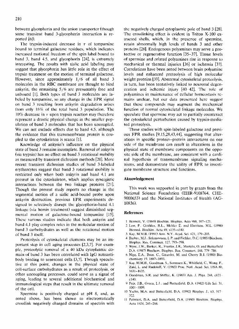
Fig. 1. Schematic diagram showing the orientation in the membrane of key molecules discussed in this study (note that only 1 /6 of band 3 molecules are coupled to spectrin via band 2.1 [1]). 3, band 3; G, glycophorin; SA, sialic acid residues; GAL, galactose residues; 4.5, band 4.5; Sp,
spectrin; 2.1, band 2.1 or ankyrin; A, actin.

207
residues of glycophorin in membrane ghosts were cova- lently and selectively spin labeled, as desired, by reduc- tive amination procedures described previously [23,24]. In the case of galactose labeling, intact cells were activated by galactose oxidase to produce an aldehyde on C-6 terminal galactose and galactosamine residues, while ghosts were used in the sialic acid activation process with NalO 4. Protein concentrations were as- sayed by the method of Lowry [26].
Spin labeling of cytoskeletal proteins The labeling solution consisted of 51 /zM MAL-6 in
5P8, well below the 1 mM level of SH-specific reagents required to disrupt the spectrin tetramer-dimer equi- librium [27]. Thiol groups of spectrin were labeled by incubating 1 ml ghosts (at 3 mg pro te in /ml ghosts) with 10 ml MAL-6 solution at 4°C for 14-16 h as previously described [22].
TPCK-trypsin treatment Ghosts were washed twice in 5P8 and twice in 10
mM Tris (pH 7.4) to remove excess spin label and to prepare the membranes for trypsin treatment.
Ice-cold solutions of TPCK-trypsin and trypsin in- hibitor were prepared fresh by dissolving these agents in 10 mM Tris (pH 7.4). Ghosts were assayed for total protein content and pipetted into 5 ml culture tubes suspended in an ice bath. Ice-cold TPCK-trypsin was introduced into ghost suspensions on ice to give a final concentration of 1 mg trypsin: 300 mg membrane pro- tein. At specific time periods, the trypsin was deacti- vated by addition of at least 10-fold excess of trypsin inhibitor. Ghosts were washed once more in 5P8, as- sayed for protein content, and aliquots withdrawn for EPR analysis or SDS-PAGE.
EPR / SDS-PA GE EPR spectroscopy was performed at a protein con-
centration of 2.0-2.5 m g /m l in a flat quartz aqueous sample cell, using a Bruker 300 ESP spectrometer equipped with computerized data acquisition and anal- ysis capabilities (microwave frequency = 10 GHz, mi- crowave power = 10 mW, modulation frequency = 100 kHz, modulation amplitude = 0.481 G, time constant = 1.28 ms). PAGE was performed according to the method of Fairbanks et al. [29], using a continuous buffer system and a 4.6% acrylamide gel, and protein bands were visualized by staining with Coomassie bril- liant blue R-250.
Results
MAL-6 labeled spectrin / SDS-PAGE The physical state of cytoskeletal proteins of ery-
throcyte membranes was monitored by EPR using the protein specific spin label MAL-6 [22]. By use of selec- tive isolation experiments and studies employing anti- bodies to MAL-6 [4,22], it is reported that up to 90% of the spin label intensity of MAL-6 is found on spectrin, the major cytoskeletal protein. The relevant EPR parameter measured is the ratio of the spectral amplitude of the M I = + 1 low-field weakly immobi- lized line (W) and that of the M I = 6-1 low-field strongly immobilized line (S), which is referred to as the W/S ratio (Fig. 2). Changes in the W/S ratio are known to be strong indicators of perturbations in the normal in te rac t ions of cy toske le ta l e l emen t s [15,22,25,28]. Previous studies have shown that de- creased cytoskeletal p ro te in -p ro te in interactions caused by hemin [15], polyphosphates [5] or increased dimeric spectrin content [25] 'loosen' the conformation
Spermine treatment Fresh spermine solutions (pH 8.0), were prepared at
8 mM by dissolving spermine in 5P8 buffer and titrat- ing with a measured volume of 1.0 M HC1. Typically, a 100 ml solution of 8 mM spermine required 0.8-1.2 ml HC1 to reach the desired pH. An ionic strength control solution was prepared by addition of a NaC1 aliquot (0.5 M) to an appropriate volume of 5P8 [28]. Solutions so prepared were kept on ice until ready to use.
Several minutes prior to spectral acquisition, 1 vol- ume of 8 mM spermine was added to 3 volumes of spin-labeled ghosts to yield a final spermine concentra- tion of 2.0 mM and a final protein concentration of 2 mg/ml . Ionic strength controls were prepared by sub- stituting the NaC1 control solution for spermine. 'Ghost only' controls were prepared by substituting 5P8 for spermine or NaCI. Ghost suspensions thus treated were allowed to equilibrate to room temperature for 15 min prior to spectral acquisition.
I
I ,o° I l]e
V Fig. 2. Typical EPR spectrum of MAL-6 covalently attached to erythrocyte membrane proteins, showing strongly (S) and weakly (W) immobilized components of the M 1 = + 1 low-field resonance
line (box).

208
__2401 - I , , o L . . . . . . . . . . . . . . . p , o . _ o o o 3 _ _ p , o . o o o 2 _. l
e,, o
¢J 2OO "6
180 _o
180 rr 0~ 140
120
100 6 10 20
time of reaction (aeconcla)
Fig. 3. EPR analysis of the W/S ratio as a function of TPCK-trypsin treatment. P-values computed by one-tailed Student's t-test. Error
bars indicate S.E.
of spectrin and decrease steric hindrance to spin label motion resulting in an increase in the W/S ratio of MAL-6.
Fig. 3 summarizes the effect of trypsin to decrease cytoskeletal pro te in-pro te in interactions as judged by EPR. A significant increase was seen in the W/S ratio of MAL-6 labeled ghosts following as little as 5 s TPCK-trypsin incubation at 0°C (mean increase over controls +_ S.D. = 56% +_ 43%, P < 0.004, n = 8). The W/S ratio of MAL-6 labeled cytoskeletal proteins in- creased to approx. 200% control after 10 s exposure to TPCK-trypsin at 0°C, and showed no further increase at 20 s, suggesting that most of the ankyrin was de- pleted from the membrane between 5 and 10 s.
This suggestion was confirmed by SDS-PAGE. In-
spection of Coomassie blue stained SDS-PAGE gels revealed some loss of the ankyrin band at 5 s (Fig. 4, lane C), with almost total loss of band 2.1 after 20 s trypsin t reatment (Fig. 4, lane D). Loss of the poorly resolved 2.1 band was accompanied by the loss of 'syndein' bands in the 2.2-2.3 region (Fig. 4). In some cases new, lightly stained bands appeared lower in the gel, just above band 3 in the trypsinized samples (not shown). There was no apparent loss of major bands, except 2.1, at any of the time intervals tested in this experiment. These observations agree closely with those of Cherry and co-workers [20].
Temparnine labeled glycophorin Previous EPR studies of the specific disruption of
the band 4.1-glycophorin linkage by hemin suggested that the physical state of cell-surface sialic acid was altered [15]. We wondered if breakage of the linkage between band 2.1 and a fraction of the band 3 molecules would affect sialic acid motion as well. The motion of the spin label covalently bound to glycophorin was characterized from the spectra like those previously published from our laboratory [23] by an apparent correlation time, r, which can be thought of as the time required to complete rotation through one radian. The r parameter was calculated from the well-established equation:
Fig. 4. SDS-PAGE profile of high-molecular weight cytoskeletal components a spectrin (1), /3 spectrin (2) and ankyrin (band 2.1). Lanes A, B: controls; lane C: 5 s incubation with TPCK-trypsin; lane D: 20 s incubation with TPCK-trypsin as described in text.

where W 0 is the peak-to-peak linewidth of the M I = 0 central line and A(n) refers to the peak-to-peak ampli- tude of the M~ = + 1, 0, or - 1 lines.
The r parameter of tempamine bound to sialic acid on glycophorin in ghosts subjected to 5 s trypsin incu- bation did not differ significantly from controls (mean% control + S.D. = 97.9 + 7.01, P > 0.622, n = 4, Table I). Furthermore, there were no significant effects ob- served even after 20 s incubation with the proteinase (mean% control + S.D. = 106 + 12.3, n = 4, Table I). These results suggest that sialic acid motion on gly- cophorin is unaffected by disruption of the band 3- spectrin linkage upon proteolysis of ankyrin.
Tempamine labeled terminal galactose residues Since essentially all of the terminal galactose
residues specifically spin labeled by reductive amina- tion are located on band 3, band 4.5, and glycophorin [24], and the above-mentioned results suggested that sialic acid on glycophorin was unaffected by trypsin treatment, we reasoned that trypsin cleavage of ankyrin on the cytoplasmic side of a fraction of these trans- membrane proteins may affect the motion of terminal galactose residues on the opposite side of band 3 and band 4.5. Incubation of ghosts with TPCK-trypsin for 5-10 s was found to cause a significant decrease in r relative to controls (mean% control + S.D. = 90.4 + 9.29, P < 0.005, n = 12; Table I), suggesting increased motion and an altered physical state of galactose in cell-surface glycoconjugates after rupture of the band 3-spectrin linkage. As noted above, SDS-PAGE analy- sis showed no damage to band 3 or band 4.5 as a result of the trypsin treatment described here.
Spermine effect Previous studies from our laboratory suggested that
spermine addition to ghosts electrostatically cross-links negatively charged spectrin to the negatively charged cytoplasmic pole of band 3, thereby increasing pro-
TABLE I
Effect of TPCK-trypsin treatment on "r of tempamine covalently bound to cell-surface carbohydrates
P-values calculated by two-tailed Student's t- test. n.s. = not signifi- cant; n.c. = not calculated.
Carbohydrate ~" (% control) n P spin labeled mean +_ S.D.
Sialic acid control 100 +_ 0.0 4 5 s r × n 97.9_+ 7.0 4 20 s r × n 105.9___ 12.3 4
Galactose control 100 _+ 0.0 12 5 - 1 0 s r x n 90.4_+ 9.3 12 2 0 s r x n 88.1+ 3.2 2
n.s. n.s.
0.005 n . c .
160 t-- pcO.O001 --~
209
J
v trypsin trypsin*spermlne spermine
Fig. 5. Effect of spermine addition to untreated ghosts and to ghosts previously treated with TPCK-trypsin. P values calculated by one-
tailed Student's t-test. Error bars indicate S.E.
te in-protein interactions [28]. We wondered if sper- mine would 'repair ' the connection between band 3 and spectrin caused by trypsin cleavage of ankyrin.
Fig. 5 shows that the W/S ratio of MAL-6 bound to spectrin was significantly reduced after spermine addi- tion to ghosts previously treated with TPCK-trypsin (P < 0.0001), suggesting that spermine partially re- stored cytoskeletal prote in-prote in interactions as judged by EPR. The W/S ratio of MAL-6 obtained in this proteinase plus spermine system was some 20% greater than that obtained by addition of spermine to unproteolyzed control membranes (P < 0.0001, Fig. 5). Spermine addition to galactose-labeled ghosts previ- ously treated with trypsin showed no effect [ r% con- trol with trypsin treatment, 82.4% (n = 2); ~-% control with spermine addition after trypsin treatment, 84.2% (n = 2)]. This result, coupled with the MAL-6 studies, suggests that the primary effect of this polyamine is to alter the conformation of spectrin upon crosslinking to band 3.
Discussion
The results of our proteolysis experiments closely resemble those of Clague et al. [20], who succeeded in depleting ghosts of ankyrin with a 370:1 (w/w) mem- brane protein/ trypsin ratio and 20 s trypsin incuba- tion. However, other researchers have reported much slower kinetics for this reaction [13]. Although we have no explanation for the results in this latter study, we have noted that the magnitude of the observed W/S increaseof the spin label attached to cytoskeletal pro- teins following trypsin treatment depends somewhat upon the age and batch of proteinase used.
It is not surprising that degradation of ankyrin has no observable effect on the motion of the tempamine spin label bound to the external oligosaccharides of glycophorin: this sialoglycoprotein is not bound directly to ankyrin, and there is probably no permanent bond
210
between glycophorin and the anion transporter (though some transient band 3-glycophorin interaction is re- ported [6]).
The trypsin-induced decrease in r of tempamine bound to terminal galactose residues, which indicates increased motional f reedom of the spin label bound to band 3, band 4.5, and glycophorin [24], is extremely interesting. The results with sialic acid labeling may suggest that glycophorin has little role in the effect of trypsin t reatment on the motion of terminal galactose. However, since approximately 1 / 6 of all band 3 molecules in the RBC membrane are thought to bind ankyrin, the remaining 5 / 6 are presumably free and unbound [1]. Both types of band 3 molecules are la- beled by tempamine, so any change in the EPR signal on band 3 resulting from ankyrin degradation arises from only 16% of the total band 3 population. The 10% decrease in z upon trypsin reaction may therefore represent a drastic physical change in the smaller pop- ulation of band 3 molecules that has been uncoupled. We can not exclude effects due to band 4.5, although the evidence that this t ransmembrane protein is cou- pled to the cytoskeleton is scarce [1].
Knowledge of ankyrin's influence on the physical state of band 3 remains incomplete. Removal of ankyrin (via trypsin) has no effect on band 3 rotational mobility as measured by transient dichroism methods [20]. More recent transient dichroism studies of band 3-labeled erythrocytes suggest that band 3 rotational mobility is restricted only when both ankyrin and band 4.1 are present in the cytoskeleton, which implies synergistic interactions between the two linkage proteins [21]. Though the present study reports no change in the segmental motion of a sialic acid-bound probe upon ankyrin destruction, previous EPR experiments de- signed to selectively disrupt the glycophorin-band 4.1 linkage (via hemin treatment) suggest diminished seg- mental motion of galactose-bound tempamine [15]. These various studies indicate that both ankyrin and band 4.1 play complex roles in the molecular motion of band 3 carbohydrates as well as the rotational motion of band 3 itself.
Proteolysis of cytoskeletal elements may be an im- portant step in cell aging processes [2,3,7]. For exam- ple, proteolytic removal of a 40 kDa cytoplasmic do- main of band 3 has been correlated with lgG autoanti- body binding to senescent cells [3,7]. Though specula- tive at this point, changes in the physical state of cell-surface carbohydrates as a result of proteolysis, or other uncoupling processes, could serve as a signal of aging, leading to several additional biochemical and immunological steps that result in the ultimate removal of the cell.
Spermine is positively charged at pH 8, and, as noted above, has been shown to electrostatically crosslink negatively charged domains of spectrin with
the negatively charged cytoplasmic pole of band 3 [28]. The crosslinking effect is evident in Triton X-100 ex- tracted shells, which, in the presence of spermine, retain abnormally high levels of bands 3 and other proteins [28]. Endogenous polyamines may serve a pro- tective or regenerative function [35-37]. Tissue levels of spermine and related polyamines rise in response to mechanical or thermal injuries [36] or ischemia [37]. Correlations have been noted between brain polyamine levels and enhanced proteolysis of high molecular weight proteins [39]. Abnormal cytoskeletal proteolysis, in turn, has been tentatively linked to neuronal degen- eration and ischemic injury [40-42]. The role of polyamines in maintenance of cellular homeostasis re- mains unclear, but our data presented here suggest that these compounds may augment the mechanical function of normal cytoskeletal linkage molecules. We speculate that spermine may act to partially counteract the cytoskeletal perturbation caused by trypsin-media- ted proteolysis.
These studies with spin-labeled galactose and previ- ous EPR studies [9,15,28,43,44], suggesting that alter- ations in specific pro te in-pro te in interactions on one side of the membrane can result in alterations in the physical state of membrane components on the oppo- site side of the membrane, are consistent with a cardi- nal hypothesis of t ransmembrane signaling mecha- nisms, and demonstrate the utility of EPR to investi- gate membrane structure and functions.
Aknowledgement
This work was supported in part by grants from the National Science Foundation (EHR-9108764, CHE- 9000653) and the National Institutes of Health (AG- 10836).
References
l Bennett, V. (1989) Biochim. Biophys. Acta 988, 107-121. 2 Low, P., Geahlen, R.L., Mehler E. and Harrison, M.L. (1990)
Biomed. Biochim. Acta 49, s135-s140. 3 Kay, M.M.B. (1991) Ann. N.Y. Acad. Sci. 621, 179-203. 4 Barker, M.J., Solomonson, L.P. and Eichler, D.C. (1985) Biochem.
Biophys. Res. Commun. 127, 793-798. 5 Wyse, J.W., Barker, R., Franko, J.R., Martelo, O. and Butterfield
D.A. (1987) Biochem. Biophys. Res. Commun. 144, 779-786. 6 Nigg, E.A., Bron, C., Girardet, M. and Cherry R.J. (1980) Bio-
chemistry 19, 1887-1893. 7 Kay, M.M.B., Goodman, S., Sorensen K., Whitfield, C., Wong, P.,
Zaki, L. and Rudoloff, V. (1983) Proc. Natl. Acad. Sci. USA 80, 1631-1635.
8 Goodman, S.R. and Shifter, K. (1983) Am, J. Phys. 244, c121- c140.
9 Feix, J.B., Green, L.L. and Butterfield, D.A. (1982) Life Sci. 31, 1001-1009.
10 Yacko, M.A. and Butterfield, D.A. (1992) Biophys. J., 63, 317- 322.
11 Palmieri, D.A. and Butterfield, D.A. (1990) Biochim. Biophys. Acta 1024, 285-288.

12 Butterfield, D.A. and Rangachari, A. (1992) Biochim. Biophys. Res. Commun. 185, 596-603.
13 Jinbu, Y., Sato, S., Nakao, T., Nakao, M., Tsukita, S., Tsukita, S. and Ishikawa, H. (1984) Biochim. Biophys. Acta 773, 237-245.
14 Liu, S.C., Derick, L.H., Duquette, M.A. and Palek, J. (1989) Eur. J. Cell Biol. 49, 358-365.
15 Wyse, J.W. and Butterfield, D.A. (1989) Biochim. Biophys. Acta 979, 121-126.
16 Liu, S.C., Derick, L.H., Agre, P. and Palek, J. (1990) Blood 76, 198-205.
17 Cohen, A.M., Lawler, J., Liu, S.C., Prichal, J.T., Gualtieri, R.J., Brain, M.C., Dacie, J.V. and Palek, J. (1988) N. Eng. J. Med. 318, 230.
18 Markesbery, W.R., Leung, P.K. and Butterfield, D.A. (1980) J. Neurol. Sci. 45, 323-330.
19 Butterfield, D.A., Rangachari, A., Isbell, D.T. and Umhauer, S.A. (1992) Applied Magn. Reson., in press.
20 Clague, M.J., Harrison, J.P. and Cherry, R.J. (1989) Biochim. Biophys. Acta 981, 43-50.
21 Wyatt, K. and Cherry, R.J. (1992) Biochim. Biophys. Acta 1103, 327-330.
22 Butterfield, D.A. (1982) in Biological Magnetic Resonance (Berliner, L.J. and Reuben, J., eds.), Vol. 4, pp. 1-78.
23 Feix, J.B. and Butterfield, D.A. (1980) FEBS Lett. 115, 185-188. 24 Farmer, B.T. and Butterfield, D.A. (1984) J. Biochem. Biophys.
Methods 10, 111-120. 25 Farmer, B.T., Harmon, T.M. and Butterfield, D.A. (1985)
Biochim. Biophys. Acta 821,420-430. 26 Lowry, O.H., Rosebrough, N.J., Farr, A.L. and Randall, R.J.
(1951) J. Biol. Chem. 193, 265-275. 27 Smith, D.K. and Palek, J. (1983) Blood 62, 1190-96.
211
28 Wyse, J. and Butterfield, D.A. (1988) Biochim. Biophys. Acta 941, 141-149.
29 Fairbanks, G., Steck, T. and Wallach, D.F.H. (1971) Biochemistry 10, 2606-2616.
30 Bennett, V. and Hu, R. (1991) J. Biol. Chem. 266, 18200-18205. 31 Masliah, E., Hansen, M., Albright, T. and Terry, R.D. (1991)
Neurosci. Lett. 129, 1-5. 32 Truscott, R.J.W., Marcantonio, J.M., Tomlinson, J. and Duncan,
G. (1989) Biochem. Biophys. Res. Commun. 162, 1472-1477. 33 Backman, L., Pekrun, A. and Gratzer, W.B. (1991) J. Biol. Chem.
266, 3835-3840. 34 Rochel, S.M. and Margolis, F.L. (1980) J. Neurochem. 35, 85(I-
860. 35 Tetzlaff, W. and Kreutzberg, G.W. (1985) Exp. Neurol. 89, 679-
688. 36 Edbladh, M., Edstrom, A. and Persson, L. (1990) Exp. Neurol.
107, 63-68. 37 Dienel, G.A. and Cruz, N.F. (1984) J. Neurochem. 42, 913-25. 38 Paschen, W., Hallmayer, J. and Mies, G. (1987) Neurochem.
Pathol. 7, 143-156. 39 Najm, I., Vanderklish, P., Etebaryi, A., Lynch, G. and Baudry, M.
(1991) J. Neurochem. 57, 1151-1158. 40 Seubert, P., Lee, K. and Lynch, G. (1989) Brain Res. 492,
366-370. 41 Lee, K.S., Frank, S., Vanderklish, P., Arai, A. and Lynch, G.
(1991) Proc. Natl. Acad. Sci. USA 88, 7233-7237. 42 Peterson, C., Vanderklish, P., Seubert, P., Cotman, C. and Lynch,
G. (1991) Neurosci. Lett. 121,239-243. 43 Butterfield, D.A. (1990) J. Membr. Sci. 53, 3-17. 44 Butterfield, D.A. (1989) Biological and Synthetic Membranes,
Wiley-Liss, New York.