alterations in the light transmission through single lens...
TRANSCRIPT

Alterations in the Light Transmission Through Single LensFibers During Calcium-Mediated DisintegrativeGlobulization
Aruni Bhatnagar,* Prajay Dhir,* Le-Fei Wang,* Naseem H. Ansari* WooKuen Lo,fand Satish K. Srivastava*
Purpose. The purpose of this study was to examine changes in the light transmission throughsingle cortical fibers isolated from the rat lens during the process of disintegrative globulization.
Methods. Single cortical fibers were isolated from adult rat lens by treatment with trypsin ina solution containing 10 mM HEPES, 10 mM EDTA, and 280 mM sucrose (pH 7.4, 300 to310 mOsm) HEPES-EDTA-sucrose (HES) solution. The isolated fibers were illuminated bya white light source, and the light transmission through the fibers was collected by a charge-coupled device camera and quantified by digital image analysis. In some experiments, thinsections of fixed lens cells were examined using transmission electron microscopy.
Results. Enzymatic dissociation of the lens yielded elongated fibers, which, in the presence ofRinger's solution (containing 2 mM Ca2+), underwent disintegrative globulization. Measure-ments of light transmission through elongated fibers suspended in HES solution showedmaximal transmission at the center of the fiber. Exposure of the cortical fibers to Ringer'ssolution led to biphasic changes in the intensity of the transmitted light. Within 5 to 10minutes of exposure to Ringer's solution, a general decrease in the light transmission acrossthe long axis of the fiber was observed. Extended superfusion led to a local, apparent increasein light transmission corresponding to the formation of membrane blebs and globules. Imagesof disintegrated globules focused above their equator showed bright halos with dark centralzones. In electron micrographs, the single fibers showed uniform electron density. No signifi-cant inhomogeneities or precipitation of intracellular crystallins was observed in globulesgenerated from fiber cells exposed to Ringer's solution; in addition, no high molecular weightprotein aggregates were found in the globules.
Conclusions. Exposure to calcium alters the light-transmitting properties of isolated corticalfibers. The initial decrease in the average light transmittance of the fiber appears to besecondary to cell swelling and may relate to protein-based opacification. An apparent increasein light transmission through calcium-generated globules is likely because of the Becke linegenerated by a mismatch between the refractive index of the medium and the globule cyto-plasm and accentuated by the transition from rod-shaped to spheroidal morphology. InvestOphthalmol Vis Sci. 1997; 38:586-592.
1 he clarity of the ocular lens has been suggested tobe the result of the tight packing of variably dediffer-enti ated fiber cells containing a high concentration
From the *Defmrtment of Human Biological Chemistry and Genetics, University ofTexas Medical Branch, Galveston, and the ̂ Department of Anatomy, MorehoiiseSchool of Medicine Atlanta, Georgia.Supported by the National Institutes of Health grants EY 10677 (SKS) andEY05314 (WKIJ.Submitted for publication March 26, 1996; revised October 9, 1996; acceptedOctober 11, 1996.Proprietary interest category: N.Reprint requests: Satish K. Srivastava, Department of Human Biological Chemistryand Genetics, University of Texas Medical Branch, 619 Basic Science Building,Galveston, TX 77555-0647.
of crystallins.1'2 The short-range interaction of crys-tallins in the fiber cells minimizes light scattering andpromotes undistorted transmission.2 Disruption of theshort-range order of the crystallins, either by syneresis3
or phase separation,4'5 has been shown to increaselight scattering and has been suggested to contributeto opacification of the lens in some forms of experi-mental and clinical cataract. However, several studiesof the morphology of human and animal cortical cata-ract show that the light-scattering centers are associ-ated with areas containing membranous beads or glob-ules.6"8 The origin of such globules has not been iden-
586Investigative Ophthalmology & Visual Science, March 1997, Vol. 38, No. 3Copyright © Association for Research in Vision and Ophthalmology
Downloaded From: http://iovs.arvojournals.org/pdfaccess.ashx?url=/data/journals/iovs/933197/ on 06/04/2018

Light Transmission Through Single Cortical Fibers 587
tified, but it has been speculated that they arise fromthe disintegration of cortical fibers.8
The formation of globules may be the underlyingreason for increased light scattering because that de-stroys the tight packing of cortical fibers and disruptsthe quasiperiodic membrane stacking essential forlens transparency. Moreover, if zones of globular de-posit are indeed responsible for opacification of thelens, identification of the origin of these globules andinvestigations into the pathophysiological mecha-nisms that lead to globule formation will be useful indeveloping an understanding of the cellular mecha-nisms of cataractogenesis.
The complex architecture of the intact lens doesnot permit the easy access required to examine themetabolism and physiology of cortical fibers. We have,therefore, developed methods to isolate viable, singlecortical fibers9 to study the physiological and patho-logic changes in the structure and function of corticalfibers under well-defined experimental conditions.
Such preparations allow direct investigation of thecortical fibers without the complexities arising fromintrafiber communications and epithelial regulation.We observe that superfusion of isolated fibers withRinger's solution leads to disintegrative globulization.9
This process appears to be mediated by an increasedinflux of calcium from the medium because globuliza-tion is delayed by decreasing the concentration of cal-cium in the external medium.9 Calcium-mediated dis-integration could be delayed by protease inhibitors,suggesting a role for enhanced (calcium-mediated)proteolysis.10 Interestingly, calcium-induced disinte-gration does not lead to cell lysis and fragmentationbut does result in the formation of resealed, discreteglobules strikingly similar to those observed at thelight-scattering centers of cortical cataract.6"8
The current study was undertaken examine thechanges in light transmission of isolated, single corti-cal fibers during the process of calcium-mediated glo-bulization. Our results suggest a complex series ofchanges in light transmission during disintegrativeglobulization, which may contribute to the opacifica-tion of lens during cataractogenesis.
MATERIALS AND METHODS
Sprague-Dawley rats (weight range, 200 to 250 g)were housed in accordance with the institutionalguidelines and killed by a single intraperitoneal injec-tion of sodium pentobarbital. All animals were treatedaccording to the ARVO Statement for the Use of Ani-mals in Ophthalmic and Vision Research. Eyeballswere removed and immersed in Ringer's solution(composition in mM: NaCl 150, KC1 5.4, MgCl2 2,CaCl2 2, HEPES 10, pH 7.4, adjusted with NaOH).The osmolarity of the solution was 300 to 310 mOsm.Eyeballs were dissected, and the lenses were removed
with the capsule intact. The procedure for isolationof single-lens fibers has been described.9 Briefly, theisolated lenses were incubated with 300-310 mOsmHEPES-EDTA-sucrose (HES) solution (compositionin mM: sucrose 280, Na-EDTA 10, HEPES 10, pH 7.4)containing 0.6 mg trypsin/ml (Sigma Chemical, St.Louis, MO) for 15 minutes at 33°C, after which thesolution was replenished. The lenses were reincubatedin fresh solution at 32°C for 6 minutes, after which thetemperature of the medium was increased gradually ata rate of 0.8°C/minute for 6 minutes and maintainedat 37°C for an additional 3 minutes. After incubation,the lenses were decapsulated, and the epithelium wasremoved. The lenses were rotated gently at a rate of1 revolution/second in HES solution for 15 to 20 min-utes at room temperature. The suspension was re-moved and plated on a coverslip and was superfusedgently with HES solution to remove trypsin.
Measurement of Light Transmission
Isolated fiber cells were plated on a coverslip at thebottom of a tissue bath maintained at 35°C using anopen perfusion chamber, equipped with a Peltier heat-ing element (PDMI-2; Medical Systems, Greenvale,NY). The perfusion chamber was mounted on theheadstage of an inverted microscope (Nikon [Tokyo,Japan] Diaphot 300). Cortical fibers were illuminatedfrom above using a 6 V halogen lamp, and the transmit-ted light was collected by a charge-coupled device cam-era (model #48153100; Cohu, San Diego, CA) con-nected to the side port of the microscope by a 0.7X C-mount adaptor. Images were acquired digitally usingImage-1 acquisition system and software (version 4.0;Universal Imaging, West Chester, PA) and were dis-played on an RGB monitor (NEC Multisyn 5FGe; NECTechnologies, Boxborough, MA).
Thin-Section Electron Microscopy
Isolated lens fiber cells in small centrifuge tubes fromcontrol and experimental groups were fixed in an im-proved fixative containing 2.5% glutaraldehyde, 0.1 Mcacodylate buffer (pH 7.3), 50 mM L-lysine, and 1%tannic acid for 2 hours or overnight at room tempera-ture.11 After a brief rinse in 0.1 M cacodylate buffer, theisolated fiber cells were centrifuged down and postfixedin 1% aqueous OsO4 for 1 hour at room temperature,rinsed in distilled water, and stained en bloc with 0.5%uranyl acetate in 0.15 M NaCl overnight at 4°C. Isolatedfiber cells were dehydrated through graded ethanol andpropylene oxide and embedded in Polybed 812 resin(Polysciences, Warrington, PA). Thin sections (1 fim)were cut with a glass knife, stained with 1% toluidineblue, and examined under a light microscope. Thin sec-tions (80 nm) were cut with a diamond knife, stainedwith 5% uranyl acetate and then by Reynold's lead ci-trate, and examined in aJEOL (Peabody, MA) 1200EXelectron microscope.
Downloaded From: http://iovs.arvojournals.org/pdfaccess.ashx?url=/data/journals/iovs/933197/ on 06/04/2018
588 Investigative Ophthalmology & Visual Science, March 1997, Vol. 38, No. 3
RESULTS
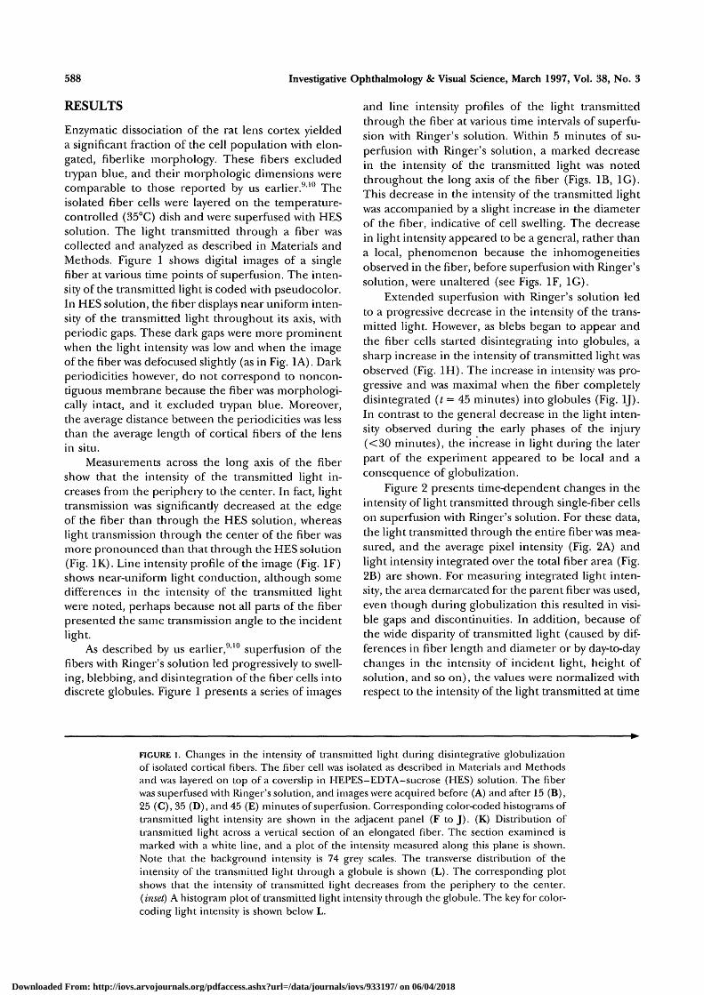
Enzymatic dissociation of the rat lens cortex yieldeda significant fraction of the cell population with elon-gated, fiberlike morphology. These fibers excludedtrypan blue, and their morphologic dimensions werecomparable to those reported by us earlier.910 Theisolated fiber cells were layered on the temperature-controlled (35°C) dish and were superfused with HESsolution. The light transmitted through a fiber wascollected and analyzed as described in Materials andMethods. Figure 1 shows digital images of a singlefiber at various time points of superfusion. The inten-sity of the transmitted light is coded with pseudocolor.In HES solution, the fiber displays near uniform inten-sity of the transmitted light throughout its axis, withperiodic gaps. These dark gaps were more prominentwhen the light intensity was low and when the imageof the fiber was defocused slightly (as in Fig. 1A). Darkperiodicities however, do not correspond to noncon-tiguous membrane because the fiber was morphologi-cally intact, and it excluded trypan blue. Moreover,the average distance between the periodicities was lessthan the average length of cortical fibers of the lensin situ.
Measurements across the long axis of the fibershow that the intensity of the transmitted light in-creases from the periphery to the center. In fact, lighttransmission was significandy decreased at the edgeof the fiber than through the HES solution, whereaslight transmission through the center of the fiber wasmore pronounced than that through the HES solution(Fig. IK). Line intensity profile of the image (Fig. IF)shows near-uniform light conduction, although somedifferences in the intensity of the transmitted lightwere noted, perhaps because not all parts of the fiberpresented the same transmission angle to the incidentlight.
As described by us earlier,910 superfusion of thefibers with Ringer's solution led progressively to swell-ing, blebbing, and disintegration of the fiber cells intodiscrete globules. Figure 1 presents a series of images
and line intensity profiles of the light transmittedthrough the fiber at various time intervals of superfu-sion with Ringer's solution. Within 5 minutes of su-perfusion with Ringer's solution, a marked decreasein the intensity of die transmitted light was notedthroughout the long axis of the fiber (Figs. IB, 1G).This decrease in the intensity of the transmitted lightwas accompanied by a slight increase in the diameterof the fiber, indicative of cell swelling. The decreasein light intensity appeared to be a general, rather thana local, phenomenon because the inhomogeneitiesobserved in the fiber, before superfusion with Ringer'ssolution, were unaltered (see Figs. IF, 1G).
Extended superfusion with Ringer's solution ledto a progressive decrease in the intensity of the trans-mitted light. However, as blebs began to appear andthe fiber cells started disintegrating into globules, asharp increase in the intensity of transmitted light wasobserved (Fig. 1H). The increase in intensity was pro-gressive and was maximal when the fiber completelydisintegrated (t = 45 minutes) into globules (Fig. 1J).In contrast to the general decrease in the light inten-sity observed during the early phases of the injury(<30 minutes), the increase in light during the laterpart of the experiment appeared to be local and aconsequence of globulization.
Figure 2 presents time-dependent changes in theintensity of light transmitted through single-fiber cellson superfusion with Ringer's solution. For these data,the light transmitted through the entire fiber was mea-sured, and the average pixel intensity (Fig. 2A) andlight intensity integrated over the total fiber area (Fig.2B) are shown. For measuring integrated light inten-sity, the area demarcated for the parent fiber was used,even though during globulization this resulted in visi-ble gaps and discontinuities. In addition, because ofthe wide disparity of transmitted light (caused by dif-ferences in fiber length and diameter or by day-to-daychanges in the intensity of incident light, height ofsolution, and so on), the values were normalized withrespect to the intensity of the light transmitted at time
FIGURE l. Changes in the intensity of transmitted light during disintegrative globulizationof isolated cortical fibers. The fiber cell was isolated as described in Materials and Methodsand was layered on top of a coverslip in HEPES-EDTA-sue rose (HES) solution. The fiberwas superfused with Ringer's solution, and images were acquired before (A) and after 15 (B),25 (C), 35 (D), and 45 (E) minutes of superfusion. Corresponding color-coded histograms oftransmitted light intensity are shown in the adjacent panel (F to J). (K) Distribution oftransmitted light across a vertical section of an elongated fiber. The section examined ismarked with a white line, and a plot of the intensity measured along this plane is shown.Note that the background intensity is 74 grey scales. The transverse distribution of theintensity of the transmitted light through a globule is shown (L). The corresponding plotshows that the intensity of transmitted light decreases from the periphery to die center.(inset) A histogram plot of tiansmitted light intensity through the globule. The key for color-coding light intensity is shown below L.
Downloaded From: http://iovs.arvojournals.org/pdfaccess.ashx?url=/data/journals/iovs/933197/ on 06/04/2018

light Transmission Through Single Cortical Fibers 589
zero—that is, at the beginning of the superfusion withRinger's solution. The average light intensity showedan initial decrease, which was followed by a sharp in-crease after 40 minutes of superfusion. The intensityof the integrated light shows the converse.
Image analysis of single globules indicates thatthe intensity of light transmitted through the globulesdecreases from the periphery to the center (Fig. 1L).This is exactly opposite to die spatial distribution ofthe intensity of the light transmitted through elon-
Downloaded From: http://iovs.arvojournals.org/pdfaccess.ashx?url=/data/journals/iovs/933197/ on 06/04/2018

590 Investigative Ophthalmology 8c Visual Science, March 1997, Vol. 38, No. 3
A Average
Integrated
Time (min) FIGURE 2. Time-dependent changes in the intensicy of trans- mitted light through single cortical fibers of ocular lens. Individual fibcrs were dissociated using trypsin as described in Matci-ials and Methods. Average (intensity per pixel, A) or integrated (intensity Tron~ the: entire fiber, B) light inten- sity was measured using the configura~ion shown in Figure I , in HEPES-EDTA-si~crose solution ( l = 01, and at thc indicated times after superfilsion with Ringer's solution. A representative set of images is shown in Figure 2. Data are shown as discrctc points, and the cutves are the best fit OF two polynomials to the data. Note that the t d t ~ e s were t~o~*malized with respect to the intensity of the respective fibcr at t = 0 (that is, before exposure to Ringer's solution). Thus, transmittance before exposure to Ringer's solution is expressed as 100%. Thc concentration of calcium in Ring- er's solution was 2 mM.
gated fibers, which showed maximal transmittance near the center (Figs. lL, 1K). Thus, in the globules, the periphery was bright and the center was dark (Fig. lL, inset); nevertheless, the light transmitted by the globule was more intense than the surro~~nding me- di~11-n.
To investigate f ~ ~ r t h e r the alterations caused by globulization, the structure of the isolated fibers was studied by transmission electron microscopy. Figure 3A shows a representative electron micrograph of a fiber cell fixed after exposure to the HES solution. Note that the single fiber cell shows uniform electron density, similar to that observed in thin sections of the intact lens cortex. Figure 3B shows two globules
generated o n exposure to Ringer's solution. The cyto- plasm of each globule shows no marked inhomogenei- ties or precipitation of intracellular crystallins. More- over, the cytoplas~n sholvs no indication of the forma- tion of high niolecular weight protein aggregates.
DISCUSSION
Several studies have reported that discrete membra- nous globules are present at the light-scattering cen- ters of supranuclear Nevertheless, the ori- gin of these globules is unclear, and i t is ~lnknown how such globules are formecl during cataractogene- sis. Our studies with isolated fiber cells show that S L K ~
globules arise spontaneously from disintegrative glo- bulization of elongated fibers exposed to medium con- taining high concentration of extracellular calcium." The process of globulization appears to be mediated by a large calcium influx and the consequent activa- tion of proteolytic enzymes such as ~ a l ~ a i n . ' ~ These results are in agreeinen t with those of in vivo studies that suggest a critical role of calcium h~rneostasis'~
FIGURE 3. (A) Thin-section eleccl-on micrograph showing an isolated single fiber cell after exposure to HEPES-EDTA- sucrose solution (control). The fiber cell is elongated, and the entire cytoplasm exhibits a honmgenous electron den- sity of crystalline proteins and cell matrix. (B) Transmission electron micrograph shows discrete globules formed on su- perfusion with the Ringer's solution containing 2 mM Calf. The entire cytoplasm of each globule displays a homogenous electron density, without protein aggregation. Bar = 2 pm.
Downloaded From: http://iovs.arvojournals.org/pdfaccess.ashx?url=/data/journals/iovs/933197/ on 06/04/2018

Light Transmission Through Single Cortical Fibers 591
and the proposed association between the activationof proteases13 and cataractogenesis.
Exposure of the isolated fibers to Ringer's solu-tion led to an initial decrease in the average lightintensity transmitted through the fiber. As indicatedin Results, this decrease in transmitted light was gen-eral and was evident throughout the length of thefiber. The initial response of the fiber to Ringer's solu-tion is an increase in cell volume (swelling) caused byincreased hydration. This is consistent with the in-crease in the integrated light intensity, presumablyalso caused by an increase in the fiber area. Increasedlens hydration has been shown to be associated withthe early stages of several forms of cataract.1415 Al-though the mechanisms of this hydration are not wellunderstood, it appears likely to have been caused bya net gain of Na+ and Cl~ ions by the fiber (Donnanswelling). The process of globulization appears to re-sult from calcium-induced proteolysis (vide supra),which could involve progressive degradation of thecytoskeleton.16 This process probably also induces per-meability changes so that a positive feedback loop isestablished, giving rise to catastrophic changes (thatis, disintegration into discrete globules).
The observed decrease in average light transmis-sion in the initial stages may result from progressivedilution of crystallins because of increased influx ofwater and cell swelling. In vitro experiments show thatlight scattering increases as the concentration of crys-tallins is increased up to approximately 0.12 g/ml;then it decreases.2 Concentrated (0.12 to 0.3 g/ml)solutions of crystallins transmit more light, probablybecause of increased short-range, liquidlike organiza-tion of the crystallins.2 Therefore, it follows that swell-ing or increased hydration of the lens fibers may altercrystallin organization, which may contribute to theobserved decrease in transmitted light during the pre-globulization stages (Fig. 1). Changes in light trans-mission could be secondary to an increase in the diam-eter of the fiber cell that increases the light path.However, changes in the light path length alone can-not account for our observations because it appearsunlikely that path length could change without alter-ing the concentration of intracellular crystallins; thisassumes that no significant de novo synthesis of crys-tallins occurs during globulization.
The formation of globules from fiber cells ex-posed to calcium-containing medium (Ringer's solu-tion) was associated with an increase in the averageintensity of the transmitted light. The increase in thetransmitted light was more pronounced than the earlydecrease discussed and apparently continues after theglobules have detached completely from the parentfiber. The corresponding decrease in the integratedlight intensity is apparently caused by die discontinu-ities caused by globulization. The increase in transmit-ted light on globulization appears paradoxical, but it
Focal length giving morediffuse central image
Focal length giving brightcentral image
/Tl WCorrect focallevel
/ M \A \X Focal length
giving halo
FIGURE 4. Formation of Becke line during measurements oflight transmission through a conducting sphere observed undera microscope, when the refractive index of die medium is lessthan that of the sphere. Adapted from Slayter EM. The determi-nation of refractive index. In: Optical Methods in Biology. NewYork: Robert E. Krieger; 1976:575-599. Reprinted by permis-sion of John Wiley & Sons, Inc.
probably does not result from the formation of highmolecular weight aggregates in the cytoplasm becauseglobule cytoplasm remains clear (Fig. 3). As pointedout by Bettleheim,5 protein aggregates must reach amolecular weight of more dian 1 million to cause sig-nificant scattering. Proteins of such high molecularweights, however, are found primarily in the nucleus.Moreover, high molecular weight proteins are precur-sors of insoluble aggregates that lead to the formationof large electron-dense particles measuring as much as500 nm in diameter.17 Clearly, no such large, electron-dense bodies are apparent in the cytoplasm of glob-ules generated from the calcium-induced disintegra-tion of isolated cortical fibers. In fact, the cytoplasmof the globules appears to be slightly more electrondense than the fiber (see Figs. 3A, 3B), which couldcontribute partially to the increase in light transmis-sion observed during the process of globulization.
The predominant cause for the observed increasein light transmission on globulization, however, ap-pears to be a mismatch between the refractive indicesof the globules and the surrounding medium. As illus-trated in Figure 4, a mismatch between the refractiveindex of a conducting sphere and the surroundingmedium gives rise to the Becke line, which appears asa halo around the image plane when it is defocusedslighdy.18 The Becke line arises when a sphere is im-mersed in a medium with a refractive index differentfrom that of the particle; at a certain focal plane, itappears outside the geometric edges of die sphere. Asan exact focus is approached, the fringe moves inwardto coincide with the edge of the sphere. Beyond thispoint, die bright region moves into the image of the
Downloaded From: http://iovs.arvojournals.org/pdfaccess.ashx?url=/data/journals/iovs/933197/ on 06/04/2018

592 Investigative Ophthalmology & Visual Science, March 1997, Vol. 38, No. 3
sphere and tends to form a bright spot at the center.As shown in Figure 1L, a distinct halo appears aroundthe globule formed from a fiber cell, which coincideswith the geometric edge of the sphere. This halo ap-pears to result from a lower refractive index of thesurrounding Ringer's solution than from the refrac-tive index of the globule. It should be pointed outthat under similar conditions (illumination by parallelrays), the tubelike fiber cell behaves as a long, cylindri-cal lens and does not deflect rays that are incidentalong its midline; maximal deviation is experiencedby rays that are incident along its sides. This is incontrast to a spherical surface, that has equal curva-ture in all directions and focuses all rays, regardlessof the angle at which the rays are incident, on thesphere. Thus, the mismatch in the refractive index,which leads to significant attenuation in light intensityonly at the edge of the fibers (Fig. IK), is accentuatedby globulization forming a bright halo surroundingthe edge of the globule (Fig. 1L).
Although the contribution of calcium-induceddisintegration of the fiber cells to alterations in thelight-transmitting properties of the lens during in vivocataractogenesis remains to be assessed, it is expectedthat the formation of globules within the lens will haveoptical effects similar to those observed in vitro. In anormal transparent lens, there is a mismatch betweenthe refractive indices of the intercellular space andthe protein-filled fibers. However, because of quasi-periodic organization of the membranes, no scatter-ing is expected except in the Bragg reflection, whichappears within a narrow cone close to the incidentlight forming lenticular halos.2 Such halos have beenobserved experimentally, and their angular spacingcorresponds to the width of the fiber.12'3 Nevertheless,the well-aligned cortical lattice gives rise to a diffrac-tion pattern that maximizes light transmission by de-structive interference over a large section of space.5 Itfollows that any variation in the membrane-stackingperiodicity, or any disruption of the balance betweenform and intrinsic birefringence, will enhance opticalanisotropy. However, the formation of globules maybe particularly damaging because, unlike elongatedfibers (or even disrupted membranes), the globulesfocus light locally, and this interrupts the specularreflection by tubelike fiber cells. Moreover, becauseswelling is an early step in the process, globulizationenhances the difference in the refractive index be-tween intracellular and extracellular space. This leadsto a synergetic increase in light scattering caused bythe dispersion of refractive indices and to the efficientlocal focusing of light by the spherical globules.
Key Words
calcium, globulization, lens fiber cells, light transmission,transmission electron microscopy
Acknowledgments
The authors thank Mr. Adell Mills for technical assistancewith electron microscopy and Drs. Albert Zacarias and Mas-soud Motamedi for helpful discussions.
References
1. Trokel S. The physical basis of lens transparency. In-vest Ophthalmol 1962; 1:493-501.
2. Tardieu A, Delaye M. Eye lens proteins and transparency:From light transmission theory to solution x-ray structuralanalysis. Ann Rev Biophys Own. 1988; 17:47-70.
3. Bettelheim FA. Syneresis and its possible role in cata-ractogenesis. ExpEyeRes. 1979;28:189-197.
4. Delaye M, Clark JI, Benedek GB. Identification of thescattering elements responsible for lens opacificationin cold cataract. Biophys J. 1982; 37:647-656.
5. Bettelheim FA. Physical basis of lens transparency. In:Maisel H, ed. Ocular Lens: Structure, Function and Pathol-ogy. New York: Marcel Dekker; 1985;256-300.
6. Harding CV, Susan SR, Lo WK, Bobrow WFS, Maisel H,Chylack LT. The structure of the human cataractouslens. In: Maisel H, ed. Ocular Ijms: SMicture, Function andPathology. New York: Marcel Dekker; 1985; 367-404.
7. TrevithickJR, Robertson J, Mitton KP.JVitamin E andthe eye. In: Packer L, FuchsJ, eds. Vitamin E in Healthand Disease. New York: Marcel Dekker; 1993; 873-928.
8. Mackic JB, Ross-Cisneros FN, McComb JG, et al. Ga-lactose-induced cataract formation in guinea-pigs:Morphologic changes and accumulation of galactitol.Invest Ophthalmol Vis Sci. 1994;35:804-810.
9. Bhatnagar A, Ansari NH, Wang LF, Khanna P, WangCS, Srivastava SK. Calcium-mediated disintegrativeglobulization of isolated ocular lens fibers mimic cata-ractogenesis. ExpEyeRes. 1995;61:303-310.
10. Wang LF, Bhatnagar A, Ansari NH, Dhir P, SrivastavaSK. Mechanism of calcium-induced disintegrative glo-bulization of rat lens fiber cells. Invest Ophthalmol VisSci. 1996; 37:915-922.
11. Lo WK. Adherens junctions in the ocular lens of vari-ous species: Ultrastructural analysis with improvedfixation. Cell Tissue Res. 1988;254:31-40.
12. Hightower KR. Cytotoxic effects of internal calcium onlens physiology: A review. CwrEyeRes. 1985; 4:453-459.
13. Shearer TR, David LL, Anderson RS, Azuma M. Reviewof selenite cataract. ExpEyeRes. 1992; 11:357-369.
14. Mizuno A, Ozaki Y. Aging and cataractous process ofdie lens detected by laser Raman spectroscopy. LensEye ToxRes. 1991;8:177-187.
15. Hightower K, McCreadyJ. Physiological effects of UVBirradiation on cultured rabbit lens. Invest OphthalmolVis Sci. 1992;33:1783-1787.
16. Truscott RJW, Marcantonio JM, Tomlinson J, DuncanG. Calcium-induced opacification and proteolysis in theintact rat lens. Invest Ophthalmol Vis Sci. 1990;31:2405-2411.
17. Kramps HA, Stols AL, Hoenders HJ. On the quater-nary structure of high-molecular-weight proteins fromthe bovine eye lens. Eur JBiochem. 1975;50:503-509.
18. Slayter EM. The determination of refractive index. In:Optical Methods in Biology. New York: Robert E. Krieger;1976:575-599.
Downloaded From: http://iovs.arvojournals.org/pdfaccess.ashx?url=/data/journals/iovs/933197/ on 06/04/2018