alterations of cholinergic … · bacopa monnieri (bm), an indian herb extensively used in...
TRANSCRIPT

Available Online through
www.ijpbs.com (or) www.ijpbsonline.com IJPBS |Volume 3| Issue 2 |APR-JUN |2013|286-292
Research Article
Biological Sciences
International Journal of Pharmacy and Biological Sciences (e-ISSN: 2230-7605)
Wudayagiri Rajendra* et al Int J Pharm Biol Sci www.ijpbs.com or www.ijpbsonline.com
Pag
e28
6
ALTERATIONS OF CHOLINERGIC NEUROTRANSMISSION IN ROTENONE INDUCED
PARKINSON’S DISEASE: PROTECTIVE ROLE OF BACOPA MONNIERI
Gunduluru Swathi, Cherukupalle Bhuvaneswar & Wudayagiri Rajendra*
Department of Zoology, Division of Molecular Biology, Sri Venkateswara University,
Tirupati-517502. A.P. INDIA.
*Corresponding Author Email: [email protected]
ABSTRACT Parkinson’s disease (PD) is the second most common neurodegenerative disorder. Dopaminergic anti-parkinsonian
medications, cause drug-induced development of disabling motor complications in majority of patients with PD.
Bacopa monnieri (BM), an Indian herb extensively used in Ayurveda, was tested on cholinergic system in Rotenone
(RT) induced rat model of PD. Cholinergic system is affected due to imbalance between Acetylcholine (ACh) and
Dopamine (DA) neurotransmitters. In the experiment conducted rats were divided into four groups of six in each
group, group 1 received Saline water (1 ml/kg), group 2 received RT (2.5 mg/kg) through i.p. route administration
for 60 days to induce PD. The third group received BM extract (180 mg/kg/day) for 20 days orally before induction
of PD and group 4 received Levodopa (LD) (10 mg/kg/day) orally which is referred as reference control. The
Acetylcholine (ACh) and Acetylcholinesterase (AchE) had been elevated and depleted respectively when PD was
induced. In LD and BM treated rats the Ach was decreased and AchE was increased when compared to the PD
induced rats. Our results suggest the ability of BM extract to modulate cholinergic system in different brain
regions of PD induced rats.
KEY WORDS Parkinson’s disease (PD), Bacopa monnieri (BM), Rotenone (RT), Levodopa (LD), Acetylcholinesterase (AChE),
Acetylcholine (ACh).
INTRODUCTION
PD is a chronically progressive, age-related
neurodegenerative disease characterized by
progressive resting tremor, rigidity, bradykinesia, gait
disturbance, postural instability and dementia. A
major neuro-pathological feature is PD is
neurodegeneration in substantia nigra pars compacta
(SNpc), by loss of DA neurons. In PD, cognitive
dysfunction may be related to impairment of the
ascending cholinergic system [1]. The key feature of
cognitive profiles in PD is to executive dysfunction
that has difficulty in tasks that require generation of
cognitive sequencing. Cholinergic fibres originate
from the brainstem and the basal forebrain is
impaired in dementia associated with Lewy bodies
which can be a consequence of Parkinson’s disease
[2]. Although a neural basis for cognitive dysfunctions
in PD remains unknown, pathological and functional
neuroimaging studies suggest that the cholinergic
system arising from the basal forebrain has an
important role in cognitive functions of PD patients
[3]. Moreover, the loss of functioning in the
dopaminergic system of the substantia nigra and the
loss of cells in the cholinergic pathways in the nucleus
basalis are responsible for the most significant
cognitive deficits in PD [4]. Chronic use of current
anti-parkinsonian medications including Levodopa
therapy causes disabling abnormal involuntary
movements known as drug-induced dyskinesias in the
majority of PD patients [5,6].
There are significant number of evidences indicating
the oxidative stress involvment in the

Available Online through
www.ijpbs.com (or) www.ijpbsonline.com IJPBS |Volume 3| Issue 2 |APR-JUN |2013|286-292
International Journal of Pharmacy and Biological Sciences (e-ISSN: 2230-7605)
Wudayagiri Rajendra* et al Int J Pharm Biol Sci www.ijpbs.com or www.ijpbsonline.com
Pag
e28
7
pathophysiology of these diseases which can induce
neuronal damages, modulate intracellular signaling
and ultimately leading to neuronal death by apoptosis
or necrosis[7]. Thus, antioxidants have been studied
for their effectiveness in reducing these deleterious
effects and neuronal death in many in vitro and in
vivo studies 8. Hence there is a need to find out
newer pharmacologically active agents obtained from
natural sources as plant extracts, without side effects
and can act as natural antioxidants. Bacopa monnieri
(BM), a medicinal plant commonly known as Brahmi
in Sanskrit, has been used in the indigenous systems
of medicine reported for its pharmacological roles as
memory enhancer [9], cognition-enhancer, [10-12],
antidepressant and also antioxidant properties [13].
In this present investigation BM was used in
treatment of induced PD.
MATERIALS AND METHODS
Collection of plant material:
Bacopa monnieri plant used in this work was collected
in bulk from Tirumala Hills, Andhra Pradesh in India
and authenticated by qualified botanist at
Department of Botany, Sri Venkateswara University,
Tirupati, Andhra Pradesh in India.
Extract Preparation:
The whole plant material was collected and shade-
dried to powder. The plant material was percolated
with circulating 95% ethanol (200 ml) for three
rounds. The residue was extracted twice using the
same procedure. The extract was filtered and
concentrated under reduced pressure in the Buchi
rotavapour yielding a greenish-black sticky residue.
Finally the extract was freeze- dried and was used for
further studies.
Experimental design:
The present work was conducted on male Wistar rats
weighing 150±25g, they were maintained at a
temperature of 25±5⁰C and relative humidity of 45-
55% with 12:12 h dark: light cycle. The rats were
maintained according to the ethical guidelines for
animal protection and welfare bearing
no.04a/a/CPCSEA/IAEC/08-09/SVU/Zool/WR-
GS/dt.1.9.2009.
GROUP I: Served as normal control group, received
vehicle (1.0 ml/kg/day) i.p. for 60 days.
GROUP II: PD was induced by RT (emulsified in natural
oil to a concentration of 2.5 mg/ml), given i.p. route
administration (2.5 mg/kg/day) for 60 days [14].
GROUP III: RT -induced PD rats were treatedwith BM
extract (180 mg/kg/day) orally for 80 days, started
before 20 days from induction of PD.
GROUP IV: RT -induced PD rats were treated with LD
(10 mg/kg/day) orally started after 20 days from
induction of PD [15].
The development of PD was detected after 20 days
from induction with rotenone, by occurrence of
tremors and exhibiting specific symptoms such as
bradykinesia and rigidity in rats. The treatment with
BM extract was started 20 days before induction of
PD and LD was started after 20 days from induction of
PD and continued for 60 days. After stipulated
duration, the animals were sacrificed by cervical
dislocation and the brain regions [Cerebral cortex
(CC), Cerebellum (CB), Mid brain(MB) and Pons-
Medulla (PM)] were immediately isolated, frozen in
liquid nitrogen and were stored at -40⁰C until further
analysis.
Biochemical Analysis:
The level of ACh content and activity of AChE were
estimated by the method of Hestrin (1949) as given
by Augustinson (1957) [16] and with slight
modification of the Ellman et al., 1961 [17]
respectively in different brain regions of control and
experimental animals.
Statistical Analyses:
Values of the measured parameters were expressed
as mean ± SEM. One way- ANOVA (F value) was used
to test the significance of difference among more
than two arithmetic means, followed by Post-hoc test
(Scheffe multiple comparison) to test the difference
between each two means. The significance was
considered at p values < 0.05. All the statistical
analyses were processed using Statistical Program of
Social Sciences (SPSS) for Windows, version 11.5.
RESULTS
The levels of Acetylcholine (ACh) content and the
activity of Acetylcholinesterase (AChE) in different
brain regions Cerbral cortex (CC), Cerebellum (CB),
Mid brain (MB) and Pons medulla (PM) of control and
experimental rats were represented in (Table 1 and 2;
Fig 1 and 2).
Available Online through
www.ijpbs.com (or) www.ijpbsonline.com IJPBS |Volume 3| Issue 2 |APR-JUN |2013|286-292
International Journal of Pharmacy and Biological Sciences (e-ISSN: 2230-7605)
Wudayagiri Rajendra* et al Int J Pharm Biol Sci www.ijpbs.com or www.ijpbsonline.com
Pag
e28
8
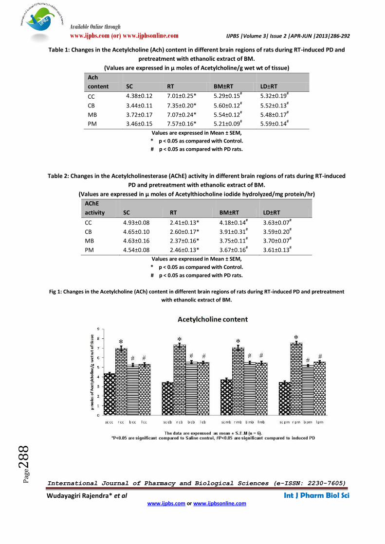
Table 1: Changes in the Acetylcholine (Ach) content in different brain regions of rats during RT-induced PD and
pretreatment with ethanolic extract of BM.
(Values are expressed in µ moles of Acetylcholine/g wet wt of tissue)
Ach
content SC RT BM±RT LD±RT
CC 4.38±0.12 7.01±0.25* 5.29±0.15# 5.32±0.19
#
CB 3.44±0.11 7.35±0.20* 5.60±0.12# 5.52±0.13
#
MB 3.72±0.17 7.07±0.24* 5.54±0.12# 5.48±0.17#
PM 3.46±0.15 7.57±0.16* 5.21±0.09# 5.59±0.14#
Values are expressed in Mean ± SEM,
* p < 0.05 as compared with Control.
# p < 0.05 as compared with PD rats.
Table 2: Changes in the Acetylcholinesterase (AChE) activity in different brain regions of rats during RT-induced
PD and pretreatment with ethanolic extract of BM.
(Values are expressed in µ moles of Acetylthiocholine iodide hydrolyzed/mg protein/hr)
AChE
activity SC RT BM±RT LD±RT
CC 4.93±0.08 2.41±0.13* 4.18±0.14# 3.63±0.07#
CB 4.65±0.10 2.60±0.17* 3.91±0.31# 3.59±0.20#
MB 4.63±0.16 2.37±0.16* 3.75±0.11# 3.70±0.07
#
PM 4.54±0.08 2.46±0.13* 3.67±0.16# 3.61±0.13#
Values are expressed in Mean ± SEM,
* p < 0.05 as compared with Control.
# p < 0.05 as compared with PD rats.
Fig 1: Changes in the Acetylcholine (ACh) content in different brain regions of rats during RT-induced PD and pretreatment
with ethanolic extract of BM.

Available Online through
www.ijpbs.com (or) www.ijpbsonline.com IJPBS |Volume 3| Issue 2 |APR-JUN |2013|286-292
International Journal of Pharmacy and Biological Sciences (e-ISSN: 2230-7605)
Wudayagiri Rajendra* et al Int J Pharm Biol Sci www.ijpbs.com or www.ijpbsonline.com
Pag
e28
9
Fig 2: Changes in the Acetylcholinesterase (AChE) activity in different brain regions of rats during RT-induced PD and
pretreatment with ethanolic extract of BM.
In RT-induced rats, AChE activity (P < 0.05) was
significantly depleted, whereas ACh content (P < 0.05)
was elevated in all the brain regions, when compared
with control rats. Pretreatment with BM extract and
treatment with LD caused significant elevation of
AChE activity (P < 0.05) and depletion of ACh content
(P < 0.05) were observed in different brain regions,
when compared to RT induced PD rats.
DISCUSSION
The central cholinergic system is considered to be the
most important neurotransmitter involved in the
regulation of cognitive functions [18]. The main
neurotransmitter in cholinergic system is ACh,
important in motor control, the autonomic, the
enteric, and in the central nervous system (CNS). In
cholinergic neurotransmission, the transmitted signal
is terminated by cleavage of the transmitter, ACh,
yielding acetate and choline. This cleavage is
mediated by acetylcholinesterase (AChE), an enzyme
of the α/β-fold family of proteins. The present study
was designed to explore the protective effect BM on
Cholinergic system (ACh content and AChE activity) in
the brain of induced PD rats. The main feature of PD
is relatively selective nigrostriatal dopaminergic
degeneration. The interaction between molecules
of DA and AChE resulted not only in modification
of catecholamine oxidation, but caused the
inactivation of AChE catalytic activity as well [19]
which occurred mainly due to the direct
interaction of a quinone or semiquinone oxidation
products with the enzyme when induced by RT.
Reactive oxygen species (ROS) are generated during
dopamine metabolism and by mitochondrial
respiration, which are shown to cause protein
damage [20]. In this study, decline in AChE activity in
PD induced rats as compared to the control rats was
observed in different regions of brain. The AChE
activity is an important marker of cholinergic
neurotransmission dysfunction. In our study all the
brain regions of RT induced PD rats showed the
decreased activity of AChE compared to controls, by
which we can conclude the progression of PD by
cholinergic neurotransmission dysfunction.
Numerous reports in literature show decrease in
AChE activity is reflected by acetylcholine content in
the brain [21]. Imaging studies [22] agree with post-
mortem evidence suggesting that basal forebrain
cholinergic system degeneration appears early in PD
and worsens coincident with the appearance of
dementia. Early cholinergic denervation in PD without
dementia appears to be heterogeneous and may
make specific contributions to the PD. Apart from
well-known cognitive and behavioral deficits; central
in particular limbic, cholinergic denervation may be
Available Online through
www.ijpbs.com (or) www.ijpbsonline.com IJPBS |Volume 3| Issue 2 |APR-JUN |2013|286-292
International Journal of Pharmacy and Biological Sciences (e-ISSN: 2230-7605)
Wudayagiri Rajendra* et al Int J Pharm Biol Sci www.ijpbs.com or www.ijpbsonline.com
Pag
e29
0
associated with progressive deficits of odor
identification in PD.
The main motor symptoms of PD as rigidity/tremors
and muscle fatigness may be due to the abnormal
increase of ACh content in induced PD rats (Table 1)
when compared to control rats. The loss of
dopaminergic inhibition for increased cholinergic
activity in the striatum causes an imbalance between
dopaminergic and cholinergic modulation of the
striatal output to the motor program may be due to
increased level of ACh (Table 1; Fig 1) which causes
overactivity and due to continuous stimulation
without inhibition leading to the characteristic
symptoms of tremor, rigidity and muscle fatigness
leading to postural instability.
According to Henk Konings (1995) [23] observations
cholinergic dysfunction exists in PD patients with
cognitive impairment. The increment of ACh (Table 1)
correlates with decrement of AChE (Table 2)
expressed as alterations in both motor and cognitive
behaviors respectively. Although the major factors
involved in PD declines remain to be specified,
oxidative stress has been mainly implicated in
cognitive impairment and neuropathologic disorders
[24]. The RT induces complex I inhibition which causes
the symptoms of PD, and increases the oxidative
stress.The central nervous system is particularly more
vulnerable to oxidative damage because of its high
oxygen consumption, high tissue concentration of
iron and relatively low levels of some antioxidants
system [25]. This implication has led to the notion
that antioxidant defense mechanisms in the brain are
not sufficient to prevent PD which increases in
oxidative damage. The dietary intake of a variety of
antioxidants might be beneficial for preserving brain
functions and maintaining protective role to prevent
progression of PD.
The ethanolic extract of BM is reported to be rich in
saponins, the saponins in general possess antioxidant
activity. BM pretreatment in the present study
resulted in declined the content of ACh (Table 1; Fig
1) in PD induced rats (Group III) as compared to the
PD induced rats (Group II).which clearly indicates
reduction of motor complications. Pretreatment with
BM significantly increased the AChE activity in the
brain of induced PD rat (Table 2; Fig 2).The BM
extract group showed that it had significance
difference compared to RT induced PD which
indicates its protective effect on PD induced rats.
Which clearly indicates that progression of PD had
been slowed down in case of BM pretreated group
compared to untreated PD group. It also showed that
there was no significance differences compared to
controls, showing recovery of this group of rats from
PD. The results of BM were on par with LD treated
group which specify the protective role of BM. This
shows the improvement of motor and cognitive
functions in pretreated BM and LD treated rats. The
BM treated group results are similar to the results of
LD treated group, showing that it can act as
antiparkinsonian agent with antioxidant properties
and/or its effect on the cholinergic system.
Chowdhuri et al., (2002) [26] have demonstrated that
Brahmi extract modulated the expression of
important enzymes involved in generation and
scavenging of reactive oxygen in the brain. Bacopa’s
antioxidant action and free radical scavenging
activity,especially in memory related structures in the
brain including the hippocampus [9] Oral
administration of BM extract markedly reduced the
memory deficits as well as acetylcholine concentra-
tions, choline acetylase activity, and muscuranic
receptor binding in the hippocampus and frontal
cortex [9]. From the present results coupled with
earlier reports it is obvious that the saponins which
are present in BM extract might have offered a
protective role from the oxidative stress and toxicity
caused during RT-induced PD.
The study concludes that BM extract administration is
effective in enhancing AChE and also demonstrates
inhibition of ACh in PD induced rats. Hence, BM
administration might serve useful in reversing PD
symptoms. The cholinergic/cognitive enhancing
properties of BM ethanolic extract warrants further
studies with larger group of animals, on its individual
active constituents and their mechanism of action.
The rapid development in the field of animal models
hopefully lead to an improved understanding of the
pathophysiology of PD, and finally permit a rational
designing of novel therapeutic strategies for PD.
Available Online through
www.ijpbs.com (or) www.ijpbsonline.com IJPBS |Volume 3| Issue 2 |APR-JUN |2013|286-292
International Journal of Pharmacy and Biological Sciences (e-ISSN: 2230-7605)
Wudayagiri Rajendra* et al Int J Pharm Biol Sci www.ijpbs.com or www.ijpbsonline.com
Pag
e29
1
ACKNOWLEDGEMENTS
One of the authors (G. Swathi) is grateful to CSIR-UGC
(NET) (India) for providing Junior Research Fellowship.
REFERENCES 1. Lange KW, Wells FR, Jenner P, Marsden CD. Altered
muscarinic and nicotinic receptor densities in cortical
and subcortical brain regions in Parkinson's disease. J
Neurochem, 1993; 60:197–203.
2. Perry EK, Irving D, Kerwin JM, McKeith I, Thompson P,
Collerton D, Fairbairn A, Ince PG, Morris CM, Cheng AV
et al., Cholinergic transmitter and neurotrophic
activities in Lewy body dementia: similarity to
Parkinson's and distinction from Alzheimer disease.
Alzheimer Dis. Assoc. Disord, 1993; 7, 69-79.
3. Caviness JN, Driver-Dunckley E, Connor DJ, Sabbagh
MN, Hentz JG, Noble B, Evidente VG, Shill HA, Adler
CH. Defining mild cognitive impairment in Parkinson’s
disease. Mov. Disord. 2007; 22, 1272–1277.
4. Ferreri F, Agbokou C, and Gauthier S. Recognition and
management of neuropsychiatric complications in
Parkinson's disease. CMAJ 2006; 175(12): 1545–1552.
5. Deogaonkar M, Subramanian T. Pathophysiological
basis of drug-induced dyskinesias in Parkinson’s
disease. Brain Res Rev 2005; 50:156-68.
6. Obeso JA, Olanow CW, Nutt JG. Levodopa motor
complications in Parkinson’s disease. Trends Neurosci
23(10 Suppl): 2000; S2-7.
7. Palla `SM, & Camins A. Molecular and biochemical
features inAlzheimer’s disease. Current Pharmaceutical
Design, 2006; 12: 4389–4408.
8. Ramassamy C. Emerging role of polyphenolic
compounds in the treatment of neurodegenerative
diseases: A review of their intracellular targets.
European Journal of Pharmacology, 2006; 545, 51–64.
9. Bhattacharya SK, Bhattacharya A, Kumar A, & Ghosal S.
Antioxidant activity of Bacopa monniera in rat frontal
cortex, striatum and hippocampus. Phytotherapy
Research, 2000; 14(3), 174-179.
10. Das A, Shanker G, Nath C, Pal R, Singh S, Singh
HK. A comparative study in rodents of
standardized extracts of Bacopa monniera and
Ginkgo biloba anticholinesterase and cognitive
enhancing activities. Pharmacol Biochem Behav
2002; 73:893-900.
11. Singh HK, Dhawan BN. Neuropsychopharmacoloical
effects of the Ayurvedic nootropic Bacopa monnieri
Linn (Brahmi). Indian J Pharmacol 1997; 29:S359-S365.
12. Sumathi T, Nayeem M, Balakrisshna K, Veluchamy G,
Devarraj NS. Alcoholic extract of Bacopa monniera
reduces the in vitro effects of morphine withdrawal in
guinea-pig ileum. J Ethnopharmacol 2002; 82:75-81.
13. Sairam K, Rao CV, Babu MD, Goel RK. Prophylactic and
curative effects of Bacopa monniera in gastric ulcer
models. Phytomedicine 2001; 8:423-430.
14. Alam M, Schmidt WJ. Rotenone destroys
dopaminergic neurons and induces parkinsonian
symptoms in rats. Behav Brain Res 2002; 136(1):317-
324.
15. Alam M, Schmidt WJ. L-DOPA reverses the
hypokinetic behaviour and rigidity in rotenone-treated
rats. Behav Brain Res 2004; 153(2):439-446.
16. Augustinson KB. Assay methods for cholinesterases.
In: Methods of Biochemical Analysis (D. Glick, Ed.), Vol.
5. Interscience Publishers Inc., New York, pp. 1957; 1-
63.
17. Ellman GL, Courtney KL, Andres VJr Featherstone RM.
A new and rapid colorimetric determination of
acetylcholinesterase activity. Biochem. Pharmacol.
1961; 7: 88-95.
18. Kim HK, Kim M, Kim S, Kim M, & Chung JH. Effects of
green tea polyphenol on cognitive and
acetylcholinesterase activities.Bioscience,
Biotechnology, and Biochemistry, 2004; 68:1977–79.
19. Klegeris A, Korkina LG, Greenfield SA. A possible
interaction between acetylcholinesterase and
dopamine molecules during autoxidation of the amine.
Free Radic. Biol. Med. 1995; 18: 223–230.
20. Simpson JA, Narita S, Gieseg S, Gebicki S, Gebicki
JM, Dean RT. Long-lived reactive species on free-
radical- damaged proteins. Biochem. J., 1992;
282:621-624;
21. Das A, Dikshit M, Nath C. Profile of
acetylcholinesterase in brain regions of male and
female rats of adult and old age. Life Sci. 2001; 68 (13):
1545–1555.
22. Oikawa H, Sasaki M, Ehara S, Abe T. Substantia
innominata: MR findings in Parkinson’s disease.
Neuroradiology 2004; 46: 817–821.
23. Henk Konings C, Kuiper MA, Mulder C, Calliauw J,
Wolters ECh. CSF acetylcholinesterase in Parkinson
disease: decreased enzyme activity and
immunoreactivity in demented patients. Short
communication. Clinica Chimica Acta 1995; 235: 101-
105.
24. Murali G, Panneerselvam C. Age-associated oxidative
macromolecular damages in rat brain regions: role of
glutathione monoester. J. Gerontol. A Biol. Sci Med.
Sci, 2007; 62:824–830.
25. Droge W, Schipper HM. Oxidative stress and aberrant
signaling in aging and cognitive decline. Aging Cell.
2007; 6: 361–370.

Available Online through
www.ijpbs.com (or) www.ijpbsonline.com IJPBS |Volume 3| Issue 2 |APR-JUN |2013|286-292
International Journal of Pharmacy and Biological Sciences (e-ISSN: 2230-7605)
Wudayagiri Rajendra* et al Int J Pharm Biol Sci www.ijpbs.com or www.ijpbsonline.com
Pag
e29
2
26. Chowdhuri DK, Parmar D, Kakkar P, Shukla R, Seth PK,
& Srimal RC. Antistress effects of bacosides of Bacopa
monnierei: Modulation of Hsp70 expression,
superoxide dismutase and cytochrome P450 activity in
rat brain. Phytotherapy Research, 2002; 16: 639-645.
*Corresponding Author: Wudayagiri Rajendra* Professor, Department of Zoology, Division of Molecular Biology, Sri Venkateswara University, Tirupati-517502. INDIA. E-mail: [email protected] Mobile: +91 9849667236
© 2013; JP RESEARCH Publishers This is an Open Access article distributed under the terms of the Creative Commons Attribution License which permits unrestricted use, distribution, and reproduction in any medium, provided the original work is properly cited.—IJPBS--