alternative models for studying aspergilli dr peter warn school of translational medicine university...
TRANSCRIPT
Alternative models for studying Aspergilli
Dr Peter Warn
School of Translational Medicine
University of Manchester

First The Good News
• This should be the only slide you need to take notes from.
• This presentation will be available at http://www.aspergillus.org.uk/
• Any SOPs referred to will be available through the same link
• Additional SOPs will be available through the IAAM website http://www.sacmm.org/iaam.html

Why do we need models of aspergillosis?
To provide a bridge between in vitro studies and clinical research– Models have been the bedrock of research
under pinning many research areasUnderstanding Innate and adaptive immunityPathogenesisVirulenceDrug discovery
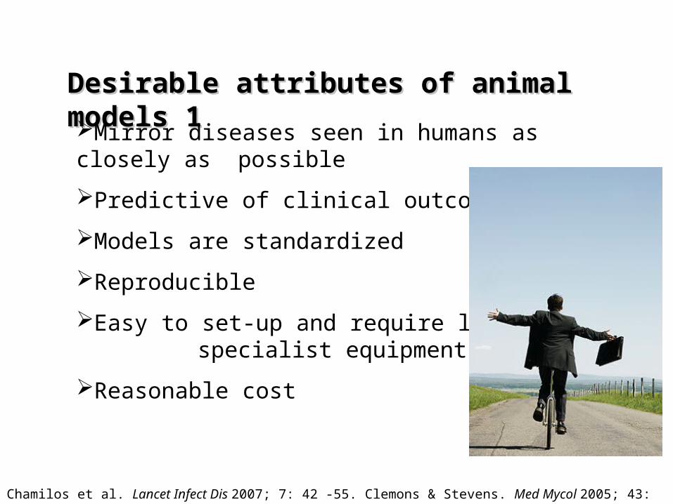
Desirable attributes of animal Desirable attributes of animal models 1models 1Mirror diseases seen in humans as closely as possible
Predictive of clinical outcomes
Models are standardized
Reproducible
Easy to set-up and require little specialist equipment
Reasonable cost
Chamilos et al. Lancet Infect Dis 2007; 7: 42 -55. Clemons & Stevens. Med Mycol 2005; 43: S101-10.

Desirable attributes of animal Desirable attributes of animal models 2models 2Amenable to studies including
* Evaluation of therapeutics
* Evaluation of host response
* Evaluation of pathogen virulence
factors
* Assessment of in vivo gene
expression
Chamilos et al. Lancet Infect Dis 2007; 7: 42 -55. Clemons & Stevens. Med Mycol 2005; 43: S101-10.

Weaknesses of Animal ModelsWeaknesses of Animal Models
Will never fully replicate human disease
No single model answers all questions
May not mimic all structural features
e.g. the structure of mouse lung
Additional effort with drug studies to ‘humanize’ PK and metabolic effects
Animal models can be acute and expensive
Chamilos et al. Lancet Infect Dis 2007; 7: 42 -55. Clemons & Stevens. Med Mycol 2005; 43: S101-10.

Potential sites of infection in mammals
Intravenous/disseminated
Intranasal/sinus
Air Pocket Subcutaneous chamber
Oral
GI tract
Peritoneal?
Vaginal
Claw/nail
Inhaled or tracheal
Skin and hair
Eyes
BladderFootpad
Heart valve
Modulators of fungal infection – host factors• Age of animal – in general younger animals more susceptible
• Genetic background inbred v outbred – only mice
• Immune statusImmunocompetent:
Immunocompromised: neutropenic vs. non-neutropenic
•Tissue damage
• Sex - Hormone status
• Site of infection - route of infection/ method of infection
• Pre exposure to whole fungi- hyphae or spores – immune status
• Sensitization with fungal allergens

Modulators of fungal infection – fungal factors
• Inoculum level
• Stage of growth
• Lag / log /stationary
• Infection form
•Spore v hyphae
• Intrinsic virulence factors of the fungus
• Virulence factors suitable for infection site
• Time between infection and treatment

Housing and HusbandryClean dedicated animal housing
Day/Night light cycles
Controlled temperature/humidity
Room sterilization possible between models
Waste disposal

Housing and Husbandry

Immunosuppression Normally required to establish an infection at the site of interest
Make a model more ‘reproducible’
More closely replicate human disease
Cytotoxic drugs (render animals neutropenic)
Steroids (inhibit functions of immune cells)
Hormones (change conditions at site of infection)
Irradiation (render animals neutropenic)
“Knock-out” / transgenic strains (potential to effect immune function/receptors/ cytokine response etc)
Model types Lethal v non-lethal models
Lethal models:
Animals challenged with increasing inocula till death occurs. Outcome is time of death
Death can be due to multiple causes
Non-lethal models:
Animals infected with lower doses to develop a persistent high level infection with very mild symptoms
Samples can be collected at defined time-points allowing multiple surrogate markers of disease
Infected at a site which avoids systemic dissemination.

Review of the available modelsReview of the available models
Disseminated infection
Intravenous: The “unnatural” model
Easy model for lethal infection in mice and other species
Targets kidneys and spleen, much less the lungs – some strains invade brain – 2o effects can occur
Easy model for antifungal therapy
Can be easily modified to examine pathogen specific virulence factors
Bypasses many stages in the infection process

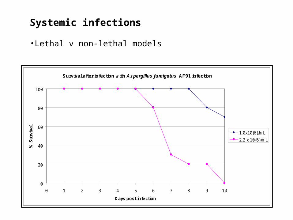
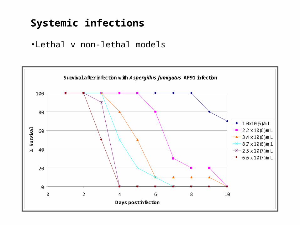
Systemic infections
•Lethal v non-lethal models
Survival after infection with Aspergillus fumigatus AF91 infection
0
20
40
60
80
100
0 1 2 3 4 5 6 7 8 9 10
Days post infection
% S
urv
ival
1.0x10(6)/mL
Systemic infections
•Lethal v non-lethal models
Survival after infection with Aspergillus fumigatus AF91 infection
0
20
40
60
80
100
0 1 2 3 4 5 6 7 8 9 10
Days post infection
% S
urv
ival
1.0x10(6)/mL
2.2 x 10(6)/mL
Systemic infections
•Lethal v non-lethal models
Survival after infection with Aspergillus fumigatus AF91 infection
0
20
40
60
80
100
0 2 4 6 8 10
Days post infection
% S
urv
iva
l
1.0x10(6)/mL
2.2 x 10(6)/mL
3.4 x 10(6)/mL
8.7 x 10(6)/ml
2.5 x 10(7)/mL
6.6 x 10(7)/mL

Systemic infections
Endpoints
Death
Surrogate marker of imminent death (hypothermia/ torticollis/renal failure)
Euthanize animals at specific time-points
Organ culture (quantitative) over a predefined time range
Measurement of fungal products e.g. Chitin, Galactomannan
Measurement of fungal burden by qPCR (either DNA or RNA)/ assessment of fungal gene expression
•Lethal v non-lethal models

ReviewReview of the available modelsof the available modelsMice versus other rodents
Advantages:
Can study disease in mice with specific host immune defects…potentially identifying the most critical
Can study disease in large numbers of fairly uniform inbred animals … increasing reproducibility of results
Less space for housing
Cost
Ease of handling
Disadvantages:
Serial sampling not usually possible
Lung remodelling/airway narrowing differs from larger animals
Drugs are cleared from mice far more rapidly than in humans
Course of disease generally very acute, leading to death or recovery
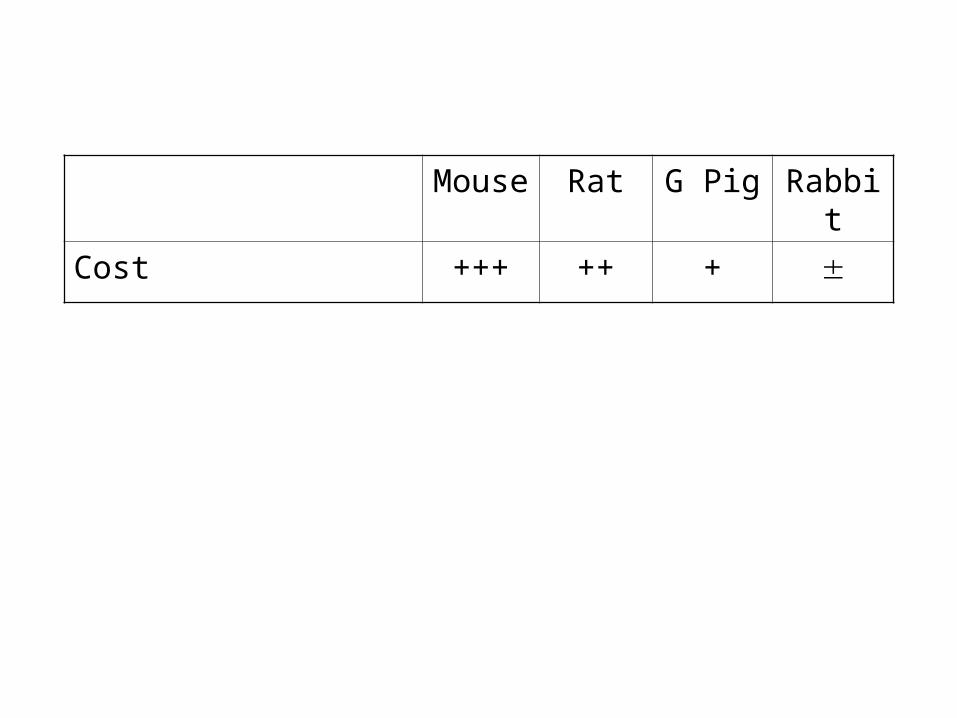
Mouse
Rat G Pig Rabbit
Cost +++ ++ +

Mouse
Rat G Pig Rabbit
Cost +++ ++ + Housing +++ ++ ++

Mouse
Rat G Pig Rabbit
Cost +++ ++ +
Housing +++ ++ ++
Availability in bulk +++ +++ + +

Mouse
Rat G Pig Rabbit
Cost +++ ++ +
Housing +++ ++ ++
Availability in bulk +++ +++ + +
Ease of handling +++ ++ ++ +

Mouse
Rat G Pig Rabbit
Cost +++ ++ +
Housing +++ ++ ++
Availability in bulk +++ +++ + +
Ease of handling +++ ++ ++ +
Daily blood samples - ++ - +++

Mouse
Rat G Pig Rabbit
Cost +++ ++ +
Housing +++ ++ ++
Availability in bulk +++ +++ + +
Ease of handling +++ ++ ++ +
Daily blood samples - ++ - +++
Human lung structure
- + ++ ++
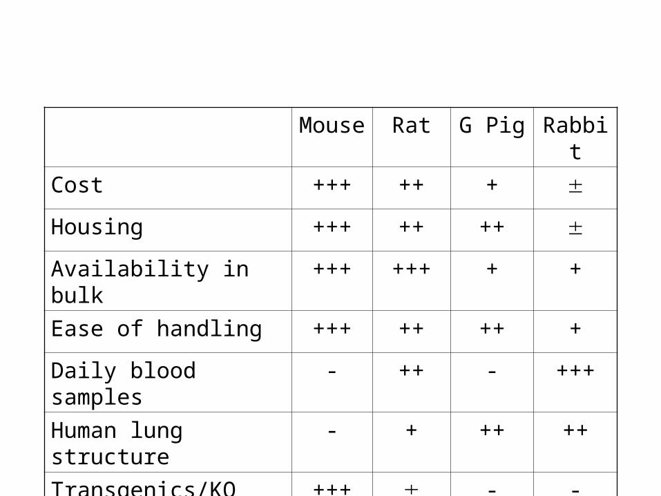
Mouse
Rat G Pig Rabbit
Cost +++ ++ + Housing +++ ++ ++ Availability in bulk +++ +++ + +
Ease of handling +++ ++ ++ +
Daily blood samples - ++ - +++
Human lung structure
- + ++ ++
Transgenics/KO strain
+++ - -

Models of localized infectionsModels of localized infections
a) Invasive Pulmonary aspergillosisMost models of IPA use infection by direct intranasal/intratracheal inoculation
• Mice are anaesthetized and conidia suspension inhaled
• Rats, Guinea pigs & Rabbits infected via tracheostomy/ intubation
Advantages
Relatively cost effective
Little specialist equipment required
Possible to infect large numbers from a single organism stock
Possible to test multiple strains in a single model

Models of localized infectionsModels of localized infections
a) Invasive Pulmonary aspergillosisMost models of IPA use infection by direct intranasal/intratracheal inoculation
• Mice are anaesthetized and conidia suspension inhaled
• Rats, Guinea pigs & Rabbits infected via tracheostomy/ intubation
Drawbacks
Enormous mouse-mouse variation - direct methods better
Inter-laboratory studies difficult
Distribution may not be equal between lobes
Inoculum delivered in liquid – assumption that all of the inoculum delivered to lungs
Some animals develop bacterial pneumonia
Animals develop disease in trachea or sinuses
Therapeutic studies difficult

Piggott and Emmons Adapted Inhalation chamber
Hinners Inhalation Chamber
SIDRANSKY and FRIEDMAN chamber
SIDRANSKYand FRIEDMAN. 1959 Am.J.Pathol. 35:169-183.
Models of localized infectionsModels of localized infectionsa) Invasive Pulmonary aspergillosis
There have been several attempts to standardize delivery of spores but none have been widely accepted

• Development and standardization of aerosol challenge model of invasive pulmonary aspergillosis• Mouse, rat, guinea pig
• Provide samples and resources to other investigators
• Supported by NIH / NIAID
• UTHSCSA / Harbor-UCLA / University of Manchesterhttp://www.sacmm.org/iaam.html

IPA Inoculation Chambers
Acrylic chamber• Conidia delivered via small
particle nebulizer• Consistent inoculum level• 1 hour exposure
Madison chamber• Sealed chamber• Simultaneous exposure of
large number of different species
• Adjust inocula sizes and exposure period

IPA Inoculation Chambers – Mice, rats and guinea pigs

IPA Inoculation Chambers – Mice, rats and guinea pigs
Suitable for:
40 mice
12 rats
8 guinea pigs
Difficult to clean after and between runs
We use vaporized formaldehyde OR VHP
Multiple strains = chambers needed
Models of localized infectionsModels of localized infections
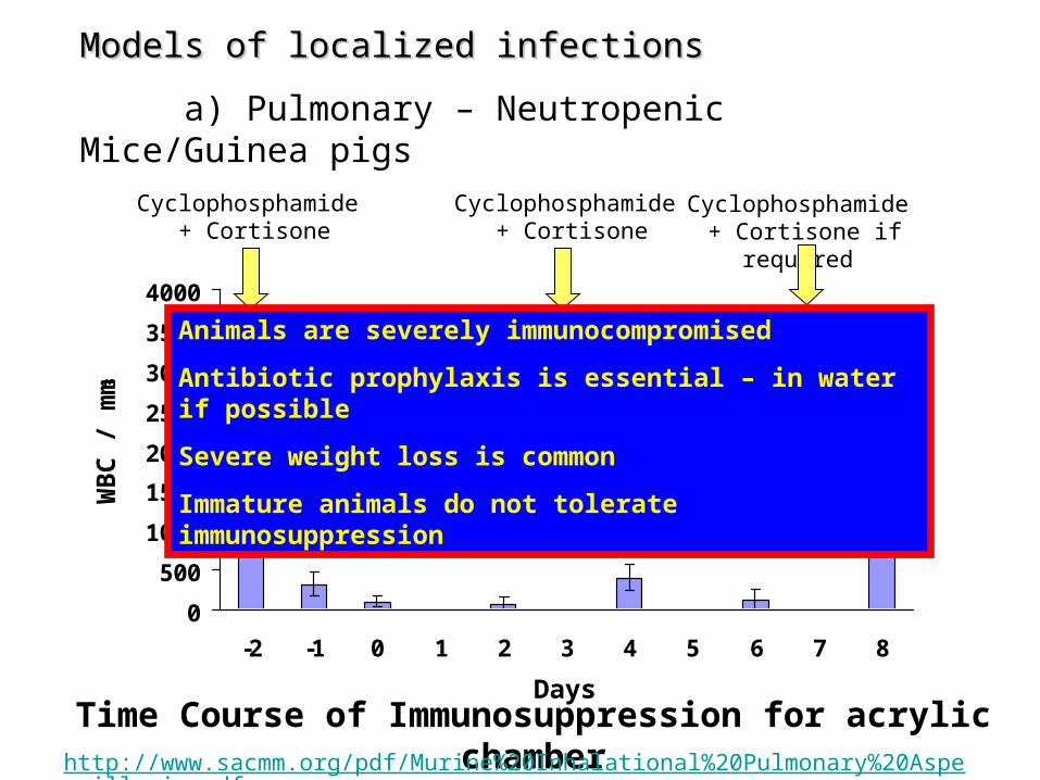
a) Pulmonary – Neutropenic Mice/Guinea pigs
0
500
1000
1500
2000
2500
3000
3500
4000
-2 -1 0 1 2 3 4 5 6 7 8
WB
C /
mm3
Days
Cyclophosphamide + Cortisone
Cyclophosphamide + Cortisone
Infect
Time Course of Immunosuppression for acrylic chamber
Cyclophosphamide + Cortisone if required
Animals are severely immunocompromised
Antibiotic prophylaxis is essential – in water if possible
Severe weight loss is common
Immature animals do not tolerate immunosuppression
http://www.sacmm.org/pdf/Murine%20Inhalational%20Pulmonary%20Aspergillosis.pdf
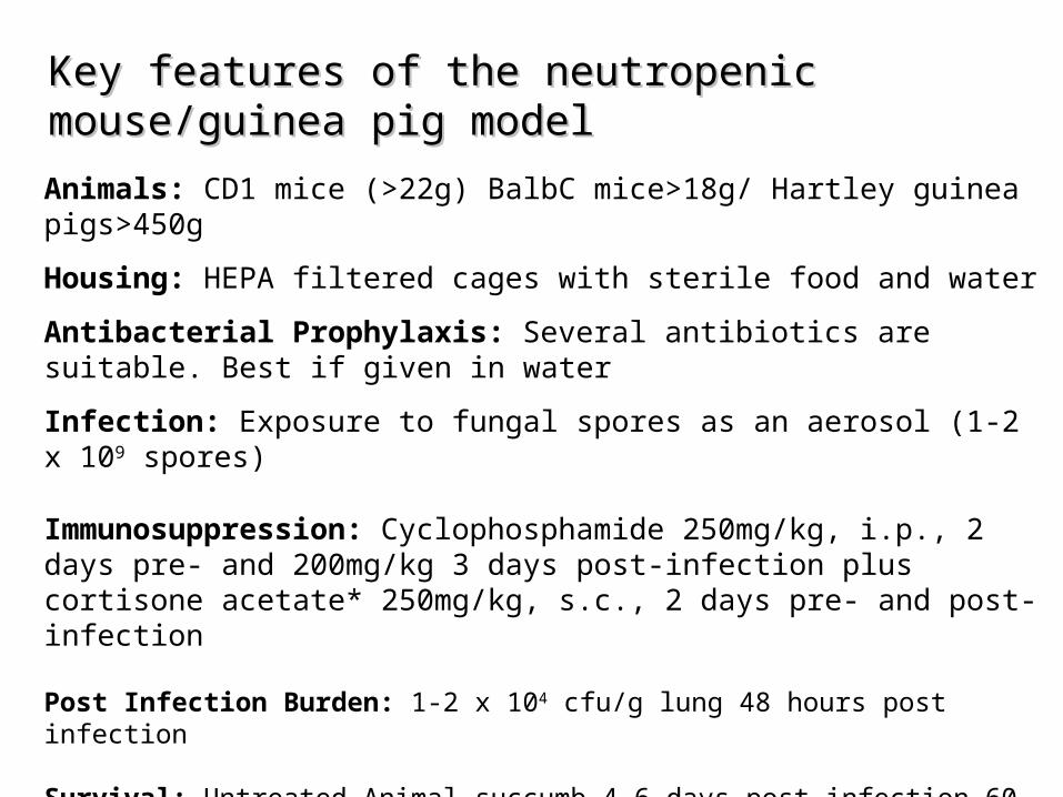
Key features of the neutropenic Key features of the neutropenic mouse/guinea pig modelmouse/guinea pig model
Animals: CD1 mice (>22g) BalbC mice>18g/ Hartley guinea pigs>450g
Housing: HEPA filtered cages with sterile food and water
Antibacterial Prophylaxis: Several antibiotics are suitable. Best if given in water
Infection: Exposure to fungal spores as an aerosol (1-2 x 10⁹ spores)
Immunosuppression: Cyclophosphamide 250mg/kg, i.p., 2 days pre- and 200mg/kg 3 days post-infection plus cortisone acetate* 250mg/kg, s.c., 2 days pre- and post-infection
Post Infection Burden: 1-2 x 10⁴ cfu/g lung 48 hours post infection
Survival: Untreated Animal succumb 4-6 days post infection 60-80% (mice) 100% (guinea pigs) mortality)
*Cortisone acetate is given as a suspension. Has batch variability. Remains as solid beneath skin throughout model
Models of localized Models of localized infectionsinfections
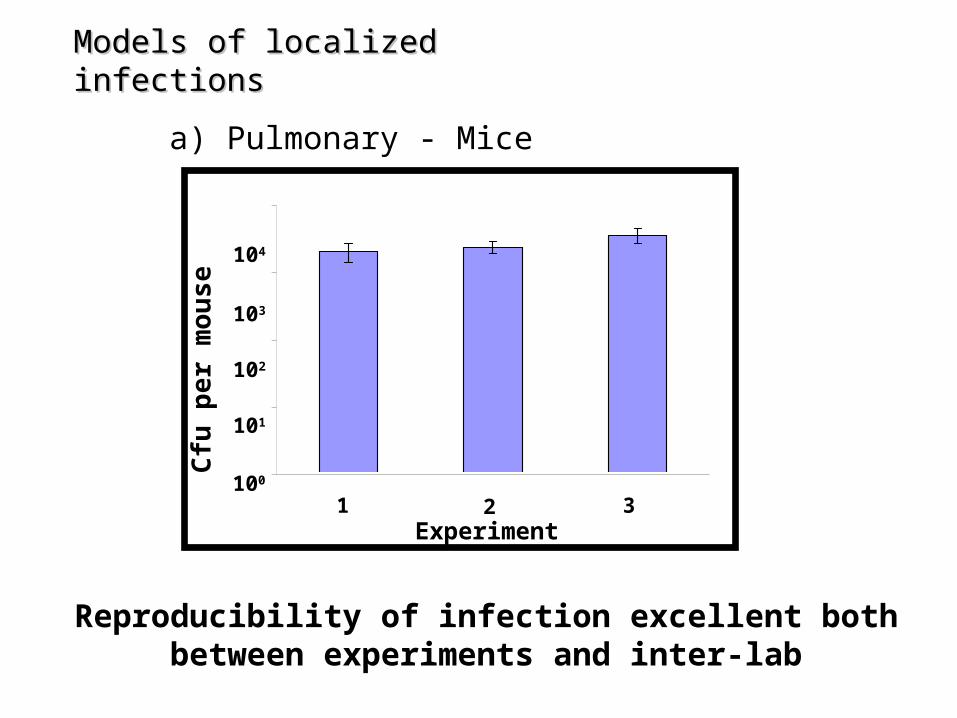
a) Pulmonary - Mice
1
10
100
1000
10000
1 2 3
101
102
103
104
Cfu
per
mou
se
1 2 3Experiment
100
Reproducibility of infection excellent both between experiments and inter-lab

Models of localized infectionsModels of localized infections
a) Pulmonary – Neutropenic Mice
0%
10%
20%
30%
40%
50%
60%
70%
80%
90%
100%
0 2 4 6 8 10 12 14
UNINFECTED
INFECTED
Murine Inhalational Model - Outcomes
Note- This model does not lead to 100% mortality
Note- There is occasionally loss of controls (steroids)

Models of localized infectionsModels of localized infections
a) Pulmonary – Neutropenic Rats
0
500
1000
1500
2000
2500
3000
3500
4000
-2 -1 0 1 2 3 4 5 6 7 8
WB
C /
mm
3
Days
Cyclophosphamide +Long acting steroid
Cyclophosphamide
Infect by aerosol
Time Course of IPA Models
Treatment
Tissue burden mice euthanized
90-100% Untreated mice die
Tissue burden rats euthanized
100% of untreated rats die
Prednisolone in a depo formulation is used IM
Daily tail vein bleeds are possible (~1ml)
Antibiotic prophylaxis is essential – in water if possible
Severe weight loss is common
Rats need a long acclimatization period

Key features of the neutropenic rat Key features of the neutropenic rat ModelModel
Animals: Sprague Dawley rats, Male 225-250g
Housing: HEPA filtered cages with sterile food and water
Antibacterial Prophylaxis: Baytril (enrofloxacin), 4 days pre-infection to prevent secondary bacterial pneumonia & urinary tract infection.
Infection: Exposure to fungal spores as an aerosol (1 x 10⁹ spores)
Immunosuppression: Cyclophosphamide 75mg/kg, i.p., 2 days pre- and post-infection plus Depo-medrone (prednisolone) 15mg/kg, i.m., 2 days pre-infection
Post Infection Burden: 3 x 10⁴ cfu/g lung 48 hours post infection
Survival: Untreated Animal succumb 4-6 days post infection (100% mortality)

Models of localized infectionsModels of localized infections
a) Pulmonary – Neutropenic Rat
Rats immunosupressed with 75mg/kg cyclophosphamide and 10mg/kg depo-medrone
0
20
40
60
80
100
0 1 2 3 4 5 6 7 8
Days post Infection
% S
urvi
val
Infected
Uninfected
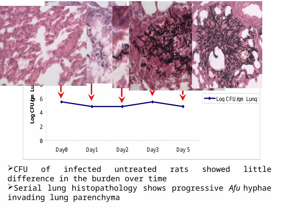
CFU of infected untreated rats showed little difference in the burden over time Serial lung histopathology shows progressive Afu hyphae invading lung parenchyma
Aspergillus Burden Changes During Infection
Log CFU/gm Lung Over Time
0
2
4
6
8
10
12
Day0 Day1 Day2 Day3 Day 5
Log
CFU
/gm
Lun
g
Log CFU/gm Lung

Characteristic disease progression in ratsCharacteristic disease progression in rats
Experimental endpoints: Major causes of death
Weight loss >25%
Laboured breathing
Bloody nasal discharge
Unable to reach food and water

Models of localized infectionsModels of localized infections
a) Pulmonary – Non-neutropenic Rats
0
500
1000
1500
2000
2500
3000
3500
4000
-4 -2 -1 0 1 2 3 4 5 6 7 8
Days
WBC
/mm
3
200mg/kg
Cortisone acetate
Infection by aerosol
200mg/kg
Cortisone acetate
Antibacterial prophylaxis
The non-neutropenic model is similar in rats and mice
The dose of cortisone is limited by toxicity
Antibiotic prophylaxis is essential – in water if possible
Severe weight loss is common
Uninfected animals have large numbers of white cells in lungs at the end of the study
*Danger of Pneumocystis in rats*
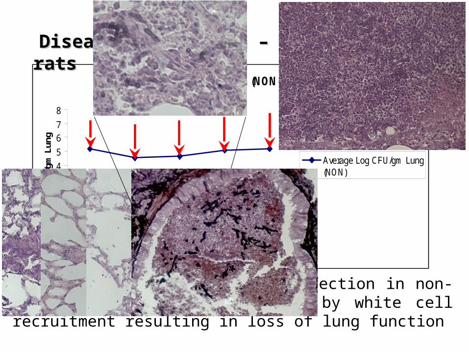
Lung Burden Progression (NON-Neutropenic)
0
12
34
5
67
8
4h 24h 48h 72h 96h
Hours Post-Infection
Log
CFU
/gm
Lun
g
Average Log CFU/gm Lung(NON)
Disease Progression – Non-neutropenic ratsDisease Progression – Non-neutropenic rats
The lung pathology following infection in non-neutropenic hosts is dominated by white cell recruitment resulting in loss of lung function

Neutropenic vs. Non-neutropenicCharacteristic Glucocorticosteroids Neutropenia
Cellular trafficking BAL
Rapid and extensive increase in PMN
No PMN influx
Cytokines BAL TNF-α and IL-10 low to undetected
TNF-α and IL-10 high
Histological features Diffuse and extensive consolidation and inflammation
Limited consolidation, necrosis with hyphae
Fungal elements Small numbers of conidia and poorly germinated hyphae
Extensive angioinvasive hyphae
Amphotericin efficacy
No survival improvement Survival improvement with high dose AmB/AmBisome
Dominant mechanisms
Adverse host inflammation Unimpeded fungal growth / invasion
Berenguer et al. Am J Resp Crit Care Med 1995; 152: 1079.Balloy et al. Infect Immun 2005; 73: 494.
Wiederhold N TIMM 2009

Models of localized infectionsModels of localized infections
a) Pulmonary – Chronic infection mice
C57BL/6 mice infected intratracheally with 1 x 10⁵ spores of A. fumigatus embedded in agarose.
Disease is restricted to the lungs with no tissue invasion. Infections possible
for >20 days
Don Sheppard IAAM Workshop 2008

Chronic Aspergillus Models - Tissue Chambers
Osmotic membrane
Chambers (1cm x 0.3cm) inserted subcutaneously
Aspergillus is separated from cellular responses and unable to invade beyond the chamber.
Sampling possible though silicon membrane
Complex ‘biofilms’ develop in chamber.
Suitable for antifungal efficacy/ development of resistance/ host adaption studies
Silicon rubber membrane
1cm
Animal need ~1 week recovery post surgery
Antibiotic prophylaxis post-op
Chambers can remain in situ for up to 6 weeks
Volume recovered during sampling is small
No time to discuss other models
• Rabbits – great for drug and imaging studies
• Transgenic/knockout mouse models – fantastic for understanding disease mechanisms
• Non-mammalian hosts
• Sinus models
• Allergy/Asthma

Acknowledgements
• Andrew Sharp• Raghdaa Shrief• Jayesh Majithiya• Joanne Slater• David Denning • University of Manchester• IAAM Contract team• Fungal Research Trust