american society of neurorehabilitation - ghajar
TRANSCRIPT
http://nnr.sagepub.com
Neurorehabilitation and Neural Repair
DOI: 10.1177/1545968308315600 2008; 22; 217 Neurorehabil Neural Repair
Jamshid Ghajar and Richard B. Ivry The Predictive Brain State: Timing Deficiency in Traumatic Brain Injury?
http://nnr.sagepub.com/cgi/content/abstract/22/3/217 The online version of this article can be found at:
Published by:
http://www.sagepublications.com
On behalf of:
American Society of Neurorehabilitation
can be found at:Neurorehabilitation and Neural Repair Additional services and information for
http://nnr.sagepub.com/cgi/alerts Email Alerts:
http://nnr.sagepub.com/subscriptions Subscriptions:
http://www.sagepub.com/journalsReprints.navReprints:
http://www.sagepub.com/journalsPermissions.navPermissions:
http://nnr.sagepub.com/cgi/content/refs/22/3/217SAGE Journals Online and HighWire Press platforms):
(this article cites 108 articles hosted on the Citations
© 2008 American Society of Neurorehabilitation. All rights reserved. Not for commercial use or unauthorized distribution. by on May 9, 2008 http://nnr.sagepub.comDownloaded from

Point of View: Directions for Research
The Predictive Brain State: Timing Deficiency inTraumatic Brain Injury?
Jamshid Ghajar, MD, PhD, Richard B. Ivry, PhD, and the Cognitive andNeurobiological Research Consortium
Copyright © 2008 The American Society of Neurorehabilitation 217
Attention and memory deficits observed in traumatic braininjury (TBI) are postulated to result from the shearing ofwhite matter connections between the prefrontal cortex, pari-etal lobe, and cerebellum that are critical in the generation,maintenance, and precise timing of anticipatory neural activ-ity. These fiber tracts are part of a neural network that gener-ates predictions of future states and events, processes that arerequired for optimal performance on attention and workingmemory tasks. The authors discuss the role of this anticipa-tory neural system for understanding the varied symptomsand potential rehabilitation interventions for TBI.
Preparatory neural activity normally allows the efficientintegration of sensory information with goal-based represen-tations. It is postulated that an impairment in the generationof this activity in traumatic brain injury (TBI) leads to per-formance variability as the brain shifts from a predictive toreactive mode. This dysfunction may constitute a fundamen-tal defect in TBI as well as other attention disorders, causingworking memory deficits, distractibility, a loss of goal-ori-ented behavior, and decreased awareness.
“The future is not what is coming to meet us, but whatwe are moving forward to meet.”
⎯⎯Jean-Marie Guyau1
Key Words: Attention—mild traumatic brain injury—headinjury—concussion—post-concussive symptoms—diffuse axonalinjury—neuropsychological tests.
TBI is a global health problem in terms of inci-dence, cost, and impact on daily living.2
Worldwide, an estimated 57 million individualshave been hospitalized with TBI.3 The Glasgow Coma
Scale (GCS) has come to serve as a standardized assess-ment instrument to classify the severity of TBI, withscores ranging from a low of 3 (comatose) to 15 (awakeand following commands). The GCS score is a reliablemeasure of the level of consciousness, but it is of ques-tionable utility for assessing current cognitive status andpredicting longer-term cognitive sequelae. Additionalassessments are especially critical given that more than80% of individuals classified as mild TBI or concussionhave GCS scores between 13 and 15. In the United States,medical center emergency rooms report approximately1.74 million cases of mild TBI.4 These estimates fail toinclude unrecognized or unreported TBI cases that maynumber up to 3 million3,5 for sports-related injuries andup to 40% for personnel participating in current mili-tary engagements.6,7
Mild TBI or concussion with a normal GCS scoreimplies little or no brain injury; indeed, CT imagingstudies reveal traumatic lesions in less than 15% ofpatients.8 Cognitive sequelae, which are difficult to mea-sure, are often ascribed to the traumatic event or pre-morbid factors.9 However, we postulate that subtle brainwhite matter tract lesions that are not detected by stan-dard forms of neuroimaging may lead to chronic diffi-culties in complex cognitive tasks. These white mattertract lesions are located predominantly in anterior cere-bral areas, which are selectively vulnerable to rotationalshearing injury. Furthermore, these fiber tracts are partof a neural network that generates predictions of futurestates and events, processes that are required for optimalperformance on attention and working memory tasks.We hypothesize that impairments in this anticipatoryprocess likely contribute to the fact that approximately20% of individuals with TBI end up being classified aschronically disabled.10
Unless otherwise specified, we use the term “TBI” todenote mild TBI patients and survivors of more severeTBI who are awake and responsive. Although we expectthe general ideas advanced here are relevant for under-standing the cognitive deficits in all forms of TBI, muchof the discussion in the literature has centered onaccounting for the cognitive deficits that persist even inindividuals with relatively mild overt symptoms.
From the Brain Trauma Foundation and the Department ofNeurological Surgery, Weill Medical College of Cornell University, NewYork, New York (JG), and the Department of Psychology, University ofCalifornia, Berkeley (RBI).
Address correspondence to Jamshid Ghajar, Brain Trauma Foundation,708 3rd Ave, Suite 1810, New York, NY 10017. E-mail: [email protected]. Neurorehabil Neural Repair 2008;22:217–227.
DOI: 10.1177/1545968308315600
© 2008 American Society of Neurorehabilitation. All rights reserved. Not for commercial use or unauthorized distribution. by on May 9, 2008 http://nnr.sagepub.comDownloaded from

THE PREDICTIVE BRAIN STATE
Currently, there is no causative explanation for theattention deficits and persistence of symptoms in TBIpatients.11,12 An extensive review of TBI by Alexander13
finds no single psychological, physiologic, somatic, ordemographic factor that can account for persistentpostconcussive symptoms (PCS). Many PCS symptomsare equally compatible with diagnoses of depression,anxiety, and chronic pain as they are with disorders ofcognition. The lack of a clear understanding of themechanisms underlying concussive symptoms hasimportant consequences for the treatment of these indi-viduals. For example, clear assessment guidelines arelacking to help determine when it is appropriate for ath-letes with concussive symptoms to resume sports, inlarge part because there is no consensus on objectivemeasures to assess the level of cognitive function atwhich there remains a high vulnerability for reinjury.14
A widely accepted view is that frontal lobe damageunderlies the cognitive deficits associated with TBI. Thisview is consistent, to some degree, with the central roleattributed to this brain region in functions related toattention and working memory. However, frontal lobelesion volumes in TBI patients do not correlate withneuropsychological deficits.15 This observation, coupledwith more sophisticated models of cognition, suggeststhat these complex processes involve distributed neuralnetworks and that the effects of TBI reflect disruption ofthese networks.16 While TBI certainly results in frontal-like symptoms, one goal in this review is to encourage abroader conceptualization of how TBI might disruptcontrol processes. In particular, we outline a network-based hypothesis that emphasizes an impairment inanticipatory control, a form of prediction. Rather thanattribute this process to a single neural region such asthe frontal lobe, we consider how this predictive aspectof brain function emerges through the interactions offrontal and parietal regions, as well as the cerebellum.
A core premise we develop in this article is that oneaspect of attention emerges from the generation ofmoment-to-moment expectancies about the immediatefuture. To be clear, we argue that the causal link here isnot that sensory expectancy requires attention; rather,we propose that attention is the consequence of, orrather, is manifest as expectancies of the future. Theseexpectancies are a core feature of goal-oriented behav-ior, providing predictions to be compared with sensoryfeedback. These expectancies, based on prior experi-ence, are essential for ensuring the fluid operation ofcognitive functions, allowing them to be anticipatoryrather than reactive. One consequence of successfulanticipation is that performance becomes less variable.By accurately anticipating the future, the performeris less likely to miss task-relevant information or be
distracted by irrelevant information, 2 prominentsources of errors. By anticipation, the individual is “pay-ing attention,” creating a future-oriented brain state.
Anticipatory Neural Network
Prediction is a general feature of cognition, operat-ing over multiple time scales. When driving, we mayanticipate the actions of an oncoming car as well asthink about the location of a lunch-stop, 100 milesdown the highway. Within the framework of our modelof TBI-related cognitive deficits, we limit our discussionto the saliency of short-range predictions that requirethe comparison of expectancies and feedback with real-time properties. We hypothesize that these temporalpredictions emerge from the interactions of a networkinvolving frontal, parietal, and cerebellar areas (Table1). Could one prominent source of performance vari-ability in TBI be related to an impaired ability to gener-ate and/or maintain predictive states? Our premise isthat disruption of the connectivity between these areasaffects this predictive capability and, as such, results indeficits in goal-oriented behavior arising from problemswith attention and working memory.
One way to examine the neural underpinnings ofprediction is to study the activation and coherence ofactivation in and between cortical areas that arerequired for anticipatory activity in attention and work-ing memory. Studies of spatial attention frequently usetasks in which a cue, indicating either the probable loca-tion of a stimulus or serving solely as an alerting signal,is followed after a short delay by an imperative stimulus.Neuroimaging studies consistently reveal that, followingthe cue, a network encompassing the dorsolateral pre-frontal cortex (PFC) and parietal lobe is engaged, mostprominently in the right hemisphere.17-19 In addition tothis cerebral cortical network, anticipation also engagesthe cerebellum; functional magnetic resonance imaging(fMRI) studies consistently show activation of the cere-bellum during the interval following attentional cues(Figure 1).20-22 This activation is not dependent on thepresence of movement, although it may require thepreparation of a potential response.
Electrophysiological markers of anticipation associ-ated with the PFC include the readiness potential (RP)23
on tasks in which the movement is self-initiated and con-tingent negative variation (CNV)24 when the response isinitiated following an imperative stimulus. In TBIpatients, the CNV amplitude is decreased. Reactiontimes become more variable, and errors in performancecorrelate with the amplitude of the CNV.25 There are alsoreported reductions or absence of the RP in TBI patientsin self-initiated movements (Figure 2),26 similar to that
Ghajar, Ivry
218 Neurorehabilitation and Neural Repair 22(3); 2008 © 2008 American Society of Neurorehabilitation. All rights reserved. Not for commercial use or unauthorized distribution.
by on May 9, 2008 http://nnr.sagepub.comDownloaded from
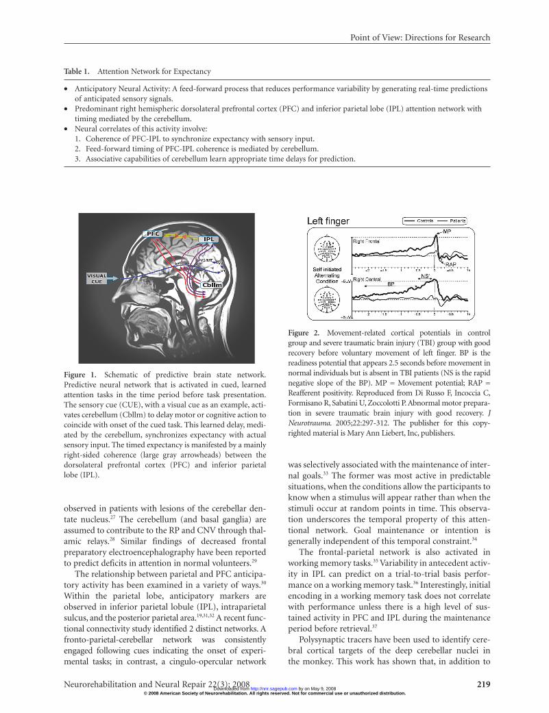
observed in patients with lesions of the cerebellar den-tate nucleus.27 The cerebellum (and basal ganglia) areassumed to contribute to the RP and CNV through thal-amic relays.28 Similar findings of decreased frontalpreparatory electroencephalography have been reportedto predict deficits in attention in normal volunteers.29
The relationship between parietal and PFC anticipa-tory activity has been examined in a variety of ways.30
Within the parietal lobe, anticipatory markers areobserved in inferior parietal lobule (IPL), intraparietalsulcus, and the posterior parietal area.19,31,32 A recent func-tional connectivity study identified 2 distinct networks. Afronto-parietal-cerebellar network was consistentlyengaged following cues indicating the onset of experi-mental tasks; in contrast, a cingulo-opercular network
was selectively associated with the maintenance of inter-nal goals.33 The former was most active in predictablesituations, when the conditions allow the participants toknow when a stimulus will appear rather than when thestimuli occur at random points in time. This observa-tion underscores the temporal property of this atten-tional network. Goal maintenance or intention isgenerally independent of this temporal constraint.34
The frontal-parietal network is also activated inworking memory tasks.35 Variability in antecedent activ-ity in IPL can predict on a trial-to-trial basis perfor-mance on a working memory task.36 Interestingly, initialencoding in a working memory task does not correlatewith performance unless there is a high level of sus-tained activity in PFC and IPL during the maintenanceperiod before retrieval.37
Polysynaptic tracers have been used to identify cere-bral cortical targets of the deep cerebellar nuclei inthe monkey. This work has shown that, in addition to
Point of View: Directions for Research
Neurorehabilitation and Neural Repair 22(3); 2008 219
Table 1. Attention Network for Expectancy
• Anticipatory Neural Activity: A feed-forward process that reduces performance variability by generating real-time predictionsof anticipated sensory signals.
• Predominant right hemispheric dorsolateral prefrontal cortex (PFC) and inferior parietal lobe (IPL) attention network withtiming mediated by the cerebellum.
• Neural correlates of this activity involve:1. Coherence of PFC-IPL to synchronize expectancy with sensory input.2. Feed-forward timing of PFC-IPL coherence is mediated by cerebellum.3. Associative capabilities of cerebellum learn appropriate time delays for prediction.
Figure 1. Schematic of predictive brain state network.Predictive neural network that is activated in cued, learnedattention tasks in the time period before task presentation.The sensory cue (CUE), with a visual cue as an example, acti-vates cerebellum (Cbllm) to delay motor or cognitive action tocoincide with onset of the cued task. This learned delay, medi-ated by the cerebellum, synchronizes expectancy with actualsensory input. The timed expectancy is manifested by a mainlyright-sided coherence (large gray arrowheads) between thedorsolateral prefrontal cortex (PFC) and inferior parietallobe (IPL).
Figure 2. Movement-related cortical potentials in controlgroup and severe traumatic brain injury (TBI) group with goodrecovery before voluntary movement of left finger. BP is thereadiness potential that appears 2.5 seconds before movement innormal individuals but is absent in TBI patients (NS is the rapidnegative slope of the BP). MP = Movement potential; RAP =Reafferent positivity. Reproduced from Di Russo F, Incoccia C,Formisano R, Sabatini U, Zoccolotti P. Abnormal motor prepara-tion in severe traumatic brain injury with good recovery. JNeurotrauma. 2005;22:297-312. The publisher for this copy-righted material is Mary Ann Liebert, Inc, publishers.
© 2008 American Society of Neurorehabilitation. All rights reserved. Not for commercial use or unauthorized distribution. by on May 9, 2008 http://nnr.sagepub.comDownloaded from
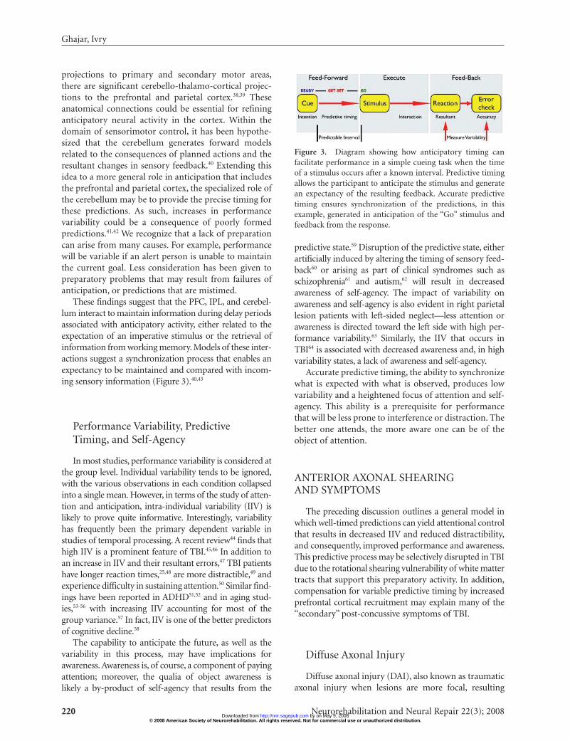
projections to primary and secondary motor areas,there are significant cerebello-thalamo-cortical projec-tions to the prefrontal and parietal cortex.38,39 Theseanatomical connections could be essential for refininganticipatory neural activity in the cortex. Within thedomain of sensorimotor control, it has been hypothe-sized that the cerebellum generates forward modelsrelated to the consequences of planned actions and theresultant changes in sensory feedback.40 Extending thisidea to a more general role in anticipation that includesthe prefrontal and parietal cortex, the specialized role ofthe cerebellum may be to provide the precise timing forthese predictions. As such, increases in performancevariability could be a consequence of poorly formedpredictions.41,42 We recognize that a lack of preparationcan arise from many causes. For example, performancewill be variable if an alert person is unable to maintainthe current goal. Less consideration has been given topreparatory problems that may result from failures ofanticipation, or predictions that are mistimed.
These findings suggest that the PFC, IPL, and cerebel-lum interact to maintain information during delay periodsassociated with anticipatory activity, either related to theexpectation of an imperative stimulus or the retrieval ofinformation from working memory. Models of these inter-actions suggest a synchronization process that enables anexpectancy to be maintained and compared with incom-ing sensory information (Figure 3).40,43
Performance Variability, PredictiveTiming, and Self-Agency
In most studies, performance variability is considered atthe group level. Individual variability tends to be ignored,with the various observations in each condition collapsedinto a single mean. However, in terms of the study of atten-tion and anticipation, intra-individual variability (IIV) islikely to prove quite informative. Interestingly, variabilityhas frequently been the primary dependent variable instudies of temporal processing. A recent review44 finds thathigh IIV is a prominent feature of TBI.45,46 In addition toan increase in IIV and their resultant errors,47 TBI patientshave longer reaction times,25,48 are more distractible,49 andexperience difficulty in sustaining attention.50 Similar find-ings have been reported in ADHD51,52 and in aging stud-ies,53-56 with increasing IIV accounting for most of thegroup variance.57 In fact, IIV is one of the better predictorsof cognitive decline.58
The capability to anticipate the future, as well as thevariability in this process, may have implications forawareness. Awareness is, of course, a component of payingattention; moreover, the qualia of object awareness islikely a by-product of self-agency that results from the
predictive state.59 Disruption of the predictive state, eitherartificially induced by altering the timing of sensory feed-back60 or arising as part of clinical syndromes such asschizophrenia61 and autism,62 will result in decreasedawareness of self-agency. The impact of variability onawareness and self-agency is also evident in right parietallesion patients with left-sided neglect—less attention orawareness is directed toward the left side with high per-formance variability.63 Similarly, the IIV that occurs inTBI64 is associated with decreased awareness and, in highvariability states, a lack of awareness and self-agency.
Accurate predictive timing, the ability to synchronizewhat is expected with what is observed, produces lowvariability and a heightened focus of attention and self-agency. This ability is a prerequisite for performancethat will be less prone to interference or distraction. Thebetter one attends, the more aware one can be of theobject of attention.
ANTERIOR AXONAL SHEARINGAND SYMPTOMS
The preceding discussion outlines a general model inwhich well-timed predictions can yield attentional controlthat results in decreased IIV and reduced distractibility,and consequently, improved performance and awareness.This predictive process may be selectively disrupted in TBIdue to the rotational shearing vulnerability of white mattertracts that support this preparatory activity. In addition,compensation for variable predictive timing by increasedprefrontal cortical recruitment may explain many of the“secondary” post-concussive symptoms of TBI.
Diffuse Axonal Injury
Diffuse axonal injury (DAI), also known as traumaticaxonal injury when lesions are more focal, resulting
Ghajar, Ivry
220 Neurorehabilitation and Neural Repair 22(3); 2008
Figure 3. Diagram showing how anticipatory timing canfacilitate performance in a simple cueing task when the timeof a stimulus occurs after a known interval. Predictive timingallows the participant to anticipate the stimulus and generatean expectancy of the resulting feedback. Accurate predictivetiming ensures synchronization of the predictions, in thisexample, generated in anticipation of the “Go” stimulus andfeedback from the response.
© 2008 American Society of Neurorehabilitation. All rights reserved. Not for commercial use or unauthorized distribution. by on May 9, 2008 http://nnr.sagepub.comDownloaded from
from rotational shear forces65-67 is common in TBI68 andmay account for persistent cognitive deficits and symp-toms.69,70 This pattern of injury, distinguished fromfocal contusion without shearing, is characterized bydamage to anterior axons at the gray/white matter junc-tion of the cerebral hemispheres, corpus callosum, andin severe cases, dorsolateral midbrain. These patterns ofshearing are often accompanied by similar damage inthe superior cerebellar peduncles.71,72 Deep white matterabnormalities on magnetic resonance imaging (MRI)are associated with poorer neuropsychological test per-formances and poor long-term outcome.73 The severityof deep white matter tract injury may explain manyTBI-induced cognitive deficits, ranging from mild con-cussion to the vegetative state.74,75
The normal CT or MRI scans observed in the major-ity of cases of mild TBI may belie microscopic axonalpathology. The sensitivity of most MRI structural stud-ies is insufficient to detect white matter tract disruptionor small areas of edema. Postmortem analysis, whenconducted within a few hours of severe TBI, showsextensive microscopic hemorrhages and severing ofnerve fibers in pons, midbrain, and corpus callosum,consistent with shearing forces.76 Even in TBI cases clas-sified as mild (in which the patients died of other causes),white matter microhemorrhages in the brain stem, cere-bral hemispheres, and corpus callosum are evident.77
DAI and Deficits in Attention andWorking Memory
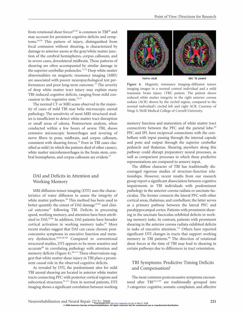
MRI diffusion tensor imaging (DTI) uses the charac-teristics of water diffusion to assess the integrity ofwhite matter pathways.78 This method has been used tobetter quantify the extent of DAI damage79-82 and clini-cal outcome11 following TBI. Deficits in processingspeed, working memory, and attention have been attrib-uted to DAI.83,84 In addition, DAI patients have broadercortical activation in working memory tasks.85 Morerecent studies suggest that DAI can cause chronic post-concussive symptoms in executive function and mem-ory dysfunction.69,81,86-88 Compared to conventionalstructural studies, DTI appears to be more sensitive andaccurate89 in correlating pathology with attention andmemory deficits (Figure 4).90,111 These observations sug-gest that white matter shear injury in TBI plays a promi-nent causal role in the observed cognitive deficits.
As revealed by DTI, the predominant sites for mildTBI axonal shearing are located in anterior white mattertracts connecting PFC with posterior cortical regions andsubcortical structures.91,111 Even in normal patients, DTIimaging shows a significant correlation between working
memory function and maturation of white matter tractconnectivity between the PFC and the parietal lobe.92
PFC and IPL have reciprocal connections with the cere-bellum with input passing through the internal capsuleand pons and output through the superior cerebellarpeduncle and thalamus. Shearing anywhere along thispathway could disrupt preparatory neural processes, aswell as comparison processes in which these predictiverepresentations are compared to sensory input.
The diffuse character of TBI has traditionally dis-couraged rigorous studies of structure-function rela-tionships. However, recent results from our researchgroup report a significant dissociation between cognitiveimpairments in TBI individuals with predominantpathology in the anterior corona radiata or uncinate fas-ciculus. The former connects the lateral PFC with othercortical areas, thalamus, and cerebellum; the latter servesas a primary pathway between the lateral PFC andparahippocampal cortex. Patients with prominent shear-ing in the uncinate fasciculus exhibited deficits in work-ing memory tasks. In contrast, patients with prominentshearing in the anterior corona radiata exhibited deficitsin tasks of executive attention.111 Others have reportedsignificant DTI changes in tracts that support workingmemory in TBI patients.90 The direction of rotationalshear forces at the time of TBI may lead to shearing incertain pathways due to differences in tract orientation.
TBI Symptoms: Predictive Timing Deficitsand Compensation?
The most common postconcussive symptoms encoun-tered after TBI10,13,93 are traditionally grouped into3 categories: cognitive, somatic complaints, and affective
Point of View: Directions for Research
Neurorehabilitation and Neural Repair 22(3); 2008 221
Figure 4. Magnetic resonance imaging–diffusion tensorimaging images in a normal control individual and a mildtraumatic brain injury (TBI) patient. The patient showsreduced white matter integrity in the right anterior coronaradiata (ACR) shown by the circled region, compared to thenormal individual’s circled left and right ACR. Courtesy ofNiogi S, Weill Medical College of Cornell University.
© 2008 American Society of Neurorehabilitation. All rights reserved. Not for commercial use or unauthorized distribution. by on May 9, 2008 http://nnr.sagepub.comDownloaded from
complaints (Table 2). Cognitive function is a major pre-dictor of poor outcome in TBI patients.94 In particular,working memory is vulnerable95,96 and likely underliesmany of the prominent problems associated with dailylife activities that require planning and problem solv-ing.97 Similar neuropsychological deficiencies are seen inchildren with TBI.98 Some symptoms of TBI may resultfrom damage to core cognitive processes, whereas othersmay be due to compensatory mechanisms recruited tominimize the ensuing performance variability.
The performance of individuals with TBI is especiallyvulnerable to tasks that tax attention, such as when task-relevant and task-irrelevant stimuli are intermixed,96 or ifthere is an increase in task difficulty. Activation patterns inchronic patients with TBI tend to be abnormal on tasks ofexecutive function. These abnormalities may be manifestas more diffuse patterns of cortical recruitment85 or asgreater increases in prefrontal activity despite perfor-mance levels that are below that observed in controls.99
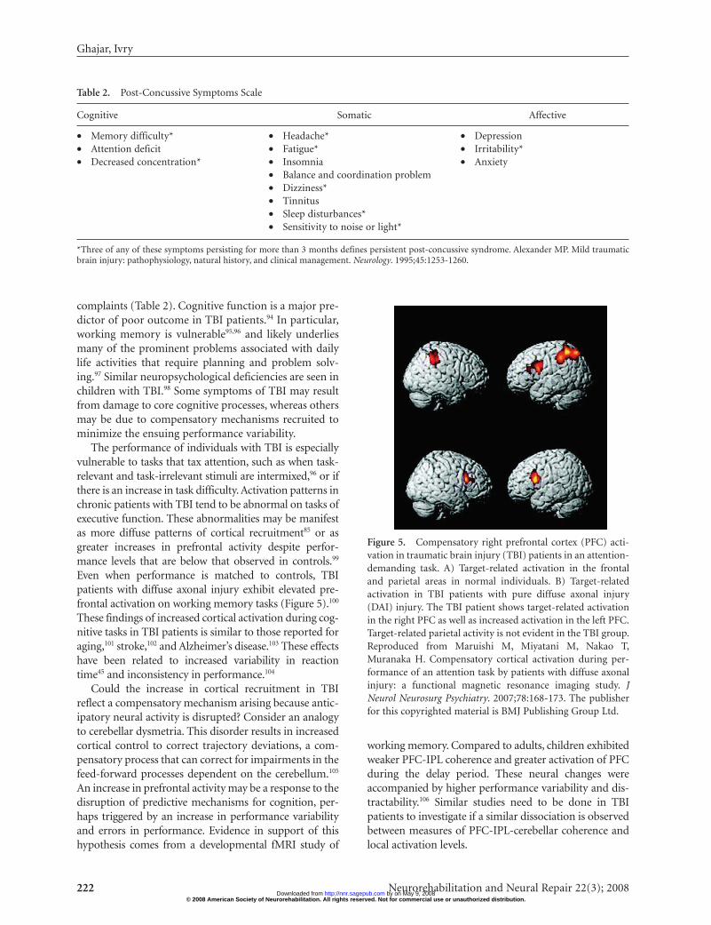
Even when performance is matched to controls, TBIpatients with diffuse axonal injury exhibit elevated pre-frontal activation on working memory tasks (Figure 5).100
These findings of increased cortical activation during cog-nitive tasks in TBI patients is similar to those reported foraging,101 stroke,102 and Alzheimer’s disease.103 These effectshave been related to increased variability in reactiontime45 and inconsistency in performance.104
Could the increase in cortical recruitment in TBIreflect a compensatory mechanism arising because antic-ipatory neural activity is disrupted? Consider an analogyto cerebellar dysmetria. This disorder results in increasedcortical control to correct trajectory deviations, a com-pensatory process that can correct for impairments in thefeed-forward processes dependent on the cerebellum.105
An increase in prefrontal activity may be a response to thedisruption of predictive mechanisms for cognition, per-haps triggered by an increase in performance variabilityand errors in performance. Evidence in support of thishypothesis comes from a developmental fMRI study of
working memory. Compared to adults, children exhibitedweaker PFC-IPL coherence and greater activation of PFCduring the delay period. These neural changes wereaccompanied by higher performance variability and dis-tractability.106 Similar studies need to be done in TBIpatients to investigate if a similar dissociation is observedbetween measures of PFC-IPL-cerebellar coherence andlocal activation levels.
Ghajar, Ivry
222 Neurorehabilitation and Neural Repair 22(3); 2008
Table 2. Post-Concussive Symptoms Scale
Cognitive Somatic Affective
• Memory difficulty*• Attention deficit• Decreased concentration*
• Headache*• Fatigue*• Insomnia• Balance and coordination problem• Dizziness*• Tinnitus• Sleep disturbances*• Sensitivity to noise or light*
• Depression• Irritability*• Anxiety
*Three of any of these symptoms persisting for more than 3 months defines persistent post-concussive syndrome. Alexander MP. Mild traumaticbrain injury: pathophysiology, natural history, and clinical management. Neurology. 1995;45:1253-1260.
Figure 5. Compensatory right prefrontal cortex (PFC) acti-vation in traumatic brain injury (TBI) patients in an attention-demanding task. A) Target-related activation in the frontaland parietal areas in normal individuals. B) Target-relatedactivation in TBI patients with pure diffuse axonal injury(DAI) injury. The TBI patient shows target-related activationin the right PFC as well as increased activation in the left PFC.Target-related parietal activity is not evident in the TBI group.Reproduced from Maruishi M, Miyatani M, Nakao T,Muranaka H. Compensatory cortical activation during per-formance of an attention task by patients with diffuse axonalinjury: a functional magnetic resonance imaging study. JNeurol Neurosurg Psychiatry. 2007;78:168-173. The publisherfor this copyrighted material is BMJ Publishing Group Ltd.
© 2008 American Society of Neurorehabilitation. All rights reserved. Not for commercial use or unauthorized distribution. by on May 9, 2008 http://nnr.sagepub.comDownloaded from

Classifying symptoms based on the framework out-lined in this article would posit that postconcussivesymptoms encountered after a TBI could be grouped intoprimary and secondary causes (Table 3). Primarysymptoms arise from TBI axonal shearing in white mattertracts connecting the PFC-IPL-cerebellar network leadingto decreased PFC-IPL coherence (Figure 6). The result-ing increase in variability would be manifest as increaseddistractibility, working memory deficits, and problemswith balance and coordination. An inability to anticipatesensory events, resulting in a temporal mismatch of sen-sory expectation to actual sensory input, could lead todizziness, tinnitus, and sensory hypersensitivity. Self-agency would be abnormal due to the de-synchroniza-tion between actions and their resulting consequences.
Secondary symptoms might be a consequence ofheightened activation of the PFC. This might arise from anincrease in error signals and performance variability. PFCrecruitment, serving as a compensatory mechanism, couldhelp bridge the moment-to-moment temporal discrepan-cies. The expended effort might underlie fatigue, headache,irritability, anxiety, and when prolonged, depression.
Variability As a Metric of Prediction
If a consequence of accurate anticipation is a reduc-tion in performance variability, then variability can beused as a metric to assess this aspect of attention. By def-inition, attention varies over time. Relatively continuousand extended measures of performance should be usedto gauge moment-to-moment fluctuations in perfor-mance within individuals. Tasks used to assess IIV havetypically involved relatively discrete measures such asreaction time,25,45 allowing measurements, at best, everyfew seconds. Such measures may be relatively insensitiveto lapses in attention.
We have employed a task in which we measure circu-lar smooth pursuit eye movements during a predictivetracking task, lasting 2.5 seconds using a high-resolution
camera sampling performance at 500 Hz over blocks of20 trials. The predictive nature of this task engages thePFC-parietal-cerebellar network, similar to that observedin studies of spatial attention. The variability in the dif-ference between eye and target position, as well as veloc-ity error (the difference between target velocity and eyevelocity), both correlate with performance on a workingmemory task.107,108 In addition, TBI patients with chroniccognitive symptoms had higher variability and a persis-tent lag in eye position compared to control individuals.
Further studies have demonstrated that adding aconcurrent secondary task (short-term verbal memory)during the smooth pursuit tracking task produces dif-ferent effects on velocity error variability in control andTBI patients. In TBI patients, there is a significantincrease in variability as the difficulty of the secondarytask increases. In contrast, control individuals showed adecrease in velocity error variability when the length ofthe word list was increased, a counterintuitive finding,yet one that is also observed in other attention-loading
Point of View: Directions for Research
Neurorehabilitation and Neural Repair 22(3); 2008 223
Figure 6. Figure showing predominant focal areas of diffuseaxonal injury (DAI).
Table 3. Post-Concussive Symptoms
Primary Symptoms – Related to Secondary Symptoms – Related To PFCPredictive Timing Deficit Compensation and Error Signaling
• Decreased attention• Decreased memory• Decreased concentration• Balance and coordination problem• Decreased awareness• Dizziness• Tinnitus• Sensory sensitivity
• Fatigue• Headache• Insomnia• Irritability• Anxiety• Depression
© 2008 American Society of Neurorehabilitation. All rights reserved. Not for commercial use or unauthorized distribution. by on May 9, 2008 http://nnr.sagepub.comDownloaded from
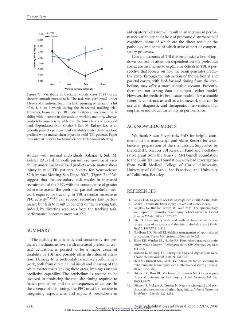
studies with normal individuals (Ghajar J, Suh M,Kolster RA, et al. Smooth pursuit eye movement vari-ability under dual-task load predicts white matter shearinjury in mild TBI patients. Society for Neuroscience37th Annual Meeting. San Diego 2007) (Figure 7).109 Wesuggest that the secondary task results in increasedrecruitment of the PFC, with the consequence of greatercoherence across the prefrontal-parietal-cerebellar net-work required for tracking. In TBI, a similar increase inPFC activity85,100,110 can support secondary task perfor-mance but fails to result in benefits on the tracking task.Indeed, by diverting resources from the tracking task,performance becomes more variable.
SUMMARY
The inability to efficiently and consistently use pre-dictive mechanisms, even with increased prefrontal cor-tical activation, is posited to be a major cause ofdisability in TBI, and possibly other disorders of atten-tion. Damage to a prefrontal-parietal-cerebellum net-work, both from direct neural insult and shearing of thewhite matter tracts linking these areas, impinges on thispredictive capability. The cerebellum is posited to beinvolved in producing the requisite timing required tomatch predictions and the consequences of actions. Inthe absence of this timing, the PFC must be reactive inintegrating expectancies and input. A breakdown in
anticipatory behavior will result in an increase in perfor-mance variability and a host of profound disturbances ofcognition, some of which are the direct result of thepathology and some of which arise as part of compen-satory processes.
Current accounts of TBI that emphasize a loss of top-down control of attention dependent on the prefrontalcortex are insufficient to explain the deficits in TBI. A per-spective that focuses on how the brain generates predic-tive states through the interaction of the prefrontal andparietal cortex, with feed-forward timing from the cere-bellum, may offer a more complete account. Presently,there are not strong data to support either model.However, the predictive brain state model offers a testablescientific construct, as well as a framework that can beuseful in diagnostic and therapeutic interventions thatemphasize individual variability in performance.
ACKNOWLEDGMENTS
We thank Susan Fitzpatrick, PhD, for helpful com-ments on the manuscript and Akiyo Kodera for assis-tance in preparation of the manuscript. Supported bythe Rachel L. Mellon TBI Research Fund and a collabo-rative grant from the James S. McDonnell Foundationto the Brain Trauma Foundation, with lead investigatorsfrom Weill Medical College of Cornell University,University of California, San Francisco, and Universityof California, Berkeley.
REFERENCES
1. Guyau J-M. La genèse de l’idée de temps. Paris: Félix Alcan; 1890.2. Ghajar J. Traumatic brain injury. Lancet. 2000;356:923-929.3. Langlois JA, Rutland-Brown W, Wald MM. The epidemiology
and impact of traumatic brain injury: a brief overview. J HeadTrauma Rehabil. 2006;21:375-378.
4. Fife D. Head injury with and without hospital admission:comparisons of incidence and short-term disability. Am J PublicHealth. 1987;77:810-812.
5. Goldberg LD, Dimeff RJ. Sideline management of sport-relatedconcussions. Sports Med Arthrosc. 2006;14:199-205.
6. Taber KH, Warden DL, Hurley RA. Blast-related traumatic braininjury: what is known? J Neuropsychiatry Clin Neurosci. 2006;18:141-145.
7. Warden D. Military TBI during the Iraq and Afghanistan wars.J Head Trauma Rehabil. 2006;21:398-402.
8. Stein SC, Burnett MG, Glick HA. Indications for CT scanning inmild traumatic brain injury: a cost-effectiveness study. J Trauma.2006;61:558-566.
9. Dikmen SS, Ross BL, Machamer JE, Temkin NR. One year psy-chosocial outcome in head injury. J Int Neuropsychol Soc.1995;1:67-77.
10. Dikmen S, McLean A, Temkin N. Neuropsychological and psy-chosocial consequences of minor head injury. J Neurol NeurosurgPsychiatry. 1986;49:1227-1232.
Ghajar, Ivry
224 Neurorehabilitation and Neural Repair 22(3); 2008
Figure 7. Variability of tracking velocity error (VE) duringcircular smooth pursuit task. The task was performed under4 levels of attentional load in a task requiring rehearsal of a listof 0, 1, 5, or 9 words during the 30-second tracking trial.Traumatic brain injury (TBI) patients show an increase in vari-ability with increases in demands on working memory, whereascontrols become less variable over the lower levels of increasedload. Reproduced from Ghajar J, Suh M, Kolster RA, et al.Smooth pursuit eye movement variability under dual-task loadpredicts white matter shear injury in mild TBI patients. Paperpresented at: Society for Neuroscience 37th Annual Meeting.
© 2008 American Society of Neurorehabilitation. All rights reserved. Not for commercial use or unauthorized distribution. by on May 9, 2008 http://nnr.sagepub.comDownloaded from

11. Schaefer PW, Huisman TA, Sorensen AG, Gonzalez RG, SchwammLH. Diffusion-weighted MR imaging in closed head injury: highcorrelation with initial Glasgow coma scale score and score onmodified Rankin scale at discharge. Radiology. 2004;233:58-66.
12. Huisman TA, Schwamm LH, Schaefer PW, et al. Diffusion tensorimaging as potential biomarker of white matter injury in diffuseaxonal injury. AJNR Am J Neuroradiol. 2004;25:370-376.
13. Alexander MP. Mild traumatic brain injury: pathophysiology, naturalhistory, and clinical management. Neurology. 1995;45:1253-1260.
14. Concussion (mild traumatic brain injury) and the team physician:a consensus statement. Med Sci Sports Exerc. 2006;38:395-399.
15. Scheibel RS, Newsome M, Steinberg J, Pearson D, Rauch R, MaoH, Levin H. Altered brain activation during cognitive control inpatients with moderate to severe traumatic brain injury.Neurorehabil Neural Repair. 2007;21:36-45.
16. Knight RT. Neuroscience. Neural networks debunk phrenology.Science. 2007;316:1578-1579.
17. Posner MI, Walker JA, Friedrich FJ, Rafal RD. Effects of parietalinjury on covert orienting of attention. J Neurosci. 1984;4:1863-1874.
18. Thiel CM, Zilles K, Fink GR. Cerebral correlates of alerting, ori-enting and reorienting of visuospatial attention: an event-relatedfMRI study. Neuroimage. 2004;21:318-328.
19. Naghavi HR, Nyberg L. Common fronto-parietal activity inattention, memory, and consciousness: shared demands on inte-gration? Conscious Cogn. 2005;14:390-425.
20. Kim SG, Ugurbil K, Strick PL. Activation of a cerebellar outputnucleus during cognitive processing. Science. 1994;265:949-951.
21. Tomasi D, Ernst T, Caparelli EC, Chang L. Practice-inducedchanges of brain function during visual attention: a parametricfMRI study at 4 Tesla. Neuroimage. 2004;23:1414-1421.
22. Macar F, Anton JL, Bonnet M, Vidal F. Timing functions of thesupplementary motor area: an event-related fMRI study. BrainRes Cogn Brain Res. 2004;21:206-215.
23. Cunnington R, Windischberger C, Deecke L, Moser E. Thepreparation and readiness for voluntary movement: a high-fieldevent-related fMRI study of the Bereitschafts-BOLD response.Neuroimage. 2003;20:404-412.
24. Walter WG, Cooper R, Aldridge VJ, McCallum WC, Winter AL.Contingent negative variation: an electric sign of sensorimotorassociation and expectancy in the human brain. Nature.1964;203:380-384.
25. Segalowitz SJ, Dywan J, Unsal A. Attentional factors in responsetime variability after traumatic brain injury: an ERP study. J IntNeuropsychol Soc. 1997;3:95-107.
26. Di Russo F, Incoccia C, Formisano R, Sabatini U, Zoccolotti P.Abnormal motor preparation in severe traumatic brain injurywith good recovery. J Neurotrauma. 2005;22:297-312.
27. Kitamura J, Shabasaki H, Terashi A, Tashima K. Cortical poten-tials preceding voluntary finger movement in patients with focalcerebellar lesion. Clin Neurophysiol. 1999;110:126-132.
28. Paradiso G, Cunic D, Chen R. Involvement of subcortical struc-tures in the preparation of self-paced movement. J Psychophysiol.2004;18:130-139.
29. Padilla ML, Wood RA, Hale LA, Knight RT. Lapses in a pre-frontal-extrastriate preparatory attention network predict mis-takes. J Cogn Neurosci. 2006;18:1477-1487.
30. Nagai Y, Critchley HD, Featherstone E, Fenwick PB, Trimble MR,Dolan RJ. Brain activity relating to the contingent negative vari-ation: an fMRI investigation. Neuroimage. 2004;21:1232-1241.
31. Knight RT. Distributed cortical network for visual attention.J Cogn Neurosci. 1997;9:75-91.
32. Wiese H, Stude P, Nebel K, Forsting M, de Greiff A. Prefrontalcortex activity in self-initiated movements is condition-specific,but not movement-related. Neuroimage. 2005;28:691-697.
33. Dosenbach NU, Fair DA, Miezin FM, et al. Distinct brain net-works for adaptive and stable task control in humans. Proc NatlAcad Sci U S A. 2007;104:11073-11078.
34. Manly T, Owen AM, McAvinue L, et al. Enhancing the sensitiv-ity of a sustained attention task to frontal damage: convergentclinical and functional imaging evidence. Neurocase. 2003;9:340-349.
35. LaBar KS,Gitelman DR,Parrish TB,Mesulam M.Neuroanatomic over-lap of working memory and spatial attention networks: a functionalMRI comparison within subjects. Neuroimage. 1999; 10:695-704.
36. Sapir A, d’Avossa G, McAvoy M, Shulman GL, Corbetta M. Brainsignals for spatial attention predict performance in a motion discrimination task. Proc Natl Acad Sci U S A. 2005;102:17810-17815.
37. Pessoa L, Gutierrez E, Bandettini P, Ungerleider L. Neural corre-lates of visual working memory: fMRI amplitude predicts taskperformance. Neuron. 2002;35:975-987.
38. Dum RP, Strick PL. An unfolded map of the cerebellar dentatenucleus and its projections to the cerebral cortex. J Neurophysiol.2003;89:634-639.
39. Middleton FA, Strick PL. Cerebellar projections to the prefrontalcortex of the primate. J Neurosci. 2001;21:700-712.
40. Blakemore SJ, Sirigu A. Action prediction in the cerebellum andin the parietal lobe. Exp Brain Res. 2003;153:239-245.
41. Mauk MD, Medina JF, Nores WL, Ohyama T. Cerebellar func-tion: coordination, learning or timing? Curr Biol. 2000;10:R522-R525.
42. Mehta B, Schaal S. Forward models in visuomotor control. JNeurophysiol. 2002;88:942-953.
43. Volker S, Gross H-M. Neural anticipative architecture for expec-tation driven perception. Proceedings of the 2001 IEEE Systems,Man, and Cybernetics Conference; 2001.
44. MacDonald SW, Nyberg L, Backman L. Intra-individual variabil-ity in behavior: links to brain structure, neurotransmission andneuronal activity. Trends Neurosci. 2006;29:474-480.
45. Stuss DT, Pogue J, Buckle L, Bondar J. Characterization of stabil-ity of performance in patients with traumatic brain injury: vari-ability and consistency on reaction time test. Neuropsychology.1994;8:316-324.
46. Collins LF, Long CJ. Visual reaction time and its relationship toneuropsychological test performance. Arch Clin Neuropsychol.1996;11:613-623.
47. Dockree PM, Bellgrove MA, O’Keeffe F M, et al. Sustained atten-tion in traumatic brain injury and healthy controls: enhancedsensitivity with dual-task load. Exp Brain Res. 2006;168:218-219.
48. Hetherington CR, Stuss DT, Finlayson MA. Reaction time andvariability 5 and 10 years after traumatic brain injury. Brain Inj.1996;10:473-486.
49. Mangels JA, Craik FI, Levine B, Schwartz ML, Stuss DT. Effects ofdivided attention on episodic memory in chronic traumatic braininjury: a function of severity and strategy. Neuropsychologia.2002;40:2369-2385.
50. Whyte J, Polansky M, Fleming M, Coslett HB, Cavallucci C.Sustained arousal and attention after traumatic brain injury.Neuropsychologia. 1995;33:797-813.
51. Castellanos FX, Sonuga-Barke EJ, Scheres A, Di Martino A, HydeC, Walters JR. Varieties of attention-deficit/hyperactivity disorder-related intra-individual variability. Biol Psychiatry. 2005;57:1416-1423.
52. Klein C, Wendling K, Huettner P, Ruder H, Peper M. Intra-subject variability in attention-deficit hyperactivity disorder. BiolPsychiatry. 2006;60:1088-1097.
53. Hultsch DF, MacDonald SW, Dixon RA. Variability in reactiontime performance of younger and older adults. J Gerontol BPsychol Sci Soc Sci. 2002;57:P101-P115.
Point of View: Directions for Research
Neurorehabilitation and Neural Repair 22(3); 2008 225 © 2008 American Society of Neurorehabilitation. All rights reserved. Not for commercial use or unauthorized distribution.
by on May 9, 2008 http://nnr.sagepub.comDownloaded from

54. Martin M, Hofer SM. Intraindividual variability, change, andaging: conceptual and analytical issues. Gerontology. 2004;50:7-11.
55. Nesselroade JR, Salthouse TA. Methodological and theoreticalimplications of intraindividual variability in perceptual-motor performance. J Gerontol B Psychol Sci Soc Sci. 2004;59:P49-P55.
56. Gorus E, De Raedt R, Mets T. Diversity, dispersion and inconsis-tency of reaction time measures: effects of age and task complex-ity. Aging Clin Exp Res. 2006;18:407-417.
57. Strauss E, MacDonald SW, Hunter M, Moll A, Hultsch DF.Intraindividual variability in cognitive performance in threegroups of older adults: cross-domain links to physical status andself-perceived affect and beliefs. J Int Neuropsychol Soc. 2002;8:893-906.
58. MacDonald SW, Hultsch DF, Dixon RA. Performance variabilityis related to change in cognition: evidence from the VictoriaLongitudinal Study. Psychol Aging. 2003;18:510-523.
59. Jeannerod M. The mechanism of self-recognition in humans.Behav Brain Res. 2003;142:1-15.
60. Blakemore SJ, Frith CD, Wolpert DM. Spatio-temporal predic-tion modulates the perception of self-produced stimuli. J CognNeurosci. 1999;11:551-559.
61. Posada A, Franck N, Georgieff N, Jeannerod M. Anticipatingincoming events: an impaired cognitive process in schizophrenia.Cognition. 2001;81:209-225.
62. Schmitz C, Martineau J, Barthélémy C, Assaiante C. Motor con-trol and children with autism: deficit of anticipatory function?Neurosci Lett. 2003;348:17-20.
63. Anderson B, Mennemeier M, Chatterjee A. Variability not ability:another basis for performance decrements in neglect.Neuropsychologia.2000;38:785-796.
64. Prigatano GP. Disturbances of self-awareness and rehabilitationof patients with traumatic brain injury: a 20-year perspective. JHead Trauma Rehabil. 2005;20:19-29.
65. Gennarelli TA, Graham DI. Neuropathology of the head injuries.Semin Clin Neuropsychiatry. 1998;3:160-175.
66. Povlishock JT, Katz DI. Update of neuropathology and neurolog-ical recovery after traumatic brain injury. J Head Trauma Rehabil.2005;20:76-94.
67. Smith DH, Meaney DF, Shull WH. Diffuse axonal injury in headtrauma. J Head Trauma Rehabil. 2003;18:307-316.
68. Medana IM, Esiri MM. Axonal damage: a key predictor of out-come in human CNS diseases. Brain. 2003;126:515-530.
69. Salmond CH, Menon DK, Chatfield DA, et al. Diffusion tensorimaging in chronic head injury survivors: correlations withlearning and memory indices. Neuroimage. 2006;29:117-124.
70. Schmithorst VJ, Wilke M, Dardzinski BJ, Holland SK. Cognitivefunctions correlate with white matter architecture in a normalpediatric population: a diffusion tensor MRI study. Hum BrainMapp. 2005;26:139-147.
71. Gennarelli TA, Thibault LE, Adams JH, Graham DI, ThompsonCJ, Marcincin RP. Diffuse axonal injury and traumatic coma inthe primate. Ann Neurol. 1982;12:564-574.
72. Raghupathi R, Margulies SS. Traumatic axonal injury after closedhead injury in the neonatal pig. J Neurotrauma. 2002;19:843-853.
73. Wilson JT, Pettigrew LE, Teasdale GM. Structured interviewsfor the Glasgow Outcome Scale and the extended GlasgowOutcome Scale: guidelines for their use. J Neurotrauma. 1998;15:573-585.
74. Ross BL, Temkin NR, Newell D, Dikmen SS. Neuropsychologicaloutcome in relation to head injury severity. Contributions ofcoma length and focal abnormalities. Am J Phys Med Rehabil.1994;73:341-347.
75. Kampfl A, Schmutzhard E, Franz G, et al. Prediction of recoveryfrom post-traumatic vegetative state with cerebral magnetic-res-onance imaging. Lancet. 1998;351:1763-1767.
76. Oppenheimer DR. Microscopic lesions in the brain followinghead injury. J Neurol Neurosurg Psychiatry. 1968;31:299-306.
77. Blumbergs PC, Scott G, Manavis J, Wainwright H, Simpson DA,McLean AJ. Staining of amyloid precursor protein to studyaxonal damage in mild head injury. Lancet. 1994;344:1055-1056.
78. Basser PJ, Pierpaoli C. Microstructural and physiological featuresof tissues elucidated by quantitative-diffusion-tensor MRI. JMagn Reson B. 1996;111:209-219.
79. Arfanakis K, Haughton VM, Carew JD, Rogers BP, Dempsey RJ,Meyerand ME. Diffusion tensor MR imaging in diffuse axonalinjury. AJNR Am J Neuroradiol. 2002;23:794-802.
80. Huisman TA. Diffusion-weighted imaging: basic concepts andapplication in cerebral stroke and head trauma. Eur Radiol.2003;13:2283-2297.
81. Huisman TA, Schwamm LH, Schaefer PW, et al. Diffusion tensorimaging as potential biomarker of white matter injury in diffuseaxonal injury. AJNR Am J Neuroradiol. 2004;25:370-376.
82. Lee ZI, Byun WM, Jang SH, Ahn SH, Moon HK, Chang Y.Diffusion tensor magnetic resonance imaging of microstructuralabnormalities in children with brain injury. Am J Phys MedRehabil. 2003;82:556-559.
83. Wallesch CW, Curio N, Galazky I, Jost S, Synowitz H. The neu-ropsychology of blunt head injury in the early postacute stage:effects of focal lesions and diffuse axonal injury. J Neurotrauma.2001;18:11-20.
84. Wallesch CW, Curio N, Kutz S, Jost S, Bartels C, Synowitz H.Outcome after mild-to-moderate blunt head injury: effects offocal lesions and diffuse axonal injury. Brain Inj. 2001;15:401-412.
85. Christodoulou C, DeLuca J, Ricker JH, et al. Functional magneticresonance imaging of working memory impairment after trau-matic brain injury. J Neurol Neurosurg Psychiatry. 2001;71:161-168.
86. Fork M, Bartels C, Ebert AD, Grubich C, Synowitz H, WalleschCW. Neuropsychological sequelae of diffuse traumatic braininjury. Brain Inj. 2005;19:101-108.
87. Himanen L, Portin R, Isoniemi H, Helenius H, Kurki T, TenovuoO. Cognitive functions in relation to MRI findings 30 years aftertraumatic brain injury. Brain Inj. 2005;19:93-100.
88. Himanen L, Portin R, Isoniemi H, Helenius H, Kurki T, TenovuoO. Longitudinal cognitive changes in traumatic brain injury: a30-year follow-up study. Neurology. 2006;66:187-192.
89. Mac Donald CL, Dikranian K, Song SK, Bayly PV, HoltzmanDM, Brody DL. Detection of traumatic axonal injury with diffu-sion tensor imaging in a mouse model of traumatic brain injury.Exp Neurol. 2007;205:116-131.
90. Salmond CH, Menon DK, Chatfield DA, et al. Diffusion tensorimaging in chronic head injury survivors: correlations withlearning and memory indices. Neuroimage. 2005;29:117-124.
91. Inglese M, Makani S, Johnson G, et al. Diffuse axonal injury inmild traumatic brain injury: a diffusion tensor imaging study. JNeurosurg. 2005;103:298-303.
92. Olesen PJ, Nagy Z, Westerberg H, Klingberg T. Combined analy-sis of DTI and fMRI data reveals a joint maturation of white andgrey matter in a fronto-parietal network. Brain Res Cogn BrainRes. 2003;18:48-57.
93. Martinez P, Richters JE. The NIMH community violence project:II. Children’s distress symptoms associated with violence expo-sure. Psychiatry. 1993;56:22-35.
94. Kaplan CP, Corrigan JD. The relationship between cognition andfunctional independence in adults with traumatic brain injury.Arch Phys Med Rehabil. 1994;75:643-647.
95. Levin HS, Gary HE Jr, Eisenberg HM, et al. Neurobehavioral out-come 1 year after severe head injury. Experience of the TraumaticComa Data Bank. J Neurosurg. 1990;73:699-709.
96. Stuss DT, Ely P, Hugenholtz H, et al. Subtle neuropsychologicaldeficits in patients with good recovery after closed head injury.Neurosurgery. 1985;17:41-47.
Ghajar, Ivry
226 Neurorehabilitation and Neural Repair 22(3); 2008 © 2008 American Society of Neurorehabilitation. All rights reserved. Not for commercial use or unauthorized distribution.
by on May 9, 2008 http://nnr.sagepub.comDownloaded from
97. Drake AI, Gray N, Yoder S, Pramuka M, Llewellyn M. Factors pre-dicting return to work following mild traumatic brain injury: adiscriminant analysis. J Head Trauma Rehabil. 2000;15:1103-1112.
98. Ewing-Cobbs L, Fletcher JM, Levin HS, Francis DJ, Davidson K,Miner ME. Longitudinal neuropsychological outcome in infantsand preschoolers with traumatic brain injury. J Int NeuropsycholSoc. 1997;3:581-591.
99. Levine B, Cabeza R, McIntosh AR, Black SE, Grady CL, Stuss DT.Functional reorganisation of memory after traumatic braininjury: a study with H(2)(15)0 positron emission tomography. JNeurol Neurosurg Psychiatry. 2002;73:173-181.
100. Maruishi M, Miyatani M, Nakao T, Muranaka H. Compensatorycortical activation during performance of an attention task bypatients with diffuse axonal injury: a functional magnetic resonanceimaging study. J Neurol Neurosurg Psychiatry. 2007;78:168-173.
101. Cabeza R, Grady CL, Nyberg L, et al. Age-related differences inneural activity during memory encoding and retrieval: a positronemission tomography study. J Neurosci. 1997;17:391-400.
102. Lotze M, Grodd W, Rodden F, Schonle P, Katdatzke B, Cohen LG.Neuroimaging patterns associated with motor control in trau-matic brain injury. Neurorehabil Neural Repair. 2006;20:14-23.
103. Woodard JL, Grafton ST, Votaw JR, Green RC, Dobraski ME,Hoffman JM. Compensatory recruitment of neural resourcesduring overt rehearsal of word lists in Alzheimer’s disease.Neuropsychology. 1998;12:491-504.
104. Bleiberg J, Garmoe WS, Halpern EL, Reeves DL, Nadler JD.Consistency of within-day and across-day performance after
mild brain injury. Neuropsychiatr Neuropsychol Behav Neurol. 1997;10:247-253.
105. Timmann D, Citron R, Watts S, Hore J. Increased variabilityin finger position occurs throughout overarm throws madeby cerebellar and unskilled subjects. J Neurophysiol. 2001;86:2690-2702.
106. Olesen PJ, Macoveanu J, Tegner J, Klingberg T. Brain activityrelated to working memory and distraction in children andadults. Cereb Cortex. 2007;17:1047-1054.
107. Suh M, Kolster R, Sarkar R, McCandliss B, Ghajar J. Deficits inpredictive smooth pursuit after mild traumatic brain injury.Neurosci Lett. 2006;401:108-113.
108. Suh M, Basu S, Kolster R, Sarkar R, McCandliss B, Ghajar J. Increasedoculomotor deficits during target blanking as an indicator of mildtraumatic brain injury. Neurosci Lett. 2006;410: 203-207.
109. Stuss DT, Murphy KJ, Binns MA, Alexander MP. Staying on thejob: the frontal lobes control individual performance variability.Brain. 2003;126:2363-2380.
110. McAllister TW, Saykin AJ, Flashman LA, et al. Brain activationduring working memory 1 month after mild traumatic braininjury: a functional MRI study. Neurology. 1999;53:1300-1308.
111. Niogi SN, Mukherjee P, Ghajar J, Johnson C, Kolster RA, Sarkar R,Lee H, Meeker M, Zimmerman RD, Manley GT, McCandliss BD.Extent of Microstructural White Matter Injury in PostconcussiveSyndrome Correlates with Impaired Cognitive Reaction Time:A 3T Diffusion Tensor Imaging Study of Mild Traumatic BrainInjury. AJNR Am J Neuroradiol. 2008 Feb 26; [Epub ahead of print]
Point of View: Directions for Research
Neurorehabilitation and Neural Repair 22(3); 2008 227 © 2008 American Society of Neurorehabilitation. All rights reserved. Not for commercial use or unauthorized distribution.
by on May 9, 2008 http://nnr.sagepub.comDownloaded from