computational neurorehabilitation: modeling plasticity and...
TRANSCRIPT
REVIEW Open Access
Computational neurorehabilitation:modeling plasticity and learning to predictrecoveryDavid J. Reinkensmeyer1*, Etienne Burdet2, Maura Casadio3, John W. Krakauer4, Gert Kwakkel5,6,7,Catherine E. Lang8, Stephan P. Swinnen9,10, Nick S. Ward11 and Nicolas Schweighofer12
Abstract
Despite progress in using computational approaches to inform medicine and neuroscience in the last 30 years,there have been few attempts to model the mechanisms underlying sensorimotor rehabilitation. We argue that afundamental understanding of neurologic recovery, and as a result accurate predictions at the individual level, willbe facilitated by developing computational models of the salient neural processes, including plasticity and learningsystems of the brain, and integrating them into a context specific to rehabilitation. Here, we therefore discussComputational Neurorehabilitation, a newly emerging field aimed at modeling plasticity and motor learning tounderstand and improve movement recovery of individuals with neurologic impairment. We first explain how theemergence of robotics and wearable sensors for rehabilitation is providing data that make development andtesting of such models increasingly feasible. We then review key aspects of plasticity and motor learning that suchmodels will incorporate. We proceed by discussing how computational neurorehabilitation models relate to thecurrent benchmark in rehabilitation modeling – regression-based, prognostic modeling. We then critically discussthe first computational neurorehabilitation models, which have primarily focused on modeling rehabilitation of theupper extremity after stroke, and show how even simple models have produced novel ideas for futureinvestigation. Finally, we conclude with key directions for future research, anticipating that soon we will see theemergence of mechanistic models of motor recovery that are informed by clinical imaging results and driven bythe actual movement content of rehabilitation therapy as well as wearable sensor-based records of daily activity.
Keywords: Neurorehabilitation, Computational modeling, Motor control, Plasticity, Motor learning, Stroke recovery
BackgroundNature of the problem and definition of computationalneurorehabilitationMobility-related disability arising from neurologic injuryis a worldwide problem of pressing concern. For ex-ample, 16.9 million people suffer a first stroke eachyear, resulting in about 33 million survivors of strokewho are currently alive, making stroke one of the maincauses of acquired adult disability [1]. Up to 74 % of strokesurvivors worldwide require some assistance from care-givers for their basic activities of daily living (ADL) [2].
Disabling disorders such as stroke can be classified withinthe World Health Organization’s International Classifica-tion of Functioning, Disability, and Health (ICF) frame-work, which highlights the multi-tiered effect of stroke onthe individual in terms of pathology (disease or diagnosis),impairment (symptoms and signs), activity limitations(disability), and participation restriction (handicap) (seeFig. 1 in refs [3, 4]). The present paper argues thatmechanism-based, computational modeling of neuroreh-abilitation (Fig. 1) will be a valuable tool for improving re-habilitation strategies and furthering the recovery ofindividuals with neurologic injury at all of these levels.At the onset, we define several terms that we will use
throughout the paper, which provide a conceptualframework for computational neurorehabilitation. Wewill use the term “recovery” to describe improvements
* Correspondence: [email protected] of Anatomy and Neurobiology, Mechanical and AerospaceEngineering, Biomedical Engineering, and Physical Medicine andRehabilitation, University of California, Irvine, USAFull list of author information is available at the end of the article
© 2016 Reinkensmeyer et al. Open Access This article is distributed under the terms of the Creative Commons Attribution 4.0International License (http://creativecommons.org/licenses/by/4.0/), which permits unrestricted use, distribution, andreproduction in any medium, provided you give appropriate credit to the original author(s) and the source, provide a link tothe Creative Commons license, and indicate if changes were made. The Creative Commons Public Domain Dedication waiver(http://creativecommons.org/publicdomain/zero/1.0/) applies to the data made available in this article, unless otherwise stated.
Reinkensmeyer et al. Journal of NeuroEngineering and Rehabilitation (2016) 13:42 DOI 10.1186/s12984-016-0148-3

in movement ability over time, resulting in improvementsat any of the ICF levels, regardless of how these improve-ments occurred. Note that this definition of recovery issimilar to that used in [5, 6], but different from that pro-posed in [4], where the term is restricted to improvementsresulting from restitution of normative biological struc-tures and functions; we feel that “restitution” is indeed themore natural term for this more specific concept (we alsofound ourselves using the term “true recovery” in ourdiscussions; others use the term “repair”.) Thus, for thepurposes of this paper, we follow the nomenclature in[5, 6], in which recovery occurs through restitution,but also through compensation, which we define asuse of biological structures and/or function differentfrom those originally used before the injury to achieve amovement goal. Easy-to-understand examples of compen-sation are, after a stroke, using the less-affected side toperform tasks that one normally would have done withthe more-affected side, or, reaching forward by leaning anabnormal amount with the trunk rather than using theusual amount of shoulder and elbow extension [7]. Notethat the different modes of recovery (restitution and com-pensation) may occur concurrently at different levels ofanalysis. For example, more normal movement behavior,
which appears as biomechanical restitution, may resultfrom leveraging residual neural substrate, a form of neuralcompensation.We use the terms “learning” and “plasticity” as follows
(and here, we are referring to motor system learning andplasticity). If people with or without a neurologic injurytrain at a motor task their ability to perform the task willimprove through normal skill acquisition [8]. Thisprocess of “motor learning” is dependent on plasticity,both in health and disease. In chronic stroke patients,training of appropriate tasks can therefore lead to im-proved function (Fig. 1b) [9]. However, the anatomy of thedamage sets a limit on how much impairments, such asdegraded force production capability, can be reduced inthe chronic phase. Therefore functional improvements inthis phase often appear to be due to learning compensa-tion techniques [10], although targeted training may allowmodest reduction in specific impairments, assessed quan-titatively (e.g. [8, 11–13]). The early post-stroke period isinteresting in that there are a number of injury-inducedchanges in the potential for plasticity, including, for ex-ample, exuberant production then activity-based pruningof new synapses [14], that may last several months, caus-ing spontaneous biological recovery (see below) [10]. At
A B
C D
Fig. 1 a General framework of computational neurorehabilitation models. Such models predict patient functional outcomes by drivingcomputational representations of plasticity and learning with sensorimotor activity achieved in rehabilitation therapy and/or throughout thecourse of daily life. b Computational neurorehabilitation models presume that rehabilitation modulates both spontaneous biological recovery andmotor learning, leading to improvements in both impaired limb motor control and compensatory movement strategies. Shown here is anestimate of the dose-response effect arising from additional therapy time, obtained by plotting effect sizes of 30 studies of upper and lowerextremity rehabilitation therapy after stroke involving 1750 total participants as a function of the number of additional training hours ΔΤime. Notein this study there was no significant effect of the time the therapy was delivered after stroke (i.e. soon after stroke or in the chronic state). From[9]. Used with permission. c Computational neurorehabilitation models are becoming increasingly feasible in part because of a large influx ofdetailed kinematic data characterizing the content and outcomes of therapy, which is being obtained from robotic devices, such as Pneu-WREXshown here [218] and wearable sensors. Both individuals consented to the publication of this image. d Example of a computational neurorehabilitationmodel [112]. This model simplified neurorehabilitation dynamics by assuming that a reward-based learning mechanism determines the probabilities ofusing the impaired or unimpaired arms after stroke, and that a separate, error-based learning mechanism accounts for improvements in motor controlthrough practice. The model predicts that if a patient reaches a threshold of recovery, then he or she will enter a positive cycle of using and furtherretraining their impaired arm through spontaneous activity in daily life, a prediction supported by data from the EXCITE clinical trial. Used with permission
Reinkensmeyer et al. Journal of NeuroEngineering and Rehabilitation (2016) 13:42 Page 2 of 25

least in animal models, motor training during this periodappears to lead to a more rapid and generalized improve-ment in function through reduced impairment [10]. Anumber of therapeutic interventions under investigationcan be thought of as attempts to prolong or even re-openthis ‘critical period’ of plasticity, for example drugs such asfluoxetine [15, 16], non-invasive brain stimulation [14],enriched environments [17] and aerobic exercise [18]. Aswe will see below, so far, the interaction of this criticalperiod with rehabilitation has not yet been well exploredin computational neurorehabilitation, but it is an import-ant target for modeling.To make a computational analogy, recovery can be
viewed as a constrained optimization problem. Theamount and type of anatomical injury define the initialconstraints. Unique forms of plasticity present early afterinjury, driven in part by experience and amenable totherapeutic interventions, act to alter the constraints,especially in early recovery. Motor learning is like theoptimization itself. Motor learning that finds solutionssimilar to those used before the injury results in “restitu-tion”, while motor learning that finds new solutions(which are potentially local minima) results in “compensa-tion”. At present, it appears that intensive motor trainingduring the early period of spontaneous biological recoverymay be best suited for optimization of both surviving andnew networks that results in substantial recovery of motorperformance.The idea of mathematically modeling sensorimotor re-
covery is not new. For example, as reviewed below, thereis a rich history of research in prognostic models thattake as inputs patient clinical features, baseline measure-ments of behavior, and/or brain imaging measurements,then predict functional outcomes at future time points
using regression techniques (see reviews: [19, 20]). Thereare also models that have focused on altered networkdynamics following injury (e.g. [21–24]), and now, thefirst few models that have incorporated specific aspectsof rehabilitation into their dynamics (see below and re-lated reviews [25, 26]). What is new about the computa-tional neurorehabilitation approach is that it attempts tomathematically model the mechanisms underlying therehabilitation process itself in order to understand therecovery of motor behavior, again via both restitutionand compensation.Specifically, we define computational neurorehabilitation
models as models with three key features (Fig. 1a). Here,we describe these features in the context of sensorimotorrehabilitation, although the features can be broadened todescribe other aspects of rehabilitation.First, such models take as input quantitative descrip-
tions of sensorimotor activity, achieved during therapysessions and/or throughout the day via spontaneous useof the limbs. These descriptions quantify dose and alsothe specific features of practice. Such data can be gener-ated by simulations of training sessions, but are also in-creasingly available from actual training sessions usingrobotic devices (Fig. 1c) and wearable sensors (Fig. 2).The fact that computational neurorehabilitation modelsare driven by sensorimotor activity reflects the fundamen-tal premise of these models, that training can improve re-covery after neurologic injury. While there is considerablevariability to the way this premise works out in practice,overall it is well supported by a (noisy) dose-response ef-fect of rehabilitation therapy after stroke that has beendocumented in several systematic reviews [9, 27–30](Fig. 1b). Essential to generating quantitative descriptionsof the amount and quality of rehabilitation training, which
A B
Fig. 2 Example of wearable sensing for quantifying the daily sensorimotor activity that stimulates plasticity. a The Manumeter is an example of adevice that monitors arm, wrist, and finger movements during daily activities [77]. The wristband is equipped with a tri-axial accelerometer to quantifymovement of the arm, and thus could be used to produce data such as that shown in b. The wristband also contains a pair of magnetometers thatquantify movement of the wrist and fingers by sensing the magnetic field changes due to a magnetic ring worn on the finger. From: [219]; Used withpermission. b Bilateral upper limb daily activity from one individual with a stroke (ARAT score = 10) who wore a commercial accelerometer on eachwrist for a 24 h period. The y-axis shows the magnitude of bilateral activity obtained by summing at each time point the vector magnitude ofthe acceleration of each upper limb, when each was moving over a threshold value. The x-axis shows the ratio of these two values, quantifying thecontribution of each limb to the activity. Each point represents data from a one second time period throughout the day. For individuals without astroke, these plots are symmetrical, like evergreen trees, indicating the bimanual nature of most functional activity. From [74]; Used with permission
Reinkensmeyer et al. Journal of NeuroEngineering and Rehabilitation (2016) 13:42 Page 3 of 25

can then be used as inputs in computational neurorehabil-itation models, are measurements of both motor and sen-sory activity, which often are strongly coupled.Second, computational neurorehabilitation models ex-
plicitly model computational mechanisms of activity-dependent plasticity. Here, we define “activity-dependentplasticity” as changes in the motor system that are causedby sensorimotor activity. Motor learning, which dependson activity-dependent plasticity, is often the basis for re-covery through compensation (for example [31]) and hasbeen the primary focus of the initial models reviewedbelow. Other forms of plasticity are also relevant to recov-ery, and these may not cause motor learning, as is clearfrom studies of neuronal changes early after neural injury[31]. Computational neurorehabilitation models have in-ternal states that have a biological or functional meaningand are dynamical in nature (e.g. Fig. 1d). This distin-guishes these models from the input/output type modelsthat have been developed for prognostic regression(as reviewed below), or models with arbitrary internalstates that are not linked to neuro-recovery mechanisms.Third, computational neurorehabilitation models pro-
duce as outputs quantitative variables that vary withtime and that relate to functional outcomes. Exampleoutputs for computational neurorehabilitation models ofarm recovery after stroke are predictions of the changesin Fugl-Meyer Motor Score, or changes in detailedquantitative measures of arm function, such as armmovement kinematics or changes in statistical patternsof the daily amount of use of the arm.
How will computational models of neurorehabilitation beuseful?We foresee three main uses for computational models ofneurorehabilitation. First, such models will provide arigorous methodology for understanding mechanisms ofrecovery, that is, the biological entities and processesthat implement recovery. As we survey in the next sec-tion, much is now known about various neurobiologicalprocesses important to effective rehabilitation. However,what is lacking is the integration of the processes, whichoperate at widely different spatial and temporal scales.Developing computational models of rehabilitation willforce researchers to make these processes and their dy-namic interactions more concrete. We thus believe thatcomputational neurorehabilitation will become essentialfor providing frameworks to organize a diverse andgrowing body of data. Such multi-level computationalapproaches are already playing important roles in fieldssuch as HIV [32, 33] and cancer treatment [34].The second use of these models will be to aid in design-
ing new clinical experiments. Currently, optimization of re-habilitation proceeds slowly in a trial-and-error fashionthat is overly dependent on large and very expensive
clinical trials that include multiple testing points for eachparticipant. As in engineering design, a mechanistic,mathematical model of the system of interest willallow the effect of variations in rehabilitative parametersto be simulated, allowing a means to design potentiallymore effective experiments. Initial examples of this ap-proach have already been demonstrated in the related fieldof motor adaptation, in which computational models ofadaptation have been used to conceptualize behavioral en-vironments that accelerate an individual’s ability to learn,e.g., [35], although adaptation is a somewhat limited typeof motor learning to study for rehabilitation purposes.Nevertheless, we expect, by analogy, that new mathemat-ical models of behavioral interventions relevant for re-habilitation will provide a means to conceptualize anddesign studies that can potentially enhance recovery. Useof models will also help guide collection of the types ofdata that can answer important mechanistic questions.The third use of computational neurorehabilitation
models relates to the second but extends it, and is tooptimize therapy selection for individual patients, interms of dosage, timing, scheduling, and content. Howmuch therapy should patient X receive? At what timeand according to what schedule should this therapy beprovided? What movements should he or she practicewith what sort of instructions and feedback? Currently,treatment modality and dose are mainly determinedbased on clinical opinion or historical precedent. In somecases, data from clinical trials influence these choices, butthese data reflect averages from large groups of patients.Computational models of stroke recovery will enhanceprecision medicine and improve stratified trials by allow-ing better selection of patients for specific evidence-basedtherapies as well as optimizing the dosage of such therap-ies. For example, based on current knowledge about thepredictive value of the shoulder-abduction-finger-exten-sion (SAFE) model for the upper paretic limb within 72 hpost stroke, a prognostic algorithm for selecting evidencebased therapies was recently developed as a smartphoneapp [36]. We expect in the future, that these computerizedprognostic algorithms for improving task-specific treat-ments may be further optimized by additional informationfrom neuroimaging [37] and more sensitive informationfrom kinematic assays about quality of motor performancepost stroke [8]. Computational neurorehabilitation modelswill further enhance these efforts by incorporating explicitrepresentations of plasticity and learning mechanisms, po-tentially improving predictive capability.The idea for this review resulted from a small collo-
quium on computational neurorehabilitation sponsored bythe Borchard Foundation in July 2013 in France. At thismeeting, researchers from complementary disciplines, in-cluding neuroscience, movement science, rehabilitation,neurology, robotics, and engineering, overviewed the latest
Reinkensmeyer et al. Journal of NeuroEngineering and Rehabilitation (2016) 13:42 Page 4 of 25

data available to develop such mechanistic models, andcritically evaluated several first modeling attempts that areavailable. Based on our interactions, we argue thatprinciple guided neurologic recovery and, as a result, ac-curate predictions at the individual level will be facilitatedif algorithmic computational models of learning behavior,and eventually of fine-level neural processes, are developedand integrated into a context specific to rehabilitation. Todevelop this argument, we first review here modelelements for computational neurorehabilitation, and thenreview the current gold standard in rehabilitation modeling– prognostic regression models (Table 1). We finally reviewseveral initial computational neurorehabilitation models,before concluding by summarizing the state of the fieldand identifying needed directions for future research.
ReviewModel elements for computational neurorehabilitationThis section reviews the key elements needed to constructa computational neurorehabilitation model, which are A)a quantitative description of the sensorimotor activity thatthe patient experiences; B) a computational model of theplasticity mediating recovery; and C) a quantitative de-scription of the patient’s behavioral outcomes. To providea specific context for the discussion, we again concentrateon strokes affecting motor control of the upper extremity,as much of the initial work in computational neuroreh-abilitation is being done in this area.
Inputs: sensorimotor activityModeling activity-dependent plasticity requires a quanti-tative description of activity that stimulates plasticity.Historically, sensorimotor activity during neurorehabil-itation has been characterized in research studies andclinical practice primarily by the amount of time spentin assigned therapy sessions [9, 27]. It is also possible tosimulate training sessions, in order to derive theoreticalinputs for models, as has been done for most initialmodels described below. However, one reason that com-putational neurorehabilitation models have the potentialto soon become much more elaborate and powerful isthat researchers are beginning to quantify more preciselythe sensorimotor activity that a patient experiences.There has been increased interest in quantitative, obser-vational studies, and in new sensing technologies, in-cluding robotics and wearable sensors.
Observational studies of rehabilitation therapy Re-cent observational studies found that although strokepatients spend approximately 47 min in occupationaltherapy each day in early rehabilitation, only 4–11 minof this time is focused on upper extremity rehabilitation[38, 39]. Distinguishing between total therapy time andactive movement time is essential for accurately drivingcomputational neurorehabilitation models. Another fun-damental question that was only recently answered is“How many practice movements are typically made dur-ing rehabilitation therapy?” For the upper extremity afterstroke, a study of 162 rehabilitation sessions in sevensites yielded an average of 32 functionally orientedmovements [40]. Notably, this number of movementsper session is an order of magnitude less than the num-ber of movements per session that has been shown toinduce motor plasticity in animal models [40]. There isevidence that upper extremity interventions can be de-signed to provide such a larger number of repetitionswithout increasing therapy duration [41–43].
Quantification of sensorimotor activity duringtherapy Use of robotics and sensor-based therapies, in-cluding virtual rehabilitation [44, 45] and exergaming/serious games [46], has grown rapidly in both rehabilita-tion research and practice in the last 20 years [47–52] al-though the overall percentage of clinics using these newtechnologies is still relatively low [53]. The primary mo-tivation for developing these technologies is to provide agreater dose of therapy, but an important secondarybenefit relevant to computational neurorehabilitationmodels is that these technologies can continuouslymeasure the sensorimotor activity of the patient duringtherapy. For example, a robotic device that assists intherapy of the upper extremity (Fig. 1c) can measure theforces and motions that a patient experiences during
Table 1 Organization of this review
Introduction
Nature of the problem and definition of computationalneurorehabilitation
How will computational models of neurorehabilitation be useful?
Review
I. Model elements for computational neurorehabilitation
A. Inputs: Sensorimotor Activity
B. Innards: Modeling activity-dependent plasticity
C. Outputs: Functional outcomes and kinematics
II. The Current Modeling Benchmark: Prognostic Regression Models
A. Predicting outcome post stroke with baseline behavioral measures
B. Predicting outcome post-stroke with brain imaging measures
C. Predicting treatment effects
III. Computational neurorehabilitation models
A. Reaching the threshold for recovery in bilateral hand use
B. Recovering from weakness via reinforcement learning
C. Robot assistance, retention, and learning predicts recovery
D. Understanding interactions between function and use
E. Modeling the effect of assistance-as-needed
F. Patient-trainer dynamics as an optimization
Conclusions
Reinkensmeyer et al. Journal of NeuroEngineering and Rehabilitation (2016) 13:42 Page 5 of 25

training, providing insight into both the motor com-mands and intrinsic biological feedback that result fromthose commands. Such a device can also record any ex-ternal, augmented sensory feedback – visual, audio, andhaptic – that the patient experiences during training,since this feedback is provided by the device itself (as-suming the therapist is not also providing feedback).Quantifying feedback content as well as movement itselfis important because feedback powerfully modulatesmotor learning [54] and rehabilitation [55].Examples of the type of data available from robotic and
sensor-based therapy devices include the number of move-ments made and the trajectories achieved while makingthese movements. Other key variables relate to kinetics,such as the forces applied by the robot to the patient[56, 57] or by the patient to the robot [58], or amount ofpositive and negative work done on the patient duringtherapy with the device [59, 60]. Isolated sensors can alsoquantify the physical interaction forces and motions thattherapists apply during hands-on therapy [61]. Such bio-mechanical measurements can be combined with mea-sures of Electromyography (EMG) to generate estimatesof muscle activity during training, and, increasingly, brainimaging techniques, including Electroencephalography(EEG) [62], Near-Infrared Spectroscopy (NIRS) [63], andfunctional Magnetic Resonance Imaging (fMRI) [64], toprovide insight into brain activity during training.
Quantification of sensorimotor activity during dailyactivities In rehabilitation research there has been anincreasing recognition that the sensorimotor activity ex-perienced during therapy is only one part of the totalsensorimotor activity that drives recovery, or, put an-other way, that daily use of the arm likely also plays amajor role in aiding recovery [65]. Again, new technolo-gies, in this case wearable sensors, are now making itpossible to quantify this daily activity beyond how it wasdone in the past [66, 67], i.e. through patient self-reportscales such as the Motor Activity Log [68]. The primaryapproach being used so far for the upper extremity iswrist accelerometry, in which a three-axis accelerometeris embedded in a wristband [69–72].Wrist accelerometry is typically used to detect the
amount of time spent moving the arm using a threshold-ing approach [73]. If sensors are worn on both arms, theamount of bimanual activity can be quantified, and theactivity of the stroke-impaired arm can be compared tothat of the less affected arm [74]. Indeed most humanmotor activity seems to be bimanual in nature [74, 75],which has implications for how computational neuroreh-abilitation models should be structured. New wearablesensing approaches are making it possible to non-obtrusively quantify finger and hand activity as well asgross arm movement during daily life [76, 77].
Innards: modeling activity-dependent plasticityGiven a quantitative description of sensorimotor activityduring stroke recovery, a computational neurorehabilita-tion model uses this description to drive a mathematicalmodel of activity-dependent plasticity mechanisms. Here,we briefly overview two types of activity-dependentplasticity that will play a key role in computationalneurorehabilitation models – one related to spontan-eous biological recovery, and one related to motorlearning. For reviews see [78–80]. Note that for ease ofpresentation we speak of plasticity and learning rules as ifthey were independent form the model structure, but formost models they will not be. The model will need toconsider how the necessary anatomical and functionalstructures support learning and plasticity, regardlessof the abstraction level of the model.
Spontaneous biological recovery and activity-dependent plasticity Many initial performance changesafter stroke are attributed to “spontaneous biological re-covery”, a term that implies that this recovery is auto-matic, although spontaneous biological recovery isalmost certainly modulated by and requires behavior formaximal expression [10, 14]. Animal models indicatethat spontaneous biological recovery is aided by a sig-nificantly altered tissue microenvironment triggered bythe injury, in which, for example, a different profile ofgenes is expressed compared to during normal motorlearning [31]. Spontaneous biological recovery also in-volves both reduction of the ischemic penumbra andbrain reorganization in areas both near the lesion andfarther away [81]. Spontaneous biological recovery ismaximally expressed in the first several weeks post-stroke, and tapers off over months [10, 14, 20, 82]. Brainreorganization processes underlying this spontaneous re-covery are thought to be driven by homeostatic mecha-nisms, Hebbian-like processes driven by long-termpotentiation (LTP) [83, 84], as well as spine, dendriticand axonal forms of structural plasticity.Soon after stroke, abnormal cortical patterns of excita-
tion and inhibition occur both near [84–87]) and farfrom the lesion [88]. Homeostatic plasticity, which isubiquitous in the brain, acts to maintain desired firingrates and patterns [81]. After a lesion, because of the lossof interneuronal connections, the activities of neuronsneighboring the lesions, or neurons previously connectedto neurons within the lesion, are affected. Homeostaticplasticity may be crucial for network recovery, as mea-sured by re-establishment of lesion-affected inputs [89]. Inaddition, sensorimotor activity might modulate thishomeostatic plasticity, which is of importance for compu-tational neurorehabilitation models, as it is one exampleof how sensorimotor activity appears to modulate spon-taneous recovery [6, 31, 89, 90].
Reinkensmeyer et al. Journal of NeuroEngineering and Rehabilitation (2016) 13:42 Page 6 of 25

LTP, LTD and neural structural plasticity such asdendritic and axonal sprouting, are also modulated bysensorimotor activity, and also change as a function oftime. Following stroke, some features of brain functionrevert to those seen at an early stage of development,with the subsequent process of “recovery recapitulatingontogeny” [91], but there is also a distinct, age-related pat-tern of gene expression, a “recovery transcriptome” [92].Genetic changes in the perilesional area allow for a win-dow of increased plasticity that makes it easier for theperilesional neurons to modify existing connections andform new ones in response to sensorimotor activity [81].Increased LTP may also potentially lead to maladaptiveplasticity and poor cortical reorganization if existinginputs are further strengthened at the expense of thereemergence of weak afferent synapses [89]. In summary,underlying mechanisms assumed to contribute to thenon-linear time course of recovery of movement in thefirst 3 months after stroke presumably reflect theinteraction between a period of heightened plasticitymechanisms, occurring in a limited time window, andsensorimotor activity [81, 93–95]. A practical implicationis that, when new patterns of movement that are a conse-quence of specific combinations of muscle weakness (e.g.increased trunk flexion and abduction of the shoulderduring reaching) are attempted repeatedly during thisperiod of heightened plasticity, they may become the new‘norm’ – hence patients get stuck in local minima. Further,use of the less-impaired arm may subvert the heightenedplasticity of the stroke-affected hemisphere, preventing itfrom improving the paretic arm function [14].There are as yet few computational models of spon-
taneous biological recovery, much less of the interactionbetween spontaneous biological recovery and sensori-motor activity. Computational models of the effects ofstroke to date have primarily focused on the networkeffects of deleting cells or of altered connectivity. Forexample, one early model used a difference-of-Gaussiansconnectivity pattern to explain rapid changes in the sizeof cellular receptive fields after stroke lesions [96, 97].Other models have studied interhemispheric effects oflesions [23, 24], and used connectome data to modelbrain regions as graphical network nodes, evaluating theeffects of node deletion on network dynamics [98–100].One of the first models to study the effects of networkchanges on movement kinematics evaluated the effect oflesion size on post-stroke reach variability using corticalcells that were tuned to preferred reach directions, butdid not simulate plastic processes after lesion [101]. Arecent model studied the interaction between homeo-static plasticity and Hebbian-plasticity after stroke in thesomatosensory cortex, and suggests that after a lesion, adelay preceding rehabilitation would allow a return ofhomeostatically-determined desired firing in cells
neighboring the lesions, and thus may allow a faster net-work recovery in the rehabilitation training compared tono delay [89]. It will be increasingly important to comparemodels that incorporate spontaneous biological recoverymechanisms to ones that do not, to determine how mod-eling these phenomena improves explanatory power. Newanalytical approaches to examine structural and functionalconnectivity within well-defined macroscopic brain net-works, as briefly reviewed in Section II C below, will in-creasingly play a role, and will integrate plasticity ruleswith the necessary anatomical and functional structures.
Motor learning Although some aspects of neuralreorganization involved in spontaneous recovery arisebecause of the unique biological state caused by injury,other aspects of neural reorganization that contribute torecovery relate to normal motor learning mechanisms[78, 79, 102]. It has been argued, in fact, that much ofthe recovery seen during the chronic phase of stroke isdue to compensation, as defined in the introduction,which is enabled by motor learning [31]. In this section,we briefly survey several models and features of motorlearning relevant to computational neurorehabilitationmodels.
Forms of learning There is a long-history of research inartificial intelligence linking different types of feedbackto three forms of learning: unsupervised (or Hebbian)learning, supervised learning, and reinforcement learn-ing, or, more simply paraphrased, learning features andrepresentations, learning from errors, and learning fromrewards [103]. To learn a motor task, the learner needsfeedback, exteroceptive, interoceptive, or both; as a re-sult, in addition to unsupervised learning, supervised orreinforcement forms of learning are implicated in re-habilitation. Indeed, feedback, including the content andfrequency of feedback, is known to modulate learningand rehabilitation efficacy [54, 55]. Different forms oflearning have been associated with different biologicalsubstrates, although there is not a one-to-one mappingand the picture is still unfolding [103]. For example, aform of unsupervised learning is LTP in pyramidal neu-rons [104]. Supervised learning-like plasticity has beenfound to occur in the cerebellum [105, 106]. Some formsof reinforcement learning depend to a large extent onthe dopaminergic system [107, 108], with the basal gan-glia [109] and in particular the nucleus accumbens [110]also playing roles. Note that supervised learning is linkedto the concept of Knowledge of Performance (KP)and reinforcement learning to the concept of Knowledgeof Results (KR) in the motor learning and rehabilitationliterature [54], although KP and KR likely also both act asreward signals for reinforcement learning, thus blurringthis distinction.
Reinkensmeyer et al. Journal of NeuroEngineering and Rehabilitation (2016) 13:42 Page 7 of 25

Unsupervised learning is related to the concept of use-dependent learning, which refers to the phenomenonthat the motor system can modify its performancethrough pure repetition of movements, without externalfeedback as to the success or failure of the movement[103, 111]. Several initial models of network dynamicsafter stroke incorporate unsupervised learning (see review[25]). Unsupervised learning, together with homeo-static plasticity, likely plays a role in map and neuralreorganization post-stroke, and presumably in de-creasing movement variability and thereby improvingfunctional performance [89, 112].
Supervised learning and arm adaptation A large num-ber of studies of motor learning in the last 20 years havefocused on elucidating aspects of supervised learning byobserving the adaptation of arm movements in visuo-motor rotations or force fields produced by robotic inter-faces. It is still unclear how meaningful such studies ofmotor adaptation are for stroke rehabilitation, but theyhave inspired at least two innovative rehabilitation para-digms – error augmentation [113] and split-belt treadmilladaptation [114]. Such studies have also generated someof the first and now fairly sophisticated mathematicalmodels of motor learning, and thus serve as an examplefor how the development of computational models instroke movement rehabilitation might proceed. Further,adaptation studies are relevant because individuals with aneurologic injury can adapt to predictable perturbations(e.g. [115, 116]), and likely continue to use motor adapta-tion to recalibrate limb systems in daily life (e.g. when theyput on new shoes of a different weight, enter a swimmingpool, or experience muscle fatigue [117]).Adaptation studies have shown that humans interact
with novel environments by minimizing error (e) relativeto the planned movement, and effort (u) [118, 119].This can be modeled as the minimization of the costfunction.
V ¼ αe2 þ βu2;α; β > 0: ð1Þ
A key recent result is that a simple neural algorithm,which is a “sunken-v”, muscle-specific activation updatelaw that relates the error experienced in muscle coordi-nates to the change in in muscle activation on the nextmovement trial, implements this minimization, whilesimultaneously shaping arm impedance to the task athand [118–120]. Another factor involved in movementgeneration is that subjects tend to minimize time tocomplete an action, which stands in tradeoff with therequired effort [121].
Time-scales Learning occurs at multiple time scales asshort as 10s of seconds [122, 123] and as long as several
years [54]. Multiple time-scales are also evident in thelearning-performance distinction [102, 124], which willimpact how models of recovery are structured. This sortof multiple time-constant dynamic also characterizes abroad range of motor learning literature encompassing abroader variety of tasks. Motor adaptation studies alsoshed light on multiple time scales, as motor adaptationoccurs via simultaneous update of a fast process, whichcontributes to fast initial learning, and a slow process,which correlates with long-term retention [122]; theseprocesses appear to be organized in parallel [125]. Linearmodels with two time-constants implemented using astate space representation can account for a range of dataon motor adaptation such as anterograde interference,spontaneous recovery, and savings under some conditions.Addition of non-linearities in the model allow for multipleadaptation and savings after washout [125, 126].Whereas behavioral observations suggest that at least
two learning processes are involved in adaptation, it isunclear how many distinct memories the brain actuallyupdates. In addition, it is unclear whether these putativemultiple motor memories reside within a single neuralsystem that contains a distribution of possible timescales,or in qualitatively distinguishable neural systems. A recentstudy addressed these issues using a model-based fMRIapproach [127]. The behavioral data of subjects adaptingto two opposing visuo-motor perturbations were first usedto derive a large number of possible memory “states”, eachwith different dynamics, which were then correlated withneural activities. Regional specificity to timescales wereidentified. In particular, the activity in inferior parietal re-gion and in the anterior-medial cerebellum was associatedwith memories for intermediate and long timescales, re-spectively. A sparse singular value decomposition analysisof variability in specificities to timescales over the brainidentified four components, two fast, one middle, and oneslow, each associated with different brain networks. Then,a multivariate decoding analysis showed that activity pat-terns in the anterior-medial cerebellum progressively rep-resented the two rotations. These results thus support theexistence of brain regions associated with multiple time-scales in adaptation and a role of the cerebellum in storingmultiple internal models.Note that these multiple-time constant models assume
error-based learning mechanisms. A recent summary ofbehavioral evidence concluded that while there are atleast two components of motor adaptation in responseto perturbations, they cannot be fully characterized byfirst order processes driven by error. For example, theslow process is implicit and learns form errors, while thefast process is explicit and is sensitive to success andfailure, among other key differences [128]. The evidencefor reward-based and use-dependent mechanisms inmotor adaptation suggest they operate at multiple time
Reinkensmeyer et al. Journal of NeuroEngineering and Rehabilitation (2016) 13:42 Page 8 of 25

constants as well, and are likely to be of more relevanceto restitution rather than compensation [129–131].Such state-space models can account for short-term
motor adaptation as well as multiple task learning andthe contextual interference effect in post-stroke individ-uals [132, 133]. At least one initial computational neuror-ehabilitation model successfully used state-space modelsinspired by supervised learning data [56]. In addition, ro-botically amplifying errors can help stroke patients elimin-ate steady state directional reaching errors [116]. Further,training with amplified errors may increase arm move-ment recovery after chronic stroke [113]. The beneficialeffects of sensory augmentation may be due to the largererror available to the brain for perception and for learning.A recent study however suggests that augmenting errorscan decrease motivation in a way that persists beyond theexperience of the augmented errors [134], and motivationplays a key role in neurorehabilitation [112].
Reinforcement learning However, as mentioned above,the relevance of mechanisms of supervised learning offorce fields and rotations to rehabilitation is limited, inpart because rehabilitation, like motor skill learning, ap-pears to be characterized by gradual improvement with-out a clear directional error signal, as when one tries toperform a fast movement or loses balance [130]. Thistype of learning appears to be better characterized byreinforcement learning. Reinforcement learning theory[135] provides a framework for learning a “control pol-icy” that maps states of the world to the actions that theagent should take in those states to maximize expectedfuture rewards (or equivalently minimize future costsuch as effort). Crucial to reward-based learning is theconcept of exploration or search, which is necessary be-cause there is no teacher. Instead, the learner must learnby trial and error. Compared to supervised learning,much less is known about how humans make use ofreinforcement learning in learning motor behaviors; theissue has been explored to a larger extent in decisionmaking. However, as we describe below, this searchmetaphor has been used successfully to simulate strokerehabilitation, and replicate several key behavioral recov-ery observations [112, 136].Note that both supervised and reinforcement learning
likely operate simultaneously as both error and rewardfeedback are often available [137]. For instance for fastreaching to targets by unimpaired subjects, it has beenshown that different time constants of learning, andforgetting, may be associated with supervised andreinforcement learning [130]. In rehabilitation therapy,receiving error feedback from a therapist can be rewardingand reinforces behavior. In the absence of externalfeedback, the learner still has access to intrinsic feedbackand this can strongly promote self-learning. Thus, what is
presumed to be unsupervised learning can be insteadreinforcement learning driven by self-generated feedback.Also, it is the self-generated feedback that the patientneeds to rely on when returning to his or her homeenvironment. Accordingly, whenever external feedbackis provided, it is important not to become too dependenton this source of augmented information by graduallyweaning the learner from external feedback during practice,i.e. to learn to rely on self-generated feedback [137, 138].Humans do not always appear to minimize error or
maximize future rewards, however. In some instances,humans tend to perform a motor task by using the samestrategy as they had used in previous trials, even if theyhad previously experienced a strategy using much lesseffort [139–142]. This suggests that rather than attempt-ing global minimization of effort, the sensorimotor systemmight rather repeat a strategy that it knows will achievethe goal, a finding with implications for modeling use ofcompensatory movements by stroke patients.
Smoothness, generalization, and synergies Severaladditional key aspects of motor learning that computationalneurorehabilitation models will ultimately need to accountfor are the importance of sub-movements, generalization,and suppression of undesirable synergies. It is well knownthat the movements of individuals with stroke exhibitdecreased smoothness. Variations in smoothness can bemodeled as arising from patterns of stereotypical sub-movements, which may be neural “building blocks” thatrehabilitation training must reassemble [143].Generalization refers to the concept that training on
one task can improve performance on other tasks. Pat-terns of generalization are complex, in that generalizationhas been found to be limited in some conditions [143],but rather broad in others [54]. For example, after trainingto reach in one direction with a planar robotic perturb-ation, there is little transfer to other directions [144], butrelatively broad generalization across certain arm postures[145]. The concept that motor generalization is rather lim-ited has helped drive a strong focus on task-specific train-ing after stroke [146]. However, a key qualifier of thisconcept is that the organization of practice may determinehow much generalization occurs. If one trains one specifictask or task variant, there may be little transfer. Howeverif many task variants are practiced, transfer will likely belarger. This is called the variability of practice hypothesis[147], and has clear relevance for computational neuror-ehabilitation models.Finally, motor learning not only involves building new
action patterns but also suppressing or modulating pre-existing patterns or synergies. This is clear in bimanualskill learning in which the learner gradually overcomesthe effect of pre-existing preferred coordination modes(such as in-phase and anti-phase patterns) that are part
Reinkensmeyer et al. Journal of NeuroEngineering and Rehabilitation (2016) 13:42 Page 9 of 25

of the intrinsic dynamics of the system in order to ac-quire new coordination modes (such as less intrinsic,relative phase patterns) [75]. Like any motor learner,individuals with a stroke may be more constrained bypreferred coordination modes and/or by basic synergiesthat need to be overcome to develop skill. Similarly,previously acquired coordination modes can hamper theacquisition of new coordination modes, a phenomenoncalled negative transfer [148]. Further, neural damage itselfmay fundamentally constrain the solutions possible formotor learning.
Outputs: functional outcomes and kinematicsCurrently, in rehabilitation, behavior is usually describedwith relatively coarse scales, which often sum scores ofperformance on many tasks, and are typically taken atwidely spaced time points. Ideally, computational neu-rorehabilitation models will bridge the causal link be-tween network plasticity and behavioral changes, whichwill require higher resolution measurements of behaviorat many repeated time points.Note that data sets that evaluate outcomes differ from
the data sets discussed in Section I A in that they quan-tify how much and how well a patient can move, ratherthan the total amount and features of rehabilitationtraining activity. There can be overlap between the twodata sets, however, in that measurements made duringtraining can be used to assess movement outcomes, andmeasurements made of movement during daily life canbe used to quantify both training inputs (inasmuch asdaily movement serves a training function), as well asserve as a way to quantify outcomes. For instance, kine-matic measurements from a robotic rehabilitation deviceobtained during the course of robotic therapy have beenshown to predict standard functional outcomes, withoutthe need for dedicated assessment procedures [149].Higher resolution outcomes data are becoming avail-
able through detailed kinematic studies of upper extrem-ity movement in stroke recovery. In one study thatserves as an example, patients with active proximal anddistal limb movement within the first 2 weeks afterstroke participating in the VECTORS trial were studiedwith kinematics and electromyography, identifying defi-cits in movement accuracy, reduced muscle efficiency,delayed muscle onsets, and a reduced ability to modulatemuscle activity [150, 151]. Within the first 3 monthsafter stroke, muscle onset times and percentage ofmuscle capabilities were similar to a neurologically-intact control group, but deficits in the ability to modu-late muscle activity remained [151], including an inabil-ity to efficiently open and close the fingers on a targetobject [152, 153]. No computational neurorehabilitationmodels have yet to our knowledge attempted to modelthese outcomes.
Other longitudinal movement data from multiple labsaround the world are accumulating [7, 154, 155]. For ex-ample, a recent kinematic study with intensive repeatedmeasurements in the first months post stroke used prin-cipal components analysis to show that individuals withstroke learn to dissociate shoulder and elbow move-ments mainly in the early phase post-stroke, but do notachieve fully dissociated movements even at 26 weeks[7]. Likewise, recovery in smoothness in reaching andhand aperture was mainly predicted by progress of timealone and almost plateaued within the first 8 weeks poststroke [156]. Again, no models that incorporate plasticitymechanisms have yet attempted to model these findings.Ideally, motion capture data sets would include the ef-
fect of different interventions. For example, there is anongoing debate on the issue of whether recovery of func-tional movement is best achieved through restitution(such as reaching with normal kinematics) or compensa-tion (such as using the less affected extremity or leaningforward with the trunk) [4, 157, 158]. At the presenttime, there are only small amounts of movement datacollected pre- and post-intervention to address thisissue. In a pilot trial of intensive, progressive, task-specific upper extremity training for people with stroke[41], kinematic and kinetic movement data were exam-ined pre- and post-intervention to examine how move-ment changed [159]. The results suggest that recovery offunction via restitution versus compensation is not anall-or-none phenomenon, but varies within and acrossindividuals. All patients demonstrated improvements infunction on clinical scales. In contrast, some movementvariables in some subjects indicated restitution of nor-mal movement patterns, while other variables in thesame or different subjects indicated the adoption ofcompensatory movement patterns [159].Just as wearable sensors will drive computational neu-
rorehabilitation models with data from self-training ofthe arm during home exercise or daily life, they will pro-vide the descriptors of movement recovery that themodels seeks to predict. Such sensors will provide dataat a much finer temporal resolution than previous clin-ical data, which typically are obtained only at baseline,post-intervention, and at one or two follow-ups. Thisfact, along with the fact that the sensors provide kine-matic data, will facilitate simulation of neural networkscontrolling movement recovery. Such technology-basedmeasurements are also being found to map well to clinicaloutcome scales [149, 160–162]. Thus, these measureshave validity in terms of established clinical measures,while enhancing the interpretation of these measures, fa-cilitating more fine-grain modeling, and developing newmeasures. Further, sensor-based measures may catch im-provements or differences in behavior when clinical as-sessment suffers from lack of resolution or floor/ceiling
Reinkensmeyer et al. Journal of NeuroEngineering and Rehabilitation (2016) 13:42 Page 10 of 25

effects, e.g. [163]. Again, we are at a propitious time forcomputational neurorehabilitation because of the rapidrise of new wearable sensing technologies, the data fromwhich can be used to quantify functional outcomes im-portant to patients in clinicians.
The current modeling benchmark: prognostic regressionmodelsThere is a rich body of work on statistical modeling ofstroke recovery using regression models. A primary mo-tivation for this work has been to develop prognosticmodels that support clinical decision making withregards to early stroke management, rehabilitation goals,and discharge planning [20]. Such models take a “black-box” approach, seeking to identify the mapping betweeninputs, such as behavioral status and brain structure andfunction soon after stroke, and patient outcomes, suchas long-term functional recovery. Such models can bedriven solely by behavioral data combined with clinicaldescriptors of patients, by neurophysiological data (e.g.EMG, (f )MRI and Transcranial Magnetic Stimulation),or by both. These models usually do not have a priorihypotheses regarding the basis of this mapping inspecific plasticity mechanisms, but they form a keybenchmark against which the prognostic power ofcomputational neurorehabilitation for clinical decision-making must be tested. That is, a key question is: “Willadding mechanistic details provide additional predictivepower useful for clinical practice?” Accordingly, this sec-tion provides a brief overview of prognostic regressionmodels.
Predicting outcome post stroke with baseline behavioralmeasuresProspective cohort studies suggest that 33 to 66 % ofstroke patients with a paretic upper limb do not showany recovery in upper limb function 6 months afterstroke [164, 165]. In contrast, depending on the outcomemeasures used, 5 to 20 % will achieve full recovery of ac-tivities at 6 months. Multivariate regression models thatare aimed to predict these outcomes are based on identi-fying variables (i.e. “markers” or “predictors”), usuallymeasured at baseline, that are linearly or logistically as-sociated with patient outcome at a later time post stroke.Baseline behavior markers that have been found to beuseful in such models are voluntary movement abilitymeasured at key joints, initial gross severity of stroke,initial disability, initial severity of motor deficits (e.g.Fugl-Meyer motor scores), and initial kinematics ofreaching movements [7].Two independently conducted prospective cohort
studies showed that 98 % of individuals who preservesome voluntary finger extension and some shoulder ab-duction when assessed within the first 72 h post stroke
regain some function at 6 months [20, 166, 167]. How-ever, only 25 % of patients without voluntary control at72 h regained some function at 6 months. The smallproportion of false positives (≈2 %) and relatively largeproportion of false negatives (≈25 %) in the SAFE model(Shoulder Abduction Finger Extension) [167] suggeststhat this clinical model may be too pessimistic in identi-fying patients who are likely not to recover meaningfulfunction. The preservation or early return of some fingerextension most likely reflects the necessity of some fi-bers of the corticospinal tract system in the affectedhemisphere to remain intact in order to activate musclesof the forearm and hand [166, 168].Numerous studies have also shown that the initial
overall severity of stroke measured within 72 h afterstroke onset, for example by using the NIH Stroke Scale[169] and initial disability, for example measured withthe Barthel Index [170], are highly associated with thefinal outcome at 6 months measured with the NIHStroke Scale, Barthel Index, or Functional IndependenceMeasure. Retesting the Barthel Index at regular intervalssignificantly improved the model accuracy. These find-ings indicate that the timing of clinical assessment poststroke is an important factor that defines the accuracy ofpredicting final outcome [20].The upper extremity Fugl-Meyer score, an assessment
of arm movement ability in which various test move-ments are scored on a three point ordinal scale and thenthe component scores are summed to form a singlenumber, also predicts motor recovery. In 2008, it wasshown that approximately 70–80 % of stroke patientsfollow a “proportional recovery rule”, recovering about70 % of their maximal potential recovery at 3 monthsbased on the initial Fugl-Meyer motor scale [171–173](Fig. 3). This can be expressed as:
ΔFM≈0:7 � 66 ‐ FMinitialð Þ þ 0:4 ð2Þ
where ΔFM is the predicted change in upper extremityFugl-Meyer score at 6 months and FMinitial is the scoremeasured within 72 h. This rules suggests either that 1)patients receive a dose of therapy proportional to theirimpairment, 2) some basal amount of rehabilitation isrequired for spontaneous recovery, or 3) current re-habilitation does not strongly modulate impairment re-covery [10], hypotheses that could be explored withcomputational neurorehabilitation models. In addition,at lower (i.e., more severe) values of FMinitial, this rela-tionship is not as accurate, with approximately 20–30 %of patients showing a much smaller ΔFM than that pre-dicted by the model [171–173]. Outliers not fitting theline of proportional fixed recovery suffered from moresevere hemiparesis and multimodal impairments such assensory deficits and neglect [133].
Reinkensmeyer et al. Journal of NeuroEngineering and Rehabilitation (2016) 13:42 Page 11 of 25
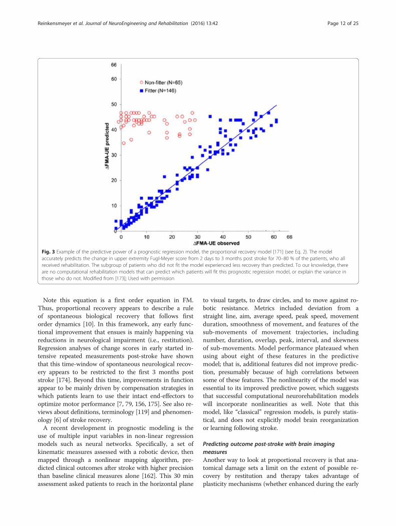
Note this equation is a first order equation in FM.Thus, proportional recovery appears to describe a ruleof spontaneous biological recovery that follows firstorder dynamics [10]. In this framework, any early func-tional improvement that ensues is mainly happening viareductions in neurological impairment (i.e., restitution).Regression analyses of change scores in early started in-tensive repeated measurements post-stroke have shownthat this time-window of spontaneous neurological recov-ery appears to be restricted to the first 3 months poststroke [174]. Beyond this time, improvements in functionappear to be mainly driven by compensation strategies inwhich patients learn to use their intact end-effectors tooptimize motor performance [7, 79, 156, 175]. See also re-views about definitions, terminology [119] and phenomen-ology [6] of stroke recovery.A recent development in prognostic modeling is the
use of multiple input variables in non-linear regressionmodels such as neural networks. Specifically, a set ofkinematic measures assessed with a robotic device, thenmapped through a nonlinear mapping algorithm, pre-dicted clinical outcomes after stroke with higher precisionthan baseline clinical measures alone [162]. This 30 minassessment asked patients to reach in the horizontal plane
to visual targets, to draw circles, and to move against ro-botic resistance. Metrics included deviation from astraight line, aim, average speed, peak speed, movementduration, smoothness of movement, and features of thesub-movements of movement trajectories, includingnumber, duration, overlap, peak, interval, and skewnessof sub-movements. Model performance plateaued whenusing about eight of these features in the predictivemodel; that is, additional features did not improve predic-tion, presumably because of high correlations betweensome of these features. The nonlinearity of the model wasessential to its improved predictive power, which suggeststhat successful computational neurorehabilitation modelswill incorporate nonlinearities as well. Note that thismodel, like “classical” regression models, is purely statis-tical, and does not explicitly model brain reorganizationor learning following stroke.
Predicting outcome post-stroke with brain imagingmeasuresAnother way to look at proportional recovery is that ana-tomical damage sets a limit on the extent of possible re-covery by restitution and therapy takes advantage ofplasticity mechanisms (whether enhanced during the early
Fig. 3 Example of the predictive power of a prognostic regression model, the proportional recovery model [171] (see Eq. 2). The modelaccurately predicts the change in upper extremity Fugl-Meyer score from 2 days to 3 months post stroke for 70–80 % of the patients, who allreceived rehabilitation. The subgroup of patients who did not fit the model experienced less recovery than predicted. To our knowledge, thereare no computational rehabilitation models that can predict which patients will fit this prognostic regression model, or explain the variance inthose who do not. Modified from [173]; Used with permission
Reinkensmeyer et al. Journal of NeuroEngineering and Rehabilitation (2016) 13:42 Page 12 of 25

period of spontaneous biological recovery or normal inthe chronic phase) to help patients achieve the maximumrecovery possible. Predictive models thus need to includeneuro-anatomical variables [176]. However, given thatlevel of initial severity is the best predictor of final outcome,we need to ask whether brain imaging data can improvemodel performance. In other words, we are interested inthe factors that determine recovery over and above thosethat cause impairment, as these are different things [172].Residual structural and functional architecture after
stroke can also be used to estimate clinical outcome.Diffusion tensor imaging (DTI) is able to assess integrityof white matter tracts and when measured within3 weeks of subcortical stroke, corticospinal tract (CST)integrity correlates with both initial and 6 month upperlimb impairment [177]. In a separate study, damage tothe CST at the posterior limb of the internal capsule12 h post-stroke correlated well with motor impairmentat 30 and 90 days [178]. Stinear and colleagues have pro-posed an algorithm for sequentially combining simpleclinical, TMS, and DTI measures to predict upper limbfunction [168]. The PREP (Predicting REcovery Poten-tial) algorithm was tested in a sample of 40 sub-acutestroke patients and performed well in predicting motorfunction based on Action Research Arm Test scores at12 weeks post-stroke. The performance of DTI in thissetting should be improved by making the tracts specificto particular functions e.g. upper limb [179] and develop-ing ways for the assessment of tract integrity to be done ina standardised [180] and automatic [181] manner.Corticospinal tract integrity correlates with initial
upper limb severity, which explains why it often corre-lates with final outcome. However, it remains unclearwhether it can explain outcomes over and above initialimpairment. The role of intact cortical regions in support-ing motor recovery is unknown, but has been explored inlanguage recovery. Predicting language outcome and re-covery after stroke (PLORAS) uses the whole structuralbrain scan from which voxel-wise estimates of the likeli-hood of damaged tissue are derived [182]. This ‘lesion-map’ for each patient is added to (i) time since stroke and(ii) a detailed assessment of various language capabilities.Using a machine learning approach, a new subject's lesionimage is compared with those from all the other patientsalready in the database to find one with a similar lesion.The language scores for all the similar patients are plottedover time, enabling the time course of recovery forthe new patient to be estimated [183]. The potentialfor such an approach extends to many domains includingmotor and cognitive outcomes. Using this type of neuro-imaging complex biomarker discovery [184] it should bepossible to provide accurate prognostic models allowingaccurate goal setting in neurorehabilitation and stratifica-tion in clinical trials [185]. Note that for goal setting in
neurorehabilitation, for any patients for whom a givenmodel predicts little potential benefit of treatment, futureresearch will hopefully reveal new, modifiable factors thatcan be targeted for such patients.Multivariate machine learning approaches have also
been applied to functional MRI data to predict outcome.For example, fMRI data acquired within 2 weeks ofstroke in patients with aphasia was used to predict out-come in language [186]. Accuracy in predicting goodand bad outcome at 6 months was 76 % and improvedto 86 % when age and baseline language impairmentwere added to the classification model. In the motordomain, fMRI data acquired in the first few days afterstroke has been used to try to predict a subsequentchange in motor performance [172]. Using a multivariateanalysis, a specific pattern of activated voxels was identi-fied as highly predictive of clinical change over the subse-quent 3 months, a finding that was independent of initialstroke severity and lesion volume. Anatomical hypothesescould not be tested using this multivariate approach – thestudy simply indicated that predictive signal was presentin a pattern of activation.
Predicting treatment effectsPredicting outcome will be useful for clinical and re-search stratification, but what a clinician would like toknow is what are the chances of a patient responding toa specific intervention. Stinear and colleagues [187] setout to determine whether characterising the state of themotor system would help in predicting an individualpatient's capacity for further functional improvement atleast 6 months post-stroke in a subsequent motor prac-tice programme. A combination of TMS, structural andfunctional MRI was used to suggest a method for deter-mining who would respond to training. This approachhas also been used to predict likely response to robotictraining, with both structural [188] and functional im-aging [189] data making some contribution.When it comes to assessing the effects of treatments
thought to enhance the potential for experiencedependent plasticity, less work has been done. Currently,there is a problematic explanatory gap between molecu-lar (from animal studies) and behavioral (from humanstudies) accounts of the mechanisms of recovery afterstroke. Lack of progression of knowledge from animalmodels to benefit for human stroke patients has ledto the search for ways to study these mechanisms inhuman subjects. There are exciting advances in howhuman neuroimaging data can be analyzed that sug-gest a way forward. Specifically, it is now possible toexamine changes in organization of the human brainafter stroke at multiple levels of brain architecture,ranging from large-scale networks to alterations insynaptic physiology.
Reinkensmeyer et al. Journal of NeuroEngineering and Rehabilitation (2016) 13:42 Page 13 of 25

A number of analytical approaches are available toexamine connectivity within well-defined macroscopicbrain networks. For example, graph theory can be usedto determine ‘efficiency’ of information transfer aroundsmall world networks such as the brain [190]. Thisallows inferences to be made about functional connectivityat the level of whole brain, hemisphere or specified net-work level and can be applied to fMRI, EEG, and MEGdata. Dynamic Causal Modeling (DCM) is a methodof analyzing data from a dynamic system such as thebrain. Bayesian inversion of a specified anatomicalmodel given the empirical data allows the determinationof model parameters, which reflect effective connectivitybetween brain regions (i.e. the task-dependent influ-ence of one brain region on another) [191]. DCM ofinduced responses acquired with MEG [192] is particularlyappealing as it allows partitioning of the effective couplingbetween regions at the same spectral frequency (linearcoupling) and across frequencies (non-linear coupling).Non-linear coupling in particular is important in functionalintegration [193].Another recently developed approach to studying
brain dynamics after stroke is to use patient-specificstructural connectivity data obtained from MRI to set-up an individualized Virtual Brain model of the patient.By optimizing model parameters, such as long-rangecoupling or local inhibition, in order to match restingstate BOLD signals, insight can be gained into how thoseparameters vary with different types of stroke [194].At the mesoscopic scale, the spectral characteristics of
brain oscillations measured with MEG (or EEG) in thegamma and beta frequency are dependent on the bal-ance of activity between populations of excitatory (gluta-matergic) pyramidal cells and inhibitory (GABAergic)interneurons [195] and are candidate biomarkers of thepotential for both local- and network-plasticity [196].More recently, it has become possible to define plausiblebiophysical DCMs to examine mesoscopic interactionsbetween populations of excitatory and inhibitory cells inspecific cortical regions using data from MEG [197].This approach has been validated using local field po-tentials in animal preparations where independentpharmacological/microdialysis assays have served to cor-roborate modelling results [198]. A recent example ofhow DCM of canonical microcircuits can provide mech-anistic inferences is the finding that psilocybin, a 5HT-2Aagonist, reduced oscillatory power in posterior associationcortex by increasing excitability of deep-layer pyramidalneurons [199].This range of neuroimaging tools and computational
approaches will provide the appropriate intermediatelevel of description with which to bridge the gap be-tween what we know about recovery after stroke fromanimal models compared to what we know from studies
of behavior in humans. A more detailed knowledge ofhow these processes are related to impairment and re-covery following stroke will provide a mechanisticframework for understanding how to treat patients moreeffectively. It will open the way for functional brain im-aging to become a clinically useful tool in rehabilitation,particularly for our ability to predict outcomes and re-sponse to novel plasticity enhancing therapies.In summary, there is a rich history and promising fu-
ture to using behavioural status and brain structure andfunction soon after stroke, to predict long-term motorrecovery. Prognostic regression models can inform com-putational neurorehabilitation models and are a keybenchmark against which the predictive power of com-putational neurorehabilitation will need to be tested.
Computational neurorehabilitation modelsAs described above, we use the term “computational neu-rorehabilitation” to refer to the emergence of theory-driven,mechanistic dynamical models that naturally encode timein differential equations and model recovery of motor be-havior using internal states that have a physiologic meaning.These models differ from the prognostic regression modelsdescribed in the previous section primarily because they in-corporate mechanistic dynamical models of plasticity andlearning mechanisms underlying recovery. In this section,we critically review a number of recent computational neu-rorehabilitation models (Table 2).
Reaching the threshold for recovery in bilateral hand useA key aspect of stroke motor recovery is that individualswith a stroke can elect not to use their impaired arm,since they usually have a relatively unaffected arm toperform most needed tasks (i.e. they can compensate).This “learned non-use” may logically be expected to con-tribute to loss of motor control of the hemiparetic arm(just as an athlete or musician who stops practicing be-comes rusty), although to our knowledge there is no re-search that has yet documented this loss. Han et al.developed one of the first computational neurorehabil-itation models to study the interactions between adap-tive decision making related to learned non-use andmotor relearning after a simulated motor cortex lesion[112] (Fig. 1c). The inputs to the model are targets forbilateral reaching practice and the outputs are the choiceof arm to use to reach to the given target, and the kine-matic accuracy of the reaching movement. The modelincorporates a reward-based learning mechanism forarm selection, and an error-based learning mechanismfor refining the neural population code in primary motorcortex that specifies reach direction.This model predicts a loss of motor cortex representa-
tion without rehabilitation, and a reversal of cortical repre-sentational loss with rehabilitation (cf. [93]). Furthermore,
Reinkensmeyer et al. Journal of NeuroEngineering and Rehabilitation (2016) 13:42 Page 14 of 25
the model predicts that if spontaneous recovery, motortraining, or both, bring function above a certain threshold,then training can be stopped, as the repeated spontaneousarm use provides a form of motor learning that furtherbootstraps function and spontaneous use (Fig. 4a), that is,a “virtual cycle” is entered. Below this threshold, motortraining is in vain: the model exhibits learned non-use, andcompensatory movements performed with the less im-paired hand are reinforced; that is, a “viscious cycle” is en-tered. Evidence for this threshold prediction at the group
level was subsequently found in data from the EXCITEclinical trial, a large study of constraint-induced movementtherapy after stroke [65].One clinical impact of the model is that it suggests a
novel therapeutic paradigm, the “Train-Wait-Train”paradigm, that not only would test the threshold hypoth-esis, but also which could potentially be a more cost-effective way to deliver rehabilitation therapy. For ex-ample, in computer simulations, a virtual patient wasfirst given a set of 200 trials of therapy, simulated byforced use of the affected arm. Then the patient experi-enced repeated cycles of 1000 trials in a free arm choicecondition (wait), followed by 100 treatment trials (train).The initial training trials were not sufficient to reachthreshold, and spontaneous use stayed low. After furthertraining sessions, however, the simulated patient enteredthe “virtuous circle.” Experiments are currently testingthis Train-Wait-Train paradigm.The model also provides insights into the time constants
of stroke motor recovery. The main recovery time constantwas on the order of thousands of trials. However, the timeconstant controlling the change in the decision to use onehand or the other was much smaller, and as a result, themodel developed learned non-use soon after stroke. Be-cause how fast learned non-use actually develops afterstroke is unknown, hand choice and kinematic data col-lected soon after stroke would lead to better parameter es-timation and hence better predictions from these models.This model, however, has a number of limitations that
need to be addressed to better understand and predictindividual recovery as a function of use and motor train-ing. For example, the model is simply bi-stable: eitherthe patient is “above threshold” and fully recovers, orbelow and does not. In addition, the model ignores in-teractions between the two cortices. An extension of thismodel, which includes inter-hemispheric inhibition, hasbeen proposed to account for the beneficial effects of bi-manual training compared to uni-manual training [200].Further, the model is only concerned with recovery ofthe control of movement direction, with no attentionpaid to arm muscle activity, other kinematic features, orfunctional upper extremity behaviors.
Recovering from weakness via reinforcement learningMotor recovery after stroke is characterized by a seeminglydisparate set of behavioral and brain imaging observations,many reviewed above, but could these observations arisefrom a few fundamental features of human sensorimotorplasticity? Reinkensmeyer et al. approached this questionby focusing on the modeling of the recovery of distal upperlimb strength, the rationale being that strength stronglypredicts upper extremity functional activity [136, 201]. Theinputs to the model are attempts to flex the wrist, and theoutput is the flexion force achieved. For the motor system
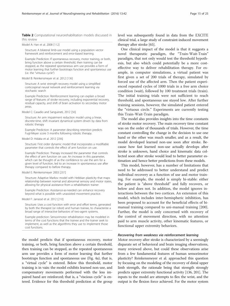
Table 2 Computational neurorehabilitation models discussed inthis review
Model A: Han et al. 2008 [112]
Structure: A bilateral limb-use model using a population vectorframework and reinforcement and error-based learning.
Example Prediction: If spontaneous recovery, motor training, or both,bring function above a certain threshold, then training can bestopped, as the repeated spontaneous arm use provides a form ofmotor learning that further bootstraps function and spontaneous use(i.e. the “virtuous cycle”)
Model B: Reinkensmeyer et al. 2012 [136]
Structure: A wrist strength recovery model using a simplifiedcorticospinal neural network and reinforcement learning viastochastic search
Example Prediction: Reinforcement learning can explain a broadrange of features of stroke recovery, including exponential recovery,residual capacity, and shift of brain activation to secondary motorareas.
Model C: Casadio and Sanguineti, 2012 [56]
Structure: An arm impairment reduction model using a linear,discrete-time, shift invariant dynamical system driven by data fromrobotic therapy
Example Prediction: A parameter describing retention predictsFugl-Meyer score 3 months following robotic therapy.
Model D: Hidaka et al. 2012 [206]
Structure: First order dynamic model that incorporates a modifiableparameter that controls the effect of arm function on use.
Example Prediction: Therapy increased the parameter that controlsthe effect of arm function on use. An increase in this parameter,which can be thought of as the confidence to use the arm for agiven level of function, led to an increase in spontaneous use aftertherapy compared to before therapy.
Model E: Reinkensmeyer 2003 [207]
Structure: Adaptive Markov model with Hebbian plasticity that mapsrelationship between normal and abnormal sensory and motor states,allowing for physical assistance from a rehabilitation trainer
Example Prediction: Assistance-as-needed can enhance recoverybeyond what is possible with unassisted movement practice.
Model F: Jarrassé et al. 2012 [210]
Structure: Uses a cost function with error and effort terms, generatedby both the therapist (or robot) and human trainee, to characterize abroad range of interactive behaviors of two-agent systems.
Example prediction: Sensorimotor rehabilitation may be modeled interms of the cost functions that the trainee and the trainer seek toimplement, as well as the algorithms they use to implement thosecost functions.
Reinkensmeyer et al. Journal of NeuroEngineering and Rehabilitation (2016) 13:42 Page 15 of 25

to recover wrist flexion strength, the model assumes thatthe motor system must learn appropriate activation of re-sidual corticospinal cells (i.e. those preserved after thestroke – thus, within the terminology adopted in this paperthis is again a study of neural compensation not restitu-tion) via repeated movement attempts followed by rewardfeedback. A simplified corticospinal network model wasdeveloped based on several key observations from primateneurophysiological experiments – that corticospinal cellactivity sums to create a net excitatory flexor or extensordrive to a joint, that different corticospinal cells have differ-ent “gains” for exciting motoneuronal pools, and that therelationship between a corticospinal cell’s activity and itsindividual contribution to muscle force is linear up to apeak firing rate, then saturates for higher activity levels. Fi-nally, the model assumed that the key underlying mechan-ism of plasticity driving strength recovery after stroke wasa reinforcement learning mechanism, in which the sensori-motor system modifies corticospinal cell activations basedon repetitive movement experiences, using stochastic ran-dom search, such that limb force output is maximized.
This model predicts a broad range of the features ofstroke recovery. It predicts the dose-response curve ofrehabilitation, and putative modifiers of this curve, suchas that the same dose of exercise has a smaller effectwhen given to a more severely impaired patient, and thatthe same dose of exercise given early has a larger differ-ential effect than when given later. This latter findingsuggests that increased sub-acute plasticity may arise inpart due to normative, compensatory network learningdynamics rather than solely as a function of the stroke-altered tissue microenvironment.Further, and unlike the Han et al. model reviewed
above, the model predicts exponential-like strength re-covery curves that never quite reach an asymptote, butinstead exhibit a residual capacity for further recoverywith further movement practice, as has been observedexperimentally [202]. At the level of structural brain im-aging observations, the model predicts that patients with alarger residual corticospinal network will recover more, aknown stroke phenomenon [203]. It also predicts a keyfunctional imaging observation, which is that movement-
A
C D
B
Fig. 4 Examples of computational neurorehabilitation approaches and results. a A key output of the Han et al. model [112] is the predictedspontaneous use of the impaired hand, shown here as a percent of all movement trials in a bimanual choice task. Each curve represents theevolution of spontaneous use given the number of rehabilitation practice trials, shown as a number on the far right of each curve. Spontaneoususe increases only when enough rehabilitation practice trials are performed to reach a threshold. From [112]; used with permission. b A keyoutput of the Casadio et al. model [56], which used data from a robotic therapy trial, is that the retention parameter in the model, measuredthrough a trial-to-trial analysis, predicts the change in Fugl-Meyer score at 3 months for these chronic stroke participants. c The Reinkensmeyer etal. model [136] assumes that wrist force is produced by the summed effect of corticospinal cells targeting motor neuronal pools. Each cellcontributes an incremental force proportional to its firing rate, up to a saturation level. Cell firing rate changes by a random amount from trial totrial; activation patterns that produce more force are remembered for future use, thus implementing a reinforcement learning paradigm. d In theReinkensmeyer et al. model, the probability that a single neuron will contribute to an increase in force on a new trial depends on whether theneuron is strongly or weakly connected to the motor neuronal pool. Strongly connected cells have a greater probability of producing a largerincrease. In addition, when cells become saturated, they can only decrease force production. Thus, an increasing number of saturated cellsincreasingly blocks further optimization, leaving a residual capacity for further increases in force
Reinkensmeyer et al. Journal of NeuroEngineering and Rehabilitation (2016) 13:42 Page 16 of 25
related activation in secondary motor areas increases fol-lowing a stroke that damages output from the primarymotor area [204].Mathematically, two key mechanisms drive the ob-
served network dynamics. First, the saturation in the in-dividual relationships between corticospinal cell firingrates and their contribution to muscle force slows laterlearning, thus leaving room for residual capacity (Fig. 4d).This is because force results from the summed contribu-tions of many cells. Once the activations of a subset ofthese cells have evolved in the correct direction tomaximize force output, and become saturated, then thoseneurons can no longer contribute to further increases inforce. As saturated neurons randomly vary their outputfrom trial to trial, they exhibit an increased probability ofdecreasing the force produced on subsequent trials, sincethey can now only vary their output in the direction coun-terproductive to further force increase. Second, the activa-tions of more strongly connected cells tend to be modifiedfirst, as changes in their activations are more likely to in-crease force output (Fig. 4c).A key clinical implication of the model is that, if a sin-
gle learning mechanism – reinforcement learning viastochastic search – can explain such a broad range offeatures of stroke recovery, then perhaps this mechanismshould be targeted in rehabilitation training to facilitatethe search process. Adding variability and chances forexploration to training regimens and providing aug-mented feedback on the teaching signal are two possibleways to achieve this; a large literature on reinforcementlearning in computer science may also be brought tobear on rehabilitation protocol optimization (see above).The model predicts another unexpected way that animproved search might be achieved, an “Inhibit-Train-Release” paradigm, which is to temporarily inhibitcells that are already optimized (perhaps using a localizedinhibition scheme, such as infused muscimol or focusedtranscranial magnetic stimulation, for example), train theremaining cells, and then release the inhibited cells.Stochastic search tends to optimize cells with strongereffects on muscles, and once optimized these cells blockfurther optimization as explained above. The model pre-dicts that temporarily inhibiting optimized cells will allownon-optimized cell activations to become optimized.As discussed above, this model successfully accounts
for a large number of existing neural and behavioraldata. However, it has several limitations. It has cartoon-like complexity, modeling only a single flexor and exten-sor muscle, and simplifying corticospinal activation tostatic input-output relationships. In addition, it does notmodel spontaneous recovery or restitution, as we have de-fined them here. Further, while severe weakness precludescontrol, mild to moderate weakness is less well correlatedwith motor control. For example, the ipsilateral limb after
stroke shows abnormal motor control but strength is nor-mal [205], so motor control requires more than strength.
Robot assistance, retention, and learning predicts recoveryModels such as the Han et al. model or the Reinkens-meyer model above are “qualitative”, i.e., they make gen-eral qualitative predictions of recovery. Parameters inthese models are “hand-tuned”. As such, they are onlyloosely based on actual data and cannot be used to pre-dict recovery of individual patients. As reviewed above,during rehabilitative training mediated by a robot it ispossible to observe and record the patient’s performanceas well as the level of assistance that the patient received.Can the trial-to-trial evolution in movement success dur-ing rehabilitation be modeled, and, if so, what are the keylearning parameters needed to describe this evolution?Casadio et al. [56] developed the first computationalmodel driven by data from actual robotic training sessions,which describes the trial-by-trial evolution of the recoveryprocess induced by robotic training. This model providesinsights into the role of assistive force in the recoveryprocess, and the extent to which learned changes in vol-untary control decay over time and transfer to subsequenttraining sessions.The model characterizes the recovery process related
to robot-assisted training for improving arm extensionin chronic stroke survivors as a linear, discrete-time,shift invariant dynamical system. The model posits thatmotor performance is a function of a voluntary controlcomponent, an assistive force component, provided ei-ther by a robot or a therapist, and a performance noiseterm. Thus, the level of physical assistance provided dur-ing rehabilitative training is taken as one of the system’sinputs. The voluntary control component is the internalstate of the model and its temporal evolution is describedas the combination of three additive terms: a ‘memory’ or‘retention term’, accounting for how much voluntary con-trol depends on previous experience; an ‘assistance’ com-ponent expressed as the magnitude of the assistive force;and a learning component that accounts for how muchvoluntary control on the actual trial is affected by the pre-vious value of the driving signal. The input signals thatdrive recovery are movement error or a performancemeasure. A process noise term accounts for the portion ofthe recovery not due to these three terms.The most striking result from this model is that the re-
tention parameter predicts the percent change of theFugl-Meyer score at the 3-month point following theend of the robotic treatment (Fig. 4b). This result, whichneeds to be confirmed with a larger cohort, is potentiallyimportant for the individualization of training. After oneor few initial training sessions with the robot, the modelcan be fitted to the data. Then, by examining the reten-tion parameter, one could potentially determine who will
Reinkensmeyer et al. Journal of NeuroEngineering and Rehabilitation (2016) 13:42 Page 17 of 25

benefit from additional robotic motor training. Notehowever that this model is based on a set up in whichthe arm is constrained to move in two dimensions withshoulder and elbow movements. Thus, here again, forexample, the issue of restitution versus compensation isdifficult to address and the conclusions are limited tosimplified movements at only two joints.
Understanding interactions between function and useIn the same vein of using computational models for pre-dicting individual recovery, Schweighofer and colleaguesdeveloped a first-order dynamical model of stroke recov-ery with longitudinal data from participants receivingconstraint-induced movement therapy in the EXCITEclinical trial [206]. The goal of the model was to betterunderstand the interactions between arm function anduse in human post-stroke following therapy. The modelshows the existence of time-varying interactions betweenself-reported amount of daily arm use and recovery offunction via a dynamical model of stroke recovery in the2 years following therapy. The time constants in thismodel were on the order of several weeks. Of most im-portance, by comparing the model parameters beforeand after constraint-induced movement therapy inter-vention in participants receiving the intervention 1 yearafter randomization, therapy increased the parameterthat controls the effect of arm function on use. An in-crease in this parameter, which can be thought of as theconfidence to use the arm for a given level of function,led to an increase in self-reported spontaneous use aftertherapy compared to before therapy. Thus, here, the pa-rameters of the model can be viewed as “states” thathave physiological meaning, which may expedite thetesting of new experimental hypotheses.However, as in the previous model of Casadio et al.,
this is a highly simplified model of recovery. It containsonly a few parameters and two time constants, one forfunctional recovery and the other for hand use. The reasonfor this simplicity is the very sparse data set on which themodel was built. Increasing the complexity of the model,and thus the number of parameters was not possible be-cause this would have led to overfitting, i.e., the modelwould fit the data very well but would not generalize.
Modeling the effect of assistance-as-neededRehabilitation therapy often involves interactions betweenpatient and a trainer, be it a rehabilitation therapist, a ro-botic device, or a computer game. Modeling the patient-trainer interaction could potentially provide insight intomovement recovery.Reinkensmeyer used an adaptive Markov model to
examine the role of external mechanical assistance froma robotic device or therapist in promoting movement re-covery [207]. Following research in gait training after
spinal cord injury, the model assumes that motor con-trol is characterized by repeated transitions between sen-sory and motor states; for example, sensing of full hipextension at the end of stance triggers leg flexion. It fur-ther grossly characterizes these states as abnormal andnormal sensory states (e.g. appropriate or inappropriatehip extension), and abnormal and normal motor states(effective or ineffective leg flexion). The model uses aHebbian-inspired model of plasticity in which the transi-tions between specific sensory and specific motor statesbecome more reliable with repetitive activation of thattransition. The action of a skilled external trainer (robotor human) who is assisting in movement is modeled as amediated increase in the probability of transferring to astate of normal motor output (e.g. the trainer helps gen-erate hip extension sensory inputs to enable effective legflexion). Assistance-as-needed is simulated by mediatingthis transfer only when the patient is in an abnormalmotor state, while assistance-always is simulated by me-diating this increase on every movement repetition.The model predicts that assistance-as-needed can en-
hance recovery beyond what is possible with unassistedpractice, that assistance-always is not as effective asassistance-as-needed, that the trainer’s skill in assistingtoward normal motor output matters in reinforcing nor-mal state transitions, and that assistance is not usefulwhen sensory input is less directly coupled to motoroutput. While these predictions may sound somewhatintuitive, this is perhaps because they mirror opinionsthat are often expressed in the current clinical milieu.The model in this case served to verify that these opin-ions could be mathematically supported relying on asimple but plausible plasticity rule.
Patient-trainer dynamics as an optimizationAnother approach toward modeling patient-trainer dy-namics is based on what was first developed as a model ofhuman movement adaptation. As briefly described above,the motor system uses a sunken-v muscle adaption rule toalter force, impedance, and trajectory, explaining a widevariety of experimental findings of reaching in dynamicenvironments [119, 208]. This update rule can be viewedas the motor system implementing a greedy minimizationof a cost function of error and effort [118]. If the patient’smotor system is minimizing a cost function from move-ment to movement, then, what cost function might thetrainer minimize to assist in learning? A gradient descentof a similar cost function of error and effort was shown toprovide an efficient assistive controller for rehabilitation[209]. Jarrassé et al. expanded and formalized this ap-proach, providing a framework for the description and im-plementation of a broad range of interactive behaviors oftwo agents (such as a patient and a robotic therapist) per-forming a joint motor task, including rehabilitation [210].
Reinkensmeyer et al. Journal of NeuroEngineering and Rehabilitation (2016) 13:42 Page 18 of 25

For example, one may assume that a robotic trainer justmoves a patients’ limb while this patient is passive. Tomodel this situation, the trainer would take all of the effortoff of the patient by minimizing its cost function:
Vt ¼ αe2 þ βu2;α; β > 0; ð3Þwhere e and u correspond to the patient’s error andeffort state as observed by the trainer. However, individ-uals with stroke improve their motor function more whenactively attempting to move, rather than relying on therobot to move their arm [211]. To avoid the patient be-coming passive, the trainer can modify its cost function to
Vt ¼ αe2 þ βtut2; α; βt > 0 ð4Þ
i.e. minimize its own effort ut2. If one assumes that the
patient behaves according to the sunken-v adaptationrule, if the trainer is slightly more lazy (i.e. βt > β), it willhelp the patient fulfill the task (as the trainer also has tominimize e2) only if the patient is not able to do soby itself, and disappear if the patient can do it. Thus,this method and the cost function of Eq. (4) can beused to design assist-as-needed control of rehabilita-tion robots. In terms of computational neurorehabil-itation, this work suggests that sensorimotorrehabilitation may be able to be modeled in terms ofthe cost functions that the trainee and the trainerseek to implement, as well as the algorithms they useto implement those cost functions.
ConclusionsWe contend that the models reviewed in the previoussection are evidence that a fundamental understandingof neurologic recovery will be facilitated by modelingmotor learning and plasticity itself in a context specificto rehabilitation. Even the initial, simplified modelspresented are generating novel ideas concerning themechanisms of recovery in rehabilitation, and thusare suggesting important directions for future experi-mental research.
Novel ideas from initial models and their limitationsFor example, the Han et al. model discussed above sug-gests the existence of a threshold of movement abilitythat allows patients to enter a “virtuous” cycle of activity-dependent plasticity; this premise is now being tested in aclinical trial. The Reinkensmeyer et al. model suggeststhat a large number of existing behavioral and neuraldata can be accounted for if the motor system uses areinforcement-learning paradigm – stochastic search – tooptimize neural activations; this suggests increasing re-search into techniques to enhance reinforcement learningin order to enhance rehabilitation therapy, a propositionthat is only beginning to be explored. The Casadio et al.
model showed that a short-term measure of retention inrobotic training can accurately predict how much long-term recovery is possible with such training duringchronic stroke, suggesting a novel assay for patient selec-tion for robotic therapy. Other interesting ideas generatedby the initial models reviewed above are that a key waythat rehabilitation therapy may function is by modulatinga parameter that controls the effect of arm function onuse, and that trainer-trainee interactions can be character-ized using parsimonious cost functions.Even given their utility in generating such novel ideas
concerning rehabilitation, these initial models clearlyhave shortcomings. They greatly simplify human move-ment and the rehabilitation process. They ignore the factthat every day activity relies on bimanual motor func-tion. They selectively model only one or two learningand plasticity mechanisms. They focus on motor learn-ing rather than plasticity mechanisms associated withrestitution, as yet neglecting the rich literature on spon-taneous biological recovery, which may ultimately offergreater potential for recovery. They have not yet beenused to replicate, let alone extend, the findings achievedwith prognostic regression models. None have yet used across-validation procedure in order to see how much amodel based on the data from a pool of patients can ex-plain the behavior of patients outside of that pool. Inaddition, sensorimotor control is just one of many do-mains addressed by neurorehabilitation.
The forthcoming data revolutionAnother key argument that this review has attempted tomake is that the time is propitious for continued develop-ment of computational neurorehabilitation models. As wesurveyed above, the mathematical tools for developingsuch models are already at least partly available because ofthe past several decades of work in the field of computa-tional neuroscience and machine learning. Further, as wealso reviewed above, large-scale longitudinal data for eachpatient and for a large number of patients are now possibleto obtain with robotic devices and wearable sensors. Thiswill allow the development of more elaborate, physics-based models that predict recovery at a fine temporal andspatial resolution. Note that conventional motor learningexperiments have generally failed to even come close tothe number of training trials that gives rise to the high levelexpertise in sports and work-related skills. With instru-mented rehabilitation and wearable sensors, this situationis now set to change, at least in the context of motor learn-ing during rehabilitation. A key challenge for the future ishow to utilize these longitudinal data that are collected ondifferent samples, with different methods of motion cap-ture, and different quantifications of movement into a co-herent package such that they can inform models ofmovement control over the course of recovery after stroke.
Reinkensmeyer et al. Journal of NeuroEngineering and Rehabilitation (2016) 13:42 Page 19 of 25
The Casadio et al. model described above initiates thedata-driven approach, as it is the first to use trial-to-trialdata from actual robotic training to predict changes inFugl-Meyer scores, suggesting that retention-related pa-rameters correlate with impairment recovery. Followingthis approach, we predict that soon we will see theemergence of large scale mechanistic models of motorrecovery that are driven by the actual movement contentof rehabilitation therapy as well as records of daily activity.Ideally, one would use the same data set from hundredsor even thousands of well-characterized patients, so thatcomputational model output is realistic and appropriate.In addition, the models will increasingly be informed
by automatic analysis of MRI scans routinely obtainedafter stroke, since, as we reviewed above, brain anatomicalinformation is needed because identical behavior can arisethrough largely different neural processes. A range ofnovel neuroimaging tools and computational methods, in-cluding analysis of grey and white matter structures andstructural and functional connectivity [212], will providean intermediate level of description with which to bridgethe gap between what we know about recovery afterstroke from animal models compared to what we knowfrom studies of behavior in humans.
Complexification and utilityClearly, initial computational neurorehabilitation modelsare vast simplifications of a very complex process. Whilesimplification and abstraction are often virtues in model-ing, with richer data sets, it will be possible to increasethe complexity of computational neurorehabilitationmodels with less risk of overfitting. Models containing amultiple joint system, such as the whole arm or botharms, will be important for understanding compensa-tion. Upper extremity motor control during real-world,free-living activity involves the movement of all the seg-ments in order to position and orient the limb and tointeract with objects [213, 214]. If a computational modelutilizes only a few of the segments, then the model outputwill provide only a limited view of the actual solution, es-sentially ignoring the degrees of freedom problem under-scored by Bernstein. Models that can help understand orpredict control processes during naturalistic actions willbe of high value for the field of neurorehabilitation.Further, it is currently difficult to experimentally study
the interacting effects between different forms of learn-ing, such as supervised, unsupervised, and reinforcementlearning, including the role of the timing of rehabilita-tion on these processes [81, 215]. There appear to bemethods also to induce beneficial plasticity beyond taskrepetition, such as the possible enhancing of effectsof non-invasive brain stimulation on motor learning[216, 217]. Computational neurorehabilitation modelscan incorporate multiple levels of plasticity and learning,
as well as plasticity-enhancing effects of techniques suchas electrical stimulation, and even psychological effectsimportant to rehabilitation, helping understand these in-teractions in computer simulations to guide future experi-mental work. This is important, as one theoretically couldvet hypotheses in an efficient and cost-effective manner,rather than relying solely on randomized, controlled trials,which are costly and time consuming.A key question, of course, is whether the incorporation
of plasticity and learning mechanisms, along with in-ternal physiological states, into models will improveupon the predictive capability now possible with prog-nostic regression models. We contend they have a goodchance to, because they are more likely to isolate the keypredictive variables of interest, since these variableslikely relate to physiologic function, and computationalneurorehabilitation models seek to make just such vari-ables explicit. Such variables likely vary from patient-to-patient as well, suggesting that their isolation will improveindividualized predictions.If so, computational models of neurorehabilitation
should ultimately improve rehabilitation for individualswith neurologic injuries. We expect that computationalmodels of recovery, based on early clinical data, kinematicperformance, and routine scans, will provide the basis forfuture clinical software that suggests timing, dosage, andcontent of therapy. Such an approach will transform neu-rorehabilitation by guiding clinicians, patients, and healthproviders in the optimization of treatments.
Competing interestsThe authors declare that they have no competing interests.
Authors’ contributionsAll authors participated in drafting the manuscript. DR and NS also editedindividual author contributions into the manuscript. All authors read andapproved the final manuscript.
AcknowledgmentsThe authors would like to thank the Borchard Foundation for supporting thiswork.DJR was supported by grant NIH R01 HD062744EB was supported by grants EU-FP7 ICT-601003 BALANCE, ICT-2013-10SYMBITRON, and EU-H2020 ICT-644727 COGIMON.CEL was supported by grant NIH R01 HD068290MC was supported by grant FP7-PEOPLE-2012-CIG-334201 (REMAKE)NS was supported by grant NIH R01 HD065438.
Author details1Departments of Anatomy and Neurobiology, Mechanical and AerospaceEngineering, Biomedical Engineering, and Physical Medicine andRehabilitation, University of California, Irvine, USA. 2Department ofBioengineering, Imperial College of Science, Technology and Medicine,London, UK. 3Department Informatics, Bioengineering, Robotics and SystemsEngineering, University of Genoa, Genoa, Italy. 4Departments of Neurologyand Neuroscience, John Hopkins University School of Medicine, Baltimore,MD, USA. 5Department of Rehabilitation Medicine, MOVE Research InstituteAmsterdam, VU University Medical Center, Amsterdam, The Netherlands.6Reade, Centre for Rehabilitation and Rheumatology, Amsterdam, TheNetherlands. 7Department of Physical Therapy and Human MovementSciences, Northwestern University, Chicago, IL, USA. 8Department ofNeurology, Program in Physical Therapy, Program in Occupational Therapy,
Reinkensmeyer et al. Journal of NeuroEngineering and Rehabilitation (2016) 13:42 Page 20 of 25

Washington University School of Medicine, St Louis, MO, USA. 9Departmentof Kinesiology, KU Leuven Movement Control & Neuroplasticity ResearchGroup, Leuven, KU, Belgium. 10Leuven Research Institute for Neuroscience &Disease (LIND), KU, Leuven, Belgium. 11Sobell Department of MotorNeuroscience and UCLPartners Centre for Neurorehabilitation, UCL Instituteof Neurology, Queen Square, London, UK. 12Division of Biokinesiology andPhysical Therapy, University of Southern California, Los Angeles, USA.
Received: 19 November 2015 Accepted: 13 April 2016
References1. Feigin VL, Forouzanfar MH, Krishnamurthi R, Mensah GA, Connor M, Bennett
DA, Moran AE, Sacco RL, Anderson L, Truelsen T, O’Donnell M,Venketasubramanian N, Barker-Collo S, Lawes CMM, Wang W, Shinohara Y,Witt E, Ezzati M, Naghavi M, Murray C. Global and regional burden of strokeduring 1990–2010: findings from the Global Burden of Disease Study 2010.Lancet. 2014;383(9913):245–55.
2. Miller EL, Murray L, Richards L, Zorowitz RD, Bakas T, Clark P, Billinger SA.Comprehensive overview of nursing and interdisciplinary rehabilitation careof the stroke patient: a scientific statement from the American HeartAssociation. Stroke. 2010;41(10):2402–48.
3. Langhorne P, Bernhardt J, Kwakkel G. Stroke rehabilitation. Lancet. 2011;377(9778):1693–702.
4. Levin MF, Kleim JA, Wolf SL. What do motor ‘recovery’ and ‘compensation’mean in patients following stroke? Neurorehabil Neural Repair.2009;23(4):313–9.
5. Langhorne P, Coupar F, Pollock A. Motor recovery after stroke: a systematicreview. Lancet Neurol. 2009;8(8):741–54.
6. Buma F, Kwakkel G, Ramsey N. Understanding upper limb recovery afterstroke. Restor Neurol Neurosci. 2013;31(6):707–22.
7. van Kordelaar J, van Wegen EEH, Nijland RHM, Daffertshofer A, Kwakkel G.Understanding adaptive motor control of the paretic upper limb early poststroke:the EXPLICIT-stroke program. Neurorehabil Neural Repair. 2013;27(9):854–63.
8. Kitago T, Goldsmith J, Harran M, Kane L, Berard J, Huang S, Ryan SL,Mazzoni P, Krakauer JW, Huang VS. Robotic therapy for chronic stroke:general recovery of impairment or improved task-specific skill?J Neurophysiol. 2015;114(3):1885–94.
9. Lohse KR, Lang CE, Boyd LA. Is more better? Using metadata to exploredose-response relationships in stroke rehabilitation. Stroke. 2014;45(7):2053–8.
10. Krakauer JW, Carmichael ST, Corbett D, Wittenberg GF. Gettingneurorehabilitation right: what can be learned from animal models?Neurorehabil Neural Repair. 2012;26(8):923–31.
11. Milot MH, Spencer SJ, Chan V, Allington JP, Klein J, Chou C, Bobrow JE,Cramer SC, Reinkensmeyer DJ. Re-training upper extremity movement withthe robotic exoskeleton BONES after chronic stroke: a crossover pilot studyevaluating the functional outcomes of more naturalistic robotic movementtraining. J Neuroeng Rehabil. 2013;10:112.
12. Awad LN, Reisman DS, Pohlig RT, and S. A. Binder-Macleod. Reducing TheCost of Transport and Increasing Walking Distance After Stroke: ARandomized Controlled Trial on Fast Locomotor Training Combined WithFunctional Electrical Stimulation. Neurorehabil. Neural Repair. 2015. pii:1545968315619696. [Epub ahead of print]
13. Clark DJ, Neptune RR, Behrman AL, Kautz SA. Locomotor Adaptability TaskPromotes Intense and Task-Appropriate Output From the Paretic LegDuring Walking. Arch Phys Med Rehabil. 2016;97(3):493–6.
14. Jones TA, Adkins DL. Motor System Reorganization After Stroke: Stimulatingand Training Toward Perfection. Physiology (Bethesda). 2015;30(5):358–70.
15. Chollet F, Tardy J, Albucher J-F, Thalamas C, Berard E, Lamy C, et al.Fluoxetine for motor recovery after acute ischaemic stroke (FLAME): arandomised placebo-controlled trial. Lancet Neurol. 2011;10(2):123–30.
16. Ng KL, Gibson EM, Hubbard R, Yang J, Caffo B, O’Brien RJ, Krakauer JW, ZeilerSR. Fluoxetine Maintains a State of Heightened Responsiveness to MotorTraining Early After Stroke in a Mouse Model. Stroke. 2015;46(10):2951–60.
17. Janssen H, Bernhardt J, Collier JM, Sena ES, McElduff P, Attia J, Pollack M,Howells DW, Nilsson M, Calford MB, Spratt NJ. An enriched environmentimproves sensorimotor function post-ischemic stroke. Neurorehabil NeuralRepair. 2010;24(9):802–13.
18. Tiozzo E, Youbi M, Dave K, Perez-Pinzon M, Rundek T, Sacco RL,Loewenstein D, Lewis JE, Wright CB. Aerobic, Resistance, and CognitiveExercise Training Poststroke. Stroke. 2015;46(7):2012–6.
19. Veerbeek JM, Kwakkel G, van Wegen EEH, Ket JCF, Heymans MW. EarlyPrediction of Outcome of Activities of Daily Living After Stroke: A SystematicReview. Stroke. 2011;42(5):1482–8.
20. Kwakkel G, Kollen BJ. Predicting activities after stroke: what is clinicallyrelevant? Int J Stroke. 2013;8(1):25–32.
21. Robertson IH, Murre JM. Rehabilitation of brain damage: brain plasticity andprinciples of guided recovery. Psychol Bull. 1999;125(5):544–75.
22. Goodall S, Reggia JA, Chen Y, Ruppin E, Whitney C. A computational modelof acute focal cortical lesions. Stroke. 1997;28(1):101–9.
23. Reggia JA. Neurocomputational models of the remote effects of focal braindamage. Med Eng Phys. 2004;26(9):711–22.
24. Levitan S, Reggia JA. Interhemispheric effects on map organizationfollowing simulated cortical lesions. Artif Intell Med. 1999;17(1):59–85.
25. Casadio M, Tamagnone I, Summa S, Sanguineti V. Neuromotor recoveryfrom stroke: computational models at central, functional, and musclesynergy level. Front Comput Neurosci. 2013;7:97.
26. Aimone JB, Weick JP. Perspectives for computational modeling of cellreplacement for neurological disorders. Front Comput Neurosci. 2013;7:150.
27. Kwakkel G, van Peppen R, Wagenaar RC, Wood Dauphinee S, Richards C,Ashburn A, Miller K, Lincoln N, Partridge C, Wellwood I, Langhorne P. Effectsof augmented exercise therapy time after stroke: a meta-analysis. Stroke.2004;35(11):2529–39.
28. Langhorne P, Wagenaar R, Partridge C. Physiotherapy after stroke: more isbetter? Physiother Res Int. 1996;1(2):75–88.
29. Veerbeek JM, Koolstra M, Ket JCF, van Wegen EEH, Kwakkel G. Effectsof augmented exercise therapy on outcome of gait and gait-relatedactivities in the first 6 months after stroke: a meta-analysis. Stroke.2011;42(11):3311–5.
30. Veerbeek JM, van Wegen E, van Peppen R, van der Wees PJ, Hendriks E,Rietberg M, Kwakkel G. What is the evidence for physical therapypoststroke? A systematic review and meta-analysis. PLoS One.2014;9(2):e87987.
31. Krakauer JW. The applicability of motor learning to neurorehabilitation. In:Oxford Textbook of Neurorehabilitation. 2015. p. 55.
32. Tan WY, Ye Z. Estimation of HIV infection and incubation via state spacemodels. Math Biosci. 2000;167(1):31–50.
33. Cazelles B, Chau NP. Using the Kalman filter and dynamic models to assessthe changing HIV/AIDS epidemic. Math Biosci. 1997;140(2):131–54.
34. Venkatakrishnan K, Friberg LE, Ouellet D, Mettetal JT, Stein A, Trocóniz IF,Bruno R, Mehrotra N, Gobburu J, Mould DR. Optimizing oncologytherapeutics through quantitative translational and clinical pharmacology:challenges and opportunities. Clin Pharmacol Ther. 2015;97(1):37–54.
35. Emken JL, Reinkensmeyer DJ. Robot-enhanced motor learning: acceleratinginternal model formation during locomotion by transient dynamicamplification. IEEE Trans Neural Sys Rehab Eng. 2005;13(1):33–9.
36. Wolf SL, Kwakkel G, Bayley M, McDonnell MN. Best practice for arm recoverypost stroke: an international application. Physiotherapy. 2016;102(1):1–4.
37. Ramos-Murguialday A, Broetz D, Rea M, Läer L, Yilmaz O, Brasil FL, Liberati G,Curado MR, Garcia-Cossio E, Vyziotis A, Cho W, Agostini M, Soares E, SoekadarS, Caria A, Cohen LG, Birbaumer N. Brain-machine interface in chronic strokerehabilitation: a controlled study. Ann Neurol. 2013;74(1):100–8.
38. Bernhardt J, Chan J, Nicola I, Collier JM. Little therapy, little physical activity:rehabilitation within the first 14 days of organized stroke unit care. J RehabilMed. 2007;39(1):43–8.
39. Harris JE, Eng JJ, Miller WC, Dawson AS. A self-administered GradedRepetitive Arm Supplementary Program (GRASP) improves arm functionduring inpatient stroke rehabilitation: a multi-site randomized controlledtrial. Stroke. 2009;40(6):2123–8.
40. Lang CE, Macdonald JR, Reisman DS, Boyd L, Jacobson KT, Schindler-IvensSM, et al. Observation of amounts of movement practice provided duringstroke rehabilitation. Arch Phys Med Rehabil. 2009;90(10):1692–8.
41. Birkenmeier RL, Prager EM, Lang CE. Translating animal doses of task-specific training to people with chronic stroke in 1-hour therapy sessions: aproof-of-concept study. Neurorehabil Neural Repair. 2010;24(7):620–35.
42. Waddell KJ, Birkenmeier RL, Moore JL, Hornby TG, Lang CE. Feasibility ofhigh-repetition, task-specific training for individuals with upper-extremityparesis. Am J Occup Ther. 2014;68(4):444–53.
43. Park H, Kim S, Winstein CJ, Gordon J, Schweighofer N. Short-Duration andIntensive Training Improves Long-Term Reaching Performance in IndividualsWith Chronic Stroke. Neurorehabil Neural Repair. 2015. pii:1545968315606990. [Epub ahead of print].
Reinkensmeyer et al. Journal of NeuroEngineering and Rehabilitation (2016) 13:42 Page 21 of 25

44. Turolla A, Dam M, Ventura L, Tonin P, Agostini M, Zucconi C, Kiper P, CagninA, Piron L. Virtual reality for the rehabilitation of the upper limb motorfunction after stroke: a prospective controlled trial. J Neuroeng Rehabil.2013;10:85.
45. Molina KI, Ricci NA, de Moraes SA, Perracini MR. Virtual reality using gamesfor improving physical functioning in older adults: a systematic review.J Neuroeng Rehabil. 2014;11(1):156.
46. van Diest M, Lamoth CJC, Stegenga J, Verkerke GJ, Postema K. Exergamingfor balance training of elderly: state of the art and future developments.J Neuroeng Rehabil. 2013;10:101.
47. Kwakkel G, Kollen BJ, Krebs HI. Effects of robot-assisted therapy on upperlimb recovery after stroke: a systematic review. Neurorehabil Neural Repair.2008;22(2):111–21.
48. Levin MF, Weiss PL, Keshner EA. Emergence of Virtual Reality as a Tool forUpper Limb Rehabilitation: Incorporation of Motor Control and MotorLearning Principles. Phys Ther. 2014;95(3):415–25.
49. Reinkensmeyer DJ, Boninger ML. Technologies and combination therapiesfor enhancing movement training for people with a disability. J NeuroengRehabil. 2012;9:17.
50. Balasubramanian S, Klein J, Burdet E. Robot-assisted rehabilitation of handfunction. Curr Opin Neurol. 2010;23(6):661–70.
51. Maciejasz P, Eschweiler J, Gerlach-Hahn K, Jansen-Troy A, Leonhardt S.A survey on robotic devices for upper limb rehabilitation. J NeuroengRehabil. 2014;11:3.
52. Basteris A, Nijenhuis SM, Stienen AHA, Buurke JH, Prange GB,Amirabdollahian F. Training modalities in robot-mediated upper limbrehabilitation in stroke: a framework for classification based on a systematicreview. J Neuroeng Rehabil. 2014;11:111.
53. Chen CC, Bode RK. Factors influencing therapist’s decision-making in theacceptance of new technology devices in stroke rehabilitation. Am J PhysMed Rehabil. 2011;90:415–25.
54. Schmidt RA. Motor control and learning: A behavioral emphasis.Champaign, Il USA: Human Kinetics Publishers; 2005.
55. Giggins OM, Persson UM, Caulfield B. Biofeedback in rehabilitation. J NeuroengRehabil. 2013;10:60.
56. Casadio M, Sanguineti V. Learning, retention, and slacking: a model of thedynamics of recovery in robot therapy. IEEE Trans Neural Syst Rehabil Eng.2012;20(4):286–96.
57. Reinkensmeyer DJ, Maier MA, Guigon E, Chan V, Akoner OM, Wolbrecht ET,Cramer SC, Bobrow JE. Do Robotic and Non-Robotic Arm MovementTraining Drive Motor Recovery after Stroke by a Common NeuralMechanism? Experimental Evidence and a Computational Model. ProcIEEE Eng Med Biol Conf. 2009;2009:2439–41.
58. Ellis MD, Sukal-Moulton TM, Dewald JPA. Impairment-Based 3-D RoboticIntervention Improves Upper Extremity Work Area in Chronic Stroke:Targeting Abnormal Joint Torque Coupling With Progressive ShoulderAbduction Loading. IEEE Trans Robot. 2009;25(3):549–55.
59. Lum PS, Burgar CG, Kenney DE, Van der Loos HF. Quantification of forceabnormalities during passive and active-assisted upper-limb reachingmovements in post-stroke hemiparesis. Ieee Trans Biomed Eng.1999;46(6):652–62.
60. Lum PS, Burgar CG, Shor PC. Evidence for improved muscle activationpatterns after retraining of reaching movements with the MIME roboticsystem in subjects with post-stroke hemiparesis. IEEE Trans Neural SystRehabil Eng. 2004;12(2):186–94.
61. Galvez JA, Budovitch A, Harkema SJ, Reinkensmeyer DJ. Trainer variabilityduring step training after spinal cord injury: Implications for robotic gaittraining device design. J Rehabil Res Dev. 2011;48(2):147–59.
62. Formaggio E, Storti SF, Boscolo Galazzo I, Gandolfi M, Geroin C, Smania N,Spezia L, Waldner A, Fiaschi A, Manganotti P. Modulation of event-relateddesynchronization in robot-assisted hand performance: brain oscillatorychanges in active, passive and imagined movements. J Neuroeng Rehabil.2013;10(1):24.
63. Zimmermann R, Marchal-Crespo L, Edelmann J, Lambercy O, Fluet M-C,Riener R, Wolf M, Gassert R. Detection of motor execution using ahybrid fNIRS-biosignal BCI: a feasibility study. J Neuroeng Rehabil.2013;10(1):4.
64. Gassert R, Burdet E, Chinzei K. Opportunities and challenges in MR-compatible robotics: reviewing the history, mechatronic components,and future directions of this technology. IEEE Eng Med Biol Mag. 2008;27(3):15–22.
65. Schweighofer N, Han CE, Wolf SL, Arbib MA, Winstein CJ. A functionalthreshold for long-term use of hand and arm function can be determined:predictions from a computational model and supporting data from theExtremity Constraint-Induced Therapy Evaluation (EXCITE) Trial. Phys Ther.2009;89(12):1327–36.
66. Patel S, Park H, Bonato P, Chan L, Rodgers M. A review of wearable sensorsand systems with application in rehabilitation. J Neuroeng Rehabil. 2012;9:21.
67. Steins D, Dawes H, Esser P, Collett J. Wearable accelerometry-based technologycapable of assessing functional activities in neurological populations incommunity settings: a systematic review. J Neuroeng Rehabil. 2014;11:36.
68. van der Lee JH, Beckerman H, Knol DL, de Vet HCW, Bouter LM. Clinimetricproperties of the Motor Activity Log for the assessment of arm use inhemiparetic patients. Stroke. 2004;35(6):1404–10.
69. Lang CE, Wagner JM, Edwards DF, Dromerick AW. Upper Extremity Use inPeople with Hemiparesis in the First Few Weeks After Stroke. J Neurol PhysTher. 2007;31(2):56–63.
70. Bailey RR, Birkenmeier RL, Lang CE. Real-world affected upper limb activityin chronic stroke: an examination of potential modifying factors. Top StrokeRehabil. 2015;22:26–33.
71. Uswatte G, Miltner WH, Foo B, Varma M, Moran S, Taub E. Objectivemeasurement of functional upper-extremity movement using accelerometerrecordings transformed with a threshold filter. Stroke. 2000;31(3):662–7.
72. Rand D, Eng JJ. Predicting daily use of the affected upper extremity 1 yearafter stroke. J Stroke Cerebrovasc Dis. 2015;24(2):274–83.
73. van der Pas SC, Verbunt JA, Breukelaar DE, van Woerden R, Seelen HA.Assessment of arm activity using triaxial accelerometry in patients with astroke. Arch Phys Med Rehabil. 2011;92(9):1437–42.
74. Bailey RR, Klaesner JW, Lang CE. Quantifying Real-World Upper-Limb Activityin Nondisabled Adults and Adults With Chronic Stroke. Neurorehabil NeuralRepair. 2015;29(10):969–78.
75. Swinnen SP. Intermanual coordination: from behavioural principles toneural-network interactions. Nat Rev Neurosci. 2002;3(5):348–59.
76. Kortier HG, Sluiter VI, Roetenberg D, Veltink PH. Assessment of hand kinematicsusing inertial and magnetic sensors. J Neuroeng Rehabil. 2014;11:70.
77. N. Friedman, J. B. Rowe, D. J. Reinkensmeyer, M. Bachman. The manumeter:A wearable device for monitoring daily use of the wrist and fingers.J Biomed Heal Informatics. epub ahead, 2014.
78. Krakauer JW. Motor learning: its relevance to stroke recovery andneurorehabilitation. Curr Opin Neurol. 2006;19(1):84–90.
79. Kitago T, Krakauer JW. Motor learning principles for neurorehabilitation.Handb Clin Neurol. 2013;110:93–103.
80. Huang VSC, Krakauer JW. Robotic neurorehabilitation: a computationalmotor learning perspective. J Neuroeng Rehabil. 2009;6(1):5.
81. Murphy TH, Corbett D. Plasticity during stroke recovery: from synapse tobehaviour. Nat Rev Neurosci. 2009;10(12):861–72.
82. Duncan PW, Goldstein LB, Matchar D, Divine GW, Feussner J. Measurementof motor recovery after stroke. Stroke. 1992;23:1084–9.
83. Feldman DE, Brecht M. Map plasticity in somatosensory cortex. Science.2005;310(5749):810–5.
84. Clarkson AN, Huang BS, Macisaac SE, Mody I, Carmichael ST. Reducingexcessive GABA-mediated tonic inhibition promotes functional recoveryafter stroke. Nature. 2010;468(7321):305–9.
85. Schiene K, Bruehl C, Zilles K, Qü M, Hagemann G, Kraemer M, WitteOW. Neuronal hyperexcitability and reduction of GABAA-receptorexpression in the surround of cerebral photothrombosis. J Cereb BloodFlow Metab. 1996;16(5):906–14.
86. Qü M, Mittmann T, Luhmann HJ, Schleicher A, Zilles K. Long-termchanges of ionotropic glutamate and GABA receptors after unilateralpermanent focal cerebral ischemia in the mouse brain. Neuroscience.1998;85(1):29–43.
87. Liepert J, Storch P, Fritsch A, Weiller C. Motor cortex disinhibition in acutestroke. Clin Neurophysiol. 2000;111(4):671–6.
88. Witte OW, Buchkremer-Ratzmann I, Schiene K, Neumann-Haefelin T,Hagemann G, Kraemer M, Zilles K, Freund HJ. Lesion-induced networkplasticity in remote brain areas. Trends Neurosci. 1997;20(8):348–9.
89. Bains AS, Schweighofer N. Time-sensitive reorganization of the somatosensorycortex poststroke depends on interaction between Hebbian andhomeoplasticity: a simulation study. J Neurophysiol. 2014;112(12):3240–50.
90. Schmidt A, Wellmann J, Schilling M, Strecker J-K, Sommer C, Schäbitz W-R,Diederich K, Minnerup J. Meta-analysis of the efficacy of different trainingstrategies in animal models of ischemic stroke. Stroke. 2014;45(1):239–47.
Reinkensmeyer et al. Journal of NeuroEngineering and Rehabilitation (2016) 13:42 Page 22 of 25

91. Cramer SC, Chopp M. Recovery recapitulates ontogeny. Trends Neurosci.2000;23(6):265–71.
92. Li S, Overman JJ, Katsman D, Kozlov SV, Donnelly CJ, Twiss JL, Giger RJ,Coppola G, Geschwind DH, Carmichael ST. An age-related sproutingtranscriptome provides molecular control of axonal sprouting after stroke.Nat Neurosci. 2010;13(12):1496–504.
93. Nudo RJ, Wise BM, SiFuentes F, Milliken GW. Neural substrates for theeffects of rehabilitative training on motor recovery after ischemic infarct.Science (80-). 1996;272:1791–4.
94. Zeiler SR, Krakauer JW. The interaction between training and plasticity in thepoststroke brain. Curr Opin Neurol. 2013;26(6):609–16.
95. Nudo RJ. Recovery after damage to motor cortical areas. Curr OpinNeurobiol. 1999;9(6):740–7.
96. Lytton WW, Stark JM, Yamasaki DS, Sober SJ. Computer models of strokerecovery: implications for neurorehabilitation. Neuroscientist. 1999;5:100–11.
97. Sober SJ, Stark JM, Yamasaki DS, Lytton WW. Receptive field changes afterstrokelike cortical ablation: a role for activation dynamics. J Neurophysiol.1997;78(6):3438–43.
98. Alstott J, Breakspear M, Hagmann P, Cammoun L, Sporns O. Modeling theimpact of lesions in the human brain. PLoS Comput Biol. 2009;5(6):e1000408.
99. Honey CJ, Sporns O. Dynamical consequences of lesions in corticalnetworks. Hum Brain Mapp. 2008;29(7):802–9.
100. Rubinov M, McIntosh AR, Valenzuela MJ, Breakspear M. Simulation ofneuronal death and network recovery in a computational model ofdistributed cortical activity. Am J Geriatr Psychiatry. 2009;17(3):210–7.
101. Reinkensmeyer DJ, Iobbi MG, Kahn LE, Kamper DG, Takahashi CD. Modelingreaching impairment after stroke using a population vector model ofmovement control that incorporates neural firing rate variability. NeuralComput. 2003;15(11):2619–42.
102. Winstein CJ. Knowledge of results and motor learning–implications forphysical therapy. Phys Ther. 1991;71(2):140–9.
103. Wolpert DM, Diedrichsen J, Flanagan JR. Principles of sensorimotor learning.Nat Rev Neurosci. 2011;12:739–51.
104. Rioult-Pedotti MS, Friedman D, Donoghue JP. Learning-induced LTP inneocortex. Science (80-). 2000;290(5491):533–6.
105. Schweighofer N, Doya K, Kuroda S. Cerebellar aminergic neuromodulation:towards a functional understanding. Brain Res Brain Res Rev.2004;44(2–3):103–16.
106. Kawato M, Kuroda S, Schweighofer N. Cerebellar supervised learningrevisited: biophysical modeling and degrees-of-freedom control. Curr OpinNeurobiol. 2011;21(5):791–800.
107. Schultz W. Predictive reward signal of dopamine neurons. J Neurophysiol.1998;80(1):1–27.
108. Doya K. Complementary roles of basal ganglia and cerebellum in learningand motor control. Curr Opin Neurobiol. 2000;10(6):732–9.
109. Fee MS, Goldberg JH. A hypothesis for basal ganglia-dependentreinforcement learning in the songbird. Neuroscience. 2011;198:152–70.
110. Kelley AE, Smith-Roe SL, Holahan MR. Response-reinforcement learning isdependent on N-methyl-D-aspartate receptor activation in the nucleusaccumbens core. Proc Natl Acad Sci U S A. 1997;94(22):12174–9.
111. Bains A, Schweighofer N. “Robust Use-Dependent Learning in ArmMovements,” TCMC proceedings, Chicago. 2015. Accessed at: http://www.seas.harvard.edu/motorlab/acmc/openconf.php.
112. Han CE, Arbib MA, Schweighofer N. Stroke Rehabilitation Reaches aThreshold. PLoS Comput Biol. 2008;4(8):e1000133.
113. Abdollahi F, Case ED, Listenberger M, Kenyon RV, Kovic M, Bogey R,Hedeker D, Jovanovic B, Patton JL. Error augmentation enhancing armrecovery in individuals with chronic hemiparetic stroke: a randomizedcrossover design. Neurorehabili Neural Repair (NNR). 2014;28(2):120–8.
114. Reisman DS, Wityk R, Silver K, Bastian AJ. Locomotor adaptation on asplit-belt treadmill can improve walking symmetry post-stroke. Brain.2007;130(Pt 7):1861–72.
115. Takahashi CD, Reinkensmeyer DJ. Hemiparetic stroke impairs anticipatorycontrol of arm movement. Exp Brain Res. 2003;149(2):131–40.
116. Patton JL, Kovic M, Mussa-Ivaldi FA. Custom-designed haptic training forrestoring reaching ability to individuals with poststroke hemiparesis.J Rehabil Res Dev. 2006;43(5):643–56.
117. Takahashi CD, Nemet D, Rose-Gottron CM, Larson JK, Cooper DM,Reinkensmeyer DJ. Effect of muscle fatigue on internal model formationand retention during reaching with the arm. J Appl Physiol. 2006;100(2):695–706.
118. Emken JL, Benitez R, Sideris A, Bobrow JE, Reinkensmeyer DJ. Motoradaptation as a greedy optimization of error and effort. J Neurophysiol.2007;97:3997–4006.
119. Franklin DW, Burdet E, Tee KP, Osu R, Chew CM, Milner TE, Kawato M.CNS learns stable, accurate, and efficient movements using a simplealgorithm. J Neurosci. 2008;28(44):11165–73.
120. Franklin DW, Osu R, Burdet E, Kawato M, Milner TE. Adaptation to stable andunstable dynamics achieved by combined impedance control and inversedynamics model. J Neurophysiol. 2003;90:3270–82.
121. Rigoux L, Guigon E. A model of reward- and effort-based optimal decisionmaking and motor control. PLoS Comput Biol. 2012;8(10):e1002716.
122. Smith MA, Ghazizadeh A, Shadmehr R. Interacting adaptive processes withdifferent timescales underlie short-term motor learning. PLoS Biol. 2006;4(6):e179.
123. Kording KP, Tenenbaum JB, Shadmehr R. The dynamics of memory as aconsequence of optimal adaptation to a changing body. Nat Neurosci.2007;10(6):779–86.
124. Cahill L, McGaugh JL, Weinberger NM. The neurobiology of learning andmemory: some reminders to remember. Trends Neurosci. 2001;24(10):578–81.
125. Lee J-Y, Schweighofer N. Dual adaptation supports a parallel architecture ofmotor memory. J Neurosci. 2009;29(33):10396–404.
126. Zarahn E, Weston GD, Liang J, Mazzoni P, Krakauer JW. Explaining savingsfor visuomotor adaptation: linear time-invariant state-space models are notsufficient. J Neurophysiol. 2008;100(5):2537–48.
127. S. S. Kim, K. Ogawa, J. Lv, N. Schweighofer, H. Imamizu. Multiple time constants insensorimotor adaptation: a model-based fMRI study. PLOS Biol. in press, 2015.
128. Huberdeau DM, Krakauer JW, Haith AM. Dual-process decomposition inhuman sensorimotor adaptation. Curr Opin Neurobiol. 2015;33:71–7.
129. Huang V, Haith A, Mazzoni P, Krakauer J. Rethinking motor learning andsavings in adaptation paradigms: model-free memory for successful actionscombines with internal models. Neuron. 2011;70:787–801.
130. Shmuelof L, Huang VS, Haith AM, Delnicki RJ, Mazzoni P, Krakauer JW.Overcoming motor ‘forgetting’ through reinforcement of learned actions.J Neurosci. 2012;32(42):14617–21.
131. Galea JM, Mallia E, Rothwell J, Diedrichsen J. The dissociable effects ofpunishment and reward on motor learning. Nat Neurosc. 2015;18:597–602.
132. Schweighofer N, Lee J-Y, Goh H-T, Choi Y, Kim SS, Stewart JC, Lewthwaite R,Winstein CJ. Mechanisms of the contextual interference effect in individualspoststroke. J Neurophysiol. 2011;106(5):2632–41.
133. Scheidt RA, Stoeckmann T. Reach adaptation and final position control amidenvironmental uncertainty after stroke. J Neurophysiol. 2007;97(4):2824–36.
134. Duarte JE, Reinkensmeyer DJ. Effects of robotically modulating kinematic variabilityon motor skill learning and motivation. J Neurophysiol. 2015;113(7):2682–91.
135. Sutton RS, Barto AG. Reinforcement learning: An introduction. Cambridge:MIT Press; 1998.
136. Reinkensmeyer DJ, Guigon E, Maier MA. A computational model of use-dependent motor recovery following stroke: optimizing corticospinalactivations via reinforcement learning can explain residual capacity andother strength recovery dynamics. Neural Netw. 2012;29–30:60–9.
137. Izawa J, Shadmehr R. Learning from sensory and reward prediction errorsduring motor adaptation. PLoS Comput Biol. 2011;7(3):e1002012.
138. Salmoni AW, Schmidt RA, Walter CB. Knowledge of results and motorlearning: a review and critical reappraisal. Psychol Bull. 1984;95(3):355–86.
139. Ganesh G, Haruno M, Kawato M, Burdet E. Motor memory and localminimization of error and effort, not global optimization, determine motorbehavior. J Neurophysiol. 2010;104(1):382–90.
140. de Rugy A, Loeb GE, Carroll TJ. Muscle coordination is habitual rather thanoptimal. J Neurosci. 2012;32(21):7384–91.
141. Kistemaker DA, Wong JD, Gribble PL. The cost of moving optimally:kinematic path selection. J Neurophysiol. 2014;112(8):1815–24.
142. Kistemaker DA, Wong JD, Gribble PL. The central nervous system does notminimize energy cost in arm movements. J Neurophysiol. 2010;104(6):2985–94.
143. Rohrer B, Fasoli S, Krebs HI, Hughes R, Volpe B, Frontera WR, et al. Movementsmoothness changes during stroke recovery. J Neurosci. 2002;22(18):8297–304.
144. Gandolfo F, Mussa-Ivaldi FA, Bizzi E. Motor learning by field approximation.Proc Natl Acad Sci U S A. 1996;93(9):3843–6.
145. Shadmehr R, Mussa-Ivaldi FA. Adaptive representation of dynamics duringlearning of a motor task. J Neurosci. 1994;14(5):3208–24.
146. French B, Thomas LH, Leathley MJ, Sutton CJ, McAdam J, Forster A,Langhorne P, Price CI, Walker A, Watkins CL. Repetitive task training forimproving functional ability after stroke. Cochrane Database Syst Rev.2007;4(4):CD006073.
Reinkensmeyer et al. Journal of NeuroEngineering and Rehabilitation (2016) 13:42 Page 23 of 25
147. Schmidt RA. A schema theory of discrete motor skill learning. Psychol Rev.1975;82(4):225–60.
148. Brashers-Krug T, Shadmehr R, Bizzi E. Consolidation in human motormemory. Nature. 1996;382(6588):252–5.
149. Zariffa J, Kapadia N, Kramer JL, Taylor P, Alizadeh-Meghrazi M, Zivanovic V,Albisser U, Willms R, Townson A, Curt A, Popovic MR, Steeves JD.Relationship between clinical assessments of function and measurementsfrom an upper-limb robotic rehabilitation device in cervical spinal cordinjury. IEEE Trans Neural Syst Rehabil Eng. 2012;20(3):341–50.
150. Dromerick AW, Lang CE, Birkenmeier RL, Wagner JM, Miller JP, Videen TO,Powers WJ, Wolf SL, Edwards DF. Very Early Constraint-Induced Movementduring Stroke Rehabilitation (VECTORS): A single-center RCT. Neurology.2009;73(3):195–201.
151. Wagner JM, Dromerick AW, Sahrmann SA, Lang CE. Upper extremity muscleactivation during recovery of reaching in subjects with post-strokehemiparesis. Clin Neurophysiol. 2007;118(1):164–76.
152. Lang CE, Wagner JM, Bastian AJ, Hu Q, Edwards DF, Sahrmann SA,Dromerick AW. Deficits in grasp versus reach during acute hemiparesis. ExpBrain Res. 2005;166(1):126–36.
153. Lang CE, DeJong SL, Beebe JA. Recovery of thumb and finger extensionand its relation to grasp performance after stroke. J Neurophysiol.2009;102(1):451–9.
154. Cheung VCK, Turolla A, Agostini M, Silvoni S, Bennis C, Kasi P, Paganoni S,Bonato P, Bizzi E. Muscle synergy patterns as physiological markers of motorcortical damage. Proc Natl Acad Sci U S A. 2012;109(36):14652–6.
155. Ellis MD, Kottink AIR, Prange GB, Rietman JS, Buurke JH, Dewald JPA. Quantifyingloss of independent joint control in acute stroke with a robotic evaluation ofreaching workspace. Conf Proc IEEE Eng Med Biol Soc. 2011;2011:8231–4.
156. van Kordelaar J, van Wegen E, Kwakkel G. Impact of time on quality ofmotor control of the paretic upper limb after stroke. Arch Phys MedRehabil. 2014;95(2):338–44.
157. Michaelsen SM, Dannenbaum R, Levin MF. Task-specific training with trunkrestraint on arm recovery in stroke: randomized control trial. Stroke.2006;37(1):186–92.
158. Roby-Brami A, Feydy A, Combeaud M, Biryukova EV, Bussel B, Levin MF.Motor compensation and recovery for reaching in stroke patients. ActaNeurol Scand. 2003;107(5):369–81.
159. DeJong SL, Birkenmeier RL, Lang CE. Person-specific changes in motorperformance accompany upper extremity functional gains after stroke.J Appl Biomech. 2012;28(3):304–16.
160. Del Din S, Patel S, Cobelli C, Bonato P. Estimating Fugl-Meyer clinical scoresin stroke survivors using wearable sensors. Conf Proc IEEE Eng Med Biol Soc.2011;2011:5839–42.
161. Friedman N, Chan V, Reinkensmeyer AN, Beroukhim A, Zambrano G,Bachman M, Reinkensmeyer DJ. Retraining and assessing hand movementafter stroke using the MusicGlove: Comparison with conventional handtherapy and isometric grip training. J Neuroeng Rehabil Res. 2014;11:76.
162. Krebs HI, Krams M, Agrafiotis DK, DiBernardo A, Chavez JC, Littman GS, YangE, Byttebier G, Dipietro L, Rykman A, McArthur K, Hajjar K, Lees KR, Volpe BT.Robotic measurement of arm movements after stroke establishesbiomarkers of motor recovery. Stroke. 2014;45(1):200–4.
163. van Dokkum L, Hauret I, Mottet D, Froger J, Métrot J, Laffont I. Thecontribution of kinematics in the assessment of upper limb motor recoveryearly after stroke. Neurorehabil Neural Repair. 2014;28(1):4–12.
164. Sunderland A, Tinson DJ, Bradley EL, Fletcher D, Langton Hewer R, WadeDT. Enhanced physical therapy improves recovery of arm function afterstroke. A randomized controlled trial. J Neurol Neurosurgery Psychiatry.1992;55:530–5.
165. Wade DT, Langton-Hewer R, Wood VA, Skilbeck CE, Ismail HM. Thehemiplegic arm after stroke: measurement and recovery. J NeurolNeurosurg Psychiatry. 1983;46(6):521–4.
166. Stinear C. Prediction of recovery of motor function after stroke. LancetNeurol. 2010;9(12):1228–32.
167. Nijland RHM, van Wegen EEH, Harmeling-van der Wel BC, Kwakkel G.Presence of finger extension and shoulder abduction within 72 hoursafter stroke predicts functional recovery: early prediction of functionaloutcome after stroke: the EPOS cohort study. Stroke.2010;41(4):745–50.
168. Stinear CM, Barber PA, Petoe M, Anwar S, Byblow WD. The PREP algorithmpredicts potential for upper limb recovery after stroke. Brain A J Neurol.2012;135(Pt 8):2527–35.
169. Kwakkel G, Veerbeek JM, van Wegen EEH, Nijland R, Harmeling-van der WelBC, Dippel DWJ. Predictive value of the NIHSS for ADL outcome afterischemic hemispheric stroke: does timing of early assessment matter?J Neurol Sci. 2010;294(1–2):57–61.
170. Kwakkel G, Veerbeek JM, Harmeling-van der Wel BC, van Wegen E, KollenBJ. Diagnostic accuracy of the Barthel Index for measuring activities of dailyliving outcome after ischemic hemispheric stroke: does early poststroketiming of assessment matter? Stroke. 2011;42(2):342–6.
171. Prabhakaran S, Zarahn E, Riley C, Speizer A, Chong JY, Lazar RM, Marshall RS,Krakauer JW. Inter-individual variability in the capacity for motor recoveryafter ischemic stroke. Neurorehabil Neural Repair. 2007;22(1):64–71.
172. Zarahn E, Alon L, Ryan SL, Lazar RM, Vry M-S, Weiller C, Marshall RS, KrakauerJW. Prediction of motor recovery using initial impairment and fMRI 48 hpoststroke. Cereb Cortex (New York, NY 1991). 2011;21(12):2712–21.
173. Winters C, van Wegen EEH, Daffertshofer A, Kwakkel G. Generalizability ofthe Proportional Recovery Model for the Upper Extremity After an IschemicStroke. Neurorehabil Neural Repair. 2015;29(7):614–22.
174. Kwakkel G, Kollen B, Twisk J. Impact of time on improvement of outcomeafter stroke. Stroke. 2006;37(9):2348–53.
175. Kwakkel G, Veerbeek JM, van Wegen EEH, Wolf SL. Constraint-inducedmovement therapy after stroke. Lancet Neurol. 2015;14(2):224–34.
176. Ward NS. Does neuroimaging help to deliver better recovery of movementafter stroke? Curr Opin Neurol. 2015;28(4):323–9.
177. Radlinska B, Ghinani S, Leppert IR, Minuk J, Pike GB, Thiel A. Diffusion tensorimaging, permanent pyramidal tract damage, and outcome in subcorticalstroke. Neurology. 2010;75(12):1048–54.
178. Puig J, Pedraza S, Blasco G, Daunis-I-Estadella J, Prados F, Remollo S, Prats-Galino A, Soria G, Boada I, Castellanos M, Serena J. Acute damage to theposterior limb of the internal capsule on diffusion tensor tractography as anearly imaging predictor of motor outcome after stroke. AJNR Am JNeuroradiol. 2011;32(5):857–63.
179. Schulz R, Park C-H, Boudrias M-H, Gerloff C, Hummel FC, Ward NS. Assessingthe integrity of corticospinal pathways from primary and secondary corticalmotor areas after stroke. Stroke. 2012;43(8):2248–51.
180. Park C-H, Kou N, Boudrias M-H, Playford ED, Ward NS. Assessing astandardised approach to measuring corticospinal integrity after stroke withDTI. NeuroImage Clin. 2013;2:521–33.
181. Kou N, Park C, Seghier ML, Leff AP, Ward NS. Can fully automated detectionof corticospinal tract damage be used in stroke patients? Neurology. 2013;80(24):2242–5.
182. Price CJ, Seghier ML, Leff AP. Predicting language outcome and recoveryafter stroke: the PLORAS system. Nat Rev Neurol. 2010;6(4):202–10.
183. Hope TMH, Seghier ML, Leff AP, Price CJ. Predicting outcome and recoveryafter stroke with lesions extracted from MRI images. NeuroImage Clin.2013;2:424–33.
184. Atluri G, Padmanabhan K, Fang G, Steinbach M, Petrella JR, Lim K,Macdonald A, Samatova NF, Doraiswamy PM, Kumar V. Complex biomarkerdiscovery in neuroimaging data: Finding a needle in a haystack.NeuroImage Clin. 2013;3:123–31.
185. Ward NS. Getting lost in translation. Curr Opin Neurol. 2008;21(6):625–7.186. Saur D, Ronneberger O, Kümmerer D, Mader I, Weiller C, Klöppel S. Early
functional magnetic resonance imaging activations predict languageoutcome after stroke. Brain A J Neurol. 2010;133(Pt 4):1252–64.
187. Stinear CM, Barber PA, Smale PR, Coxon JP, Fleming MK, Byblow WD.Functional potential in chronic stroke patients depends on corticospinaltract integrity. Brain. 2007;130(Pt 1):170–80.
188. Riley JD, Le V, Der-Yeghiaian L, See J, Newton JM, Ward NS, Cramer SC.Anatomy of stroke injury predicts gains from therapy. Stroke.2011;42(2):421–6.
189. Cramer SC, Parrish TB, Levy RM, Stebbins GT, Ruland SD, Lowry DW, Trouard TP,Squire SW, Weinand ME, Savage CR, Wilkinson SB, Juranek J, Leu SYY, Himes DM.Predicting functional gains in a stroke trial. Stroke. 2007;38(7):2108–14.
190. Gerloff C, Hallett M. Big news from small world networks after stroke. BrainA J Neurol. 2010;133(Pt 4):952–5.
191. Friston KJ, Harrison L, Penny W. Dynamic causal modelling. Neuroimage.2003;19(4):1273–302.
192. Chen C-C, Kiebel SJ, Kilner JM, Ward NS, Stephan KE, Wang W-J, Friston KJ.A dynamic causal model for evoked and induced responses. Neuroimage.2012;59(1):340–8.
193. Friston KJ. The labile brain. I. Neuronal transients and nonlinear coupling.Philos Trans R Soc Lond B Biol Sci. 2000;355(1394):215–36.
Reinkensmeyer et al. Journal of NeuroEngineering and Rehabilitation (2016) 13:42 Page 24 of 25
194. Falcon MI, Riley JD, Jirsa V, McIntosh AR, Shereen AD, Chen EE, Solodkin A.The Virtual Brain: Modeling Biological Correlates of Recovery after ChronicStroke. Front Neurol. 2015;6:228.
195. Yamawaki N, Stanford IM, Hall SD, Woodhall GL. Pharmacologically inducedand stimulus evoked rhythmic neuronal oscillatory activity in the primarymotor cortex in vitro. Neuroscience. 2008;151(2):386–95.
196. Traub RD, Bibbig A, LeBeau FEN, Buhl EH, Whittington MA. Cellularmechanisms of neuronal population oscillations in the hippocampus invitro. Annu Rev Neurosci. 2004;27:247–78.
197. Moran RJ, Stephan KE, Kiebel SJ, Rombach N, O’Connor WT, Murphy KJ,Reilly RB, Friston KJ. Bayesian estimation of synaptic physiology from thespectral responses of neural masses. Neuroimage. 2008;42(1):272–84.
198. Moran RJ, Jung F, Kumagai T, Endepols H, Graf R, Dolan RJ, Friston KJ,Stephan KE, Tittgemeyer M. Dynamic causal models and physiologicalinference: a validation study using isoflurane anaesthesia in rodents. PLoSOne. 2011;6(8):e22790.
199. Muthukumaraswamy SD, Carhart-Harris RL, Moran RJ, Brookes MJ,Williams TM, Errtizoe D, Sessa B, Papadopoulos A, Bolstridge MK, SinghD, Feilding A, Friston KJ, Nutt DJ. Broadband cortical desynchronizationunderlies the human psychedelic state. J Neurosci Off J Soc Neurosci.2013;33(38):15171–83.
200. Takiyama K, Okada M. Recovery in stroke rehabilitation through the rotationof preferred directions induced by bimanual movements: a computationalstudy. PLoS One. 2012;7(5):e37594.
201. Patten C, Condliffe EG, Dairaghi CA, Lum PS. Concurrent neuromechanicaland functional gains following upper-extremity power training post-stroke.J Neuroeng Rehabil. 2013;10:1.
202. Page SJ, Gater DR, Bach-Y-Rita P. Reconsidering the motor recovery plateauin stroke rehabilitation. Arch Phys Med Rehabil. 2004;85(8):1377–81.
203. Brouwer BJ. Hand function and motor cortical output poststroke: are theyrelated? Arch Phys Med Rehabil. 2006;87(5):627–34.
204. Ward NS, Newton JM, Swayne OB, Lee L, Frackowiak RS, Thompson AJ,Greenwood RJ, Rothwell JC. The relationship between brain activity andpeak grip force is modulated by corticospinal system integrity aftersubcortical stroke. Eur J Neurosci. 2007;25(6):1865–73.
205. Noskin O, Krakauer JW, Lazar RM, Festa JR, Handy C, O’Brien KA, Marshall RS.Ipsilateral motor dysfunction from unilateral stroke: implications for thefunctional neuroanatomy of hemiparesis. J Neurol Neurosurg Psychiatry.2008;79(4):401–6.
206. Hidaka Y, Han CE, Wolf SL, Winstein CJ, Schweighofer N. Use it and improveit or lose it: interactions between arm function and use in humans post-stroke. PLoS Comput Biol. 2012;8(2):e1002343.
207. Reinkensmeyer DJ. “How to Retrain Movement after Neurologic Injury: AComputational Rationale for Incorporating Robot (or Therapist) Assistance”.Proc IEEE Eng Med Biol Soc Meet. 2003;2:1479–1482.
208. Tee KP, Franklin DW, Kawato M, Milner TE, Burdet E. “Concurrent adaptationof force and impedance in the redundant muscle system. Biol Cybern. 2010;102:31–44.
209. Emken JL, Benitez R, Reinkensmeyer DJ. Human-robot cooperativemovement training: learning a novel sensory motor transformation duringwalking with robotic assistance-as-needed. J Neuroeng Rehabil. 2007;4:8.
210. Jarrassé N, Charalambous T, Burdet E. A framework to describe, analyze andgenerate interactive motor behaviors. PLoS One. 2012;7(11):e49945.
211. Hu XL, Tong K-YY, Song R, Zheng XJ, Leung WWF. A comparison betweenelectromyography-driven robot and passive motion device on wristrehabilitation for chronic stroke. Neurorehabil Neural Repair. 2009;23(8):837–46.
212. Grefkes C, Fink GR. Connectivity-based approaches in stroke and recovery offunction. Lancet Neurol. 2014;13(2):206–16.
213. Bland MD, Beebe JA, Hardwick DD, Lang CE. Restricted active range ofmotion at the elbow, forearm, wrist, or fingers decreases hand function. JHand Ther. 2008;21(3):268–74.
214. C. E. Lang, M. D. Bland, R. R. Bailey, S. Y. Schaefer, and R. L. Birkenmeier.Assessment of upper extremity impairment, function, and activityafter stroke: foundations for clinical decision making. J. Hand Ther.26 (2): 104–14. quiz 115.
215. Nahmani M, Turrigiano GG. Adult cortical plasticity following injury:Recapitulation of critical period mechanisms? Neuroscience. 2014;283:4–16.
216. Reis J, Schambra HM, Cohen LG, Buch ER, Fritsch B, Zarahn E, Celnik PA,Krakauer JW. Noninvasive cortical stimulation enhances motor skillacquisition over multiple days through an effect on consolidation. Proc NatlAcad Sci U S A. 2009;106(5):1590–5.
217. Hummel F, Celnik P, Giraux P, Floel A, Wu W-H, Gerloff C, Cohen LG. Effectsof non-invasive cortical stimulation on skilled motor function in chronicstroke. Brain. 2005;128(Pt 3):490–9.
218. Reinkensmeyer DJ, Wolbrecht ET, Chan V, Chou C, Cramer SC, Bobrow JE.Comparison of 3D, assist-as-needed robotic arm/hand movement trainingprovided with Pneu-WREX to conventional table top therapy followingchronic stroke. Am J Phys Med Rehabil. 2012;91(11 Suppl 3):S232–41.
219. J. Rowe, N. Friedman, V. Chan, S. Cramer, M. Bachman, D. J. Reinkensmeyer.The Variable Relationship between Arm and Hand Use: A Rationale forUsing Finger Magnetometry to Complement Wrist Accelerometry WhenMeasuring Daily Use of the Upper Extremity. Proc. 36th Annu. Int. IEEE EMBSConf. 2014.
• We accept pre-submission inquiries
• Our selector tool helps you to find the most relevant journal
• We provide round the clock customer support
• Convenient online submission
• Thorough peer review
• Inclusion in PubMed and all major indexing services
• Maximum visibility for your research
Submit your manuscript atwww.biomedcentral.com/submit
Submit your next manuscript to BioMed Central and we will help you at every step:
Reinkensmeyer et al. Journal of NeuroEngineering and Rehabilitation (2016) 13:42 Page 25 of 25