amino acid sequence in lysozyme - semantic scholar · amino acid sequence in lysozyme. spots caused...
TRANSCRIPT
Vol. 6o
Amino Acid Sequence in Lysozyme1. DISPLACEMENT CHROMATOGRAPHY OF PEPTIDES FROMA PARTIAL HYDROLYSATE ON ION-EXCHANGE RESINS
By ADRIENNE R. THOMPSONLow Temperature Station for Re8earch in BTiochemi8try and Biophysic8, UniverBity of Cambridge,
and Department of Scientific and Indu8trial Re8earch
(Received 16 July 1954)
It has been shown (Fraenkel-Conrat, 1950) that theenzymic activity of lysozyme depends upon thepresence of some of its amino, carboxyl and hydr-oxyl groups. A study of the structure of the lyso-zyme molecule as awhole has been undertaken in anapproach to the problem of defining that portion ofthe molecule which is concerned in its activity.The lysozyme molecule probably consists of a
single polypeptide chain (Fraenkel-Conrat, Mo-hammad, Ducay & Mecham, 1951) internally cross-
linked by disulphide bridges. The N-terminalresidue has been shown to be lysine (Green &
Schroeder, 1951; Thompson, 1951a) by the fluoro-dinitrobenzene method (Sanger, 1945), and theC-terminal residue leucine with the enzymecarboxypeptidase (Thompson, 1952a). A sequenceof four residues at the N-terminal end of the chain(Lys. Val . Phe . Gly) has been established bySchroeder (1952) by a study of the 2:4-dinitro-phenyl-(DNP-)peptides liberated on hydrolysis ofDNP-lysozyme. This sequence has been confirmedby Landmann, Drake & Dillaha (1953), whoreported that the fifth residue is serine, and byR. Acher & J. Thaureaux, whose results indicate,however, that arginine is the fifth residue (privatecommunication from Professor C. Fromageot,1953). A tripeptide sequence Arg. His. Lys and twodipeptides containing tyrosine have also beenidentified in partial hydrolysates by Acher,Thaureaux, Crocker & Fromageot (1952).The successful investigation of the sequence of
amino acids in fractions A and B of oxidized insulin(Sanger & Tuppy, 1951 a, b; Sanger & Thompson,1953a, b) suggested that a considerable portion ofthe sequence in lysozyme could be elucidated bysimilar methods.The most generally useful method for degrading
proteins to smaller peptides is partial hydrolysiswith concentrated acid at low temperatures (Synge,1943). Since the lysozyme molecule containsapproximately 130 amino acid residues, the mixtureof peptides produced on partial hydrolysis would beexpected to be much more complicated than thatfrom either of the insulin chains. Obviously a
method for the preliminary separation of thepeptides into fractions as well defined as possiblewas necessary. A preliminary experiment in whichthe peptides were separated into groups by iono-phoresis on filter paper had indicated the com-
plexity of the fractions so obtained.The successful separation ofamino acids by means
of displacement chromatography on ion-exchangeresins (Partridge & Westall, 1949; Partridge, 1949;Partridge, Brimley & Pepper, 1950; Partridge &Brimley, 1951a, b, 1952) suggested the use of suchcolumns for fractionation of the complex mixture ofpeptides in a protein hydrolysate. The high capacityof these columns is an obvious advantage.The behaviour of peptides on ion-exchange
columns had not previously been studied. But on
the assumptions that the order of displacement ofpeptides is governed by the pK value of the ionizinggroup as is the case with amino acids (Partridge &Brimley, 1951 b), and that other factors are notmuch involved, it was envisaged that effluentfractions from the column would be considerablysimpler than the original peptide mixture.The two experiments described in this paper
involving displacement chromatography on ion-exchange resins are exploratory and do not neces-
sarily give the best conditions for obtaining finalfractions for subsequent identification of thepeptides. Nevertheless, a large number of peptideswas isolated from these experiments. It was
possible to determine the structure of a number ofthem and finally deduce certain of the amino acidsequences in lysozyme. These experiments showed,however, that a method of much higher resolvingpower is required for preliminary fractionation ofthe peptides in partial hydrolysates of proteins.Further experiments have shown that elutionchromatography on ion-exchange resins (cf. Moore& Stein, 1951; Dowmont & Fruton, 1952) is verysuccessful for this purpose and will be described ina subsequent paper.A preliminary account of the present work was
presented at the 2nd International Congress ofBiochemistry in Paris (Thompson, 1952 b).
507
A. R. THOMPSON
METHODS
The lysozyme was purchased from Armour and Co. The ashcontent was 1-5% and the nitrogen content was 18-6 % ona moisture-free and ash-free basis. The moisture content wasdetermined by the method of Chibnall, Rees & Williams(1943). It was electrophoretically homogeneous in theTiselius apparatus (Thompson, 1951 a) and at least 96%pure with respect to other proteins, as shown by Dr K.Boardman of this laboratory, by elution chromatography onion-exchange resins (Tallan & Stein, 1953).
Experiment IHydrolysis of Iysozyme. Lysozyme (500 mg.) was partially
hydrolysed with 12N-HCI (4 ml.) at 370 for 7 days. HCI wasremoved by repeated evaporation in vacuo. The solution wasthen oxidized with performic acid (Sanger, 1947) to breakthe disulphide bonds between half-cystine residues, con-verting these into cysteic acid residues. The methionineresidues were converted into methionine sulphone (abbrevi-ated MetO2) by this treatment. H,02 (0-5 ml., 30%, w/v)and formic acid (4.5 ml.) were added and themixture allowedto stand 15 min. Water (5 ml.) was then added and themixture repeatedly evaporated in vacuo.
Leu
Ala
Gly
Glu
I955every 15 min. with an automatic fraction collector andtested afterwards for the presence of amino acids andpeptides with ninhydrin.
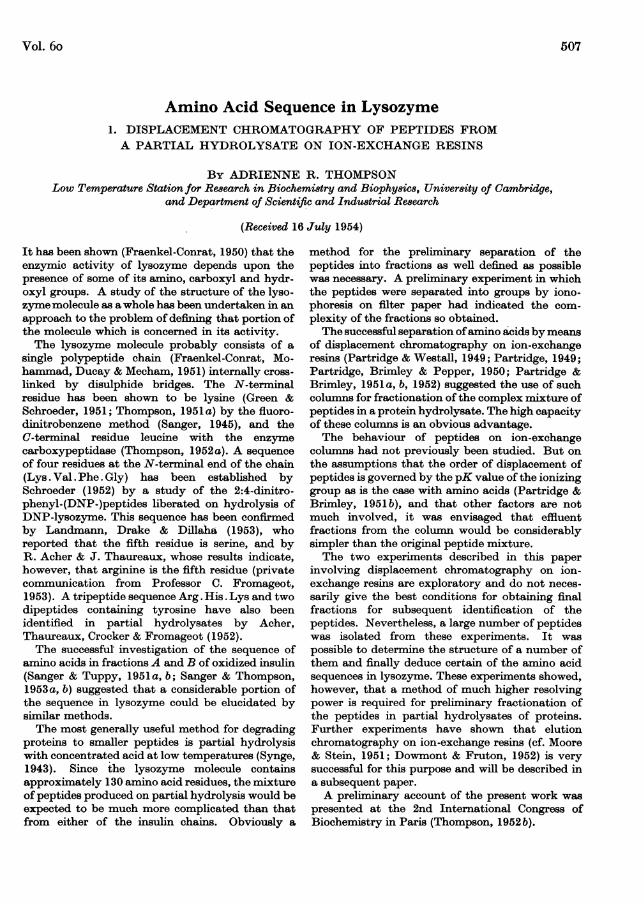
Fractions 35-75 were run on paper chromatograms inphenol-water-NH3 and butanol-acetic acid-water (Part-ridge, 1949); these are shown in Fig. 1. (The figures give pre-ponderantly the distribution of the amino acids, as peptidesgive weak colours in comparison.) The absence of spots infractions 59-68 indicates the ammonia band. Fractions70-73, following the ammonia band and corresponding toarginine in the displacement chromatogram of the aminoacids (Partridge & Brimley, 1952), were clearly differenti-ated from the rest of the material and were bulked as' arginine peptides'. The distribution of the remainder oftheamino acids and peptides was fairly satisfactory andapproached the limit of resolution for the amount of hydro-lysate, the size of column and the rather large particle sizeof the resin (100-120 mesh/in., which was the finest sul-phonated polystyrene available).The fractions were pooled as follows: 37-39; 40-43; 44-
47; 48-51; 52-58. This was done on the basis of the distribu-tion of amino acids as shown in Fig. 1.
Separation of peptides by paper chromatography. Each ofthe fractions obtained from the ion-exchange columns wasevaporated to dryness and run on duplicate two-dimensional
Leu
Val
AlaGlyLYS
40 45 50 55 40 50 60 70Fraction no. Fraction no.
Fig. 1. Experiment I. Fractionation of peptides on sulphonated-polystyrene columns. Paper chromatograms offractions developed: (a) with phenol-water-NH3; (b) with n-butanol-acetic acid-water. Control spots of aminoacids on left of figures.
Treatment with activated charcoal. An attempt was madeto remove selectively the peptides containing aromaticamino acids by adsorption on activated charcoal as de-scribed by Sanger & Tuppy (1951a). However, on subse-quent examination of this fraction, it contained littlematerial.
Displacement development ofpeptidesfrom lysozyme hydro-lysate. A column of sulphonated polystyrene (diam.0-95 cm., height 8-2 cm.) and a smaller column (diam.4 mm., height 5 cm.) mounted in series below it, were packedand regenerated as described by Partridge et al. (1950). Asmall amount of ammonia was added to the material notadsorbed on charcoal to act as 'carrier' to increase theseparation between lysine and arginine. This solution wasthen added to the top ofthe column and 0 075N-NaOH usedas displacement developer. Fractions (2 ml.) were collected
filter-paper chromatograms (Whatman no. 3). The solventswere phenol-water-NH3 (Consden, Gordon & Martin, 1944)and butanol-acetic acid-water (40: 10: 50 by vol.; Partridge,1948). The spots were detected by spraying with 0-025%ninhydrin in ethanol, cut out and eluted (cf. Sanger &Tuppy, 1951 a).
Identification of amino acid residues in peptides. Aboutone-third of each peptide was hydrolysed with 6N-HC1 for24 hr. at 1000 and the resulting amino acids identified bychromatography. Phenol-water-NH3 was used in the firstinstance and was the solvent used throughout this work forone-dimensional chromatograms. When confusion arosefrom amino acids having the same Rp, either n-butanol-acetic acid-water or tert.-pentanol-water (Consden et al.1944) was used. It is an advantage to add a trace of 8-hydroxyquinoline to the last solvent to avoid 'shadow'
508
AMINO ACID SEQUENCE IN LYSOZYME.spots caused by metal impurities in the filter paper (cf.Thompson, 1951 b).
In certain cases colour tests for histidine (Sanger &Tuppy, 1951a) and for proline (Acher, Fromageot &Jutisz, 1950) have proved valuable.N-Terminal residues of peptides with fluorodinitrobenzene.
The method of Sanger (1945) was used to identify the N-terminal amino acid in the remainder of the peptidematerial. Trimethylamine was used in place of NaHCO3 forthe condensation reaction (Sanger & Thompson, 1953a),thus avoiding the desalting procedure. After hydrolysis for8 hr. in 6N-HCI the DNP-amino acids were recovered byether extraction and identified on paper chromatogramswith tert.-pentanol buffer (pH 6.0) on buffered paper(Blackburn & Lowther, 1951). DNP-amino acids werealways run as controls oneach chromatogram. It was foundessential to run these chromatograms in the dark and atconstant temperature to achieve satisfactory results (cf.Biserte & Osteaux, 1951). DNP-Arginine and E-DNP-lysinewere always identified by similar chromatography of theyellow aqueous layer after ether extraction. The aminoacids in the aqueous phase were also identified on ordinarypaper chromatograms, generally with phenol as solvent.
C-Terminal residues with carboxypeptidase. Towards theend of this work it was found possible to use the enzymecarboxypeptidase to hydrolyse the C-terminal amino acidfrom isolated peptides in the same way as with lysozymeitself (Thompson, 1952a) and thus identify the C-terminalresidue. The method is only suitable when the peptide hasbeen shown to be pure by the DNP procedure. The peptidewas dissolved in 0-2 ml. 0-05M ammonium acetate, the pH ofwhich (8-0 in these experiments) was such as to give finalpH approx. 7-5 after addition of 0-02 ml. carboxypeptidasesuspension (10 mg./ml. water, purchased from Armour andCo.). After incubation at 370 for 16 hr. the enzyme wasinactivated and precipitated by boiling, the supernatantsolution evaporated to dryness and the liberated amino acididentified by paper chromatography. In certain cases theappearance of a second amino acid in less intensity than thefirst suggests that its position in the chain is next to theC-terminal residue.
In other cases, the terminal amino acid is not easilyliberated, e.g. glycine, lysine, proline and, probably,cysteic acid and arginine. In such cases the method fails,but the amount of material and labour thus expended issmall.
Experiment IILysozyme (500 mg.) was partially hydrolysed for 4 days.
The conditions for hydrolysis, oxidation of the disulphidebonds and adsorption of the aromatic peptides on charcoalwere the same as in Expt. I.The fraction not adsorbed on charcoal was evaporated to
dryness twice to remove acid and put first through sul-phonated-polystyrene columns. It was proposed to separ-ate a fraction corresponding in position to the amino acidarginine after the ammonia band as in Expt. I and alsoa fraction corresponding to lysine. The remainder of thematerial was to be eluted and run on a Zeo-Karb 215column.
This procedure was chosen since one of the possiblelimiting factors in the degree of resolution of the peptideson sulphonated-polystyrene columns was the rather largeparticle size of the sample available. Very finely powderedZeo-Karb 215 was available and, moreover, this resin gives
rather more satisfactory separations of the non-basic aminoacids than does sulphonated polystyrene (Partridge &Brimley, 1952). However, arginine cannot be displacedfrom Zeo-Karb 215 and lysine is incompletely displaced.Hence peptide fractions corresponding to these two aminoacids were first cut from the sulphonated-polystyrenecolumns. Four columns (diam. 1-2, 0 9, 0-6, 0-4 cm. andheights 14, 6-3, 5.0 and 4 0 cm. respectively) were used inseries. There wasample capacity to allow adequate turnoverin one run. The eluate from the column, after the materialhad been applied and washed through with water, wascollected, and, on concentration, gave a weakly positiveninhydrin reaction. This fraction, termed 'cysteic peptides'corresponds in behaviour to cysteic acid, which is notadsorbed by sulphonated-polystyrene resin.
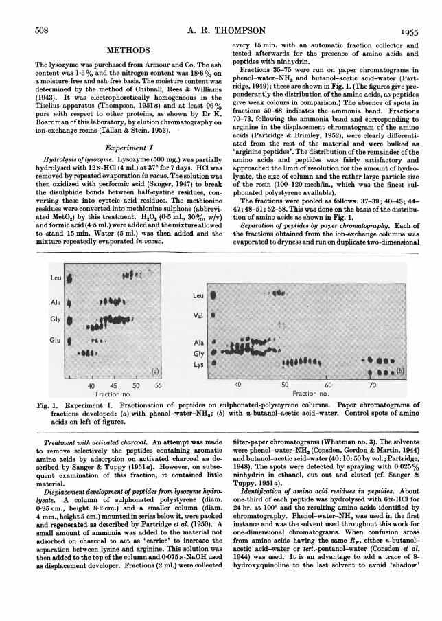
After removal of the cysteic acid fraction, the adsorbedmaterial was displaced with 0-075N-NaOH, flow rate7 ml./hr. Fractions of approx. 1-2 ml. were collected.Paper chromatography of successive fractions (as inExpt. I) is shown in Fig. 2.
Leu
Val
AlaGlyLys
10 15 LU 25 30 35Fraction no.
Fig. 2. Experiment II. Fractionation of peptides onsulphonated-polystyrene columns. Paper chromato-gram developed with n-butanol-acetic acid-water.Control spots of amino acids on left of figure.
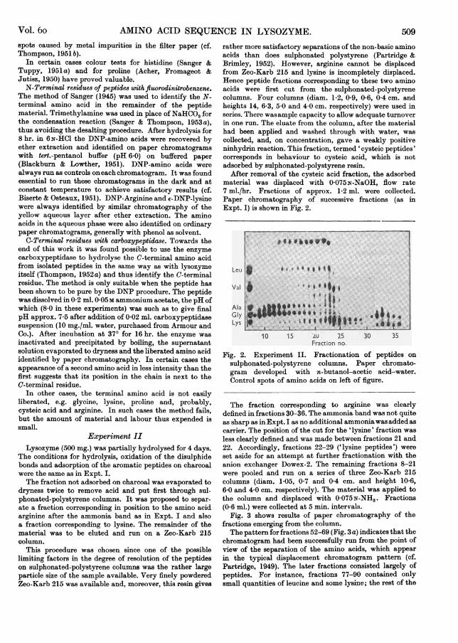
The fraction corresponding to arginine was clearlydefined in fractions 30-36. The ammonia band was not quiteas sharp as in Expt. I as no additional ammoniawas added ascarrier. The position of the cut for the 'lysine' fraction wasless clearly defined and was made between fractions 21 and22. Accordingly, fractions 22-29 ('lysine peptides') wereset aside for an attempt at further fractionation with theanion exchanger Dowex-2. The remaining fractions 8-21were pooled and run on a series of three Zeo-Karb 215columns (diam. 1-05, 0-7 and 04 cm. and height 10-6,6-0 and 4-0 cm. respectively). The material was applied tothe column and displaced with 0-075N-NH3. Fractions(0-6 ml.) were collected at 5 min. intervals.
Fig. 3 shows results of paper chromatography of thefractions emerging from the column.The pattern for fractions 52-69 (Fig. 3 a) indicates that the
chromatogram had been successfully run from the point ofview of the separation of the amino acids, which appearin the typical displacement chromatogram pattern (cf.Partridge, 1949). The later fractions consisted largely ofpeptides. For instance, fractions 77-90 contained onlysmall quantities of leucine and some lysine; the rest of the
Vol. 6o 509
A. R. THOMPSONmaterial appeared to be of peptide nature. The fractionswere pooled as follows: 52-55, 56-61, 62-66, 67-71, 72-76,77-82, 83-91. 2N-NH3 was then put through the column toremove any very strongly adsorbed peptides. These and thefractions after 91 were combined with the 'lysine peptides'fraction from sulphonated polystyrene.
I955There was some resolution on the column as shown
by paper chromatography of the fractions (Fig. 4) butthe degree of separation was disappointing. Fractionswere pooled in three groups (5-7, 8-10, 11-14 of 4-5,5*5 and 4*0 ml. respectively) which were examinedseparately.
Lou ~LAla ~~~~~~~~~~~~~~~Val
Gly
Aspd Lys
55 60 65 70 75 80 85 55 60 65 70 75 80 85Fractlon no. Fraction no.
Fig. 3. Experiment II. Fractionation of peptides on columns of Zeo-Karb 215. Paper chromatograms of fractionsdeveloped: (a) with phenol-water-NH3; (b) with n-butanol-acetic acid-water. Control spots of amino acids onleft of figure.
Leu
Val
Ala
Lys
5 10 15Fraction no.
Fig. 4. Experiment II. Fractionation of 'lysine peptides'on columns of Dowex-2. Paper chromatogram developedwith n-butanol-acetic acid-water. Control spots ofamino acids on left and right of figure.
The considerable overlap of peptides in the lysine fractionindicated that there was a large number of peptides whichbehaved similarly on cation-exchange resins.
It was hoped that, by using another property of themolecule (the ionization of the amino rather than of thecarboxyl group) a fractionation of the peptides on columnsof the anion exchanger (Dowex-2) would prove useful.Two columns (diam. 0-6 and 0-4 cm.; height, 6-7 and
5B0 cm., respectively) were prepared and operated in seriesas described by Partridge & Brimley (1951 b). Precautionswere taken to exclude CO. The material was displacedfrom the column with 0-075N-HCI.
I
t
3 ez
, 1co -cC
0-
i5
0 5 10Inches
n-Butanol-acetic acid-water
15
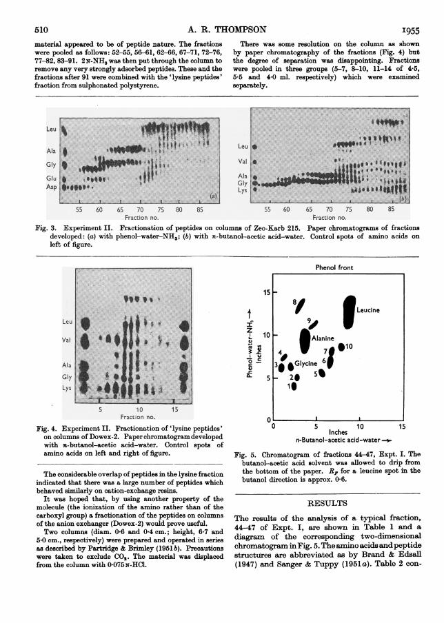
Fig. 5. Chromatogram of fractions 44-47, Expt. I. Thebutanol-acetic acid solvent was allowed to drip fromthe bottom of the paper. RF for a leucine spot in thebutanol direction is approx. 0-6.
RESULTS
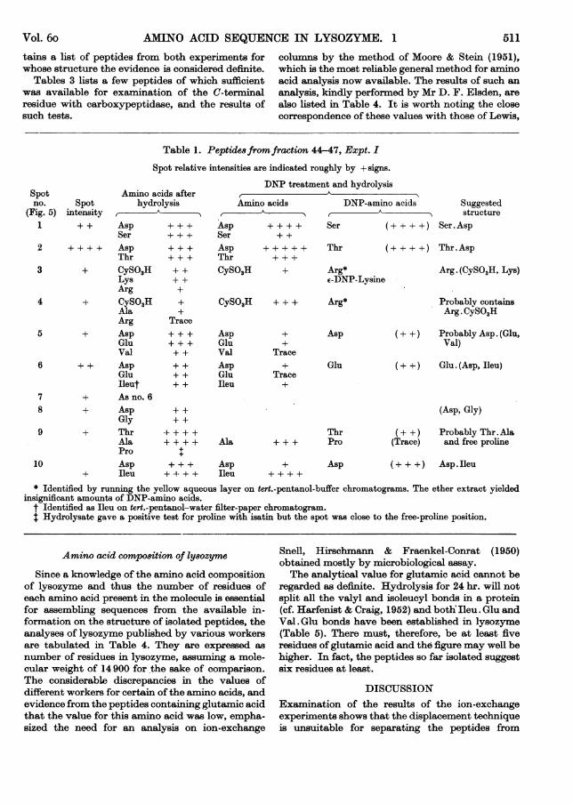
The results of the analysis of a typical fraction,44-47 of Expt. I, are shown in Table 1 and a
diagram of the corresponding two-dimensionalchromatogram inFig. 5. The amino acidsandpeptidestructures are abbreviated as by Brand & Edsall(1947) and Sanger & Tuppy (1951a). Table 2 con-
510
Phenol front
U Leucine
10 *Alanine
4 w 7910
39 .Glycine65 - 20 5
1 I
C- _I
AMINO ACID SEQUENCE IN LYSOZYME. 1
tains a list of peptides from both experiments forwhose structure the evidence is considered definite.
Tables 3 lists a few peptides of which sufficientwas available for examination of the C-terminalresidue with carboxypeptidase, and the results ofsuch tests.
columns by the method of Moore & Stein (1951),which is the most reliable general method for aminoacid analysis now available. The results of such ananalysis, kindly performed by Mr D. F. Elsden, arealso listed in Table 4. It is worth noting the closecorrespondence of these values with those of Lewis,
Table 1. Peptide8fromfraction 44-47, Expt. I
Spot relative intensities are indicated roughly by + signs.
Spotno.
(Fig. 5)Spot
intensity
Amino acids afterhydrolysis_:
DNP treatmer
Amino acids_A
1 + + Asp + + + AspSer + + + Ser
2 + + + + AspThr
3 + CySO3HLysArg
4 + CySO3HAlaArg
5 + AspGluVal
6 + + AspGluIleut
7 + As no. 68 + Asp
Gly9 + Thr
AlaPro
10 Asp+ Ileu
+++
+ +++++
Trace
AspThrCySO3H
++
nt and hydrolysis
DNP-amino acids Suggestedf
A > structureSer (+ + + +) Ser.Asp
Thr (+ + + +) Thr.Asp
+ Arg*E-DNP-Lysine
CySO3H + + + Arg*
+ + + Asp+ + + Glu+ + Val+ + Asp+ + Glu+ + Ileu
+++ +
4 + + ++
+
Trace
Trace
Ala + + +
+ + + Asp+ -1- + + Ileu
+
Asp
Glu
ThrPro
Asp
Arg. (CySO3H, Lys)
Probablv containsArg. CySO3H
(+ +) Probably Asp. (Glu,Val)
(+ +) Glu. (Asp, Ileu)
(Asp, Gly)
(+ +) Probably Thr. Ala(Trace) and free proline
(+ + +) Asp. Ileu
* Identified by running the yellow aqueous layer on tert.-pentanol-buffer chromatograms. The ether extract yieldedinsignificant amounts of DNP-amino acids.
t Identified as Ileu on tert.-pentanol-water filter-paper chromatogram.t Hydrolysate gave a positive test for proline with isatin but the spot was close to the free-proline position.
Amino acid compo8ition of ly8ozyme
Since a knowledge of the amino acid compositionof lysozyme and thus the number of residues ofeach amino acid present in the molecule is essentialfor assembling sequences from the available in-formation on the structure of isolated peptides, theanalyses of lysozyme published by various workersare tabulated in Table 4. They are expressed as
number of residues in lysozyme, assuming a mole-cular weight of 14900 for the sake of comparison.The considerable discrepancies in the values ofdifferent workers for certain of the amino acids, andevidence from the peptides containing glutamic acidthat the value for this amino acid was low, empha-sized the need for an analysis on ion-exchange
Snell, Hirschmann & Fraenkel-Conrat (1950)obtained mostly by microbiological assay.The analytical value for glutamic acid cannot be
regarded as definite. Hydrolysis for 24 hr. will notsplit all the valyl and isoleucyl bonds in a protein(cf. Harfenist & Craig, 1952) and both Ileu. Glu andVal . Glu bonds have been established in lysozyme(Table 5). There must, therefore, be at least fiveresidues of glutamic acid and the figure may well behigher. In fact, the peptides so far isolated suggestsix residues at least.
DISCUSSIONExamination of the results of the ion-exchangeexperiments shows that the displacement techniqueis unsuitable for separating the peptides from
Vol. 6o 511
A. R. THOMPSON
Table 2. Peptides recognized in effluent fractions from ion-exchange chromatograms
Italicized residues were judged C-terminal from experiments with carboxypeptidase.
Peptide Expt. Fraction Peptide Expt.Asp. Gly I 37-39 Leu. (Thr, Ala) I
II 67-71 IIII 83-91 II
Asp.Ala I 37-39 Leu or Ileu. (Glu, Ala, Leu IIII 72-76 or Ileu, Leu or Ileu)
Asp.Ileu I 37-39 Lys. (Val, Phe) II
I 40-43I 44 47 Phe.Glu II
II 4682-56 Ser.Asp III 72-76 II
Asp. (Gly, MetO2) II 56-61 Ser.Ala I
Asp. (Glu, Ala) II 77-82 II
II Lysine 8-10 Ser.Leu I
Asp. (Val, Glu) I 44-47 Ser. (Asp, Val) I
56-61 I
Asp. (Glu, Ala, Val) II 72-76 II
Ala. Leu II Lysine 11-14 Ser. (Asp, Gly) IIII
Ala.MetO2 I 48-51 IIII 72-76 IIII 77-82II 77-82 ~~~~~Ser. (Asp, Leu) II
Arg.CySO3H I 44-47Ser. (Asp, Leu) II
II 77-82 Ser.(Leu, Arg) II
II 83-91 Ser.(Gly, Leu) II
Arg.Asp I 48-51 III 52-58
Arg. (CySO3H, Glu) II 77-82 Ser. (CySO3H, Asp, Val) II
Arg. (CySO,H, Asp) I 37-39 Ser. (Gly, Ala, Leu?, Leu) III 52-58 Ser. (Asp, Gly, MetO2) II
Arg. (CySO3H, Lys) I 44 47 Ser. (Asp, Gly, Ala) II
CySO3H. (Gly, Lys) II 77-82 II
CySO3H. (Ala, Lys) II 77-82 Thr.Asp I
Gly.MetO. II 77-82 I
Gly.Lys I 52-58II Lysine residues
Glu.Ala I 48-51 Thr. Glu I
Glu.Leu I 52-58 II
II 83-91 Thr.Ala I
Glu. (Asp, Ileu) I 44 471 (Asp, Gly) II
I 48-51 I
I 52-58 II
II 72-76 Thr. (Glu, Ala) IIII 77-82 IIII Lysine 8-10
Thr. (Asp, Glu, Val) IIGlu.(Ala, Leu) II 77-82 II
II 83-91 IIII Lysine 8-10
Leu.Ala I 52-58 Thr.(Asp, Arg, Arg?) II
II 83-91 Val.Asp I
Ileu.Asp I 48-51 Val.CySO3H IILeu. Glu or Ileu. Glu II Lysine 8-10 Val. (CySO3H, Ala) II
Fraction
48-5177-8283-91
48-51
Lysine residues
83-91
44-4748-5172-76
48-5177-82
48-51
48-5177-82Lysine 11-14
67-7172-76Lysine 8-10Lysine 11-14
83-91
83-91
83-91Lysine 8-10Lysine 11-14
Cysteic
83-91
72-76
72-76Lysine 8-10
40-4344 4748-5167-7172-76Lysine 11-14
48-5172-76
48-51
Lysine 8-10Lysine 11-14
77-82Lysine 11-14
67-7172-76Lysine 8-10
83-91
40-43
Cysteic
Cysteic
'955512
Vol. 6o
Fractionof origin
I, 44-47
AMINO ACID SEQUENCE IN LYSOZYME. 1
Table 3. C-Terminal residues of peptides determined with carboxypeptidase
Peptide structure judgedfrom DNP treatment
Asp. (Glu, Val)
I, 44-47 Glu. (Asp, Ileu)I, 48-51 Glu. (Asp, Leu or Ileu)
I, 48-51 Leu. (Thr, Ala)I, 48-51 Leu or Ileu. (Glu, Ala,
Leu or Ileu, Leu or Ileu)II, 72-76 Thr. (Asp, Glu, Val)
II, 77-82 Asp. (Glu, Ala)
II, 85-91 Ser. (Gly, Ala, Leu)4
GluVal
Ileu
Amino acids liberatedby carboxypeptidase
+
+
Leu or IleuAsp
AlaLeu*AlaGluValAsp
AlaGluLeu§Ala
++
+
++++++
++
++
+
++
Structure inferred
Asp. Val. Glu
Glu.Asp.IleuGlu.Asp.Leu or Ileu
Leu.Thr.AlaLeu or Ileu (Glu,Ala, Leu or Ileu).Leu
Thr. (Asp, Val) *Glut
Asp. Glu.Ala
Ser. (Gly, Ala, Leu?) .Leu
* This spot from a butanol-acetic acid chromatogram was cut out, eluted, and, run with tert.-pentanol-water, showedLeu (+ +) Ileu (+). This is suggestive that the C-terminal residue is leucine and not isoleucine. In the same peptide theliberation of alanine in next highest intensity to leucine is strong presumptive evidence for its being next to the C-terminalresidue and hence for the sequence Leu or Ileu. (Glu, Leu or Ileu). Ala. Leu in the peptide.
t Liberation of valne in next highest intensity to glutamic acid strongly suggests the sequence Thr.Asp.Val. Glu.Possibly two leucine residues.
§ Run in butanol-acetic acid. If, as seems likely, there are two leucine residues in the peptide, the results could beinterpreted by either Ser. Gly.Ala. Leu. Leu or Ser. (Gly, Leu).Ala .Leu.
Table 4. Amino acid composition of lysozyme
The more firmly established figures in the last column are in black.
moles/14900 g. lysozyme
AlanineArginineAspartic acidCystineGlutamic acidGlycineHistidineIsoleucineLeucineLysineMethioninePhenylalanineProlineSerineThreonineTryptophanTyrosineValine
Presentwork*12-411-020-4
5-111-81-05.98*25-81-93.41*98-66*7
3-165.3
Lewis et al.(1950)
9.710*920-44-964-36
11-31.005*917-845B812-063-821.819-516-897-732-926-11
Fromageot &de Garilhe (1950)
10-21142t17.9§5.03-4
10-61-06-1§9.6§6-242-32-081-7
10 2
6-76*12-96.0§
Miust(1952)10.112-513*411-44.359.53-755-557-36-232-372-572-178-95.75
2*768-3
Preferredinteger10-1211
2055
11-12i
686
23297
35-6
* Determined after hydrolysis by refluxing for 24 hr. in 6N-HCI. Analysis by Mr D. F. Elsden.t Determined as dinitrophenylamino acids.I Monier et al. (1952).§ Personal communication quoted by Fevold (1950).
513
33 Bioch. 1955, 60
A. R. THOMPSON
partial hydrolysates of proteins. This is not due tothe difficulties involved in scaling down the size ofthe columns, as is shown by the comparativelysuccessful separation of amino acids in the mixture(cf. Fig. 3, where the strong spots represent aminoacids), but rather to the behaviour of the peptidesthemselves. The amino acids are preferentially dis-placed from the columns in their normal sequencebut the proportion of longer-chain peptides in-creases gradually throughout the chromatogram tillthe final fractions consist entirely of peptides,especially those of highest molecular weight. It isclear that the behaviour of the peptides is govemedlessby thepK values oftheir ionizing groups thanbyvan der Waals adsorptive forces between the resinand the peptide molecules (Partridge & Brimley,1951 b) which are regulated, as a first approxima-tion, by molecular weight. This adsorption is farmore serious than is indicated in Table 2, whichomits many peptides altogether since it was notpossible to determine their structure. This is be-cause, in general, the larger the peptide molecule,the more difficult it is to determine its compositionand the less there is of it in the hydrolysate. Theimportance of adsorption might have been pre-dicted from a consideration of the behaviour ofaromatic amino acids, which are displaced from ion-exchange chromatograms in a different order fromthat which would be expected on the basis of pKvalues alone.As these adsorptive forces are relatively un-
specific, it is not surprising that, when they pre-ponderate over the ion-exchange reactions, thecolumns should fail to differentiate between largenumbers of peptides of similar molecular size. Thishelps to explain why, to take two examples fromExpt. I where the amino acids themselves werereasonably differentiated, Asp . Ileu should occur infractions 37-39, 40-43, 44-47 and 48-51 and Glu.-(Asp.lieu) in fractions 44-47, 48-51 and 52-58.Unfortunately, the distribution of any one peptidebetween several (ion-exchange) fractions not onlycomplicated the fractions but also furnished less ofthe peptide to work with. This lack of materialsometimes made it impossible to obtain confir-matory evidence in doubtful cases, hence withseveral peptides no definite result was obtained. It ispossible that, on ion-exchange columns operated athigher temperatures, a reduction of the effect of theadsorptive forces might permit a more typical ion-exchange behaviour and give more useful separa-tions (cf. Partridge & Brimley, 1951a), but thisinvolves the risk that some decomposition of thepeptide material might occur.
Despite the fact that displacement chromato-graphy on ion-exchange columns proved disappoint-ing as a method for the preliminary separation ofpeptides resulting from the hydrolysis of lysozyme,
it was possible in these two experiments to deter-mine the structure of a considerable number ofpeptides and, from these, to deduce several probablesequences. These are listed in Table 5, and beneaththem the structures of the peptides from which theevidence for them is derived.
Table 5. Probable peptide 8equences in ly8ozyme
1. Ser. Asp. Gly. MetO.Ser. (Asp, Gly, MetO2)Ser. (Asp. Gly)
Asp.(Gly, MetO2)Ser. Asp Gly. MetO2
Asp. Gly2. Thr. Glu.Ala
Thr. (Glu, Ala)Thr. Glu
Glu. Ala3. Thr. Asp.Val. Glu. Ala
Thr. (Asp, Val, Glu, Ala)*Thr. (Asp, Val, Glu)
Asp, (Val, Glu, Ala)Thr. (Asp, Val)*
Asp. (Val, Glu)Thr. Asp Glu. Ala
Val. Glu*4. Leu.Thr. Ala
Leu.(Thr, Ala)Thr. Ala
5. Ileu. Glu. Leu. Ala. LeuLeu or. (Glu, Leu or, Ala, Leu)Ileu Ileu
Ileu. (Glu, Leu)*Glu. (Leu, Ala)
Leu or. Glu. Leu. AlaIleu
Glu. Leu Ala. Leu6. Asp. Glu.Ala
Asp. (Glu, Ala)Glu. Ala
7. Glu. Asp. IleuGlu. (Asp, Ileu)
Asp. Ileu* Evidence for the presence of these peptides was ob-
tained by using elution chromatography on ion-exchangeresins in recent experiments which will be described in alater paper. The C-terminal residue of Glu. (Leu, Ala) wasalso shown to be alanine using carboxypeptidase.
Sequence (1) Ser .Asp . Gly MetO2 follows directlyfrom the fact that lysozyme contains only twomethionine residues (see analysis in Table 4) andsince peptide Ala . MetO2 has been well established,any peptide containing MetO2 and not preceded byAla must belong to the other MetO2 sequence.
Sequence (2) Thr - Glu .Ala. On partial hydrolysisof the peptide Thr. (Glu, Ala) and chromatography,a spot appeared which was slow to develop a nin-hydrin colour, consisted of Thr (trace) and Glu(+ +), and corresponded in position to Thr.Glu.This establishes the sequence Thr . Glu .Ala.
514 I955
Vol. 6o AMINO ACID SEQUENCE IN LYSOZYME. 1 515Sequence (3) Thr.Asp.Val.Glu.Ala. The se-
quence Thr . Asp. Val. Glu was well established bytwo peptides where carboxypeptidase was used todetermine the C-terminal residue. Alanine isestablished as the next residue by the pentapeptideThr. (Asp, Val, Glu, Ala).
Sequence (4) Leu. Thr .Ala. This sequence isdefinitely established despite the fact that the bondinvolving the amino group of the threonine isunbroken, which is unusual as such bonds, in-volving serine and threonine, are generally the mostlabile to acid hydrolysis. The occurrence of thepeptide Leu. (Thr, Ala) on at least three occasionsleaves no doubt that it is genuinely present in thehydrolysate. It is, however, the only case en-countered in these experiments of a peptide con-taining serine or threonine bound through its aminogroup.
It was clear from the results ofhydrolysis that thepeptide represented by sequence (5) had one or twoleucine residues in addition to an isoleucine residue.One of these three residues was N-terminal and oneof the leucines C-terminal, with a suggestion thatalanine preceded it. The peptide Ileu. (Glu, Leu)showed that Ileuwas N-terminal and confirmed thatthe second and third residues were Glu and Leu(order unknown). The correct order was actuallyGlu. Leu as shown by the peptide Glu. (Leu, Ala)which also confirmed the position of the alanineresidue. All possible dipeptides consistent with thispentapeptide sequence have been identified.The tripeptide sequences (6) and (7) were estab-
lished by identification of both N- and C-terminalresidues.
SUMMARY
1. An attempt has been made to simplify themixture of peptides produced on partial acidhydrolysis of lysozyme through fractionation bydisplacement chromatography on -ion-exchangeresins.
2. The peptides in the fractions so obtained havebeen separated by two-dimensional paper chro-matography. The resulting peptide spots have beeneluted and analysed for the constituent aminoacids, the N-terminal residue and, in certain cases,the C-terminal residue.
3. The peptides whose structure is known arelisted. From this information it has been possibleto assemble a number of probable amino acidsequences as they occur in the lysozyme molecule.
The author wishes to thank Dr S. M. Partridge for usefulsuggestions on the use and operation of ion-exchangecolumns, and also Dr F. Sanger, Dr E. 0. P. Thompson andDr J. Brooks for many helpful discussions.Mr D. F. Elsden carried out the analysis of lysozyme,
Miss J. E. Dixon assisted in the experiments and Mr D. P.Gatherum prepared the photographs.
The work described in this paper was carried out as partofthe programme ofthe Food Investigation Organization ofthe Department of Scientific and Industrial Research.
REFERENCESAcher, R., Fromageot, C. & Jutisz, M. (1950). Biochim.
biophy8. Acta, 5, 81.Acher, R., Thaureaux, J., Crocker, C. & Fromageot, C.
(1952). Biochim. biophys. Acta, 9, 339.Biserte, G. & Osteaux, R. (1951). Bull. Soc. Chim. biol.,
Pari8, 33, 50.Blackburn, S. & Lowther, A. G. (1951). Biochem. J. 48, 126.Brand, E. & Edsall, J. T.(1947). Annu. Rev. Biochem. 16,224.Chibnall, A. C., Rees, M. W. & Williams, E. F. (1943).
Biochem. J. 37, 354.Consden, R., Gordon, A. H. & Martin, A. J. P. (1944).
Biochem. J. 38, 224.Dowmont, Y. P. & Fruton, J. S. (1952). J. biol. Chem. 197,
271.Fevold, H. L. (1950). Advanc. Protein Chem. 6, 187.Fraenkel-Conrat, H. (1950). Arch. Biochem. 27, 109.Fraenkel-Conrat, H., Mohammad, A., Ducay, E. D. &Mecham, D. K. (1951). J. Amer. chem. Soc. 78, 625.
Fromageot, C. & de Garilhe, M. P. (1950). Biochim .6iophy8.Acta, 4, 509.
Green, F. C. & Schroeder, W. A. (1951). J. Amer. chem. Soc.73, 1385.
Harfenist, E. J. & Craig, L. C. (1952). J. Amer. chem. Soc.74, 4216.
Landmann, W. A., Drake, M. P. & Dillaha, J. (1953).J. Amer. chem. Soc. 75, 3638.
Lewis, J. C., Snell, N. S., Hirschmann, D. J. & Fraenkel-Conrat, H. (1950). J. biol. Chem. 186, 23.
Mills, G. L. (1952). Biochem. J. 50, 707.Monier, R., Gendron, Y., Jutisz, M. & Fromageot, C. (1952).
Biochim. biophys. Acta, 8, 588.Moore, S. & Stein, W. H. (1951). J. biol. Chem. 192, 663.Partridge, S. M. (1948). Biochem. J. 42, 238.Partridge, S. M. (1949). Biochem. J. 44, 521.Partridge, S. M. & Brimley, R. C. (1951 a). Biochem. J. 48,
313.Partridge, S. M. & Brimley, R. C. (1951 b). Biochem. J. 49,
153.Partridge, S. M. & Brimley, R. C. (1952). Biochem. J. 51,
628.Partridge, S. M., Brimley, R. C. & Pepper, K. W. (1950).
Biochem. J. 46, 334.Partridge, S. M. & Westall, R. G. (1949). Biochem. J. 44,418.Sanger, F. (1945). Biochem. J. 39, 507.Sanger, F. (1947). Nature, Loud., 160, 295.Sanger, F. & Thompson, E. 0. P. (1953a). Biochem. J. 58,
353.Sanger, F. & Thompson, E. 0. P. (1953b). Biochem. J. 53,
366.Sanger, F. & Tuppy, H. (1951 a). Biochem. J. 49, 463.Sanger, F. & Tuppy, H. (1951 b). Biochem. J. 49, 481.Schroeder, W. A. (1952). J. Amer. chem. Soc. 74, 5118.Synge, R. L. M. (1943). Chem. Rev. 32, 135.Tallan, H. H. & Stein, W. H. (1953). J. biol. Chem. 200,507.Thompson, A. R. (1951 a). Nature, Lond., 168, 390.Thompson, A. R. (1951 b). Aust. J. Sci. Res. B, 4, 180.Thompson, A. R. (1952 a). Nature, Lond., 169, 495.Thompson, A. R. (1952b). Resumes des Communications,2nd Int. Congr. Biochem., Paris, p. 195.
33-2