an analysis of x-ray induced chromosomal … · an analysis of x-ray induced chromosomal...
TRANSCRIPT
AN ANALYSIS OF X-RAY INDUCED CHROMOSOMAL ABERRATIONS IN TRADESCANTIA
KARL SAX Arnold Arboretum, Harvard University, Cambridge, Massachusetts
Received August 19, 1939
HE cause and nature of chromosomal alterations has become an im- T portant genetic problem since it has been shown that both mutation and speciation often are associated with structural alterations of the chromosomes (DOBZHANSKY 1937). The frequency of spontaneous struc- tural changes is too low in most organisms to permit a statistical analysis of types or frequencies of alterations, although recently GILES (1940) has been able to analyze a considerable number of natural chromosomal aber- rations in Tradescantia hybrids. In most respects these natural aberrations resemble those induced by X-rays, and although the spontaneous breaks are not caused by radiation, the same secondary factors seem to be in- volved in producing the various types and frequencies of aberrations. The X-ray induced aberrations can be produced in large numbers under con- trolled conditions, permitting adequate statistical analyses of types of aberrations and their frequency in relation to dosage, temperature, time of exposure, and radiation intensity.
The microspores of diploid species of Tradescantia provide excellent material for an analysis of X-ray induced chromosomal aberrations. The six chromosomes are large and the nuclear cycle can be timed rather ac- curately. During the summer months the microspore nucleus remains in the resting stage for about five days after the microspore is formed follow- ing meiosis. At about 30 hours before metaphase the chromosomes begin splitting, and a t 23 to 24 hours before metaphase all chromosomes are split to form the sister chromatids. Thus the nuclear cycle from micro- spore formation to metaphase requires about six days. During the winter months the nuclear cycle is nearly twice as long and the prophase stage may begin at about 6-70 hours before metaphase. For an analysis of chromatid breaks induced a t prophase the cells were fixed 24 hours after raying. Microspores fixed five days after raying were irradiated in the resting stage. Preparations were made a t 48 hours after raying during the winter months, and 30 hours after raying during the summer months, for the analysis of very early prophase stages. The analyses of dosage, the time factor, and temperature effects were based on experiments done dur- ing the winter months. The division figures obtained provide a true random sample of aberrations because there is no chance of elimination of chromo- somes or chromosome fragments.
The microspore cells were smeared, fixed in alcohol-acetic for a few GENETICS 15: 41 Jan. Ig4o

42 KARL SAX
minutes, and stained with aceto-carmine. Several hundred division figures usually were obtained from each flower bud, and preparations with less than about a hundred metaphase or anaphase figures were discarded. Since there are six chromosomes in each microspore each preparation permitted the anlysis of from 600 to 2 0 0 0 chromosomes. The results to be presented are based on an analysis of more than 300,000 chromosomes.
The statistical treatment of the data presents a difficult problem. Most of the individual values given in the various tables are based on the average frequencies on three slides, and the total number of chromosomes is from 3,000 to 6,000. Neither the number of slides nor the number of chromo- somes provides the correct value of n for a critical analysis of variation. Accordingly we have taken the deviation from the mean, in percent of the mean, for each series of observations involving three or more slides each, and have obtained the standard deviation and probable error from these figures. The value of n was taken as the number of series times 1z-I
slides examined for each series. The generalized probable error is found to be about IO percent of the mean. Many of the experiments were re- peated several times with comparable results.
TYPES OF CHROMOSOMAL ABERRATIONS
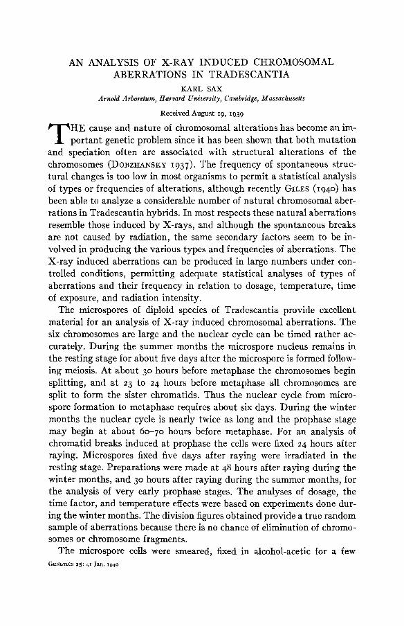
Two general types of chromosomal aberrations are induced by X-rays, namely, chromatid breaks, and chromosome breaks. The chromatid breaks are induced a t prophase when each chromosome consists of two sister chromatids, while the chromosome breaks are those induced during the resting stage when the chromosomes are in the form of single threads. Each of these general types of aberrations may be further classified into two groups: the aberrations caused by a single break which are referred to as one-hit breaks, and aberrations involving breaks in two different chromo- somes or different loci of the same chromosomes which are referred to as two-hit aberrations. The origin and nature of these four classes of chromo- somal aberrations is shown diagramatically in figure I.
The one-hit chromatid breaks may involve one or both chromatids. The terminal deletion of one of the two chromatids (Plate I, figs. E and F) results in a chromatid fragment which is usually not included in either daughter nucleus a t telophase. Often, however, the fragment appears to be separated from the centric arm only by an achromatic lesion, suggesting a partial breakage or imperfect reunion. Although these one-hit terminal deletions are rather frequent they cannot be scored accurately and have not been included in the analysis of X-ray breaks. The most frequent type of one-hit chromatid aberration is a break in both chromatids at the same locus followed by lateral fusion to produce a dicentric chromatid and an acentric U-shaped fragment (Plate I, B). At anaphase the dicentric chro-
INDUCED ABERRATIONS I N TRADESCANTIA 43
matid forms a single bridge and the fragment tends to straighten out (Plate I, C). Such fragments are rarely included in the daughter nuclei. The ends of the fragment are the normal ends of sister chromatids (cf. figure I).
The two-hit chromatid aberrations are of various types. The most fre- quent types are fusions between broken chromatids of two different chro- mosomes. These consist of reciprocal chromatid exchanges, producing a
aberrations I Chromatid aberri I I
Pre-meta a Ana hase Pro hase Pre-meta ha:
~IGURE th the types of chromosomal aberrations induced by X-rays at prophase and at the resting stage. Only the types which can be recognized at metaphase or anaphase have been in- cluded, since they are the types used in the various analyses.
pseudo-crossover type of configuration (Plate I, D), or the broken ends may fuse to form a chromatid bridge between two chromosomes accom- panied by an acentric fragment (Plate I, E). Other types have been found but they are rare. Occasionally the corresponding arms of a single chromo- some are of unequal length at anaphase (Plate I, F) indicating an inter- calary duplication such as KAUFMANN and BATE (1938) have described in Drosophila. Fusions between breaks in each of the two arms of a single chromosome have produced a few chromatid rings, and in rare cases an acentric ring is produced from two breaks in a single arm.
44 KARL S A X
The chromosome aberrations include both one-hit and two-hit types, The one-hit aberrations include both intercalary and terminal deficiencies. The loss of a terminal segment of a chromosome results in a shortened arm with no fusion of the ends of sister chromatids, and the acentric fragment consists of two separate chromatids at metaphase (Plate I, G). The small intercalary deletions presumably are deficiencies produced by breaks in adjacent relic spirals during the resting stage (figure I). These deleted fragments are small (Plate I, G and N) and have not been included in the analysis of chromosome breaks. Most of these deficiencies have been found to be two-hit aberrations (Rick, unpublished observations).
The two-hit chromosome aberrations include reciprocal interchange or fusion between different chromosomes and fusion or exchange between the arms of the same chromosome. Occasionally a relatively large intercalary deletion is produced resulting in an acentric ring fragment. Reciprocal interchange between arms of different chromosomes can be detected only when the exchange is unequal (Plate I, J), and such exchanges are infre- quent. Fusions between broken ends of two chromosomes result in a di- centric chromosome and an acentric fragment (Plate I, H and I). The
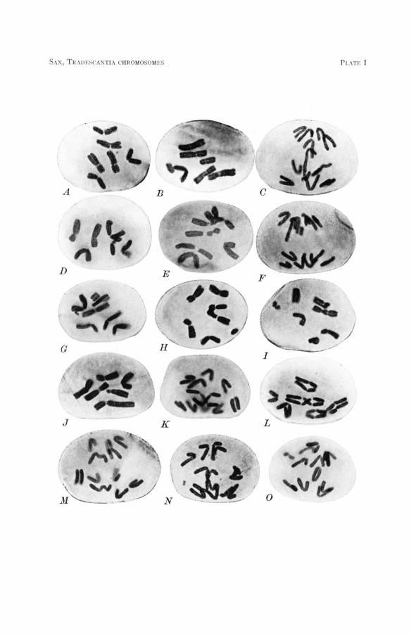
DESCRIPTION OF PLATE I Photographs of Tradescantia microspore chromosomes showing the types of aberrations in-
duced by X-rays. Compare with diagrams in figure I.
FIGURE A.-Untreated,-six normal chromosomes at metaphase. FIGURE B.-A one-hit chromatid aberration induced at prophase. Lateral fusions have occurred
between the ends of the broken sister chromatids to produce a shortened arm with terminal fusion of sister chromatids and a U-shaped acentric fragment. Metaphase.
FIGURE C.-The same at anaphase showing the chromatid bridge and partially straightened U-shaped fragment.
FIGURE D.-A two-hit chromatid aberration. Reciprocal interchange between chromatids of dif- ferent chromosomes.
FIGURE E.-The same, but with chromatids fused to form a chromatid bridge between chromo- somes. Also a one-hit chromatid aberration-terminal deletion of only one of the two sister chromatids.
FIGURE F.-An intercalary chromatid duplication and deficiency involving the sister chromatids of one arm. Results in unequal arms at anaphase. Also a single chromatid terminal dele- tion.
FIGURE G.-Two one-hit chromosome aberrations. One is a terminal deletion of most of one arm, and the other a very small interstitial deletion.
FIGURE H.-A two-hit chromosome aberration. A dicentric chromosome and the accompanying fragment. Note relational coiling between centromeres.
FIGURE 1.-Two two-hit chromosome aberrations. A dicentric and a locked ring with their ac- companying fragments.
FIGURE J.-Unequal interchange between two chromosomes. FIGURES K-N.-Behavior of dicentric chromosomes at anaphase. Also a terminal chromosome
FIGURE 0.-A continuous ring chromosome at anaphase. Also somatic non-disjunction of two deletion and an interstitial deletion in figure 14.
anaphase chromosomes.
S.\s, l'n \ i w w . i s T r , \ ( m " m n \ w s r I. \TI' I
INDUCED ABERRATIONS IN TRADESCANTIA 45
broken ends of the terminal deletions fuse to produce a fragment consisting of parts of two chromosomes. The ends of the fragment are the normal ends of the two arms. The relational coiling between centromeres of the dicentric chromosome persists to metaphase and at anaphase the dicentric chromatids may separate freely, form an X-shaped bridge, or interlock (Plate I, K-N). Fusion between breaks in the two arms of the same chro- mosome results in a ring chromosome and an acentric fragment (Plate I, I). The ends of the fragment are the normal ends of the two arms. These ring chromosomes may separate freely a t anaphase, they may be locked, or they may form a single large ring (Plate I, 0). Occasionally a ring chromo- some is locked around its rod fragment.
Combinations of chromosome and chromatid breaks may occur in the same cell or even in the same chromosome, but they are very rare. The individual chromosomes must split very rapidly a t prophase (SAX and
Other types of chromatid and chromosome aberrations undoubtedly occur, but inversions, transpositions or intra-chromosomal translocations, and simple intercalary translocations, either cannot be detected, or cannot be distinguished from reciprocal translocations. The types of aberrations included in the subsequent analyses consist of the one-hit chromatid breaks involving both chromatids, two-hit chromatid breaks which pro- duce exchange or dicentric chromatids, and the two-hit chromosome breaks resulting in dicentric and ring chromosomes. These constitute most of the visible chromosomal alterations induced by X-ray doses used in the following experiments.
MATHER 1939).
FREQUENCIES OF TYPES OF ABERRATIONS IN RELATION TO
SPATIAL ARRANGEMENT OF THE CHROMOSOMES
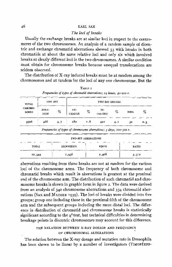
The relative frequencies of one-hit and two-hit aberrations vary with the X-ray dosage and will be considered later. The various types of two-hit aberrations, both chromosome and chromatid, appear in the same relative frequencies a t different dosages, even though the actual frequencies in- crease exponentially with dosage. The relative frequencies of the two-hit aberrations provide evidence regarding the conditions and limitations of fusion between broken ends of chromatids and chromosomes. The fre- quencies of types of chromatid and chromosome alterations are shown in table I. The dicentric chromatids are more than twice as frequent as the exchange chromatids. The exchange types cannot ordinarily be recognized at anaphase but most of these observations were made at metaphase so that the 2 : I ratio is approximately correct. The ratio of dicentric and ring chromatids is about 13 to I , but for the chromosome aberrations the ratio of dicentrics and rings is 3.3 to I.
46 KARL SAX
The loci of breaks Usually the exchange breaks are at similar loci in respect to the centro-
meres of the two chromosomes. An analysis of a random sample of dicen- tric and exchange chromatid aberrations showed 33 with breaks in both chromatids at about the same relative loci and only six which involved breaks a t clearly different loci in the two chromosomes. A similar condition must obtain for chromosome breaks because unequal translocations are seldom observed.
The distribution of X-ray induced breaks must be a t random among the chromosomes and at random for the loci of any one chromosome. But the
TABLE I
Frequencies of types of chromatid aberrations; 24 hours, 40-200 r.
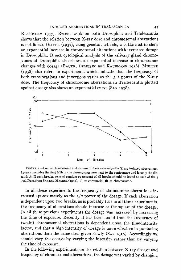
aberrations resulting from these breaks are not a t random for the various loci of the chromosome arms. The frequency of both chromosome and chromatid breaks which result in aberrations is greatest at the proximal end of the chromosome arm. The distribution of such chromatid and chro- mosome breaks is shown in graphic form in figure 2 . The data were derived from an analysis of 340 chromosome aberrations and 334 chromatid aber- rations (SAX and MATHER 1939). The loci of breaks were divided into five groups; group one including those in the proximal fifth of the chromosome arm and the subsequent groups including the more distal loci. The differ- ence in distribution of chromatid and chromosome breaks is statistically significant according to the x2 test, but technical difficulties in determining breakage points in dicentric chromosomes may account for this difference.
THE RELATION BETWEEN X-RAY DOSAGE AND FREQUENCY
OF CHROMOSOMAL ALTERATIONS
The relation between the X-ray dosage and mutation rate in Drosophila has been shown to be linear by a number of investigators (TIMOF~~EFF-
INDUCED ABERRATIONS I N TRADESCANTIA 47
RESSOVSKY 1937). Recent work on both Drosophila and Tradescantia shows that the relation between X-ray dose and chromosomal aberrations is not linear. OLIVER (1932)~ using genetic methods, was the first to show an exponential increase in chromosomal alterations with increased dosage in Drosophila. Direct cytological analysis of the salivary gland chromo- somes of Drosophila also shows an exponential increase in chromosome changes with dosage (BAUER, DEMEREC and KAUFMANN 1938). MULLER (1938) also refers to experiments which indicate that the frequency of both translocations and inversions varies as the 3/2 power of the X-ray dose. The frequency of chromosome aberrations in Tradescantia plotted against dosage also shows an exponential curve (SAX 1938).
30
U) s
m + 0
).
0 c al
U
20
FIGURE loci of chromosome and chromatid breaks involved in X-ray induced aberrations. Locus I includes the first fifth of the chromosome arm next to the centromere and locus 5 the dis- tal fifth. If such breaks were a t random 2 0 percent of all breaks should be found a t each of the 5 loci. Data from SAX and MATAER (1939). 0 = chromatid. 0 = chromosome.
In all these experiments the frequency of chromosome aberrations in- creased approximately as the 312 power of the dosage. If each aberration is dependent upon two breaks, as is probably true in all these experiments, the frequency of aberrations should increase as the square of the dosage. In all these previous experiments the dosage was increased by increasing the time of exposure. Recently it has been found that the frequency of two-hit chromosomal aberrations is dependent upon the time-intensity factor, and that a high intensity of dosage is more effective in producing aberrations than the same dose given slowly (SAX 1939). Accordingly we should vary the dosage by varying the intensity rather than by varying the time of exposure.
In the following experiments on the relation between X-ray dosage and frequency of chromosomal aberrations, the dosage was varied by changing
48 KARL SAX
the distance between the target and the flower buds according to the in- verse square law, so that variation in dosage was given without changing the exposure time. The minimum distance used was 24 cm, but even at this distance there is some variation in dosage, due to slight differences in the positions of the young buds in the inflorescences. At greater distances this experimental error is decreased. The source of the X-rays was a Coolidge tube with a tungsten target, operated at IO ma, 134 K.V. with no screen.
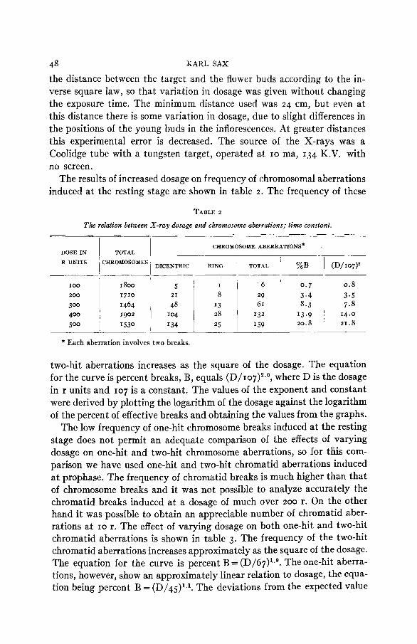
The results of increased dosage on frequency of chromosomal aberrations induced a t the resting stage are shown in table 2 . The frequency of these
DOSE I N TOTAL
R UNITS i CHROMOSOMES
TABLE 2
The relation between X-ray dosage and chromosome aberrations; time constant.
CHROMOSOME ABERRATIONS*
RING 1 TOTAL 1 %'oB 1 (D/I07)' DICENTRIC ~
I O 0 I I800 5 200 1710 21
300 1464 48 400 1902 '04 500 I530 134
I 6 I 0.7 0 . 8 8 29 3 . 4 3.5
13 61 8 .3 7 .8 28 14 .0 25 I59 20.8 21.8
two-hit aberrations increases as the square of the dosage. The equation for the curve is percent breaks, B, equals (D/107)~ .~ , where D is the dosage in r units and 107 is a constant. The values of the exponent and constant were derived by plotting the logarithm of the dosage against the logarithm of the percent of effective breaks and obtaining the values from the graphs.
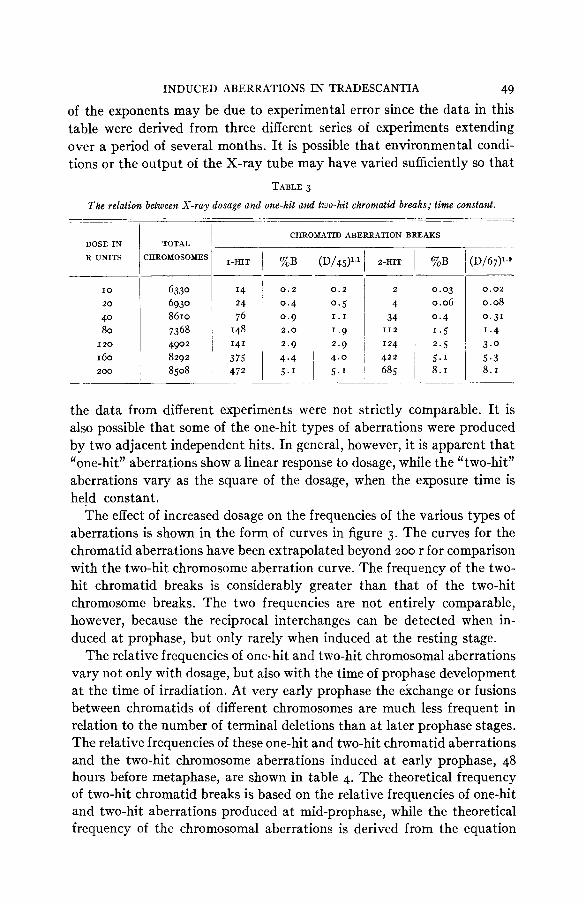
The low frequency of one-hit chromosome breaks induced at the resting stage does not permit an adequate comparison of the effects of varying dosage on one-hit and two-hit chromosome aberrations, so for this com- parison we have used one-hit and two-hit chromatid aberrations induced a t prophase. The frequency of chromatid breaks is much higher than that of chromosome breaks and it was not possible to analyze accurately the chromatid breaks induced at a dosage of much over 2 0 0 r. On the other hand it was possible to obtain an appreciable number of chromatid aber- rations at IO r. The effect of varying dosage on both one-hit and two-hit chromatid aberrations is shown in table 3 . The frequency of the two-hit chromatid aberrations increases approximately as the square of the dosage. The equation for the curve is percent B = (D/67)1.g. The one-hit aberra- tions, however, show an approximately linear relation to dosage, the equa- tion being percent B = (D/45)1.1. The deviations from the expected value
INDUCED ABERRATIONS IN TRADESCANTIA 49
of the exponents may be due to experimental error since the data in this table were derived from three different series of experiments extending over a period of several months. It is possible that environmental condi- tions or the output of the X-ray tube may have varied sufficiently so that
TABLE 3
The relation between X-ray dosage and one-hit and two-hit chromatid breaks; time constant.
6330 6930 8610 7368
- _~____
DOSE IN
R UNITS
I4 24 76 148
10
20
40 80
I 60 I20
2 0 0
ZHROMOSOMES I-HIT 1- CHROMATID ABERRATION BREAKS
%B
0.2
0.4 0.9
2.9 4.4 5.’
2 .o
0.03 0.06 0.4 1.5
2.5 5.1 8.1
D/67)’,9
0.02
0.08 0.31 1.4 3 . 0 5.3 8. I
the data from different experiments were not strictly comparable. It is also possible that some of the one-hit types of aberrations were produced by two adjacent independent hits. In general, however, it is apparent that “one-hit” aberrations show a linear response to dosage, while the “two-hit” aberrations vary as the square of the dosage, when the exposure time is held constant.
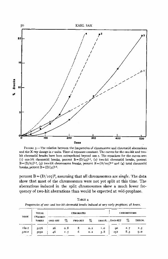
The effect of increased dosage on the frequencies of the various types of aberrations is shown in the form of curves in figure 3 . The curves for the chromatid aberrations have been extrapolated beyond 2 0 0 r for comparison with the two-hit chromosome aberration curve. The frequency of the two- hit chromatid breaks is considerably greater than that of the two-hit chromosome breaks. The two frequencies are not entirely comparable, however, because the reciprocal interchanges can be detected when in- duced at prophase, but only rarely when induced at the resting stage.
The relative frequencies of one-hit and two-hit chromosomal aberrations vary not only with dosage, but also with the time of prophase development at the time of irradiation. At very early prophase the exchange or fusions between chromatids of different chromosomes are much less frequent in relation to the number of terminal deletions than at later prophase stages. The relative frequencies of these one-hit and two-hit chromatid aberrations and the two-hit chromosome aberrations induced a t early prophase, 48 hours before metaphase, are shown in table 4. The theoretical frequency of two-hit chromatid breaks is based on the relative frequencies of one-hit and two-hit aberrations produced at mid-prophase, while the theoretical frequency of the chromosomal aberrations is derived from the equation
Dose
FIGU.RE 3.-The relation between the frequencies of chromosome and chromatid aberrations and the X-ray dosage in r units. Time of exposure constant. The curves for the one-hit and two- hit chromatid breaks have been extrapolated beyond 200 r. The equations for the curves are: (I) one-hit chromatid breaks, percent B = (D/45)lJ, (2) two-hit chromatid breaks, percent B= (D/67)1.9, (3) two-hit chromosome breaks, percent B = (D/107)~.0 and (4) total chromatid breaks, percent B = (D/37)13.
percent B = (D/107)~, assuming that all chromosomes are single. The data show that most of the chromosomes were not yet split at this time. The aberrations induced in the split chromosomes show a much lower fre- quency of two-hit aberrations than would be expected at mid-prophase.
TABLE 4 Frqltencies of one- and two-hit chromatid breaks induced at very early prophase; 48 hours.
TOTAL I CHROMATID 1 CHROMOSOME
SOMES ONE-HIT % TWO-HIT % THEOR. ITWO-HIT % THEOR.
DOSE CHROMO- , 90 2 .7 2 . 3
252 8.3 9.0 160r I 3276 1 26 0.8 8 320 r 3030 46 1.5 6
INDUCED ABERRATIONS IN TRADESCANTIA 5 1
The time-intensity factor An analysis of the time-intensity factor in the X-ray production of vari-
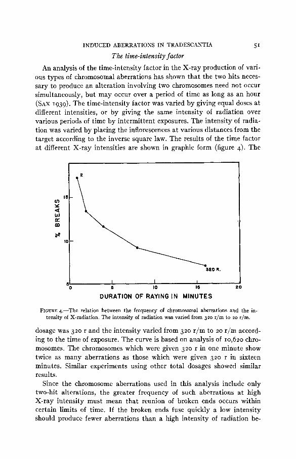
ous types of chromosomal aberrations has shown that the two hits neces- sary to produce an alteration involving two chromosomes need not occur simultaneously, but may occur over a period of time as long as an hour (SAX 1939). The time-intensity factor was varied by giving equal doses at different intensities, or by giving the same intensity of radiation over various periods of time by intermittent exposures. The intensity of radia- tion was varied by placing the inflorescences at various distances from the target according to the inverse square law. The results of the time factor at different X-ray intensities are shown in graphic form (figure 4). The
I I I 1 5 IO I5 20
5 0
DURATION OF RAYING I N MINUTES
FIGURE 4.-The relation between the frequency of chromosomal aberrations and the in- tensity of X-radiation. The intensity of radiation was varied from 320 r/m to 20 r/m.
dosage was 320 r and the intensity varied from 320 r/m to 20 r/m accord- ing to the time of exposure. The curve is based on analysis of 10,620 chro- mosomes. The chromosomes which were given 320 r in one minute show twice as many aberrations as those which were given 320 r in sixteen minutes. Similar experiments using other total dosages showed similar results.
Since the chromosome aberrations used in this analysis include only two-hit alterations, the greater frequency of such aberrations at high X-ray intensity must mean that reunion of broken ends occurs within certain limits of time. If the broken ends fuse quickly a low intensity should produce fewer aberrations than a high intensity of radiation be-
52 KARL SAX
cause a break in one chromosome would heal before a second break oc- curred in an adjacent chromosome, and no aberration would be produced. But if the broken ends remain in an unstable condition for a long time and
0 25 5 0 75 100 I25
REST PERIODS I N MINUTES
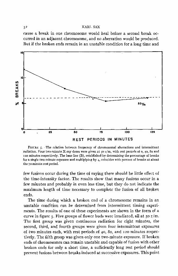
FIGURE 5.-The relation between frequency of chromosomal aberrations and intermittent radiation. Four two-minute X-ray doses were given at 50 r/m, with rest periods of 0,40, 80 and 120 minutes respectively. The base line (B), established by determining the percentage of breaks for a single two-minute exposure and multiplying by 4, coincides with percent of breaks at about the 70 minute rest period.
few fusions occur during the time of raying there should be little effect of the time-intensity factor. The results show that many fusions occur in a few minutes and probably in even less time, but they do not indicate the maximum length of time necessary to complete the fusion of all broken ends.
The time during which a broken end of a chromosome remains in an unstable condition can be determined from intermittent timing experi- ments. The results of one of these experiments are shown in the form of a curve in figure 5 . Five groups of flower buds were irradiated, all a t 50 r/m. The first group was given continuous radiation for eight minutes, the second, third, and fourth groups were given four intermittent exposures of two minutes each, with rest periods of 40, 80, and 120 minutes respec- tively. The fifth group was given only one two-minute exposure. If broken ends of chromosomes can remain unstable and capable of fusion with other broken ends for only a short time, a sufficiently long rest period should prevent fusions between breaks induced at successive exposures. This point
INDUCED ABERRATIONS IN TRADESCANTIA 53 is reached where a further increase in the rest period produces no further decline in frequency of aberrations. The base line can also be determined by multiplying the frequency of aberrations induced by a single exposure by the number of exposures used in the intermittent timing experiment. In the above experiment the base line is a t about six percent and is reached a t a rest period of somewhat more than an hour. These results show that a break induced a t one exposure period may remain open so that a broken end can fuse with another broken end produced by the second exposure an hour later, although most of the fusions occur in a considerably shorter period. Similar experiments with different X-ray intensities show similar results, although a t high intensities, where the dose is relatively high for each exposure, there is less effect of the intermittent exposures. These results are based on an analysis of more than 80,000 chromosomes.
The effect of the time-intensity factor is based on the fact that the two- hit aberrations are dependent upon two independent breaks, limited in time of occurrence. According to this interpretation the time-intensity factor should have no effect on one-hit aberrations. This assumption has been tested by comparing the effect of intermittent timing on one-hit and two-hit chromatid aberrations. As expected, the frequency of the one- hit aberrations is independent of the time factor while the frequency of the two-hit aberrations declines with increasing rest periods (SAX 1939).
THE EFFECT OF TEMPERATURE ON X-RAY INDUCED
CHROMOSOMAL ABERRATIONS
In an earlier experiment no differences in frequency of chromosomal aberrations were found when the cells were irradiated a t different tem- peratures (SAX 1938). The resu ts were not based on an analysis of in- dividual chromosomes and the frequency of aberrations included all types of alterations obtained a t various times after irradiation. More critical experiments show clearly that the temperature during and following irra- diation has a marked effect of the frequency of both one-hit and two-hit aberrations (SAX and ENZMANN 1939). The results of two of these experi- ments are shown in graphic form in figure 6.
The inflorescences were immersed in water a t the desired temperature for five to ten minutes before raying and kept just under the surface of the water during irradiation. In a few experiments the flower stalks were removed from the water and placed a t room temperature soon after ray- ing, but usually they were left in the water for an hour after raying. When pasteboard cartons were used for containers the temperature dropped gradually during this period, but similar temperature effects were ob- tained when the flowers were put in thermos bottles where the tempera- tures were maintained at a constant level during the entire period.
54 KARL SAX
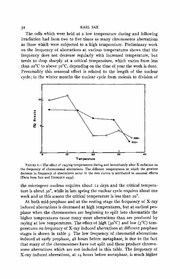
The cells which were held at a low temperature during and following irradiation had from two to five times as many chromosome aberrations as those which were subjected to a high temperature. Preliminary work on the frequency of aberrations at various temperatures shows that the frequency does not decrease regularly with increased temperature, but tends to drop sharply at a critical temperature, which varies from less than 20'C to above 3ooC, depending on the time of year the work is done. Presumably this seasonal effect is related to the length of the nuclear cycle; in the winter months the nuclear cycle from meiosis to division of
FIGURE 6.-The effect of varying temperatures during and immediately after X-radiation on the frequency of chromosomal aberrations. The different temperatures at which the greatest decrease in frequency of aberrations occur in the two curves is attributed to seasonal effects (Data from SAX and ENZMANN 1939).
the microspore nucleus requires about I 2 days and the critical tempera- ture is about 30°, while in late spring the nuclear cycle requires about one week and at this season the critical temperature is less than 20'.
At both mid-prophase and at the resting stage the frequency of X-ray induced aberrations is decreased at high temperatures, but at earliest pro- phase when the chromosomes are beginning to split into chromatids the higher temperatures cause many more aberrations than are produced by raying at low temperatures. The effect of high (30'C) and low (3OC) tem- peratures on frequency of X-ray induced aberrations at different prophase stages is shown in table 5. The low frequency of chromatid aberrations induced at early prophase, 48 hours before metaphase, is due to the fact that many of the chromosomes have not split and these produce chromo- some aberrations which are not included in this table. The frequency of X-ray induced aberrations, at 24 hours before metaphase, is much higher
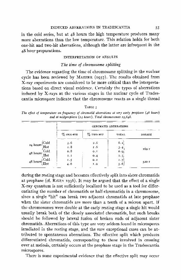
INDUCED ABERRATIONS I N TRADESCANTIA 5 5 in the cold series, but a t 48 hours the high temperature produces many more aberrations than the low temperature. This relation holds for both one-hit and two-hit aberrations, although the latter are infrequent in the 48 hour preparations.
INTERPRETATION OF RESULTS
The time of chromosome splitting The evidence regarding the time of chromosome splitting in the nuclear
cycle has been reviewed by MATHER (1937). The results obtained from X-ray experiments are considered to be more critical than the interpreta- tions based on direct visual evidence. Certainly the types of aberrations induced by X-rays at the various stages in the nuclear cycle of Trades- cantia microspore indicate that the chromosome reacts as a single thread
TABLE 5
The effect of temperature on frequency of chromatid aberrations at very early prophase (48 hours) and at midprophase (24 hours). Total chromosomes 19,848.
___ I
CHROMATID ABERRATIONS I
~ % ONE-HIT I % TWO-HIT I TOTAL DOSAGE
I
Hot I .8
48 hours
48 hours Hot I 4 .6
160 r 1 . 6 0. I
0 .4 0.2
1 . 0 320 r
during the resting stage and becomes effectively split into sister chromatids a t prophase (cf. RILEY 1936). It may be argued that the effect of a single X-ray quantum is not sufficiently localized to be used as a tool for differ- entiating the number of chromatids or half-chromatids in a chromosome, since a single “hit” can break two adjacent chromatids a t late prophase when the sister chromatids are more than a tenth of a micron apart. If the chromosomes were double a t the early resting stage a single hit would usually break both of the closely associated chromatids, but such breaks should be followed by lateral fusion of broken ends of adjacent sister chromatids. Aberrations of this type are very seldom found in microspores irradiated in the resting stage, and the rare exceptional cases can be at- tributed to spontaneous aberrations. The effective split which produces differentiated chromatids, corresponding to those involved in crossing over a t meiosis, certainly occurs a t the prophase stage in the Tradescantia microspores.
There is some experimental evidence that the effective split may occur
56 KARL SAX
at the resting stage preceding nuclear division (MATHER 1937), or even at the preceding anaphase. The behavior of the broken ends of inversion bridge chromosomes suggests that the meiotic anaphase chromosomes may be effectively split. In both Tradescantia (SAX 1937) and Zea (MCCLIN- TOCK 1938b) the break in an inversion bridge is followed by lateral fusion of sister chromatids to produce a bridge at the following division of the chromosome in the microspore. If the chromosome were single a t meiosis a single broken end would have no other broken end with which to fuse, and perhaps such a broken end would remain in an unstable condition until the chromosome is split a t the prophase stage of the microspore nucleus, a t which time the lateral fusion would occur between the broken ends of sister chromatids. But MCCLINTOCK has found that adjacent double inversion bridges behave in the same way, even though two broken ends are present in the newly formed microspore nucleus. On the other hand a break in a somatic ring chromosome of Zea is followed by fusion of broken ends of the two arms and not between ends of adjacent sister chromatids (MCCLINTOCK 1938a). This difference cannot be attributed to differences in behavior a t meiosis and mitosis because X-ray induced ab- errations in Tradescantia root tips produce double bridges, which, when broken, are followed by lateral fusion of ends of sister chromatids. There is no reason to believe that the X-ray induced and spontaneous breaks differ in their effects on subsequent fusions of broken ends of chromosomes.
The behavior of the induced ring chromosomes is of interest in relation to the nature of splitting. If these ring chromosomes induced at the resting stage are single threads they must split in different planes because the rings may be continuous or interlocked at the following metaphase. This problem has been analyzed in some detail by HUSTED (1936).
The time of effective chromosome splitting in different microspores is quite variable, ranging from about 23 to 31 hours before metaphase in Tradescantia plants grown in the field during the summer months. The splitting of the chromosomes of a single nucleus is much more limited in time, and the splitting of an individual chromosome must be very rapid since both chromosome and chromatid breaks are rarely found in the same chromosome. There is some indication that effective splitting begins a t the proximal end of the chromosome arm and progresses distally (SAX and MATHER 1939).
The frequency of types of aberrations in relation to chromosome arrangement
It has been shown that all potential fusions between broken ends of chromosomes are completed within an hour after the breaks are produced. There can be no extensive movement of the chromosomes during this
INDUCED ABERRATIONS IN TRADESCANTIA 57 time, a t most periods in the nuclear cycle, so that fusions between broken ends must be limited to those in close proximity. Certainly at the resting stage there could be no extensive movement of the chromosomes, which in the form of relic coils appear to completely fill the nucleus (BELAR 1928). With an increase in nuclear size and uncoiling of relic spirals a t prophase more movement of the chromosomes could occur, but not sufficient to permit random association of broken ends of chromatids. Accordingly, an analysis of the loci of breaks involved in fusions between different chromo- somes or chromosome arms should indicate the arrangement and relations of the chromosomes a t various phases in the nuclear cycle.
The chromosomes of Tradescantia are strongly polarized during the resting and early prophase stages so that if little chromosome movement occurs between breakage and fusion, the fusions between different chromo- some arms should occur usually a t corresponding loci in respect to the distance from the centromeres. Such a relation is found for both chromo- some and chromatid aberrations. Unequal exchanges between chromosome arms are rare, and an analysis of the fusions and exchanges between chro- matids of different chromosomes shows that the breaks have occurred a t approximately the same relative loci of both chromosomes in over 80 percent of the figures examined.
The relative frequencies of dicentric and ring chromosomes also show that proximity facilitates, or conditions, the fusions of broken ends of chromosomes. A broken end of a chromosome arm can fuse with a broken end in the other arm of the same chromosome to form a ring, or it can fuse with a break in any one of the ten arms of the other five chromosomes to form a dicentric union. Random reunion of broken ends should produce a ratio of dicentric to ring chromosomes of IO: I , but the observed ratio is 3.3 to I (table I). It is evident that the distance between breaks, both laterally and longitudinally in respect to the polarized arms of the chromo- somes, conditions or determines the occurrence of two-hit aberrations.
The spatial relationships of chromosome arms appear to change as the nuclear cycle progresses from the resting stage into prophase. At the rest- ing stage the ratio of dicentric to rings is 3.3 : I , but irradiation at prophase results in a ratio of dicentric to rings of 13 : I. This decrease in frequency of ring chromosomes at mid-prophase is attributed to the greater separation of the two arms of a single chromosome at this stage. It is known that chromosomes repel each other, especially when they become split into sister chromatids. The prophase chromosomes also become shorter and the increased size of the nucleus permits greater movement of the chromo- somes.
The relative frequencies of one-hit and two-hit aberrations induced at prophase vary with dosage as is shown in figure 3. There is, however, a
58 KART, SAX
striking difference in these frequencies between early and mid-prophase (table 4). The relative frequency of two-hit aberrations induced a t early prophase is very much less than at mid-prophase. This difference can hardly be attributed to greater repulsion between arms of chromosomes, and must be related to differences in the nature of the induced breaks. The two-hit breaks-dicentrics, exchanges and rings-induced a t mid-prophase may depend upon breaks in only one chromatid of each of the chromosome arms involved in the aberrations. The sister chromatids a t this stage are distinctly separated and breaks in only one of the two sister chromatids a t a given locus are found at metaphase. At earliest prophase, however, the two sister chromatids must be closely associated so that a single X-ray hit would usually break both chromatids a t the same locus. Such breaks would be followed by the fusion of adjacent ends of the broken chromatids in most cases, to produce a terminal deletion which is classed as a one-hit aberration. This interpretation is supported by the fact that two-hit chro- mosome aberrations, induced before chromosome splitting, but a t about the same time, show a normal frequency.
The limitations of fusions imposed by the factor of proximity must mean that most of the breaks induced by X-rays do not lead to the pro- duction of aberrations. The two broken ends simply reunite in the original position and no structural alteration of the chromosome is visible a t later stages in nuclear development. The chromosome breaks which lead to fusions between chromosome arms must constitute only a very small pro- portion of the total breaks induced, but simple breaks at the following metaphase are far less frequent than aberrations which involve two breaks in close proximity.. The one-hit breaks a t prophase are frequent only be- cause two chromatids are broken and lateral fusion of broken ends leads to a permanent aberration. The frequency of breaks which heal in the original position cannot be determined, but such breaks must be far more frequent than those which lead to gross structural changes in the chromo- somes.
A comparison of the frequencies of types of aberrations has also been used in the analysis of chromosome arrangements in Drosophila sperm (CATCHESIDE 1938; BAUER, DEMEREC and KAUFMANN 1938). The salivary gland chromosomes permit an analysis of alterations within chromosome arms, as well as between chromosome arms, although the numerous cell divisions between irradiation and chromosome analysis must produce dif- ferential elimination of the various types of aberrations. CATCHESIDE finds that exchanges within the same arm are twice as favored as those between different chromosome arms. Exchanges between arms of the same chromo- some when compared with the frequency of exchanges between arms of different chromosomes, show no significant deviation from a random dis-
INDUCED ABERRATIONS I N TRADESCANTIA 59 tribution. Similar results have been obtained by BAUER, DEMEREC, and KAUFMANN. These results must mean that a single chromosome in the Drosophila sperm is folded so that various parts of the same chromosome arm are in close proximity. Presumably, little movement is possible in the closely packed chromosomes of the sperm and induced aberrations must depend upon close proximity of the induced breaks.
The loci of breaks in relation to aberrations
It has been shown that the distribution of breaks involved in chromo- some and chromatid aberrations is not a t random for the various loci of chromosome arms. The initial breaks must be a t random, but those which are involved in chromosome alterations are most frequent a t the proximal ends of the chromosome arms. This distribution must be caused by secondary factors, and since the unequal distribution holds for one-hit chromatid breaks the secondary factor must be confined to the individual arms of the chromosomes. The greater frequency of such breaks a t the proximal end of the chromosome arm is attributed to stresses imposed by the various coiling mechanisms which would tend to throw the broken ends out of alignment so that reunion in the original position would be inhibited. The breaks induced in the chromosomes of the sperm of Drosophila show a nearly random distribution of breaks with a possible higher frequency at the distal ends (BAUER, DEMEREC and KAUFMANN 1938). Perhaps the more random distribution of effective breaks in Drosophila can be at- tributed to the close association of the chromosomes in the Drosophila sperm so that proximity of broken ends does not necessitate any appreci- able movement of broken ends in order to produce fusions between differ- en t chromosomes.
The relation between X- ray dosage and frequency of chromosomal aberrations
In both Drosophila and Tradescantia the frequency of X-ray induced breaks increases approximately as the 1.5 power of the dosage when the various doses are given at the same intensity. But since the time factor must be considered in such experiments the dosage should be varied by changing the intensity and not the time of exposure. When this is done the frequency of two-hit chromosome and chromatid aberrations in Tradescantia varies approximately as the square of the dosage. This rela- tion should be expected since the chance that two or more independent events will happen together is the product of their respective chances of happening.
The frequency of simple deletions is assumed to be induced by single hits and should show a linear relation to dosage, a t least for doses which
60 KARL SAX
produce a low frequency of breaks. The data indicate a slight exponential increase of aberrations with increased dosage which may be due to experi- mental error, or to the occasional production of the deletions by two inde- pendent hits in the two chromatids at approximately the same locus.
The number of X-ray hits or quanta necessary to produce an effect has been calculated by comparing the survival curves with those obtained from Poisson’s exponential series (TIMOF~EFF-RESSOVSKY 1937). For one- hit and two-hit chromosomal aberrations the equation can be expressed as: percent B = I -esx and percent B = I -----=(I +x), respectively, where x represents the number of effective hits. Using an arbitrary value of x of .00025 per r unit of dosage, we find that the calculated values for the one-hit aberrations are practically identical with the observed values. The calculated values of the two-hit curve also are in close agreement with the observed percentages of breaks a t the various doses, but in order to get these theoretical values, the value of x must be increased to .0025 per r unit. The dosage curves, (D/45)’.‘ and (D/67)1.9, do not indicate such different dosages for one- and two-hit aberration frequencies, as is indicated by the Poisson exponential series. It is not clear how these apparent discrepancies can be reconciled. It is evident, however, that the slopes of the two dosage curves are practically identical with the theoreti- cal curves for one-hit and two-hit effects.
In all of the various experiments on the relation between dosage and the mutation rate in Drosophila a linear relation has been found. The fre- quency of mutation is also independent of the time-intensity factor (TIMO- F~EFF-RESSOVSKY 1937). These facts indicate that most of the induced mutations are produced by single hits and that relatively few of the in- duced mutations are due to “position effects” resulting from two-hit aber- rations such as reciprocal translocations and large inversions. . The frequency of X-ray induced aberrations induced in the Drosophila
sperm must also depend on the time factor. With dosage controlled by increasing the time of exposure the relation between frequency of aberra- tions and dosage is expressed by the equation percent B=(D/K)1.5 in both Drosophila and Tradescantia; but in Tradescantia, with the time of exposure constant, the frequency of aberrations varies as the square of the dosage. Control of the time factor should also increase the exponent from 1.5 to 2.0 for the dosage-aberration curve in Drosophila.
The diJerentia1 susceptibility of chromosomes to X-rays during the nuclear cycle and in dijerent types of cells
The frequency of gross chromosomal aberrations is much greater at prophase than during the resting stage, when the same X-ray dose is used (figure 3). The equation for the relation between dosage and frequency of
INDUCED ABERRATIONS IN TRADESCANTIA 61
aberrations is: percent B = (D/37)1.5 for all the chromatid aberrations re- corded, and percent B = (D/107)~ for the chromosome aberrations. This comparison may not be entirely valid because certain types of aberrations can be detected when induced a t prophase, but not when induced a t the resting stage. In general, however, the frequency of gross alterations is greatest when the cells are rayed a t prophase. This relation is to be ex- pected for the following reasons: there are twice as many threads to be hit a t prophase; terminal deletions can be produced a t prophase by lateral fusion between broken chromatids while in the resting stage they are pro- duced only when the broken end loses its capacity for fusion; and the secondary factors of chromosome torsion and movement would be expected to be greater a t prophase.
A comparison of the frequency of chromosomal aberrations in Trades- cantia and Drosophila shows that breaks involving chromosome changes are much more frequent in Tradescantia microspores than in Drosophila sperm. Some of this difference may be attributed to inviable aberrations in Drosophila, since the chromosomes were analyzed many cell generations after raying the sperm. The data of BAUER, DEMEREC and KAUFMANN show that the frequency of breaks, in percent of total chromosomes, may be expressed by the equation, percent breaks = (D/I 150)l.j. For Trades- cantia microspores rayed a t the resting stage, with dosage varied by vary- ing the time of exposure, the equation is: percent B = (D/80)'.j (SAX 1938). These data would seem to indicate that the Tradescantia chromosomes are much more sensitive than those of Drosophila. This difference probably can be attributed to differences in chromosome size in the two genera and to differences in chromosome organization in the two types of cells. Ac- cording to METZ and BOCHE (1939) both chromosome aberrations and mutations in Sciara are less frequent than in Drosophila when given the same X-ray dosage.
The difference in the frequencies of X-ray induced chromosomal aberra- tions in different cells of the same organism should provide additional evi- dence regarding X-ray effects on the chromosome. It is known that both mutations and gross chromosomal alterations occur more frequently in the sperm of Drosophila than in the oocytes for a given X-ray dose (OLIVER 1934). In Sciara gross chromosomal alterations are readily se- cured by raying the sperm, but were not found when the egg cells were irradiated. METZ and BOCHE (1939) attribute this difference in Sciara to physical differences in the chromosomes of the two types of cells. The close proximity of the chromosomes in the sperm should facilitate illegitimate union of broken ends, while most of the chromosomes of the eggs at the time of treatment were in metaphase or anaphase of the first meiotic divi- sion. The diffuse stage a t meiotic prophase of meiosis also was found to
62 KARL SAX
be resistant to X-ray effects. METZ (1934) has suggested that the chromo- some matrix plays an important part in this differential susceptibility of chromosomes to radiation.
The differential susceptibility to X-rays of different stages of the nuclear cycle, of different cells in the same organism, and of similar cells in different organisms may depend in part on the same secondary factors such as prox- imity of chromosomes, freedom of chromosome movement, and the physi- cal condition of the chromosomes. There is, however, some evidence that differences in the internal structure of the chromosomes may be responsible for differential X-ray susceptibility. NAVASHIN and his collaborators have found that the aging of seeds increases both the mutation rate and fre- quency of spontaneous chromosomal aberrations (cf. GILES 1940). OFFER- MANN (1938) has shown that X-rays produce more mutations in old than in young sperm of Drosophila. These results suggest that aging of chromo- somes induces internal changes, probably of a chemical nature, which makes them more susceptible to both spontaneous and induced alterations.
In any analysis of differential susceptibility of different stages in the nuclear cycle it is essential to differentiate the physiological effect of X-rays from the production of mutations and gross chromosomal aberrations which lead to genetic alterations. The physiological effect of suppressed nuclear activity and clumping of the chromosomes has long been known and this reaction in plant cells has been dealt with in considerable detail by MARQUARDT (1938). At moderate X-ray doses the inhibition of nuclear development is temporary and the cell recovers. The time of recovery de- pends on the dosage (CARLSON 1938, and unpublished). Heavy X-ray dosage may cause so much clumping of the metaphase chromosomes that normal division is prevented and the effect is lethal. Maximum X-ray effects at metaphase probably can be attributed to physiological factors.
It is probable that the physiological and genetic effects can be differ- entiated by determining the temperature coefficient of the reaction to X-rays. If chromosome aberrations are responsible for cell injury the tem- perature coefficient should be one or less than one, while if physiological effects are responsible the temperature coefficient should be appreciably greater than one.
The behavior of broken eNds of chromosomes
The experimental evidence shows that the induced chromosomal aber- rations involving two chromosomes are dependent upon two independent breaks limited in both time and space. The broken ends of chromosomes either reunite in the original position or produce chromosome aberrations within a short time after the breaks are induced by X-rays, except a very small proportion of breaks producing broken ends which appear to have
INDUCED ABERRATIONS IN TRADESCANTIA 63
lost their capacity for fusion. Most of the unions occur within a few min- utes, and a t the end of about an hour all fusions are completed. This does not necessarily mean, however, that a broken end can remain in an un- stable condition for only an hour.
STADLER (1932) has assumed that an acentric fragment may be included in a daughter nucleus by chance and fuse with the original broken chromo- some after several cell generations. Such behavior is highly improbable for the following reasons: all potential fusions between broken ends are com- pleted in about an hour in the Tradescantia microspore which has a nuclear cycle of about a week; nearly all the acentric fragments found at meta- phase have no broken ends since the two ends of such fragments are the normal ends of chromosomes or chromatids; the few terminal deletions which persist until metaphase appear to have lost their capacity for fusion since chromosome fragments show no lateral fusion of sister chromatids; and very few of the various types of fragments are included in the mitotic daughter nuclei in plant cells. Acentric fragments of meiotic chromosomes often are included in the daughter nuclei in Zea (MCCLINTOCK 1938b), and in the neuroblasts of the grasshopper such fragments usually are included in the daughter nuclei (CARLSON 1938). Even in these cases re- union of acentric fragments is improbable. Moreover, according to Mc- CLINTOCK, the acentric fragments occasionally included in the Zea micro- spores act as normal chromosomes both in their cytological behavior and in their genetic influence. Thus there could be no genetic deficiency by deletion of a chromosome segment, or genetic recovery when the fragment joins the centric segment, as STADLER has postulated.
The temperature eject on induced chromosomal aberrations and mutation
According to MULLER (1935), PAPALASHVILLI has found that the fre- quency of chromosomal rearrangements in Drosophila is increased by raying the flies at low temperatures. MICKEY (1937) has found a much higher frequency of translocations in flies rayed a t I~OC than in those rayed a t 2S0-34OC. In Tradescantia microspores both chromosome and chroma- tid aberrations, induced a t either the resting or a t mid-prophase, are much more frequent in the cells rayed a t low temperatures. The variations in temperature affect both one-hit and two-hit aberrations. The decreased frequency of X-ray induced aberrations at the higher temperatures can be attributed to a more rapid fusion of broken ends or to a more relaxed condition of the chromosomes which inhibits chromosome movements. Either factor would favor the reunion of broken ends in the original posi- tion and decrease the frequency of illegitimate unions which produce the
64 KARL SAX
aberrations. I t has been shown that the frequency of the initial breaks is relatively independent of the temperature.
The reversed temperature effect on the chromatids at earliest prophase is difficult to explain. The reaction of the chromosomes cannot be due to changes in the nucleus as a whole because the single chromosomes show a decreased frequency of aberrations, and the split chromosomes an in- creased frequency of aberrations at high temperatures. An increase in the speed of union of broken ends may be involved. At earliest prophase the newly formed chromatids may be so closely associated that a break would separate the broken ends of a single chromatid further than the distance between chromatids. At mid-prophase the chromatids may be separated by a distance greater than the distance between broken ends of a chroma- tid. Rapid union of broken ends would tend to produce lateral fusions and consequently aberrations a t early prophase, but would favor reunion in the original position and consequently fewer aberrations a t later prophase stages. This interpretation could hardly be applied to the two-hit chroma- tid aberrations, which also seem to show the reversed temperature effect at early prophase, although the numbers are very low. An increase in tor- sional strain induced by high temperatures only a t earliest prophase could also be invoked to explain these results, but further work is necessary in order to determine the factors involved in the reversed temperature effect a t earliest prophase.
,
T h e nature of X-ray induced breaks
The chromosomal aberrations induced by X-rays have been attributed to fusions followed by breaks and to independent breaks followed by fu- sions between broken ends. The contact hypothesis sponsored by MULLER (1932) postulates that the chromosomes in contact or closely associated may be affected by a single X-ray hit. The breakage hypothesis, suggested by STADLER (1931), postulates that independent breaks are followed by illegitimate fusions. The analysis of X-ray induced chromosomal aberra- tions in Tradescantia confirms STADLER’S hypothesis. The dosage curves for one-hit and two-hit aberrations, the effect of the time-intensity factor, and the temperature effect, all prove that aberrations involving two chro- mosomes are dependent upon two independent hits.
The one-hit chromatid breaks appear to be caused by single X-ray “hits” since the relation between dosage and frequency of such aberrations is approximately linear. Since two sister chromatids may be broken by a single hit the ionization or excitation must cover a relatively large range because these sister chromatids are separated by a distance of a t least a tenth of a micron a t mid-prophase. The frequency of induced mutations in Drosophila is independent of X-ray wave-length (TIMOF~EFF-RESSOV-
INDUCED ABERRATIONS IN TRADESCANTIA 65 SKY 1936). The frequencies of X-ray induced chromosomal aberrations in Tradescantia also are independent of wave-length (RICK, unpublished). These results indicate that the X-ray effects are caused by ionization (GOODSPEED and UBER 1939).
The broken ends of chromosomes may remain in an unstable condition and capable of fusion for an hour or longer. I t is surprising that a molecular complex of such a nature can remain in an unstable condition for so long a period. When fusions between broken ends do occur the union appears to be quite perfect in most cases, and breaks in subsequent chromosome bridges are no more likely to occur a t the point of union than a t other loci. There must be natural breaking points in the chromosome because breaks and fusions are normal features of practically all chromosomes a t the time of meiosis. The reunion of both X-ray induced and crossover breaks ap- pears to have no effect on the physical condition of the chromosome.
About five percent of the visible chromosome alterations induced by irradiation of the resting nucleus consist of terminal deletions. The broken ends of such deletions appear to behave as normal ends. Such a behavior may be caused by breaks at loci other than the natural breaking points.
ACKNOWLEDGMENTS
This work was supported, in part, by a grant from the National Re- search Council Committee on Radiation, and by research funds of the Biological Laboratories of Harvard University. Most of the preparations were made by MARGERY POOLE, who also scored the types and frequencies of chromosomal aberrations in many of the X-ray experiments.
SUMMARY
An analysis of X-ray induced chromosomal aberrations in the micro- spores of Tradescantia has led to the following conclusions :
I . The chromosomes react to X-rays as though they were single threads during the resting stage and become effectively split to form sister chro- matids a t early prophase.
2 . The effective splitting of an individual chromosome occurs very rapidly, and there is some evidence that splitting begins a t the region of the centromere and progresses distally.
3. The simple terminal deletions are readily induced at prophase be- cause the two sister chromatids may be broken by one hit, followed by lateral fusion of broken ends of sister chromatids.
4. The frequency of these simple deletions shows an approximately linear relation to dosage. The equation for this relation is percent B =(D/K)'.l, where B is the number of effective breaks in relation to the total number of chromosomes, D the dosage in r units, and K a constant.
66 KARI, SAX
5. A simple deletion induced at the resting stage persists until meta- phase only when the broken ends lose their capacity for fusion. Such breaks are rare. The broken ends of these chromosomes appear to behave as normal ends.
6. Aberrations involving two chromosomes, or two loci in the same chromosome, are dependent upon two independent breaks, limited in both time and space.
7. The frequency of these two-hit aberrations is dependent upon the radiation intensity. High intensity is more effective than low intensity because the aberrations are dependent upon two adjacent breaks within certain limits of time. The frequency of one-hit aberrations a t a given dos- age is independent of the time-intensity factor.
8. The frequency of two-hit aberrations increases exponentially with increased dosage. When the dosage is varied by varying the time of expo- sure the relation between frequency of aberrations and dosage is percent B = (D/K)1.5, but if dosage is varied by varying the intensity the equation is percent B = (D/K)2.0.
9. Intermittent dosage experiments show that broken ends of chromo- somes may remain in an unstable condition for as long as an hour before fusion, although most fusions occur in a much shorter time.
IO. Only a small proportion of the X-ray induced breaks result in visible chromosome aberrations. Most of the broken ends reunite in the original positions with no evident alteration of the chromosome.
11. The frequency of the initial breaks induced by X-rays is relatively independent of temperature. The production of aberrations, however, is greater when the resting and prophase nuclei are irradiated a t low tem- peratures. High temperatures during or immediately following irradiation increase the reunion of broken ends in the original position thus decreasing the frequency of aberrations. This temperature effect is reversed a t very early prophase.
12. The initial breaks induced by X-rays must be distributed at ran- dom, but both the one-hit and two-hit aberrations occur more frequently a t the proximal ends of the chromosome arms. This distribution of effec- tive breaks is attributed to the effect of secondary factors.
13. Differential X-ray production of chromosomal aberrations in differ- ent phases of the nuclear cycle, in different cells of the same organism, and in similar cells of different organisms, can be attributed, in part, to secondary factors, such as proximity of chromosomes, freedom of chromo- some movement, and the physical condition of the individual chromosome.
14. The X-ray induced breaks appear to occur usually a t natural break- ing points. Most of the breaks and fusions following irradiation appear to be comparable to those occurring naturally a t the time of crossing over.
INDUCED ABERRATIONS I N TRADESCANTIA 67
ADDENDUM
I have seen, in manuscript, a paper on X-rayed Tradescantia chromo- somes submitted to “Genetics” in July 1939 by A. C. FABERGI? of Uni- versity College, London. FABERG~’S conclusions on the time factor and the temperature effect are essentially the same as those reported above. His data were not adapted to an analysis of the dosage curve, and his con- sions in this regard appeared to require reconsideration.*
LITERATURE CITED
BAUER, H., DEMEREC, M., and KAUFMANN, B. P., 1939 X-ray induced chromosomal alterations
BELAR, KARL, 1928 Die cytologischen Grundlagen der Vererbung. 41 2 pp. Berlin: Borntraeger. CARLSON, J. G., 1938 Mitotic behavior of induced chromosomal fragments lacking spindle at-
tachments in the neuroblasts of the grasshopper. Proc. Nat. Acad. Sci. Wash. 11: 500-507. 1938 Some effects of X-radiation of the neuroblast chromosomes of the grasshopper, Chorto- phuga viridifasciata. Genetics 23: 596-610.
CATCHESIDE, D. G., I938 The effect of X-ray dosage upon the frequency of induced structural changes in the chromosomes of Drosophila melanogaster. J. Genet. 36: 307-320.
DOBZHANSKY, T., 1937 Genetics and the origin of species. New York: Columbia Univ. Press. GILES, NORMAN, 1940 Spontaneous chromosome aberrations in Tradescantia. Genetics 25: 69-
GOODSPEED, T . H., and UBER, F. 11 , 1939 Radiation and plant cytogenetics. Bot. Rev. 5 : 1-48. HUSTED, L., 1936 An analysis of chromosome structure and behavior with the aid of X-ray in-
duced rearrangements. Genetics 21 : 537-553. KAUFMANN, B. P., and BATE, RUTH C., 1938 An X-ray induced intercalary duplication in Droso-
phila involving union of sister chromatids. Proc. Nat. Acad. Sci. Wash. 24: 368-371. MCCLINTOCK, BARBARA, 1938a. The production of homozygous deficient tissues with mutant
characteristics by means of the aberrant mitotic behavior of ring-shaped chromosomes. Genetics23: 315-376. 1938b The fusion of broken ends of sister half-chromatids following chromatid breakage a t meiotic anaphases. Univ. Missouri Agric. Expt. Sta. Bull. 290. 48 pp.
MATHER, K., 1937 The experimental determination of the time of chromosome doubling. Proc. Roy. Soc. London 124: 97-106.
MARQUARDT, HANS, 1938 Die Rontgenpathologie der Mitose I. und 11. Z. Bot. 32: 401-482. METZ, C. W., 1934 The r61e of the “chromosome sheath” in mitosis, and its possible relation to
phenomena of mutation. Proc. Nat. Acad. Sci. Wash. 20: 159-163. METZ, C. W., and BOCHE, R. D., 1939 Observations on the mechanism of induced chromosome
rearrangements in Sciara. Proc. Nat. Acad. Sci. Wash. 25: 280-284. MICKEY, G. H., 1938 Effect of temperature on frequency of translocations produced by X-rays.
Genetics 23: 160. (Abstract) MULLER, H. J., 1932 Further studies on the nature and causes of gene mutation. Proc. 6th Int.
Cong. Genetics I: 213-255. 1935 The present status of the mutation theory. Current Science, Special number pp. 4-15. 1938 The remaking of chromosomes. Collecting Net 13: 182-198.
OFFERMANN, C. A., 1938 Effect of ageing on the frequency of induced mutations. Genet. Soc. Rec. (Abstract) 7: 81-82.
OLIVER, C. P., 1932 An analysis of the effect of varying the duration of X-ray treatment upon the frequency of mutations. Z.i.A.V. 61 : 447-488. 1934 Radiation genetics. Quart. Rev. Biol. 9: 381-408.
in Drosophila melanogaster. Genetics 23 : 610-630.
87.
* DR. FABERG% has in the meantime withdrawn his paper from “Genetics” and has submitted i t to the “Journal of Genetics.”
68 KARL SAX
RILEY, H. P., 1936 The effect of X-rays on the chromosomes of Trudescantia gigantea. Cytologia
SAX, KARL, 1937 Chromosome behavior and nuclear development in Tradescantia. Genetics 22 : 7: 131-142.
523-533. 1938 Chromosome aberrations induced by X-rays. Genetics 23: 494-516. 1939 The time factor in X-ray production of chromosome aberrations. Proc. Nat. Acad. Sci. Wash. 25: 225-233.
SAX, KARL, and ENZMANN, E. V., 1939 The effect of temperature on the frequency of X-ray in- duced chromosome aberrations. Proc. Nat. Acad. Sci. Wash. 25: 397-405.
SAX, KARL, and MATHER, K., 1939 An X-ray analysis of progressive chromosome splitting. J. Genet. 37: 483-490.
STADLER, L. J., 1931 The experimental modification of heredity in crop plants. I. Induced chro- mosomalirregularities. Sci. Agr. 11 : 557-572. 1932 On the genetic nature of induced mutations in plants. Proc. 6th Int. Cong. Gen. I : 274-
TIMOFI$EFF-RESSOVSKY, N. W., 1937 Mutationsforschung in der Vererbungslehre. pp. 177, 094.
Steinkopff, Dresden und Leipzig.