an brief introduction to 4d ct scanning
TRANSCRIPT
1
An Brief Introduction to
4D CT Scanning
Steve B. Jiang, Ph.D.Dept of Radiation Onco logyUniv of California San Diego
AcknowledgementsPaul Keall, George Chen,Dan Low, Gig Mageras,
Ken Foster
2
Outline
� Problems with free breathing 3D scanning
� What is 4D CT? How does it work?
3
Three Types of Motio n Ar tifacts
� If CT scannin g speed << tumor motion speed,→→→→ smeared tumor imag e
� If CT scannin g speed >> tumor motion speed,→→→→ tumo r positio n and shape captured at anarbitra ry breath ing phase
� If CT scannin g speed ~ tumor motion speed,→→→→ tumo r positio n and shape heavily distor ted
4
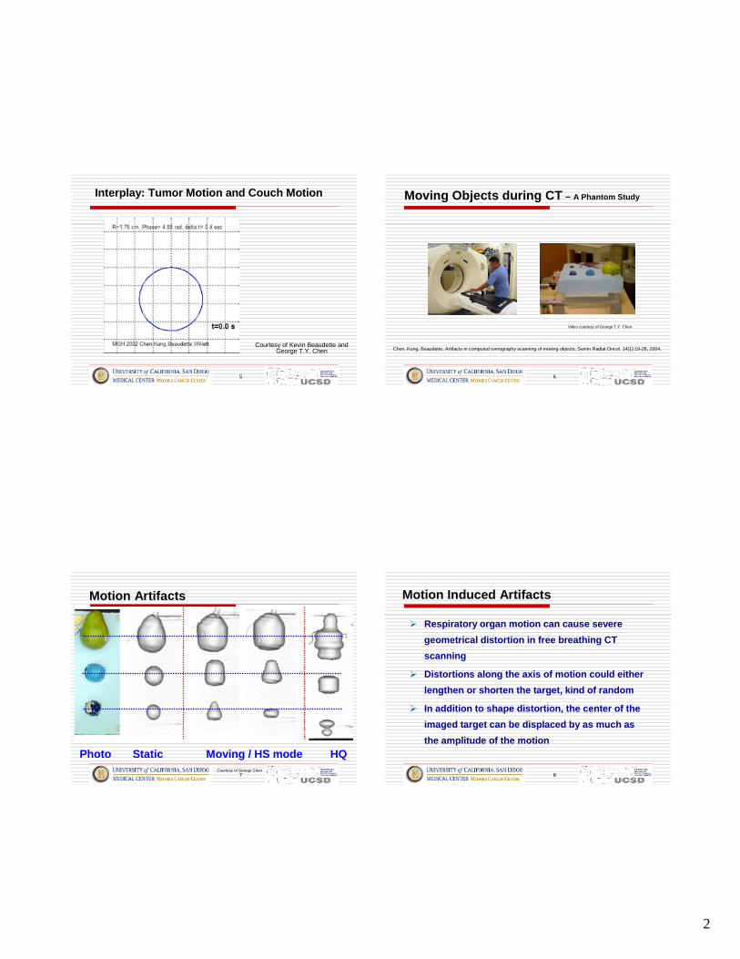
CT Art ifacts in Free Breathi ng 3D Scan
Courtesy of Eike Rietzel and George T.Y. Chen
Helica l lig ht breathing scan 4DCT – 1 phase
Tumo r
2
5
Interpla y: Tumor Motion and Couch Motion
Courtesy of Kevin Beaudette andGeorge T.Y. Chen
6
Moving Objects dur ing CT – A Phantom Study
Video courtesy of George T.Y. Chen
Chen, Kung, Beaudette, Artifacts in computed tomography scanning of moving objects, Semin Radiat Oncol. 14(1):19-26, 2004.
7
Motion Artifac ts
Photo Static Moving / HS mode HQCourtesy of George Chen
8
Motion Induce d Ar tifac ts
� Respirato ry org an motion can cause severe
geometrical distortion in free breathing CT
scanni ng
� Dis tort ions alon g the axis of motion cou ld eith er
lengthen or sho rten the target, kind of random
� In additi on to shape distortion, the center of the
imaged target can be dis plac ed by as much as
the amplitu de of the motion

3
9
Considerations of Organ Motion in CT Sim
� Breath -hold CT scan� Volun tary breath hold
� Acti ve breathing control
� Combine inhale and exhale GTVs to get ITV
� Slow CT san� 4 seconds per slice in axial mode
� Gated CT scan� imag es at onl y 1 phase, acquisition times 4-5x longer
� 4D CT scan� 3D scans at multiple phases
10
Basic Idea of 4D CT Scan
� Over-samp lin g image s at every
position of in terest along the
patient ’s lon g axis
� Each image is tagged with
breathing signals
� Images are sorted
retro spectively based on the
correspon ding breathing signals
� Many 3-D CT sets are obtaine d, each corresponding to a parti cul arbreathing phase
� Together, they cons titute a 4-D CT set tha t cove rs the ent irebreathing cycle
Photo Courtesy of Gig Mageras
11
Resorting 500-1500 CT Slices
Courtesy of Eike Rietzel andGeorge T.Y. Chen
12
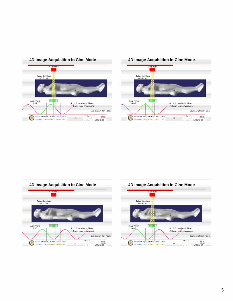
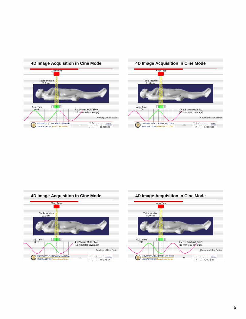
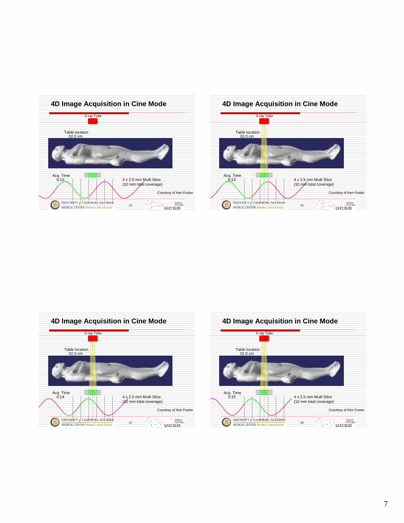
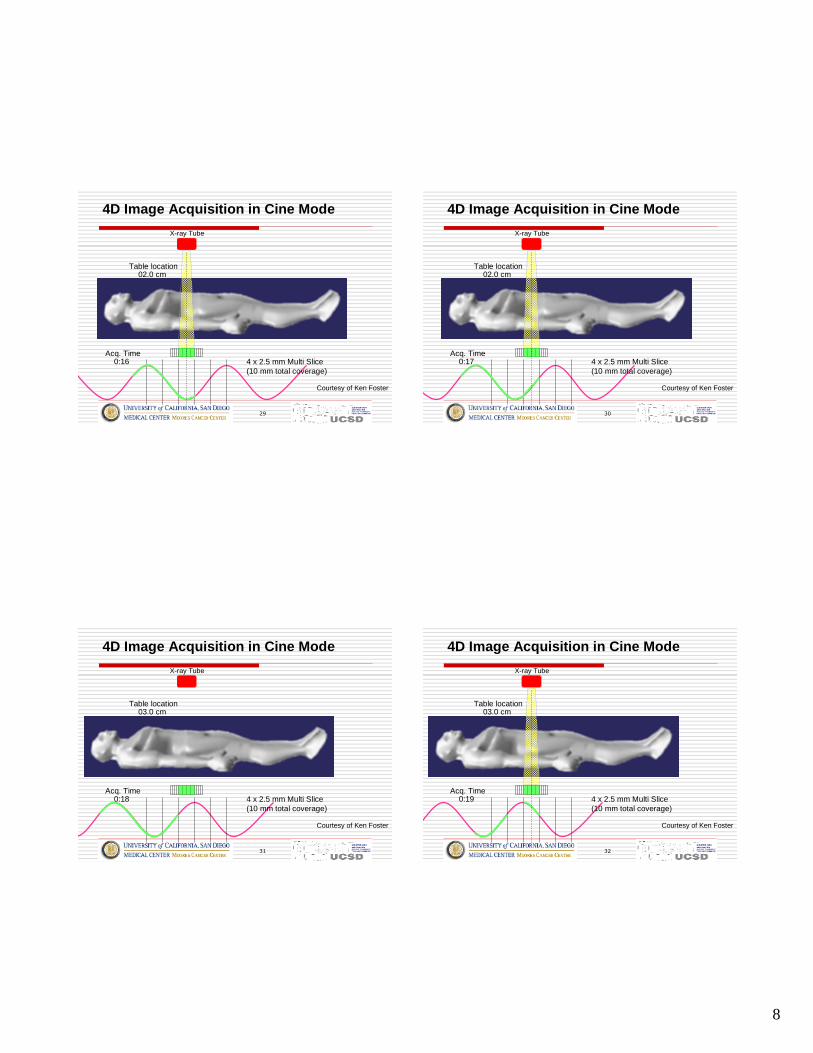
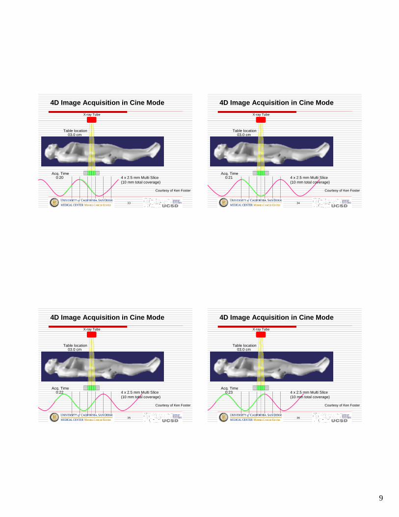
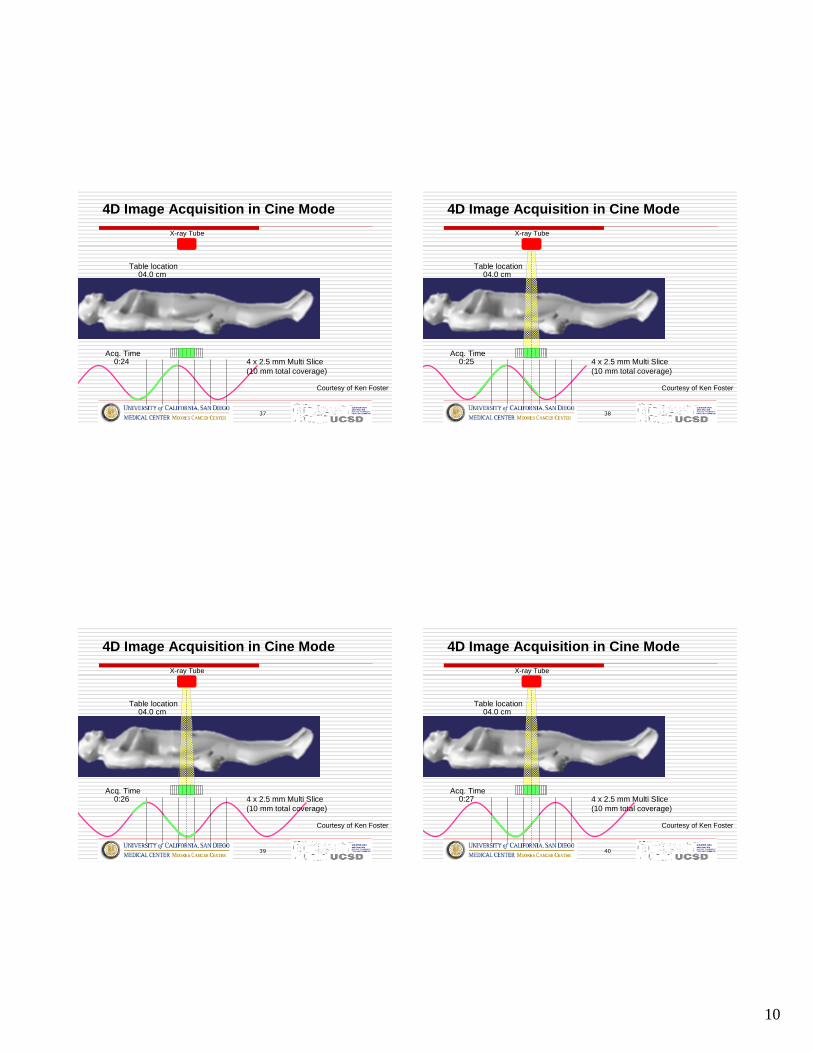
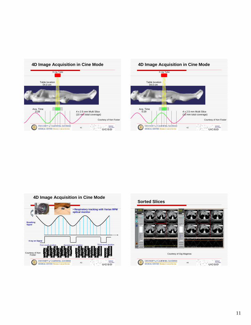
Acquisition of 4D CT Images
� Image acquisition can be performed in either
� Ciné mode: repeated image acquisiti on at each couchposition, or
� Helical mode: with small pitch
� Breathing signals can be obtained using either
� A monitor such as RPM, an abdo minal bel t, or
� variation of the patient anterio r surf ace in the CTimages
4
13
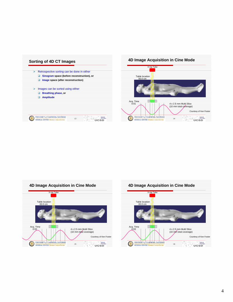
Sorting of 4D CT Images
� Retrospective sorting can be done in either
� Sinogram space (before reconstruction), or
� Image space (after reconstruction)
� Images can be sorted using either
� Breath ing phase , or
� Amplitude
14
0:01
00.0 cm
4D Image Acquisition in Cine Mode
4 x 2.5 mm Multi Slice(10 mm total coverage)
X-ray Tube
Table location
Acq. Time
Courtesy of Ken Foster
15
0:02
00.0 cm
4 x 2.5 mm Multi Slice(10 mm total coverage)
X-ray Tube
Table location
Acq. Time
Courtesy of Ken Foster
4D Image Acquisition in Cine Mode
16
0:03
00.0 cm
4 x 2.5 mm Multi Slice(10 mm total coverage)
X-ray Tube
Table location
Acq. Time
Courtesy of Ken Foster
4D Image Acquisition in Cine Mode
5
17
0:04
00.0 cm
4 x 2.5 mm Multi Slice(10 mm total coverage)
X-ray Tube
Table location
Acq. Time
Courtesy of Ken Foster
4D Image Acquisition in Cine Mode
18
0:05
00.0 cm
4 x 2.5 mm Multi Slice(10 mm total coverage)
X-ray Tube
Table location
Acq. Time
Courtesy of Ken Foster
4D Image Acquisition in Cine Mode
19
0:06
01.0 cm
4 x 2.5 mm Multi Slice(10 mm total coverage)
X-ray Tube
Table location
Acq. Time
Courtesy of Ken Foster
4D Image Acquisition in Cine Mode
20
0:07
01.0 cm
4 x 2.5 mm Multi Slice(10 mm total coverage)
X-ray Tube
Table location
Acq. Time
Courtesy of Ken Foster
4D Image Acquisition in Cine Mode
6
21
0:08
01.0 cm
4 x 2.5 mm Multi Slice(10 mm total coverage)
X-ray Tube
Table location
Acq. Time
Courtesy of Ken Foster
4D Image Acquisition in Cine Mode
22
0:09
01.0 cm
4 x 2.5 mm Multi Slice(10 mm total coverage)
X-ray Tube
Table location
Acq. Time
Courtesy of Ken Foster
4D Image Acquisition in Cine Mode
23
0:10
01.0 cm
4 x 2.5 mm Multi Slice(10 mm total coverage)
X-ray Tube
Table location
Acq. Time
Courtesy of Ken Foster
4D Image Acquisition in Cine Mode
24
0:11
01.0 cm
4 x 2.5 mm Multi Slice(10 mm total coverage)
X-ray Tube
Table location
Acq. Time
Courtesy of Ken Foster
4D Image Acquisition in Cine Mode
7
25
0:12
02.0 cm
4 x 2.5 mm Multi Slice(10 mm total coverage)
X-ray Tube
Table location
Acq. Time
Courtesy of Ken Foster
4D Image Acquisition in Cine Mode
26
0:13
02.0 cm
4 x 2.5 mm Multi Slice(10 mm total coverage)
X-ray Tube
Table location
Acq. Time
Courtesy of Ken Foster
4D Image Acquisition in Cine Mode
27
0:14
02.0 cm
4 x 2.5 mm Multi Slice(10 mm total coverage)
X-ray Tube
Table location
Acq. Time
Courtesy of Ken Foster
4D Image Acquisition in Cine Mode
28
0:15
02.0 cm
4 x 2.5 mm Multi Slice(10 mm total coverage)
X-ray Tube
Table location
Acq. Time
Courtesy of Ken Foster
4D Image Acquisition in Cine Mode
8
29
0:16
02.0 cm
4 x 2.5 mm Multi Slice(10 mm total coverage)
X-ray Tube
Table location
Acq. Time
Courtesy of Ken Foster
4D Image Acquisition in Cine Mode
30
0:17
02.0 cm
4 x 2.5 mm Multi Slice(10 mm total coverage)
X-ray Tube
Table location
Acq. Time
Courtesy of Ken Foster
4D Image Acquisition in Cine Mode
31
0:18
03.0 cm
4 x 2.5 mm Multi Slice(10 mm total coverage)
X-ray Tube
Table location
Acq. Time
Courtesy of Ken Foster
4D Image Acquisition in Cine Mode
32
0:19
03.0 cm
4 x 2.5 mm Multi Slice(10 mm total coverage)
X-ray Tube
Table location
Acq. Time
Courtesy of Ken Foster
4D Image Acquisition in Cine Mode
9
33
0:20
03.0 cm
4 x 2.5 mm Multi Slice(10 mm total coverage)
X-ray Tube
Table location
Acq. Time
Courtesy of Ken Foster
4D Image Acquisition in Cine Mode
34
0:21
03.0 cm
4 x 2.5 mm Multi Slice(10 mm total coverage)
X-ray Tube
Table location
Acq. Time
Courtesy of Ken Foster
4D Image Acquisition in Cine Mode
35
0:22
03.0 cm
4 x 2.5 mm Multi Slice(10 mm total coverage)
X-ray Tube
Table location
Acq. Time
Courtesy of Ken Foster
4D Image Acquisition in Cine Mode
36
0:23
03.0 cm
4 x 2.5 mm Multi Slice(10 mm total coverage)
X-ray Tube
Table location
Acq. Time
Courtesy of Ken Foster
4D Image Acquisition in Cine Mode
10
37
0:24
04.0 cm
4 x 2.5 mm Multi Slice(10 mm total coverage)
X-ray Tube
Table location
Acq. Time
Courtesy of Ken Foster
4D Image Acquisition in Cine Mode
38
0:25
04.0 cm
4 x 2.5 mm Multi Slice(10 mm total coverage)
X-ray Tube
Table location
Acq. Time
Courtesy of Ken Foster
4D Image Acquisition in Cine Mode
39
0:26
04.0 cm
4 x 2.5 mm Multi Slice(10 mm total coverage)
X-ray Tube
Table location
Acq. Time
Courtesy of Ken Foster
4D Image Acquisition in Cine Mode
40
0:27
04.0 cm
4 x 2.5 mm Multi Slice(10 mm total coverage)
X-ray Tube
Table location
Acq. Time
Courtesy of Ken Foster
4D Image Acquisition in Cine Mode
11
41
0:28
04.0 cm
4 x 2.5 mm Multi Slice(10 mm total coverage)
X-ray Tube
Table location
Acq. Time
Courtesy of Ken Foster
4D Image Acquisition in Cine Mode
42
0:29
04.0 cm
4 x 2.5 mm Multi Slice(10 mm total coverage)
X-ray Tube
Table location
Acq. Time
Courtesy of Ken Foster
4D Image Acquisition in Cine Mode
43
BreathingSignal
X-ray on Signal
First couch position Second couch position Third couch position
• Respir atory tracking wit h Varian RPMoptical monitor
4D Image Acquisition in Cine Mode
Courtesy of KenFoster
44
Courtesy of Gig Mageras
Sorted Slic es
12
45
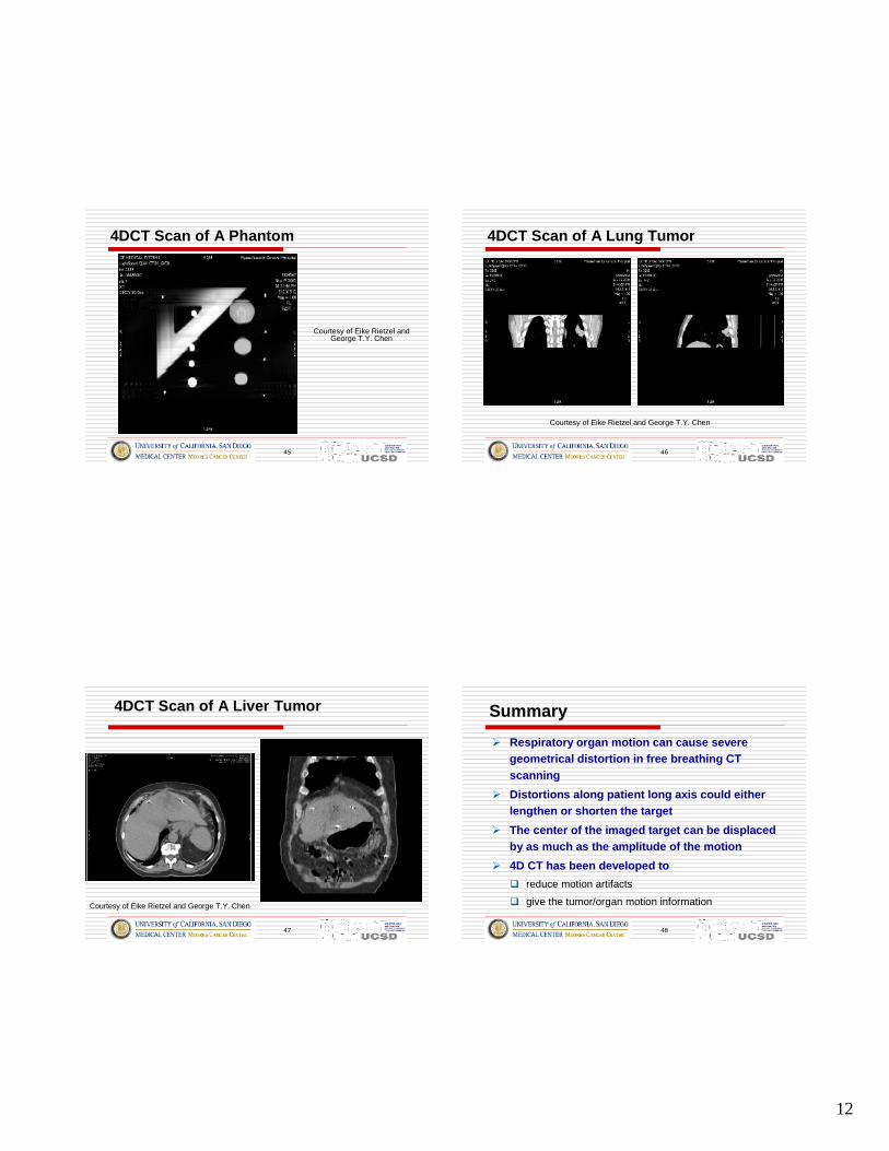
4DCT Scan of A Phantom
Courtesy of Eike Rietzel andGeorge T.Y. Chen
46
4DCT Scan of A Lung Tumo r
Courtesy of Eike Rietzel and George T.Y. Chen
47Patient 5618
4DCT Scan of A Liver Tumor
Courtesy of Eike Rietzel and George T.Y. Chen
48
Summary
� Respirato ry org an motion can cause severegeometrical distortion in free breathing CTscanni ng
� Dis tort ions alon g patient long axis could eitherlengthen or sho rten the target
� The center of the imaged target can be disp lacedby as much as the amplitud e of the motion
� 4D CT has been deve lope d to
� reduce motion artifacts
� give the tumor/organ motion information
13
49
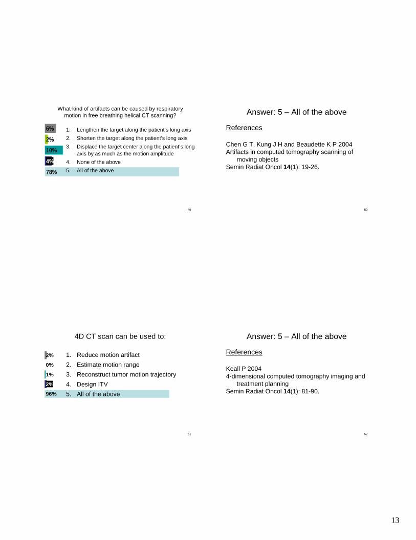
What kind of artifacts can be caused by respiratorymotion in free breathing helical CT scanning?
78%
10%
2%
6%
4%
1. Lengthen the target along the patient’s long axis
2. Shorten the target along the patient’s long axis
3. Displace the target center along the patient’s longaxis by as much as the motion amplitude
4. None of the above
5. All of the above
50
Answer: 5 – All of the above
References
Chen G T, Kung J H and Beaudette K P 2004Artifacts in computed tomography scanning of
moving objectsSemin Radiat Oncol 14(1): 19-26.
51
4D CT scan can be used to:
96%
1%
0%
2%
2%
1. Reduce motion artifact
2. Estimate motion range
3. Reconstruct tumor motion trajectory
4. Design ITV
5. All of the above
52
Answer: 5 – All of the above
References
Keall P 20044-dimensional computed tomography imaging and
treatment planningSemin Radiat Oncol 14(1): 81-90.