an evaluation of co-culture parameters effecting
TRANSCRIPT

Eastern Michigan University Eastern Michigan University
DigitalCommons@EMU DigitalCommons@EMU
Master's Theses and Doctoral Dissertations Master's Theses, and Doctoral Dissertations, and Graduate Capstone Projects
2020
An evaluation of co-culture parameters effecting antibiotic An evaluation of co-culture parameters effecting antibiotic
production in soil microbes production in soil microbes
Rebecca Lindow
Follow this and additional works at: https://commons.emich.edu/theses
Part of the Biology Commons, Microbiology Commons, and the Molecular Biology Commons
Recommended Citation Recommended Citation Lindow, Rebecca, "An evaluation of co-culture parameters effecting antibiotic production in soil microbes" (2020). Master's Theses and Doctoral Dissertations. 1036. https://commons.emich.edu/theses/1036
This Open Access Thesis is brought to you for free and open access by the Master's Theses, and Doctoral Dissertations, and Graduate Capstone Projects at DigitalCommons@EMU. It has been accepted for inclusion in Master's Theses and Doctoral Dissertations by an authorized administrator of DigitalCommons@EMU. For more information, please contact [email protected].
An Evaluation of Co-Culture Parameters Effecting Antibiotic Production in Soil
Microbes
by
Rebecca Lindow
Thesis
Submitted to the Department of Biology
Eastern Michigan University
in partial fulfillment of the requirements for the degree of
MASTER OF SCIENCE
in
Biology
Concentration in Cellular and Molecular Biology
Thesis Committee:
Paul Price, PhD, Chair
Aaron Liepman, PhD
Daniel Clemans, PhD
June 10, 2020
Ypsilanti, Michigan

ii
Acknowledgements
I would like to extend my gratitude to my advisor, Dr. Price, for his guidance in
and out of the lab. He was always willing to offer help and showed immense support
throughout this entire process. I would also like to thank my committee members, Dr.
Clemans and Dr. Liepman, for their advice and assistance during the project. My family
and friends also deserve thanks for their constant encouragement throughout this
project. In particular, Gillian Autterson deserves special recognition for her friendship
these past two years; I couldn’t have done this without her.
Funding for this project was provided by Eastern Michigan University
Department of Biology, as well as the Meta-Hellwig / Don Brown Graduate Research
Award.

iii
Abstract
The rise of infections caused by antibiotic resistant bacteria, compounded by a
reduction in antibiotic discovery and development, jeopardizes human health.
Historically, antibiotics derive from secondary metabolites produced by soil microbes in
pure culture, but recent genetic evidence suggests that microbes can produce more
secondary metabolites than are currently observed. The modified crowded plate
technique directly identifies antibiotic-producing soil microbes that were co-plated with
a target pathogen. Here, this technique was refined by testing the effect of a D-alanine
auxotrophic target pathogen rather than a prototrophic pathogen as well as
investigating conditions most conducive to antibiotic production. Antibiotic producing
conditions are most favorable with the use of a D-alanine auxotrophic pathogen that
was pre-incubated for one week. Antibiotic-producing microbes isolated using these
new parameters were cultured in single and mixed fermentations to compare secondary
metabolite production. Furthermore, mixed fermentations with multiple antibiotic
producers is an effective means to stimulate antibiotic production.
iv
Table of Contents
Acknowledgments ........................................................................................................................ ii
Abstract ........................................................................................................................................ iii
List of Tables .................................................................................................................................. v
List of Figures .............................................................................................................................. vi
INTRODUCTION ......................................................................................................................... 1
MATERIALS AND METHODS ................................................................................................ 13
RESULTS ...................................................................................................................................... 21
DISCUSSION ............................................................................................................................... 34
CONCLUSIONS ......................................................................................................................... 40
Literature Cited ........................................................................................................................... 41

v
List of Tables
Table 1. Different media types and their ingredients ............................................................ 14
Table 2. Bacterial strains used in this study ............................................................................ 14
Table 3. Primer Sequences of S. aureus Alr1, Alr2, and Pkor1 ............................................. 18
Table 4. mCPT derived isolates active against a panel of pathogens ................................. 23
Table 5. Pure cultures and their associated sequencing data ............................................... 31
Table 6. Mixed fermentations of non-producing pure culture microbes ........................... 32
vi
List of Figures
Figure 1. Antibiotic production from 1980 to 2019 .................................................................. 2
Figure 2. MRSA infection incidences ......................................................................................... 5
Figure 3. Purified antibiotic producers tested for activity against S. aureus ........................ 6
Figure 4. Bacterial D-alanine metabolic pathway .................................................................... 8
Figure 5. Growth of a D-alanine auxotroph .............................................................................. 9
Figure 6. Initial mCPT soil screen using B. subtilis as a target organism ........................... 21
Figure 7. ESKAPE pathogen safe strain screening of soil microbes .................................... 22
Figure 8. Juxtaposition of prototrophic versus auxotrophic production ........................... 25
Figure 9. Comparison of incubation period and media type on antibiotic production ... 26
Figure 10. Isolated microbes from initial mCPT soil screens were tested against B.
subtilis and S. aureus ....................................................................................................... 27
Figure 11. Co-occurrence of B. subtilis and S. aureus ............................................................. 28
Figure 12. Construction of homologous recombination plasmids for the deletion of alr1
and alr2 in S. aureus ........................................................................................................ 29
Figure 13. Antibiotic testing of chemical extracts in single- and mixed-culture
fermentations ................................................................................................................... 30
Figure 14. Chemical extracts from mixed-culture fermentations with or without
supplemental Mn ............................................................................................................ 33
1
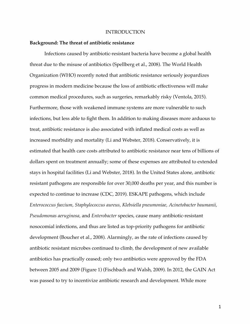
INTRODUCTION Background: The threat of antibiotic resistance
Infections caused by antibiotic-resistant bacteria have become a global health
threat due to the misuse of antibiotics (Spellberg et al., 2008). The World Health
Organization (WHO) recently noted that antibiotic resistance seriously jeopardizes
progress in modern medicine because the loss of antibiotic effectiveness will make
common medical procedures, such as surgeries, remarkably risky (Ventola, 2015).
Furthermore, those with weakened immune systems are more vulnerable to such
infections, but less able to fight them. In addition to making diseases more arduous to
treat, antibiotic resistance is also associated with inflated medical costs as well as
increased morbidity and mortality (Li and Webster, 2018). Conservatively, it is
estimated that health care costs attributed to antibiotic resistance near tens of billions of
dollars spent on treatment annually; some of these expenses are attributed to extended
stays in hospital facilities (Li and Webster, 2018). In the United States alone, antibiotic
resistant pathogens are responsible for over 30,000 deaths per year, and this number is
expected to continue to increase (CDC, 2019). ESKAPE pathogens, which include
Enterococcus faecium, Staphylococcus aureus, Klebsiella pneumoniae, Acinetobacter baumanii,
Pseudomonas aeruginosa, and Enterobacter species, cause many antibiotic-resistant
nosocomial infections, and thus are listed as top-priority pathogens for antibiotic
development (Boucher et al., 2008). Alarmingly, as the rate of infections caused by
antibiotic resistant microbes continued to climb, the development of new available
antibiotics has practically ceased; only two antibiotics were approved by the FDA
between 2005 and 2009 (Figure 1) (Fischbach and Walsh, 2009). In 2012, the GAIN Act
was passed to try to incentivize antibiotic research and development. While more
2
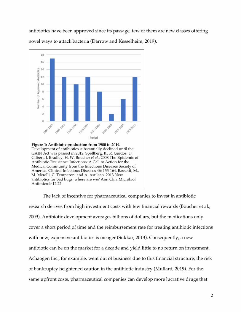
antibiotics have been approved since its passage, few of them are new classes offering
novel ways to attack bacteria (Darrow and Kesselheim, 2019).
The lack of incentive for pharmaceutical companies to invest in antibiotic
research derives from high investment costs with few financial rewards (Boucher et al.,
2009). Antibiotic development averages billions of dollars, but the medications only
cover a short period of time and the reimbursement rate for treating antibiotic infections
with new, expensive antibiotics is meager (Sukkar, 2013). Consequently, a new
antibiotic can be on the market for a decade and yield little to no return on investment.
Achaogen Inc., for example, went out of business due to this financial structure; the risk
of bankruptcy heightened caution in the antibiotic industry (Mullard, 2019). For the
same upfront costs, pharmaceutical companies can develop more lucrative drugs that
Figure 1: Antibiotic production from 1980 to 2019. Development of antibiotics substantially declined until the GAIN Act was passed in 2012. Spellberg, B., R. Guidos, D. Gilbert, J. Bradley, H. W. Boucher et al., 2008 The Epidemic of Antibiotic-Resistance Infections: A Call to Action for the Medical Community from the Infectious Diseases Society of America. Clinical Infectious Diseases 46: 155-164. Bassetti, M., M. Merelli, C. Temperoni and A. Astilean, 2013 New antibiotics for bad bugs: where are we? Ann Clin. Microbiol Antimicrob 12:22.
3
treat chronic illnesses, such as heart conditions and autoimmune diseases, where the
profit margin is substantially larger (Sukkar, 2013). Although the root cause of antibiotic
resistance must be remedied, overcoming the scientific challenges of identifying new
antibiotics is critical to confront the burgeoning threat posed by antibiotic resistance.
Currently, a variety of methods have been deployed in an effort to minimize the
spread of antibiotic resistance, including stricter control of antibiotics in humans and
livestock, and the use of vaccines. One strategy in humans requires that a proper
prescription, one that prescribes appropriate drugs and is signed by a practicing doctor,
must be present in order to administer the antibiotics (Davies and Davies, 2010). Over
the counter antibiotics are readily available in developing countries, contributing to the
remarkable resistance threat because people are able to take antibiotics for any illness
(Ayukekbong et al., 2017). Vaccines are another approach to combat resistance, but
some types cannot be administered to immunocompromised individuals as they may
fall ill. Vaccines may also be met with debate from the anti-vaccination movement
(Kata, 2010). On the other hand, the food industry uses antibiotics as a feed-additive to
increase livestock yields. Although there are regulatory practices in place to prevent
sub-therapeutic use of antibiotics in agriculture, many antibiotics can be purchased
without a veterinarian’s prescription to feed to the animal (Davies and Davies, 2010;
Sneeringer et al., 2015). The misuse of antibiotics in livestock can support resistant
bacteria that may affect humans through either contaminated meat after slaughter or
through environmental infections (CDC, 2019). With these concerns, the identification
of new antibiotics with therapeutic potential plays a strategic role in the welfare of
society.
Methicillin-resistant Staphylococcus aureus (MRSA) is of particular interest since it
is now classified as a high-priority, multi-drug resistant pathogen known to cause a
4
variety of skin and soft tissue infections (SSTI) as well as many life-threatening diseases
such as pneumonia and septic shock (Lowy, 1998). SSTI’s range from mild (impetigo) to
severe (necrotizing fasciitis) (Styjewski and Chambers, 2008). Over 120,000 cases of
MRSA are reported in the United States per year, with nearly 20,000 of these diagnoses
culminating in death; the fatalities attributed to MRSA now outnumber those from
HIV/AIDS (Boucher and Corey, 2008; CDC, 2019). Fortunately, the number of
nosocomial MRSA infections has declined because of hospital infection control;
however, community-acquired infections have remained nearly constant for the past 15
years, eliciting the threat of community-acquired MRSA infections (Figure 2) (Hassoun
et al., 2017; Mehta et al., 2014). Due to its heightened virulence, most of the community
and hospital-acquired outbreaks of MRSA derive from a single clonal lineage of MRSA;
USA300 (Tenover and Goering, 2009). The extensive antibiotic resistance of MRSA
USA300 derives from its fairly plastic genome; it has acquired an assortment of
resistance genes that aid in its survival (Kuroda et al., 2001). Thus, there is a
considerable demand to discover antibiotics effective against this pathogen in an effort
to control this pathogen.

5
Antibiotic-producing bacteria may reside in soil
Most antibiotics in use today were originally derived from soil microbes.
Penicillin, produced by the soil fungus, Penicillium, serves as the archetype of such
antibiotic exploration because it was the first true antibiotic to be discovered (Clardy et
al., 2009). Early discovery efforts generally followed the “Waksman Platform” method,
in which soil microorganisms were plated alongside pathogenic bacteria and zones of
inhibition (ZOI) were detected (Figure 3). These ZOI imply the ability of a microbe to
produce an antibiotic that prevents the growth of the pathogen. The resulting strains,
and antibiotic compounds they produced, could then be characterized (Valiquette and
Laupland, 2012). However, by the 1960s, many scientists assumed that most of the
cultivatable bacteria that could produce antibiotics had been identified (Nichols et al,
2010). Fortunately, soil is a remarkably diverse environment, as elicited by
metagenomic studies; this implies that there are a variety of bacteria living in this
Figure 2: MRSA infection incidences. While there is improvement in the number of hospital acquired infections, community acquired infections have remained stagnant. Figure adapted from Kourits AP, Hartfield K, Baggs J, et al., 2019, Vital signs: epidemdiology and recent trends in methicillin-resistance Staphylococcus aureus bloodstream infections. Morbidity and Mortality Weekly Report: 214-219
6
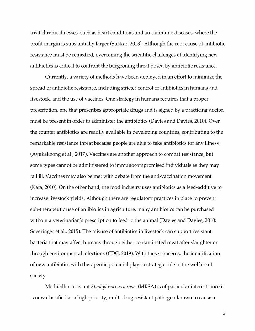
habitat that may possess novel qualities (Daniel, 2005). Because there are so many
different bacteria in the soil, there are various survival strategies prevalent, namely,
competition via antibiotic production (D’Costa et al., 2006). The crowded plate
technique (CPT), an antibiotic screening method developed by Waksman, exploits these
routine functions of bacteria. To do so, soil microbes are plated and grown for several
days, at which point the plates are observed for ZOIs. Although simplistic, this method
is limited by its lack of specificity. Furthermore, the isolated antibiotic producers from
this practice were seldom active against pathogens following purification and testing in
isolation. Soil microbes are a largely untapped source for producing novel antibiotics;
but refined methods to discover these antibiotic-producing microbes are needed.
Recently, the Price lab developed a variant of the CPT by simultaneously
inoculating a target pathogen with a diluted soil sample on the media to directly screen
for antibiotic producers that are active against a pathogen, as indicated by a ZOI. These
adjustments were termed the modified crowded plate technique (mCPT). These
Figure 3: Purified antibiotic producers tested for activity against S. aureus. Antibiotic production can be observed by the halos surrounding patches of soil microbes (see arrows: 2, 13, 14, and 16).

7
fundamental changes improved the effectiveness of the method to identify antibiotic-
producing bacteria in a complex stimulatory environment. These isolated antibiotic-
producing bacteria can then be further tested against other microbes, such as the
ESKAPE pathogens (Enterococcus faecium, Staphylococcus aureus, Klebsiella pneumoniae,
Acinetobacter baumanii, Pseudomonas aeruginosa, and Enterobacter species). However,
isolating the antibiotic producing microbe is often complicated by the presence of the
target organism; this makes it difficult to test the producer against other pathogens.
Further refinement to the mCPT could aid in establishing a more efficient antibiotic
discovery assay.
The mCPT has been introduced into two labs (BIO112 and BIO328) via the Tiny
Earth curriculum at Eastern Michigan University (EMU), allowing students to engage in
inquiry-based learning while also supplying many soil samples (Davis et al., 2017). This
educational program is partially based on the original methods, such as the “Waksman
Platform,” employed by scientists to discover antibiotics from soil microbes and it
proposes to use a crowdsourcing strategy to further explore the soil microbiome in
hopes of discovering new antibiotics (Valderrama et al., 2018; Peek et al., 2018). At
EMU, the mCPT has been intermingled into this curriculum to better exploit the routine
ecological duties of antibiotics. The Price lab advances this classroom research to
determine the novelty of the antibiotic producing microbes and their secondary
metabolites. A contingent of the Price lab is also developing new tools and
methodologies that can then be taken back into the classroom, further promoting safe
learning and discovery for students with introductory laboratory skills and experience.
D-alanine auxotrophs may function as a safe screening tool
One such approach, developed by undergraduate researchers in the Price lab at
Eastern Michigan University (EMU), involves the use of D-alanine auxotrophic bacteria
8
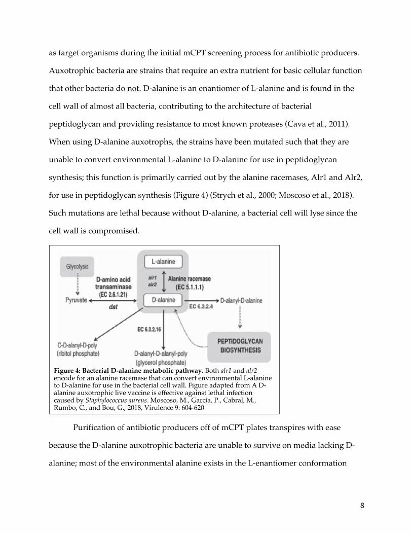
as target organisms during the initial mCPT screening process for antibiotic producers.
Auxotrophic bacteria are strains that require an extra nutrient for basic cellular function
that other bacteria do not. D-alanine is an enantiomer of L-alanine and is found in the
cell wall of almost all bacteria, contributing to the architecture of bacterial
peptidoglycan and providing resistance to most known proteases (Cava et al., 2011).
When using D-alanine auxotrophs, the strains have been mutated such that they are
unable to convert environmental L-alanine to D-alanine for use in peptidoglycan
synthesis; this function is primarily carried out by the alanine racemases, Alr1 and Alr2,
for use in peptidoglycan synthesis (Figure 4) (Strych et al., 2000; Moscoso et al., 2018).
Such mutations are lethal because without D-alanine, a bacterial cell will lyse since the
cell wall is compromised.
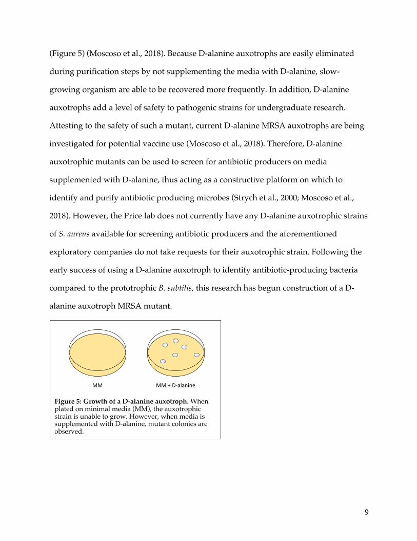
Purification of antibiotic producers off of mCPT plates transpires with ease
because the D-alanine auxotrophic bacteria are unable to survive on media lacking D-
alanine; most of the environmental alanine exists in the L-enantiomer conformation
Figure 4: Bacterial D-alanine metabolic pathway. Both alr1 and alr2 encode for an alanine racemase that can convert environmental L-alanine to D-alanine for use in the bacterial cell wall. Figure adapted from A D-alanine auxotrophic live vaccine is effective against lethal infection caused by Staphylococcus aureus. Moscoso, M., Garcia, P., Cabral, M., Rumbo, C., and Bou, G., 2018, Virulence 9: 604-620
9
(Figure 5) (Moscoso et al., 2018). Because D-alanine auxotrophs are easily eliminated
during purification steps by not supplementing the media with D-alanine, slow-
growing organism are able to be recovered more frequently. In addition, D-alanine
auxotrophs add a level of safety to pathogenic strains for undergraduate research.
Attesting to the safety of such a mutant, current D-alanine MRSA auxotrophs are being
investigated for potential vaccine use (Moscoso et al., 2018). Therefore, D-alanine
auxotrophic mutants can be used to screen for antibiotic producers on media
supplemented with D-alanine, thus acting as a constructive platform on which to
identify and purify antibiotic producing microbes (Strych et al., 2000; Moscoso et al.,
2018). However, the Price lab does not currently have any D-alanine auxotrophic strains
of S. aureus available for screening antibiotic producers and the aforementioned
exploratory companies do not take requests for their auxotrophic strain. Following the
early success of using a D-alanine auxotroph to identify antibiotic-producing bacteria
compared to the prototrophic B. subtilis, this research has begun construction of a D-
alanine auxotroph MRSA mutant.
Figure 5: Growth of a D-alanine auxotroph. When plated on minimal media (MM), the auxotrophic strain is unable to grow. However, when media is supplemented with D-alanine, mutant colonies are observed.
MM MM + D-alanine
10
Culturing techniques for antibiotic production
The looming threat of antibiotic resistance requires the construction of novel
antibiotic screening tools, as well as the efficient identification of bioactive substances.
Many bioactive substances are generated as secondary metabolites, compounds that are
not directly involved in growth and development of the cell, but they promote survival
functions within the environment, such as corporative or competitive cell-to-cell
communication within the environment, often through the use of antibiotics (Netzker et
al., 2015). Secondary metabolites are encoded by cryptic biosynthetic gene clusters
(BGCs), which in some bacteria, such as Actinomycetes, constitute roughly 5% of the
genome (Traxler et al., 2013). Many BGCs, however, are not expressed under normal
axenic laboratory conditions, including many encoding proteins that produce
antibiotics as they can be self-harming. Research focused on inducing expression of
cryptic BGCs indicates that certain ecological or chemical cues within a microbe’s
environment may be necessary to stimulate their expression (Cornforth and Foster,
2015; Van der Meij et al., 2017). Thus, in order to encourage the production of secondary
metabolites and antibiotics, culture methods need to closely mimic the complexity
experienced in a microbe’s natural environment.
Co-culturing seeks to stimulate bioactive substances by encouraging interspecies
interactions. In contrast to axenic cultures, which have been the basis for most antibiotic
discovery methods, co-cultures involve growth of different microbes together, thereby
encouraging interspecies interactions and potentially stimulating the expression of
cryptic BGCs (Van der Meij et al., 2017). Assuming that antibiotic production is the
product of a competitive environment, and many antibiotics are self-harming, then the
use of axenic cultures likely restricts antibiotic expression as interspecies interactions
that encourage antibiotic production are absent (Abruden et al., 2015; Seyedsayamdost
11
et al., 2012; Nai and Meyer, 2017). For example, some microbes, such as Streptomyces
when grown with mycolic acid-containing bacteria, require cell-to-cell contact to
produce antibiotics that are otherwise not produced in axenic cultures (Onaka et al.,
2011). Under co-culturing conditions, the expression of BGCs and the concentration of
secondary metabolites increase, suggesting that their induction and production may be
triggered by interspecies interactions, such as commensalism and competition (Ueda
and Beppu, 2016). As a testament to the impact of co-culture, antibiotic activity was
only observed in bacteria from the leaf microbiome of a plant with no known antibiotic
activity when they were grown in co-cultures (Helfrich, 2018). This implies that changes
in cultivation conditions can change the metabolic profile of a microbe (Bode et al.,
2002). Co-culturing, then, may allow for increased production of novel secondary
metabolites that can be used to combat antibiotic resistance (Onaka et al., 2011).
Study objectives
As a result of the formidable demand to discover new antibiotics effective
against multi-drug resistant pathogens, this research aims to advance the robustness of
the mCPT screening method to identify more antibiotic-producing bacteria. To do so,
the effectiveness of the auxotrophic mutants was compared to the prototrophic
pathogens, and culture conditions were optimized to improve the sensitivity of the
mCPT screening method. The differences in antibiotic production between axenic and
co-culturing methods of antibiotic production were also evaluated. Using the improved
screening methods, the antibiotic properties of the secondary metabolites, released by
antibiotic-producing bacteria grown under single and mixed fermentations, were
isolated and characterized. Construction of the D-alanine MRSA auxotroph has begun
with cloning the plasmids. This auxotroph will broaden the scope of the mCPT

12
screening method as a way to safely and effectively screen for and isolate antibiotic-
producing bacteria.
13
MATERIALS AND METHODS
Soil collection
Soil samples were collected at an air temperature of 15 ° C in May 2019 from five
locations on the Eastern Michigan University Campus in Ypsilanti, Michigan (Halle
Library, Student Center, Rec/IM, Pray Harrold, and the Science Complex). The
locations were sampled at a depth of one inch into the soil.
Soil screening
One gram of each soil sample was serially diluted 1:10,000 with ddH2O, and 100
𝜇L was plated on TYME (Table 1) pre-inoculated with B. subtilis (TE-Bs) or S. aureus
(PP667) (Table 2). Natamycin (20𝜇g/mL; NataMax SF, DuPont-Danisco USA Inc.: Kansas,
USA) was added to these plates to prevent fungal growth. For experiments involving B.
subtilis D-alanine auxotrophs (PP655), TYME media was supplemented with 100 𝜇g/mL
D-alanine (Matrix Scientific, Colombia, SC, USA) (Table 2). For experiments examining
the potential of gellan gum (VWR: Radnor, VA, USA) to generate solid media, 7 g/L
gellan gum was added in place of agar and solidified by adding CaCl2 to a final
concentration of 5 mM. These plates were grown in closed containers at 30 ° C for three
months, with antibiotic production checked at both two weeks and three months of
incubation.

14
Students in the Fall 2019 sections of Tiny Earth incorporated labs (BIO112 and
BIO328) followed the same protocol, but the BIO112 students used prototrophic B.
subtilis (TE-Bs) and the BIO328 students used auxotrophic B. subtilis (PP655) (Table 2).
Both of these courses are introductory biology labs; BIO112 is geared towards biology
majors while BIO328 is designed for nursing majors.
Table 1: Different media types and their ingredients. All chemicals were sourced from VWR: Radnor, VA, USA.
Media Type TYME EPSM TY LB
Ingredients (per 1L ddH2O)
0.5g Glucose 0.5g Yeast extract 0.5g Tryptone 0.5g Peptone 1mL KH2PO4 1mL CaCl2 1mL MgSO4 1mL Minor salts 12g Agar (for solid media)
0.5g Potato starch 0.5g Yeast extract 0.5g Tryptone 0.5g Peptone 1mL KH2PO4 1mL CaCl2 1mL MgSO4 1mL Minor salts 12g Agar (for solid media)
6g Tryptone 3g Yeast extract 0.5g CaCl2·2H2O 12g Agar (for solid media)
10g Tryptone 5g Yeast extract 10g NaCl
Table 2: Bacterial strains used in this study
Strain Genotype Source/Reference PP655 Bacillus subtilis 168 dal-1 sigB::erm [from AG232 (46)] Alan Grossman
(Massachusetts Institute of Technology)
PP665 Klebsiella pneumoniae ATCC 13883 American Type Culture Collection (ATCC)
PP666 Pseudomonas aeruginosa ATCC 27853 ATCC PP667 Staphylococcus aureus ATCC 25923 ATCC PP673 TE-Ps
Mycobacterium smegmatis MC2155 Pseudomonas putida
Miriam Braunstein (University of North Carolina at Chapel Hill) Tiny Earth
TE-Ec Escherichia coli ATCC 1775 Tiny Earth TE-Bs RN4220
Bacillus subtilis Staphylococcus aureus RN4220
Tiny Earth Eric Skaar (Vanderbilt University)

15
Screen against pathogen panel via spread patch plate technique:
Antibiotic-producing isolates identified following the mCPT screen, both at two
weeks and three months post-incubation, were partially purified and tested, via a
spread-patch assay, for secondary screening against a panel of pathogens that include:
S. aureus (PP667), B. subtilis (TE-Bs), M. smegmatis (PP673), P. putida (TE-Ps) and E. coli
(TE-Ec) (Table 2). Purified antibiotic-producing bacteria were patched on top of a target
pathogen. These plates were grown at 30 ° C for two weeks and bacteria were examined
for antibiotic production at days one, three, and seven post-inoculation.
Isolation of antibiotic producers
For antibiotic-producing isolates that presented a clear zone of inhibition (ZOI)
on any of the pathogens in the aforementioned panel, the size of the ZOI was measured
in millimeters using a ruler. The antibiotic producers were then isolated to pure culture
via three-phase streaks and frozen down in 20% glycerol and stored at -80 ° C.
Bacterial identification
Isolated colonies were identified using 16S rRNA gene sequencing colony PCR
with 1X GoTaq Green (Promega: Madison, WI, USA), 0.125 𝜇M forward primer (27F 5’
AGRGTTTGATYMTGGCTCAG 3’), 0.125 𝜇M reverse primer (1492R 5’
GGYTACCTTGTTACGACTT 3’), brought to a final volume of 10 𝜇L with ddH2O, and a
colony was mixed in. The thermocycler parameters were set as follows: initial
denaturation at 95 ° C for 2 minutes, 35 cycles of 30 seconds at 95 ° C for denaturation,
30 seconds at 50 ° C for annealing, and 1 minute at 72 ° C for extension, followed by a
final extension period of 2 minutes at 72 ° C. The PCR products were confirmed on a 1%
agarose gel containing 1X GelRed (VWR: Radnor, VA, USA). For isolates that did not
yield sufficient PCR product for Sanger sequencing, InstaGene matric (BioRad,
16
Hercules, CA, USA) was used to prepare partially purified genomic DNA for PCR
according to the manufacturer’s instructions. An ExoSAP-IT clean-up procedure was
used to prepare all PCR products for Sanger sequencing by adding 1X ExoSAP-IT
(Thermo Fisher Scientific: Waltham, MA, USA) to the PCR tube, then placing in the
thermocycler for 15 minutes at 37 ° C then 15 minutes at 80 ° C. Sanger sequencing was
performed by Eton Biosciences (Union, New Jersey, USA). Sequencing results were run
through a nucleotide BLAST analysis using the 16S rRNA sequences database to
identify the bacteria (Altschul et al., 1990).
Pure and co-cultures
A culture consisting of an antibiotic producing colony from the mCPT screen
was placed in 5 mL TYME and incubated 3-5 days at 30 ° C at 300 rpm. Subsequently, 1
mL of the overnight culture was then added to 100 mL of fresh ESPM, a TYME media
substituting 5 g/L potato starch for glucose (VWR: Radnor, VA, USA), in a 250 mL
Erlenmeyer flask covered with surgical paper (VWR: Radnor, VA, USA) (Table 1). After
three days of incubation at room temperature at 200 rpm, a sterile amberlite bag was
added to the culture.
Co-cultures followed the same steps; however, 1 mL of three randomly selected
antibiotic-producing bacteria from the mCPT screen were added to 100 mL liquid EPSM
in a single 250 mL Erlenmeyer flask, rather than one. For experiments comparing EPSM
with and without supplemental Manganese in the form of MnCl2 (1 mM final
concentration), MnCl2 was added to the culture media prior to autoclaving.
Amberlite™ bag assembly
Amberlite XAD16N resin (Sigma Aldrich, St. Louis, MO, USA) was enclosed in
small, 1 x 1 inch bags using heat sealed strips of Unitherm 1.5, a polyester/polyethylene
cloth-like material (Midwest Filtration Company, West Chester Township, OH, USA).
17
15 g/L (w/v) Amberlite™ was measured out for each culture. The Amberlite™ was
then washed with acetone three times and once with ddH2O. The new mass of the
Amberlite™ was then equally distributed amongst the bags and autoclaved prior to
addition to liquid cultures.
Chemical extractions of Amberlite™ bags:
On day 8 of incubation, Amberlite™ bags were removed from their respective
cultures, rinsed in cold water, and patted dry. The bags were then placed in pre-
weighed vials, to which 20 mL of 50/50 ethyl acetate/methanol was added. This shook
for 30 minutes at room temperature at 300 rpm, after which the Amberlite™ bags were
removed and the samples were dried in a Thermo Fisher Scientific SpeedVac at 45 ° C
for 8 hours. After reweighing the vials, the mass of the dried product was calculated
and the samples were resuspended in DMSO at 15 mg/mL to obtain the final natural
product extract.
Antibiotic activity
To test for antibiotic activity, 10 𝜇L spots of chemical extract were placed on
TYME agar plates and allowed to dry. S. aureus (PP667) or E. coli (TE-Ec) were then
inoculated over top the samples and the plates grew for 7 days at 37 ° C (Table 2). The
plates were checked at days one and seven of incubation for ZOI and signs of
developing antibiotic resistance.
Comparison of pre-incubation time and media type for mCPT screening
Auxotrophic B. subtilis streaks were grown on LB, TY, TYME, and EPSM (Table
1) for either 1 or 7 days at 30 ° C. mCPT screening plates were prepared as described
above with natamycin and D-alanine. These plates incubated for two weeks at 30 ° C.
Size and number of ZOI were then recorded.
18
Construction of a D-alanine auxotrophic S. aureus mutant:
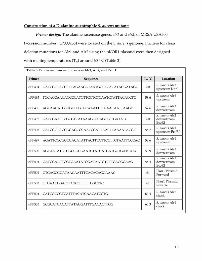
Primer design: The alanine racemase genes, alr1 and alr2, of MRSA USA300
(accession number: CP000255) were located on the S. aureus genome. Primers for clean
deletion mutations for Alr1 and Alr2 using the pKOR1 plasmid were then designed
with melting temperatures (Tm) around 60 ° C (Table 3).
Table 3: Primer sequences of S. aureus Alr1, Alr2, and Pkor1.
Primer Sequence Tm ˚C Location
oPP494 GATCGGTACCCTTAGAAGGTAATGGCTCACATACGATAGC 60 S. aureus Alr2 upstream KpnI
oPP495 TGCACCAACACCCCATGTTGCTGTCAATGTATTACACCTC 58.6 S. aureus Alr2 upstream
oPP496 AGCAACATGGTGTTGGTGCAAATTCTGAACAATTAAGT 57.6 S. aureus Alr2 downstream
oPP497 GATCGAATTCGCGTCATAAAGTGCAGTTCTGATATG 60 S. aureus Alr2 downstream EcoRI
oPP498 GATCGGTACCGGAGCCCAATCGATTAACTTAAAATACGC 58.7 S. aureus Alr1 upstream EcoRI
oPP499 AGATTCGCGGCGACATATTACTTCCTTCCTTGTAATTCCCAC 58.6 S. aureus Alr1 upstream
oPP500 AGTAATATGTCGCCGCGAATCTATCATGATGGTGATCAAC 59.9 S. aureus Alr1 downstream
oPP501 GATCGAATTCGTGAATATCGACAATGTCTTCAGGCAAG 58.4 S. aureus Alr1 downstream EcoRI
oPP502 GTGAGCGGATAACAATTTCACACAGGAAAC 61 Pkor1 Plasmid Forward
oPP503 CTGAACCGACTTCTCCTTTTTCGCTTC 61 Pkor1 Plasmid Reverse
oPP504 CATCGCCGTCATTTACATCAACATCCTG 60.4 S. aureus Alr2 check
oPP505 GCGCATCACATTATAGGATTTGACACTTGG 60.3 S. aureus Alr1 check

19
Plasmid preparation: S. aureus genomic DNA was isolated using the Purelink
Microbiome DNA Purification kit for Gram-positive organisms (Thermo Fisher
Scientific: Waltham, MA, USA) using 2 mL of a S. aureus (PP667) overnight culture
grown in LB (Table 2). The target regions were amplified via PCR at a volume of 20 𝜇L
consisting of 1.25 ng/𝜇L isolated gDNA, 10 𝜇M dNTPs (VWR: Radnor, VA, USA), 25
units per liter Q5 polymerase (NEB: Ipswich, MA, USA), 1X Q5 Buffer (NEB: Ipswich,
MA, USA), 0.5 𝜇M of each primer and brought to the final volume with ddH2O. The
thermocycler conditions were as follows: initial denaturation at 95 ° C for 2 minutes, 35
cycles of 30 seconds at 95 ° C for denaturation, 30 seconds at 55 ° C for annealing, and 1
minute at 72 ° C for extension, followed by a final extension period of 2 minutes at 72 °
C. Splicing by overlap extension (SOE) PCR using the same PCR conditions was used to
generate a single PCR product for cloning into the pKOR1 vector. Primers oPP494,
oPP495, oPP496, and oPP497 (Table 3) were used to construct the clean deletion plasmid
for alr2 and oPP498, oPP499, oPP500, and oPP501 (Table 3) for alr1. Following agarose
gel electrophoresis, PCR products were purified with Zymoclean Gel DNA Recovery
Kit using manufacturer’s instructions (Zymo Research, Irvine, CA, USA).
Plasmid Construction: pKOR1 and the corresponding SOE PCR products were
enzymatically digested with EcoRI and KpnI (NEB: Ipswich, MA, USA). Digested PCR
product was purified with Zymoclean Gel DNA Recovery Kit using manufacturer’s
instructions (Zymo Research, Irvine, CA, USA). This product was then ligated using T4
DNA ligase (NEB: Ipswich, MA, USA) and transformed into chemically competent
NEB-5𝛼 E. coli (NEB: Ipswich, MA, USA) cells following the manufacturer’s protocols.
Sanger Sequencing of Potential Clones: Colony PCR was used to amplify clean
deletion insertions into pKOR1using oPP502 and oPP503 (Table 3). Sanger sequencing,
20
as described above, was used to verify the correct insertions. Final constructs were
designated pPAP003 for the alr2 deletion construct and pPAP004 for the alr1 deletion
construct.
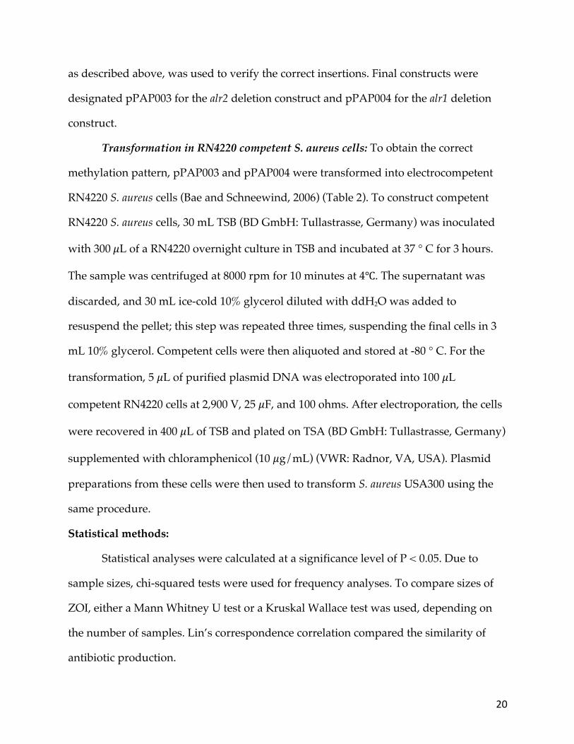
Transformation in RN4220 competent S. aureus cells: To obtain the correct
methylation pattern, pPAP003 and pPAP004 were transformed into electrocompetent
RN4220 S. aureus cells (Bae and Schneewind, 2006) (Table 2). To construct competent
RN4220 S. aureus cells, 30 mL TSB (BD GmbH: Tullastrasse, Germany) was inoculated
with 300𝜇L of a RN4220 overnight culture in TSB and incubated at 37 ° C for 3 hours.
The sample was centrifuged at 8000 rpm for 10 minutes at 4℃. The supernatant was
discarded, and 30 mL ice-cold 10% glycerol diluted with ddH2O was added to
resuspend the pellet; this step was repeated three times, suspending the final cells in 3
mL 10% glycerol. Competent cells were then aliquoted and stored at -80 ° C. For the
transformation, 5 𝜇L of purified plasmid DNA was electroporated into 100 𝜇L
competent RN4220 cells at 2,900 V, 25 𝜇F, and 100 ohms. After electroporation, the cells
were recovered in 400 𝜇L of TSB and plated on TSA (BD GmbH: Tullastrasse, Germany)
supplemented with chloramphenicol (10 𝜇g/mL) (VWR: Radnor, VA, USA). Plasmid
preparations from these cells were then used to transform S. aureus USA300 using the
same procedure.
Statistical methods:
Statistical analyses were calculated at a significance level of P < 0.05. Due to
sample sizes, chi-squared tests were used for frequency analyses. To compare sizes of
ZOI, either a Mann Whitney U test or a Kruskal Wallace test was used, depending on
the number of samples. Lin’s correspondence correlation compared the similarity of
antibiotic production.
21
RESULTS:
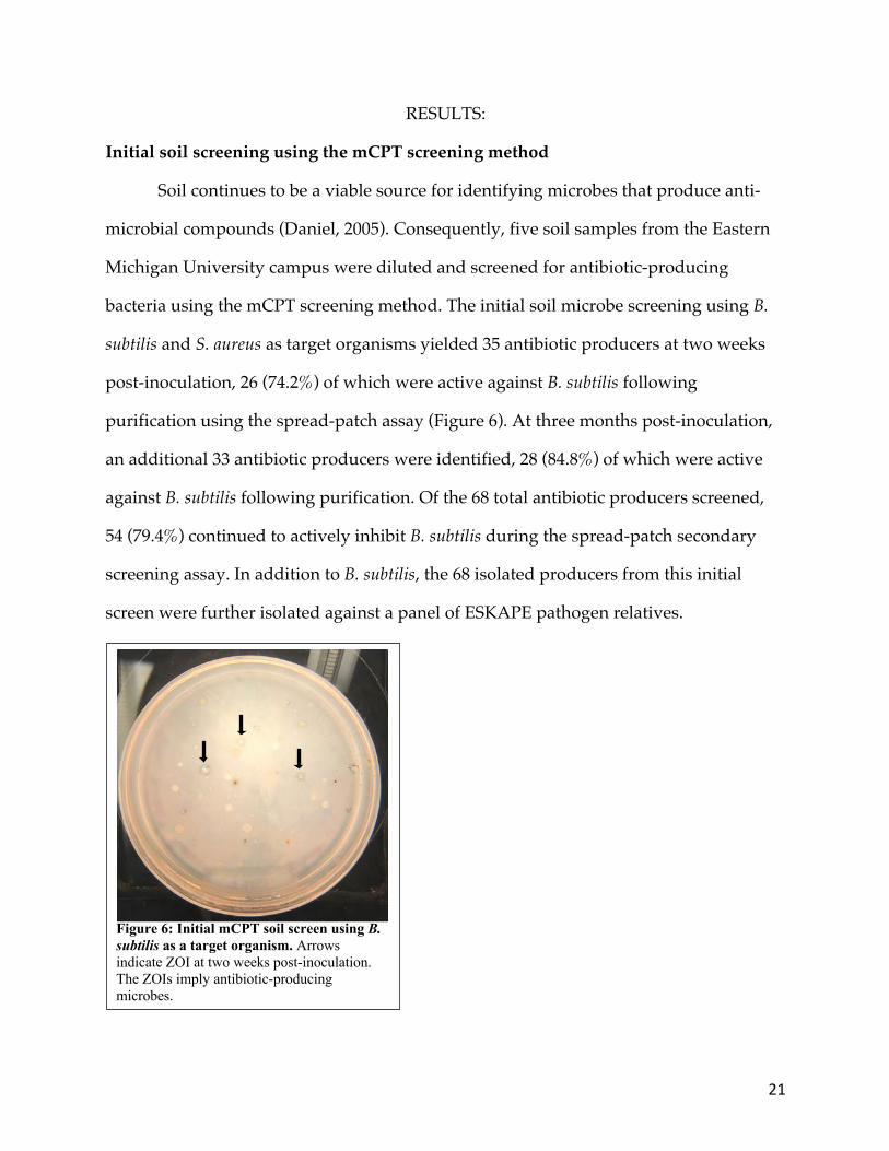
Initial soil screening using the mCPT screening method
Soil continues to be a viable source for identifying microbes that produce anti-
microbial compounds (Daniel, 2005). Consequently, five soil samples from the Eastern
Michigan University campus were diluted and screened for antibiotic-producing
bacteria using the mCPT screening method. The initial soil microbe screening using B.
subtilis and S. aureus as target organisms yielded 35 antibiotic producers at two weeks
post-inoculation, 26 (74.2%) of which were active against B. subtilis following
purification using the spread-patch assay (Figure 6). At three months post-inoculation,
an additional 33 antibiotic producers were identified, 28 (84.8%) of which were active
against B. subtilis following purification. Of the 68 total antibiotic producers screened,
54 (79.4%) continued to actively inhibit B. subtilis during the spread-patch secondary
screening assay. In addition to B. subtilis, the 68 isolated producers from this initial
screen were further isolated against a panel of ESKAPE pathogen relatives.
Figure 6: Initial mCPT soil screen using B. subtilis as a target organism. Arrows indicate ZOI at two weeks post-inoculation. The ZOIs imply antibiotic-producing microbes.
22
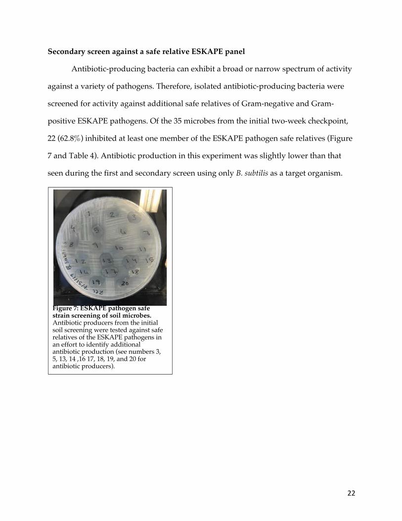
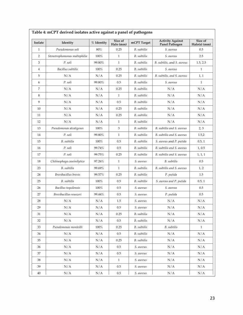
Secondary screen against a safe relative ESKAPE panel
Antibiotic-producing bacteria can exhibit a broad or narrow spectrum of activity
against a variety of pathogens. Therefore, isolated antibiotic-producing bacteria were
screened for activity against additional safe relatives of Gram-negative and Gram-
positive ESKAPE pathogens. Of the 35 microbes from the initial two-week checkpoint,
22 (62.8%) inhibited at least one member of the ESKAPE pathogen safe relatives (Figure
7 and Table 4). Antibiotic production in this experiment was slightly lower than that
seen during the first and secondary screen using only B. subtilis as a target organism.
Figure 7: ESKAPE pathogen safe strain screening of soil microbes. Antibiotic producers from the initial soil screening were tested against safe relatives of the ESKAPE pathogens in an effort to identify additional antibiotic production (see numbers 3, 5, 13, 14 ,16 17, 18, 19, and 20 for antibiotic producers).
23
Table 4: mCPT derived isolates active against a panel of pathogens
Isolate Identity % Identity Size of Halo (mm) mCPT Target Activity Against
Panel Pathogen Size of
Halo(s) (mm) 1 Pseudomonas soli 80% 0.25 B. subtilis S. aureus 0.5
2 Stenotrophomonas maltophilia 100% 1 B. subtilis S. aureus 2.5
3 P. soli 99.80% 1 B. subtilis B. subtilis, and S. aureus 1.5, 2.5
4 Bacillus subtilis 100% 0.25 B. subtilis S. aureus 1
5 N/A N/A 0.25 B. subtilis B. subtilis, and S. aureus 1, 1
6 P. soli 99.80% 0.5 B. subtilis S. aureus 1
7 N/A N/A 0.25 B. subtilis N/A N/A
8 N/A N/A 1 B. subtilis N/A N/A
9 N/A N/A 0.5 B. subtilis N/A N/A
10 N/A N/A 0.25 B. subtilis N/A N/A
11 N/A N/A 0.25 B. subtilis N/A N/A
12 N/A N/A 1 B. subtilis N/A N/A
13 Pseudomonas alcaligenes 100% 3 B. subtilis B. subtilis and S. aureus 2, 3
14 P. soli 99.80% 1 B. subtilis B. subtilis and S. aureus 1.5,2
15 B. subtilis 100% 0.5 B. subtilis S. aureus and P. putida 0.5, 1
16 P. soli 99.74% 0.5 B. subtilis B. subtilis and S. aureus 1, 0.5
17 P. soli 99.75% 0.25 B. subtilis B. subtilis and S. aureus 1, 1, 1
18 Chitinophaga caseinilytica 97.26% 1 S. aureus B. subtilis 0.5
23 B. subtilis 99.69% 1 B. subtilis B. subtilis and S. aureus 1, 2
24 Brevibacillus brevis 99.57% 0.25 B. subtilis P. putida 1.5
25 B. subtilis 100% 0.5 B. subtilis S. aureus and P. putida 0.5, 1
26 Bacillus tequilensis 100% 0.5 S. aureus S. aureus 0.5
27 Brevibacillus reuszeri 99.44% 0.5 S. aureus P. putida 0.5
28 N/A N/A 1.5 S. aureus N/A N/A
29 N/A N/A 0.5 S. aureus N/A N/A
31 N/A N/A 0.25 B. subtilis N/A N/A
32 N/A N/A 0.5 B. subtilis N/A N/A
33 Pseudomonas monteilli 100% 0.25 B. subtilis B. subtilis 1
34 N/A N/A 0.5 B. subtilis N/A N/A
35 N/A N/A 0.25 B. subtilis N/A N/A
36 N/A N/A 0.5 S. aureus N/A N/A
37 N/A N/A 0.5 S. aureus N/A N/A
38 N/A N/A 1 S. aureus N/A N/A
39 N/A N/A 0.5 S. aureus N/A N/A
40 N/A N/A 0.5 S. aureus N/A N/A
24
Optimization of the mCPT technique
Comparison of gellan gum to agar: Previous literature suggested that when
gellan gum is used as a medium solidifying agent, it is more effective for isolating
actinomycetes and stimulating antibiotic production than agar (Suzuki, 2001). Gellan
gum-based and agar-based media were compared for their effectiveness in the mCPT
screening method. While media solidified using agar yielded 35 antibiotic producers
after two weeks of incubation, gellan gum only yielded five antibiotic producers from
the same diluted soil sample. This indicates a significant difference in antibiotic
production between the two media types (Corrected 𝒳! = 56.077, 𝑝 < 0.05 (1, N =
2436)). In addition, because gellan gum has a much narrower pH range at which it
remains solid, the media becomes less solid over time, making the mCPT method more
difficult over time (Picona and Cunha, 2011).
Comparison of prototrophic and auxotrophic B. subtilis strains: The initial
antibiotic screens used a prototrophic strain of B. subtilis as the target organism in the
mCPT screen, but the Price lab had also started using a D-alanine auxotrophic mutant
of B. subtilis as the target strain to aid in the downstream purification process. This
study’s initial mCPT screen resulted in fewer antibiotic producers than those isolated
with the D-alanine auxotrophic strains of B. subtilis. Therefore, a course wide
experiment was designed to determine the potential differences between using
prototrophic and D-alanine auxotrophic strains of B. subtilis as a target organism in the
mCPT screen. Students in the BIO328 sections of the Tiny Earth lab were provided with
the D-alanine auxotrophic strain whereas the students in the BIO112 sections of the
Tiny Earth lab were provided with the prototrophic B. subtilis strain. Overall, compared
to prototrophic strains of B. subtilis, the D-alanine auxotrophic mutants produced more
25
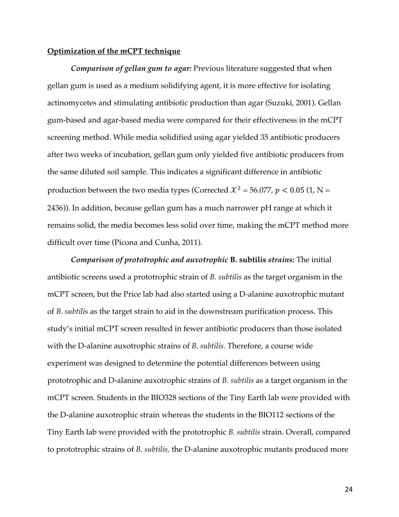
and larger ZOIs. There were 346 ZOI averaging 3.96 mm in size on the mCPT plates
using prototrophic B. subtilis. There were significantly more, and larger, halos with the
auxotrophic B. subtilis overlay with 677 halos averaging 7.20 mm in size (Corrected
𝒳!= 128.9103, 𝑝 < 0.05 (1, N = 41752); Mann-Whitney U test, Z = -8.571. n1 = 346, n2 =
677, 𝑝 < 0.05 , one-tailed) (Figure 8). This implied that there may be an interaction
between the microbes and supplemental D-alanine that might be weakening the cell
wall of the pathogen. This may make the target pathogen more susceptible to
antibiotics.
Comparison of incubation time and media type: The Price lab also observed
variation in the performance of the mCPT screening method over time. Based on these
observations, it was hypothesized that more antibiotic producers would be identified
with increasing culture age. The effects of the incubation time (one or seven days) of the
D-alanine auxotrophic mutant culture and the media type (LB, TY, TYME, or EPSM) it
was cultivated on prior to inoculating the screen were tested to determine whether pre-
incubation conditions influenced antibiotic production during the mCPT screen.
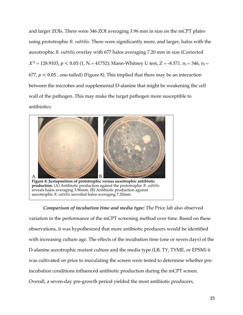
Overall, a seven-day pre-growth period yielded the most antibiotic producers,
A. B. Figure 8: Juxtaposition of prototrophic versus auxotrophic antibiotic production. (A) Antibiotic production against the prototrophic B. subtilis reveals halos averaging 3.96mm. (B) Antibiotic production against auxotrophic B. subtilis unveiled halos averaging 7.20mm.
26
regardless of media type, although there was a trend toward TY and TYME generating
more antibiotic producers (Figure 9). These data suggest that there is an interaction
between media and pre-incubation time (G-test = 316008.908, 𝑝 < 0.05 (7, N = 8176))
and the number of antibiotic producers observed during the mCPT screening method.
Overall, the largest number of antibiotic producers was identified on TYME media
when auxotrophic B. subtilis cultures were pre-grown for seven days.
Comparison of antibiotic producers ssing either B. subtilis or S. aureus as the target
organism
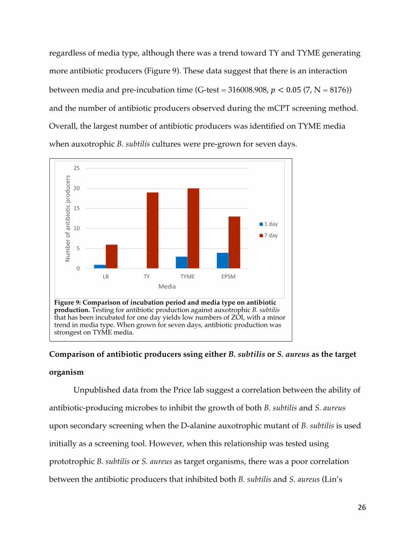
Unpublished data from the Price lab suggest a correlation between the ability of
antibiotic-producing microbes to inhibit the growth of both B. subtilis and S. aureus
upon secondary screening when the D-alanine auxotrophic mutant of B. subtilis is used
initially as a screening tool. However, when this relationship was tested using
prototrophic B. subtilis or S. aureus as target organisms, there was a poor correlation
between the antibiotic producers that inhibited both B. subtilis and S. aureus (Lin’s
Figure 9: Comparison of incubation period and media type on antibiotic production. Testing for antibiotic production against auxotrophic B. subtilis that has been incubated for one day yields low numbers of ZOI, with a minor trend in media type. When grown for seven days, antibiotic production was strongest on TYME media.
0
5
10
15
20
25
LB TY TYME EPSM
Num
ber o
f ant
ibio
tic p
rodu
cers
Media
1 day
7 day

27
correspondence; N = 35, r = -0.5525) (McBride, 2005) (Figures 10 and 11). Although not
all antibiotic producers kill both organisms all of the time, B. subtilis can still be used as
an initial tool to screen for antibiotic producers with the understanding that the isolated
antibiotic producers from such experiments may not inhibit true pathogens like S.
aureus. Consequently, the construction of a D-alanine auxotrophic mutant of S. aureus
would likely provide the added antibiotic sensitivity observed with B. subtilis while
potentially increasing the likelihood of recovering antibiotic-producing bacteria that kill
MRSA. Furthermore, an auxotrophic mutant would provide a safe way to screen for
antibiotic producers that specifically inhibit S. aureus.
Figure 10: Isolated microbes from initial soil screens were tested against B. subtilis and S. aureus. This showed whether the same antibiotic producer was able to kill both pathogens.
B. subtilis mCPT S. aureus mCPT
28
Generation of a S. aureus D-alanine auxotrophic mutant
Although there are two alanine racemase genes in B. subtilis, the mutant in the
Price lab effectively functions as an auxotroph with only the alr1 gene mutated.
However, for the purposes of the mCPT antibiotic screening tool in a classroom setting,
both alanine racemase genes, alr1 and alr2, in the S. aureus strain will be deleted using
homologous recombination with the plasmid pKOR1 (Bae and Schneewind, 2006).
Briefly, 1KB upstream and downstream of each alr gene was amplified using PCR to
increase the likelihood of a recombination event (Figure 12A). The individual upstream
and downstream PCR products were combined used splicing by overlap extension
(SOE) PCR prior to being cloned into pKOR1 and transformed into NEB-5𝛼 E. coli cells
(3.951 × 10" transformants per 𝜇g of DNA) (Figure 12B). Sanger sequencing was used
to confirm each plasmid construct. To generate the right methylation patterns, each
plasmid was then transformed into chemically competent S. aureus RN4220 cells
(transformation efficiency was 3.453 × 10# transformants per 𝜇g of DNA for alr1).
Figure 11: Co-occurrence of B. subtilis and S. aureus. Many of the antibiotic-producing bacteria that killed B. subtilis do not necessarily kill S. aureus.
0
0.1
0.2
0.3
0.4
0.5
0.6
B. subtilis S. aureus B. subtilis and S.aureus
Ratio
of a
ntib
iotic
pro
duce
rs
mCPT B. subtilis
mCPT S. aureus
mCPT B. subtilis
mCPT S. aureus
S. aureusB. subtilis B. subtilis and S. aureus
29
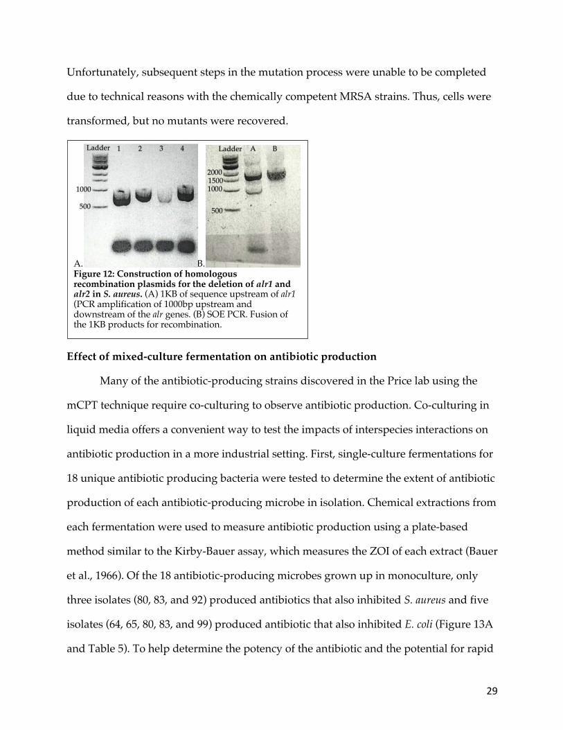
Unfortunately, subsequent steps in the mutation process were unable to be completed
due to technical reasons with the chemically competent MRSA strains. Thus, cells were
transformed, but no mutants were recovered.
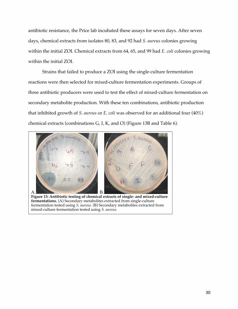
Effect of mixed-culture fermentation on antibiotic production
Many of the antibiotic-producing strains discovered in the Price lab using the
mCPT technique require co-culturing to observe antibiotic production. Co-culturing in
liquid media offers a convenient way to test the impacts of interspecies interactions on
antibiotic production in a more industrial setting. First, single-culture fermentations for
18 unique antibiotic producing bacteria were tested to determine the extent of antibiotic
production of each antibiotic-producing microbe in isolation. Chemical extractions from
each fermentation were used to measure antibiotic production using a plate-based
method similar to the Kirby-Bauer assay, which measures the ZOI of each extract (Bauer
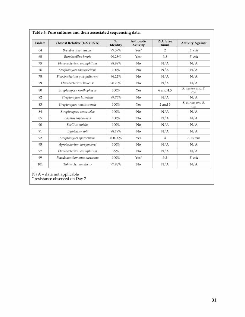
et al., 1966). Of the 18 antibiotic-producing microbes grown up in monoculture, only
three isolates (80, 83, and 92) produced antibiotics that also inhibited S. aureus and five
isolates (64, 65, 80, 83, and 99) produced antibiotic that also inhibited E. coli (Figure 13A
and Table 5). To help determine the potency of the antibiotic and the potential for rapid
A. B. Figure 12: Construction of homologous recombination plasmids for the deletion of alr1 and alr2 in S. aureus. (A) 1KB of sequence upstream of alr1 (PCR amplification of 1000bp upstream and downstream of the alr genes. (B) SOE PCR. Fusion of the 1KB products for recombination.
1000
2000
500
1000
Ladder 1 2 3 4
500
1500
A Ladder B
30
antibiotic resistance, the Price lab incubated these assays for seven days. After seven
days, chemical extracts from isolates 80, 83, and 92 had S. aureus colonies growing
within the initial ZOI. Chemical extracts from 64, 65, and 99 had E. coli colonies growing
within the initial ZOI.
Strains that failed to produce a ZOI using the single-culture fermentation
reactions were then selected for mixed-culture fermentation experiments. Groups of
three antibiotic producers were used to test the effect of mixed-culture fermentation on
secondary metabolite production. With these ten combinations, antibiotic production
that inhibited growth of S. aureus or E. coli was observed for an additional four (40%)
chemical extracts (combinations G, I, K, and O) (Figure 13B and Table 6).
A. B. Figure 13: Antibiotic testing of chemical extracts of single- and mixed-culture fermentations. (A) Secondary metabolites extracted from single-culture fermentation tested using S. aureus. (B) Secondary metabolites extracted from mixed-culture fermentation tested using S. aureus.
31
Table 5: Pure cultures and their associated sequencing data.
Isolate Closest Relative (16S rRNA) % Identity
Antibiotic Activity
ZOI Size (mm) Activity Against
64 Brevibacillus reuszeri 99.59% Yes* 2 E. coli
65 Brevibacillus brevis 99.25% Yes* 3.5 E. coli
75 Flavobacterium amniphilum 98.88% No N/A N/A
76 Streptomyces zaomyceticus 100% No N/A N/A
78 Flavobacterium quisquiliarum 96.22% No N/A N/A
79 Flavobacterium hauense 98.20% No N/A N/A
80 Streptomyces xanthophaeus 100% Yes 6 and 4.5 S. aureus and E. coli
82 Streptomyces lateritius 99.75% No N/A N/A
83 Streptomyces amritsarensis 100% Yes 2 and 3 S. aureus and E. coli
84 Streptomyces venezuelae 100% No N/A N/A
85 Bacillus toyonensis 100% No N/A N/A
90 Bacillus mobilis 100% No N/A N/A
91 Lysobacter soli 98.19% No N/A N/A
92 Streptomyces spororaveus 100.00% Yes 4 S. aureus
95 Agrobacterium larrymoorei 100% No N/A N/A
97 Flavobacterium amniphilum 99% No N/A N/A
99 Psuedoxanthomonas mexicana 100% Yes* 3.5 E. coli
101 Tahibacter aquaticus 97.98% No N/A N/A N/A – data not applicable * resistance observed on Day 7

32
Effect of manganese on antibiotic production
Manganese (Mn) has been shown to stimulate antibiotic production in various
microorganisms (Foster and Woodruff, 1945). Although TYME and EPSM media
contain Mn, its concentration is at the lower threshold (0.2 µM) for this stimulation
effect. Therefore, it was tested whether the addition of supplemental Mn (2.3 µM)
would induce antibiotic production in mixed-culture fermentations. The same mixed-
culture fermentation combinations were grown in regular EPSM and EPSM+Mn. The
Table 6: Mixed fermentations of non-producing pure culture microbes.
Letter Isolates Microbes Co-Cultured Antibiotic Activity
Activity Against
ZOI size (mm) Mn Extract Colour
A 75, 76, 78
Flavobacterium amniphilum, Streptomyces zoomyceticus, and
Flavobacterium quisquiliarum
No N/A N/A No Transparent yellow
B No N/A N/A Yes Transparent grey
C 76, 78, 79
Streptomyces zoomyceticus, Flavobacterium quisquiliarum, and Flavobacterium hauense
No N/A N/A No Transparent yellow
D No N/A N/A Yes Opaque purple
E 78, 79, 82
Flavobacterium quisquiliarum, Flavobacterium hauense, and
Streptomyces lateritius
No N/A N/A No Purple
F No N/A N/A Yes Transparent purple
G 79, 82, 75
Flavobacterium hauense, Streptomyces lateritius, and Flavobacterium amniphilum
Yes S. aureus 2.5 No Opaque purple
H Yes S. aureus 2.5 Yes Transparent grey
I 82, 75, 76
Streptomyces lateritius, Flavobacterium amniphilum,
and Streptomyces zaomyceticus
Yes S. aureus 2.5 No Transparent grey
J Yes S. aureus 2.5 Yes Transparent grey
K 84, 85, 90
Streptomyces venezuelae, Bacillus
toyonensis, and Bacillus mobilis
Yes S. aureus and E. coli 8 No Brown
L Yes S. aureus and E. coli 8 Yes Brown
M 85, 90, 91 Bacillus toyonensis, Bacillus
mobilis, and Lysobacter soli No N/A N/A No Transparent
yellow N No N/A N/A Yes Transparent
yellow / brown O
90, 91, 95 Bacillus mobilis, Lysobacter soli, and Agrobacterium larrymoorei
Yes S. aureus 2 No Transparent yellow / grey
P No N/A N/A Yes Transparent yellow
Q 91, 95, 84
Lysobacter soli, Agrobacterium larrymoorei,
And Streptomyces venezuelae
No N/A N/A No Yellow
R No N/A N/A Yes Brown
S 95, 84, 85
Agrobacterium larrymoorei, Streptomyces venezuelae, and
Bacillus toyonensis
No N/A N/A No Transparent yellow / grey
T No N/A N/A Yes Transparent yellow
N/A – data not applicable Mn – addition of Manganese
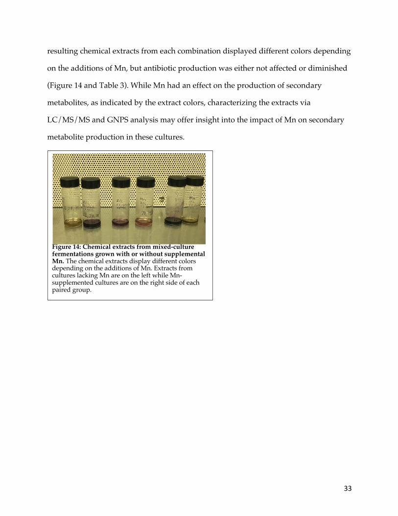
33
resulting chemical extracts from each combination displayed different colors depending
on the additions of Mn, but antibiotic production was either not affected or diminished
(Figure 14 and Table 3). While Mn had an effect on the production of secondary
metabolites, as indicated by the extract colors, characterizing the extracts via
LC/MS/MS and GNPS analysis may offer insight into the impact of Mn on secondary
metabolite production in these cultures.
Figure 14: Chemical extracts from mixed-culture fermentations grown with or without supplemental Mn. The chemical extracts display different colors depending on the additions of Mn. Extracts from cultures lacking Mn are on the left while Mn-supplemented cultures are on the right side of each paired group.

34
DISCUSSION:
This study improves the efficacy of the mCPT screening method and shows that
mixed-culture fermentation can be effective at inducing antibiotic activity when single-
culture fermentation fails to produce sufficient antibiotic activity. By optimizing growth
conditions, specifically, incubation time and media type, prior to and during the mCPT
screen, the improved conditions for enhanced antibiotic production were developed. In
addition, the results indicate that the benefits of using D-alanine auxotrophic B. subtilis
extend beyond safety and the efficiency of colony purification; they also suggest an
increased sensitivity to antibiotic producers, resulting in more and larger ZOIs. The
results also show that it is possible to use the mCPT screening method to find
antibiotics active against S. aureus; this pattern was most defined when S. aureus was
used as an initial target. Consequently, a MRSA D-alanine auxotroph is under
construction and will help broaden the range of the mCPT screening method to
specifically target MRSA. The results also suggested that otherwise cryptic BGCs
(biosynthetic gene clusters) can be stimulated under mixed-culture fermentations when
single-culture fermentation conditions fail to yield antibiotic activity.
Effectiveness of the mCPT modifications
One of the primary advantages of the mCPT screening method is the ability to
accommodate slow growing bacteria by incubating the mCPT plates for several months
(Wollheben et al., 2016; Weiner, 2000). These microbes are often outcompeted by faster
growing microbes, however, when grown over a long period of time, slow growers are
able to compete with the other microbes via the production of antibiotics (Lazzarini et
al., 2000; Wollheben et al., 2016). Slow growing bacteria are rarely represented in
antibiotic screens because they are often outcompeted or do not grow over the short

35
incubation periods normally used. Hence, the secondary metabolites of slow-growing
bacteria offer a new potential source for novel antibiotics (Wollheben et al., 2016).
Different neighboring microbes likely resulted in different external stimuli and
might explain why microbes may not have produced antibiotics against B. subtilis
during both the primary mCPT and secondary ESKAPE pathogen screens (Netzker et
al., 2015). The number of antibiotic producers observed during the mCPT decreased
during the secondary screening using the spread-patch assay with safe relatives of the
ESKAPE pathogens. Notably, many of the microbes formed ZOI during the initial
antibiotic screen using B. subtilis did not necessarily produce a ZOI during the
secondary ESKAPE pathogen screen. During the ESKAPE relative screen, the antibiotic
producing bacteria are not necessarily near the same microbes they were neighboring
during the initial mCPT screen.
Previous research has suggested that the use of gellan gum as a solidifying agent
helps stimulate secondary metabolite production in actinomycetes (Suzuki, 2001).
However, this study found that compared to agar plates, there was less growth and less
antibiotic production. In addition, gellan gum made a comparatively soft media that
was not suitable for standard plating procedures. Thus, agar yields better results for the
mCPT screening method than gellan gum. This is potentially due to the sensitivity of
gellan gum to pH (Picona and Cunha, 2011)
B. subtilis and S. aureus are both Gram-positive bacteria in the Firmicutes
phylum. Consequently, they share much of their metabolic profile and are susceptible
to many of the same antibiotics (Brown et al., 2010; Bosi et al., 2016). They have similar
cell wall structures consisting of peptidoglycan, polyribitol phosphate, and wall teichoic
acids (WTA) in both constitutes roughly 60% of the cell wall in both species. However,
the WTA is produced via different metabolic pathways (Brown et al., 2010).

36
Unpublished data from the Price lab suggests that many (about 75%) of the antibiotic-
producing microbes that inhibited B. subtilis also inhibited the growth of S. aureus.
However, a much lower proportion (28.6%) was observed in this study. Overall, these
data suggest that the mCPT can be easily adapted to find antibiotics to target particular
species.
Interestingly, when D-alanine prototrophic and auxotrophic B. subtilis strains
were directly compared using the mCPT screen, the auxotrophic mutants yielded more
antibiotic producers with larger ZOI. D-alanine is used to construct both the
crosslinking amino acids in peptidoglycan and in wall teichoic acids (WTA). WTAs
function in cell support, as well as contributing to the charge and hydrophobicity of the
cell wall (Brown et al., 2010). This, in turn, impacts the susceptibility of the microbe to
the antibiotics by influencing the binding and flow of secondary metabolites through
the cell wall. D-alanine esters are attached to WTAs in a process known as D-
alanylation (Brown et al., 2013). In lower concentrations of D-alanine, there is a decline
in D-alanylation; this alters the hydrophobicity and charge of the cell wall, thus lending
itself to increase the susceptibility of the bacteria to antibiotics (Brown et al., 2013).
When D-alanine is provided in the media, such as the case during the initial antibiotic
producer screen, both auxotrophic and prototrophic microbes will preferentially take
up the D-alanine from their environment (Khonsari and Kollmann, 2015). This results in
local D-alanine depletion on these screening plates because both the soil microbes and
the auxotrophic bacteria using the supplemental D-alanine from the media. Following
the local depletion of D-alanine from the media, prototrophic bacteria transition to
produce D-alanine from L-alanine using D-alanine racemases, continuing to divide and
grow on the plate (Moscoso et al., 2018). The auxotrophs, however, rely solely on the
supplied D-alanine in the media; when this is depleted, the auxotrophs are no longer

37
able to grow, likely have weaker cell walls, and may be lysing at a higher rate than
prototrophic bacteria. Therefore, as the concentration of D-alanine in the media
decreases, there is likely a similar decline in the robustness of the auxotrophic cell wall
because D-alanine can be incorporated into the peptidoglycan and the WTAs.
Consequently, the auxotroph may be more susceptible to antibiotics following the
primary growth of the bacteria. This may result in more and larger ZOIs because the
soil microbes are able to inhibit a weakened pathogen with less antibiotic, adding more
sensitivity to the mCPT screen. It may be beneficial to construct more auxotrophic
pathogens for use as an antibiotic screening tool because they lead to more obvious
ZOIs. In doing so, rarer, slow-growing antibiotic producers in the complex stimulatory
environment resulting from the mCPT screening method may be identified (Mehl and
Cotty, 2013). It is anticipated that the increased sensitivity of D-alanine auxotrophs and
the abundant microbe-microbe interactions will increase the ability to identify antibiotic
production from otherwise cryptic BGCs. To this end, the process of constructing a D-
alanine auxotrophic S. aureus mutant has begun; considerable progress has been made,
but mutagenesis has not yet been completed.
Liquid culturing conditions
Growth of pure cultures were considered the gold-standard for the production
and extraction of secondary metabolites because they provide more controlled growth
environments; there is also no question as to which microbe produced the secondary
metabolite (Nei and Meyer, 2017). Thus, most antibiotics to date were discovered and
produced using this type of single-culture fermentation. Unfortunately, these are
relatively barren growth conditions as there are no interspecies interactions or
competition to encourage antibiotic production, especially when most antibiotics are
also self-harming (Seyedsayamdost et al., 2012). Co-cultures, or mixed-culture

38
fermentations, provide interspecies interactions that can induce different and
potentially novel secondary metabolic pathways. To evaluate the effects of interspecies
interactions on antibiotic production, Ueda and Beppu (2016) selected strains that
showed very little antibiotic production in pure cultures and used mixed-culture
fermentations significantly increase secondary metabolite production. To help control
for the production of a desired secondary antimicrobial metabolite, only bacteria that
did not produce antibiotics in pure culture were used in the mixed-culture
fermentation. These mixed-culture fermentations with non-antibiotic producing
bacteria increased antibiotic production, supporting the notion that interspecies
interactions increase the expression of various metabolic pathways. However, this
process has largely been limited to the increasing production of known compounds;
only the Onaka lab has used this process as a mechanism for discovering new
antibiotics (Onaka et al., 2011). The dichotomy observed between antibiotic production
using the mCPT screening method and the overall lack of antibiotic production in
single-culture fermentation results presented in this study suggest that the parameters
for mixed-culture fermentation still need to be optimized. While the compounds
produced during mixed-culture fermentation are currently unknown, it is encouraging
to see increased antibiotic activity when multiple antibiotic producers are combined in
mixed-culture fermentation. This suggests that there are still likely new antibiotics to be
found and that combining multiple antibiotic-producing bacteria together during
mixed-fermentation might provide the best strategy for increasing antibiotic production
from these producers (Seyedsayamdost et al., 2012).
While this study focused on optimizing the use of multiple cultures, media
modifications are widely used to modify antibiotic production. Manganese (Mn) has
been reported to stimulate secondary metabolism, increasing antibiotic production
39
(Foster and Woodruff, 1945). Although chemical extracts from Mn and non-Mn
supplemented culture were different colors, a potential sign of differential expression of
secondary metabolites, there was little change in the size of the ZOIs. However, Mn
exists in different colors at different oxidation states, so it is possible, too, that the color
change is courtesy of a different redox environment (Willard and Greathouse, 1917).
These results suggest that there is an interaction between secondary metabolite
pathways and Mn availability. However, without characterizing the chemical extracts,
it is hard to say what effect Mn has on secondary metabolite production
D-alanine auxotroph construction
Plasmids for the MRSA D-alanine auxotroph have been constructed, but they
have not yet been transformed into MRSA. Thus far, transformations reactions have
yielded any colonies. Glycine was added to competent cells in an attempt to optimize
culture conditions and a new electroporator was purchased, but no colonies have been
observed (Cruz-Rodz and Gilmore, 1990).
One potential explanation for the lack of transformed MRSA colonies is the
importance of methylation patterns. Due to the number of restriction modification
systems in MRSA, methylation patterns of the plasmid are of utmost importance in
order to transform the plasmid into the cell (Jones et al., 2015). Despite using S. aureus
RN4220, which is supposed to help bypass some of these restriction systems in the
MRSA cell that recognize foreign DNA and restrict transformation, there was still an
issue with transformation (Jones et al., 2015). For future transformation attempts, it may
be worthwhile to investigate plasmid alterations that can circumvent restriction
modification in MRSA. One study accomplished this by using plasmids from E. coli
DC10B; this strain is able to bypass some restriction modifications in S. aureus as it does
not methylate cytosines (Jones et al., 2015).

40
CONCLUSIONS
These findings suggest the need to continue to optimize the process of
identifying and producing antibiotics from cryptic BGCs in soil microbes. There was a
notable increase in efficiency when using a D-alanine auxotrophic mutant of B. subtilis
during the initial mCPT screening to identify antibiotic producers. This success could
thus be furthered by constructing a series of D-alanine auxotrophs in addition to the
MRSA auxotroph as this will allow for specific identification of antibiotic producers that
are able to kill MRSA and other specific ESKAPE pathogens. Until these auxotrophs are
built, however, the B. subtilis and E. coli auxotrophs can continue to be used in
introductory labs as the best means of efficiently identifying antibiotic producers for
further study. Furthermore, the use of B. subtilis auxotroph in the classroom will allow
students to more easily visualize their results, potentially creating a stronger sense of
discovery and engagement with concepts taught in the lab.
Given the early success with mixed-culture fermentation using multiple
antibiotic producers, this technique may be further optimized; perhaps there are
specific interactions that can be harnessed to trigger secondary metabolite production
and specifically secondary metabolites that have antimicrobial properties. To get a
better understanding of the trends seen in the mixed-culture fermentation cultures, full
chemical profiles of the single- and mixed-culture fermentation chemical extracts can be
characterized via LC/MS/MS. Modern computational chemistry approaches such as
Global Natural Products Social (GNPS) molecular networking can be used to delineate
the novelty of these secondary metabolites and potentially useful compounds that
specifically induce antibiotic production (Wang et al., 2016).

41
Literature Cited
Abrudan, M. I., F. Smakman, A. J. Grimbergen, S. Westhoff, E. L. Miller et al., 2015
Socially mediated induction and suppression of antibiosis during bacterial
coexistence. Proceedings of the National Academy of Sciences 112: 11054.
Altschul, S.F., Gish, W., Miller, W., Myers, E.W. & Lipman, D.J. 1990 Basic local
alignment search tool. J. Mol. Biol. 215:403-410.
“Antibiotic / Antimicrobial Resistance.” Centers for Disease Control and Prevention,
Centers for Disease Control and Prevention, 14 Feb. 2020,
www.cdc.gov/drugresistance/index.html.
Ayukekbong, J. A., M. Ntemgwa and A. N. Atabe, 2017 The threat of antimicrobial
resistance in developing countries: causes and control strategies. Antimicrobial
Resistance & Infection Control 6: 47.
Bae, T., and O. Schneewind, 2006 Allelic replacement in Staphylococcus aureus with
inducible counter-selection. Plasmid 55: 58-63.
Bassetti, M., M. Merelli, C. Temperoni and A. Astilean, 2013 New antibiotics for bad
bugs: where are we? Ann Clin Microbiol Antimicrob 12:22.
Bauer, A. W., Kirby, W. M., Sherris, J.C., Turck M., Antibiotic susceptibility testing by a
standardized single disk method.
Bode, H. B., B. Bethe, R. Hofs and A. Zeeck, 2002 Big effects from small changes:
Possible ways to explore nature's chemical diversity. Chembiochem 3: 619-627.
42
Bosi, E., J. M. Monk, R. K. Aziz, M. Fondi, V. Nizet et al., 2016 Comparative genome-
scale modelling of <em>Staphylococcus aureus</em> strains identifies strain-
specific metabolic capabilities linked to pathogenicity. Proceedings of the
National Academy of Sciences 113: E3801.
Boucher, H. W., and G. R. Corey, 2008 Epidemiology of Methicillin-Resistant
Staphylococcus aureus. Clinical Infectious Diseases 46: S344-S349.
Brown, S., T. Meredith, J. Swoboda and S. Walker, 2010 Staphylococcus aureus and
Bacillus subtilis W23 make polyribitol wall teichoic acids using different
enzymatic pathways. Chemistry & biology 17: 1101-1110.
Brown, S., J. P. Santa Maria, Jr. and S. Walker, 2013 Wall teichoic acids of gram-positive
bacteria. Annual review of microbiology 67: 313-336.
Cava, F., M. A. Lam H., de Pedro, M. K., Waldor M. K., 2011 Emerging knowledge of
regulatory roles of D-amino acids in bacteria.
Clardy, J., M. A. Fischbach and C. R. Currie, 2009 The natural history of antibiotics.
Current biology : CB 19: R437-R441.
Cruz-Rodz, A. L., and M. S. Gilmore, 1990 High efficiency introduction of plasmid
DNA into glycine treated Enterococcus faecalis by electroporation. Molecular
and General Genetics MGG 224: 152-154.
Darrow, J. J., and A. S. Kesselheim, 2020 Incentivizing Antibiotic Development: Why
Isn’t the Generating Antibiotic Incentives Now (GAIN) Act Working? Open
Forum Infectious Diseases 7.

43
Cornforth, D. M., and K. R. Foster, 2015 Antibiotics and the art of bacterial war.
Proceedings of the National Academy of Sciences of the United States of
America 112: 10827-10828.
Davies, J., and D. Davies, 2010 Origins and evolution of antibiotic resistance.
Microbiology and molecular biology reviews : MMBR 74: 417-433.
Davis, E., T. Sloan, K. Aurelius, A. Barbour, E. Bodey et al., 2017 Antibiotic discovery
throughout the Small World Initiative: A molecular strategy to identify
biosynthetic gene clusters involved in antagonistic activity. MicrobiologyOpen
6: e00435.
D’Costa, V. M., K. M. McGrann, D. W. Hughes and G. D. Wright, 2006 Sampling the
Antibiotic Resistome. Science 311: 374.
Exner, M., S. Bhattacharya, B. Christiansen, J. Gebel, P. Goroncy-Bermes et al., 2017
Antibiotic resistance: What is so special about multidrug-resistant Gram-
negative bacteria? GMS hygiene and infection control 12: Doc05-Doc05.
Fischbach, M. A., and C. T. Walsh, 2009 Antibiotics for Emerging Pathogens. Science
325: 1089.
Foster, J. W., and H. B. Woodruff, 1946 Bacillin, a New Antibiotic Substance from a Soil
Isolate of Bacillus subtilis. Journal of bacteriology 51: 363-369.
Helfrich, E. J. N., C. M. Vogel, R. Ueoka, M. Schafer, F. Ryffel et al., 2018 Bipartite
interactions, antibiotic production and biosynthetic potential of the Arabidopsis
leaf microbiome. Nature Microbiology 3: 909-919.
44
Jones, M. J., N. P. Donegan, I. V. Mikheyeva and A. L. Cheung, 2015 Improving
Transformation of Staphylococcus aureus Belonging to the CC1, CC5 and CC8
Clonal Complexes. PLOS ONE 10: e0119487.
Kata, A., 2010 A postmodern Pandora's box: Anti-vaccination misinformation on the
Internet. Vaccine 28: 1709-1716.
Khonsari, A. S., and M. Kollmann, 2015 Perception and regulatory principles of
microbial growth control. PloS one 10: e0126244-e0126244.
Kourits AP, Hartfield K, Baggs J, et al., 2019, Vital signs: epidemdiology and recent
trends in methicillin-resistant Staphylococcus aureus bloodstream infections.
Morbidity and Mortality Weekly Report: 214-219.
Kuroda, M., T. Ohta, I. Uchiyama, T. Baba, H. Yuzawa et al., 2001 Whole genome
sequencing of methicillin-resistant Staphylococcus aureus. The Lancet 357: 1225-
1240.
Lazzarini, A., L. Cavaletti, G. Toppo and F. Marinelli, 2000 Rare genera of
actinomycetes as potential producers of new antibiotics. Antonie van
Leeuwenhoek 78: 399-405.
Li, B., and T. J. Webster, 2018 Bacteria antibiotic resistance: New challenges and
opportunities for implant-associated orthopedic infections. Journal of
Orthopaedic Research 36: 22-32.

45
Lowy, F. D., 1998 Staphylococcus aureus Infections. New England Journal of Medicine
339: 520-532.
McBride, G.B. A proposal for the strength-of-agreement criteria for Lin’s concordance
correlation coefficient. NIWA Client Report: HAM2005-062; National Institute of
Water & Atmospheric Research: Hamilton, New Zealand, May 2005
Mehl, H. L., and P. J. Cotty, 2013 Nutrient Environments Influence Competition among
Aspergillus flavusGenotypes. Applied and Environmental Microbiology
79: 1473.
Miller, S. I., 2016 Antibiotic Resistance and Regulation of the Gram-Negative Bacterial
Outer Membrane Barrier by Host Innate Immune Molecules. mBio 7: e01541-
01516.
Moscoso, M., P. García, M. P. Cabral, C. Rumbo and G. Bou, 2018 A D-Alanine
auxotrophic live vaccine is effective against lethal infection caused by
Staphylococcus aureus. Virulence 9: 604-620.
Mullard, A., 2019 Achaogen bankruptcy highlights antibacterial development woes.
Nature Reviews Drug Discovery 18: 411-411.
Nai, C., and V. Meyer, 2018 From Axenic to Mixed Cultures: Technological Advances
Accelerating a Paradigm Shift in Microbiology. Trends in Microbiology 26: 538-
554.
46
Netzker, T., J. Fischer, J. Weber, D. J. Mattern, C. C. Konig et al., 2015 Microbial
communication leading to the activation of silent fungal secondary metabolite
gene clusters. Frontiers in Microbiology 6: 13.
Nichols, D., N. Cahoon, E. M. Trakhtenberg, L. Pham, A. Mehta et al., 2010 Use of Ichip
for High-Throughput <em>In Situ</em> Cultivation of “Uncultivable”
Microbial Species. Applied and Environmental Microbiology 76: 2445.
Onaka, H., Y. Mori, Y. Igarashi and T. Furumai, 2011 Mycolic Acid-Containing Bacteria
Induce Natural-Product Biosynthesis in Streptomyces Species. Applied and
Environmental Microbiology 77: 400-406.
Peek, J., M. Lilic, D. Montiel, A. Milshteyn, I. Woodworth et al., 2018 Rifamycin
congeners kanglemycins are active against rifampicin-resistant bacteria via a
distinct mechanism. Nature Communications 9: 4147.
Picone, C. S. F., and R. L. Cunha, 2011 Influence of pH on formation and properties of
gellan gels. Carbohydrate Polymers 84: 662-668.
Rigali, S., F. Titgemeyer, S. Barends, S. Mulder, A. W. Thomae et al., 2008 Feast or
famine: the global regulator DasR links nutrient stress to antibiotic production
by Streptomyces. EMBO reports 9: 670-675.
Seyedsayamdost, M. R., M. F. Traxler, J. Clardy and R. Kolter, 2012 Old Meets New:
Using Interspecies Interactions to Detect Secondary Metabolite Production in
Actinomycetes, pp. 89-109 in Natural Product Biosynthesis by Microorganisms and
Plants, Pt C, edited by D. A. Hopwood. Elsevier Academic Press Inc, San Diego.

47
Spellberg, B., R. Guidos, D. Gilbert, J. Bradley, H. W. Boucher et al., 2008 The Epidemic
of Antibiotic-Resistant Infections: A Call to Action for the Medical Community
from the Infectious Diseases Society of America. Clinical Infectious Diseases
46: 155-164.
Strych, U., H.-C. Huang, K. L. Krause and M. J. Benedik, 2000 Characterization of the
Alanine Racemases from Pseudomonas aeruginosa PAO1. Current Microbiology
41: 290-294.
Tenover, F. C., and R. V. Goering, 2009 Methicillin-resistant Staphylococcus aureus
strain USA300: origin and epidemiology. Journal of Antimicrobial
Chemotherapy 64: 441-446.
Traxler, M. F., J. D. Watrous, T. Alexandrov, P. C. Dorrestein and R. Kolter, 2013
Interspecies Interactions Stimulate Diversification of the Streptomyces coelicolor
Secreted Metabolome. Mbio 4: 12.
Ueda, K., and T. Beppu, 2017 Antibiotics in microbial coculture. Journal of Antibiotics
70: 361-365.
Valderrama, M. J., B. González-Zorn, P. C. de Pablo, R. Díez-Orejas, T. Fernández-
Acero et al., 2018 Educating in antimicrobial resistance awareness: adaptation of
the Small World Initiative program to service-learning. FEMS Microbiology
Letters 365: fny161-fny161.

48
Valiquette, L., and K. B. Laupland, 2015 Digging for new solutions. The Canadian
journal of infectious diseases & medical microbiology = Journal canadien des
maladies infectieuses et de la microbiologie medicale 26: 289-290.
Van der Meij, A., S. F. Worsley, M. I. Hutchings and G. P. van Wezel, 2017 Chemical
ecology of antibiotic production by actinomycetes. Fems Microbiology Reviews
41: 392-416.
Ventola, C. L., The antibiotic resistance crisis: part 1: causes and threats.
Wang M., Carver J., Phelan V., Sanchez L., Garg N., Peng Y., Nguyen D. "Sharing and
community curation of mass spectrometry data with Global Natural Products
Social Molecular Networking." Nature biotechnology 34, no. 8 (2016): 828.
Willard, H. H., and L. H. Greathouse, 1917 The colorimetric determination of
manganese by oxidation with periodate.1. Journal of the American Chemical
Society 39: 2366-2377.